Notice (8): Undefined variable: solution_of_interest [APP/View/Products/view.ctp, line 755]Code Context<!-- BEGIN: REQUEST_FORM MODAL -->
<div id="request_formModal" class="reveal-modal medium" data-reveal aria-labelledby="modalTitle" aria-hidden="true" role="dialog">
<?= $this->element('Forms/simple_form', array('solution_of_interest' => $solution_of_interest, 'header' => $header, 'message' => $message, 'campaign_id' => $campaign_id)) ?>
$viewFile = '/home/website-server/www/app/View/Products/view.ctp'
$dataForView = array(
'language' => 'en',
'meta_keywords' => '5-hydroxymethylcytosine,monoclonal antibody ,Diagenode',
'meta_description' => '5-hydroxymethylcytosine (5-hmC) Monoclonal Antibody (mouse) validated in hMeDIP, DB and ELISA. Batch-specific data available on the website. Sample size available.',
'meta_title' => '5-hydroxymethylcytosine (5-hmC) Monoclonal Antibody (mouse) | Diagenode',
'product' => array(
'Product' => array(
'id' => '2009',
'antibody_id' => '47',
'name' => '5-hydroxymethylcytosine (5-hmC) Antibody (mouse) ',
'description' => '<p>One of the <strong>only two monoclonal antibodies raised against 5-hydroxymethylcytosine (5-hmC).</strong> 5-hmC is a recently discovered DNA modification which results from the enzymatic conversion of 5-methylcytosine into 5-hydroxymethylcytosine by the TET family of oxygenases. Preliminary results indicate that 5-hmC may have important roles distinct from 5-methylcytosine (5-mC). Although its precise role has still to be shown, early evidence suggests a few putative mechanisms that could have big implications in epigenetics.</p>
<p><strong></strong></p>',
'label1' => 'Validation Data',
'info1' => '<div class="row">
<div class="small-4 columns">
<p><img src="https://www.diagenode.com/img/product/antibodies/C15200200-fig1.png" alt="ChIP" width="160" caption="false" height="280" style="display: block; margin-left: auto; margin-right: auto;" /></p>
</div>
<div class="small-8 columns">
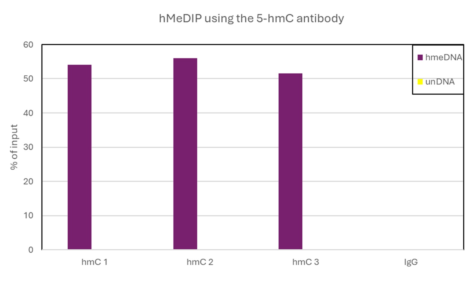
<p><small><strong> Figure 1. An hydroxymethylated DNA IP (hMeDIP) was performed using the Diagenode mouse monoclonal antibody directed against 5-hydroxymethylcytosine (Cat. No. MAb-31HMC-020, MAb-31HMC-050, MAb-31HMC-100).</strong> <br />The IgG isotype antibodies from mouse (Cat. No. kch-819-015) was used as negative control. The DNA was prepared with the GenDNA module of the hMeDIP kit and sonicated with our Bioruptor® (UCD-200/300 series) to have DNA fragments of 300-500 bp. 1 μg of human Hela cells DNA were spiked with non-methylated, methylated, and hydroxymethylated PCR fragments. The IP’d material has been analysed by qPCR using the primer pair specific for the 3 different control sequences. The obtained results show that the mouse monoclonal for 5-hmC is highly specific for this base modification (no IP with non-methylated or methylated C bases containing fragments). </small></p>
</div>
</div>
<div class="row">
<div class="small-4 columns">
<p><img src="https://www.diagenode.com/img/product/antibodies/C15200200-fig2.png" alt="ELISA" width="190" caption="false" style="display: block; margin-left: auto; margin-right: auto;" /></p>
</div>
<div class="small-8 columns">
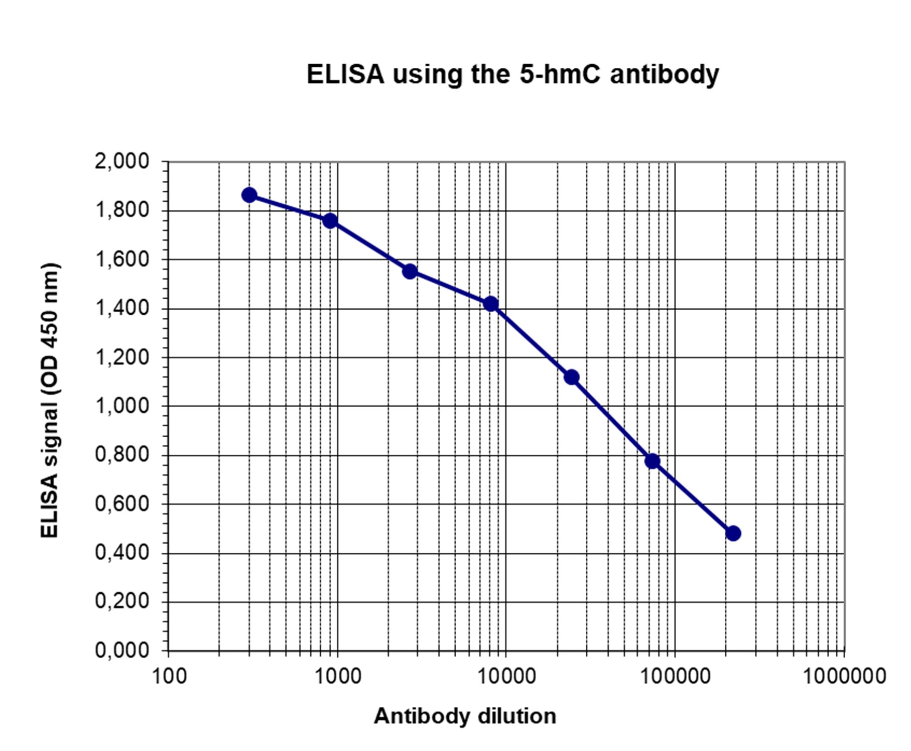
<p><small><strong> Figure 2. Determination of the 5-hmC mouse monoclonal antibody titer </strong><br />To determine the titer, an ELISA was performed using a serial dilution of the Diagenode mouse monoclonal antibody directed against 5-hmC (Cat No. MAb-31HMC-050, MAb-31HMC-100) in antigen coated wells. The antigen used was KHL coupled to 5-hmC base. By plotting the absorbance against the antibody dilution, the titer of the antibody was estimated to be 1:40,000. </small></p>
</div>
</div>
<div class="row">
<div class="small-4 columns">
<p><img src="https://www.diagenode.com/img/product/antibodies/C15200200-fig3.png" alt="Dot Blot" width="100" caption="false" height="137" style="display: block; margin-left: auto; margin-right: auto;" /></p>
</div>
<div class="small-8 columns">
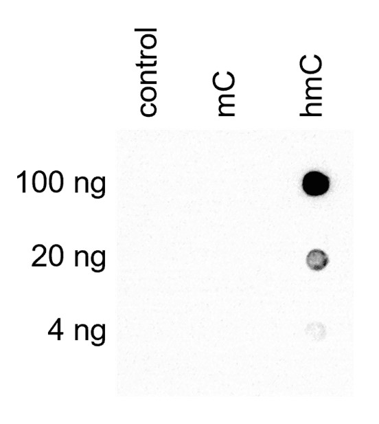
<p><small><strong> Figure 3. Dotblot analysis of the Diagenode 5-hmC mouse monoclonal antibody with the C, mC and hmC PCR controls </strong><br />200 to 2 ng (equivalent of 10 to 0.1 pmol of C-bases) of the hmC (1), mC (2) and C (3) PCR controls from the Diagenode “5-hmC, 5-mC & cytosine DNA Standard Pack” (Cat No. AF-101-0020) were spotted on a membrane (Amersham Hybond-N+). The membrane was incubated with 2 μg/ml of the mouse 5-hydroxymethylcytosine monoclonal antibody (dilution 1:500). The membranes were exposed for 30 seconds. </small></p>
</div>
</div>',
'label2' => 'Target description',
'info2' => '<p>5-hydroxymethylcytosine (5-hmC) has been recently discovered in mammalian DNA. This results from the enzymatic conversion of 5-methylcytosine into 5-hydroxymethylcytosine by the TET family of oxygenases. So far, the 5-hmC bases have been identified in Purkinje neurons, in granule cells and embryonic stem cells where they are present at high levels (up to 0,6% of total nucleotides in Purkinje cells).</p>
<p>Preliminary results indicate that 5-hmC may have important roles distinct from 5-mC. Although its precise role has still to be shown, early evidence suggests a few putative mechanisms that could have big implications in epigenetics : 5-hydroxymethylcytosine may well represent a new pathway to demethylate DNA involving a repair mechanism converting 5-hmC to cytosine and, as such open up entirely new perspectives in epigenetic studies.</p>
<p>Due to the structural similarity between 5-mC and 5-hmC, these bases are experimentally almost indistinguishable. Recent articles demonstrated that the most common approaches (e.g. enzymatic approaches, bisulfite sequencing) do not account for 5-hmC. The development of the affinity-based technologies appears to be the most powerful way to differentially and specifically enrich 5-mC and 5-hmC sequences. The results shown here illustrate the use of this unique monoclonal antibody against 5-hydroxymethylcytosine that has been fully validated in various technologies.</p>',
'label3' => '',
'info3' => '',
'format' => '50 µg/50 µl',
'catalog_number' => 'C15200200-50',
'old_catalog_number' => 'Mab-31HMC-050',
'sf_code' => 'C15200200-D001-000581',
'type' => 'FRE',
'search_order' => '03-Antibody',
'price_EUR' => '380',
'price_USD' => '380',
'price_GBP' => '340',
'price_JPY' => '59525',
'price_CNY' => '',
'price_AUD' => '950',
'country' => 'ALL',
'except_countries' => 'None',
'quote' => false,
'in_stock' => false,
'featured' => false,
'no_promo' => false,
'online' => true,
'master' => true,
'last_datasheet_update' => '0000-00-00',
'slug' => '5-hmc-monoclonal-antibody-mouse-classic-50-ug-50-ul',
'meta_title' => '5-hydroxymethylcytosine (5-hmC) Monoclonal Antibody (mouse) | Diagenode',
'meta_keywords' => '5-hydroxymethylcytosine,monoclonal antibody ,Diagenode',
'meta_description' => '5-hydroxymethylcytosine (5-hmC) Monoclonal Antibody (mouse) validated in hMeDIP, DB and ELISA. Batch-specific data available on the website. Sample size available.',
'modified' => '2022-01-05 15:12:18',
'created' => '2015-06-29 14:08:20',
'locale' => 'eng'
),
'Antibody' => array(
'host' => '*****',
'id' => '47',
'name' => '5-hmC monoclonal antibody (mouse)',
'description' => '5-hydroxymethylcytosine (5-hmC) has been recently discovered in mammalian DNA. This results from the enzymatic conversion of 5-methylcytosine into 5-hydroxymethylcytosine by the TET family of oxygenases. So far, the 5-hmC bases have been identified in Purkinje neurons, in granule cells and embryonic stem cells where they are present at high levels (up to 0,6% of total nucleotides in Purkinje cells).
Preliminary results indicate that 5-hmC may have important roles distinct from 5-mC. Although its precise role has still to be shown, early evidence suggests a few putative mechanisms that could have big implications in epigenetics : 5-hydroxymethylcytosine may well represent a new pathway to demethylate DNA involving a repair mechanism converting 5-hmC to cytosine and, as such open up entirely new perspectives in epigenetic studies.
Due to the structural similarity between 5-mC and 5-hmC, these bases are experimentally almost indistinguishable. Recent articles demonstrated that the most common approaches (e.g. enzymatic approaches, bisulfite sequencing) do not account for 5-hmC. The development of the affinity-based technologies appears to be the most powerful way to differentially and specifically enrich 5-mC and 5-hmC sequences. The results shown here illustrate the use of this unique monoclonal antibody against 5-hydroxymethylcytosine that has been fully validated in various technologies.',
'clonality' => '',
'isotype' => '',
'lot' => '001',
'concentration' => '1.0 µg/µl',
'reactivity' => 'Human, mouse, other (wide range)',
'type' => 'Monoclonal',
'purity' => 'Protein G purified',
'classification' => 'Classic',
'application_table' => '<table>
<thead>
<tr>
<th>Applications</th>
<th>Suggested dilution</th>
<th>References</th>
</tr>
</thead>
<tbody>
<tr>
<td>hMeDIP</td>
<td>2.5 μg/IP</td>
<td>Fig 1</td>
</tr>
<tr>
<td>ELISA</td>
<td>1:500</td>
<td>Fig 2</td>
</tr>
<tr>
<td>Dot Blotting</td>
<td>2 μg / ml</td>
<td>Fig 3</td>
</tr>
</tbody>
</table>',
'storage_conditions' => '',
'storage_buffer' => '',
'precautions' => 'This product is for research use only. Not for use in diagnostic or therapeutic procedures.',
'uniprot_acc' => '',
'slug' => '',
'meta_keywords' => '',
'meta_description' => '',
'modified' => '2016-08-03 10:38:14',
'created' => '0000-00-00 00:00:00',
'select_label' => '47 - 5-hmC monoclonal antibody (mouse) (001 - 1.0 µg/µl - Human, mouse, other (wide range) - Protein G purified - Mouse)'
),
'Slave' => array(
(int) 0 => array(
[maximum depth reached]
)
),
'Group' => array(
'Group' => array(
[maximum depth reached]
),
'Master' => array(
[maximum depth reached]
),
'Product' => array(
[maximum depth reached]
)
),
'Related' => array(
(int) 0 => array(
[maximum depth reached]
),
(int) 1 => array(
[maximum depth reached]
),
(int) 2 => array(
[maximum depth reached]
),
(int) 3 => array(
[maximum depth reached]
),
(int) 4 => array(
[maximum depth reached]
),
(int) 5 => array(
[maximum depth reached]
),
(int) 6 => array(
[maximum depth reached]
),
(int) 7 => array(
[maximum depth reached]
),
(int) 8 => array(
[maximum depth reached]
),
(int) 9 => array(
[maximum depth reached]
),
(int) 10 => array(
[maximum depth reached]
)
),
'Application' => array(
(int) 0 => array(
[maximum depth reached]
),
(int) 1 => array(
[maximum depth reached]
)
),
'Category' => array(
(int) 0 => array(
[maximum depth reached]
),
(int) 1 => array(
[maximum depth reached]
)
),
'Document' => array(
(int) 0 => array(
[maximum depth reached]
),
(int) 1 => array(
[maximum depth reached]
),
(int) 2 => array(
[maximum depth reached]
),
(int) 3 => array(
[maximum depth reached]
)
),
'Feature' => array(),
'Image' => array(
(int) 0 => array(
[maximum depth reached]
)
),
'Promotion' => array(),
'Protocol' => array(),
'Publication' => array(
(int) 0 => array(
[maximum depth reached]
),
(int) 1 => array(
[maximum depth reached]
),
(int) 2 => array(
[maximum depth reached]
),
(int) 3 => array(
[maximum depth reached]
),
(int) 4 => array(
[maximum depth reached]
),
(int) 5 => array(
[maximum depth reached]
),
(int) 6 => array(
[maximum depth reached]
),
(int) 7 => array(
[maximum depth reached]
),
(int) 8 => array(
[maximum depth reached]
),
(int) 9 => array(
[maximum depth reached]
),
(int) 10 => array(
[maximum depth reached]
),
(int) 11 => array(
[maximum depth reached]
),
(int) 12 => array(
[maximum depth reached]
),
(int) 13 => array(
[maximum depth reached]
),
(int) 14 => array(
[maximum depth reached]
),
(int) 15 => array(
[maximum depth reached]
),
(int) 16 => array(
[maximum depth reached]
)
),
'Testimonial' => array(),
'Area' => array(),
'SafetySheet' => array(
(int) 0 => array(
[maximum depth reached]
),
(int) 1 => array(
[maximum depth reached]
),
(int) 2 => array(
[maximum depth reached]
),
(int) 3 => array(
[maximum depth reached]
),
(int) 4 => array(
[maximum depth reached]
),
(int) 5 => array(
[maximum depth reached]
),
(int) 6 => array(
[maximum depth reached]
),
(int) 7 => array(
[maximum depth reached]
)
)
),
'meta_canonical' => 'https://www.diagenode.com/en/p/5-hmc-monoclonal-antibody-mouse-classic-50-ug-50-ul'
)
$language = 'en'
$meta_keywords = '5-hydroxymethylcytosine,monoclonal antibody ,Diagenode'
$meta_description = '5-hydroxymethylcytosine (5-hmC) Monoclonal Antibody (mouse) validated in hMeDIP, DB and ELISA. Batch-specific data available on the website. Sample size available.'
$meta_title = '5-hydroxymethylcytosine (5-hmC) Monoclonal Antibody (mouse) | Diagenode'
$product = array(
'Product' => array(
'id' => '2009',
'antibody_id' => '47',
'name' => '5-hydroxymethylcytosine (5-hmC) Antibody (mouse) ',
'description' => '<p>One of the <strong>only two monoclonal antibodies raised against 5-hydroxymethylcytosine (5-hmC).</strong> 5-hmC is a recently discovered DNA modification which results from the enzymatic conversion of 5-methylcytosine into 5-hydroxymethylcytosine by the TET family of oxygenases. Preliminary results indicate that 5-hmC may have important roles distinct from 5-methylcytosine (5-mC). Although its precise role has still to be shown, early evidence suggests a few putative mechanisms that could have big implications in epigenetics.</p>
<p><strong></strong></p>',
'label1' => 'Validation Data',
'info1' => '<div class="row">
<div class="small-4 columns">
<p><img src="https://www.diagenode.com/img/product/antibodies/C15200200-fig1.png" alt="ChIP" width="160" caption="false" height="280" style="display: block; margin-left: auto; margin-right: auto;" /></p>
</div>
<div class="small-8 columns">
<p><small><strong> Figure 1. An hydroxymethylated DNA IP (hMeDIP) was performed using the Diagenode mouse monoclonal antibody directed against 5-hydroxymethylcytosine (Cat. No. MAb-31HMC-020, MAb-31HMC-050, MAb-31HMC-100).</strong> <br />The IgG isotype antibodies from mouse (Cat. No. kch-819-015) was used as negative control. The DNA was prepared with the GenDNA module of the hMeDIP kit and sonicated with our Bioruptor® (UCD-200/300 series) to have DNA fragments of 300-500 bp. 1 μg of human Hela cells DNA were spiked with non-methylated, methylated, and hydroxymethylated PCR fragments. The IP’d material has been analysed by qPCR using the primer pair specific for the 3 different control sequences. The obtained results show that the mouse monoclonal for 5-hmC is highly specific for this base modification (no IP with non-methylated or methylated C bases containing fragments). </small></p>
</div>
</div>
<div class="row">
<div class="small-4 columns">
<p><img src="https://www.diagenode.com/img/product/antibodies/C15200200-fig2.png" alt="ELISA" width="190" caption="false" style="display: block; margin-left: auto; margin-right: auto;" /></p>
</div>
<div class="small-8 columns">
<p><small><strong> Figure 2. Determination of the 5-hmC mouse monoclonal antibody titer </strong><br />To determine the titer, an ELISA was performed using a serial dilution of the Diagenode mouse monoclonal antibody directed against 5-hmC (Cat No. MAb-31HMC-050, MAb-31HMC-100) in antigen coated wells. The antigen used was KHL coupled to 5-hmC base. By plotting the absorbance against the antibody dilution, the titer of the antibody was estimated to be 1:40,000. </small></p>
</div>
</div>
<div class="row">
<div class="small-4 columns">
<p><img src="https://www.diagenode.com/img/product/antibodies/C15200200-fig3.png" alt="Dot Blot" width="100" caption="false" height="137" style="display: block; margin-left: auto; margin-right: auto;" /></p>
</div>
<div class="small-8 columns">
<p><small><strong> Figure 3. Dotblot analysis of the Diagenode 5-hmC mouse monoclonal antibody with the C, mC and hmC PCR controls </strong><br />200 to 2 ng (equivalent of 10 to 0.1 pmol of C-bases) of the hmC (1), mC (2) and C (3) PCR controls from the Diagenode “5-hmC, 5-mC & cytosine DNA Standard Pack” (Cat No. AF-101-0020) were spotted on a membrane (Amersham Hybond-N+). The membrane was incubated with 2 μg/ml of the mouse 5-hydroxymethylcytosine monoclonal antibody (dilution 1:500). The membranes were exposed for 30 seconds. </small></p>
</div>
</div>',
'label2' => 'Target description',
'info2' => '<p>5-hydroxymethylcytosine (5-hmC) has been recently discovered in mammalian DNA. This results from the enzymatic conversion of 5-methylcytosine into 5-hydroxymethylcytosine by the TET family of oxygenases. So far, the 5-hmC bases have been identified in Purkinje neurons, in granule cells and embryonic stem cells where they are present at high levels (up to 0,6% of total nucleotides in Purkinje cells).</p>
<p>Preliminary results indicate that 5-hmC may have important roles distinct from 5-mC. Although its precise role has still to be shown, early evidence suggests a few putative mechanisms that could have big implications in epigenetics : 5-hydroxymethylcytosine may well represent a new pathway to demethylate DNA involving a repair mechanism converting 5-hmC to cytosine and, as such open up entirely new perspectives in epigenetic studies.</p>
<p>Due to the structural similarity between 5-mC and 5-hmC, these bases are experimentally almost indistinguishable. Recent articles demonstrated that the most common approaches (e.g. enzymatic approaches, bisulfite sequencing) do not account for 5-hmC. The development of the affinity-based technologies appears to be the most powerful way to differentially and specifically enrich 5-mC and 5-hmC sequences. The results shown here illustrate the use of this unique monoclonal antibody against 5-hydroxymethylcytosine that has been fully validated in various technologies.</p>',
'label3' => '',
'info3' => '',
'format' => '50 µg/50 µl',
'catalog_number' => 'C15200200-50',
'old_catalog_number' => 'Mab-31HMC-050',
'sf_code' => 'C15200200-D001-000581',
'type' => 'FRE',
'search_order' => '03-Antibody',
'price_EUR' => '380',
'price_USD' => '380',
'price_GBP' => '340',
'price_JPY' => '59525',
'price_CNY' => '',
'price_AUD' => '950',
'country' => 'ALL',
'except_countries' => 'None',
'quote' => false,
'in_stock' => false,
'featured' => false,
'no_promo' => false,
'online' => true,
'master' => true,
'last_datasheet_update' => '0000-00-00',
'slug' => '5-hmc-monoclonal-antibody-mouse-classic-50-ug-50-ul',
'meta_title' => '5-hydroxymethylcytosine (5-hmC) Monoclonal Antibody (mouse) | Diagenode',
'meta_keywords' => '5-hydroxymethylcytosine,monoclonal antibody ,Diagenode',
'meta_description' => '5-hydroxymethylcytosine (5-hmC) Monoclonal Antibody (mouse) validated in hMeDIP, DB and ELISA. Batch-specific data available on the website. Sample size available.',
'modified' => '2022-01-05 15:12:18',
'created' => '2015-06-29 14:08:20',
'locale' => 'eng'
),
'Antibody' => array(
'host' => '*****',
'id' => '47',
'name' => '5-hmC monoclonal antibody (mouse)',
'description' => '5-hydroxymethylcytosine (5-hmC) has been recently discovered in mammalian DNA. This results from the enzymatic conversion of 5-methylcytosine into 5-hydroxymethylcytosine by the TET family of oxygenases. So far, the 5-hmC bases have been identified in Purkinje neurons, in granule cells and embryonic stem cells where they are present at high levels (up to 0,6% of total nucleotides in Purkinje cells).
Preliminary results indicate that 5-hmC may have important roles distinct from 5-mC. Although its precise role has still to be shown, early evidence suggests a few putative mechanisms that could have big implications in epigenetics : 5-hydroxymethylcytosine may well represent a new pathway to demethylate DNA involving a repair mechanism converting 5-hmC to cytosine and, as such open up entirely new perspectives in epigenetic studies.
Due to the structural similarity between 5-mC and 5-hmC, these bases are experimentally almost indistinguishable. Recent articles demonstrated that the most common approaches (e.g. enzymatic approaches, bisulfite sequencing) do not account for 5-hmC. The development of the affinity-based technologies appears to be the most powerful way to differentially and specifically enrich 5-mC and 5-hmC sequences. The results shown here illustrate the use of this unique monoclonal antibody against 5-hydroxymethylcytosine that has been fully validated in various technologies.',
'clonality' => '',
'isotype' => '',
'lot' => '001',
'concentration' => '1.0 µg/µl',
'reactivity' => 'Human, mouse, other (wide range)',
'type' => 'Monoclonal',
'purity' => 'Protein G purified',
'classification' => 'Classic',
'application_table' => '<table>
<thead>
<tr>
<th>Applications</th>
<th>Suggested dilution</th>
<th>References</th>
</tr>
</thead>
<tbody>
<tr>
<td>hMeDIP</td>
<td>2.5 μg/IP</td>
<td>Fig 1</td>
</tr>
<tr>
<td>ELISA</td>
<td>1:500</td>
<td>Fig 2</td>
</tr>
<tr>
<td>Dot Blotting</td>
<td>2 μg / ml</td>
<td>Fig 3</td>
</tr>
</tbody>
</table>',
'storage_conditions' => '',
'storage_buffer' => '',
'precautions' => 'This product is for research use only. Not for use in diagnostic or therapeutic procedures.',
'uniprot_acc' => '',
'slug' => '',
'meta_keywords' => '',
'meta_description' => '',
'modified' => '2016-08-03 10:38:14',
'created' => '0000-00-00 00:00:00',
'select_label' => '47 - 5-hmC monoclonal antibody (mouse) (001 - 1.0 µg/µl - Human, mouse, other (wide range) - Protein G purified - Mouse)'
),
'Slave' => array(
(int) 0 => array(
'id' => '104',
'name' => 'C15200200',
'product_id' => '2009',
'modified' => '2016-02-19 16:52:05',
'created' => '2016-02-19 16:52:05'
)
),
'Group' => array(
'Group' => array(
'id' => '104',
'name' => 'C15200200',
'product_id' => '2009',
'modified' => '2016-02-19 16:52:05',
'created' => '2016-02-19 16:52:05'
),
'Master' => array(
'id' => '2009',
'antibody_id' => '47',
'name' => '5-hydroxymethylcytosine (5-hmC) Antibody (mouse) ',
'description' => '<p>One of the <strong>only two monoclonal antibodies raised against 5-hydroxymethylcytosine (5-hmC).</strong> 5-hmC is a recently discovered DNA modification which results from the enzymatic conversion of 5-methylcytosine into 5-hydroxymethylcytosine by the TET family of oxygenases. Preliminary results indicate that 5-hmC may have important roles distinct from 5-methylcytosine (5-mC). Although its precise role has still to be shown, early evidence suggests a few putative mechanisms that could have big implications in epigenetics.</p>
<p><strong></strong></p>',
'label1' => 'Validation Data',
'info1' => '<div class="row">
<div class="small-4 columns">
<p><img src="https://www.diagenode.com/img/product/antibodies/C15200200-fig1.png" alt="ChIP" width="160" caption="false" height="280" style="display: block; margin-left: auto; margin-right: auto;" /></p>
</div>
<div class="small-8 columns">
<p><small><strong> Figure 1. An hydroxymethylated DNA IP (hMeDIP) was performed using the Diagenode mouse monoclonal antibody directed against 5-hydroxymethylcytosine (Cat. No. MAb-31HMC-020, MAb-31HMC-050, MAb-31HMC-100).</strong> <br />The IgG isotype antibodies from mouse (Cat. No. kch-819-015) was used as negative control. The DNA was prepared with the GenDNA module of the hMeDIP kit and sonicated with our Bioruptor® (UCD-200/300 series) to have DNA fragments of 300-500 bp. 1 μg of human Hela cells DNA were spiked with non-methylated, methylated, and hydroxymethylated PCR fragments. The IP’d material has been analysed by qPCR using the primer pair specific for the 3 different control sequences. The obtained results show that the mouse monoclonal for 5-hmC is highly specific for this base modification (no IP with non-methylated or methylated C bases containing fragments). </small></p>
</div>
</div>
<div class="row">
<div class="small-4 columns">
<p><img src="https://www.diagenode.com/img/product/antibodies/C15200200-fig2.png" alt="ELISA" width="190" caption="false" style="display: block; margin-left: auto; margin-right: auto;" /></p>
</div>
<div class="small-8 columns">
<p><small><strong> Figure 2. Determination of the 5-hmC mouse monoclonal antibody titer </strong><br />To determine the titer, an ELISA was performed using a serial dilution of the Diagenode mouse monoclonal antibody directed against 5-hmC (Cat No. MAb-31HMC-050, MAb-31HMC-100) in antigen coated wells. The antigen used was KHL coupled to 5-hmC base. By plotting the absorbance against the antibody dilution, the titer of the antibody was estimated to be 1:40,000. </small></p>
</div>
</div>
<div class="row">
<div class="small-4 columns">
<p><img src="https://www.diagenode.com/img/product/antibodies/C15200200-fig3.png" alt="Dot Blot" width="100" caption="false" height="137" style="display: block; margin-left: auto; margin-right: auto;" /></p>
</div>
<div class="small-8 columns">
<p><small><strong> Figure 3. Dotblot analysis of the Diagenode 5-hmC mouse monoclonal antibody with the C, mC and hmC PCR controls </strong><br />200 to 2 ng (equivalent of 10 to 0.1 pmol of C-bases) of the hmC (1), mC (2) and C (3) PCR controls from the Diagenode “5-hmC, 5-mC & cytosine DNA Standard Pack” (Cat No. AF-101-0020) were spotted on a membrane (Amersham Hybond-N+). The membrane was incubated with 2 μg/ml of the mouse 5-hydroxymethylcytosine monoclonal antibody (dilution 1:500). The membranes were exposed for 30 seconds. </small></p>
</div>
</div>',
'label2' => 'Target description',
'info2' => '<p>5-hydroxymethylcytosine (5-hmC) has been recently discovered in mammalian DNA. This results from the enzymatic conversion of 5-methylcytosine into 5-hydroxymethylcytosine by the TET family of oxygenases. So far, the 5-hmC bases have been identified in Purkinje neurons, in granule cells and embryonic stem cells where they are present at high levels (up to 0,6% of total nucleotides in Purkinje cells).</p>
<p>Preliminary results indicate that 5-hmC may have important roles distinct from 5-mC. Although its precise role has still to be shown, early evidence suggests a few putative mechanisms that could have big implications in epigenetics : 5-hydroxymethylcytosine may well represent a new pathway to demethylate DNA involving a repair mechanism converting 5-hmC to cytosine and, as such open up entirely new perspectives in epigenetic studies.</p>
<p>Due to the structural similarity between 5-mC and 5-hmC, these bases are experimentally almost indistinguishable. Recent articles demonstrated that the most common approaches (e.g. enzymatic approaches, bisulfite sequencing) do not account for 5-hmC. The development of the affinity-based technologies appears to be the most powerful way to differentially and specifically enrich 5-mC and 5-hmC sequences. The results shown here illustrate the use of this unique monoclonal antibody against 5-hydroxymethylcytosine that has been fully validated in various technologies.</p>',
'label3' => '',
'info3' => '',
'format' => '50 µg/50 µl',
'catalog_number' => 'C15200200-50',
'old_catalog_number' => 'Mab-31HMC-050',
'sf_code' => 'C15200200-D001-000581',
'type' => 'FRE',
'search_order' => '03-Antibody',
'price_EUR' => '380',
'price_USD' => '380',
'price_GBP' => '340',
'price_JPY' => '59525',
'price_CNY' => '',
'price_AUD' => '950',
'country' => 'ALL',
'except_countries' => 'None',
'quote' => false,
'in_stock' => false,
'featured' => false,
'no_promo' => false,
'online' => true,
'master' => true,
'last_datasheet_update' => '0000-00-00',
'slug' => '5-hmc-monoclonal-antibody-mouse-classic-50-ug-50-ul',
'meta_title' => '5-hydroxymethylcytosine (5-hmC) Monoclonal Antibody (mouse) | Diagenode',
'meta_keywords' => '5-hydroxymethylcytosine,monoclonal antibody ,Diagenode',
'meta_description' => '5-hydroxymethylcytosine (5-hmC) Monoclonal Antibody (mouse) validated in hMeDIP, DB and ELISA. Batch-specific data available on the website. Sample size available.',
'modified' => '2022-01-05 15:12:18',
'created' => '2015-06-29 14:08:20'
),
'Product' => array(
(int) 0 => array(
[maximum depth reached]
),
(int) 1 => array(
[maximum depth reached]
)
)
),
'Related' => array(
(int) 0 => array(
'id' => '1885',
'antibody_id' => null,
'name' => 'Auto hMeDIP kit x16 (monoclonal mouse antibody)',
'description' => '<p><span>The Auro hMeDIP kit is designed for enrichment of hydroxymethylated DNA from fragmented genomic DNA samples for use in genome-wide methylation analysis. It features</span><span> a highly specific monoclonal antibody against </span><span>5-hydroxymethylcytosine (5-hmC) for the immunoprecipitation of hydroxymethylated DNA</span><span>. It includes control DNA and primers to assess the effiency of the assay. </span><span>Performing hydroxymethylation profiling with the hMeDIP kit is fast, reliable and highly specific.</span></p>',
'label1' => ' Characteristics',
'info1' => '<ul style="list-style-type: disc;">
<li><span>Robust enrichment & immunoprecipitation of hydroxymethylated DNA</span></li>
<li>Highly specific monoclonal antibody against 5-hmC<span> for reliable, reproducible results</span></li>
<li>Including control DNA and primers to <span>monitor the efficiency of the assay</span>
<ul style="list-style-type: circle;">
<li>5-hmC, 5-mC and unmethylated DNA sequences and primer pairs</li>
<li>Mouse primer pairs against Sfi1 targeting hydroxymethylated gene in mouse</li>
</ul>
</li>
</ul>
<ul style="list-style-type: disc;">
<li>Improved single-tube, magnetic bead-based protocol</li>
</ul>',
'label2' => '',
'info2' => '',
'label3' => '',
'info3' => '',
'format' => '16 rxns',
'catalog_number' => 'C02010034',
'old_catalog_number' => '',
'sf_code' => 'C02010034-',
'type' => 'RFR',
'search_order' => '04-undefined',
'price_EUR' => '630',
'price_USD' => '690',
'price_GBP' => '580',
'price_JPY' => '98690',
'price_CNY' => '',
'price_AUD' => '1725',
'country' => 'ALL',
'except_countries' => 'Japan',
'quote' => false,
'in_stock' => false,
'featured' => false,
'no_promo' => false,
'online' => true,
'master' => true,
'last_datasheet_update' => '0000-00-00',
'slug' => 'auto-hmedip-kit-x16-monoclonal-mouse-antibody-16-rxns',
'meta_title' => 'Auto hMeDIP kit x16 (monoclonal mouse antibody)',
'meta_keywords' => '',
'meta_description' => 'Auto hMeDIP kit x16 (monoclonal mouse antibody)',
'modified' => '2021-01-18 10:37:19',
'created' => '2015-06-29 14:08:20',
'ProductsRelated' => array(
[maximum depth reached]
),
'Image' => array(
[maximum depth reached]
)
),
(int) 1 => array(
'id' => '1882',
'antibody_id' => null,
'name' => 'hMeDIP kit x16 (monoclonal mouse antibody)',
'description' => '<p><a href="https://www.diagenode.com/files/products/kits/hMeDIP_kit_manual.pdf"><img src="https://www.diagenode.com/img/buttons/bt-manual.png" /></a></p>
<p><span>The hMeDIP kit is designed for enrichment of hydroxymethylated DNA from fragmented genomic DNA<span><span> </span>samples for use in genome-wide methylation analysis. It features</span></span><span> a highly specific monoclonal antibody against </span>5-hydroxymethylcytosine (5-hmC) for the immunoprecipitation of hydroxymethylated DNA<span>. It includes control DNA and primers to assess the effiency of the assay. </span>Performing hydroxymethylation profiling with the hMeDIP kit is fast, reliable and highly specific.</p>
<p><em>Looking for hMeDIP-seq protocol? <a href="https://go.diagenode.com/l/928883/2022-01-07/2m1ht" target="_blank" title="Contact us">Contact us</a></em></p>
<p><span></span></p>
<p><span></span></p>',
'label1' => 'Characteristics',
'info1' => '<ul style="list-style-type: disc;">
<li><span>Robust enrichment & immunoprecipitation of hydroxymethylated DNA</span></li>
<li>Highly specific monoclonal antibody against 5-hmC<span> for reliable, reproducible results</span></li>
<li>Including control DNA and primers to <span>monitor the efficiency of the assay</span>
<ul style="list-style-type: circle;">
<li>hmeDNA and unmethylated DNA sequences and primer pairs</li>
<li>Mouse primer pairs against Sfi1 targeting hydroxymethylated gene in mouse</li>
</ul>
</li>
<li>Improved single-tube, magnetic bead-based protocol</li>
</ul>',
'label2' => '',
'info2' => '',
'label3' => '',
'info3' => '',
'format' => '16 rxns',
'catalog_number' => 'C02010031',
'old_catalog_number' => 'AF-110-0016',
'sf_code' => 'C02010031-',
'type' => 'RFR',
'search_order' => '04-undefined',
'price_EUR' => '630',
'price_USD' => '690',
'price_GBP' => '580',
'price_JPY' => '98690',
'price_CNY' => '',
'price_AUD' => '1725',
'country' => 'ALL',
'except_countries' => 'None',
'quote' => false,
'in_stock' => false,
'featured' => true,
'no_promo' => false,
'online' => true,
'master' => true,
'last_datasheet_update' => '0000-00-00',
'slug' => 'hmedip-kit-x16-monoclonal-mouse-antibody-16-rxns',
'meta_title' => 'hMeDIP kit x16 (monoclonal mouse antibody)',
'meta_keywords' => '',
'meta_description' => 'hMeDIP kit x16 (monoclonal mouse antibody)',
'modified' => '2023-04-20 16:12:48',
'created' => '2015-06-29 14:08:20',
'ProductsRelated' => array(
[maximum depth reached]
),
'Image' => array(
[maximum depth reached]
)
),
(int) 2 => array(
'id' => '1880',
'antibody_id' => null,
'name' => 'MagMeDIP Kit',
'description' => '<p><a href="https://www.diagenode.com/files/products/kits/magmedip-kit-manual-C02010020-21.pdf"><img src="https://www.diagenode.com/img/buttons/bt-manual.png" /></a></p>
<p> </p>
<div class="small-12 medium-4 large-4 columns"><center></center><center></center><center></center><center><a href="https://www.ncbi.nlm.nih.gov/pubmed/30429608" target="_blank"><img src="https://www.diagenode.com/img/banners/banner-nature-publication-580.png" alt="Click here to read more about MeDIP " caption="false" width="80%" /></a></center></div>
<div class="small-12 medium-8 large-8 columns">
<h3 style="text-align: justify;">Sensitive tumour detection and classification using plasma cell-free DNA methylomes<br /><a href="https://www.ncbi.nlm.nih.gov/pubmed/30429608" target="_blank">Read the publication</a></h3>
<h3 class="c-article-title u-h1" data-test="article-title" itemprop="name headline" style="text-align: justify;">Preparation of cfMeDIP-seq libraries for methylome profiling of plasma cell-free DNA<br /><a href="https://www.nature.com/articles/s41596-019-0202-2" target="_blank" title="cfMeDIP-seq Nature Method">Read the method</a></h3>
</div>
<p></p>
<p></p>
<p></p>
<div class="row">
<div class="small-12 medium-4 large-4 columns"><center>
<script>// <![CDATA[
var date = new Date(); var heure = date.getHours(); var jour = date.getDay(); var semaine = Math.floor(date.getDate() / 7) + 1; if (jour === 2 && ( (heure >= 9 && heure < 9.5) || (heure >= 18 && heure < 18.5) )) { document.write('<a href="https://us02web.zoom.us/j/85467619762"><img src="https://www.diagenode.com/img/epicafe-ON.gif"></a>'); } else { document.write('<a href="https://go.diagenode.com/l/928883/2023-04-26/3kq1v"><img src="https://www.diagenode.com/img/epicafe-OFF.png"></a>'); }
// ]]></script>
</center></div>
<div class="small-12 medium-8 large-8 columns"><br />
<p>Perform <strong>MeDIP</strong> (<strong>Me</strong>thylated <strong>D</strong>NA <strong>I</strong>mmuno<strong>p</strong>recipitation) followed by qPCR or NGS to estimate DNA methylation status of your sample using a highly sensitive 5-methylcytosine antibody. Our MagMeDIP kit contains high quality reagents to get the highest enrichment of methylated DNA with an optimized user-friendly protocol.</p>
</div>
</div>
<h3><span>Features</span></h3>
<ul>
<li>Starting DNA amount: <strong>10 ng – 1 µg</strong></li>
<li>Content: <strong>all reagents included</strong> for DNA extraction, immunoprecipitation (including the 5-mC antibody, spike-in controls and their corresponding qPCR primer pairs) as well as DNA isolation after IP.</li>
<li>Application: <strong>qPCR</strong> and <strong>NGS</strong></li>
<li>Robust method, <strong>superior enrichment</strong>, and easy-to-use protocol</li>
<li><strong>High reproducibility</strong> between replicates and repetitive experiments</li>
<li>Compatible with <strong>all species </strong></li>
</ul>',
'label1' => 'MagMeDIP workflow',
'info1' => '<p>DNA methylation occurs primarily as 5-methylcytosine (5-mC), and the Diagenode MagMeDIP Kit takes advantage of a specific antibody targeting this 5-mC to immunoprecipitate methylated DNA, which can be thereafter directly analyzed by qPCR or Next-Generation Sequencing (NGS).</p>
<h3><span>How it works</span></h3>
<p>In brief, after the cell collection and lysis, the genomic DNA is extracted, sheared, and then denatured. In the next step the antibody directed against 5 methylcytosine and antibody binding beads are used for immunoselection and immunoprecipitation of methylated DNA fragments. Then, the IP’d methylated DNA is isolated and can be used for any subsequent analysis as qPCR, amplification, hybridization on microarrays or next generation sequencing.</p>
<center><img src="https://www.diagenode.com/img/product/kits/MagMeDIP-workflow.png" width="70%" alt="5-methylcytosine" caption="false" /></center>
<script async="" src="https://edge.fullstory.com/s/fs.js" crossorigin="anonymous"></script>
<script src="chrome-extension://hhojmcideegachlhfgfdhailpfhgknjm/web_accessible_resources/index.js"></script>',
'label2' => 'MeDIP-qPCR',
'info2' => '<p>The kit MagMeDIP contains all reagents necessary for a complete MeDIP-qPCR workflow. Two MagMeDIP protocols have been validated: for manual processing as well as for automated processing, using the Diagenode’s IP-Star Compact Automated System (please refer to the kit manual).</p>
<ul>
<li><strong>Complete kit</strong> including DNA extraction module, IP antibody and reagents, DNA isolation buffer</li>
<li><strong>Quality control of the IP:</strong> due to methylated and unmethylated DNA spike-in controls and their associated qPCR primers</li>
<li><strong>Easy to use</strong> with user-friendly magnetic beads and rack</li>
<li><strong>Highly validated protocol</strong></li>
<li>Automated protocol supplied</li>
</ul>
<center><img src="https://www.diagenode.com/img/product/kits/fig1-magmedipkit.png" width="85%" alt="Methylated DNA Immunoprecipitation" caption="false" /></center>
<p style="font-size: 0.9em;"><em><strong>Figure 1.</strong> Immunoprecipitation results obtained with Diagenode MagMeDIP Kit</em></p>
<p style="font-size: 0.9em;">MeDIP assays were performed manually using 1 µg or 50 ng gDNA from blood cells with the MagMeDIP kit (Diagenode). The IP was performed with the Methylated and Unmethylated spike-in controls included in the kit, together with the human DNA samples. The DNA was isolated/purified using DIB. Afterwards, qPCR was performed using the primer pairs included in this kit.</p>
<script async="" src="https://edge.fullstory.com/s/fs.js" crossorigin="anonymous"></script>
<script src="chrome-extension://hhojmcideegachlhfgfdhailpfhgknjm/web_accessible_resources/index.js"></script>',
'label3' => 'MeDIP-seq',
'info3' => '<p>For DNA methylation analysis on the whole genome, MagMeDIP kit can be coupled with Next-Generation Sequencing. To perform MeDIP-sequencing we recommend the following strategy:</p>
<ul style="list-style-type: circle;">
<li>Choose a library preparation solution which is compatible with the starting amount of DNA you are planning to use (from 10 ng to 1 μg). It can be a home-made solution or a commercial one.</li>
<li>Choose the indexing system that fits your needs considering the following features:</li>
<ul>
<ul>
<ul>
<li>Single-indexing, combinatorial dual-indexing or unique dual-indexing</li>
<li>Number of barcodes</li>
<li>Full-length adaptors containing the barcodes or barcoding at the final amplification step</li>
<li>Presence / absence of Unique Molecular Identifiers (for PCR duplicates removal)</li>
</ul>
</ul>
</ul>
<li>Standard library preparation protocols are compatible with double-stranded DNA only, therefore the first steps of the library preparation (end repair, A-tailing, adaptor ligation and clean-up) will have to be performed on sheared DNA, before the IP.</li>
</ul>
<p style="padding-left: 30px;"><strong>CAUTION:</strong> As the immunoprecipitation step occurs at the middle of the library preparation workflow, single-tube solutions for library preparation are usually not compatible with MeDIP-sequencing.</p>
<ul style="list-style-type: circle;">
<li>For DNA isolation after the IP, we recommend using the <a href="https://www.diagenode.com/en/p/ipure-kit-v2-x24" title="IPure kit v2">IPure kit v2</a> (available separately, Cat. No. C03010014) instead of DNA isolation Buffer.</li>
</ul>
<ul style="list-style-type: circle;">
<li>Perform library amplification after the DNA isolation following the standard protocol of the chosen library preparation solution.</li>
</ul>
<h3><span>MeDIP-seq workflow</span></h3>
<center><img src="https://www.diagenode.com/img/product/kits/MeDIP-seq-workflow.png" width="110%" alt="MagMeDIP qPCR Kit x10 workflow" caption="false" /></center>
<h3><span>Example of results</span></h3>
<center><img src="https://www.diagenode.com/img/product/kits/medip-specificity.png" alt="MagMeDIP qPCR Kit Result" caption="false" width="951" height="488" /></center>
<p></p>
<p style="font-size: 0.9em;"><strong>Figure 1. qPCR analysis of external spike-in DNA controls (methylated and unmethylated) after IP.</strong> Samples were prepared using 1μg – 100ng -10ng sheared human gDNA with the MagMeDIP kit (Diagenode) and a commercially available library prep kit. DNA isolation after IP has been performed with IPure kit V2 (Diagenode).</p>
<p></p>
<p></p>
<center><img src="https://www.diagenode.com/img/product/kits/medip-saturation-analysis.png" alt=" MagMeDIP kit " caption="false" width="951" height="461" /></center>
<p></p>
<p style="font-size: 0.9em;"><strong>Figure 2. Saturation analysis.</strong> Clean reads were aligned to the human genome (hg19) using Burrows-Wheeler aligner (BWA) algorithm after which duplicated and unmapped reads were removed resulting in a mapping efficiency >98% for all samples. Quality and validity check of the mapped MeDIP-seq data was performed using MEDIPS R package. Saturation plots show that all sets of reads have sufficient complexity and depth to saturate the coverage profile of the reference genome and that this is reproducible between replicates and repetitive experiments (data shown for 50 ng gDNA input: left panel = replicate a, right panel = replicate b).</p>
<p></p>
<p></p>
<center><img src="https://www.diagenode.com/img/product/kits/medip-libraries-prep.png" alt="MagMeDIP x10 " caption="false" width="951" height="708" /></center>
<p></p>
<p style="font-size: 0.9em;"><strong>Figure 3. Sequencing profiles of MeDIP-seq libraries prepared from different starting amounts of sheared gDNA on the positive and negative methylated control regions.</strong> MeDIP-seq libraries were prepared from decreasing starting amounts of gDNA (1 μg (green), 50 ng (red), and 10ng (blue)) originating from human blood with the MagMeDIP kit (Diagenode) and a commercially available library prep kit. DNA isolation after IP has been performed with IPure kit V2 (Diagenode). IP and corresponding INPUT samples were sequenced on Illumina NovaSeq SP with 2x50 PE reads. The reads were mapped to the human genome (hg19) with bwa and the alignments were loaded into IGV (the tracks use an identical scale). The top IGV figure shows the TSH2B (also known as H2BC1) gene (marked by blue boxes in the bottom track) and its surroundings. The TSH2B gene is coding for a histone variant that does not occur in blood cells, and it is known to be silenced by methylation. Accordingly, we see a high coverage in the vicinity of this gene. The bottom IGV figure shows the GADPH locus (marked by blue boxes in the bottom track) and its surroundings. The GADPH gene is a highly active transcription region and should not be methylated, resulting in no reads accumulation following MeDIP-seq experiment.</p>
<p></p>
<ul>
<ul>
<script async="" src="https://edge.fullstory.com/s/fs.js" crossorigin="anonymous"></script>
<script src="chrome-extension://hhojmcideegachlhfgfdhailpfhgknjm/web_accessible_resources/index.js"></script>
</ul>
</ul>',
'format' => '48 rxns (IP)',
'catalog_number' => 'C02010021',
'old_catalog_number' => 'mc-magme-048',
'sf_code' => 'C02010021-',
'type' => 'RFR',
'search_order' => '04-undefined',
'price_EUR' => '745',
'price_USD' => '750',
'price_GBP' => '680',
'price_JPY' => '116705',
'price_CNY' => '',
'price_AUD' => '1875',
'country' => 'ALL',
'except_countries' => 'None',
'quote' => false,
'in_stock' => false,
'featured' => true,
'no_promo' => false,
'online' => true,
'master' => true,
'last_datasheet_update' => '0000-00-00',
'slug' => 'magmedip-kit-x48-48-rxns',
'meta_title' => 'MagMeDIP Kit for efficient immunoprecipitation of methylated DNA | Diagenode',
'meta_keywords' => '',
'meta_description' => 'Perform Methylated DNA Immunoprecipitation (MeDIP) to estimate DNA methylation status of your sample using highly specific 5-mC antibody. This kit allows the preparation of cfMeDIP-seq libraries.',
'modified' => '2023-07-06 14:15:10',
'created' => '2015-06-29 14:08:20',
'ProductsRelated' => array(
[maximum depth reached]
),
'Image' => array(
[maximum depth reached]
)
),
(int) 3 => array(
'id' => '1888',
'antibody_id' => null,
'name' => 'Auto MethylCap kit',
'description' => '<p>The Auto MethylCap kit allows to specifically capture DNA fragments containing methylated CpGs. The assay is based on the affinity purification of methylated DNA using methyl-CpG-binding domain (MBD) of human MeCP2 protein. This procedure has been optimized to perform automated immunoprecipitation of chromatin using the <a href="https://www.diagenode.com/en/p/sx-8g-ip-star-compact-automated-system-1-unit">IP-Star® Compact Automated System</a> enabling highly reproducible results and allowing for high throughput.</p>',
'label1' => ' Characteristics',
'info1' => '<ul>
<li><strong>Fast & sensitive capture</strong> of methylated DNA</li>
<li><strong>High capture efficiency</strong></li>
<li><strong>Differential fractionation</strong> of methylated DNA by CpG density (3 eluted fractions)</li>
<li><strong>Automation compatibility</strong><strong></strong>
<h3>MBD-seq allows for detection of genomic regions with different CpG density</h3>
<p><img src="https://www.diagenode.com/img/product/kits/mbd_results1.png" alt="MBD-sequencing results have been validated by bisulfite sequencing" style="display: block; margin-left: auto; margin-right: auto;" /></p>
<p><strong>F</strong><strong>igure 1.</strong><span> </span>Using the MBD approach, two methylated regions were detected in different elution fractions according to their methylated CpG density (A). Low, Medium and High refer to the sequenced DNA from different elution fractions with increasing salt concentration. Methylated patterns of these two different methylated regions were validated by bisulfite conversion assay (B).<br /><strong></strong></p>
</li>
</ul>',
'label2' => '',
'info2' => '',
'label3' => '',
'info3' => '',
'format' => '48 rxns',
'catalog_number' => 'C02020011',
'old_catalog_number' => 'AF-Auto01-0048',
'sf_code' => 'C02020011-',
'type' => 'RFR',
'search_order' => '04-undefined',
'price_EUR' => '740',
'price_USD' => '695',
'price_GBP' => '675',
'price_JPY' => '115920',
'price_CNY' => '',
'price_AUD' => '1738',
'country' => 'ALL',
'except_countries' => 'Japan',
'quote' => false,
'in_stock' => false,
'featured' => false,
'no_promo' => false,
'online' => true,
'master' => true,
'last_datasheet_update' => '0000-00-00',
'slug' => 'auto-methylcap-kit-x48-48-rxns',
'meta_title' => 'Auto MethylCap kit x48',
'meta_keywords' => '',
'meta_description' => 'Auto MethylCap kit x48',
'modified' => '2020-09-17 13:33:12',
'created' => '2015-06-29 14:08:20',
'ProductsRelated' => array(
[maximum depth reached]
),
'Image' => array(
[maximum depth reached]
)
),
(int) 4 => array(
'id' => '1887',
'antibody_id' => null,
'name' => 'MethylCap kit',
'description' => '<p>The MethylCap kit allows to specifically capture DNA fragments containing methylated CpGs. The assay is based on the affinity purification of methylated DNA using methyl-CpG-binding domain (MBD) of human MeCP2 protein. The procedure has been adapted to both manual process or <a href="https://www.diagenode.com/en/p/sx-8g-ip-star-compact-automated-system-1-unit">IP-Star® Compact Automated System</a>. Libraries of captured methylated DNA can be prepared for next-generation sequencing (NGS) by combining MBD technology with the <a href="https://www.diagenode.com/en/p/microplex-lib-prep-kit-v3-48-rxns">MicroPlex Library Preparation Kit v3</a>.</p>',
'label1' => 'Characteristics',
'info1' => '<ul style="list-style-type: circle;">
<li><strong>Fast & sensitive capture</strong> of methylated DNA</li>
<li><strong>High capture efficiency</strong></li>
<li><strong>Differential fractionation</strong> of methylated DNA by CpG density (3 eluted fractions)</li>
<li><strong>On-day protocol</strong></li>
<li><strong>NGS compatibility</strong></li>
</ul>
<h3>MBD-seq allows for detection of genomic regions with different CpG density</h3>
<p><img src="https://www.diagenode.com/img/product/kits/mbd_results1.png" alt="MBD-sequencing results have been validated by bisulfite sequencing" style="display: block; margin-left: auto; margin-right: auto;" /></p>
<p><strong></strong></p>
<p><strong></strong><strong>F</strong><strong>igure 1.</strong> Using the MBD approach, two methylated regions were detected in different elution fractions according to their methylated CpG density (A). Low, Medium and High refer to the sequenced DNA from different elution fractions with increasing salt concentration. Methylated patterns of these two different methylated regions were validated by bisulfite conversion assay (B).</p>',
'label2' => '',
'info2' => '',
'label3' => '',
'info3' => '',
'format' => '48 rxns',
'catalog_number' => 'C02020010',
'old_catalog_number' => 'AF-100-0048',
'sf_code' => 'C02020010-',
'type' => 'RFR',
'search_order' => '04-undefined',
'price_EUR' => '740',
'price_USD' => '695',
'price_GBP' => '675',
'price_JPY' => '115920',
'price_CNY' => '',
'price_AUD' => '1738',
'country' => 'ALL',
'except_countries' => 'None',
'quote' => false,
'in_stock' => false,
'featured' => true,
'no_promo' => false,
'online' => true,
'master' => true,
'last_datasheet_update' => '0000-00-00',
'slug' => 'methylcap-kit-x48-48-rxns',
'meta_title' => 'MethylCap kit x48',
'meta_keywords' => '',
'meta_description' => 'MethylCap kit x48',
'modified' => '2021-02-19 17:55:38',
'created' => '2015-06-29 14:08:20',
'ProductsRelated' => array(
[maximum depth reached]
),
'Image' => array(
[maximum depth reached]
)
),
(int) 5 => array(
'id' => '1892',
'antibody_id' => null,
'name' => 'Premium Bisulfite kit',
'description' => '<p style="text-align: center;"><a href="https://www.diagenode.com/files/products/kits/Premium_Bisulfite_kit_manual.pdf"><img src="https://www.diagenode.com/img/buttons/bt-manual.png" /></a></p>
<p style="text-align: center;"><strong>Make your Bisulfite conversion now in only 60 minutes !</strong></p>
<p>Diagenode's Premium Bisulfite Kit rapidly converts DNA through bisulfite treatment. Our conversion reagent is added directly to DNA, requires no intermediate steps, and results in high yields of DNA ready for downstream analysis methods including PCR and Next-Generation Sequencing.</p>',
'label1' => '',
'info1' => '',
'label2' => '',
'info2' => '',
'label3' => '',
'info3' => '',
'format' => '50 rxns',
'catalog_number' => 'C02030030',
'old_catalog_number' => '',
'sf_code' => 'C02030030-',
'type' => 'REF',
'search_order' => '04-undefined',
'price_EUR' => '255',
'price_USD' => '240',
'price_GBP' => '230',
'price_JPY' => '39945',
'price_CNY' => '',
'price_AUD' => '600',
'country' => 'ALL',
'except_countries' => 'None',
'quote' => false,
'in_stock' => false,
'featured' => true,
'no_promo' => false,
'online' => true,
'master' => true,
'last_datasheet_update' => '0000-00-00',
'slug' => 'premium-bisulfite-kit-50-rxns',
'meta_title' => 'Premium Bisulfite kit',
'meta_keywords' => '',
'meta_description' => 'Premium Bisulfite kit',
'modified' => '2023-04-20 16:13:50',
'created' => '2015-06-29 14:08:20',
'ProductsRelated' => array(
[maximum depth reached]
),
'Image' => array([maximum depth reached])
),
(int) 6 => array(
'id' => '2362',
'antibody_id' => '428',
'name' => 'TET2 Antibody',
'description' => '<p><span>Polyclonal antibody raised in rabbit against <strong>TET2 (tet oncogene family member 2)</strong>, using a recombinant protein.</span></p>',
'label1' => 'Validation Data',
'info1' => '<div class="row">
<div class="small-4 columns">
<p><img src="https://www.diagenode.com/img/product/antibodies/C15410255-TET2-Fig4.jpg" alt="TET2 Antibody ChIP Grade" width="284" height="208" /></p>
</div>
<div class="small-8 columns">
<p><small><strong> Figure 1. TET2 ChIP results</strong><br /> ChIP was performed with U2OS chromatin extract and 5 μg of either control rabbit IgG or TET2 antibody. The precipitated DNA was detected by PCR with primer set targeting to CCND2. </small></p>
</div>
</div>
<div class="row">
<div class="small-4 columns">
<p><img src="https://www.diagenode.com/img/product/antibodies/C15410255-TET2-Fig1.jpg" alt="TET2 Antibody validated in Immunoprecipitates" width="284" height="345" /></p>
</div>
<div class="small-8 columns">
<p><small><strong> Figure 2. TET2 IP results</strong> TET2 antibody immunoprecipitates TET2 protein in IP experiments. IP samples: 30 μg whole cell extract of TET2-transfected 293T cells. A. Control with 3 μg of preimmune Rabbit IgG B. Immunoprecipitation of TET2 protein by 3 μg TET2 antibody (Cat. No. C15410255) 5 % SDS-PAGE The immunoprecipitated TET2 protein was detected by TET2 antibody diluted 1:3,000. </small></p>
</div>
</div>
<div class="row">
<div class="small-4 columns">
<p><img src="https://www.diagenode.com/img/product/antibodies/C15410255-TET2-Fig2.jpg" alt="TET2 Antibody validated in Immunofluorescent" width="284" height="112" /></p>
</div>
<div class="small-8 columns">
<p><small><strong> Figure 3. TET2 IF results</strong> TET2 antibody detects TET2 protein in the nucleus by immunofluorescent analysis. Sample: HeLa cells were fixed in 4% paraformaldehyde at RT for 15 min. Green: TET2 protein stained by TET2 antibody (Cat. No. C15410255) diluted 1:500. Blue: Hoechst 33342 staining. </small></p>
</div>
</div>
<div class="row">
<div class="small-4 columns">
<p><img src="https://www.diagenode.com/img/product/antibodies/C15410255-TET2-Fig3.jpg" alt="TET2 Antibody validated in Western Blot" width="150" height="258" /></p>
</div>
<div class="small-8 columns">
<p><small><strong> Figure 4. TET2 Western blot results</strong> TET2 antibody detects TET2 protein by Western blot analysis. A. 30 μg 293T whole cell extract B. 30 μg whole cell extract of human TET2-transfected 293T cells 5 % SDS-PAGE TET2 antibody (Cat. No. C15410255) dilution: 1:5000. </small></p>
</div>
</div>',
'label2' => 'Target description',
'info2' => '<p>TET2 (UniProt/Swiss-Prot entry Q6N021) is a methylcytosine dioxygenase that catalyzes the conversion of 5-methylcytosine to 5-hydroxymethylcytosine (5-hmC). 5-hmC has been recently discovered in mammalian DNA and is abundant in Purkinje neurons, granule cells, embryonic stem cells, and brain tissue, especially in areas that are associated with higher cognitive function. Although its precise role has still to be shown, recent studies indicate that 5-hmC plays important roles distinct from 5-mC. Early evidence suggests that 5-hmC may represent a new pathway to demethylate DNA involving a repair mechanism converting 5-hmC to cytosine. Mutations in TET2 have been associated with myeloproliferative diseases such as essential thrombocythemia, polycythemia vera and primary myelofibrosis.</p>',
'label3' => '',
'info3' => '',
'format' => '100 μl',
'catalog_number' => 'C15410255-100',
'old_catalog_number' => '',
'sf_code' => 'C15410255-D001-001161',
'type' => 'FRE',
'search_order' => '03-Antibody',
'price_EUR' => '395',
'price_USD' => '410',
'price_GBP' => '345',
'price_JPY' => '61875',
'price_CNY' => '',
'price_AUD' => '1025',
'country' => 'ALL',
'except_countries' => 'None',
'quote' => false,
'in_stock' => false,
'featured' => false,
'no_promo' => false,
'online' => true,
'master' => true,
'last_datasheet_update' => '0000-00-00',
'slug' => 'tet2-polyclonal-antibody-classic-100-mg',
'meta_title' => 'TET2 Antibody - ChIP Grade (C15410255) | Diagenode',
'meta_keywords' => '',
'meta_description' => 'TET2 (Tet oncogene family member 2) Polyclonal Antibody validated in ChIP-qPCR, IP, WB and IF.',
'modified' => '2022-01-05 15:05:23',
'created' => '2015-06-29 14:08:20',
'ProductsRelated' => array(
[maximum depth reached]
),
'Image' => array(
[maximum depth reached]
)
),
(int) 7 => array(
'id' => '2429',
'antibody_id' => '429',
'name' => 'TET3 Antibody ',
'description' => '<p><span>Polyclonal antibody raised in rabbit against TET3 (Tet Methylcytosine Dioxygenase 3), using 4 KLH-conjugated synthetic peptides containing sequences from different parts of the protein.</span></p>',
'label1' => 'Validation Data',
'info1' => '<div class="row">
<div class="small-5 columns">
<p><img src="https://www.diagenode.com/img/product/antibodies/C15410311-ELISA.jpg" alt="ELISA" height="301" width="400" /></p>
</div>
<div class="small-7 columns">
<p><small><strong>Figure 1. Determination of the antibody titer</strong><br />To determine the titer of the antibody, an ELISA was performed using a serial dilution of Diagenode antibody directed against mouse TET3 (cat. No. C15410311). The plates were coated with the peptides used for immunization of the rabbit. By plotting the absorbance against the antibody dilution (Figure 2), the titer of the antibody was estimated to be 1:20,300.</small></p>
</div>
</div>
<div class="row">
<div class="small-5 columns">
<p class="text-center"><img src="https://www.diagenode.com/img/product/antibodies/C15410311-WB.jpg" alt="Western blot" height="167" width="123" /></p>
</div>
<div class="small-7 columns">
<p><small> <strong>Figure 2. Western blot analysis using the Diagenode antibody directed against TET3</strong><br />Whole cell extracts (25 μg) from Jurkat cells were analysed by Western blot using the Diagenode antibody against TET3 (cat. No. C15410311) diluted 1:1,000 in TBS-Tween containing 5% skimmed milk. The position of the protein of interest is indicated on the right; the marker (in kDa) is shown on the left.</small></p>
</div>
</div>
<div class="row">
<div class="small-5 columns">
<p class="text-center"><img src="https://www.diagenode.com/img/product/antibodies/C15410311-WB2.jpg" alt="Western blot" height="185" width="142" /></p>
</div>
<div class="small-7 columns">
<p><small> <strong>Figure 3. Western blot analysis using the Diagenode antibody directed against TET3</strong><br /> Whole cell extracts (25 μg) from Jurkat cells were analysed by Western blot using the Diagenode antibody against TET3 (cat. No. C15410311) diluted 1:200 in TBS- Tween containing 5% skimmed milk. Lane 2 shows the results after incubation of the antibody with the immunizing peptides. The position of the protein of interest is indicated on the right; the marker (in kDa) is shown on the left.</small></p>
</div>
</div>',
'label2' => 'Target description',
'info2' => '<p>TET3 (UniProtKB/Swiss-Prot entry O43151) is a member of the ten-eleven translocation (TET) gene family which play a role in the DNA methylation process. It catalyzes the conversion of the modified genomic base 5-methylcytosine (5mC) into 5-hydroxymethylcytosine (5hmC) which is the first step in demethylation of the DNA. TET3 may therefore play an important role in gene activation and plays a key role in epigenetic chromatin reprogramming in the zygote following fertilization. Diseases associated with TET3 include acute myeloid leukemia.</p>',
'label3' => '',
'info3' => '',
'format' => '50 μg',
'catalog_number' => 'C15410311',
'old_catalog_number' => '',
'sf_code' => 'C15410311-D001-000581',
'type' => 'FRE',
'search_order' => '03-Antibody',
'price_EUR' => '260',
'price_USD' => '260',
'price_GBP' => '245',
'price_JPY' => '40730',
'price_CNY' => '',
'price_AUD' => '650',
'country' => 'ALL',
'except_countries' => 'None',
'quote' => false,
'in_stock' => false,
'featured' => false,
'no_promo' => false,
'online' => true,
'master' => true,
'last_datasheet_update' => '0000-00-00',
'slug' => 'tet3-polyclonal-antibody-pioneer-50-mg',
'meta_title' => 'TET3 Polyclonal Antibody | Diagenode',
'meta_keywords' => '',
'meta_description' => 'TET3 (Tet Methylcytosine Dioxygenase 3) Polyclonal Antibody validated in WB and ELISA. Batch-specific data available on the website. ',
'modified' => '2022-01-05 16:06:44',
'created' => '2015-06-29 14:08:20',
'ProductsRelated' => array(
[maximum depth reached]
),
'Image' => array(
[maximum depth reached]
)
),
(int) 8 => array(
'id' => '1980',
'antibody_id' => '630',
'name' => '5-methylcytosine (5-mC) Antibody - clone 33D3',
'description' => '<p><span>Monoclonal antibody raised in mouse against </span><b>5-mC</b><span><span> </span>(</span><b>5-methylcytosine</b><span>) conjugated to ovalbumine (</span><b>33D3 clone</b><span>).</span></p>',
'label1' => 'Validation Data',
'info1' => '<div class="row">
<div class="small-12 columns">
<p class="text-center"><img src="https://www.diagenode.com/img/product/antibodies/C15200081_ChIPSeq-A.png" alt="5-mC (5-methylcytosine) Antibody validated in MeDIP-seq" caption="false" width="886" height="173" /></p>
<p class="text-center"><img src="https://www.diagenode.com/img/product/antibodies/C15200081_ChIPSeq-B.png" alt="5-mC (5-methylcytosine) Antibody validated in MeDIP-seq" caption="false" width="886" height="184" /></p>
</div>
</div>
<div class="row">
<div class="small-12 columns">
<p><small><strong>Figure 1. MeDIP-seq with the Diagenode monoclonal antibody directed against 5-mC</strong><br /> Genomic DNA from E14 ES cells was sheared with the Bioruptor® to generate random fragments (size range 300 to 700 bp). One µg of the fragmented DNA was ligated to Illumina adapters and the resulting DNA was used for a standard MeDIP assay, using 2 µg of the Diagenode monoclonal against 5-mC (Cat. No. C15200081). After recovery of the methylated DNA, Illumina sequencing libraries were generated and sequenced on an Illumina Genome Analyzer according to the manufacturer’s instructions. Figure 1A and 1B show Genome browser views of CA simple repeat elements with read distributions specific for 5-mC at 2 gene locations (SigleC15 and Mfsd4). Visual inspection of the peak profiles in a genome browser reveals high enrichment of CA simple repeats in affinity-enriched methylated fragments after MeDIP with the Diagenode 5-mC monoclonal antibody.</small></p>
</div>
</div>
<div class="row">
<div class="small-5 columns">
<p><img src="https://www.diagenode.com/img/product/antibodies/C15200081_medip.png" alt="5-mC (5-methylcytosine) Antibody validated in MeDIP" caption="false" width="355" height="372" /></p>
</div>
<div class="small-7 columns">
<p><small><strong>Figure 2. MeDIP results obtained with the Diagenode monoclonal antibody directed against 5-mC</strong><br /> MeDIP (Methylated DNA immunoprecipitation) was performed on 1 µg fragmented human genomic DNA using 0.2 µg of the Diagenode monoclonal antibody against 5-mC (cat. No. C15200081) and the MagMeDIP Kit (cat. No. C02010021). The fragmented DNA was spiked with the internal controls present in the kit (methylated DNA (meDNA) as a positive and unmethylated DNA (unDNA) as a negative control) prior to performing the IP. QPCR was performed with optimized primer sets, included in the kit, specific for the methylated and unmethylated DNA controls, and for a known methylated (TSH2B) and unmethylated (GAPDH) genomic region. Figure 2 shows the recovery expressed as a % of input (the relative amount of immunoprecipitated DNA compared to input DNA after qPCR analysis).</small></p>
</div>
</div>
<div class="row">
<div class="small-3 columns">
<p><img src="https://www.diagenode.com/img/product/antibodies/C15200081_Dotblot.png" alt=" 5-mC (5-methylcytosine) Antibody validated in dot blot" caption="false" width="201" height="196" /></p>
</div>
<div class="small-9 columns">
<p><small><strong>Figure 3. Dot blot analysis using the Diagenode monoclonal antibody directed against 5-mC</strong><br />To demonstrate the specificity of the Diagenode antibody against 5-mC (cat. No. C15200081), a Dot blot analysis was performed using the hmC, mC and C controls from the Diagenode “5-hmC, 5-mC & cytosine DNA Standard Pack” (cat. No. C02040010). One hundred to 4 ng (equivalent of 5 to 0.2 pmol of C-bases) of the controls were spotted on a membrane. Figure 3 shows a high specificity of the antibody for the methylated control.</small></p>
</div>
</div>
<div class="row">
<div class="small-12 columns"><center><img src="https://www.diagenode.com/img/product/antibodies/C15200081_IF1.png" alt="5-mC (5-methylcytosine) Antibody for immunofluorescence" height="121" width="500" caption="false" /></center></div>
</div>
<div class="row">
<div class="small-12 columns">
<p><small><strong>Figure 4. Immunofluorescence using the Diagenode monoclonal antibody directed against 5-mC</strong><br />HeLa cells were stained with the Diagenode antibody against 5-mC (Cat. No. C15200081) and with DAPI. Cells were fixed with 4% formaldehyde for 10’ and blocked with PBS/TX-100 containing 1% BSA. The cells were immunofluorescently labelled with the 5-mC antibody (middle) diluted 1:500 in blocking solution followed by an anti-mouse antibody conjugated to Alexa594. The left panel shows staining of the nuclei with DAPI. A merge of the two stainings is shown on the right.</small></p>
</div>
</div>
<!--
<div class="row">
<div class="small-12 columns"><center><img src="https://www.diagenode.com/img/product/antibodies/C15200081_SPR.png" alt="5-methylcytosine (5-mC) Antibody" surface="" plasmon="" resonance="" caption="false" width="700" height="372" /></center></div>
</div>
<div class="row">
<div class="small-12 columns">
<p><small><strong>Figure 5. Surface plasmon resonance (SPR) analysis of the the Diagenode monoclonal antibody directed against 5-mC</strong><br />A synthesized biotin-labeled 5-mC conjugate was immobilized on a CM4 BIAcore sensorchip (GE Healthcare, France). Briefly, two flowcells were prepared by sequential injections of EDC/NHS, streptavidin, and ethanolamine. One of these flowcells served as negative control (biotinylated spacer without 5-mC), while biotinylated 5-mC conjugate was injected in the other one, to get an immobilization level of 55 response units (RU). All SPR experiments were performed, using HBS-N buffer (10 mM HEPES,150 mM NaCl, pH 7.4), at a flow rate of 5 µl/min. Interaction assays involved injections of 2 different dilutions of the Diagenode 5-mC monoclonal antibody (Cat. No. C15200081) over the biotinylated 5-mC conjugate and negative control surfaces, followed by a 3 min washing step with HBS-N buffer to allow dissociation of the complexes formed. At the end of each cycle, the streptavidin surface was regenerated by injection of 0.1M citric acid (pH=3).</small></p>
<p><small>The sensorgrams correspond to the biotinylated 5-mC conjugate surface signal subtracted with the negative control. Data from the sensorgrams that reached binding equilibrium were used for Scatchard analysis. The value of the dissociation constant (kd) obtained by global fitting and 1:1 Langmuir model is 65 nM.</small></p>
</div>
</div>-->',
'label2' => '',
'info2' => '',
'label3' => '',
'info3' => '',
'format' => '100 µg',
'catalog_number' => 'C15200081-100',
'old_catalog_number' => 'MAb-081-100',
'sf_code' => 'C15200081-D001-000526',
'type' => 'FRE',
'search_order' => '03-Antibody',
'price_EUR' => '505',
'price_USD' => '575',
'price_GBP' => '450',
'price_JPY' => '79110',
'price_CNY' => '0',
'price_AUD' => '1438',
'country' => 'ALL',
'except_countries' => 'None',
'quote' => false,
'in_stock' => false,
'featured' => false,
'no_promo' => false,
'online' => true,
'master' => true,
'last_datasheet_update' => 'October 27, 2020',
'slug' => '5-mc-monoclonal-antibody-33d3-premium-100-ug-50-ul',
'meta_title' => '5-methylcytosine (5-mC) Antibody - clone 33D3 (C15200081) | Diagenode',
'meta_keywords' => '5-methylcytosine (5-mC),monoclonal antibody,Methylated DNA Immunoprecipitation',
'meta_description' => '5-methylcytosine (5-mC) Monoclonal Antibody, clone 33D3 validated in MeDIP-seq, MeDIP, DB and IF. Batch-specific data available on the website. Sample size available.',
'modified' => '2023-05-17 10:08:33',
'created' => '2015-06-29 14:08:20',
'ProductsRelated' => array(
[maximum depth reached]
),
'Image' => array(
[maximum depth reached]
)
),
(int) 9 => array(
'id' => '2280',
'antibody_id' => '234',
'name' => '5-Carboxylcytosine (5-caC) Antibody ',
'description' => '<div data-canvas-width="124.25999999999996" style="left: 329.401px; top: 425.793px; font-size: 15px; font-family: sans-serif; transform: scaleX(1.0021);">Polyclonal antibody raised in rabbit against 5-Carboxylcytosine (5ca-CMP monophosphate) conjugated to BSA.</div>
<p><span> </span></p>
<p><strong></strong></p>',
'label1' => 'Validation Data',
'info1' => '<div class="row">
<div class="small-3 columns">
<p><img src="https://www.diagenode.com/img/product/antibodies/C15410204-Dotblot.jpg" alt="Dot blot" style="display: block; margin-left: auto; margin-right: auto;" /></p>
</div>
<div class="small-9 columns">
<p><small><strong> Fig. 1. Dot blot analysis using the Diagenode antibody directed against 5-caC</strong><br /> To demonstrate the specificity of the Diagenode antibody against 5-caC (cat. No. pAb-CaC-020/050), a Dot Blot analysis was performed using synthetic oligonucleotides containing different modified C-bases (indicated in red). 125 and 25 ng of the respective oligo’s were bound to a Streptavindin-coated multi-well plate. The antibody was used at a dilution of 1:1,000. The binding of antibody to the DNA was measured by ECL chemiluminescence. Figure 1 shows a high specificity of the antibody for the carboxylated cytosine. </small></p>
</div>
</div>
<div class="row">
<div class="small-4 columns">
<p><img src="https://www.diagenode.com/img/product/antibodies/C15410204-Immunostaining.jpg" alt="Immunofluorescence" style="display: block; margin-left: auto; margin-right: auto;" /></p>
</div>
<div class="small-8 columns">
<p><small><strong> Fig. 2. Immunofluorescence assay using the Diagenode antibody directed against 5-caC</strong><br /> 293T cells were transfected with either the mouse FLAG-tagged wild-type Tet1 (Tet1 CD) or the catalytically inactive FLAG-tagged C-terminal domain of Tet1 (Tet1 mCD) and stained with the Diagenode antibody against 5-caC (cat. No. pAb-CaC-020/050), diluted 1:500, and with an anti-FLAG antibody, followed by DAPI counterstaining. </small></p>
</div>
</div>
<div class="row">
<div class="small-4 columns">
<p><img src="https://www.diagenode.com/img/product/antibodies/C15410204-chip.jpg" alt="Immunoprecipitation" style="display: block; margin-left: auto; margin-right: auto;" /></p>
</div>
<div class="small-8 columns">
<p><small><strong> Fig. 3. Immunoprecipitation using the Diagenode antibody directed against 5-caC</strong><br /> Immunoprecipitation was performed with the Diagenode antibody against 5-caC (cat. No. pAb-CaC-020/050) on 2 μg of J1 ES genomic DNA, spiked with 1 pg of a control DNA fragment (approximately 700 bp from the RFP (Ring finger protein) gene) containing different cytosine modifications. The mC and hmC control DNA was generated by PCR with the corresponding nucleotide. The caC control fragment was obtained by in vitro methylation using M.SssI methyltransferase followed by oxidation with purified Tet2. The IP’d DNA was subsequently anaysed by qPCR using primers specific for the control DNA fragments and for GAPDH, used as a negative control. Figure 3 shows the enrichment calculated as the ratio of the recovery of the control DNA versus the recovery of the GAPDH negative control. </small></p>
</div>
</div>',
'label2' => 'Target description',
'info2' => '<p>Until recently, 5-methylcytosine (5-mC) was the only known modification of DNA for epigenetic regulation. In 2009, however, a second methylated cytosine, 5-hydroxymethylcytosine (5-hmC) was discovered. This new modified base (also called the Sixth base) is generated by enzymatic conversion of 5-mC into 5-hmC by the TET family of oxygenases.</p>
<p>Recent results indicate that 5-hmC plays important roles distinct from 5-mC. Although its precise role has still to be shown, early evidence suggests that 5-hmC may well represent a new pathway to demethylate DNA involving a repair mechanism converting 5-hmC to cytosine. This pathway could involve further oxidation of the hydroxymethyl group to a formyl or carboxyl group followed by either deformylation or decarboxylation. The carboxyl and formyl groups of 5-Formylcytosine (5-fC) and 5-Carboxylcytosine (5-caC) could be enzymatically removed without excision of the base.</p>
<p>Due to their structural similarity, the different modified cytosine analogues are difficult to discriminate. The development of highly specific affinity-based reagents, such as antibodies, appears to be the most powerful way to differentially and specifically enrich 5-mC and 5-hmC sequences. We previously released highly specific antibodies directed against 5-mC and 5-hmC. Now, we also present a unique rabbit polyclonal antibody against 5-Carboxycytosine.</p>',
'label3' => '',
'info3' => '',
'format' => '100 µg',
'catalog_number' => 'C15410204-100',
'old_catalog_number' => 'pAb-caC-100',
'sf_code' => 'C15410204-D001-000526',
'type' => 'FRE',
'search_order' => '03-Antibody',
'price_EUR' => '380',
'price_USD' => '380',
'price_GBP' => '340',
'price_JPY' => '59525',
'price_CNY' => '',
'price_AUD' => '950',
'country' => 'ALL',
'except_countries' => 'None',
'quote' => false,
'in_stock' => false,
'featured' => false,
'no_promo' => false,
'online' => true,
'master' => true,
'last_datasheet_update' => '0000-00-00',
'slug' => '5-cac-polyclonal-antibody-classic-100-ug',
'meta_title' => '5-Carboxylcytosine (5-caC) Polyclonal Antibody | Diagenode',
'meta_keywords' => 'Immunoprecipitation,5-Carboxylcytosine (5-caC),polyclonal antibody',
'meta_description' => '5-Carboxylcytosine (5-caC) Polyclonal Antibody validated in DB, IF and IP. Batch-specific data available on the website. Sample size available',
'modified' => '2024-01-17 20:11:37',
'created' => '2015-06-29 14:08:20',
'ProductsRelated' => array(
[maximum depth reached]
),
'Image' => array(
[maximum depth reached]
)
),
(int) 10 => array(
'id' => '2136',
'antibody_id' => '440',
'name' => '5-formylcytosine (5-fC) Antibody ',
'description' => '<p><span>Polyclonal antibody raised in rabbit against 5-formylcytosine (5-fC) conjugated to KLH.</span></p>',
'label1' => 'Validation Data',
'info1' => '<div class="row">
<div class="small-4 columns">
<p><img src="https://www.diagenode.com/img/product/antibodies/C15310200-DIP.png" alt="DIP" height="433" width="400" /></p>
</div>
<div class="small-8 columns">
<p><small><strong>Figure 1. DIP results obtained with the Diagenode antibody directed against 5-fC</strong><br />HEK293 cells were transfected with a reporter gene and hydroxymethylated in vitro with either a pCAG expression vector containing the TET2 catalytic domain (TET2cd) or a negative control pCAG vector. DIP assays were performed on 4 μg of sheared and denatured DNA using 3 μl of the Diagenode antibody against 5-fC (Cat. No. C15310200) in a total of 500 μl IP buffer. QPCR was performed with primers specific for the reporter gene. Figure 1 shows the recovery, expressed as a % of input (mean +standard deviation of 3 different experiments).</small></p>
</div>
</div>
<div class="row">
<div class="small-4 columns">
<p><img src="https://www.diagenode.com/img/product/antibodies/C15310200-fig1.jpg" alt="ELISA" height="277" width="379" /></p>
</div>
<div class="small-8 columns">
<p><small><strong>Figure 2. Determination of the titer</strong><br />To determine the titer of the antibody, an ELISA was performed using a serial dilution of the Diagenode antibody directed against 5-fC (Cat. No. C15310200). The plates were coated with the immunogen. By plotting the absorbance against the antibody dilution (Figure 2), the titer of the antibody was estimated to be >1:100,000.</small></p>
</div>
</div>',
'label2' => 'Target description',
'info2' => '<p>Until a few years ago, 5-methylcytosine (5-mC) was the only known modification of DNA for epigenetic regulation. In 2009, however, a second methylated cytosine, 5-hydroxymethylcytosine (5-hmC) was discovered. This new modified base is generated by enzymatic conversion of 5-mC into 5-hmC by the TET family of oxygenases.</p>
<p>Recent results indicate that 5-hmC plays important roles distinct from 5-mC. Although its precise role has still to be shown, early evidence suggests that 5-hmC may well represent a new pathway to demethylate DNA involving a repair mechanism converting 5-hmC to cytosine. As such it may play a role in the regulation of gene activity. This pathway includes further oxidation of the hydroxymethyl group to a formyl or carboxyl group, both catalyzed by TET oxygenases. The formyl and carboxyl groups of 5-Formylcytosine (5-fC) and 5-Carboxylcytosine (5-caC) can be enzymatically removed without excision of the base.</p>
<p>Due to their structural similarity, the different modified cytosine analogues are difficult to discriminate. The development of highly specific affinity-based reagents, such as antibodies, appears to be the most powerful way to differentially and specifically enrich 5-mC and 5-hmC sequences. We previously released highly specific antibodies directed against 5-mC, 5-hmC and 5-caC. Now, we also present a unique rabbit polyclonal antibody against 5-fC.</p>',
'label3' => '',
'info3' => '',
'format' => '100 µl',
'catalog_number' => 'C15310200',
'old_catalog_number' => '',
'sf_code' => 'C15310200-D001-001161',
'type' => 'FRE',
'search_order' => '03-Antibody',
'price_EUR' => '380',
'price_USD' => '380',
'price_GBP' => '340',
'price_JPY' => '59525',
'price_CNY' => '',
'price_AUD' => '950',
'country' => 'ALL',
'except_countries' => 'Japan',
'quote' => false,
'in_stock' => false,
'featured' => false,
'no_promo' => false,
'online' => true,
'master' => true,
'last_datasheet_update' => '0000-00-00',
'slug' => '5-formylcytosine-polyclonal-antibody-classic-100-ul',
'meta_title' => '5-formylcytosine (5-fC) Polyclonal Antibody | Diagenode',
'meta_keywords' => '5-formylcytosine (5-fC), polyclonal antibody,Diagenode',
'meta_description' => '5-formylcytosine (5-fC) Polyclonal Antibody validated in DIP and ELISA. Batch-specific data available on the website. Sample size available.',
'modified' => '2023-01-30 14:16:16',
'created' => '2015-06-29 14:08:20',
'ProductsRelated' => array(
[maximum depth reached]
),
'Image' => array(
[maximum depth reached]
)
)
),
'Application' => array(
(int) 0 => array(
'id' => '28',
'position' => '10',
'parent_id' => '40',
'name' => 'DB',
'description' => '<p>Dot blotting</p>',
'in_footer' => false,
'in_menu' => false,
'online' => true,
'tabular' => true,
'slug' => 'dot-blotting',
'meta_keywords' => 'Dot blotting,Monoclonal & Polyclonal antibody,',
'meta_description' => 'Diagenode offers Monoclonal & Polyclonal antibodies for Dot blotting applications',
'meta_title' => 'Dot blotting Antibodies - Monoclonal & Polyclonal antibody | Diagenode',
'modified' => '2016-01-13 14:40:49',
'created' => '2015-07-08 13:45:05',
'ProductsApplication' => array(
[maximum depth reached]
)
),
(int) 1 => array(
'id' => '20',
'position' => '10',
'parent_id' => '40',
'name' => 'ELISA',
'description' => '<div class="row">
<div class="small-12 medium-12 large-12 columns">Enzyme-linked immunosorbent assay.</div>
</div>',
'in_footer' => false,
'in_menu' => false,
'online' => true,
'tabular' => true,
'slug' => 'elisa-antibodies',
'meta_keywords' => ' ELISA Antibodies,Monoclonal antibody, Polyclonal antibody',
'meta_description' => 'Diagenode offers Monoclonal & Polyclonal antibodies for ELISA applications',
'meta_title' => 'ELISA Antibodies - Monoclonal & Polyclonal antibody | Diagenode',
'modified' => '2016-01-13 12:21:41',
'created' => '2014-07-08 08:13:28',
'ProductsApplication' => array(
[maximum depth reached]
)
)
),
'Category' => array(
(int) 0 => array(
'id' => '23',
'position' => '40',
'parent_id' => '4',
'name' => 'DNA modifications',
'description' => '<p><span style="font-weight: 400;">T</span><span style="font-weight: 400;">he pattern of <strong>DNA modifications</strong> is critical for genome stability and the control of gene expression in the cell. Methylation of 5-cytosine (5-mC), one of the best-studied epigenetic marks, is carried out by the <strong>DNA methyltransferases</strong> DNMT3A and B and DNMT1. DNMT3A and DNMT3B are responsible for </span><i><span style="font-weight: 400;">de novo</span></i><span style="font-weight: 400;"> DNA methylation, whereas DNMT1 maintains existing methylation. 5-mC undergoes active demethylation which is performed by the <strong>Ten-Eleven Translocation</strong> (TET) familly of DNA hydroxylases. The latter consists of 3 members TET1, 2 and 3. All 3 members catalyze the conversion of <strong>5-methylcytosine</strong> (5-mC) into <strong>5-hydroxymethylcytosine</strong> (5-hmC), and further into <strong>5-formylcytosine</strong> (5-fC) and <strong>5-carboxycytosine</strong> (5-caC). 5-fC and 5-caC can be converted to unmodified cytosine by <strong>Thymine DNA Glycosylase</strong> (TDG). It is not yet clear if 5-hmC, 5-fC and 5-caC have specific functions or are simply intermediates in the demethylation of 5-mC.</span></p>
<p><span style="font-weight: 400;">DNA methylation is generally considered as a repressive mark and is usually associated with gene silencing. It is essential that the balance between DNA methylation and demethylation is precisely maintained. Dysregulation of DNA methylation may lead to many different human diseases and is often observed in cancer cells.</span></p>
<p><span style="font-weight: 400;">Diagenode offers highly validated antibodies against different proteins involved in DNA modifications as well as against the modified bases allowing the study of all steps and intermediates in the DNA methylation/demethylation pathway:</span></p>
<p><img src="https://www.diagenode.com/img/categories/antibodies/dna-methylation.jpg" height="599" width="816" /></p>
<p><strong>Diagenode exclusively sources the original 5-methylcytosine monoclonal antibody (clone 33D3).</strong></p>
<p>Check out the list below to see all proposed antibodies for DNA modifications.</p>
<p>Diagenode’s highly validated antibodies:</p>
<ul>
<li>Highly sensitive and specific</li>
<li>Cost-effective (requires less antibody per reaction)</li>
<li>Batch-specific data is available on the website</li>
<li>Expert technical support</li>
<li>Sample sizes available</li>
<li>100% satisfaction guarantee</li>
</ul>',
'no_promo' => false,
'in_menu' => true,
'online' => true,
'tabular' => false,
'hide' => true,
'all_format' => false,
'is_antibody' => true,
'slug' => 'dna-methylation-antibody',
'cookies_tag_id' => null,
'meta_keywords' => 'Monoclonal Antibodies,Polyclonal antibodies,DNA methylation,Diagenode',
'meta_description' => 'Diagenode offers Monoclonal and Polyclonal antibodies for DNA Methylation. The pattern of DNA modifications is critical for genome stability and the control of gene expression in the cell. ',
'meta_title' => 'DNA modifications - Monoclonal and Polyclonal Antibodies for DNA Methylation | Diagenode',
'modified' => '2020-08-03 15:25:37',
'created' => '2014-07-31 03:51:53',
'ProductsCategory' => array(
[maximum depth reached]
),
'CookiesTag' => array([maximum depth reached])
),
(int) 1 => array(
'id' => '103',
'position' => '0',
'parent_id' => '4',
'name' => 'All antibodies',
'description' => '<p><span style="font-weight: 400;">All Diagenode’s antibodies are listed below. Please, use our Quick search field to find the antibody of interest by target name, application, purity.</span></p>
<p><span style="font-weight: 400;">Diagenode’s highly validated antibodies:</span></p>
<ul>
<li>Highly sensitive and specific</li>
<li>Cost-effective (requires less antibody per reaction)</li>
<li>Batch-specific data is available on the website</li>
<li>Expert technical support</li>
<li>Sample sizes available</li>
<li>100% satisfaction guarantee</li>
</ul>',
'no_promo' => false,
'in_menu' => true,
'online' => true,
'tabular' => false,
'hide' => true,
'all_format' => false,
'is_antibody' => true,
'slug' => 'all-antibodies',
'cookies_tag_id' => null,
'meta_keywords' => 'Antibodies,Premium Antibodies,Classic,Pioneer',
'meta_description' => 'Diagenode Offers Strict quality standards with Rigorous QC and validated Antibodies. Classified based on level of validation for flexibility of Application. Comprehensive selection of histone and non-histone Antibodies',
'meta_title' => 'Diagenode's selection of Antibodies is exclusively dedicated for Epigenetic Research | Diagenode',
'modified' => '2019-07-03 10:55:44',
'created' => '2015-11-02 14:49:22',
'ProductsCategory' => array(
[maximum depth reached]
),
'CookiesTag' => array([maximum depth reached])
)
),
'Document' => array(
(int) 0 => array(
'id' => '437',
'name' => 'Datasheet 5hmC MAb-31HMC-050',
'description' => '<p>Monoclonal antibody raised in mouse against 5-hydroxymethylcytosine conjugated to BSA.</p>',
'image_id' => null,
'type' => 'Datasheet',
'url' => 'files/products/antibodies/Datasheet_5hmC_MAb-31HMC-050.pdf',
'slug' => 'datasheet-5hmc-mab-31hmc-050',
'meta_keywords' => '',
'meta_description' => '',
'modified' => '2016-07-08 15:52:40',
'created' => '2015-07-07 11:47:44',
'ProductsDocument' => array(
[maximum depth reached]
)
),
(int) 1 => array(
'id' => '5',
'name' => 'Exclusive Highly Specific Kits Antibodies for DNA HydroxyMethylation Studies',
'description' => '<p>Cytosine hydroxymethylation was recently discovered as an important epigenetic mechanism. This cytosine base modification results from the enzymatic conversion of 5-methylcytosine (5-mC) into 5-hydroxymethylcytosine (5-hmC) by the TET family of oxygenases. Though the precise role of 5-hmC is the subject of intense research and debate, early studies strongly indicate that it is also involved in gene regulation and in numerous important biological processes including embryonic development, cellular differentiation, stem cell reprogramming and carcinogenesis.</p>
<p>The study of 5-hmC has long been limited due to the lack of high quality, validated tools and technologies that discriminate hydroxymethylation from methylation in regulating gene expression. The use of highly specific antibodies against 5-hmC for the immunoprecipitation of hydroxymethylated DNA offers a reliable solution for hydroxymethylation profiling.</p>
<p></p>',
'image_id' => null,
'type' => 'Poster',
'url' => 'files/posters/Exclusive_Highly_Specific_Kits_Antibodies_for_DNA_HydroxyMethylation_Studies_Poster.pdf',
'slug' => 'exclusive-highly-specific-kits-antibodies-for-dna-hydroxymethylation-studies-poster',
'meta_keywords' => '',
'meta_description' => '',
'modified' => '2020-11-23 17:39:14',
'created' => '2015-07-03 16:05:15',
'ProductsDocument' => array(
[maximum depth reached]
)
),
(int) 2 => array(
'id' => '38',
'name' => 'Epigenetic Antibodies Brochure',
'description' => '<p>More than in any other immuoprecipitation assays, quality antibodies are critical tools in many epigenetics experiments. Since 10 years, Diagenode has developed the most stringent quality production available on the market for antibodies exclusively focused on epigenetic uses. All our antibodies have been qualified to work in epigenetic applications.</p>',
'image_id' => null,
'type' => 'Brochure',
'url' => 'files/brochures/Epigenetic_Antibodies_Brochure.pdf',
'slug' => 'epigenetic-antibodies-brochure',
'meta_keywords' => '',
'meta_description' => '',
'modified' => '2016-06-15 11:24:06',
'created' => '2015-07-03 16:05:27',
'ProductsDocument' => array(
[maximum depth reached]
)
),
(int) 3 => array(
'id' => '11',
'name' => 'Antibodies you can trust',
'description' => '<p style="text-align: justify;"><span>Epigenetic research tools have evolved over time from endpoint PCR to qPCR to the analyses of large sets of genome-wide sequencing data. ChIP sequencing (ChIP-seq) has now become the gold standard method for chromatin studies, given the accuracy and coverage scale of the approach over other methods. Successful ChIP-seq, however, requires a higher level of experimental accuracy and consistency in all steps of ChIP than ever before. Particularly crucial is the quality of ChIP antibodies. </span></p>',
'image_id' => null,
'type' => 'Poster',
'url' => 'files/posters/Antibodies_you_can_trust_Poster.pdf',
'slug' => 'antibodies-you-can-trust-poster',
'meta_keywords' => '',
'meta_description' => '',
'modified' => '2015-10-01 20:18:31',
'created' => '2015-07-03 16:05:15',
'ProductsDocument' => array(
[maximum depth reached]
)
)
),
'Feature' => array(),
'Image' => array(
(int) 0 => array(
'id' => '250',
'name' => 'product/antibodies/antibody.png',
'alt' => 'Mouse IgG',
'modified' => '2020-11-27 07:00:09',
'created' => '2015-07-17 10:12:18',
'ProductsImage' => array(
[maximum depth reached]
)
)
),
'Promotion' => array(),
'Protocol' => array(),
'Publication' => array(
(int) 0 => array(
'id' => '4980',
'name' => '5-Hydroxymethylcytosine in circulating cell-free DNA as a potential diagnostic biomarker for SLE ',
'authors' => 'Xinya Tong et al.',
'description' => '<div id="sec-1" class="subsection">
<p id="p-2"><strong>Background</strong><span> </span>SLE is a complex autoimmune disease with heterogeneous manifestations and unpredictable outcomes. Early diagnosis is challenging due to non-specific symptoms, and current treatments only manage symptoms. Epigenetic alternations, including 5-Hydroxymethylome (5hmC) modifications, are important contributors to SLE pathogenesis. However, the 5hmC modification status in circulating cell-free DNA (cfDNA) of patients with SLE remains largely unexplored. We investigated the distribution of 5hmC in cfDNA of patients with SLE and healthy controls (HCs), and explored its potential as an SLE diagnosis marker.</p>
</div>
<div id="sec-2" class="subsection">
<p id="p-3"><strong>Methods</strong><span> </span>We used 5hmC-Seal to generate genome-wide 5hmC profiles of plasma cfDNA and bioinformatics analysis to screen differentially hydroxymethylated regions (DhMRs). In vitro mechanistic exploration was conducted to investigate the regulatory effect of CCCTC-binding factor (CTCF) in 5hmC candidate biomarkers.</p>
</div>
<div id="sec-3" class="subsection">
<p id="p-4"><strong>Results</strong><span> </span>We found distinct differences in genomic regions and 5hmC modification motif patterns between patients with SLE and HCs, varying with disease progression. Increased 5hmC modification enrichment was detected in SLE. Additionally, we screened 151 genes with hyper-5hmC, which are significantly involved in SLE-related processes, and 5hmC-modified<span> </span><em>BCL2</em>,<span> </span><em>CD83</em>,<span> </span><em>ETS1</em><span> </span>and<span> </span><em>GZMB</em><span> </span>as SLE biomarkers. Our findings suggest that CTCF regulates 5hmC modification of these genes by recruiting TET (ten-eleven translocation) protein, and CTCF knockdown affected the protein expression of these genes in vitro.</p>
</div>
<div id="sec-4" class="subsection">
<p id="p-5"><strong>Conclusions</strong><span> </span>Our findings demonstrate the increased 5hmC distribution in plasma cfDNA in different disease activity in patients with SLE compared with HCs and relating DhMRs involved in SLE-associated pathways. Furthermore, we identified a panel of SLE relevant biomarkers, and these viewpoints could provide insight into the pathogenesis of SLE.</p>
</div>',
'date' => '2024-10-04',
'pmid' => 'https://pubmed.ncbi.nlm.nih.gov/39366755/',
'doi' => '10.1136/lupus-2024-001286',
'modified' => '2024-10-10 14:35:36',
'created' => '2024-10-10 14:35:36',
'ProductsPublication' => array(
[maximum depth reached]
)
),
(int) 1 => array(
'id' => '4979',
'name' => 'RNA m5C oxidation by TET2 regulates chromatin state and leukaemogenesis',
'authors' => 'Zhongyu Zou et al. ',
'description' => '<p><span>Mutation of tet methylcytosine dioxygenase 2 (encoded by </span><i>TET2</i><span>) drives myeloid malignancy initiation and progression</span><sup><a data-track="click" data-track-action="reference anchor" data-track-label="link" data-test="citation-ref" title="Tefferi, A., Lim, K. H. & Levine, R. Mutation in TET2 in myeloid cancers. N. Engl. J. Med. 361, 1117 (2009)." href="https://www.nature.com/articles/s41586-024-07969-x#ref-CR1" id="ref-link-section-d100968202e609">1</a>,<a data-track="click" data-track-action="reference anchor" data-track-label="link" data-test="citation-ref" title="Jankowska, A. M. et al. Loss of heterozygosity 4q24 and TET2 mutations associated with myelodysplastic/myeloproliferative neoplasms. Blood 113, 6403–6410 (2009)." href="https://www.nature.com/articles/s41586-024-07969-x#ref-CR2" id="ref-link-section-d100968202e609_1">2</a>,<a data-track="click" data-track-action="reference anchor" data-track-label="link" data-test="citation-ref" aria-label="Reference 3" title="Langemeijer, S. M. et al. Acquired mutations in TET2 are common in myelodysplastic syndromes. Nat. Genet. 41, 838–842 (2009)." href="https://www.nature.com/articles/s41586-024-07969-x#ref-CR3" id="ref-link-section-d100968202e612">3</a></sup><span>. TET2 deficiency is known to cause a globally opened chromatin state and activation of genes contributing to aberrant haematopoietic stem cell self-renewal</span><sup><a data-track="click" data-track-action="reference anchor" data-track-label="link" data-test="citation-ref" aria-label="Reference 4" title="Moran-Crusio, K. et al. Tet2 loss leads to increased hematopoietic stem cell self-renewal and myeloid transformation. Cancer Cell 20, 11–24 (2011)." href="https://www.nature.com/articles/s41586-024-07969-x#ref-CR4" id="ref-link-section-d100968202e616">4</a>,<a data-track="click" data-track-action="reference anchor" data-track-label="link" data-test="citation-ref" aria-label="Reference 5" title="Lopez-Moyado, I. F. et al. Paradoxical association of TET loss of function with genome-wide DNA hypomethylation. Proc. Natl Acad. Sci. USA 116, 16933–16942 (2019)." href="https://www.nature.com/articles/s41586-024-07969-x#ref-CR5" id="ref-link-section-d100968202e619">5</a></sup><span>. However, the open chromatin observed in TET2-deficient mouse embryonic stem cells, leukaemic cells and haematopoietic stem and progenitor cells</span><sup><a data-track="click" data-track-action="reference anchor" data-track-label="link" data-test="citation-ref" aria-label="Reference 5" title="Lopez-Moyado, I. F. et al. Paradoxical association of TET loss of function with genome-wide DNA hypomethylation. Proc. Natl Acad. Sci. USA 116, 16933–16942 (2019)." href="https://www.nature.com/articles/s41586-024-07969-x#ref-CR5" id="ref-link-section-d100968202e623">5</a></sup><span><span> </span>is inconsistent with the designated role of DNA 5-methylcytosine oxidation of TET2. Here we show that chromatin-associated retrotransposon RNA 5-methylcytosine (m</span><sup>5</sup><span>C) can be recognized by the methyl-CpG-binding-domain protein MBD6, which guides deubiquitination of nearby monoubiquitinated Lys119 of histone H2A (H2AK119ub) to promote an open chromatin state. TET2 oxidizes m</span><sup>5</sup><span>C and antagonizes this MBD6-dependent H2AK119ub deubiquitination. TET2 depletion thereby leads to globally decreased H2AK119ub, more open chromatin and increased transcription in stem cells.<span> </span></span><i>TET2-</i><span>mutant human leukaemia becomes dependent on this gene activation pathway, with<span> </span></span><i>MBD6</i><span><span> </span>depletion selectively blocking proliferation of<span> </span></span><i>TET2</i><span>-mutant leukaemic cells and largely reversing the haematopoiesis defects caused by<span> </span></span><i>Tet2</i><span><span> </span>loss in mouse models. Together, our findings reveal a chromatin regulation pathway by TET2 through retrotransposon RNA m</span><sup>5</sup><span>C oxidation and identify the downstream MBD6 protein as a feasible target for developing therapies specific against<span> </span></span><i>TET2</i><span><span> </span>mutant malignancies.</span></p>',
'date' => '2024-10-02',
'pmid' => 'https://www.nature.com/articles/s41586-024-07969-x',
'doi' => 'https://doi.org/10.1038/s41586-024-07969-x',
'modified' => '2024-10-10 14:31:29',
'created' => '2024-10-10 14:31:29',
'ProductsPublication' => array(
[maximum depth reached]
)
),
(int) 2 => array(
'id' => '4850',
'name' => 'Bioengineering novel AAV9-mGULO-GT for multi-disease gene therapy:Targeting mutated GULO expression to cure scurvy and brain diseases.',
'authors' => 'Liu J. et al.',
'description' => '<p>Current clinical breakthroughs in gene therapy have brought adeno-associated virus (AAV) vectors to the forefront of gene delivery systems. Vitamin C deficiency due to GULO mutations is a genetic disorder affecting guinea pigs and humans. In our study, we used AAV9-mGULO GT to deliver the mouse GULO gene to guinea pigs and restore Vc synthesis in affected tissues, including the liver and brain. AAV9-mGULO-GT treatment significantly improved survival rates and bone health compared to non-treated and Vc-treated groups. Dot blot analysis confirmed restored Vc content in various parts of the brain. Additionally, micro-CT imaging showcased significant enhancements in bone mineral density, content, width, and cortical thickness. Further, RNA sequencing and immunological studies of organs validated the successful restoration of Vc synthesis. These findings highlight the potential of AAV9- mGULO-GT as a therapeutic option for GULO-related scurvy and other genetic disorders. The success of our study underscores the importance of advanced targeting and gene rescue systems in developing effective therapies for genetic disorders in clinical applications.</p>',
'date' => '2023-06-01',
'pmid' => 'https://doi.org/10.21203%2Frs.3.rs-3028525%2Fv1',
'doi' => '10.21203/rs.3.rs-3028525/v1',
'modified' => '2023-08-01 14:23:57',
'created' => '2023-08-01 15:59:38',
'ProductsPublication' => array(
[maximum depth reached]
)
),
(int) 3 => array(
'id' => '4674',
'name' => 'Methylation and expression of glucocorticoid receptor exon-1 variants andFKBP5 in teenage suicide-completers.',
'authors' => 'Rizavi H. et al.',
'description' => '<p>A dysregulated hypothalamic-pituitary-adrenal (HPA) axis has repeatedly been demonstrated to play a fundamental role in psychiatric disorders and suicide, yet the mechanisms underlying this dysregulation are not clear. Decreased expression of the glucocorticoid receptor (GR) gene, which is also susceptible to epigenetic modulation, is a strong indicator of impaired HPA axis control. In the context of teenage suicide-completers, we have systematically analyzed the 5'UTR of the GR gene to determine the expression levels of all GR exon-1 transcript variants and their epigenetic state. We also measured the expression and the epigenetic state of the FK506-binding protein 51 (FKBP5/FKBP51), an important modulator of GR activity. Furthermore, steady-state DNA methylation levels depend upon the interplay between enzymes that promote DNA methylation and demethylation activities, thus we analyzed DNA methyltransferases (DNMTs), ten-eleven translocation enzymes (TETs), and growth arrest- and DNA-damage-inducible proteins (GADD45). Focusing on both the prefrontal cortex (PFC) and hippocampus, our results show decreased expression in specific GR exon-1 variants and a strong correlation of DNA methylation changes with gene expression in the PFC. FKBP5 expression is also increased in both areas suggesting a decreased GR sensitivity to cortisol binding. We also identified aberrant expression of DNA methylating and demethylating enzymes in both brain regions. These findings enhance our understanding of the complex transcriptional regulation of GR, providing evidence of epigenetically mediated reprogramming of the GR gene, which could lead to possible epigenetic influences that result in lasting modifications underlying an individual's overall HPA axis response and resilience to stress.</p>',
'date' => '2023-02-01',
'pmid' => 'https://www.ncbi.nlm.nih.gov/pubmed/36781843',
'doi' => '10.1038/s41398-023-02345-1',
'modified' => '2023-04-14 09:26:37',
'created' => '2023-02-28 12:19:11',
'ProductsPublication' => array(
[maximum depth reached]
)
),
(int) 4 => array(
'id' => '4823',
'name' => 'Gene body DNA hydroxymethylation restricts the magnitude oftranscriptional changes during aging.',
'authors' => 'Occean J. R. et al.',
'description' => '<p>DNA hydroxymethylation (5hmC) is the most abundant oxidative derivative of DNA methylation (5mC) and is typically enriched at enhancers and gene bodies of transcriptionally active and tissue-specific genes. Although aberrant genomic 5hmC has been implicated in many age-related diseases, the functional role of the modification in aging remains largely unknown. Here, we report that 5hmC is stably enriched in multiple aged organs. Using the liver and cerebellum as model organs, we show that 5hmC accumulates in gene bodies associated with tissue-specific function and thereby restricts the magnitude of gene expression changes during aging. Mechanistically, we found that 5hmC decreases binding affinity of splicing factors compared to unmodified cytosine and 5mC, and is correlated with age-related alternative splicing events, suggesting RNA splicing as a potential mediator of 5hmC’s transcriptionally restrictive function. Furthermore, we show that various age-related contexts, such as prolonged quiescence and senescence, are partially responsible for driving the accumulation of 5hmC with age. We provide evidence that this age-related function is conserved in mouse and human tissues, and further show that the modification is altered by regimens known to modulate lifespan. Our findings reveal that 5hmC is a regulator of tissue-specific function and may play a role in regulating longevity.</p>',
'date' => '2023-02-01',
'pmid' => 'https://www.ncbi.nlm.nih.gov/pubmed/36824863',
'doi' => '10.1101/2023.02.15.528714',
'modified' => '2023-06-14 08:39:26',
'created' => '2023-06-13 22:16:30',
'ProductsPublication' => array(
[maximum depth reached]
)
),
(int) 5 => array(
'id' => '4569',
'name' => 'The age of bone marrow dictates the clonality of smooth muscle-derivedcells in atherosclerotic plaques.',
'authors' => 'Kabir I. et al.',
'description' => '<p>Aging is the predominant risk factor for atherosclerosis, the leading cause of death. Rare smooth muscle cell (SMC) progenitors clonally expand giving rise to up to ~70\% of atherosclerotic plaque cells; however, the effect of age on SMC clonality is not known. Our results indicate that aged bone marrow (BM)-derived cells non-cell autonomously induce SMC polyclonality and worsen atherosclerosis. Indeed, in myeloid cells from aged mice and humans, TET2 levels are reduced which epigenetically silences integrin β3 resulting in increased tumor necrosis factor [TNF]-α signaling. TNFα signals through TNF receptor 1 on SMCs to promote proliferation and induces recruitment and expansion of multiple SMC progenitors into the atherosclerotic plaque. Notably, integrin β3 overexpression in aged BM preserves dominance of the lineage of a single SMC progenitor and attenuates plaque burden. Our results demonstrate a molecular mechanism of aged macrophage-induced SMC polyclonality and atherogenesis and suggest novel therapeutic strategies.</p>',
'date' => '2023-01-01',
'pmid' => 'https://www.ncbi.nlm.nih.gov/pubmed/36743663',
'doi' => '10.1038/s43587-022-00342-5',
'modified' => '2023-04-14 09:03:36',
'created' => '2023-02-21 09:59:46',
'ProductsPublication' => array(
[maximum depth reached]
)
),
(int) 6 => array(
'id' => '3790',
'name' => 'Relationship between osteoporosis and osteoarthritis based on DNA methylation',
'authors' => 'Ying Li, Bing Xie, Zhiqiang Jiang, Binbin Yuan',
'description' => '<p>: The aim of this study was to investigate the relationship between osteoporosis and osteoarthritis by analyzing the DNA methylation in osteoporosis and osteoarthritis. The cancellous bone specimens were collected from a total of 12 hospitalized patients and divided into the osteoporosis group (OA), the osteoarthritis group (OP), the osteoporosis combined with osteoarthritis group (OA & OP), and the normal control group (N). The cancellous bone specimens of each group were detected and the differences in gene expression profiles by the MeDIP-chip technique were compared. Compared with Group OA & OP, the methylation levels in Group OA and Group OP were statistically higher, P < 0.05. In the microarray analysis, a total of 1,222 sites occurred hypermethylation. The analysis targeting the differentially expressed genes between Group OA & OP and Group N revealed that group OA and group OP had 4 common genes: PPIL3, NIF3L1, SMTN, and CALHM2. The level of genomic methylation is lower in the patients with osteoporosis and/or osteoarthritis. The common difference between osteoarthritis and osteoporosis is reflected in some specific promoters, which may participate in the processes of diseases through different pathways.</p>',
'date' => '2019-09-15',
'pmid' => 'http://www.ijcep.com/files/ijcep0098041.pdf',
'doi' => '/',
'modified' => '2019-12-05 11:53:16',
'created' => '2019-12-02 15:25:44',
'ProductsPublication' => array(
[maximum depth reached]
)
),
(int) 7 => array(
'id' => '3706',
'name' => 'TET3 prevents terminal differentiation of adult NSCs by a non-catalytic action at Snrpn.',
'authors' => 'Montalbán-Loro R, Lozano-Ureña A, Ito M, Krueger C, Reik W, Ferguson-Smith AC, Ferrón SR',
'description' => '<p>Ten-eleven-translocation (TET) proteins catalyze DNA hydroxylation, playing an important role in demethylation of DNA in mammals. Remarkably, although hydroxymethylation levels are high in the mouse brain, the potential role of TET proteins in adult neurogenesis is unknown. We show here that a non-catalytic action of TET3 is essentially required for the maintenance of the neural stem cell (NSC) pool in the adult subventricular zone (SVZ) niche by preventing premature differentiation of NSCs into non-neurogenic astrocytes. This occurs through direct binding of TET3 to the paternal transcribed allele of the imprinted gene Small nuclear ribonucleoprotein-associated polypeptide N (Snrpn), contributing to transcriptional repression of the gene. The study also identifies BMP2 as an effector of the astrocytic terminal differentiation mediated by SNRPN. Our work describes a novel mechanism of control of an imprinted gene in the regulation of adult neurogenesis through an unconventional role of TET3.</p>',
'date' => '2019-04-12',
'pmid' => 'http://www.pubmed.gov/30979904',
'doi' => '10.1038/s41467-019-09665-1',
'modified' => '2019-07-05 14:37:26',
'created' => '2019-07-04 10:42:34',
'ProductsPublication' => array(
[maximum depth reached]
)
),
(int) 8 => array(
'id' => '3171',
'name' => 'miR-30a as Potential Therapeutics by Targeting TET1 through Regulation of Drp-1 Promoter Hydroxymethylation in Idiopathic Pulmonary Fibrosis',
'authors' => 'Zhang S. et al.',
'description' => '<p>Several recent studies have indicated that miR-30a plays critical roles in various biological processes and diseases. However, the mechanism of miR-30a participation in idiopathic pulmonary fibrosis (IPF) regulation is ambiguous. Our previous study demonstrated that miR-30a may function as a novel therapeutic target for lung fibrosis by blocking mitochondrial fission, which is dependent on dynamin-related protein1 (Drp-1). However, the regulatory mechanism between miR-30a and Drp-1 is yet to be investigated. Additionally, whether miR-30a can act as a potential therapeutic has not been verified in vivo. In this study, the miR-30a expression in IPF patients was evaluated. Computational analysis and a dual-luciferase reporter assay system were used to identify the target gene of miR-30a, and cell transfection was utilized to confirm this relationship. Ten-eleven translocation 1 (TET1) was validated as a direct target of miR-30a, and miR-30a mimic and inhibitor transfection significantly reduced and increased the TET1 protein expression, respectively. Further experimentation verified that the TET1 siRNA interference could inhibit Drp-1 promoter hydroxymethylation. Finally, miR-30a agomir was designed and applied to identify and validate the therapeutic effect of miR-30a in vivo. Our study demonstrated that miR-30a could inhibit TET1 expression through base pairing with complementary sites in the 3'untranslated region to regulate Drp-1 promoter hydroxymethylation. Furthermore, miR-30a could act as a potential therapeutic target for IPF.</p>',
'date' => '2017-03-15',
'pmid' => 'https://www.ncbi.nlm.nih.gov/pubmed/28294974',
'doi' => '',
'modified' => '2017-05-10 16:15:16',
'created' => '2017-05-10 16:15:16',
'ProductsPublication' => array(
[maximum depth reached]
)
),
(int) 9 => array(
'id' => '2863',
'name' => 'Dynamic interplay between locus-specific DNA methylation and hydroxymethylation regulates distinct biological pathways in prostate carcinogenesis',
'authors' => 'Kamdar SN, Ho LT, Kron KJ, Isserlin R, van der Kwast T, Zlotta AR, Fleshner NE, Bader G, Bapat B',
'description' => '<div id="__sec1" class="sec sec-first">
<h3>Background</h3>
<p id="__p1" class="p p-first-last">Despite the significant global loss of DNA hydroxymethylation marks in prostate cancer tissues, the locus-specific role of hydroxymethylation in prostate tumorigenesis is unknown. We characterized hydroxymethylation and methylation marks by performing whole-genome next-generation sequencing in representative normal and prostate cancer-derived cell lines in order to determine functional pathways and key genes regulated by these epigenomic modifications in cancer.</p>
</div>
<div id="__sec2" class="sec">
<h3>Results</h3>
<p id="__p2" class="p p-first-last">Our cell line model shows disruption of hydroxymethylation distribution in cancer, with global loss and highly specific gain in promoter and CpG island regions. Significantly, we observed locus-specific retention of hydroxymethylation marks in specific intronic and intergenic regions which may play a novel role in the regulation of gene expression in critical functional pathways, such as BARD1 signaling and steroid hormone receptor signaling in cancer. We confirm a modest correlation of hydroxymethylation with expression in intragenic regions in prostate cancer, while identifying an original role for intergenic hydroxymethylation in differentially expressed regulatory pathways in cancer. We also demonstrate a successful strategy for the identification and validation of key candidate genes from differentially regulated biological pathways in prostate cancer.</p>
</div>
<div id="__sec3" class="sec">
<h3>Conclusions</h3>
<p id="__p3" class="p p-first-last">Our results indicate a distinct function for aberrant hydroxymethylation within each genomic feature in cancer, suggesting a specific and complex role for the deregulation of hydroxymethylation in tumorigenesis, similar to methylation. Subsequently, our characterization of key cellular pathways exhibiting dynamic enrichment patterns for methylation and hydroxymethylation marks may allow us to identify differentially epigenetically modified target genes implicated in prostate cancer tumorigenesis.</p>
</div>',
'date' => '2016-03-15',
'pmid' => 'http://www.ncbi.nlm.nih.gov/pmc/articles/PMC4791926/',
'doi' => '10.1186/s13148-016-0195-4',
'modified' => '2016-03-31 14:49:24',
'created' => '2016-03-21 10:18:12',
'ProductsPublication' => array(
[maximum depth reached]
)
),
(int) 10 => array(
'id' => '2837',
'name' => 'Hydroxymethylation of microRNA-365-3p Regulates Nociceptive Behaviors via Kcnh2',
'authors' => 'Pan Z, Zhang M, Ma T, Xue Z-Y, Li G-F, Hao L-Y, Zhu L-J, Li Y-Q, Ding H-L, Cao J-L',
'description' => '<p id="p-3">DNA 5-hydroxylmethylcytosine (5hmC) catalyzed by ten-eleven translocation methylcytosine dioxygenase (TET) occurs abundantly in neurons of mammals. However, the <em>in vivo</em> causal link between TET dysregulation and nociceptive modulation has not been established. Here, we found that spinal TET1 and TET3 were significantly increased in the model of formalin-induced acute inflammatory pain, which was accompanied with the augment of genome-wide 5hmC content in spinal cord. Knockdown of spinal TET1 or TET3 alleviated the formalin-induced nociceptive behavior and overexpression of spinal TET1 or TET3 in naive mice produced pain-like behavior as evidenced by decreased thermal pain threshold. Furthermore, we found that TET1 or TET3 regulated the nociceptive behavior by targeting microRNA-365-3p (miR-365-3p). Formalin increased 5hmC in the miR-365-3p promoter, which was inhibited by knockdown of TET1 or TET3 and mimicked by overexpression of TET1 or TET3 in naive mice. Nociceptive behavior induced by formalin or overexpression of spinal TET1 or TET3 could be prevented by downregulation of miR-365-3p, and mimicked by overexpression of spinal miR-365-3p. Finally, we demonstrated that a potassium channel, voltage-gated eag-related subfamily H member 2 (<em>Kcnh2</em>), validated as a target of miR-365-3p, played a critical role in nociceptive modulation by spinal TET or miR-365-3p. Together, we concluded that TET-mediated hydroxymethylation of miR-365-3p regulates nociceptive behavior via <em>Kcnh2</em>.</p>
<p id="p-4"><strong>SIGNIFICANCE STATEMENT</strong> Mounting evidence indicates that epigenetic modifications in the nociceptive pathway contribute to pain processes and analgesia response. Here, we found that the increase of 5hmC content mediated by TET1 or TET3 in miR-365-3p promoter in the spinal cord is involved in nociceptive modulation through targeting a potassium channel, <em>Kcnh2</em>. Our study reveals a new epigenetic mechanism underlying nociceptive information processing, which may be a novel target for development of antinociceptive drugs.</p>',
'date' => '2016-03-02',
'pmid' => 'http://www.jneurosci.org/content/36/9/2769.short',
'doi' => '10.1523/JNEUROSCI.3474-15.2016',
'modified' => '2016-03-08 10:40:48',
'created' => '2016-03-08 10:40:48',
'ProductsPublication' => array(
[maximum depth reached]
)
),
(int) 11 => array(
'id' => '2815',
'name' => 'The dual specificity phosphatase 2 gene is hypermethylated in human cancer and regulated by epigenetic mechanisms',
'authors' => 'Tanja Haag, Antje M. Richter, Martin B. Schneider, Adriana P. Jiménez and Reinhard H. Dammann',
'description' => '<p><span>Dual specificity phosphatases are a class of tumor-associated proteins involved in the negative regulation of the MAP kinase pathway. Downregulation of the </span><em xmlns="" class="EmphasisTypeItalic">dual specificity phosphatase 2</em><span> (</span><em xmlns="" class="EmphasisTypeItalic">DUSP2</em><span>) has been reported in cancer. Epigenetic silencing of tumor suppressor genes by abnormal promoter methylation is a frequent mechanism in oncogenesis. It has been shown that the epigenetic factor CTCF is involved in the regulation of tumor suppressor genes.</span></p>
<p><span>We analyzed the promoter hypermethylation of <em xmlns="" class="EmphasisTypeItalic">DUSP2</em><span> in human cancer, including primary Merkel cell carcinoma by bisulfite restriction analysis and pyrosequencing. Moreover we analyzed the impact of a DNA methyltransferase inhibitor (5-Aza-dC) and CTCF on the epigenetic regulation of </span><em xmlns="" class="EmphasisTypeItalic">DUSP2</em><span> by qRT-PCR, promoter assay, chromatin immuno-precipitation and methylation analysis.</span></span></p>
<p><span><span>Here we report a significant tumor-specific hypermethylation of <em xmlns="" class="EmphasisTypeItalic">DUSP2</em><span> in primary Merkel cell carcinoma (</span><em xmlns="" class="EmphasisTypeItalic">p</em><span> = 0.05). An increase in methylation of </span><em xmlns="" class="EmphasisTypeItalic">DUSP2</em><span> was also found in 17 out of 24 (71 %) cancer cell lines, including skin and lung cancer. Treatment of cancer cells with 5-Aza-dC induced </span><em xmlns="" class="EmphasisTypeItalic">DUSP2</em><span> expression by its promoter demethylation, Additionally we observed that CTCF induces </span><em xmlns="" class="EmphasisTypeItalic">DUSP2</em><span> expression in cell lines that exhibit silencing of </span><em xmlns="" class="EmphasisTypeItalic">DUSP2</em><span>. This reactivation was accompanied by increased CTCF binding and demethylation of the </span><em xmlns="" class="EmphasisTypeItalic">DUSP2</em><span> promoter.</span></span></span></p>
<p><span><span><span>Our data show that aberrant epigenetic inactivation of <em xmlns="" class="EmphasisTypeItalic">DUSP2</em><span> occurs in carcinogenesis and that CTCF is involved in the epigenetic regulation of </span><em xmlns="" class="EmphasisTypeItalic">DUSP2</em><span> expression.</span></span></span></span></p>',
'date' => '2016-02-01',
'pmid' => 'http://pubmed.gov/26833217',
'doi' => '10.1186/s12885-016-2087-6',
'modified' => '2016-03-09 16:34:51',
'created' => '2016-02-09 14:47:36',
'ProductsPublication' => array(
[maximum depth reached]
)
),
(int) 12 => array(
'id' => '2604',
'name' => 'Single-Base Resolution Analysis of 5-Formyl and 5-Carboxyl Cytosine Reveals Promoter DNA Methylation Dynamics.',
'authors' => 'Neri F, Incarnato D, Krepelova A, Rapelli S, Anselmi F, Parlato C, Medana C, Dal Bello F, Oliviero S',
'description' => '<p>Ten eleven translocation (Tet) proteins oxidize 5-methylcytosine (5mC) to 5-hydroxymethylcytosine (5hmC), 5-formylcytosine (5fC), and 5-carboxylcytosine (5caC). 5fC and 5caC can be further excised by thymine-DNA glycosylase (Tdg). Here, we present a genome-wide approach, named methylation-assisted bisulfite sequencing (MAB-seq), that enables single-base resolution mapping of 5fC and 5caC and measures their abundance. Application of this method to mouse embryonic stem cells (ESCs) shows the occurrence of 5fC and 5caC residues on the hypomethylated promoters of highly expressed genes, which is increased upon Tdg silencing, revealing active DNA demethylation on these promoters. Genome-wide mapping of Tdg reveals extensive colocalization with Tet1 on active promoters. These regions were found to be methylated by Dnmt1 and Dnmt3a and demethylated by a Tet-dependent mechanism. Our work demonstrates the DNA methylation dynamics that occurs on the promoters of the expressed genes and provides a genomic reference map of 5fC and 5caC in ESCs.</p>',
'date' => '2015-02-04',
'pmid' => 'https://www.ncbi.nlm.nih.gov/pubmed/25660018',
'doi' => '',
'modified' => '2016-04-04 10:37:14',
'created' => '2015-07-24 15:39:05',
'ProductsPublication' => array(
[maximum depth reached]
)
),
(int) 13 => array(
'id' => '1715',
'name' => 'Dynamic reprogramming of 5-hydroxymethylcytosine during early porcine embryogenesis.',
'authors' => 'Cao Z, Zhou N, Zhang Y, Zhang Y, Wu R, Li Y, Zhang Y, Li N',
'description' => 'DNA active demethylation is an important epigenetic phenomenon observed in porcine zygotes, yet its molecular origins are unknown. Our results show that 5-methylcytosine (5mC) converts into 5-hydroxymethylcytosine (5hmC) during the first cell cycle in porcine in vivo fertilization (IVV), IVF, and SCNT embryos, but not in parthenogenetically activated embryos. Expression of Ten-Eleven Translocation 1 (TET1) correlates with this conversion. Expression of 5mC gradually decreases until the morula stage; it is only expressed in the inner cell mass, but not trophectoderm regions of IVV and IVF blastocysts. Expression of 5mC in SCNT embryos is ectopically distinct from that observed in IVV and IVF embryos. In addition, 5hmC expression was similar to that of 5mC in IVV cleavage-stage embryos. Expression of 5hmC remained constant in IVF and SCNT embryos, and was evenly distributed among the inner cell mass and trophectoderm regions derived from IVV, IVF, and SCNT blastocysts. Ten-Eleven Translocation 3 was highly expressed in two-cell embryos, whereas TET1 and TET2 were highly expressed in blastocysts. These data suggest that TET1-catalyzed 5hmC may be involved in active DNA demethylation in porcine early embryos. In addition, 5mC, but not 5hmC, participates in the initial cell lineage specification in porcine IVV and IVF blastocysts. Last, SCNT embryos show aberrant 5mC and 5hmC expression during early porcine embryonic development.',
'date' => '2013-11-11',
'pmid' => 'https://www.ncbi.nlm.nih.gov/pubmed/24315686',
'doi' => '',
'modified' => '2015-07-24 15:39:01',
'created' => '2015-07-24 15:39:01',
'ProductsPublication' => array(
[maximum depth reached]
)
),
(int) 14 => array(
'id' => '1485',
'name' => 'Vitamin C induces Tet-dependent DNA demethylation and a blastocyst-like state in ES cells.',
'authors' => 'Blaschke K, Ebata KT, Karimi MM, Zepeda-Martínez JA, Goyal P, Mahapatra S, Tam A, Laird DJ, Hirst M, Rao A, Lorincz MC, Ramalho-Santos M',
'description' => 'DNA methylation is a heritable epigenetic modification involved in gene silencing, imprinting, and the suppression of retrotransposons. Global DNA demethylation occurs in the early embryo and the germ line, and may be mediated by Tet (ten eleven translocation) enzymes, which convert 5-methylcytosine (5mC) to 5-hydroxymethylcytosine (5hmC). Tet enzymes have been studied extensively in mouse embryonic stem (ES) cells, which are generally cultured in the absence of vitamin C, a potential cofactor for Fe(II) 2-oxoglutarate dioxygenase enzymes such as Tet enzymes. Here we report that addition of vitamin C to mouse ES cells promotes Tet activity, leading to a rapid and global increase in 5hmC. This is followed by DNA demethylation of many gene promoters and upregulation of demethylated germline genes. Tet1 binding is enriched near the transcription start site of genes affected by vitamin C treatment. Importantly, vitamin C, but not other antioxidants, enhances the activity of recombinant Tet1 in a biochemical assay, and the vitamin-C-induced changes in 5hmC and 5mC are entirely suppressed in Tet1 and Tet2 double knockout ES cells. Vitamin C has a stronger effect on regions that gain methylation in cultured ES cells compared to blastocysts, and in vivo are methylated only after implantation. In contrast, imprinted regions and intracisternal A particle retroelements, which are resistant to demethylation in the early embryo, are resistant to vitamin-C-induced DNA demethylation. Collectively, the results of this study establish vitamin C as a direct regulator of Tet activity and DNA methylation fidelity in ES cells.',
'date' => '2013-08-08',
'pmid' => 'https://www.ncbi.nlm.nih.gov/pubmed/23812591',
'doi' => '',
'modified' => '2015-07-24 15:39:00',
'created' => '2015-07-24 15:39:00',
'ProductsPublication' => array(
[maximum depth reached]
)
),
(int) 15 => array(
'id' => '546',
'name' => '5-Hydroxymethylcytosine is associated with enhancers and gene bodies in human embryonic stem cells.',
'authors' => 'Stroud H, Feng S, Morey Kinney S, Pradhan S, Jacobsen SE',
'description' => 'BACKGROUND: 5-Hydroxymethylcytosine (5hmC) was recently found to be abundantly present in certain cell types, including embryonic stem cells. There is growing evidence that TET proteins, which convert 5-methylcytosine (5mC) to 5hmC, play important biological roles. To further understand the function of 5hmC, an analysis of the genome-wide localization of this mark is required. RESULTS: Here, we have generated a genome-wide map of 5hmC in human embryonic stem cells by hmeDIP-seq, in which hydroxymethyl-DNA immunoprecipitation is followed by massively parallel sequencing. We found that 5hmC is enriched in enhancers as well as in gene bodies, suggesting a potential role for 5hmC in gene regulation. Consistent with localization of 5hmC at enhancers, 5hmC was significantly enriched in histone modifications associated with enhancers, such as H3K4me1 and H3K27ac. 5hmC was also enriched in other protein-DNA interaction sites, such as OCT4 and NANOG binding sites. Furthermore, we found that 5hmC regions tend to have an excess of G over C on one strand of DNA. CONCLUSIONS: Our findings suggest that 5hmC may be targeted to certain genomic regions based both on gene expression and sequence composition.',
'date' => '2011-06-20',
'pmid' => 'https://www.ncbi.nlm.nih.gov/pubmed/21689397',
'doi' => '',
'modified' => '2015-07-24 15:38:57',
'created' => '2015-07-24 15:38:57',
'ProductsPublication' => array(
[maximum depth reached]
)
),
(int) 16 => array(
'id' => '488',
'name' => '5-Hydroxymethylcytosine in the mammalian zygote is linked with epigenetic reprogramming.',
'authors' => 'Wossidlo M, Nakamura T, Lepikhov K, Marques CJ, Zakhartchenko V, Boiani M, Arand J, Nakano T, Reik W, Walter J',
'description' => 'The epigenomes of early mammalian embryos are extensively reprogrammed to acquire a totipotent developmental potential. A major initial event in this reprogramming is the active loss/demethylation of 5-methylcytosine (5mC) in the zygote. Here, we report on findings that link this active demethylation to molecular mechanisms. We detect 5-hydroxymethylcytosine (5hmC) as a novel modification in mouse, bovine and rabbit zygotes. On zygotic development 5hmC accumulates in the paternal pronucleus along with a reduction of 5mC. A knockdown of the 5hmC generating dioxygenase Tet3 simultaneously affects the patterns of 5hmC and 5mC in the paternal pronucleus. This finding links the loss of 5mC to its conversion into 5hmC. The maternal pronucleus seems to be largely protected against this mechanism by PGC7/Dppa3/Stella, as in PGC7 knockout zygotes 5mC also becomes accessible to oxidation into 5hmC. In summary, our data suggest an important role of 5hmC and Tet3 for DNA methylation reprogramming processes in the mammalian zygote.',
'date' => '2011-03-15',
'pmid' => 'https://www.ncbi.nlm.nih.gov/pubmed/21407207',
'doi' => '',
'modified' => '2015-07-24 15:38:57',
'created' => '2015-07-24 15:38:57',
'ProductsPublication' => array(
[maximum depth reached]
)
)
),
'Testimonial' => array(),
'Area' => array(),
'SafetySheet' => array(
(int) 0 => array(
'id' => '358',
'name' => '5-hmC antibody mouse SDS GB en',
'language' => 'en',
'url' => 'files/SDS/5-hmC/SDS-C15200200-5-hydroxymethylcytosine_5-hmC_Antibody_mouse_-GB-en-GHS_2_0.pdf',
'countries' => 'GB',
'modified' => '2020-06-09 15:40:05',
'created' => '2020-06-09 15:40:05',
'ProductsSafetySheet' => array(
[maximum depth reached]
)
),
(int) 1 => array(
'id' => '360',
'name' => '5-hmC antibody mouse SDS US en',
'language' => 'en',
'url' => 'files/SDS/5-hmC/SDS-C15200200-5-hydroxymethylcytosine_5-hmC_Antibody_mouse_-US-en-GHS_2_0.pdf',
'countries' => 'US',
'modified' => '2020-06-09 15:41:08',
'created' => '2020-06-09 15:41:08',
'ProductsSafetySheet' => array(
[maximum depth reached]
)
),
(int) 2 => array(
'id' => '354',
'name' => '5-hmC antibody mouse SDS BE nl',
'language' => 'nl',
'url' => 'files/SDS/5-hmC/SDS-C15200200-5-hydroxymethylcytosine_5-hmC_Antibody_mouse_-BE-nl-GHS_2_0.pdf',
'countries' => 'BE',
'modified' => '2020-06-09 15:38:03',
'created' => '2020-06-09 15:38:03',
'ProductsSafetySheet' => array(
[maximum depth reached]
)
),
(int) 3 => array(
'id' => '353',
'name' => '5-hmC antibody mouse SDS BE fr',
'language' => 'fr',
'url' => 'files/SDS/5-hmC/SDS-C15200200-5-hydroxymethylcytosine_5-hmC_Antibody_mouse_-BE-fr-GHS_2_0.pdf',
'countries' => 'BE',
'modified' => '2020-06-09 15:37:34',
'created' => '2020-06-09 15:37:34',
'ProductsSafetySheet' => array(
[maximum depth reached]
)
),
(int) 4 => array(
'id' => '357',
'name' => '5-hmC antibody mouse SDS FR fr',
'language' => 'fr',
'url' => 'files/SDS/5-hmC/SDS-C15200200-5-hydroxymethylcytosine_5-hmC_Antibody_mouse_-FR-fr-GHS_2_0.pdf',
'countries' => 'FR',
'modified' => '2020-06-09 15:39:38',
'created' => '2020-06-09 15:39:38',
'ProductsSafetySheet' => array(
[maximum depth reached]
)
),
(int) 5 => array(
'id' => '356',
'name' => '5-hmC antibody mouse SDS ES es',
'language' => 'es',
'url' => 'files/SDS/5-hmC/SDS-C15200200-5-hydroxymethylcytosine_5-hmC_Antibody_mouse_-ES-es-GHS_2_0.pdf',
'countries' => 'ES',
'modified' => '2020-06-09 15:39:09',
'created' => '2020-06-09 15:39:09',
'ProductsSafetySheet' => array(
[maximum depth reached]
)
),
(int) 6 => array(
'id' => '355',
'name' => '5-hmC antibody mouse SDS DE de',
'language' => 'de',
'url' => 'files/SDS/5-hmC/SDS-C15200200-5-hydroxymethylcytosine_5-hmC_Antibody_mouse_-DE-de-GHS_2_0.pdf',
'countries' => 'DE',
'modified' => '2020-06-09 15:38:35',
'created' => '2020-06-09 15:38:35',
'ProductsSafetySheet' => array(
[maximum depth reached]
)
),
(int) 7 => array(
'id' => '359',
'name' => '5-hmC antibody mouse SDS JP ja',
'language' => 'ja',
'url' => 'files/SDS/5-hmC/SDS-C15200200-5-hydroxymethylcytosine_5-hmC_Antibody_mouse_-JP-ja-GHS_3_0.pdf',
'countries' => 'JP',
'modified' => '2020-06-09 15:40:36',
'created' => '2020-06-09 15:40:36',
'ProductsSafetySheet' => array(
[maximum depth reached]
)
)
)
)
$meta_canonical = 'https://www.diagenode.com/en/p/5-hmc-monoclonal-antibody-mouse-classic-50-ug-50-ul'
$country = 'US'
$countries_allowed = array(
(int) 0 => 'CA',
(int) 1 => 'US',
(int) 2 => 'IE',
(int) 3 => 'GB',
(int) 4 => 'DK',
(int) 5 => 'NO',
(int) 6 => 'SE',
(int) 7 => 'FI',
(int) 8 => 'NL',
(int) 9 => 'BE',
(int) 10 => 'LU',
(int) 11 => 'FR',
(int) 12 => 'DE',
(int) 13 => 'CH',
(int) 14 => 'AT',
(int) 15 => 'ES',
(int) 16 => 'IT',
(int) 17 => 'PT'
)
$outsource = false
$other_formats = array(
(int) 0 => array(
'id' => '2008',
'antibody_id' => '46',
'name' => '5-hydroxymethylcytosine (5-hmC) Antibody (mouse) ',
'description' => '<p><span>One of the </span><strong>only two monoclonal antibodies raised against 5-hydroxymethylcytosine (5-hmC).</strong><span> 5-hmC is a recently discovered DNA modification which results from the enzymatic conversion of 5-methylcytosine into 5-hydroxymethylcytosine by the TET family of oxygenases. Preliminary results indicate that 5-hmC may have important roles distinct from 5-methylcytosine (5-mC). Although its precise role has still to be shown, early evidence suggests a few putative mechanisms that could have big implications in epigenetics.</span></p>
<p><strong></strong></p>',
'label1' => 'Validation Data',
'info1' => '<div class="row">
<div class="small-4 columns">
<p><img src="https://www.diagenode.com/img/product/antibodies/C15200200-fig1.png" alt="ChIP" caption="false" width="180" height="315" style="display: block; margin-left: auto; margin-right: auto;" /></p>
</div>
<div class="small-8 columns">
<p><small><strong> Figure 1. An hydroxymethylated DNA IP (hMeDIP) was performed using the Diagenode mouse monoclonal antibody directed against 5-hydroxymethylcytosine (Cat. No. MAb-31HMC-020, MAb-31HMC-050, MAb-31HMC-100).</strong> <br />The IgG isotype antibodies from mouse (Cat. No. kch-819-015) was used as negative control. The DNA was prepared with the GenDNA module of the hMeDIP kit and sonicated with our Bioruptor® (UCD-200/300 series) to have DNA fragments of 300-500 bp. 1 μg of human Hela cells DNA were spiked with non-methylated, methylated, and hydroxymethylated PCR fragments. The IP’d material has been analysed by qPCR using the primer pair specific for the 3 different control sequences. The obtained results show that the mouse monoclonal for 5-hmC is highly specific for this base modification (no IP with non-methylated or methylated C bases containing fragments). </small></p>
</div>
</div>
<div class="row">
<div class="small-4 columns">
<p><img src="https://www.diagenode.com/img/product/antibodies/C15200200-fig2.png" alt="ELISA" width="200" height="181" caption="false" style="display: block; margin-left: auto; margin-right: auto;" /></p>
</div>
<div class="small-8 columns">
<p><small><strong> Figure 2. Determination of the 5-hmC mouse monoclonal antibody titer </strong><br />To determine the titer, an ELISA was performed using a serial dilution of the Diagenode mouse monoclonal antibody directed against 5-hmC (Cat No. MAb-31HMC-050, MAb-31HMC-100) in antigen coated wells. The antigen used was KHL coupled to 5-hmC base. By plotting the absorbance against the antibody dilution, the titer of the antibody was estimated to be 1:40,000. </small></p>
</div>
</div>
<div class="row">
<div class="small-4 columns">
<p><img src="https://www.diagenode.com/img/product/antibodies/C15200200-fig3.png" alt="Dot Blot" caption="false" style="display: block; margin-left: auto; margin-right: auto;" width="110" height="150" /></p>
</div>
<div class="small-8 columns">
<p><small><strong> Figure 3. Dotblot analysis of the Diagenode 5-hmC mouse monoclonal antibody with the C, mC and hmC PCR controls </strong><br />200 to 2 ng (equivalent of 10 to 0.1 pmol of C-bases) of the hmC (1), mC (2) and C (3) PCR controls from the Diagenode “5-hmC, 5-mC & cytosine DNA Standard Pack” (Cat No. AF-101-0020) were spotted on a membrane (Amersham Hybond-N+). The membrane was incubated with 2 μg/ml of the mouse 5-hydroxymethylcytosine monoclonal antibody (dilution 1:500). The membranes were exposed for 30 seconds. </small></p>
</div>
</div>',
'label2' => '',
'info2' => '',
'label3' => '',
'info3' => '',
'format' => '20 µg/20 µl',
'catalog_number' => 'C15200200-20',
'old_catalog_number' => 'Mab-31HMC-020',
'sf_code' => 'C15200200-D001-000831',
'type' => 'FRE',
'search_order' => '03-Antibody',
'price_EUR' => '120',
'price_USD' => '120',
'price_GBP' => '110',
'price_JPY' => '18800',
'price_CNY' => '',
'price_AUD' => '300',
'country' => 'ALL',
'except_countries' => 'None',
'quote' => false,
'in_stock' => false,
'featured' => false,
'no_promo' => false,
'online' => true,
'master' => false,
'last_datasheet_update' => '0000-00-00',
'slug' => '5-hmc-monoclonal-antibody-mouse-classic-20-ug-20-ul',
'meta_title' => '5-hmC Monoclonal Antibody (mouse) | Diagenode',
'meta_keywords' => '',
'meta_description' => '5-hydroxymethylcytosine (5-hmC) Monoclonal Antibody (mouse) validated in hMeDIP, DB and ELISA. Batch-specific data available on the website. Sample size available.',
'modified' => '2022-01-05 15:12:32',
'created' => '2015-06-29 14:08:20',
'ProductsGroup' => array(
'id' => '114',
'product_id' => '2008',
'group_id' => '104'
)
),
(int) 1 => array(
'id' => '2010',
'antibody_id' => '48',
'name' => '5-hydroxymethylcytosine (5-hmC) Antibody (mouse) ',
'description' => '<p>One of the <strong>only two monoclonal antibodies raised against 5-hydroxymethylcytosine (5-hmC).</strong> 5-hmC is a recently discovered DNA modification which results from the enzymatic conversion of 5-methylcytosine into 5-hydroxymethylcytosine by the TET family of oxygenases. Preliminary results indicate that 5-hmC may have important roles distinct from 5-methylcytosine (5-mC). Although its precise role has still to be shown, early evidence suggests a few putative mechanisms that could have big implications in epigenetics.</p>
<p><strong></strong></p>',
'label1' => 'Validation Data',
'info1' => '<div class="row">
<div class="small-4 columns">
<p><img src="https://www.diagenode.com/img/product/antibodies/C15200200-fig1.png" alt="ChIP" width="180" height="315" caption="false" style="display: block; margin-left: auto; margin-right: auto;" /></p>
</div>
<div class="small-8 columns">
<p><small><strong> Figure 1. An hydroxymethylated DNA IP (hMeDIP) was performed using the Diagenode mouse monoclonal antibody directed against 5-hydroxymethylcytosine (Cat. No. MAb-31HMC-020, MAb-31HMC-050, MAb-31HMC-100).</strong> <br />The IgG isotype antibodies from mouse (Cat. No. kch-819-015) was used as negative control. The DNA was prepared with the GenDNA module of the hMeDIP kit and sonicated with our Bioruptor® (UCD-200/300 series) to have DNA fragments of 300-500 bp. 1 μg of human Hela cells DNA were spiked with non-methylated, methylated, and hydroxymethylated PCR fragments. The IP’d material has been analysed by qPCR using the primer pair specific for the 3 different control sequences. The obtained results show that the mouse monoclonal for 5-hmC is highly specific for this base modification (no IP with non-methylated or methylated C bases containing fragments). </small></p>
</div>
</div>
<div class="row">
<div class="small-4 columns">
<p><img src="https://www.diagenode.com/img/product/antibodies/C15200200-fig2.png" alt="ELISA" width="190" height="173" caption="false" style="display: block; margin-left: auto; margin-right: auto;" /></p>
</div>
<div class="small-8 columns">
<p><small><strong> Figure 2. Determination of the 5-hmC mouse monoclonal antibody titer </strong><br />To determine the titer, an ELISA was performed using a serial dilution of the Diagenode mouse monoclonal antibody directed against 5-hmC (Cat No. MAb-31HMC-050, MAb-31HMC-100) in antigen coated wells. The antigen used was KHL coupled to 5-hmC base. By plotting the absorbance against the antibody dilution, the titer of the antibody was estimated to be 1:40,000. </small></p>
</div>
</div>
<div class="row">
<div class="small-4 columns">
<p><img src="https://www.diagenode.com/img/product/antibodies/C15200200-fig3.png" alt="Dot Blot" width="100" caption="false" style="display: block; margin-left: auto; margin-right: auto;" /></p>
</div>
<div class="small-8 columns">
<p><small><strong> Figure 3. Dotblot analysis of the Diagenode 5-hmC mouse monoclonal antibody with the C, mC and hmC PCR controls </strong><br />200 to 2 ng (equivalent of 10 to 0.1 pmol of C-bases) of the hmC (1), mC (2) and C (3) PCR controls from the Diagenode “5-hmC, 5-mC & cytosine DNA Standard Pack” (Cat No. AF-101-0020) were spotted on a membrane (Amersham Hybond-N+). The membrane was incubated with 2 μg/ml of the mouse 5-hydroxymethylcytosine monoclonal antibody (dilution 1:500). The membranes were exposed for 30 seconds. </small></p>
</div>
</div>',
'label2' => '',
'info2' => '',
'label3' => '',
'info3' => '',
'format' => '100 µg/100 µl',
'catalog_number' => 'C15200200-100',
'old_catalog_number' => 'Mab-31HMC-100',
'sf_code' => 'C15200200-D001-000526',
'type' => 'FRE',
'search_order' => '03-Antibody',
'price_EUR' => '500',
'price_USD' => '500',
'price_GBP' => '450',
'price_JPY' => '78325',
'price_CNY' => '',
'price_AUD' => '1250',
'country' => 'ALL',
'except_countries' => 'None',
'quote' => false,
'in_stock' => false,
'featured' => false,
'no_promo' => false,
'online' => true,
'master' => false,
'last_datasheet_update' => '0000-00-00',
'slug' => '5-hmc-monoclonal-antibody-mouse-classic-100-ug-100-ul',
'meta_title' => '5-hmC Monoclonal Antibody (mouse) | Diagenode',
'meta_keywords' => '',
'meta_description' => '5-hydroxymethylcytosine (5-hmC) Monoclonal Antibody (mouse) validated in hMeDIP, DB and ELISA. Batch-specific data available on the website. Sample size available.',
'modified' => '2022-01-05 15:12:47',
'created' => '2015-06-29 14:08:20',
'ProductsGroup' => array(
'id' => '115',
'product_id' => '2010',
'group_id' => '104'
)
)
)
$pro = array(
'id' => '2010',
'antibody_id' => '48',
'name' => '5-hydroxymethylcytosine (5-hmC) Antibody (mouse) ',
'description' => '<p>One of the <strong>only two monoclonal antibodies raised against 5-hydroxymethylcytosine (5-hmC).</strong> 5-hmC is a recently discovered DNA modification which results from the enzymatic conversion of 5-methylcytosine into 5-hydroxymethylcytosine by the TET family of oxygenases. Preliminary results indicate that 5-hmC may have important roles distinct from 5-methylcytosine (5-mC). Although its precise role has still to be shown, early evidence suggests a few putative mechanisms that could have big implications in epigenetics.</p>
<p><strong></strong></p>',
'label1' => 'Validation Data',
'info1' => '<div class="row">
<div class="small-4 columns">
<p><img src="https://www.diagenode.com/img/product/antibodies/C15200200-fig1.png" alt="ChIP" width="180" height="315" caption="false" style="display: block; margin-left: auto; margin-right: auto;" /></p>
</div>
<div class="small-8 columns">
<p><small><strong> Figure 1. An hydroxymethylated DNA IP (hMeDIP) was performed using the Diagenode mouse monoclonal antibody directed against 5-hydroxymethylcytosine (Cat. No. MAb-31HMC-020, MAb-31HMC-050, MAb-31HMC-100).</strong> <br />The IgG isotype antibodies from mouse (Cat. No. kch-819-015) was used as negative control. The DNA was prepared with the GenDNA module of the hMeDIP kit and sonicated with our Bioruptor® (UCD-200/300 series) to have DNA fragments of 300-500 bp. 1 μg of human Hela cells DNA were spiked with non-methylated, methylated, and hydroxymethylated PCR fragments. The IP’d material has been analysed by qPCR using the primer pair specific for the 3 different control sequences. The obtained results show that the mouse monoclonal for 5-hmC is highly specific for this base modification (no IP with non-methylated or methylated C bases containing fragments). </small></p>
</div>
</div>
<div class="row">
<div class="small-4 columns">
<p><img src="https://www.diagenode.com/img/product/antibodies/C15200200-fig2.png" alt="ELISA" width="190" height="173" caption="false" style="display: block; margin-left: auto; margin-right: auto;" /></p>
</div>
<div class="small-8 columns">
<p><small><strong> Figure 2. Determination of the 5-hmC mouse monoclonal antibody titer </strong><br />To determine the titer, an ELISA was performed using a serial dilution of the Diagenode mouse monoclonal antibody directed against 5-hmC (Cat No. MAb-31HMC-050, MAb-31HMC-100) in antigen coated wells. The antigen used was KHL coupled to 5-hmC base. By plotting the absorbance against the antibody dilution, the titer of the antibody was estimated to be 1:40,000. </small></p>
</div>
</div>
<div class="row">
<div class="small-4 columns">
<p><img src="https://www.diagenode.com/img/product/antibodies/C15200200-fig3.png" alt="Dot Blot" width="100" caption="false" style="display: block; margin-left: auto; margin-right: auto;" /></p>
</div>
<div class="small-8 columns">
<p><small><strong> Figure 3. Dotblot analysis of the Diagenode 5-hmC mouse monoclonal antibody with the C, mC and hmC PCR controls </strong><br />200 to 2 ng (equivalent of 10 to 0.1 pmol of C-bases) of the hmC (1), mC (2) and C (3) PCR controls from the Diagenode “5-hmC, 5-mC & cytosine DNA Standard Pack” (Cat No. AF-101-0020) were spotted on a membrane (Amersham Hybond-N+). The membrane was incubated with 2 μg/ml of the mouse 5-hydroxymethylcytosine monoclonal antibody (dilution 1:500). The membranes were exposed for 30 seconds. </small></p>
</div>
</div>',
'label2' => '',
'info2' => '',
'label3' => '',
'info3' => '',
'format' => '100 µg/100 µl',
'catalog_number' => 'C15200200-100',
'old_catalog_number' => 'Mab-31HMC-100',
'sf_code' => 'C15200200-D001-000526',
'type' => 'FRE',
'search_order' => '03-Antibody',
'price_EUR' => '500',
'price_USD' => '500',
'price_GBP' => '450',
'price_JPY' => '78325',
'price_CNY' => '',
'price_AUD' => '1250',
'country' => 'ALL',
'except_countries' => 'None',
'quote' => false,
'in_stock' => false,
'featured' => false,
'no_promo' => false,
'online' => true,
'master' => false,
'last_datasheet_update' => '0000-00-00',
'slug' => '5-hmc-monoclonal-antibody-mouse-classic-100-ug-100-ul',
'meta_title' => '5-hmC Monoclonal Antibody (mouse) | Diagenode',
'meta_keywords' => '',
'meta_description' => '5-hydroxymethylcytosine (5-hmC) Monoclonal Antibody (mouse) validated in hMeDIP, DB and ELISA. Batch-specific data available on the website. Sample size available.',
'modified' => '2022-01-05 15:12:47',
'created' => '2015-06-29 14:08:20',
'ProductsGroup' => array(
'id' => '115',
'product_id' => '2010',
'group_id' => '104'
)
)
$edit = ''
$testimonials = ''
$featured_testimonials = ''
$related_products = '<li>
<div class="row">
<div class="small-12 columns">
<a href="/en/p/auto-hmedip-kit-x16-monoclonal-mouse-antibody-16-rxns"><img src="/img/product/kits/methyl-kit-icon.png" alt="Methylation kit icon" class="th"/></a> </div>
<div class="small-12 columns">
<div class="small-6 columns" style="padding-left:0px;padding-right:0px;margin-top:-6px;margin-left:-1px">
<span class="success label" style="">C02010034</span>
</div>
<div class="small-6 columns text-right" style="padding-left:0px;padding-right:0px;margin-top:-6px">
<!--a href="#" style="color:#B21329"><i class="fa fa-info-circle"></i></a-->
<!-- BEGIN: ADD TO CART MODAL --><div id="cartModal-1885" class="reveal-modal small" data-reveal aria-labelledby="modalTitle" aria-hidden="true" role="dialog">
<form action="/en/carts/add/1885" id="CartAdd/1885Form" method="post" accept-charset="utf-8"><div style="display:none;"><input type="hidden" name="_method" value="POST"/></div><input type="hidden" name="data[Cart][product_id]" value="1885" id="CartProductId"/>
<div class="row">
<div class="small-12 medium-12 large-12 columns">
<p>Add <input name="data[Cart][quantity]" placeholder="1" value="1" min="1" style="width:60px;display:inline" type="number" id="CartQuantity" required="required"/> <strong> Auto hMeDIP kit x16 (monoclonal mouse antibody)</strong> to my shopping cart.</p>
<div class="row">
<div class="small-6 medium-6 large-6 columns">
<button class="alert small button expand" onclick="$(this).addToCart('Auto hMeDIP kit x16 (monoclonal mouse antibody)',
'C02010034',
'690',
$('#CartQuantity').val());" name="checkout" id="checkout" value="checkout" type="submit">Checkout</button> </div>
<div class="small-6 medium-6 large-6 columns">
<button class="alert small button expand" onclick="$(this).addToCart('Auto hMeDIP kit x16 (monoclonal mouse antibody)',
'C02010034',
'690',
$('#CartQuantity').val());" name="keepshop" id="keepshop" type="submit">Keep shopping</button> </div>
</div>
</div>
</div>
</form><a class="close-reveal-modal" aria-label="Close">×</a></div><!-- END: ADD TO CART MODAL --><a href="#" id="auto-hmedip-kit-x16-monoclonal-mouse-antibody-16-rxns" data-reveal-id="cartModal-1885" class="" style="color:#B21329"><i class="fa fa-cart-plus"></i></a>
</div>
</div>
<div class="small-12 columns" >
<h6 style="height:60px">Auto hMeDIP kit x16 (monoclonal mouse antibody)</h6>
</div>
</div>
</li>
<li>
<div class="row">
<div class="small-12 columns">
<a href="/en/p/hmedip-kit-x16-monoclonal-mouse-antibody-16-rxns"><img src="/img/product/kits/methyl-kit-icon.png" alt="Methylation kit icon" class="th"/></a> </div>
<div class="small-12 columns">
<div class="small-6 columns" style="padding-left:0px;padding-right:0px;margin-top:-6px;margin-left:-1px">
<span class="success label" style="">C02010031</span>
</div>
<div class="small-6 columns text-right" style="padding-left:0px;padding-right:0px;margin-top:-6px">
<!--a href="#" style="color:#B21329"><i class="fa fa-info-circle"></i></a-->
<!-- BEGIN: ADD TO CART MODAL --><div id="cartModal-1882" class="reveal-modal small" data-reveal aria-labelledby="modalTitle" aria-hidden="true" role="dialog">
<form action="/en/carts/add/1882" id="CartAdd/1882Form" method="post" accept-charset="utf-8"><div style="display:none;"><input type="hidden" name="_method" value="POST"/></div><input type="hidden" name="data[Cart][product_id]" value="1882" id="CartProductId"/>
<div class="row">
<div class="small-12 medium-12 large-12 columns">
<p>Add <input name="data[Cart][quantity]" placeholder="1" value="1" min="1" style="width:60px;display:inline" type="number" id="CartQuantity" required="required"/> <strong> hMeDIP kit x16 (monoclonal mouse antibody)</strong> to my shopping cart.</p>
<div class="row">
<div class="small-6 medium-6 large-6 columns">
<button class="alert small button expand" onclick="$(this).addToCart('hMeDIP kit x16 (monoclonal mouse antibody)',
'C02010031',
'690',
$('#CartQuantity').val());" name="checkout" id="checkout" value="checkout" type="submit">Checkout</button> </div>
<div class="small-6 medium-6 large-6 columns">
<button class="alert small button expand" onclick="$(this).addToCart('hMeDIP kit x16 (monoclonal mouse antibody)',
'C02010031',
'690',
$('#CartQuantity').val());" name="keepshop" id="keepshop" type="submit">Keep shopping</button> </div>
</div>
</div>
</div>
</form><a class="close-reveal-modal" aria-label="Close">×</a></div><!-- END: ADD TO CART MODAL --><a href="#" id="hmedip-kit-x16-monoclonal-mouse-antibody-16-rxns" data-reveal-id="cartModal-1882" class="" style="color:#B21329"><i class="fa fa-cart-plus"></i></a>
</div>
</div>
<div class="small-12 columns" >
<h6 style="height:60px">hMeDIP kit x16 (monoclonal mouse antibody)</h6>
</div>
</div>
</li>
<li>
<div class="row">
<div class="small-12 columns">
<a href="/en/p/magmedip-kit-x48-48-rxns"><img src="/img/product/kits/C02010021-magmedip-qpcr.jpg" alt="MagMeDIP qPCR Kit box" class="th"/></a> </div>
<div class="small-12 columns">
<div class="small-6 columns" style="padding-left:0px;padding-right:0px;margin-top:-6px;margin-left:-1px">
<span class="success label" style="">C02010021</span>
</div>
<div class="small-6 columns text-right" style="padding-left:0px;padding-right:0px;margin-top:-6px">
<!--a href="#" style="color:#B21329"><i class="fa fa-info-circle"></i></a-->
<!-- BEGIN: ADD TO CART MODAL --><div id="cartModal-1880" class="reveal-modal small" data-reveal aria-labelledby="modalTitle" aria-hidden="true" role="dialog">
<form action="/en/carts/add/1880" id="CartAdd/1880Form" method="post" accept-charset="utf-8"><div style="display:none;"><input type="hidden" name="_method" value="POST"/></div><input type="hidden" name="data[Cart][product_id]" value="1880" id="CartProductId"/>
<div class="row">
<div class="small-12 medium-12 large-12 columns">
<p>Add <input name="data[Cart][quantity]" placeholder="1" value="1" min="1" style="width:60px;display:inline" type="number" id="CartQuantity" required="required"/> <strong> MagMeDIP Kit</strong> to my shopping cart.</p>
<div class="row">
<div class="small-6 medium-6 large-6 columns">
<button class="alert small button expand" onclick="$(this).addToCart('MagMeDIP Kit',
'C02010021',
'750',
$('#CartQuantity').val());" name="checkout" id="checkout" value="checkout" type="submit">Checkout</button> </div>
<div class="small-6 medium-6 large-6 columns">
<button class="alert small button expand" onclick="$(this).addToCart('MagMeDIP Kit',
'C02010021',
'750',
$('#CartQuantity').val());" name="keepshop" id="keepshop" type="submit">Keep shopping</button> </div>
</div>
</div>
</div>
</form><a class="close-reveal-modal" aria-label="Close">×</a></div><!-- END: ADD TO CART MODAL --><a href="#" id="magmedip-kit-x48-48-rxns" data-reveal-id="cartModal-1880" class="" style="color:#B21329"><i class="fa fa-cart-plus"></i></a>
</div>
</div>
<div class="small-12 columns" >
<h6 style="height:60px">MagMeDIP Kit</h6>
</div>
</div>
</li>
<li>
<div class="row">
<div class="small-12 columns">
<a href="/en/p/auto-methylcap-kit-x48-48-rxns"><img src="/img/product/kits/methyl-kit-icon.png" alt="Methylation kit icon" class="th"/></a> </div>
<div class="small-12 columns">
<div class="small-6 columns" style="padding-left:0px;padding-right:0px;margin-top:-6px;margin-left:-1px">
<span class="success label" style="">C02020011</span>
</div>
<div class="small-6 columns text-right" style="padding-left:0px;padding-right:0px;margin-top:-6px">
<!--a href="#" style="color:#B21329"><i class="fa fa-info-circle"></i></a-->
<!-- BEGIN: ADD TO CART MODAL --><div id="cartModal-1888" class="reveal-modal small" data-reveal aria-labelledby="modalTitle" aria-hidden="true" role="dialog">
<form action="/en/carts/add/1888" id="CartAdd/1888Form" method="post" accept-charset="utf-8"><div style="display:none;"><input type="hidden" name="_method" value="POST"/></div><input type="hidden" name="data[Cart][product_id]" value="1888" id="CartProductId"/>
<div class="row">
<div class="small-12 medium-12 large-12 columns">
<p>Add <input name="data[Cart][quantity]" placeholder="1" value="1" min="1" style="width:60px;display:inline" type="number" id="CartQuantity" required="required"/> <strong> Auto MethylCap kit</strong> to my shopping cart.</p>
<div class="row">
<div class="small-6 medium-6 large-6 columns">
<button class="alert small button expand" onclick="$(this).addToCart('Auto MethylCap kit',
'C02020011',
'695',
$('#CartQuantity').val());" name="checkout" id="checkout" value="checkout" type="submit">Checkout</button> </div>
<div class="small-6 medium-6 large-6 columns">
<button class="alert small button expand" onclick="$(this).addToCart('Auto MethylCap kit',
'C02020011',
'695',
$('#CartQuantity').val());" name="keepshop" id="keepshop" type="submit">Keep shopping</button> </div>
</div>
</div>
</div>
</form><a class="close-reveal-modal" aria-label="Close">×</a></div><!-- END: ADD TO CART MODAL --><a href="#" id="auto-methylcap-kit-x48-48-rxns" data-reveal-id="cartModal-1888" class="" style="color:#B21329"><i class="fa fa-cart-plus"></i></a>
</div>
</div>
<div class="small-12 columns" >
<h6 style="height:60px">Auto MethylCap kit</h6>
</div>
</div>
</li>
<li>
<div class="row">
<div class="small-12 columns">
<a href="/en/p/methylcap-kit-x48-48-rxns"><img src="/img/product/kits/methyl-kit-icon.png" alt="Methylation kit icon" class="th"/></a> </div>
<div class="small-12 columns">
<div class="small-6 columns" style="padding-left:0px;padding-right:0px;margin-top:-6px;margin-left:-1px">
<span class="success label" style="">C02020010</span>
</div>
<div class="small-6 columns text-right" style="padding-left:0px;padding-right:0px;margin-top:-6px">
<!--a href="#" style="color:#B21329"><i class="fa fa-info-circle"></i></a-->
<!-- BEGIN: ADD TO CART MODAL --><div id="cartModal-1887" class="reveal-modal small" data-reveal aria-labelledby="modalTitle" aria-hidden="true" role="dialog">
<form action="/en/carts/add/1887" id="CartAdd/1887Form" method="post" accept-charset="utf-8"><div style="display:none;"><input type="hidden" name="_method" value="POST"/></div><input type="hidden" name="data[Cart][product_id]" value="1887" id="CartProductId"/>
<div class="row">
<div class="small-12 medium-12 large-12 columns">
<p>Add <input name="data[Cart][quantity]" placeholder="1" value="1" min="1" style="width:60px;display:inline" type="number" id="CartQuantity" required="required"/> <strong> MethylCap kit</strong> to my shopping cart.</p>
<div class="row">
<div class="small-6 medium-6 large-6 columns">
<button class="alert small button expand" onclick="$(this).addToCart('MethylCap kit',
'C02020010',
'695',
$('#CartQuantity').val());" name="checkout" id="checkout" value="checkout" type="submit">Checkout</button> </div>
<div class="small-6 medium-6 large-6 columns">
<button class="alert small button expand" onclick="$(this).addToCart('MethylCap kit',
'C02020010',
'695',
$('#CartQuantity').val());" name="keepshop" id="keepshop" type="submit">Keep shopping</button> </div>
</div>
</div>
</div>
</form><a class="close-reveal-modal" aria-label="Close">×</a></div><!-- END: ADD TO CART MODAL --><a href="#" id="methylcap-kit-x48-48-rxns" data-reveal-id="cartModal-1887" class="" style="color:#B21329"><i class="fa fa-cart-plus"></i></a>
</div>
</div>
<div class="small-12 columns" >
<h6 style="height:60px">MethylCap kit</h6>
</div>
</div>
</li>
<li>
<div class="row">
<div class="small-12 columns">
<a href="/en/p/premium-bisulfite-kit-50-rxns"><img src="/img/grey-logo.jpg" alt="default alt" class="th"/></a> </div>
<div class="small-12 columns">
<div class="small-6 columns" style="padding-left:0px;padding-right:0px;margin-top:-6px;margin-left:-1px">
<span class="success label" style="">C02030030</span>
</div>
<div class="small-6 columns text-right" style="padding-left:0px;padding-right:0px;margin-top:-6px">
<!--a href="#" style="color:#B21329"><i class="fa fa-info-circle"></i></a-->
<!-- BEGIN: ADD TO CART MODAL --><div id="cartModal-1892" class="reveal-modal small" data-reveal aria-labelledby="modalTitle" aria-hidden="true" role="dialog">
<form action="/en/carts/add/1892" id="CartAdd/1892Form" method="post" accept-charset="utf-8"><div style="display:none;"><input type="hidden" name="_method" value="POST"/></div><input type="hidden" name="data[Cart][product_id]" value="1892" id="CartProductId"/>
<div class="row">
<div class="small-12 medium-12 large-12 columns">
<p>Add <input name="data[Cart][quantity]" placeholder="1" value="1" min="1" style="width:60px;display:inline" type="number" id="CartQuantity" required="required"/> <strong> Premium Bisulfite kit</strong> to my shopping cart.</p>
<div class="row">
<div class="small-6 medium-6 large-6 columns">
<button class="alert small button expand" onclick="$(this).addToCart('Premium Bisulfite kit',
'C02030030',
'240',
$('#CartQuantity').val());" name="checkout" id="checkout" value="checkout" type="submit">Checkout</button> </div>
<div class="small-6 medium-6 large-6 columns">
<button class="alert small button expand" onclick="$(this).addToCart('Premium Bisulfite kit',
'C02030030',
'240',
$('#CartQuantity').val());" name="keepshop" id="keepshop" type="submit">Keep shopping</button> </div>
</div>
</div>
</div>
</form><a class="close-reveal-modal" aria-label="Close">×</a></div><!-- END: ADD TO CART MODAL --><a href="#" id="premium-bisulfite-kit-50-rxns" data-reveal-id="cartModal-1892" class="" style="color:#B21329"><i class="fa fa-cart-plus"></i></a>
</div>
</div>
<div class="small-12 columns" >
<h6 style="height:60px">Premium Bisulfite kit</h6>
</div>
</div>
</li>
<li>
<div class="row">
<div class="small-12 columns">
<a href="/en/p/tet2-polyclonal-antibody-classic-100-mg"><img src="/img/product/antibodies/ab-chip-icon.png" alt="Antibody ChIP icon" class="th"/></a> </div>
<div class="small-12 columns">
<div class="small-6 columns" style="padding-left:0px;padding-right:0px;margin-top:-6px;margin-left:-1px">
<span class="success label" style="">C15410255-100</span>
</div>
<div class="small-6 columns text-right" style="padding-left:0px;padding-right:0px;margin-top:-6px">
<!--a href="#" style="color:#B21329"><i class="fa fa-info-circle"></i></a-->
<!-- BEGIN: ADD TO CART MODAL --><div id="cartModal-2362" class="reveal-modal small" data-reveal aria-labelledby="modalTitle" aria-hidden="true" role="dialog">
<form action="/en/carts/add/2362" id="CartAdd/2362Form" method="post" accept-charset="utf-8"><div style="display:none;"><input type="hidden" name="_method" value="POST"/></div><input type="hidden" name="data[Cart][product_id]" value="2362" id="CartProductId"/>
<div class="row">
<div class="small-12 medium-12 large-12 columns">
<p>Add <input name="data[Cart][quantity]" placeholder="1" value="1" min="1" style="width:60px;display:inline" type="number" id="CartQuantity" required="required"/> <strong> TET2 Antibody</strong> to my shopping cart.</p>
<div class="row">
<div class="small-6 medium-6 large-6 columns">
<button class="alert small button expand" onclick="$(this).addToCart('TET2 Antibody',
'C15410255-100',
'410',
$('#CartQuantity').val());" name="checkout" id="checkout" value="checkout" type="submit">Checkout</button> </div>
<div class="small-6 medium-6 large-6 columns">
<button class="alert small button expand" onclick="$(this).addToCart('TET2 Antibody',
'C15410255-100',
'410',
$('#CartQuantity').val());" name="keepshop" id="keepshop" type="submit">Keep shopping</button> </div>
</div>
</div>
</div>
</form><a class="close-reveal-modal" aria-label="Close">×</a></div><!-- END: ADD TO CART MODAL --><a href="#" id="tet2-polyclonal-antibody-classic-100-mg" data-reveal-id="cartModal-2362" class="" style="color:#B21329"><i class="fa fa-cart-plus"></i></a>
</div>
</div>
<div class="small-12 columns" >
<h6 style="height:60px">TET2 Antibody</h6>
</div>
</div>
</li>
<li>
<div class="row">
<div class="small-12 columns">
<a href="/en/p/tet3-polyclonal-antibody-pioneer-50-mg"><img src="/img/product/antibodies/antibody.png" alt="Mouse IgG" class="th"/></a> </div>
<div class="small-12 columns">
<div class="small-6 columns" style="padding-left:0px;padding-right:0px;margin-top:-6px;margin-left:-1px">
<span class="success label" style="">C15410311</span>
</div>
<div class="small-6 columns text-right" style="padding-left:0px;padding-right:0px;margin-top:-6px">
<!--a href="#" style="color:#B21329"><i class="fa fa-info-circle"></i></a-->
<!-- BEGIN: ADD TO CART MODAL --><div id="cartModal-2429" class="reveal-modal small" data-reveal aria-labelledby="modalTitle" aria-hidden="true" role="dialog">
<form action="/en/carts/add/2429" id="CartAdd/2429Form" method="post" accept-charset="utf-8"><div style="display:none;"><input type="hidden" name="_method" value="POST"/></div><input type="hidden" name="data[Cart][product_id]" value="2429" id="CartProductId"/>
<div class="row">
<div class="small-12 medium-12 large-12 columns">
<p>Add <input name="data[Cart][quantity]" placeholder="1" value="1" min="1" style="width:60px;display:inline" type="number" id="CartQuantity" required="required"/> <strong> TET3 Antibody </strong> to my shopping cart.</p>
<div class="row">
<div class="small-6 medium-6 large-6 columns">
<button class="alert small button expand" onclick="$(this).addToCart('TET3 Antibody ',
'C15410311',
'260',
$('#CartQuantity').val());" name="checkout" id="checkout" value="checkout" type="submit">Checkout</button> </div>
<div class="small-6 medium-6 large-6 columns">
<button class="alert small button expand" onclick="$(this).addToCart('TET3 Antibody ',
'C15410311',
'260',
$('#CartQuantity').val());" name="keepshop" id="keepshop" type="submit">Keep shopping</button> </div>
</div>
</div>
</div>
</form><a class="close-reveal-modal" aria-label="Close">×</a></div><!-- END: ADD TO CART MODAL --><a href="#" id="tet3-polyclonal-antibody-pioneer-50-mg" data-reveal-id="cartModal-2429" class="" style="color:#B21329"><i class="fa fa-cart-plus"></i></a>
</div>
</div>
<div class="small-12 columns" >
<h6 style="height:60px">TET3 Antibody </h6>
</div>
</div>
</li>
<li>
<div class="row">
<div class="small-12 columns">
<a href="/en/p/5-mc-monoclonal-antibody-33d3-premium-100-ug-50-ul"><img src="/img/product/antibodies/antibody.png" alt="Mouse IgG" class="th"/></a> </div>
<div class="small-12 columns">
<div class="small-6 columns" style="padding-left:0px;padding-right:0px;margin-top:-6px;margin-left:-1px">
<span class="success label" style="">C15200081-100</span>
</div>
<div class="small-6 columns text-right" style="padding-left:0px;padding-right:0px;margin-top:-6px">
<!--a href="#" style="color:#B21329"><i class="fa fa-info-circle"></i></a-->
<!-- BEGIN: ADD TO CART MODAL --><div id="cartModal-1980" class="reveal-modal small" data-reveal aria-labelledby="modalTitle" aria-hidden="true" role="dialog">
<form action="/en/carts/add/1980" id="CartAdd/1980Form" method="post" accept-charset="utf-8"><div style="display:none;"><input type="hidden" name="_method" value="POST"/></div><input type="hidden" name="data[Cart][product_id]" value="1980" id="CartProductId"/>
<div class="row">
<div class="small-12 medium-12 large-12 columns">
<p>Add <input name="data[Cart][quantity]" placeholder="1" value="1" min="1" style="width:60px;display:inline" type="number" id="CartQuantity" required="required"/> <strong> 5-methylcytosine (5-mC) Antibody - clone 33D3</strong> to my shopping cart.</p>
<div class="row">
<div class="small-6 medium-6 large-6 columns">
<button class="alert small button expand" onclick="$(this).addToCart('5-methylcytosine (5-mC) Antibody - clone 33D3',
'C15200081-100',
'575',
$('#CartQuantity').val());" name="checkout" id="checkout" value="checkout" type="submit">Checkout</button> </div>
<div class="small-6 medium-6 large-6 columns">
<button class="alert small button expand" onclick="$(this).addToCart('5-methylcytosine (5-mC) Antibody - clone 33D3',
'C15200081-100',
'575',
$('#CartQuantity').val());" name="keepshop" id="keepshop" type="submit">Keep shopping</button> </div>
</div>
</div>
</div>
</form><a class="close-reveal-modal" aria-label="Close">×</a></div><!-- END: ADD TO CART MODAL --><a href="#" id="5-mc-monoclonal-antibody-33d3-premium-100-ug-50-ul" data-reveal-id="cartModal-1980" class="" style="color:#B21329"><i class="fa fa-cart-plus"></i></a>
</div>
</div>
<div class="small-12 columns" >
<h6 style="height:60px">5-methylcytosine (5-mC) Antibody - clone 33D3</h6>
</div>
</div>
</li>
<li>
<div class="row">
<div class="small-12 columns">
<a href="/en/p/5-cac-polyclonal-antibody-classic-100-ug"><img src="/img/product/antibodies/antibody.png" alt="Mouse IgG" class="th"/></a> </div>
<div class="small-12 columns">
<div class="small-6 columns" style="padding-left:0px;padding-right:0px;margin-top:-6px;margin-left:-1px">
<span class="success label" style="">C15410204-100</span>
</div>
<div class="small-6 columns text-right" style="padding-left:0px;padding-right:0px;margin-top:-6px">
<!--a href="#" style="color:#B21329"><i class="fa fa-info-circle"></i></a-->
<!-- BEGIN: ADD TO CART MODAL --><div id="cartModal-2280" class="reveal-modal small" data-reveal aria-labelledby="modalTitle" aria-hidden="true" role="dialog">
<form action="/en/carts/add/2280" id="CartAdd/2280Form" method="post" accept-charset="utf-8"><div style="display:none;"><input type="hidden" name="_method" value="POST"/></div><input type="hidden" name="data[Cart][product_id]" value="2280" id="CartProductId"/>
<div class="row">
<div class="small-12 medium-12 large-12 columns">
<p>Add <input name="data[Cart][quantity]" placeholder="1" value="1" min="1" style="width:60px;display:inline" type="number" id="CartQuantity" required="required"/> <strong> 5-Carboxylcytosine (5-caC) Antibody </strong> to my shopping cart.</p>
<div class="row">
<div class="small-6 medium-6 large-6 columns">
<button class="alert small button expand" onclick="$(this).addToCart('5-Carboxylcytosine (5-caC) Antibody ',
'C15410204-100',
'380',
$('#CartQuantity').val());" name="checkout" id="checkout" value="checkout" type="submit">Checkout</button> </div>
<div class="small-6 medium-6 large-6 columns">
<button class="alert small button expand" onclick="$(this).addToCart('5-Carboxylcytosine (5-caC) Antibody ',
'C15410204-100',
'380',
$('#CartQuantity').val());" name="keepshop" id="keepshop" type="submit">Keep shopping</button> </div>
</div>
</div>
</div>
</form><a class="close-reveal-modal" aria-label="Close">×</a></div><!-- END: ADD TO CART MODAL --><a href="#" id="5-cac-polyclonal-antibody-classic-100-ug" data-reveal-id="cartModal-2280" class="" style="color:#B21329"><i class="fa fa-cart-plus"></i></a>
</div>
</div>
<div class="small-12 columns" >
<h6 style="height:60px">5-Carboxylcytosine (5-caC) Antibody </h6>
</div>
</div>
</li>
<li>
<div class="row">
<div class="small-12 columns">
<a href="/en/p/5-formylcytosine-polyclonal-antibody-classic-100-ul"><img src="/img/product/antibodies/antibody.png" alt="Mouse IgG" class="th"/></a> </div>
<div class="small-12 columns">
<div class="small-6 columns" style="padding-left:0px;padding-right:0px;margin-top:-6px;margin-left:-1px">
<span class="success label" style="">C15310200</span>
</div>
<div class="small-6 columns text-right" style="padding-left:0px;padding-right:0px;margin-top:-6px">
<!--a href="#" style="color:#B21329"><i class="fa fa-info-circle"></i></a-->
<!-- BEGIN: ADD TO CART MODAL --><div id="cartModal-2136" class="reveal-modal small" data-reveal aria-labelledby="modalTitle" aria-hidden="true" role="dialog">
<form action="/en/carts/add/2136" id="CartAdd/2136Form" method="post" accept-charset="utf-8"><div style="display:none;"><input type="hidden" name="_method" value="POST"/></div><input type="hidden" name="data[Cart][product_id]" value="2136" id="CartProductId"/>
<div class="row">
<div class="small-12 medium-12 large-12 columns">
<p>Add <input name="data[Cart][quantity]" placeholder="1" value="1" min="1" style="width:60px;display:inline" type="number" id="CartQuantity" required="required"/> <strong> 5-formylcytosine (5-fC) Antibody </strong> to my shopping cart.</p>
<div class="row">
<div class="small-6 medium-6 large-6 columns">
<button class="alert small button expand" onclick="$(this).addToCart('5-formylcytosine (5-fC) Antibody ',
'C15310200',
'380',
$('#CartQuantity').val());" name="checkout" id="checkout" value="checkout" type="submit">Checkout</button> </div>
<div class="small-6 medium-6 large-6 columns">
<button class="alert small button expand" onclick="$(this).addToCart('5-formylcytosine (5-fC) Antibody ',
'C15310200',
'380',
$('#CartQuantity').val());" name="keepshop" id="keepshop" type="submit">Keep shopping</button> </div>
</div>
</div>
</div>
</form><a class="close-reveal-modal" aria-label="Close">×</a></div><!-- END: ADD TO CART MODAL --><a href="#" id="5-formylcytosine-polyclonal-antibody-classic-100-ul" data-reveal-id="cartModal-2136" class="" style="color:#B21329"><i class="fa fa-cart-plus"></i></a>
</div>
</div>
<div class="small-12 columns" >
<h6 style="height:60px">5-formylcytosine (5-fC) Antibody </h6>
</div>
</div>
</li>
'
$related = array(
'id' => '2136',
'antibody_id' => '440',
'name' => '5-formylcytosine (5-fC) Antibody ',
'description' => '<p><span>Polyclonal antibody raised in rabbit against 5-formylcytosine (5-fC) conjugated to KLH.</span></p>',
'label1' => 'Validation Data',
'info1' => '<div class="row">
<div class="small-4 columns">
<p><img src="https://www.diagenode.com/img/product/antibodies/C15310200-DIP.png" alt="DIP" height="433" width="400" /></p>
</div>
<div class="small-8 columns">
<p><small><strong>Figure 1. DIP results obtained with the Diagenode antibody directed against 5-fC</strong><br />HEK293 cells were transfected with a reporter gene and hydroxymethylated in vitro with either a pCAG expression vector containing the TET2 catalytic domain (TET2cd) or a negative control pCAG vector. DIP assays were performed on 4 μg of sheared and denatured DNA using 3 μl of the Diagenode antibody against 5-fC (Cat. No. C15310200) in a total of 500 μl IP buffer. QPCR was performed with primers specific for the reporter gene. Figure 1 shows the recovery, expressed as a % of input (mean +standard deviation of 3 different experiments).</small></p>
</div>
</div>
<div class="row">
<div class="small-4 columns">
<p><img src="https://www.diagenode.com/img/product/antibodies/C15310200-fig1.jpg" alt="ELISA" height="277" width="379" /></p>
</div>
<div class="small-8 columns">
<p><small><strong>Figure 2. Determination of the titer</strong><br />To determine the titer of the antibody, an ELISA was performed using a serial dilution of the Diagenode antibody directed against 5-fC (Cat. No. C15310200). The plates were coated with the immunogen. By plotting the absorbance against the antibody dilution (Figure 2), the titer of the antibody was estimated to be >1:100,000.</small></p>
</div>
</div>',
'label2' => 'Target description',
'info2' => '<p>Until a few years ago, 5-methylcytosine (5-mC) was the only known modification of DNA for epigenetic regulation. In 2009, however, a second methylated cytosine, 5-hydroxymethylcytosine (5-hmC) was discovered. This new modified base is generated by enzymatic conversion of 5-mC into 5-hmC by the TET family of oxygenases.</p>
<p>Recent results indicate that 5-hmC plays important roles distinct from 5-mC. Although its precise role has still to be shown, early evidence suggests that 5-hmC may well represent a new pathway to demethylate DNA involving a repair mechanism converting 5-hmC to cytosine. As such it may play a role in the regulation of gene activity. This pathway includes further oxidation of the hydroxymethyl group to a formyl or carboxyl group, both catalyzed by TET oxygenases. The formyl and carboxyl groups of 5-Formylcytosine (5-fC) and 5-Carboxylcytosine (5-caC) can be enzymatically removed without excision of the base.</p>
<p>Due to their structural similarity, the different modified cytosine analogues are difficult to discriminate. The development of highly specific affinity-based reagents, such as antibodies, appears to be the most powerful way to differentially and specifically enrich 5-mC and 5-hmC sequences. We previously released highly specific antibodies directed against 5-mC, 5-hmC and 5-caC. Now, we also present a unique rabbit polyclonal antibody against 5-fC.</p>',
'label3' => '',
'info3' => '',
'format' => '100 µl',
'catalog_number' => 'C15310200',
'old_catalog_number' => '',
'sf_code' => 'C15310200-D001-001161',
'type' => 'FRE',
'search_order' => '03-Antibody',
'price_EUR' => '380',
'price_USD' => '380',
'price_GBP' => '340',
'price_JPY' => '59525',
'price_CNY' => '',
'price_AUD' => '950',
'country' => 'ALL',
'except_countries' => 'Japan',
'quote' => false,
'in_stock' => false,
'featured' => false,
'no_promo' => false,
'online' => true,
'master' => true,
'last_datasheet_update' => '0000-00-00',
'slug' => '5-formylcytosine-polyclonal-antibody-classic-100-ul',
'meta_title' => '5-formylcytosine (5-fC) Polyclonal Antibody | Diagenode',
'meta_keywords' => '5-formylcytosine (5-fC), polyclonal antibody,Diagenode',
'meta_description' => '5-formylcytosine (5-fC) Polyclonal Antibody validated in DIP and ELISA. Batch-specific data available on the website. Sample size available.',
'modified' => '2023-01-30 14:16:16',
'created' => '2015-06-29 14:08:20',
'ProductsRelated' => array(
'id' => '3794',
'product_id' => '2009',
'related_id' => '2136'
),
'Image' => array(
(int) 0 => array(
'id' => '250',
'name' => 'product/antibodies/antibody.png',
'alt' => 'Mouse IgG',
'modified' => '2020-11-27 07:00:09',
'created' => '2015-07-17 10:12:18',
'ProductsImage' => array(
[maximum depth reached]
)
)
)
)
$rrbs_service = array(
(int) 0 => (int) 1894,
(int) 1 => (int) 1895
)
$chipseq_service = array(
(int) 0 => (int) 2683,
(int) 1 => (int) 1835,
(int) 2 => (int) 1836,
(int) 3 => (int) 2684,
(int) 4 => (int) 1838,
(int) 5 => (int) 1839,
(int) 6 => (int) 1856
)
$labelize = object(Closure) {
}
$old_catalog_number = ' <span style="color:#CCC">(Mab-31HMC-100)</span>'
$country_code = 'US'
$other_format = array(
'id' => '2010',
'antibody_id' => '48',
'name' => '5-hydroxymethylcytosine (5-hmC) Antibody (mouse) ',
'description' => '<p>One of the <strong>only two monoclonal antibodies raised against 5-hydroxymethylcytosine (5-hmC).</strong> 5-hmC is a recently discovered DNA modification which results from the enzymatic conversion of 5-methylcytosine into 5-hydroxymethylcytosine by the TET family of oxygenases. Preliminary results indicate that 5-hmC may have important roles distinct from 5-methylcytosine (5-mC). Although its precise role has still to be shown, early evidence suggests a few putative mechanisms that could have big implications in epigenetics.</p>
<p><strong></strong></p>',
'label1' => 'Validation Data',
'info1' => '<div class="row">
<div class="small-4 columns">
<p><img src="https://www.diagenode.com/img/product/antibodies/C15200200-fig1.png" alt="ChIP" width="180" height="315" caption="false" style="display: block; margin-left: auto; margin-right: auto;" /></p>
</div>
<div class="small-8 columns">
<p><small><strong> Figure 1. An hydroxymethylated DNA IP (hMeDIP) was performed using the Diagenode mouse monoclonal antibody directed against 5-hydroxymethylcytosine (Cat. No. MAb-31HMC-020, MAb-31HMC-050, MAb-31HMC-100).</strong> <br />The IgG isotype antibodies from mouse (Cat. No. kch-819-015) was used as negative control. The DNA was prepared with the GenDNA module of the hMeDIP kit and sonicated with our Bioruptor® (UCD-200/300 series) to have DNA fragments of 300-500 bp. 1 μg of human Hela cells DNA were spiked with non-methylated, methylated, and hydroxymethylated PCR fragments. The IP’d material has been analysed by qPCR using the primer pair specific for the 3 different control sequences. The obtained results show that the mouse monoclonal for 5-hmC is highly specific for this base modification (no IP with non-methylated or methylated C bases containing fragments). </small></p>
</div>
</div>
<div class="row">
<div class="small-4 columns">
<p><img src="https://www.diagenode.com/img/product/antibodies/C15200200-fig2.png" alt="ELISA" width="190" height="173" caption="false" style="display: block; margin-left: auto; margin-right: auto;" /></p>
</div>
<div class="small-8 columns">
<p><small><strong> Figure 2. Determination of the 5-hmC mouse monoclonal antibody titer </strong><br />To determine the titer, an ELISA was performed using a serial dilution of the Diagenode mouse monoclonal antibody directed against 5-hmC (Cat No. MAb-31HMC-050, MAb-31HMC-100) in antigen coated wells. The antigen used was KHL coupled to 5-hmC base. By plotting the absorbance against the antibody dilution, the titer of the antibody was estimated to be 1:40,000. </small></p>
</div>
</div>
<div class="row">
<div class="small-4 columns">
<p><img src="https://www.diagenode.com/img/product/antibodies/C15200200-fig3.png" alt="Dot Blot" width="100" caption="false" style="display: block; margin-left: auto; margin-right: auto;" /></p>
</div>
<div class="small-8 columns">
<p><small><strong> Figure 3. Dotblot analysis of the Diagenode 5-hmC mouse monoclonal antibody with the C, mC and hmC PCR controls </strong><br />200 to 2 ng (equivalent of 10 to 0.1 pmol of C-bases) of the hmC (1), mC (2) and C (3) PCR controls from the Diagenode “5-hmC, 5-mC & cytosine DNA Standard Pack” (Cat No. AF-101-0020) were spotted on a membrane (Amersham Hybond-N+). The membrane was incubated with 2 μg/ml of the mouse 5-hydroxymethylcytosine monoclonal antibody (dilution 1:500). The membranes were exposed for 30 seconds. </small></p>
</div>
</div>',
'label2' => '',
'info2' => '',
'label3' => '',
'info3' => '',
'format' => '100 µg/100 µl',
'catalog_number' => 'C15200200-100',
'old_catalog_number' => 'Mab-31HMC-100',
'sf_code' => 'C15200200-D001-000526',
'type' => 'FRE',
'search_order' => '03-Antibody',
'price_EUR' => '500',
'price_USD' => '500',
'price_GBP' => '450',
'price_JPY' => '78325',
'price_CNY' => '',
'price_AUD' => '1250',
'country' => 'ALL',
'except_countries' => 'None',
'quote' => false,
'in_stock' => false,
'featured' => false,
'no_promo' => false,
'online' => true,
'master' => false,
'last_datasheet_update' => '0000-00-00',
'slug' => '5-hmc-monoclonal-antibody-mouse-classic-100-ug-100-ul',
'meta_title' => '5-hmC Monoclonal Antibody (mouse) | Diagenode',
'meta_keywords' => '',
'meta_description' => '5-hydroxymethylcytosine (5-hmC) Monoclonal Antibody (mouse) validated in hMeDIP, DB and ELISA. Batch-specific data available on the website. Sample size available.',
'modified' => '2022-01-05 15:12:47',
'created' => '2015-06-29 14:08:20',
'ProductsGroup' => array(
'id' => '115',
'product_id' => '2010',
'group_id' => '104'
)
)
$img = 'banners/banner-cut_tag-chipmentation-500.jpg'
$label = '<img src="/img/banners/banner-customizer-back.png" alt=""/>'
$application = array(
'id' => '20',
'position' => '10',
'parent_id' => '40',
'name' => 'ELISA',
'description' => '<div class="row">
<div class="small-12 medium-12 large-12 columns">Enzyme-linked immunosorbent assay.</div>
</div>',
'in_footer' => false,
'in_menu' => false,
'online' => true,
'tabular' => true,
'slug' => 'elisa-antibodies',
'meta_keywords' => ' ELISA Antibodies,Monoclonal antibody, Polyclonal antibody',
'meta_description' => 'Diagenode offers Monoclonal & Polyclonal antibodies for ELISA applications',
'meta_title' => 'ELISA Antibodies - Monoclonal & Polyclonal antibody | Diagenode',
'modified' => '2016-01-13 12:21:41',
'created' => '2014-07-08 08:13:28',
'ProductsApplication' => array(
'id' => '1880',
'product_id' => '2009',
'application_id' => '20'
)
)
$slugs = array(
(int) 0 => 'elisa-antibodies'
)
$applications = array(
'id' => '20',
'position' => '10',
'parent_id' => '40',
'name' => 'ELISA',
'description' => '<div class="row">
<div class="small-12 medium-12 large-12 columns">Enzyme-linked immunosorbent assay.</div>
</div>',
'in_footer' => false,
'in_menu' => false,
'online' => true,
'tabular' => true,
'slug' => 'elisa-antibodies',
'meta_keywords' => ' ELISA Antibodies,Monoclonal antibody, Polyclonal antibody',
'meta_description' => 'Diagenode offers Monoclonal & Polyclonal antibodies for ELISA applications',
'meta_title' => 'ELISA Antibodies - Monoclonal & Polyclonal antibody | Diagenode',
'modified' => '2016-01-13 12:21:41',
'created' => '2014-07-08 08:13:28',
'locale' => 'eng'
)
$description = '<div class="row">
<div class="small-12 medium-12 large-12 columns">Enzyme-linked immunosorbent assay.</div>
</div>'
$name = 'ELISA'
$document = array(
'id' => '11',
'name' => 'Antibodies you can trust',
'description' => '<p style="text-align: justify;"><span>Epigenetic research tools have evolved over time from endpoint PCR to qPCR to the analyses of large sets of genome-wide sequencing data. ChIP sequencing (ChIP-seq) has now become the gold standard method for chromatin studies, given the accuracy and coverage scale of the approach over other methods. Successful ChIP-seq, however, requires a higher level of experimental accuracy and consistency in all steps of ChIP than ever before. Particularly crucial is the quality of ChIP antibodies. </span></p>',
'image_id' => null,
'type' => 'Poster',
'url' => 'files/posters/Antibodies_you_can_trust_Poster.pdf',
'slug' => 'antibodies-you-can-trust-poster',
'meta_keywords' => '',
'meta_description' => '',
'modified' => '2015-10-01 20:18:31',
'created' => '2015-07-03 16:05:15',
'ProductsDocument' => array(
'id' => '2154',
'product_id' => '2009',
'document_id' => '11'
)
)
$sds = array(
'id' => '359',
'name' => '5-hmC antibody mouse SDS JP ja',
'language' => 'ja',
'url' => 'files/SDS/5-hmC/SDS-C15200200-5-hydroxymethylcytosine_5-hmC_Antibody_mouse_-JP-ja-GHS_3_0.pdf',
'countries' => 'JP',
'modified' => '2020-06-09 15:40:36',
'created' => '2020-06-09 15:40:36',
'ProductsSafetySheet' => array(
'id' => '707',
'product_id' => '2009',
'safety_sheet_id' => '359'
)
)
$publication = array(
'id' => '488',
'name' => '5-Hydroxymethylcytosine in the mammalian zygote is linked with epigenetic reprogramming.',
'authors' => 'Wossidlo M, Nakamura T, Lepikhov K, Marques CJ, Zakhartchenko V, Boiani M, Arand J, Nakano T, Reik W, Walter J',
'description' => 'The epigenomes of early mammalian embryos are extensively reprogrammed to acquire a totipotent developmental potential. A major initial event in this reprogramming is the active loss/demethylation of 5-methylcytosine (5mC) in the zygote. Here, we report on findings that link this active demethylation to molecular mechanisms. We detect 5-hydroxymethylcytosine (5hmC) as a novel modification in mouse, bovine and rabbit zygotes. On zygotic development 5hmC accumulates in the paternal pronucleus along with a reduction of 5mC. A knockdown of the 5hmC generating dioxygenase Tet3 simultaneously affects the patterns of 5hmC and 5mC in the paternal pronucleus. This finding links the loss of 5mC to its conversion into 5hmC. The maternal pronucleus seems to be largely protected against this mechanism by PGC7/Dppa3/Stella, as in PGC7 knockout zygotes 5mC also becomes accessible to oxidation into 5hmC. In summary, our data suggest an important role of 5hmC and Tet3 for DNA methylation reprogramming processes in the mammalian zygote.',
'date' => '2011-03-15',
'pmid' => 'https://www.ncbi.nlm.nih.gov/pubmed/21407207',
'doi' => '',
'modified' => '2015-07-24 15:38:57',
'created' => '2015-07-24 15:38:57',
'ProductsPublication' => array(
'id' => '613',
'product_id' => '2009',
'publication_id' => '488'
)
)
$externalLink = ' <a href="https://www.ncbi.nlm.nih.gov/pubmed/21407207" target="_blank"><i class="fa fa-external-link"></i></a>'
include - APP/View/Products/view.ctp, line 755
View::_evaluate() - CORE/Cake/View/View.php, line 971
View::_render() - CORE/Cake/View/View.php, line 933
View::render() - CORE/Cake/View/View.php, line 473
Controller::render() - CORE/Cake/Controller/Controller.php, line 963
ProductsController::slug() - APP/Controller/ProductsController.php, line 1052
ReflectionMethod::invokeArgs() - [internal], line ??
Controller::invokeAction() - CORE/Cake/Controller/Controller.php, line 491
Dispatcher::_invoke() - CORE/Cake/Routing/Dispatcher.php, line 193
Dispatcher::dispatch() - CORE/Cake/Routing/Dispatcher.php, line 167
[main] - APP/webroot/index.php, line 118
Notice (8): Undefined variable: header [APP/View/Products/view.ctp, line 755]Code Context<!-- BEGIN: REQUEST_FORM MODAL -->
<div id="request_formModal" class="reveal-modal medium" data-reveal aria-labelledby="modalTitle" aria-hidden="true" role="dialog">
<?= $this->element('Forms/simple_form', array('solution_of_interest' => $solution_of_interest, 'header' => $header, 'message' => $message, 'campaign_id' => $campaign_id)) ?>
$viewFile = '/home/website-server/www/app/View/Products/view.ctp'
$dataForView = array(
'language' => 'en',
'meta_keywords' => '5-hydroxymethylcytosine,monoclonal antibody ,Diagenode',
'meta_description' => '5-hydroxymethylcytosine (5-hmC) Monoclonal Antibody (mouse) validated in hMeDIP, DB and ELISA. Batch-specific data available on the website. Sample size available.',
'meta_title' => '5-hydroxymethylcytosine (5-hmC) Monoclonal Antibody (mouse) | Diagenode',
'product' => array(
'Product' => array(
'id' => '2009',
'antibody_id' => '47',
'name' => '5-hydroxymethylcytosine (5-hmC) Antibody (mouse) ',
'description' => '<p>One of the <strong>only two monoclonal antibodies raised against 5-hydroxymethylcytosine (5-hmC).</strong> 5-hmC is a recently discovered DNA modification which results from the enzymatic conversion of 5-methylcytosine into 5-hydroxymethylcytosine by the TET family of oxygenases. Preliminary results indicate that 5-hmC may have important roles distinct from 5-methylcytosine (5-mC). Although its precise role has still to be shown, early evidence suggests a few putative mechanisms that could have big implications in epigenetics.</p>
<p><strong></strong></p>',
'label1' => 'Validation Data',
'info1' => '<div class="row">
<div class="small-4 columns">
<p><img src="https://www.diagenode.com/img/product/antibodies/C15200200-fig1.png" alt="ChIP" width="160" caption="false" height="280" style="display: block; margin-left: auto; margin-right: auto;" /></p>
</div>
<div class="small-8 columns">
<p><small><strong> Figure 1. An hydroxymethylated DNA IP (hMeDIP) was performed using the Diagenode mouse monoclonal antibody directed against 5-hydroxymethylcytosine (Cat. No. MAb-31HMC-020, MAb-31HMC-050, MAb-31HMC-100).</strong> <br />The IgG isotype antibodies from mouse (Cat. No. kch-819-015) was used as negative control. The DNA was prepared with the GenDNA module of the hMeDIP kit and sonicated with our Bioruptor® (UCD-200/300 series) to have DNA fragments of 300-500 bp. 1 μg of human Hela cells DNA were spiked with non-methylated, methylated, and hydroxymethylated PCR fragments. The IP’d material has been analysed by qPCR using the primer pair specific for the 3 different control sequences. The obtained results show that the mouse monoclonal for 5-hmC is highly specific for this base modification (no IP with non-methylated or methylated C bases containing fragments). </small></p>
</div>
</div>
<div class="row">
<div class="small-4 columns">
<p><img src="https://www.diagenode.com/img/product/antibodies/C15200200-fig2.png" alt="ELISA" width="190" caption="false" style="display: block; margin-left: auto; margin-right: auto;" /></p>
</div>
<div class="small-8 columns">
<p><small><strong> Figure 2. Determination of the 5-hmC mouse monoclonal antibody titer </strong><br />To determine the titer, an ELISA was performed using a serial dilution of the Diagenode mouse monoclonal antibody directed against 5-hmC (Cat No. MAb-31HMC-050, MAb-31HMC-100) in antigen coated wells. The antigen used was KHL coupled to 5-hmC base. By plotting the absorbance against the antibody dilution, the titer of the antibody was estimated to be 1:40,000. </small></p>
</div>
</div>
<div class="row">
<div class="small-4 columns">
<p><img src="https://www.diagenode.com/img/product/antibodies/C15200200-fig3.png" alt="Dot Blot" width="100" caption="false" height="137" style="display: block; margin-left: auto; margin-right: auto;" /></p>
</div>
<div class="small-8 columns">
<p><small><strong> Figure 3. Dotblot analysis of the Diagenode 5-hmC mouse monoclonal antibody with the C, mC and hmC PCR controls </strong><br />200 to 2 ng (equivalent of 10 to 0.1 pmol of C-bases) of the hmC (1), mC (2) and C (3) PCR controls from the Diagenode “5-hmC, 5-mC & cytosine DNA Standard Pack” (Cat No. AF-101-0020) were spotted on a membrane (Amersham Hybond-N+). The membrane was incubated with 2 μg/ml of the mouse 5-hydroxymethylcytosine monoclonal antibody (dilution 1:500). The membranes were exposed for 30 seconds. </small></p>
</div>
</div>',
'label2' => 'Target description',
'info2' => '<p>5-hydroxymethylcytosine (5-hmC) has been recently discovered in mammalian DNA. This results from the enzymatic conversion of 5-methylcytosine into 5-hydroxymethylcytosine by the TET family of oxygenases. So far, the 5-hmC bases have been identified in Purkinje neurons, in granule cells and embryonic stem cells where they are present at high levels (up to 0,6% of total nucleotides in Purkinje cells).</p>
<p>Preliminary results indicate that 5-hmC may have important roles distinct from 5-mC. Although its precise role has still to be shown, early evidence suggests a few putative mechanisms that could have big implications in epigenetics : 5-hydroxymethylcytosine may well represent a new pathway to demethylate DNA involving a repair mechanism converting 5-hmC to cytosine and, as such open up entirely new perspectives in epigenetic studies.</p>
<p>Due to the structural similarity between 5-mC and 5-hmC, these bases are experimentally almost indistinguishable. Recent articles demonstrated that the most common approaches (e.g. enzymatic approaches, bisulfite sequencing) do not account for 5-hmC. The development of the affinity-based technologies appears to be the most powerful way to differentially and specifically enrich 5-mC and 5-hmC sequences. The results shown here illustrate the use of this unique monoclonal antibody against 5-hydroxymethylcytosine that has been fully validated in various technologies.</p>',
'label3' => '',
'info3' => '',
'format' => '50 µg/50 µl',
'catalog_number' => 'C15200200-50',
'old_catalog_number' => 'Mab-31HMC-050',
'sf_code' => 'C15200200-D001-000581',
'type' => 'FRE',
'search_order' => '03-Antibody',
'price_EUR' => '380',
'price_USD' => '380',
'price_GBP' => '340',
'price_JPY' => '59525',
'price_CNY' => '',
'price_AUD' => '950',
'country' => 'ALL',
'except_countries' => 'None',
'quote' => false,
'in_stock' => false,
'featured' => false,
'no_promo' => false,
'online' => true,
'master' => true,
'last_datasheet_update' => '0000-00-00',
'slug' => '5-hmc-monoclonal-antibody-mouse-classic-50-ug-50-ul',
'meta_title' => '5-hydroxymethylcytosine (5-hmC) Monoclonal Antibody (mouse) | Diagenode',
'meta_keywords' => '5-hydroxymethylcytosine,monoclonal antibody ,Diagenode',
'meta_description' => '5-hydroxymethylcytosine (5-hmC) Monoclonal Antibody (mouse) validated in hMeDIP, DB and ELISA. Batch-specific data available on the website. Sample size available.',
'modified' => '2022-01-05 15:12:18',
'created' => '2015-06-29 14:08:20',
'locale' => 'eng'
),
'Antibody' => array(
'host' => '*****',
'id' => '47',
'name' => '5-hmC monoclonal antibody (mouse)',
'description' => '5-hydroxymethylcytosine (5-hmC) has been recently discovered in mammalian DNA. This results from the enzymatic conversion of 5-methylcytosine into 5-hydroxymethylcytosine by the TET family of oxygenases. So far, the 5-hmC bases have been identified in Purkinje neurons, in granule cells and embryonic stem cells where they are present at high levels (up to 0,6% of total nucleotides in Purkinje cells).
Preliminary results indicate that 5-hmC may have important roles distinct from 5-mC. Although its precise role has still to be shown, early evidence suggests a few putative mechanisms that could have big implications in epigenetics : 5-hydroxymethylcytosine may well represent a new pathway to demethylate DNA involving a repair mechanism converting 5-hmC to cytosine and, as such open up entirely new perspectives in epigenetic studies.
Due to the structural similarity between 5-mC and 5-hmC, these bases are experimentally almost indistinguishable. Recent articles demonstrated that the most common approaches (e.g. enzymatic approaches, bisulfite sequencing) do not account for 5-hmC. The development of the affinity-based technologies appears to be the most powerful way to differentially and specifically enrich 5-mC and 5-hmC sequences. The results shown here illustrate the use of this unique monoclonal antibody against 5-hydroxymethylcytosine that has been fully validated in various technologies.',
'clonality' => '',
'isotype' => '',
'lot' => '001',
'concentration' => '1.0 µg/µl',
'reactivity' => 'Human, mouse, other (wide range)',
'type' => 'Monoclonal',
'purity' => 'Protein G purified',
'classification' => 'Classic',
'application_table' => '<table>
<thead>
<tr>
<th>Applications</th>
<th>Suggested dilution</th>
<th>References</th>
</tr>
</thead>
<tbody>
<tr>
<td>hMeDIP</td>
<td>2.5 μg/IP</td>
<td>Fig 1</td>
</tr>
<tr>
<td>ELISA</td>
<td>1:500</td>
<td>Fig 2</td>
</tr>
<tr>
<td>Dot Blotting</td>
<td>2 μg / ml</td>
<td>Fig 3</td>
</tr>
</tbody>
</table>',
'storage_conditions' => '',
'storage_buffer' => '',
'precautions' => 'This product is for research use only. Not for use in diagnostic or therapeutic procedures.',
'uniprot_acc' => '',
'slug' => '',
'meta_keywords' => '',
'meta_description' => '',
'modified' => '2016-08-03 10:38:14',
'created' => '0000-00-00 00:00:00',
'select_label' => '47 - 5-hmC monoclonal antibody (mouse) (001 - 1.0 µg/µl - Human, mouse, other (wide range) - Protein G purified - Mouse)'
),
'Slave' => array(
(int) 0 => array(
[maximum depth reached]
)
),
'Group' => array(
'Group' => array(
[maximum depth reached]
),
'Master' => array(
[maximum depth reached]
),
'Product' => array(
[maximum depth reached]
)
),
'Related' => array(
(int) 0 => array(
[maximum depth reached]
),
(int) 1 => array(
[maximum depth reached]
),
(int) 2 => array(
[maximum depth reached]
),
(int) 3 => array(
[maximum depth reached]
),
(int) 4 => array(
[maximum depth reached]
),
(int) 5 => array(
[maximum depth reached]
),
(int) 6 => array(
[maximum depth reached]
),
(int) 7 => array(
[maximum depth reached]
),
(int) 8 => array(
[maximum depth reached]
),
(int) 9 => array(
[maximum depth reached]
),
(int) 10 => array(
[maximum depth reached]
)
),
'Application' => array(
(int) 0 => array(
[maximum depth reached]
),
(int) 1 => array(
[maximum depth reached]
)
),
'Category' => array(
(int) 0 => array(
[maximum depth reached]
),
(int) 1 => array(
[maximum depth reached]
)
),
'Document' => array(
(int) 0 => array(
[maximum depth reached]
),
(int) 1 => array(
[maximum depth reached]
),
(int) 2 => array(
[maximum depth reached]
),
(int) 3 => array(
[maximum depth reached]
)
),
'Feature' => array(),
'Image' => array(
(int) 0 => array(
[maximum depth reached]
)
),
'Promotion' => array(),
'Protocol' => array(),
'Publication' => array(
(int) 0 => array(
[maximum depth reached]
),
(int) 1 => array(
[maximum depth reached]
),
(int) 2 => array(
[maximum depth reached]
),
(int) 3 => array(
[maximum depth reached]
),
(int) 4 => array(
[maximum depth reached]
),
(int) 5 => array(
[maximum depth reached]
),
(int) 6 => array(
[maximum depth reached]
),
(int) 7 => array(
[maximum depth reached]
),
(int) 8 => array(
[maximum depth reached]
),
(int) 9 => array(
[maximum depth reached]
),
(int) 10 => array(
[maximum depth reached]
),
(int) 11 => array(
[maximum depth reached]
),
(int) 12 => array(
[maximum depth reached]
),
(int) 13 => array(
[maximum depth reached]
),
(int) 14 => array(
[maximum depth reached]
),
(int) 15 => array(
[maximum depth reached]
),
(int) 16 => array(
[maximum depth reached]
)
),
'Testimonial' => array(),
'Area' => array(),
'SafetySheet' => array(
(int) 0 => array(
[maximum depth reached]
),
(int) 1 => array(
[maximum depth reached]
),
(int) 2 => array(
[maximum depth reached]
),
(int) 3 => array(
[maximum depth reached]
),
(int) 4 => array(
[maximum depth reached]
),
(int) 5 => array(
[maximum depth reached]
),
(int) 6 => array(
[maximum depth reached]
),
(int) 7 => array(
[maximum depth reached]
)
)
),
'meta_canonical' => 'https://www.diagenode.com/en/p/5-hmc-monoclonal-antibody-mouse-classic-50-ug-50-ul'
)
$language = 'en'
$meta_keywords = '5-hydroxymethylcytosine,monoclonal antibody ,Diagenode'
$meta_description = '5-hydroxymethylcytosine (5-hmC) Monoclonal Antibody (mouse) validated in hMeDIP, DB and ELISA. Batch-specific data available on the website. Sample size available.'
$meta_title = '5-hydroxymethylcytosine (5-hmC) Monoclonal Antibody (mouse) | Diagenode'
$product = array(
'Product' => array(
'id' => '2009',
'antibody_id' => '47',
'name' => '5-hydroxymethylcytosine (5-hmC) Antibody (mouse) ',
'description' => '<p>One of the <strong>only two monoclonal antibodies raised against 5-hydroxymethylcytosine (5-hmC).</strong> 5-hmC is a recently discovered DNA modification which results from the enzymatic conversion of 5-methylcytosine into 5-hydroxymethylcytosine by the TET family of oxygenases. Preliminary results indicate that 5-hmC may have important roles distinct from 5-methylcytosine (5-mC). Although its precise role has still to be shown, early evidence suggests a few putative mechanisms that could have big implications in epigenetics.</p>
<p><strong></strong></p>',
'label1' => 'Validation Data',
'info1' => '<div class="row">
<div class="small-4 columns">
<p><img src="https://www.diagenode.com/img/product/antibodies/C15200200-fig1.png" alt="ChIP" width="160" caption="false" height="280" style="display: block; margin-left: auto; margin-right: auto;" /></p>
</div>
<div class="small-8 columns">
<p><small><strong> Figure 1. An hydroxymethylated DNA IP (hMeDIP) was performed using the Diagenode mouse monoclonal antibody directed against 5-hydroxymethylcytosine (Cat. No. MAb-31HMC-020, MAb-31HMC-050, MAb-31HMC-100).</strong> <br />The IgG isotype antibodies from mouse (Cat. No. kch-819-015) was used as negative control. The DNA was prepared with the GenDNA module of the hMeDIP kit and sonicated with our Bioruptor® (UCD-200/300 series) to have DNA fragments of 300-500 bp. 1 μg of human Hela cells DNA were spiked with non-methylated, methylated, and hydroxymethylated PCR fragments. The IP’d material has been analysed by qPCR using the primer pair specific for the 3 different control sequences. The obtained results show that the mouse monoclonal for 5-hmC is highly specific for this base modification (no IP with non-methylated or methylated C bases containing fragments). </small></p>
</div>
</div>
<div class="row">
<div class="small-4 columns">
<p><img src="https://www.diagenode.com/img/product/antibodies/C15200200-fig2.png" alt="ELISA" width="190" caption="false" style="display: block; margin-left: auto; margin-right: auto;" /></p>
</div>
<div class="small-8 columns">
<p><small><strong> Figure 2. Determination of the 5-hmC mouse monoclonal antibody titer </strong><br />To determine the titer, an ELISA was performed using a serial dilution of the Diagenode mouse monoclonal antibody directed against 5-hmC (Cat No. MAb-31HMC-050, MAb-31HMC-100) in antigen coated wells. The antigen used was KHL coupled to 5-hmC base. By plotting the absorbance against the antibody dilution, the titer of the antibody was estimated to be 1:40,000. </small></p>
</div>
</div>
<div class="row">
<div class="small-4 columns">
<p><img src="https://www.diagenode.com/img/product/antibodies/C15200200-fig3.png" alt="Dot Blot" width="100" caption="false" height="137" style="display: block; margin-left: auto; margin-right: auto;" /></p>
</div>
<div class="small-8 columns">
<p><small><strong> Figure 3. Dotblot analysis of the Diagenode 5-hmC mouse monoclonal antibody with the C, mC and hmC PCR controls </strong><br />200 to 2 ng (equivalent of 10 to 0.1 pmol of C-bases) of the hmC (1), mC (2) and C (3) PCR controls from the Diagenode “5-hmC, 5-mC & cytosine DNA Standard Pack” (Cat No. AF-101-0020) were spotted on a membrane (Amersham Hybond-N+). The membrane was incubated with 2 μg/ml of the mouse 5-hydroxymethylcytosine monoclonal antibody (dilution 1:500). The membranes were exposed for 30 seconds. </small></p>
</div>
</div>',
'label2' => 'Target description',
'info2' => '<p>5-hydroxymethylcytosine (5-hmC) has been recently discovered in mammalian DNA. This results from the enzymatic conversion of 5-methylcytosine into 5-hydroxymethylcytosine by the TET family of oxygenases. So far, the 5-hmC bases have been identified in Purkinje neurons, in granule cells and embryonic stem cells where they are present at high levels (up to 0,6% of total nucleotides in Purkinje cells).</p>
<p>Preliminary results indicate that 5-hmC may have important roles distinct from 5-mC. Although its precise role has still to be shown, early evidence suggests a few putative mechanisms that could have big implications in epigenetics : 5-hydroxymethylcytosine may well represent a new pathway to demethylate DNA involving a repair mechanism converting 5-hmC to cytosine and, as such open up entirely new perspectives in epigenetic studies.</p>
<p>Due to the structural similarity between 5-mC and 5-hmC, these bases are experimentally almost indistinguishable. Recent articles demonstrated that the most common approaches (e.g. enzymatic approaches, bisulfite sequencing) do not account for 5-hmC. The development of the affinity-based technologies appears to be the most powerful way to differentially and specifically enrich 5-mC and 5-hmC sequences. The results shown here illustrate the use of this unique monoclonal antibody against 5-hydroxymethylcytosine that has been fully validated in various technologies.</p>',
'label3' => '',
'info3' => '',
'format' => '50 µg/50 µl',
'catalog_number' => 'C15200200-50',
'old_catalog_number' => 'Mab-31HMC-050',
'sf_code' => 'C15200200-D001-000581',
'type' => 'FRE',
'search_order' => '03-Antibody',
'price_EUR' => '380',
'price_USD' => '380',
'price_GBP' => '340',
'price_JPY' => '59525',
'price_CNY' => '',
'price_AUD' => '950',
'country' => 'ALL',
'except_countries' => 'None',
'quote' => false,
'in_stock' => false,
'featured' => false,
'no_promo' => false,
'online' => true,
'master' => true,
'last_datasheet_update' => '0000-00-00',
'slug' => '5-hmc-monoclonal-antibody-mouse-classic-50-ug-50-ul',
'meta_title' => '5-hydroxymethylcytosine (5-hmC) Monoclonal Antibody (mouse) | Diagenode',
'meta_keywords' => '5-hydroxymethylcytosine,monoclonal antibody ,Diagenode',
'meta_description' => '5-hydroxymethylcytosine (5-hmC) Monoclonal Antibody (mouse) validated in hMeDIP, DB and ELISA. Batch-specific data available on the website. Sample size available.',
'modified' => '2022-01-05 15:12:18',
'created' => '2015-06-29 14:08:20',
'locale' => 'eng'
),
'Antibody' => array(
'host' => '*****',
'id' => '47',
'name' => '5-hmC monoclonal antibody (mouse)',
'description' => '5-hydroxymethylcytosine (5-hmC) has been recently discovered in mammalian DNA. This results from the enzymatic conversion of 5-methylcytosine into 5-hydroxymethylcytosine by the TET family of oxygenases. So far, the 5-hmC bases have been identified in Purkinje neurons, in granule cells and embryonic stem cells where they are present at high levels (up to 0,6% of total nucleotides in Purkinje cells).
Preliminary results indicate that 5-hmC may have important roles distinct from 5-mC. Although its precise role has still to be shown, early evidence suggests a few putative mechanisms that could have big implications in epigenetics : 5-hydroxymethylcytosine may well represent a new pathway to demethylate DNA involving a repair mechanism converting 5-hmC to cytosine and, as such open up entirely new perspectives in epigenetic studies.
Due to the structural similarity between 5-mC and 5-hmC, these bases are experimentally almost indistinguishable. Recent articles demonstrated that the most common approaches (e.g. enzymatic approaches, bisulfite sequencing) do not account for 5-hmC. The development of the affinity-based technologies appears to be the most powerful way to differentially and specifically enrich 5-mC and 5-hmC sequences. The results shown here illustrate the use of this unique monoclonal antibody against 5-hydroxymethylcytosine that has been fully validated in various technologies.',
'clonality' => '',
'isotype' => '',
'lot' => '001',
'concentration' => '1.0 µg/µl',
'reactivity' => 'Human, mouse, other (wide range)',
'type' => 'Monoclonal',
'purity' => 'Protein G purified',
'classification' => 'Classic',
'application_table' => '<table>
<thead>
<tr>
<th>Applications</th>
<th>Suggested dilution</th>
<th>References</th>
</tr>
</thead>
<tbody>
<tr>
<td>hMeDIP</td>
<td>2.5 μg/IP</td>
<td>Fig 1</td>
</tr>
<tr>
<td>ELISA</td>
<td>1:500</td>
<td>Fig 2</td>
</tr>
<tr>
<td>Dot Blotting</td>
<td>2 μg / ml</td>
<td>Fig 3</td>
</tr>
</tbody>
</table>',
'storage_conditions' => '',
'storage_buffer' => '',
'precautions' => 'This product is for research use only. Not for use in diagnostic or therapeutic procedures.',
'uniprot_acc' => '',
'slug' => '',
'meta_keywords' => '',
'meta_description' => '',
'modified' => '2016-08-03 10:38:14',
'created' => '0000-00-00 00:00:00',
'select_label' => '47 - 5-hmC monoclonal antibody (mouse) (001 - 1.0 µg/µl - Human, mouse, other (wide range) - Protein G purified - Mouse)'
),
'Slave' => array(
(int) 0 => array(
'id' => '104',
'name' => 'C15200200',
'product_id' => '2009',
'modified' => '2016-02-19 16:52:05',
'created' => '2016-02-19 16:52:05'
)
),
'Group' => array(
'Group' => array(
'id' => '104',
'name' => 'C15200200',
'product_id' => '2009',
'modified' => '2016-02-19 16:52:05',
'created' => '2016-02-19 16:52:05'
),
'Master' => array(
'id' => '2009',
'antibody_id' => '47',
'name' => '5-hydroxymethylcytosine (5-hmC) Antibody (mouse) ',
'description' => '<p>One of the <strong>only two monoclonal antibodies raised against 5-hydroxymethylcytosine (5-hmC).</strong> 5-hmC is a recently discovered DNA modification which results from the enzymatic conversion of 5-methylcytosine into 5-hydroxymethylcytosine by the TET family of oxygenases. Preliminary results indicate that 5-hmC may have important roles distinct from 5-methylcytosine (5-mC). Although its precise role has still to be shown, early evidence suggests a few putative mechanisms that could have big implications in epigenetics.</p>
<p><strong></strong></p>',
'label1' => 'Validation Data',
'info1' => '<div class="row">
<div class="small-4 columns">
<p><img src="https://www.diagenode.com/img/product/antibodies/C15200200-fig1.png" alt="ChIP" width="160" caption="false" height="280" style="display: block; margin-left: auto; margin-right: auto;" /></p>
</div>
<div class="small-8 columns">
<p><small><strong> Figure 1. An hydroxymethylated DNA IP (hMeDIP) was performed using the Diagenode mouse monoclonal antibody directed against 5-hydroxymethylcytosine (Cat. No. MAb-31HMC-020, MAb-31HMC-050, MAb-31HMC-100).</strong> <br />The IgG isotype antibodies from mouse (Cat. No. kch-819-015) was used as negative control. The DNA was prepared with the GenDNA module of the hMeDIP kit and sonicated with our Bioruptor® (UCD-200/300 series) to have DNA fragments of 300-500 bp. 1 μg of human Hela cells DNA were spiked with non-methylated, methylated, and hydroxymethylated PCR fragments. The IP’d material has been analysed by qPCR using the primer pair specific for the 3 different control sequences. The obtained results show that the mouse monoclonal for 5-hmC is highly specific for this base modification (no IP with non-methylated or methylated C bases containing fragments). </small></p>
</div>
</div>
<div class="row">
<div class="small-4 columns">
<p><img src="https://www.diagenode.com/img/product/antibodies/C15200200-fig2.png" alt="ELISA" width="190" caption="false" style="display: block; margin-left: auto; margin-right: auto;" /></p>
</div>
<div class="small-8 columns">
<p><small><strong> Figure 2. Determination of the 5-hmC mouse monoclonal antibody titer </strong><br />To determine the titer, an ELISA was performed using a serial dilution of the Diagenode mouse monoclonal antibody directed against 5-hmC (Cat No. MAb-31HMC-050, MAb-31HMC-100) in antigen coated wells. The antigen used was KHL coupled to 5-hmC base. By plotting the absorbance against the antibody dilution, the titer of the antibody was estimated to be 1:40,000. </small></p>
</div>
</div>
<div class="row">
<div class="small-4 columns">
<p><img src="https://www.diagenode.com/img/product/antibodies/C15200200-fig3.png" alt="Dot Blot" width="100" caption="false" height="137" style="display: block; margin-left: auto; margin-right: auto;" /></p>
</div>
<div class="small-8 columns">
<p><small><strong> Figure 3. Dotblot analysis of the Diagenode 5-hmC mouse monoclonal antibody with the C, mC and hmC PCR controls </strong><br />200 to 2 ng (equivalent of 10 to 0.1 pmol of C-bases) of the hmC (1), mC (2) and C (3) PCR controls from the Diagenode “5-hmC, 5-mC & cytosine DNA Standard Pack” (Cat No. AF-101-0020) were spotted on a membrane (Amersham Hybond-N+). The membrane was incubated with 2 μg/ml of the mouse 5-hydroxymethylcytosine monoclonal antibody (dilution 1:500). The membranes were exposed for 30 seconds. </small></p>
</div>
</div>',
'label2' => 'Target description',
'info2' => '<p>5-hydroxymethylcytosine (5-hmC) has been recently discovered in mammalian DNA. This results from the enzymatic conversion of 5-methylcytosine into 5-hydroxymethylcytosine by the TET family of oxygenases. So far, the 5-hmC bases have been identified in Purkinje neurons, in granule cells and embryonic stem cells where they are present at high levels (up to 0,6% of total nucleotides in Purkinje cells).</p>
<p>Preliminary results indicate that 5-hmC may have important roles distinct from 5-mC. Although its precise role has still to be shown, early evidence suggests a few putative mechanisms that could have big implications in epigenetics : 5-hydroxymethylcytosine may well represent a new pathway to demethylate DNA involving a repair mechanism converting 5-hmC to cytosine and, as such open up entirely new perspectives in epigenetic studies.</p>
<p>Due to the structural similarity between 5-mC and 5-hmC, these bases are experimentally almost indistinguishable. Recent articles demonstrated that the most common approaches (e.g. enzymatic approaches, bisulfite sequencing) do not account for 5-hmC. The development of the affinity-based technologies appears to be the most powerful way to differentially and specifically enrich 5-mC and 5-hmC sequences. The results shown here illustrate the use of this unique monoclonal antibody against 5-hydroxymethylcytosine that has been fully validated in various technologies.</p>',
'label3' => '',
'info3' => '',
'format' => '50 µg/50 µl',
'catalog_number' => 'C15200200-50',
'old_catalog_number' => 'Mab-31HMC-050',
'sf_code' => 'C15200200-D001-000581',
'type' => 'FRE',
'search_order' => '03-Antibody',
'price_EUR' => '380',
'price_USD' => '380',
'price_GBP' => '340',
'price_JPY' => '59525',
'price_CNY' => '',
'price_AUD' => '950',
'country' => 'ALL',
'except_countries' => 'None',
'quote' => false,
'in_stock' => false,
'featured' => false,
'no_promo' => false,
'online' => true,
'master' => true,
'last_datasheet_update' => '0000-00-00',
'slug' => '5-hmc-monoclonal-antibody-mouse-classic-50-ug-50-ul',
'meta_title' => '5-hydroxymethylcytosine (5-hmC) Monoclonal Antibody (mouse) | Diagenode',
'meta_keywords' => '5-hydroxymethylcytosine,monoclonal antibody ,Diagenode',
'meta_description' => '5-hydroxymethylcytosine (5-hmC) Monoclonal Antibody (mouse) validated in hMeDIP, DB and ELISA. Batch-specific data available on the website. Sample size available.',
'modified' => '2022-01-05 15:12:18',
'created' => '2015-06-29 14:08:20'
),
'Product' => array(
(int) 0 => array(
[maximum depth reached]
),
(int) 1 => array(
[maximum depth reached]
)
)
),
'Related' => array(
(int) 0 => array(
'id' => '1885',
'antibody_id' => null,
'name' => 'Auto hMeDIP kit x16 (monoclonal mouse antibody)',
'description' => '<p><span>The Auro hMeDIP kit is designed for enrichment of hydroxymethylated DNA from fragmented genomic DNA samples for use in genome-wide methylation analysis. It features</span><span> a highly specific monoclonal antibody against </span><span>5-hydroxymethylcytosine (5-hmC) for the immunoprecipitation of hydroxymethylated DNA</span><span>. It includes control DNA and primers to assess the effiency of the assay. </span><span>Performing hydroxymethylation profiling with the hMeDIP kit is fast, reliable and highly specific.</span></p>',
'label1' => ' Characteristics',
'info1' => '<ul style="list-style-type: disc;">
<li><span>Robust enrichment & immunoprecipitation of hydroxymethylated DNA</span></li>
<li>Highly specific monoclonal antibody against 5-hmC<span> for reliable, reproducible results</span></li>
<li>Including control DNA and primers to <span>monitor the efficiency of the assay</span>
<ul style="list-style-type: circle;">
<li>5-hmC, 5-mC and unmethylated DNA sequences and primer pairs</li>
<li>Mouse primer pairs against Sfi1 targeting hydroxymethylated gene in mouse</li>
</ul>
</li>
</ul>
<ul style="list-style-type: disc;">
<li>Improved single-tube, magnetic bead-based protocol</li>
</ul>',
'label2' => '',
'info2' => '',
'label3' => '',
'info3' => '',
'format' => '16 rxns',
'catalog_number' => 'C02010034',
'old_catalog_number' => '',
'sf_code' => 'C02010034-',
'type' => 'RFR',
'search_order' => '04-undefined',
'price_EUR' => '630',
'price_USD' => '690',
'price_GBP' => '580',
'price_JPY' => '98690',
'price_CNY' => '',
'price_AUD' => '1725',
'country' => 'ALL',
'except_countries' => 'Japan',
'quote' => false,
'in_stock' => false,
'featured' => false,
'no_promo' => false,
'online' => true,
'master' => true,
'last_datasheet_update' => '0000-00-00',
'slug' => 'auto-hmedip-kit-x16-monoclonal-mouse-antibody-16-rxns',
'meta_title' => 'Auto hMeDIP kit x16 (monoclonal mouse antibody)',
'meta_keywords' => '',
'meta_description' => 'Auto hMeDIP kit x16 (monoclonal mouse antibody)',
'modified' => '2021-01-18 10:37:19',
'created' => '2015-06-29 14:08:20',
'ProductsRelated' => array(
[maximum depth reached]
),
'Image' => array(
[maximum depth reached]
)
),
(int) 1 => array(
'id' => '1882',
'antibody_id' => null,
'name' => 'hMeDIP kit x16 (monoclonal mouse antibody)',
'description' => '<p><a href="https://www.diagenode.com/files/products/kits/hMeDIP_kit_manual.pdf"><img src="https://www.diagenode.com/img/buttons/bt-manual.png" /></a></p>
<p><span>The hMeDIP kit is designed for enrichment of hydroxymethylated DNA from fragmented genomic DNA<span><span> </span>samples for use in genome-wide methylation analysis. It features</span></span><span> a highly specific monoclonal antibody against </span>5-hydroxymethylcytosine (5-hmC) for the immunoprecipitation of hydroxymethylated DNA<span>. It includes control DNA and primers to assess the effiency of the assay. </span>Performing hydroxymethylation profiling with the hMeDIP kit is fast, reliable and highly specific.</p>
<p><em>Looking for hMeDIP-seq protocol? <a href="https://go.diagenode.com/l/928883/2022-01-07/2m1ht" target="_blank" title="Contact us">Contact us</a></em></p>
<p><span></span></p>
<p><span></span></p>',
'label1' => 'Characteristics',
'info1' => '<ul style="list-style-type: disc;">
<li><span>Robust enrichment & immunoprecipitation of hydroxymethylated DNA</span></li>
<li>Highly specific monoclonal antibody against 5-hmC<span> for reliable, reproducible results</span></li>
<li>Including control DNA and primers to <span>monitor the efficiency of the assay</span>
<ul style="list-style-type: circle;">
<li>hmeDNA and unmethylated DNA sequences and primer pairs</li>
<li>Mouse primer pairs against Sfi1 targeting hydroxymethylated gene in mouse</li>
</ul>
</li>
<li>Improved single-tube, magnetic bead-based protocol</li>
</ul>',
'label2' => '',
'info2' => '',
'label3' => '',
'info3' => '',
'format' => '16 rxns',
'catalog_number' => 'C02010031',
'old_catalog_number' => 'AF-110-0016',
'sf_code' => 'C02010031-',
'type' => 'RFR',
'search_order' => '04-undefined',
'price_EUR' => '630',
'price_USD' => '690',
'price_GBP' => '580',
'price_JPY' => '98690',
'price_CNY' => '',
'price_AUD' => '1725',
'country' => 'ALL',
'except_countries' => 'None',
'quote' => false,
'in_stock' => false,
'featured' => true,
'no_promo' => false,
'online' => true,
'master' => true,
'last_datasheet_update' => '0000-00-00',
'slug' => 'hmedip-kit-x16-monoclonal-mouse-antibody-16-rxns',
'meta_title' => 'hMeDIP kit x16 (monoclonal mouse antibody)',
'meta_keywords' => '',
'meta_description' => 'hMeDIP kit x16 (monoclonal mouse antibody)',
'modified' => '2023-04-20 16:12:48',
'created' => '2015-06-29 14:08:20',
'ProductsRelated' => array(
[maximum depth reached]
),
'Image' => array(
[maximum depth reached]
)
),
(int) 2 => array(
'id' => '1880',
'antibody_id' => null,
'name' => 'MagMeDIP Kit',
'description' => '<p><a href="https://www.diagenode.com/files/products/kits/magmedip-kit-manual-C02010020-21.pdf"><img src="https://www.diagenode.com/img/buttons/bt-manual.png" /></a></p>
<p> </p>
<div class="small-12 medium-4 large-4 columns"><center></center><center></center><center></center><center><a href="https://www.ncbi.nlm.nih.gov/pubmed/30429608" target="_blank"><img src="https://www.diagenode.com/img/banners/banner-nature-publication-580.png" alt="Click here to read more about MeDIP " caption="false" width="80%" /></a></center></div>
<div class="small-12 medium-8 large-8 columns">
<h3 style="text-align: justify;">Sensitive tumour detection and classification using plasma cell-free DNA methylomes<br /><a href="https://www.ncbi.nlm.nih.gov/pubmed/30429608" target="_blank">Read the publication</a></h3>
<h3 class="c-article-title u-h1" data-test="article-title" itemprop="name headline" style="text-align: justify;">Preparation of cfMeDIP-seq libraries for methylome profiling of plasma cell-free DNA<br /><a href="https://www.nature.com/articles/s41596-019-0202-2" target="_blank" title="cfMeDIP-seq Nature Method">Read the method</a></h3>
</div>
<p></p>
<p></p>
<p></p>
<div class="row">
<div class="small-12 medium-4 large-4 columns"><center>
<script>// <![CDATA[
var date = new Date(); var heure = date.getHours(); var jour = date.getDay(); var semaine = Math.floor(date.getDate() / 7) + 1; if (jour === 2 && ( (heure >= 9 && heure < 9.5) || (heure >= 18 && heure < 18.5) )) { document.write('<a href="https://us02web.zoom.us/j/85467619762"><img src="https://www.diagenode.com/img/epicafe-ON.gif"></a>'); } else { document.write('<a href="https://go.diagenode.com/l/928883/2023-04-26/3kq1v"><img src="https://www.diagenode.com/img/epicafe-OFF.png"></a>'); }
// ]]></script>
</center></div>
<div class="small-12 medium-8 large-8 columns"><br />
<p>Perform <strong>MeDIP</strong> (<strong>Me</strong>thylated <strong>D</strong>NA <strong>I</strong>mmuno<strong>p</strong>recipitation) followed by qPCR or NGS to estimate DNA methylation status of your sample using a highly sensitive 5-methylcytosine antibody. Our MagMeDIP kit contains high quality reagents to get the highest enrichment of methylated DNA with an optimized user-friendly protocol.</p>
</div>
</div>
<h3><span>Features</span></h3>
<ul>
<li>Starting DNA amount: <strong>10 ng – 1 µg</strong></li>
<li>Content: <strong>all reagents included</strong> for DNA extraction, immunoprecipitation (including the 5-mC antibody, spike-in controls and their corresponding qPCR primer pairs) as well as DNA isolation after IP.</li>
<li>Application: <strong>qPCR</strong> and <strong>NGS</strong></li>
<li>Robust method, <strong>superior enrichment</strong>, and easy-to-use protocol</li>
<li><strong>High reproducibility</strong> between replicates and repetitive experiments</li>
<li>Compatible with <strong>all species </strong></li>
</ul>',
'label1' => 'MagMeDIP workflow',
'info1' => '<p>DNA methylation occurs primarily as 5-methylcytosine (5-mC), and the Diagenode MagMeDIP Kit takes advantage of a specific antibody targeting this 5-mC to immunoprecipitate methylated DNA, which can be thereafter directly analyzed by qPCR or Next-Generation Sequencing (NGS).</p>
<h3><span>How it works</span></h3>
<p>In brief, after the cell collection and lysis, the genomic DNA is extracted, sheared, and then denatured. In the next step the antibody directed against 5 methylcytosine and antibody binding beads are used for immunoselection and immunoprecipitation of methylated DNA fragments. Then, the IP’d methylated DNA is isolated and can be used for any subsequent analysis as qPCR, amplification, hybridization on microarrays or next generation sequencing.</p>
<center><img src="https://www.diagenode.com/img/product/kits/MagMeDIP-workflow.png" width="70%" alt="5-methylcytosine" caption="false" /></center>
<script async="" src="https://edge.fullstory.com/s/fs.js" crossorigin="anonymous"></script>
<script src="chrome-extension://hhojmcideegachlhfgfdhailpfhgknjm/web_accessible_resources/index.js"></script>',
'label2' => 'MeDIP-qPCR',
'info2' => '<p>The kit MagMeDIP contains all reagents necessary for a complete MeDIP-qPCR workflow. Two MagMeDIP protocols have been validated: for manual processing as well as for automated processing, using the Diagenode’s IP-Star Compact Automated System (please refer to the kit manual).</p>
<ul>
<li><strong>Complete kit</strong> including DNA extraction module, IP antibody and reagents, DNA isolation buffer</li>
<li><strong>Quality control of the IP:</strong> due to methylated and unmethylated DNA spike-in controls and their associated qPCR primers</li>
<li><strong>Easy to use</strong> with user-friendly magnetic beads and rack</li>
<li><strong>Highly validated protocol</strong></li>
<li>Automated protocol supplied</li>
</ul>
<center><img src="https://www.diagenode.com/img/product/kits/fig1-magmedipkit.png" width="85%" alt="Methylated DNA Immunoprecipitation" caption="false" /></center>
<p style="font-size: 0.9em;"><em><strong>Figure 1.</strong> Immunoprecipitation results obtained with Diagenode MagMeDIP Kit</em></p>
<p style="font-size: 0.9em;">MeDIP assays were performed manually using 1 µg or 50 ng gDNA from blood cells with the MagMeDIP kit (Diagenode). The IP was performed with the Methylated and Unmethylated spike-in controls included in the kit, together with the human DNA samples. The DNA was isolated/purified using DIB. Afterwards, qPCR was performed using the primer pairs included in this kit.</p>
<script async="" src="https://edge.fullstory.com/s/fs.js" crossorigin="anonymous"></script>
<script src="chrome-extension://hhojmcideegachlhfgfdhailpfhgknjm/web_accessible_resources/index.js"></script>',
'label3' => 'MeDIP-seq',
'info3' => '<p>For DNA methylation analysis on the whole genome, MagMeDIP kit can be coupled with Next-Generation Sequencing. To perform MeDIP-sequencing we recommend the following strategy:</p>
<ul style="list-style-type: circle;">
<li>Choose a library preparation solution which is compatible with the starting amount of DNA you are planning to use (from 10 ng to 1 μg). It can be a home-made solution or a commercial one.</li>
<li>Choose the indexing system that fits your needs considering the following features:</li>
<ul>
<ul>
<ul>
<li>Single-indexing, combinatorial dual-indexing or unique dual-indexing</li>
<li>Number of barcodes</li>
<li>Full-length adaptors containing the barcodes or barcoding at the final amplification step</li>
<li>Presence / absence of Unique Molecular Identifiers (for PCR duplicates removal)</li>
</ul>
</ul>
</ul>
<li>Standard library preparation protocols are compatible with double-stranded DNA only, therefore the first steps of the library preparation (end repair, A-tailing, adaptor ligation and clean-up) will have to be performed on sheared DNA, before the IP.</li>
</ul>
<p style="padding-left: 30px;"><strong>CAUTION:</strong> As the immunoprecipitation step occurs at the middle of the library preparation workflow, single-tube solutions for library preparation are usually not compatible with MeDIP-sequencing.</p>
<ul style="list-style-type: circle;">
<li>For DNA isolation after the IP, we recommend using the <a href="https://www.diagenode.com/en/p/ipure-kit-v2-x24" title="IPure kit v2">IPure kit v2</a> (available separately, Cat. No. C03010014) instead of DNA isolation Buffer.</li>
</ul>
<ul style="list-style-type: circle;">
<li>Perform library amplification after the DNA isolation following the standard protocol of the chosen library preparation solution.</li>
</ul>
<h3><span>MeDIP-seq workflow</span></h3>
<center><img src="https://www.diagenode.com/img/product/kits/MeDIP-seq-workflow.png" width="110%" alt="MagMeDIP qPCR Kit x10 workflow" caption="false" /></center>
<h3><span>Example of results</span></h3>
<center><img src="https://www.diagenode.com/img/product/kits/medip-specificity.png" alt="MagMeDIP qPCR Kit Result" caption="false" width="951" height="488" /></center>
<p></p>
<p style="font-size: 0.9em;"><strong>Figure 1. qPCR analysis of external spike-in DNA controls (methylated and unmethylated) after IP.</strong> Samples were prepared using 1μg – 100ng -10ng sheared human gDNA with the MagMeDIP kit (Diagenode) and a commercially available library prep kit. DNA isolation after IP has been performed with IPure kit V2 (Diagenode).</p>
<p></p>
<p></p>
<center><img src="https://www.diagenode.com/img/product/kits/medip-saturation-analysis.png" alt=" MagMeDIP kit " caption="false" width="951" height="461" /></center>
<p></p>
<p style="font-size: 0.9em;"><strong>Figure 2. Saturation analysis.</strong> Clean reads were aligned to the human genome (hg19) using Burrows-Wheeler aligner (BWA) algorithm after which duplicated and unmapped reads were removed resulting in a mapping efficiency >98% for all samples. Quality and validity check of the mapped MeDIP-seq data was performed using MEDIPS R package. Saturation plots show that all sets of reads have sufficient complexity and depth to saturate the coverage profile of the reference genome and that this is reproducible between replicates and repetitive experiments (data shown for 50 ng gDNA input: left panel = replicate a, right panel = replicate b).</p>
<p></p>
<p></p>
<center><img src="https://www.diagenode.com/img/product/kits/medip-libraries-prep.png" alt="MagMeDIP x10 " caption="false" width="951" height="708" /></center>
<p></p>
<p style="font-size: 0.9em;"><strong>Figure 3. Sequencing profiles of MeDIP-seq libraries prepared from different starting amounts of sheared gDNA on the positive and negative methylated control regions.</strong> MeDIP-seq libraries were prepared from decreasing starting amounts of gDNA (1 μg (green), 50 ng (red), and 10ng (blue)) originating from human blood with the MagMeDIP kit (Diagenode) and a commercially available library prep kit. DNA isolation after IP has been performed with IPure kit V2 (Diagenode). IP and corresponding INPUT samples were sequenced on Illumina NovaSeq SP with 2x50 PE reads. The reads were mapped to the human genome (hg19) with bwa and the alignments were loaded into IGV (the tracks use an identical scale). The top IGV figure shows the TSH2B (also known as H2BC1) gene (marked by blue boxes in the bottom track) and its surroundings. The TSH2B gene is coding for a histone variant that does not occur in blood cells, and it is known to be silenced by methylation. Accordingly, we see a high coverage in the vicinity of this gene. The bottom IGV figure shows the GADPH locus (marked by blue boxes in the bottom track) and its surroundings. The GADPH gene is a highly active transcription region and should not be methylated, resulting in no reads accumulation following MeDIP-seq experiment.</p>
<p></p>
<ul>
<ul>
<script async="" src="https://edge.fullstory.com/s/fs.js" crossorigin="anonymous"></script>
<script src="chrome-extension://hhojmcideegachlhfgfdhailpfhgknjm/web_accessible_resources/index.js"></script>
</ul>
</ul>',
'format' => '48 rxns (IP)',
'catalog_number' => 'C02010021',
'old_catalog_number' => 'mc-magme-048',
'sf_code' => 'C02010021-',
'type' => 'RFR',
'search_order' => '04-undefined',
'price_EUR' => '745',
'price_USD' => '750',
'price_GBP' => '680',
'price_JPY' => '116705',
'price_CNY' => '',
'price_AUD' => '1875',
'country' => 'ALL',
'except_countries' => 'None',
'quote' => false,
'in_stock' => false,
'featured' => true,
'no_promo' => false,
'online' => true,
'master' => true,
'last_datasheet_update' => '0000-00-00',
'slug' => 'magmedip-kit-x48-48-rxns',
'meta_title' => 'MagMeDIP Kit for efficient immunoprecipitation of methylated DNA | Diagenode',
'meta_keywords' => '',
'meta_description' => 'Perform Methylated DNA Immunoprecipitation (MeDIP) to estimate DNA methylation status of your sample using highly specific 5-mC antibody. This kit allows the preparation of cfMeDIP-seq libraries.',
'modified' => '2023-07-06 14:15:10',
'created' => '2015-06-29 14:08:20',
'ProductsRelated' => array(
[maximum depth reached]
),
'Image' => array(
[maximum depth reached]
)
),
(int) 3 => array(
'id' => '1888',
'antibody_id' => null,
'name' => 'Auto MethylCap kit',
'description' => '<p>The Auto MethylCap kit allows to specifically capture DNA fragments containing methylated CpGs. The assay is based on the affinity purification of methylated DNA using methyl-CpG-binding domain (MBD) of human MeCP2 protein. This procedure has been optimized to perform automated immunoprecipitation of chromatin using the <a href="https://www.diagenode.com/en/p/sx-8g-ip-star-compact-automated-system-1-unit">IP-Star® Compact Automated System</a> enabling highly reproducible results and allowing for high throughput.</p>',
'label1' => ' Characteristics',
'info1' => '<ul>
<li><strong>Fast & sensitive capture</strong> of methylated DNA</li>
<li><strong>High capture efficiency</strong></li>
<li><strong>Differential fractionation</strong> of methylated DNA by CpG density (3 eluted fractions)</li>
<li><strong>Automation compatibility</strong><strong></strong>
<h3>MBD-seq allows for detection of genomic regions with different CpG density</h3>
<p><img src="https://www.diagenode.com/img/product/kits/mbd_results1.png" alt="MBD-sequencing results have been validated by bisulfite sequencing" style="display: block; margin-left: auto; margin-right: auto;" /></p>
<p><strong>F</strong><strong>igure 1.</strong><span> </span>Using the MBD approach, two methylated regions were detected in different elution fractions according to their methylated CpG density (A). Low, Medium and High refer to the sequenced DNA from different elution fractions with increasing salt concentration. Methylated patterns of these two different methylated regions were validated by bisulfite conversion assay (B).<br /><strong></strong></p>
</li>
</ul>',
'label2' => '',
'info2' => '',
'label3' => '',
'info3' => '',
'format' => '48 rxns',
'catalog_number' => 'C02020011',
'old_catalog_number' => 'AF-Auto01-0048',
'sf_code' => 'C02020011-',
'type' => 'RFR',
'search_order' => '04-undefined',
'price_EUR' => '740',
'price_USD' => '695',
'price_GBP' => '675',
'price_JPY' => '115920',
'price_CNY' => '',
'price_AUD' => '1738',
'country' => 'ALL',
'except_countries' => 'Japan',
'quote' => false,
'in_stock' => false,
'featured' => false,
'no_promo' => false,
'online' => true,
'master' => true,
'last_datasheet_update' => '0000-00-00',
'slug' => 'auto-methylcap-kit-x48-48-rxns',
'meta_title' => 'Auto MethylCap kit x48',
'meta_keywords' => '',
'meta_description' => 'Auto MethylCap kit x48',
'modified' => '2020-09-17 13:33:12',
'created' => '2015-06-29 14:08:20',
'ProductsRelated' => array(
[maximum depth reached]
),
'Image' => array(
[maximum depth reached]
)
),
(int) 4 => array(
'id' => '1887',
'antibody_id' => null,
'name' => 'MethylCap kit',
'description' => '<p>The MethylCap kit allows to specifically capture DNA fragments containing methylated CpGs. The assay is based on the affinity purification of methylated DNA using methyl-CpG-binding domain (MBD) of human MeCP2 protein. The procedure has been adapted to both manual process or <a href="https://www.diagenode.com/en/p/sx-8g-ip-star-compact-automated-system-1-unit">IP-Star® Compact Automated System</a>. Libraries of captured methylated DNA can be prepared for next-generation sequencing (NGS) by combining MBD technology with the <a href="https://www.diagenode.com/en/p/microplex-lib-prep-kit-v3-48-rxns">MicroPlex Library Preparation Kit v3</a>.</p>',
'label1' => 'Characteristics',
'info1' => '<ul style="list-style-type: circle;">
<li><strong>Fast & sensitive capture</strong> of methylated DNA</li>
<li><strong>High capture efficiency</strong></li>
<li><strong>Differential fractionation</strong> of methylated DNA by CpG density (3 eluted fractions)</li>
<li><strong>On-day protocol</strong></li>
<li><strong>NGS compatibility</strong></li>
</ul>
<h3>MBD-seq allows for detection of genomic regions with different CpG density</h3>
<p><img src="https://www.diagenode.com/img/product/kits/mbd_results1.png" alt="MBD-sequencing results have been validated by bisulfite sequencing" style="display: block; margin-left: auto; margin-right: auto;" /></p>
<p><strong></strong></p>
<p><strong></strong><strong>F</strong><strong>igure 1.</strong> Using the MBD approach, two methylated regions were detected in different elution fractions according to their methylated CpG density (A). Low, Medium and High refer to the sequenced DNA from different elution fractions with increasing salt concentration. Methylated patterns of these two different methylated regions were validated by bisulfite conversion assay (B).</p>',
'label2' => '',
'info2' => '',
'label3' => '',
'info3' => '',
'format' => '48 rxns',
'catalog_number' => 'C02020010',
'old_catalog_number' => 'AF-100-0048',
'sf_code' => 'C02020010-',
'type' => 'RFR',
'search_order' => '04-undefined',
'price_EUR' => '740',
'price_USD' => '695',
'price_GBP' => '675',
'price_JPY' => '115920',
'price_CNY' => '',
'price_AUD' => '1738',
'country' => 'ALL',
'except_countries' => 'None',
'quote' => false,
'in_stock' => false,
'featured' => true,
'no_promo' => false,
'online' => true,
'master' => true,
'last_datasheet_update' => '0000-00-00',
'slug' => 'methylcap-kit-x48-48-rxns',
'meta_title' => 'MethylCap kit x48',
'meta_keywords' => '',
'meta_description' => 'MethylCap kit x48',
'modified' => '2021-02-19 17:55:38',
'created' => '2015-06-29 14:08:20',
'ProductsRelated' => array(
[maximum depth reached]
),
'Image' => array(
[maximum depth reached]
)
),
(int) 5 => array(
'id' => '1892',
'antibody_id' => null,
'name' => 'Premium Bisulfite kit',
'description' => '<p style="text-align: center;"><a href="https://www.diagenode.com/files/products/kits/Premium_Bisulfite_kit_manual.pdf"><img src="https://www.diagenode.com/img/buttons/bt-manual.png" /></a></p>
<p style="text-align: center;"><strong>Make your Bisulfite conversion now in only 60 minutes !</strong></p>
<p>Diagenode's Premium Bisulfite Kit rapidly converts DNA through bisulfite treatment. Our conversion reagent is added directly to DNA, requires no intermediate steps, and results in high yields of DNA ready for downstream analysis methods including PCR and Next-Generation Sequencing.</p>',
'label1' => '',
'info1' => '',
'label2' => '',
'info2' => '',
'label3' => '',
'info3' => '',
'format' => '50 rxns',
'catalog_number' => 'C02030030',
'old_catalog_number' => '',
'sf_code' => 'C02030030-',
'type' => 'REF',
'search_order' => '04-undefined',
'price_EUR' => '255',
'price_USD' => '240',
'price_GBP' => '230',
'price_JPY' => '39945',
'price_CNY' => '',
'price_AUD' => '600',
'country' => 'ALL',
'except_countries' => 'None',
'quote' => false,
'in_stock' => false,
'featured' => true,
'no_promo' => false,
'online' => true,
'master' => true,
'last_datasheet_update' => '0000-00-00',
'slug' => 'premium-bisulfite-kit-50-rxns',
'meta_title' => 'Premium Bisulfite kit',
'meta_keywords' => '',
'meta_description' => 'Premium Bisulfite kit',
'modified' => '2023-04-20 16:13:50',
'created' => '2015-06-29 14:08:20',
'ProductsRelated' => array(
[maximum depth reached]
),
'Image' => array([maximum depth reached])
),
(int) 6 => array(
'id' => '2362',
'antibody_id' => '428',
'name' => 'TET2 Antibody',
'description' => '<p><span>Polyclonal antibody raised in rabbit against <strong>TET2 (tet oncogene family member 2)</strong>, using a recombinant protein.</span></p>',
'label1' => 'Validation Data',
'info1' => '<div class="row">
<div class="small-4 columns">
<p><img src="https://www.diagenode.com/img/product/antibodies/C15410255-TET2-Fig4.jpg" alt="TET2 Antibody ChIP Grade" width="284" height="208" /></p>
</div>
<div class="small-8 columns">
<p><small><strong> Figure 1. TET2 ChIP results</strong><br /> ChIP was performed with U2OS chromatin extract and 5 μg of either control rabbit IgG or TET2 antibody. The precipitated DNA was detected by PCR with primer set targeting to CCND2. </small></p>
</div>
</div>
<div class="row">
<div class="small-4 columns">
<p><img src="https://www.diagenode.com/img/product/antibodies/C15410255-TET2-Fig1.jpg" alt="TET2 Antibody validated in Immunoprecipitates" width="284" height="345" /></p>
</div>
<div class="small-8 columns">
<p><small><strong> Figure 2. TET2 IP results</strong> TET2 antibody immunoprecipitates TET2 protein in IP experiments. IP samples: 30 μg whole cell extract of TET2-transfected 293T cells. A. Control with 3 μg of preimmune Rabbit IgG B. Immunoprecipitation of TET2 protein by 3 μg TET2 antibody (Cat. No. C15410255) 5 % SDS-PAGE The immunoprecipitated TET2 protein was detected by TET2 antibody diluted 1:3,000. </small></p>
</div>
</div>
<div class="row">
<div class="small-4 columns">
<p><img src="https://www.diagenode.com/img/product/antibodies/C15410255-TET2-Fig2.jpg" alt="TET2 Antibody validated in Immunofluorescent" width="284" height="112" /></p>
</div>
<div class="small-8 columns">
<p><small><strong> Figure 3. TET2 IF results</strong> TET2 antibody detects TET2 protein in the nucleus by immunofluorescent analysis. Sample: HeLa cells were fixed in 4% paraformaldehyde at RT for 15 min. Green: TET2 protein stained by TET2 antibody (Cat. No. C15410255) diluted 1:500. Blue: Hoechst 33342 staining. </small></p>
</div>
</div>
<div class="row">
<div class="small-4 columns">
<p><img src="https://www.diagenode.com/img/product/antibodies/C15410255-TET2-Fig3.jpg" alt="TET2 Antibody validated in Western Blot" width="150" height="258" /></p>
</div>
<div class="small-8 columns">
<p><small><strong> Figure 4. TET2 Western blot results</strong> TET2 antibody detects TET2 protein by Western blot analysis. A. 30 μg 293T whole cell extract B. 30 μg whole cell extract of human TET2-transfected 293T cells 5 % SDS-PAGE TET2 antibody (Cat. No. C15410255) dilution: 1:5000. </small></p>
</div>
</div>',
'label2' => 'Target description',
'info2' => '<p>TET2 (UniProt/Swiss-Prot entry Q6N021) is a methylcytosine dioxygenase that catalyzes the conversion of 5-methylcytosine to 5-hydroxymethylcytosine (5-hmC). 5-hmC has been recently discovered in mammalian DNA and is abundant in Purkinje neurons, granule cells, embryonic stem cells, and brain tissue, especially in areas that are associated with higher cognitive function. Although its precise role has still to be shown, recent studies indicate that 5-hmC plays important roles distinct from 5-mC. Early evidence suggests that 5-hmC may represent a new pathway to demethylate DNA involving a repair mechanism converting 5-hmC to cytosine. Mutations in TET2 have been associated with myeloproliferative diseases such as essential thrombocythemia, polycythemia vera and primary myelofibrosis.</p>',
'label3' => '',
'info3' => '',
'format' => '100 μl',
'catalog_number' => 'C15410255-100',
'old_catalog_number' => '',
'sf_code' => 'C15410255-D001-001161',
'type' => 'FRE',
'search_order' => '03-Antibody',
'price_EUR' => '395',
'price_USD' => '410',
'price_GBP' => '345',
'price_JPY' => '61875',
'price_CNY' => '',
'price_AUD' => '1025',
'country' => 'ALL',
'except_countries' => 'None',
'quote' => false,
'in_stock' => false,
'featured' => false,
'no_promo' => false,
'online' => true,
'master' => true,
'last_datasheet_update' => '0000-00-00',
'slug' => 'tet2-polyclonal-antibody-classic-100-mg',
'meta_title' => 'TET2 Antibody - ChIP Grade (C15410255) | Diagenode',
'meta_keywords' => '',
'meta_description' => 'TET2 (Tet oncogene family member 2) Polyclonal Antibody validated in ChIP-qPCR, IP, WB and IF.',
'modified' => '2022-01-05 15:05:23',
'created' => '2015-06-29 14:08:20',
'ProductsRelated' => array(
[maximum depth reached]
),
'Image' => array(
[maximum depth reached]
)
),
(int) 7 => array(
'id' => '2429',
'antibody_id' => '429',
'name' => 'TET3 Antibody ',
'description' => '<p><span>Polyclonal antibody raised in rabbit against TET3 (Tet Methylcytosine Dioxygenase 3), using 4 KLH-conjugated synthetic peptides containing sequences from different parts of the protein.</span></p>',
'label1' => 'Validation Data',
'info1' => '<div class="row">
<div class="small-5 columns">
<p><img src="https://www.diagenode.com/img/product/antibodies/C15410311-ELISA.jpg" alt="ELISA" height="301" width="400" /></p>
</div>
<div class="small-7 columns">
<p><small><strong>Figure 1. Determination of the antibody titer</strong><br />To determine the titer of the antibody, an ELISA was performed using a serial dilution of Diagenode antibody directed against mouse TET3 (cat. No. C15410311). The plates were coated with the peptides used for immunization of the rabbit. By plotting the absorbance against the antibody dilution (Figure 2), the titer of the antibody was estimated to be 1:20,300.</small></p>
</div>
</div>
<div class="row">
<div class="small-5 columns">
<p class="text-center"><img src="https://www.diagenode.com/img/product/antibodies/C15410311-WB.jpg" alt="Western blot" height="167" width="123" /></p>
</div>
<div class="small-7 columns">
<p><small> <strong>Figure 2. Western blot analysis using the Diagenode antibody directed against TET3</strong><br />Whole cell extracts (25 μg) from Jurkat cells were analysed by Western blot using the Diagenode antibody against TET3 (cat. No. C15410311) diluted 1:1,000 in TBS-Tween containing 5% skimmed milk. The position of the protein of interest is indicated on the right; the marker (in kDa) is shown on the left.</small></p>
</div>
</div>
<div class="row">
<div class="small-5 columns">
<p class="text-center"><img src="https://www.diagenode.com/img/product/antibodies/C15410311-WB2.jpg" alt="Western blot" height="185" width="142" /></p>
</div>
<div class="small-7 columns">
<p><small> <strong>Figure 3. Western blot analysis using the Diagenode antibody directed against TET3</strong><br /> Whole cell extracts (25 μg) from Jurkat cells were analysed by Western blot using the Diagenode antibody against TET3 (cat. No. C15410311) diluted 1:200 in TBS- Tween containing 5% skimmed milk. Lane 2 shows the results after incubation of the antibody with the immunizing peptides. The position of the protein of interest is indicated on the right; the marker (in kDa) is shown on the left.</small></p>
</div>
</div>',
'label2' => 'Target description',
'info2' => '<p>TET3 (UniProtKB/Swiss-Prot entry O43151) is a member of the ten-eleven translocation (TET) gene family which play a role in the DNA methylation process. It catalyzes the conversion of the modified genomic base 5-methylcytosine (5mC) into 5-hydroxymethylcytosine (5hmC) which is the first step in demethylation of the DNA. TET3 may therefore play an important role in gene activation and plays a key role in epigenetic chromatin reprogramming in the zygote following fertilization. Diseases associated with TET3 include acute myeloid leukemia.</p>',
'label3' => '',
'info3' => '',
'format' => '50 μg',
'catalog_number' => 'C15410311',
'old_catalog_number' => '',
'sf_code' => 'C15410311-D001-000581',
'type' => 'FRE',
'search_order' => '03-Antibody',
'price_EUR' => '260',
'price_USD' => '260',
'price_GBP' => '245',
'price_JPY' => '40730',
'price_CNY' => '',
'price_AUD' => '650',
'country' => 'ALL',
'except_countries' => 'None',
'quote' => false,
'in_stock' => false,
'featured' => false,
'no_promo' => false,
'online' => true,
'master' => true,
'last_datasheet_update' => '0000-00-00',
'slug' => 'tet3-polyclonal-antibody-pioneer-50-mg',
'meta_title' => 'TET3 Polyclonal Antibody | Diagenode',
'meta_keywords' => '',
'meta_description' => 'TET3 (Tet Methylcytosine Dioxygenase 3) Polyclonal Antibody validated in WB and ELISA. Batch-specific data available on the website. ',
'modified' => '2022-01-05 16:06:44',
'created' => '2015-06-29 14:08:20',
'ProductsRelated' => array(
[maximum depth reached]
),
'Image' => array(
[maximum depth reached]
)
),
(int) 8 => array(
'id' => '1980',
'antibody_id' => '630',
'name' => '5-methylcytosine (5-mC) Antibody - clone 33D3',
'description' => '<p><span>Monoclonal antibody raised in mouse against </span><b>5-mC</b><span><span> </span>(</span><b>5-methylcytosine</b><span>) conjugated to ovalbumine (</span><b>33D3 clone</b><span>).</span></p>',
'label1' => 'Validation Data',
'info1' => '<div class="row">
<div class="small-12 columns">
<p class="text-center"><img src="https://www.diagenode.com/img/product/antibodies/C15200081_ChIPSeq-A.png" alt="5-mC (5-methylcytosine) Antibody validated in MeDIP-seq" caption="false" width="886" height="173" /></p>
<p class="text-center"><img src="https://www.diagenode.com/img/product/antibodies/C15200081_ChIPSeq-B.png" alt="5-mC (5-methylcytosine) Antibody validated in MeDIP-seq" caption="false" width="886" height="184" /></p>
</div>
</div>
<div class="row">
<div class="small-12 columns">
<p><small><strong>Figure 1. MeDIP-seq with the Diagenode monoclonal antibody directed against 5-mC</strong><br /> Genomic DNA from E14 ES cells was sheared with the Bioruptor® to generate random fragments (size range 300 to 700 bp). One µg of the fragmented DNA was ligated to Illumina adapters and the resulting DNA was used for a standard MeDIP assay, using 2 µg of the Diagenode monoclonal against 5-mC (Cat. No. C15200081). After recovery of the methylated DNA, Illumina sequencing libraries were generated and sequenced on an Illumina Genome Analyzer according to the manufacturer’s instructions. Figure 1A and 1B show Genome browser views of CA simple repeat elements with read distributions specific for 5-mC at 2 gene locations (SigleC15 and Mfsd4). Visual inspection of the peak profiles in a genome browser reveals high enrichment of CA simple repeats in affinity-enriched methylated fragments after MeDIP with the Diagenode 5-mC monoclonal antibody.</small></p>
</div>
</div>
<div class="row">
<div class="small-5 columns">
<p><img src="https://www.diagenode.com/img/product/antibodies/C15200081_medip.png" alt="5-mC (5-methylcytosine) Antibody validated in MeDIP" caption="false" width="355" height="372" /></p>
</div>
<div class="small-7 columns">
<p><small><strong>Figure 2. MeDIP results obtained with the Diagenode monoclonal antibody directed against 5-mC</strong><br /> MeDIP (Methylated DNA immunoprecipitation) was performed on 1 µg fragmented human genomic DNA using 0.2 µg of the Diagenode monoclonal antibody against 5-mC (cat. No. C15200081) and the MagMeDIP Kit (cat. No. C02010021). The fragmented DNA was spiked with the internal controls present in the kit (methylated DNA (meDNA) as a positive and unmethylated DNA (unDNA) as a negative control) prior to performing the IP. QPCR was performed with optimized primer sets, included in the kit, specific for the methylated and unmethylated DNA controls, and for a known methylated (TSH2B) and unmethylated (GAPDH) genomic region. Figure 2 shows the recovery expressed as a % of input (the relative amount of immunoprecipitated DNA compared to input DNA after qPCR analysis).</small></p>
</div>
</div>
<div class="row">
<div class="small-3 columns">
<p><img src="https://www.diagenode.com/img/product/antibodies/C15200081_Dotblot.png" alt=" 5-mC (5-methylcytosine) Antibody validated in dot blot" caption="false" width="201" height="196" /></p>
</div>
<div class="small-9 columns">
<p><small><strong>Figure 3. Dot blot analysis using the Diagenode monoclonal antibody directed against 5-mC</strong><br />To demonstrate the specificity of the Diagenode antibody against 5-mC (cat. No. C15200081), a Dot blot analysis was performed using the hmC, mC and C controls from the Diagenode “5-hmC, 5-mC & cytosine DNA Standard Pack” (cat. No. C02040010). One hundred to 4 ng (equivalent of 5 to 0.2 pmol of C-bases) of the controls were spotted on a membrane. Figure 3 shows a high specificity of the antibody for the methylated control.</small></p>
</div>
</div>
<div class="row">
<div class="small-12 columns"><center><img src="https://www.diagenode.com/img/product/antibodies/C15200081_IF1.png" alt="5-mC (5-methylcytosine) Antibody for immunofluorescence" height="121" width="500" caption="false" /></center></div>
</div>
<div class="row">
<div class="small-12 columns">
<p><small><strong>Figure 4. Immunofluorescence using the Diagenode monoclonal antibody directed against 5-mC</strong><br />HeLa cells were stained with the Diagenode antibody against 5-mC (Cat. No. C15200081) and with DAPI. Cells were fixed with 4% formaldehyde for 10’ and blocked with PBS/TX-100 containing 1% BSA. The cells were immunofluorescently labelled with the 5-mC antibody (middle) diluted 1:500 in blocking solution followed by an anti-mouse antibody conjugated to Alexa594. The left panel shows staining of the nuclei with DAPI. A merge of the two stainings is shown on the right.</small></p>
</div>
</div>
<!--
<div class="row">
<div class="small-12 columns"><center><img src="https://www.diagenode.com/img/product/antibodies/C15200081_SPR.png" alt="5-methylcytosine (5-mC) Antibody" surface="" plasmon="" resonance="" caption="false" width="700" height="372" /></center></div>
</div>
<div class="row">
<div class="small-12 columns">
<p><small><strong>Figure 5. Surface plasmon resonance (SPR) analysis of the the Diagenode monoclonal antibody directed against 5-mC</strong><br />A synthesized biotin-labeled 5-mC conjugate was immobilized on a CM4 BIAcore sensorchip (GE Healthcare, France). Briefly, two flowcells were prepared by sequential injections of EDC/NHS, streptavidin, and ethanolamine. One of these flowcells served as negative control (biotinylated spacer without 5-mC), while biotinylated 5-mC conjugate was injected in the other one, to get an immobilization level of 55 response units (RU). All SPR experiments were performed, using HBS-N buffer (10 mM HEPES,150 mM NaCl, pH 7.4), at a flow rate of 5 µl/min. Interaction assays involved injections of 2 different dilutions of the Diagenode 5-mC monoclonal antibody (Cat. No. C15200081) over the biotinylated 5-mC conjugate and negative control surfaces, followed by a 3 min washing step with HBS-N buffer to allow dissociation of the complexes formed. At the end of each cycle, the streptavidin surface was regenerated by injection of 0.1M citric acid (pH=3).</small></p>
<p><small>The sensorgrams correspond to the biotinylated 5-mC conjugate surface signal subtracted with the negative control. Data from the sensorgrams that reached binding equilibrium were used for Scatchard analysis. The value of the dissociation constant (kd) obtained by global fitting and 1:1 Langmuir model is 65 nM.</small></p>
</div>
</div>-->',
'label2' => '',
'info2' => '',
'label3' => '',
'info3' => '',
'format' => '100 µg',
'catalog_number' => 'C15200081-100',
'old_catalog_number' => 'MAb-081-100',
'sf_code' => 'C15200081-D001-000526',
'type' => 'FRE',
'search_order' => '03-Antibody',
'price_EUR' => '505',
'price_USD' => '575',
'price_GBP' => '450',
'price_JPY' => '79110',
'price_CNY' => '0',
'price_AUD' => '1438',
'country' => 'ALL',
'except_countries' => 'None',
'quote' => false,
'in_stock' => false,
'featured' => false,
'no_promo' => false,
'online' => true,
'master' => true,
'last_datasheet_update' => 'October 27, 2020',
'slug' => '5-mc-monoclonal-antibody-33d3-premium-100-ug-50-ul',
'meta_title' => '5-methylcytosine (5-mC) Antibody - clone 33D3 (C15200081) | Diagenode',
'meta_keywords' => '5-methylcytosine (5-mC),monoclonal antibody,Methylated DNA Immunoprecipitation',
'meta_description' => '5-methylcytosine (5-mC) Monoclonal Antibody, clone 33D3 validated in MeDIP-seq, MeDIP, DB and IF. Batch-specific data available on the website. Sample size available.',
'modified' => '2023-05-17 10:08:33',
'created' => '2015-06-29 14:08:20',
'ProductsRelated' => array(
[maximum depth reached]
),
'Image' => array(
[maximum depth reached]
)
),
(int) 9 => array(
'id' => '2280',
'antibody_id' => '234',
'name' => '5-Carboxylcytosine (5-caC) Antibody ',
'description' => '<div data-canvas-width="124.25999999999996" style="left: 329.401px; top: 425.793px; font-size: 15px; font-family: sans-serif; transform: scaleX(1.0021);">Polyclonal antibody raised in rabbit against 5-Carboxylcytosine (5ca-CMP monophosphate) conjugated to BSA.</div>
<p><span> </span></p>
<p><strong></strong></p>',
'label1' => 'Validation Data',
'info1' => '<div class="row">
<div class="small-3 columns">
<p><img src="https://www.diagenode.com/img/product/antibodies/C15410204-Dotblot.jpg" alt="Dot blot" style="display: block; margin-left: auto; margin-right: auto;" /></p>
</div>
<div class="small-9 columns">
<p><small><strong> Fig. 1. Dot blot analysis using the Diagenode antibody directed against 5-caC</strong><br /> To demonstrate the specificity of the Diagenode antibody against 5-caC (cat. No. pAb-CaC-020/050), a Dot Blot analysis was performed using synthetic oligonucleotides containing different modified C-bases (indicated in red). 125 and 25 ng of the respective oligo’s were bound to a Streptavindin-coated multi-well plate. The antibody was used at a dilution of 1:1,000. The binding of antibody to the DNA was measured by ECL chemiluminescence. Figure 1 shows a high specificity of the antibody for the carboxylated cytosine. </small></p>
</div>
</div>
<div class="row">
<div class="small-4 columns">
<p><img src="https://www.diagenode.com/img/product/antibodies/C15410204-Immunostaining.jpg" alt="Immunofluorescence" style="display: block; margin-left: auto; margin-right: auto;" /></p>
</div>
<div class="small-8 columns">
<p><small><strong> Fig. 2. Immunofluorescence assay using the Diagenode antibody directed against 5-caC</strong><br /> 293T cells were transfected with either the mouse FLAG-tagged wild-type Tet1 (Tet1 CD) or the catalytically inactive FLAG-tagged C-terminal domain of Tet1 (Tet1 mCD) and stained with the Diagenode antibody against 5-caC (cat. No. pAb-CaC-020/050), diluted 1:500, and with an anti-FLAG antibody, followed by DAPI counterstaining. </small></p>
</div>
</div>
<div class="row">
<div class="small-4 columns">
<p><img src="https://www.diagenode.com/img/product/antibodies/C15410204-chip.jpg" alt="Immunoprecipitation" style="display: block; margin-left: auto; margin-right: auto;" /></p>
</div>
<div class="small-8 columns">
<p><small><strong> Fig. 3. Immunoprecipitation using the Diagenode antibody directed against 5-caC</strong><br /> Immunoprecipitation was performed with the Diagenode antibody against 5-caC (cat. No. pAb-CaC-020/050) on 2 μg of J1 ES genomic DNA, spiked with 1 pg of a control DNA fragment (approximately 700 bp from the RFP (Ring finger protein) gene) containing different cytosine modifications. The mC and hmC control DNA was generated by PCR with the corresponding nucleotide. The caC control fragment was obtained by in vitro methylation using M.SssI methyltransferase followed by oxidation with purified Tet2. The IP’d DNA was subsequently anaysed by qPCR using primers specific for the control DNA fragments and for GAPDH, used as a negative control. Figure 3 shows the enrichment calculated as the ratio of the recovery of the control DNA versus the recovery of the GAPDH negative control. </small></p>
</div>
</div>',
'label2' => 'Target description',
'info2' => '<p>Until recently, 5-methylcytosine (5-mC) was the only known modification of DNA for epigenetic regulation. In 2009, however, a second methylated cytosine, 5-hydroxymethylcytosine (5-hmC) was discovered. This new modified base (also called the Sixth base) is generated by enzymatic conversion of 5-mC into 5-hmC by the TET family of oxygenases.</p>
<p>Recent results indicate that 5-hmC plays important roles distinct from 5-mC. Although its precise role has still to be shown, early evidence suggests that 5-hmC may well represent a new pathway to demethylate DNA involving a repair mechanism converting 5-hmC to cytosine. This pathway could involve further oxidation of the hydroxymethyl group to a formyl or carboxyl group followed by either deformylation or decarboxylation. The carboxyl and formyl groups of 5-Formylcytosine (5-fC) and 5-Carboxylcytosine (5-caC) could be enzymatically removed without excision of the base.</p>
<p>Due to their structural similarity, the different modified cytosine analogues are difficult to discriminate. The development of highly specific affinity-based reagents, such as antibodies, appears to be the most powerful way to differentially and specifically enrich 5-mC and 5-hmC sequences. We previously released highly specific antibodies directed against 5-mC and 5-hmC. Now, we also present a unique rabbit polyclonal antibody against 5-Carboxycytosine.</p>',
'label3' => '',
'info3' => '',
'format' => '100 µg',
'catalog_number' => 'C15410204-100',
'old_catalog_number' => 'pAb-caC-100',
'sf_code' => 'C15410204-D001-000526',
'type' => 'FRE',
'search_order' => '03-Antibody',
'price_EUR' => '380',
'price_USD' => '380',
'price_GBP' => '340',
'price_JPY' => '59525',
'price_CNY' => '',
'price_AUD' => '950',
'country' => 'ALL',
'except_countries' => 'None',
'quote' => false,
'in_stock' => false,
'featured' => false,
'no_promo' => false,
'online' => true,
'master' => true,
'last_datasheet_update' => '0000-00-00',
'slug' => '5-cac-polyclonal-antibody-classic-100-ug',
'meta_title' => '5-Carboxylcytosine (5-caC) Polyclonal Antibody | Diagenode',
'meta_keywords' => 'Immunoprecipitation,5-Carboxylcytosine (5-caC),polyclonal antibody',
'meta_description' => '5-Carboxylcytosine (5-caC) Polyclonal Antibody validated in DB, IF and IP. Batch-specific data available on the website. Sample size available',
'modified' => '2024-01-17 20:11:37',
'created' => '2015-06-29 14:08:20',
'ProductsRelated' => array(
[maximum depth reached]
),
'Image' => array(
[maximum depth reached]
)
),
(int) 10 => array(
'id' => '2136',
'antibody_id' => '440',
'name' => '5-formylcytosine (5-fC) Antibody ',
'description' => '<p><span>Polyclonal antibody raised in rabbit against 5-formylcytosine (5-fC) conjugated to KLH.</span></p>',
'label1' => 'Validation Data',
'info1' => '<div class="row">
<div class="small-4 columns">
<p><img src="https://www.diagenode.com/img/product/antibodies/C15310200-DIP.png" alt="DIP" height="433" width="400" /></p>
</div>
<div class="small-8 columns">
<p><small><strong>Figure 1. DIP results obtained with the Diagenode antibody directed against 5-fC</strong><br />HEK293 cells were transfected with a reporter gene and hydroxymethylated in vitro with either a pCAG expression vector containing the TET2 catalytic domain (TET2cd) or a negative control pCAG vector. DIP assays were performed on 4 μg of sheared and denatured DNA using 3 μl of the Diagenode antibody against 5-fC (Cat. No. C15310200) in a total of 500 μl IP buffer. QPCR was performed with primers specific for the reporter gene. Figure 1 shows the recovery, expressed as a % of input (mean +standard deviation of 3 different experiments).</small></p>
</div>
</div>
<div class="row">
<div class="small-4 columns">
<p><img src="https://www.diagenode.com/img/product/antibodies/C15310200-fig1.jpg" alt="ELISA" height="277" width="379" /></p>
</div>
<div class="small-8 columns">
<p><small><strong>Figure 2. Determination of the titer</strong><br />To determine the titer of the antibody, an ELISA was performed using a serial dilution of the Diagenode antibody directed against 5-fC (Cat. No. C15310200). The plates were coated with the immunogen. By plotting the absorbance against the antibody dilution (Figure 2), the titer of the antibody was estimated to be >1:100,000.</small></p>
</div>
</div>',
'label2' => 'Target description',
'info2' => '<p>Until a few years ago, 5-methylcytosine (5-mC) was the only known modification of DNA for epigenetic regulation. In 2009, however, a second methylated cytosine, 5-hydroxymethylcytosine (5-hmC) was discovered. This new modified base is generated by enzymatic conversion of 5-mC into 5-hmC by the TET family of oxygenases.</p>
<p>Recent results indicate that 5-hmC plays important roles distinct from 5-mC. Although its precise role has still to be shown, early evidence suggests that 5-hmC may well represent a new pathway to demethylate DNA involving a repair mechanism converting 5-hmC to cytosine. As such it may play a role in the regulation of gene activity. This pathway includes further oxidation of the hydroxymethyl group to a formyl or carboxyl group, both catalyzed by TET oxygenases. The formyl and carboxyl groups of 5-Formylcytosine (5-fC) and 5-Carboxylcytosine (5-caC) can be enzymatically removed without excision of the base.</p>
<p>Due to their structural similarity, the different modified cytosine analogues are difficult to discriminate. The development of highly specific affinity-based reagents, such as antibodies, appears to be the most powerful way to differentially and specifically enrich 5-mC and 5-hmC sequences. We previously released highly specific antibodies directed against 5-mC, 5-hmC and 5-caC. Now, we also present a unique rabbit polyclonal antibody against 5-fC.</p>',
'label3' => '',
'info3' => '',
'format' => '100 µl',
'catalog_number' => 'C15310200',
'old_catalog_number' => '',
'sf_code' => 'C15310200-D001-001161',
'type' => 'FRE',
'search_order' => '03-Antibody',
'price_EUR' => '380',
'price_USD' => '380',
'price_GBP' => '340',
'price_JPY' => '59525',
'price_CNY' => '',
'price_AUD' => '950',
'country' => 'ALL',
'except_countries' => 'Japan',
'quote' => false,
'in_stock' => false,
'featured' => false,
'no_promo' => false,
'online' => true,
'master' => true,
'last_datasheet_update' => '0000-00-00',
'slug' => '5-formylcytosine-polyclonal-antibody-classic-100-ul',
'meta_title' => '5-formylcytosine (5-fC) Polyclonal Antibody | Diagenode',
'meta_keywords' => '5-formylcytosine (5-fC), polyclonal antibody,Diagenode',
'meta_description' => '5-formylcytosine (5-fC) Polyclonal Antibody validated in DIP and ELISA. Batch-specific data available on the website. Sample size available.',
'modified' => '2023-01-30 14:16:16',
'created' => '2015-06-29 14:08:20',
'ProductsRelated' => array(
[maximum depth reached]
),
'Image' => array(
[maximum depth reached]
)
)
),
'Application' => array(
(int) 0 => array(
'id' => '28',
'position' => '10',
'parent_id' => '40',
'name' => 'DB',
'description' => '<p>Dot blotting</p>',
'in_footer' => false,
'in_menu' => false,
'online' => true,
'tabular' => true,
'slug' => 'dot-blotting',
'meta_keywords' => 'Dot blotting,Monoclonal & Polyclonal antibody,',
'meta_description' => 'Diagenode offers Monoclonal & Polyclonal antibodies for Dot blotting applications',
'meta_title' => 'Dot blotting Antibodies - Monoclonal & Polyclonal antibody | Diagenode',
'modified' => '2016-01-13 14:40:49',
'created' => '2015-07-08 13:45:05',
'ProductsApplication' => array(
[maximum depth reached]
)
),
(int) 1 => array(
'id' => '20',
'position' => '10',
'parent_id' => '40',
'name' => 'ELISA',
'description' => '<div class="row">
<div class="small-12 medium-12 large-12 columns">Enzyme-linked immunosorbent assay.</div>
</div>',
'in_footer' => false,
'in_menu' => false,
'online' => true,
'tabular' => true,
'slug' => 'elisa-antibodies',
'meta_keywords' => ' ELISA Antibodies,Monoclonal antibody, Polyclonal antibody',
'meta_description' => 'Diagenode offers Monoclonal & Polyclonal antibodies for ELISA applications',
'meta_title' => 'ELISA Antibodies - Monoclonal & Polyclonal antibody | Diagenode',
'modified' => '2016-01-13 12:21:41',
'created' => '2014-07-08 08:13:28',
'ProductsApplication' => array(
[maximum depth reached]
)
)
),
'Category' => array(
(int) 0 => array(
'id' => '23',
'position' => '40',
'parent_id' => '4',
'name' => 'DNA modifications',
'description' => '<p><span style="font-weight: 400;">T</span><span style="font-weight: 400;">he pattern of <strong>DNA modifications</strong> is critical for genome stability and the control of gene expression in the cell. Methylation of 5-cytosine (5-mC), one of the best-studied epigenetic marks, is carried out by the <strong>DNA methyltransferases</strong> DNMT3A and B and DNMT1. DNMT3A and DNMT3B are responsible for </span><i><span style="font-weight: 400;">de novo</span></i><span style="font-weight: 400;"> DNA methylation, whereas DNMT1 maintains existing methylation. 5-mC undergoes active demethylation which is performed by the <strong>Ten-Eleven Translocation</strong> (TET) familly of DNA hydroxylases. The latter consists of 3 members TET1, 2 and 3. All 3 members catalyze the conversion of <strong>5-methylcytosine</strong> (5-mC) into <strong>5-hydroxymethylcytosine</strong> (5-hmC), and further into <strong>5-formylcytosine</strong> (5-fC) and <strong>5-carboxycytosine</strong> (5-caC). 5-fC and 5-caC can be converted to unmodified cytosine by <strong>Thymine DNA Glycosylase</strong> (TDG). It is not yet clear if 5-hmC, 5-fC and 5-caC have specific functions or are simply intermediates in the demethylation of 5-mC.</span></p>
<p><span style="font-weight: 400;">DNA methylation is generally considered as a repressive mark and is usually associated with gene silencing. It is essential that the balance between DNA methylation and demethylation is precisely maintained. Dysregulation of DNA methylation may lead to many different human diseases and is often observed in cancer cells.</span></p>
<p><span style="font-weight: 400;">Diagenode offers highly validated antibodies against different proteins involved in DNA modifications as well as against the modified bases allowing the study of all steps and intermediates in the DNA methylation/demethylation pathway:</span></p>
<p><img src="https://www.diagenode.com/img/categories/antibodies/dna-methylation.jpg" height="599" width="816" /></p>
<p><strong>Diagenode exclusively sources the original 5-methylcytosine monoclonal antibody (clone 33D3).</strong></p>
<p>Check out the list below to see all proposed antibodies for DNA modifications.</p>
<p>Diagenode’s highly validated antibodies:</p>
<ul>
<li>Highly sensitive and specific</li>
<li>Cost-effective (requires less antibody per reaction)</li>
<li>Batch-specific data is available on the website</li>
<li>Expert technical support</li>
<li>Sample sizes available</li>
<li>100% satisfaction guarantee</li>
</ul>',
'no_promo' => false,
'in_menu' => true,
'online' => true,
'tabular' => false,
'hide' => true,
'all_format' => false,
'is_antibody' => true,
'slug' => 'dna-methylation-antibody',
'cookies_tag_id' => null,
'meta_keywords' => 'Monoclonal Antibodies,Polyclonal antibodies,DNA methylation,Diagenode',
'meta_description' => 'Diagenode offers Monoclonal and Polyclonal antibodies for DNA Methylation. The pattern of DNA modifications is critical for genome stability and the control of gene expression in the cell. ',
'meta_title' => 'DNA modifications - Monoclonal and Polyclonal Antibodies for DNA Methylation | Diagenode',
'modified' => '2020-08-03 15:25:37',
'created' => '2014-07-31 03:51:53',
'ProductsCategory' => array(
[maximum depth reached]
),
'CookiesTag' => array([maximum depth reached])
),
(int) 1 => array(
'id' => '103',
'position' => '0',
'parent_id' => '4',
'name' => 'All antibodies',
'description' => '<p><span style="font-weight: 400;">All Diagenode’s antibodies are listed below. Please, use our Quick search field to find the antibody of interest by target name, application, purity.</span></p>
<p><span style="font-weight: 400;">Diagenode’s highly validated antibodies:</span></p>
<ul>
<li>Highly sensitive and specific</li>
<li>Cost-effective (requires less antibody per reaction)</li>
<li>Batch-specific data is available on the website</li>
<li>Expert technical support</li>
<li>Sample sizes available</li>
<li>100% satisfaction guarantee</li>
</ul>',
'no_promo' => false,
'in_menu' => true,
'online' => true,
'tabular' => false,
'hide' => true,
'all_format' => false,
'is_antibody' => true,
'slug' => 'all-antibodies',
'cookies_tag_id' => null,
'meta_keywords' => 'Antibodies,Premium Antibodies,Classic,Pioneer',
'meta_description' => 'Diagenode Offers Strict quality standards with Rigorous QC and validated Antibodies. Classified based on level of validation for flexibility of Application. Comprehensive selection of histone and non-histone Antibodies',
'meta_title' => 'Diagenode's selection of Antibodies is exclusively dedicated for Epigenetic Research | Diagenode',
'modified' => '2019-07-03 10:55:44',
'created' => '2015-11-02 14:49:22',
'ProductsCategory' => array(
[maximum depth reached]
),
'CookiesTag' => array([maximum depth reached])
)
),
'Document' => array(
(int) 0 => array(
'id' => '437',
'name' => 'Datasheet 5hmC MAb-31HMC-050',
'description' => '<p>Monoclonal antibody raised in mouse against 5-hydroxymethylcytosine conjugated to BSA.</p>',
'image_id' => null,
'type' => 'Datasheet',
'url' => 'files/products/antibodies/Datasheet_5hmC_MAb-31HMC-050.pdf',
'slug' => 'datasheet-5hmc-mab-31hmc-050',
'meta_keywords' => '',
'meta_description' => '',
'modified' => '2016-07-08 15:52:40',
'created' => '2015-07-07 11:47:44',
'ProductsDocument' => array(
[maximum depth reached]
)
),
(int) 1 => array(
'id' => '5',
'name' => 'Exclusive Highly Specific Kits Antibodies for DNA HydroxyMethylation Studies',
'description' => '<p>Cytosine hydroxymethylation was recently discovered as an important epigenetic mechanism. This cytosine base modification results from the enzymatic conversion of 5-methylcytosine (5-mC) into 5-hydroxymethylcytosine (5-hmC) by the TET family of oxygenases. Though the precise role of 5-hmC is the subject of intense research and debate, early studies strongly indicate that it is also involved in gene regulation and in numerous important biological processes including embryonic development, cellular differentiation, stem cell reprogramming and carcinogenesis.</p>
<p>The study of 5-hmC has long been limited due to the lack of high quality, validated tools and technologies that discriminate hydroxymethylation from methylation in regulating gene expression. The use of highly specific antibodies against 5-hmC for the immunoprecipitation of hydroxymethylated DNA offers a reliable solution for hydroxymethylation profiling.</p>
<p></p>',
'image_id' => null,
'type' => 'Poster',
'url' => 'files/posters/Exclusive_Highly_Specific_Kits_Antibodies_for_DNA_HydroxyMethylation_Studies_Poster.pdf',
'slug' => 'exclusive-highly-specific-kits-antibodies-for-dna-hydroxymethylation-studies-poster',
'meta_keywords' => '',
'meta_description' => '',
'modified' => '2020-11-23 17:39:14',
'created' => '2015-07-03 16:05:15',
'ProductsDocument' => array(
[maximum depth reached]
)
),
(int) 2 => array(
'id' => '38',
'name' => 'Epigenetic Antibodies Brochure',
'description' => '<p>More than in any other immuoprecipitation assays, quality antibodies are critical tools in many epigenetics experiments. Since 10 years, Diagenode has developed the most stringent quality production available on the market for antibodies exclusively focused on epigenetic uses. All our antibodies have been qualified to work in epigenetic applications.</p>',
'image_id' => null,
'type' => 'Brochure',
'url' => 'files/brochures/Epigenetic_Antibodies_Brochure.pdf',
'slug' => 'epigenetic-antibodies-brochure',
'meta_keywords' => '',
'meta_description' => '',
'modified' => '2016-06-15 11:24:06',
'created' => '2015-07-03 16:05:27',
'ProductsDocument' => array(
[maximum depth reached]
)
),
(int) 3 => array(
'id' => '11',
'name' => 'Antibodies you can trust',
'description' => '<p style="text-align: justify;"><span>Epigenetic research tools have evolved over time from endpoint PCR to qPCR to the analyses of large sets of genome-wide sequencing data. ChIP sequencing (ChIP-seq) has now become the gold standard method for chromatin studies, given the accuracy and coverage scale of the approach over other methods. Successful ChIP-seq, however, requires a higher level of experimental accuracy and consistency in all steps of ChIP than ever before. Particularly crucial is the quality of ChIP antibodies. </span></p>',
'image_id' => null,
'type' => 'Poster',
'url' => 'files/posters/Antibodies_you_can_trust_Poster.pdf',
'slug' => 'antibodies-you-can-trust-poster',
'meta_keywords' => '',
'meta_description' => '',
'modified' => '2015-10-01 20:18:31',
'created' => '2015-07-03 16:05:15',
'ProductsDocument' => array(
[maximum depth reached]
)
)
),
'Feature' => array(),
'Image' => array(
(int) 0 => array(
'id' => '250',
'name' => 'product/antibodies/antibody.png',
'alt' => 'Mouse IgG',
'modified' => '2020-11-27 07:00:09',
'created' => '2015-07-17 10:12:18',
'ProductsImage' => array(
[maximum depth reached]
)
)
),
'Promotion' => array(),
'Protocol' => array(),
'Publication' => array(
(int) 0 => array(
'id' => '4980',
'name' => '5-Hydroxymethylcytosine in circulating cell-free DNA as a potential diagnostic biomarker for SLE ',
'authors' => 'Xinya Tong et al.',
'description' => '<div id="sec-1" class="subsection">
<p id="p-2"><strong>Background</strong><span> </span>SLE is a complex autoimmune disease with heterogeneous manifestations and unpredictable outcomes. Early diagnosis is challenging due to non-specific symptoms, and current treatments only manage symptoms. Epigenetic alternations, including 5-Hydroxymethylome (5hmC) modifications, are important contributors to SLE pathogenesis. However, the 5hmC modification status in circulating cell-free DNA (cfDNA) of patients with SLE remains largely unexplored. We investigated the distribution of 5hmC in cfDNA of patients with SLE and healthy controls (HCs), and explored its potential as an SLE diagnosis marker.</p>
</div>
<div id="sec-2" class="subsection">
<p id="p-3"><strong>Methods</strong><span> </span>We used 5hmC-Seal to generate genome-wide 5hmC profiles of plasma cfDNA and bioinformatics analysis to screen differentially hydroxymethylated regions (DhMRs). In vitro mechanistic exploration was conducted to investigate the regulatory effect of CCCTC-binding factor (CTCF) in 5hmC candidate biomarkers.</p>
</div>
<div id="sec-3" class="subsection">
<p id="p-4"><strong>Results</strong><span> </span>We found distinct differences in genomic regions and 5hmC modification motif patterns between patients with SLE and HCs, varying with disease progression. Increased 5hmC modification enrichment was detected in SLE. Additionally, we screened 151 genes with hyper-5hmC, which are significantly involved in SLE-related processes, and 5hmC-modified<span> </span><em>BCL2</em>,<span> </span><em>CD83</em>,<span> </span><em>ETS1</em><span> </span>and<span> </span><em>GZMB</em><span> </span>as SLE biomarkers. Our findings suggest that CTCF regulates 5hmC modification of these genes by recruiting TET (ten-eleven translocation) protein, and CTCF knockdown affected the protein expression of these genes in vitro.</p>
</div>
<div id="sec-4" class="subsection">
<p id="p-5"><strong>Conclusions</strong><span> </span>Our findings demonstrate the increased 5hmC distribution in plasma cfDNA in different disease activity in patients with SLE compared with HCs and relating DhMRs involved in SLE-associated pathways. Furthermore, we identified a panel of SLE relevant biomarkers, and these viewpoints could provide insight into the pathogenesis of SLE.</p>
</div>',
'date' => '2024-10-04',
'pmid' => 'https://pubmed.ncbi.nlm.nih.gov/39366755/',
'doi' => '10.1136/lupus-2024-001286',
'modified' => '2024-10-10 14:35:36',
'created' => '2024-10-10 14:35:36',
'ProductsPublication' => array(
[maximum depth reached]
)
),
(int) 1 => array(
'id' => '4979',
'name' => 'RNA m5C oxidation by TET2 regulates chromatin state and leukaemogenesis',
'authors' => 'Zhongyu Zou et al. ',
'description' => '<p><span>Mutation of tet methylcytosine dioxygenase 2 (encoded by </span><i>TET2</i><span>) drives myeloid malignancy initiation and progression</span><sup><a data-track="click" data-track-action="reference anchor" data-track-label="link" data-test="citation-ref" title="Tefferi, A., Lim, K. H. & Levine, R. Mutation in TET2 in myeloid cancers. N. Engl. J. Med. 361, 1117 (2009)." href="https://www.nature.com/articles/s41586-024-07969-x#ref-CR1" id="ref-link-section-d100968202e609">1</a>,<a data-track="click" data-track-action="reference anchor" data-track-label="link" data-test="citation-ref" title="Jankowska, A. M. et al. Loss of heterozygosity 4q24 and TET2 mutations associated with myelodysplastic/myeloproliferative neoplasms. Blood 113, 6403–6410 (2009)." href="https://www.nature.com/articles/s41586-024-07969-x#ref-CR2" id="ref-link-section-d100968202e609_1">2</a>,<a data-track="click" data-track-action="reference anchor" data-track-label="link" data-test="citation-ref" aria-label="Reference 3" title="Langemeijer, S. M. et al. Acquired mutations in TET2 are common in myelodysplastic syndromes. Nat. Genet. 41, 838–842 (2009)." href="https://www.nature.com/articles/s41586-024-07969-x#ref-CR3" id="ref-link-section-d100968202e612">3</a></sup><span>. TET2 deficiency is known to cause a globally opened chromatin state and activation of genes contributing to aberrant haematopoietic stem cell self-renewal</span><sup><a data-track="click" data-track-action="reference anchor" data-track-label="link" data-test="citation-ref" aria-label="Reference 4" title="Moran-Crusio, K. et al. Tet2 loss leads to increased hematopoietic stem cell self-renewal and myeloid transformation. Cancer Cell 20, 11–24 (2011)." href="https://www.nature.com/articles/s41586-024-07969-x#ref-CR4" id="ref-link-section-d100968202e616">4</a>,<a data-track="click" data-track-action="reference anchor" data-track-label="link" data-test="citation-ref" aria-label="Reference 5" title="Lopez-Moyado, I. F. et al. Paradoxical association of TET loss of function with genome-wide DNA hypomethylation. Proc. Natl Acad. Sci. USA 116, 16933–16942 (2019)." href="https://www.nature.com/articles/s41586-024-07969-x#ref-CR5" id="ref-link-section-d100968202e619">5</a></sup><span>. However, the open chromatin observed in TET2-deficient mouse embryonic stem cells, leukaemic cells and haematopoietic stem and progenitor cells</span><sup><a data-track="click" data-track-action="reference anchor" data-track-label="link" data-test="citation-ref" aria-label="Reference 5" title="Lopez-Moyado, I. F. et al. Paradoxical association of TET loss of function with genome-wide DNA hypomethylation. Proc. Natl Acad. Sci. USA 116, 16933–16942 (2019)." href="https://www.nature.com/articles/s41586-024-07969-x#ref-CR5" id="ref-link-section-d100968202e623">5</a></sup><span><span> </span>is inconsistent with the designated role of DNA 5-methylcytosine oxidation of TET2. Here we show that chromatin-associated retrotransposon RNA 5-methylcytosine (m</span><sup>5</sup><span>C) can be recognized by the methyl-CpG-binding-domain protein MBD6, which guides deubiquitination of nearby monoubiquitinated Lys119 of histone H2A (H2AK119ub) to promote an open chromatin state. TET2 oxidizes m</span><sup>5</sup><span>C and antagonizes this MBD6-dependent H2AK119ub deubiquitination. TET2 depletion thereby leads to globally decreased H2AK119ub, more open chromatin and increased transcription in stem cells.<span> </span></span><i>TET2-</i><span>mutant human leukaemia becomes dependent on this gene activation pathway, with<span> </span></span><i>MBD6</i><span><span> </span>depletion selectively blocking proliferation of<span> </span></span><i>TET2</i><span>-mutant leukaemic cells and largely reversing the haematopoiesis defects caused by<span> </span></span><i>Tet2</i><span><span> </span>loss in mouse models. Together, our findings reveal a chromatin regulation pathway by TET2 through retrotransposon RNA m</span><sup>5</sup><span>C oxidation and identify the downstream MBD6 protein as a feasible target for developing therapies specific against<span> </span></span><i>TET2</i><span><span> </span>mutant malignancies.</span></p>',
'date' => '2024-10-02',
'pmid' => 'https://www.nature.com/articles/s41586-024-07969-x',
'doi' => 'https://doi.org/10.1038/s41586-024-07969-x',
'modified' => '2024-10-10 14:31:29',
'created' => '2024-10-10 14:31:29',
'ProductsPublication' => array(
[maximum depth reached]
)
),
(int) 2 => array(
'id' => '4850',
'name' => 'Bioengineering novel AAV9-mGULO-GT for multi-disease gene therapy:Targeting mutated GULO expression to cure scurvy and brain diseases.',
'authors' => 'Liu J. et al.',
'description' => '<p>Current clinical breakthroughs in gene therapy have brought adeno-associated virus (AAV) vectors to the forefront of gene delivery systems. Vitamin C deficiency due to GULO mutations is a genetic disorder affecting guinea pigs and humans. In our study, we used AAV9-mGULO GT to deliver the mouse GULO gene to guinea pigs and restore Vc synthesis in affected tissues, including the liver and brain. AAV9-mGULO-GT treatment significantly improved survival rates and bone health compared to non-treated and Vc-treated groups. Dot blot analysis confirmed restored Vc content in various parts of the brain. Additionally, micro-CT imaging showcased significant enhancements in bone mineral density, content, width, and cortical thickness. Further, RNA sequencing and immunological studies of organs validated the successful restoration of Vc synthesis. These findings highlight the potential of AAV9- mGULO-GT as a therapeutic option for GULO-related scurvy and other genetic disorders. The success of our study underscores the importance of advanced targeting and gene rescue systems in developing effective therapies for genetic disorders in clinical applications.</p>',
'date' => '2023-06-01',
'pmid' => 'https://doi.org/10.21203%2Frs.3.rs-3028525%2Fv1',
'doi' => '10.21203/rs.3.rs-3028525/v1',
'modified' => '2023-08-01 14:23:57',
'created' => '2023-08-01 15:59:38',
'ProductsPublication' => array(
[maximum depth reached]
)
),
(int) 3 => array(
'id' => '4674',
'name' => 'Methylation and expression of glucocorticoid receptor exon-1 variants andFKBP5 in teenage suicide-completers.',
'authors' => 'Rizavi H. et al.',
'description' => '<p>A dysregulated hypothalamic-pituitary-adrenal (HPA) axis has repeatedly been demonstrated to play a fundamental role in psychiatric disorders and suicide, yet the mechanisms underlying this dysregulation are not clear. Decreased expression of the glucocorticoid receptor (GR) gene, which is also susceptible to epigenetic modulation, is a strong indicator of impaired HPA axis control. In the context of teenage suicide-completers, we have systematically analyzed the 5'UTR of the GR gene to determine the expression levels of all GR exon-1 transcript variants and their epigenetic state. We also measured the expression and the epigenetic state of the FK506-binding protein 51 (FKBP5/FKBP51), an important modulator of GR activity. Furthermore, steady-state DNA methylation levels depend upon the interplay between enzymes that promote DNA methylation and demethylation activities, thus we analyzed DNA methyltransferases (DNMTs), ten-eleven translocation enzymes (TETs), and growth arrest- and DNA-damage-inducible proteins (GADD45). Focusing on both the prefrontal cortex (PFC) and hippocampus, our results show decreased expression in specific GR exon-1 variants and a strong correlation of DNA methylation changes with gene expression in the PFC. FKBP5 expression is also increased in both areas suggesting a decreased GR sensitivity to cortisol binding. We also identified aberrant expression of DNA methylating and demethylating enzymes in both brain regions. These findings enhance our understanding of the complex transcriptional regulation of GR, providing evidence of epigenetically mediated reprogramming of the GR gene, which could lead to possible epigenetic influences that result in lasting modifications underlying an individual's overall HPA axis response and resilience to stress.</p>',
'date' => '2023-02-01',
'pmid' => 'https://www.ncbi.nlm.nih.gov/pubmed/36781843',
'doi' => '10.1038/s41398-023-02345-1',
'modified' => '2023-04-14 09:26:37',
'created' => '2023-02-28 12:19:11',
'ProductsPublication' => array(
[maximum depth reached]
)
),
(int) 4 => array(
'id' => '4823',
'name' => 'Gene body DNA hydroxymethylation restricts the magnitude oftranscriptional changes during aging.',
'authors' => 'Occean J. R. et al.',
'description' => '<p>DNA hydroxymethylation (5hmC) is the most abundant oxidative derivative of DNA methylation (5mC) and is typically enriched at enhancers and gene bodies of transcriptionally active and tissue-specific genes. Although aberrant genomic 5hmC has been implicated in many age-related diseases, the functional role of the modification in aging remains largely unknown. Here, we report that 5hmC is stably enriched in multiple aged organs. Using the liver and cerebellum as model organs, we show that 5hmC accumulates in gene bodies associated with tissue-specific function and thereby restricts the magnitude of gene expression changes during aging. Mechanistically, we found that 5hmC decreases binding affinity of splicing factors compared to unmodified cytosine and 5mC, and is correlated with age-related alternative splicing events, suggesting RNA splicing as a potential mediator of 5hmC’s transcriptionally restrictive function. Furthermore, we show that various age-related contexts, such as prolonged quiescence and senescence, are partially responsible for driving the accumulation of 5hmC with age. We provide evidence that this age-related function is conserved in mouse and human tissues, and further show that the modification is altered by regimens known to modulate lifespan. Our findings reveal that 5hmC is a regulator of tissue-specific function and may play a role in regulating longevity.</p>',
'date' => '2023-02-01',
'pmid' => 'https://www.ncbi.nlm.nih.gov/pubmed/36824863',
'doi' => '10.1101/2023.02.15.528714',
'modified' => '2023-06-14 08:39:26',
'created' => '2023-06-13 22:16:30',
'ProductsPublication' => array(
[maximum depth reached]
)
),
(int) 5 => array(
'id' => '4569',
'name' => 'The age of bone marrow dictates the clonality of smooth muscle-derivedcells in atherosclerotic plaques.',
'authors' => 'Kabir I. et al.',
'description' => '<p>Aging is the predominant risk factor for atherosclerosis, the leading cause of death. Rare smooth muscle cell (SMC) progenitors clonally expand giving rise to up to ~70\% of atherosclerotic plaque cells; however, the effect of age on SMC clonality is not known. Our results indicate that aged bone marrow (BM)-derived cells non-cell autonomously induce SMC polyclonality and worsen atherosclerosis. Indeed, in myeloid cells from aged mice and humans, TET2 levels are reduced which epigenetically silences integrin β3 resulting in increased tumor necrosis factor [TNF]-α signaling. TNFα signals through TNF receptor 1 on SMCs to promote proliferation and induces recruitment and expansion of multiple SMC progenitors into the atherosclerotic plaque. Notably, integrin β3 overexpression in aged BM preserves dominance of the lineage of a single SMC progenitor and attenuates plaque burden. Our results demonstrate a molecular mechanism of aged macrophage-induced SMC polyclonality and atherogenesis and suggest novel therapeutic strategies.</p>',
'date' => '2023-01-01',
'pmid' => 'https://www.ncbi.nlm.nih.gov/pubmed/36743663',
'doi' => '10.1038/s43587-022-00342-5',
'modified' => '2023-04-14 09:03:36',
'created' => '2023-02-21 09:59:46',
'ProductsPublication' => array(
[maximum depth reached]
)
),
(int) 6 => array(
'id' => '3790',
'name' => 'Relationship between osteoporosis and osteoarthritis based on DNA methylation',
'authors' => 'Ying Li, Bing Xie, Zhiqiang Jiang, Binbin Yuan',
'description' => '<p>: The aim of this study was to investigate the relationship between osteoporosis and osteoarthritis by analyzing the DNA methylation in osteoporosis and osteoarthritis. The cancellous bone specimens were collected from a total of 12 hospitalized patients and divided into the osteoporosis group (OA), the osteoarthritis group (OP), the osteoporosis combined with osteoarthritis group (OA & OP), and the normal control group (N). The cancellous bone specimens of each group were detected and the differences in gene expression profiles by the MeDIP-chip technique were compared. Compared with Group OA & OP, the methylation levels in Group OA and Group OP were statistically higher, P < 0.05. In the microarray analysis, a total of 1,222 sites occurred hypermethylation. The analysis targeting the differentially expressed genes between Group OA & OP and Group N revealed that group OA and group OP had 4 common genes: PPIL3, NIF3L1, SMTN, and CALHM2. The level of genomic methylation is lower in the patients with osteoporosis and/or osteoarthritis. The common difference between osteoarthritis and osteoporosis is reflected in some specific promoters, which may participate in the processes of diseases through different pathways.</p>',
'date' => '2019-09-15',
'pmid' => 'http://www.ijcep.com/files/ijcep0098041.pdf',
'doi' => '/',
'modified' => '2019-12-05 11:53:16',
'created' => '2019-12-02 15:25:44',
'ProductsPublication' => array(
[maximum depth reached]
)
),
(int) 7 => array(
'id' => '3706',
'name' => 'TET3 prevents terminal differentiation of adult NSCs by a non-catalytic action at Snrpn.',
'authors' => 'Montalbán-Loro R, Lozano-Ureña A, Ito M, Krueger C, Reik W, Ferguson-Smith AC, Ferrón SR',
'description' => '<p>Ten-eleven-translocation (TET) proteins catalyze DNA hydroxylation, playing an important role in demethylation of DNA in mammals. Remarkably, although hydroxymethylation levels are high in the mouse brain, the potential role of TET proteins in adult neurogenesis is unknown. We show here that a non-catalytic action of TET3 is essentially required for the maintenance of the neural stem cell (NSC) pool in the adult subventricular zone (SVZ) niche by preventing premature differentiation of NSCs into non-neurogenic astrocytes. This occurs through direct binding of TET3 to the paternal transcribed allele of the imprinted gene Small nuclear ribonucleoprotein-associated polypeptide N (Snrpn), contributing to transcriptional repression of the gene. The study also identifies BMP2 as an effector of the astrocytic terminal differentiation mediated by SNRPN. Our work describes a novel mechanism of control of an imprinted gene in the regulation of adult neurogenesis through an unconventional role of TET3.</p>',
'date' => '2019-04-12',
'pmid' => 'http://www.pubmed.gov/30979904',
'doi' => '10.1038/s41467-019-09665-1',
'modified' => '2019-07-05 14:37:26',
'created' => '2019-07-04 10:42:34',
'ProductsPublication' => array(
[maximum depth reached]
)
),
(int) 8 => array(
'id' => '3171',
'name' => 'miR-30a as Potential Therapeutics by Targeting TET1 through Regulation of Drp-1 Promoter Hydroxymethylation in Idiopathic Pulmonary Fibrosis',
'authors' => 'Zhang S. et al.',
'description' => '<p>Several recent studies have indicated that miR-30a plays critical roles in various biological processes and diseases. However, the mechanism of miR-30a participation in idiopathic pulmonary fibrosis (IPF) regulation is ambiguous. Our previous study demonstrated that miR-30a may function as a novel therapeutic target for lung fibrosis by blocking mitochondrial fission, which is dependent on dynamin-related protein1 (Drp-1). However, the regulatory mechanism between miR-30a and Drp-1 is yet to be investigated. Additionally, whether miR-30a can act as a potential therapeutic has not been verified in vivo. In this study, the miR-30a expression in IPF patients was evaluated. Computational analysis and a dual-luciferase reporter assay system were used to identify the target gene of miR-30a, and cell transfection was utilized to confirm this relationship. Ten-eleven translocation 1 (TET1) was validated as a direct target of miR-30a, and miR-30a mimic and inhibitor transfection significantly reduced and increased the TET1 protein expression, respectively. Further experimentation verified that the TET1 siRNA interference could inhibit Drp-1 promoter hydroxymethylation. Finally, miR-30a agomir was designed and applied to identify and validate the therapeutic effect of miR-30a in vivo. Our study demonstrated that miR-30a could inhibit TET1 expression through base pairing with complementary sites in the 3'untranslated region to regulate Drp-1 promoter hydroxymethylation. Furthermore, miR-30a could act as a potential therapeutic target for IPF.</p>',
'date' => '2017-03-15',
'pmid' => 'https://www.ncbi.nlm.nih.gov/pubmed/28294974',
'doi' => '',
'modified' => '2017-05-10 16:15:16',
'created' => '2017-05-10 16:15:16',
'ProductsPublication' => array(
[maximum depth reached]
)
),
(int) 9 => array(
'id' => '2863',
'name' => 'Dynamic interplay between locus-specific DNA methylation and hydroxymethylation regulates distinct biological pathways in prostate carcinogenesis',
'authors' => 'Kamdar SN, Ho LT, Kron KJ, Isserlin R, van der Kwast T, Zlotta AR, Fleshner NE, Bader G, Bapat B',
'description' => '<div id="__sec1" class="sec sec-first">
<h3>Background</h3>
<p id="__p1" class="p p-first-last">Despite the significant global loss of DNA hydroxymethylation marks in prostate cancer tissues, the locus-specific role of hydroxymethylation in prostate tumorigenesis is unknown. We characterized hydroxymethylation and methylation marks by performing whole-genome next-generation sequencing in representative normal and prostate cancer-derived cell lines in order to determine functional pathways and key genes regulated by these epigenomic modifications in cancer.</p>
</div>
<div id="__sec2" class="sec">
<h3>Results</h3>
<p id="__p2" class="p p-first-last">Our cell line model shows disruption of hydroxymethylation distribution in cancer, with global loss and highly specific gain in promoter and CpG island regions. Significantly, we observed locus-specific retention of hydroxymethylation marks in specific intronic and intergenic regions which may play a novel role in the regulation of gene expression in critical functional pathways, such as BARD1 signaling and steroid hormone receptor signaling in cancer. We confirm a modest correlation of hydroxymethylation with expression in intragenic regions in prostate cancer, while identifying an original role for intergenic hydroxymethylation in differentially expressed regulatory pathways in cancer. We also demonstrate a successful strategy for the identification and validation of key candidate genes from differentially regulated biological pathways in prostate cancer.</p>
</div>
<div id="__sec3" class="sec">
<h3>Conclusions</h3>
<p id="__p3" class="p p-first-last">Our results indicate a distinct function for aberrant hydroxymethylation within each genomic feature in cancer, suggesting a specific and complex role for the deregulation of hydroxymethylation in tumorigenesis, similar to methylation. Subsequently, our characterization of key cellular pathways exhibiting dynamic enrichment patterns for methylation and hydroxymethylation marks may allow us to identify differentially epigenetically modified target genes implicated in prostate cancer tumorigenesis.</p>
</div>',
'date' => '2016-03-15',
'pmid' => 'http://www.ncbi.nlm.nih.gov/pmc/articles/PMC4791926/',
'doi' => '10.1186/s13148-016-0195-4',
'modified' => '2016-03-31 14:49:24',
'created' => '2016-03-21 10:18:12',
'ProductsPublication' => array(
[maximum depth reached]
)
),
(int) 10 => array(
'id' => '2837',
'name' => 'Hydroxymethylation of microRNA-365-3p Regulates Nociceptive Behaviors via Kcnh2',
'authors' => 'Pan Z, Zhang M, Ma T, Xue Z-Y, Li G-F, Hao L-Y, Zhu L-J, Li Y-Q, Ding H-L, Cao J-L',
'description' => '<p id="p-3">DNA 5-hydroxylmethylcytosine (5hmC) catalyzed by ten-eleven translocation methylcytosine dioxygenase (TET) occurs abundantly in neurons of mammals. However, the <em>in vivo</em> causal link between TET dysregulation and nociceptive modulation has not been established. Here, we found that spinal TET1 and TET3 were significantly increased in the model of formalin-induced acute inflammatory pain, which was accompanied with the augment of genome-wide 5hmC content in spinal cord. Knockdown of spinal TET1 or TET3 alleviated the formalin-induced nociceptive behavior and overexpression of spinal TET1 or TET3 in naive mice produced pain-like behavior as evidenced by decreased thermal pain threshold. Furthermore, we found that TET1 or TET3 regulated the nociceptive behavior by targeting microRNA-365-3p (miR-365-3p). Formalin increased 5hmC in the miR-365-3p promoter, which was inhibited by knockdown of TET1 or TET3 and mimicked by overexpression of TET1 or TET3 in naive mice. Nociceptive behavior induced by formalin or overexpression of spinal TET1 or TET3 could be prevented by downregulation of miR-365-3p, and mimicked by overexpression of spinal miR-365-3p. Finally, we demonstrated that a potassium channel, voltage-gated eag-related subfamily H member 2 (<em>Kcnh2</em>), validated as a target of miR-365-3p, played a critical role in nociceptive modulation by spinal TET or miR-365-3p. Together, we concluded that TET-mediated hydroxymethylation of miR-365-3p regulates nociceptive behavior via <em>Kcnh2</em>.</p>
<p id="p-4"><strong>SIGNIFICANCE STATEMENT</strong> Mounting evidence indicates that epigenetic modifications in the nociceptive pathway contribute to pain processes and analgesia response. Here, we found that the increase of 5hmC content mediated by TET1 or TET3 in miR-365-3p promoter in the spinal cord is involved in nociceptive modulation through targeting a potassium channel, <em>Kcnh2</em>. Our study reveals a new epigenetic mechanism underlying nociceptive information processing, which may be a novel target for development of antinociceptive drugs.</p>',
'date' => '2016-03-02',
'pmid' => 'http://www.jneurosci.org/content/36/9/2769.short',
'doi' => '10.1523/JNEUROSCI.3474-15.2016',
'modified' => '2016-03-08 10:40:48',
'created' => '2016-03-08 10:40:48',
'ProductsPublication' => array(
[maximum depth reached]
)
),
(int) 11 => array(
'id' => '2815',
'name' => 'The dual specificity phosphatase 2 gene is hypermethylated in human cancer and regulated by epigenetic mechanisms',
'authors' => 'Tanja Haag, Antje M. Richter, Martin B. Schneider, Adriana P. Jiménez and Reinhard H. Dammann',
'description' => '<p><span>Dual specificity phosphatases are a class of tumor-associated proteins involved in the negative regulation of the MAP kinase pathway. Downregulation of the </span><em xmlns="" class="EmphasisTypeItalic">dual specificity phosphatase 2</em><span> (</span><em xmlns="" class="EmphasisTypeItalic">DUSP2</em><span>) has been reported in cancer. Epigenetic silencing of tumor suppressor genes by abnormal promoter methylation is a frequent mechanism in oncogenesis. It has been shown that the epigenetic factor CTCF is involved in the regulation of tumor suppressor genes.</span></p>
<p><span>We analyzed the promoter hypermethylation of <em xmlns="" class="EmphasisTypeItalic">DUSP2</em><span> in human cancer, including primary Merkel cell carcinoma by bisulfite restriction analysis and pyrosequencing. Moreover we analyzed the impact of a DNA methyltransferase inhibitor (5-Aza-dC) and CTCF on the epigenetic regulation of </span><em xmlns="" class="EmphasisTypeItalic">DUSP2</em><span> by qRT-PCR, promoter assay, chromatin immuno-precipitation and methylation analysis.</span></span></p>
<p><span><span>Here we report a significant tumor-specific hypermethylation of <em xmlns="" class="EmphasisTypeItalic">DUSP2</em><span> in primary Merkel cell carcinoma (</span><em xmlns="" class="EmphasisTypeItalic">p</em><span> = 0.05). An increase in methylation of </span><em xmlns="" class="EmphasisTypeItalic">DUSP2</em><span> was also found in 17 out of 24 (71 %) cancer cell lines, including skin and lung cancer. Treatment of cancer cells with 5-Aza-dC induced </span><em xmlns="" class="EmphasisTypeItalic">DUSP2</em><span> expression by its promoter demethylation, Additionally we observed that CTCF induces </span><em xmlns="" class="EmphasisTypeItalic">DUSP2</em><span> expression in cell lines that exhibit silencing of </span><em xmlns="" class="EmphasisTypeItalic">DUSP2</em><span>. This reactivation was accompanied by increased CTCF binding and demethylation of the </span><em xmlns="" class="EmphasisTypeItalic">DUSP2</em><span> promoter.</span></span></span></p>
<p><span><span><span>Our data show that aberrant epigenetic inactivation of <em xmlns="" class="EmphasisTypeItalic">DUSP2</em><span> occurs in carcinogenesis and that CTCF is involved in the epigenetic regulation of </span><em xmlns="" class="EmphasisTypeItalic">DUSP2</em><span> expression.</span></span></span></span></p>',
'date' => '2016-02-01',
'pmid' => 'http://pubmed.gov/26833217',
'doi' => '10.1186/s12885-016-2087-6',
'modified' => '2016-03-09 16:34:51',
'created' => '2016-02-09 14:47:36',
'ProductsPublication' => array(
[maximum depth reached]
)
),
(int) 12 => array(
'id' => '2604',
'name' => 'Single-Base Resolution Analysis of 5-Formyl and 5-Carboxyl Cytosine Reveals Promoter DNA Methylation Dynamics.',
'authors' => 'Neri F, Incarnato D, Krepelova A, Rapelli S, Anselmi F, Parlato C, Medana C, Dal Bello F, Oliviero S',
'description' => '<p>Ten eleven translocation (Tet) proteins oxidize 5-methylcytosine (5mC) to 5-hydroxymethylcytosine (5hmC), 5-formylcytosine (5fC), and 5-carboxylcytosine (5caC). 5fC and 5caC can be further excised by thymine-DNA glycosylase (Tdg). Here, we present a genome-wide approach, named methylation-assisted bisulfite sequencing (MAB-seq), that enables single-base resolution mapping of 5fC and 5caC and measures their abundance. Application of this method to mouse embryonic stem cells (ESCs) shows the occurrence of 5fC and 5caC residues on the hypomethylated promoters of highly expressed genes, which is increased upon Tdg silencing, revealing active DNA demethylation on these promoters. Genome-wide mapping of Tdg reveals extensive colocalization with Tet1 on active promoters. These regions were found to be methylated by Dnmt1 and Dnmt3a and demethylated by a Tet-dependent mechanism. Our work demonstrates the DNA methylation dynamics that occurs on the promoters of the expressed genes and provides a genomic reference map of 5fC and 5caC in ESCs.</p>',
'date' => '2015-02-04',
'pmid' => 'https://www.ncbi.nlm.nih.gov/pubmed/25660018',
'doi' => '',
'modified' => '2016-04-04 10:37:14',
'created' => '2015-07-24 15:39:05',
'ProductsPublication' => array(
[maximum depth reached]
)
),
(int) 13 => array(
'id' => '1715',
'name' => 'Dynamic reprogramming of 5-hydroxymethylcytosine during early porcine embryogenesis.',
'authors' => 'Cao Z, Zhou N, Zhang Y, Zhang Y, Wu R, Li Y, Zhang Y, Li N',
'description' => 'DNA active demethylation is an important epigenetic phenomenon observed in porcine zygotes, yet its molecular origins are unknown. Our results show that 5-methylcytosine (5mC) converts into 5-hydroxymethylcytosine (5hmC) during the first cell cycle in porcine in vivo fertilization (IVV), IVF, and SCNT embryos, but not in parthenogenetically activated embryos. Expression of Ten-Eleven Translocation 1 (TET1) correlates with this conversion. Expression of 5mC gradually decreases until the morula stage; it is only expressed in the inner cell mass, but not trophectoderm regions of IVV and IVF blastocysts. Expression of 5mC in SCNT embryos is ectopically distinct from that observed in IVV and IVF embryos. In addition, 5hmC expression was similar to that of 5mC in IVV cleavage-stage embryos. Expression of 5hmC remained constant in IVF and SCNT embryos, and was evenly distributed among the inner cell mass and trophectoderm regions derived from IVV, IVF, and SCNT blastocysts. Ten-Eleven Translocation 3 was highly expressed in two-cell embryos, whereas TET1 and TET2 were highly expressed in blastocysts. These data suggest that TET1-catalyzed 5hmC may be involved in active DNA demethylation in porcine early embryos. In addition, 5mC, but not 5hmC, participates in the initial cell lineage specification in porcine IVV and IVF blastocysts. Last, SCNT embryos show aberrant 5mC and 5hmC expression during early porcine embryonic development.',
'date' => '2013-11-11',
'pmid' => 'https://www.ncbi.nlm.nih.gov/pubmed/24315686',
'doi' => '',
'modified' => '2015-07-24 15:39:01',
'created' => '2015-07-24 15:39:01',
'ProductsPublication' => array(
[maximum depth reached]
)
),
(int) 14 => array(
'id' => '1485',
'name' => 'Vitamin C induces Tet-dependent DNA demethylation and a blastocyst-like state in ES cells.',
'authors' => 'Blaschke K, Ebata KT, Karimi MM, Zepeda-Martínez JA, Goyal P, Mahapatra S, Tam A, Laird DJ, Hirst M, Rao A, Lorincz MC, Ramalho-Santos M',
'description' => 'DNA methylation is a heritable epigenetic modification involved in gene silencing, imprinting, and the suppression of retrotransposons. Global DNA demethylation occurs in the early embryo and the germ line, and may be mediated by Tet (ten eleven translocation) enzymes, which convert 5-methylcytosine (5mC) to 5-hydroxymethylcytosine (5hmC). Tet enzymes have been studied extensively in mouse embryonic stem (ES) cells, which are generally cultured in the absence of vitamin C, a potential cofactor for Fe(II) 2-oxoglutarate dioxygenase enzymes such as Tet enzymes. Here we report that addition of vitamin C to mouse ES cells promotes Tet activity, leading to a rapid and global increase in 5hmC. This is followed by DNA demethylation of many gene promoters and upregulation of demethylated germline genes. Tet1 binding is enriched near the transcription start site of genes affected by vitamin C treatment. Importantly, vitamin C, but not other antioxidants, enhances the activity of recombinant Tet1 in a biochemical assay, and the vitamin-C-induced changes in 5hmC and 5mC are entirely suppressed in Tet1 and Tet2 double knockout ES cells. Vitamin C has a stronger effect on regions that gain methylation in cultured ES cells compared to blastocysts, and in vivo are methylated only after implantation. In contrast, imprinted regions and intracisternal A particle retroelements, which are resistant to demethylation in the early embryo, are resistant to vitamin-C-induced DNA demethylation. Collectively, the results of this study establish vitamin C as a direct regulator of Tet activity and DNA methylation fidelity in ES cells.',
'date' => '2013-08-08',
'pmid' => 'https://www.ncbi.nlm.nih.gov/pubmed/23812591',
'doi' => '',
'modified' => '2015-07-24 15:39:00',
'created' => '2015-07-24 15:39:00',
'ProductsPublication' => array(
[maximum depth reached]
)
),
(int) 15 => array(
'id' => '546',
'name' => '5-Hydroxymethylcytosine is associated with enhancers and gene bodies in human embryonic stem cells.',
'authors' => 'Stroud H, Feng S, Morey Kinney S, Pradhan S, Jacobsen SE',
'description' => 'BACKGROUND: 5-Hydroxymethylcytosine (5hmC) was recently found to be abundantly present in certain cell types, including embryonic stem cells. There is growing evidence that TET proteins, which convert 5-methylcytosine (5mC) to 5hmC, play important biological roles. To further understand the function of 5hmC, an analysis of the genome-wide localization of this mark is required. RESULTS: Here, we have generated a genome-wide map of 5hmC in human embryonic stem cells by hmeDIP-seq, in which hydroxymethyl-DNA immunoprecipitation is followed by massively parallel sequencing. We found that 5hmC is enriched in enhancers as well as in gene bodies, suggesting a potential role for 5hmC in gene regulation. Consistent with localization of 5hmC at enhancers, 5hmC was significantly enriched in histone modifications associated with enhancers, such as H3K4me1 and H3K27ac. 5hmC was also enriched in other protein-DNA interaction sites, such as OCT4 and NANOG binding sites. Furthermore, we found that 5hmC regions tend to have an excess of G over C on one strand of DNA. CONCLUSIONS: Our findings suggest that 5hmC may be targeted to certain genomic regions based both on gene expression and sequence composition.',
'date' => '2011-06-20',
'pmid' => 'https://www.ncbi.nlm.nih.gov/pubmed/21689397',
'doi' => '',
'modified' => '2015-07-24 15:38:57',
'created' => '2015-07-24 15:38:57',
'ProductsPublication' => array(
[maximum depth reached]
)
),
(int) 16 => array(
'id' => '488',
'name' => '5-Hydroxymethylcytosine in the mammalian zygote is linked with epigenetic reprogramming.',
'authors' => 'Wossidlo M, Nakamura T, Lepikhov K, Marques CJ, Zakhartchenko V, Boiani M, Arand J, Nakano T, Reik W, Walter J',
'description' => 'The epigenomes of early mammalian embryos are extensively reprogrammed to acquire a totipotent developmental potential. A major initial event in this reprogramming is the active loss/demethylation of 5-methylcytosine (5mC) in the zygote. Here, we report on findings that link this active demethylation to molecular mechanisms. We detect 5-hydroxymethylcytosine (5hmC) as a novel modification in mouse, bovine and rabbit zygotes. On zygotic development 5hmC accumulates in the paternal pronucleus along with a reduction of 5mC. A knockdown of the 5hmC generating dioxygenase Tet3 simultaneously affects the patterns of 5hmC and 5mC in the paternal pronucleus. This finding links the loss of 5mC to its conversion into 5hmC. The maternal pronucleus seems to be largely protected against this mechanism by PGC7/Dppa3/Stella, as in PGC7 knockout zygotes 5mC also becomes accessible to oxidation into 5hmC. In summary, our data suggest an important role of 5hmC and Tet3 for DNA methylation reprogramming processes in the mammalian zygote.',
'date' => '2011-03-15',
'pmid' => 'https://www.ncbi.nlm.nih.gov/pubmed/21407207',
'doi' => '',
'modified' => '2015-07-24 15:38:57',
'created' => '2015-07-24 15:38:57',
'ProductsPublication' => array(
[maximum depth reached]
)
)
),
'Testimonial' => array(),
'Area' => array(),
'SafetySheet' => array(
(int) 0 => array(
'id' => '358',
'name' => '5-hmC antibody mouse SDS GB en',
'language' => 'en',
'url' => 'files/SDS/5-hmC/SDS-C15200200-5-hydroxymethylcytosine_5-hmC_Antibody_mouse_-GB-en-GHS_2_0.pdf',
'countries' => 'GB',
'modified' => '2020-06-09 15:40:05',
'created' => '2020-06-09 15:40:05',
'ProductsSafetySheet' => array(
[maximum depth reached]
)
),
(int) 1 => array(
'id' => '360',
'name' => '5-hmC antibody mouse SDS US en',
'language' => 'en',
'url' => 'files/SDS/5-hmC/SDS-C15200200-5-hydroxymethylcytosine_5-hmC_Antibody_mouse_-US-en-GHS_2_0.pdf',
'countries' => 'US',
'modified' => '2020-06-09 15:41:08',
'created' => '2020-06-09 15:41:08',
'ProductsSafetySheet' => array(
[maximum depth reached]
)
),
(int) 2 => array(
'id' => '354',
'name' => '5-hmC antibody mouse SDS BE nl',
'language' => 'nl',
'url' => 'files/SDS/5-hmC/SDS-C15200200-5-hydroxymethylcytosine_5-hmC_Antibody_mouse_-BE-nl-GHS_2_0.pdf',
'countries' => 'BE',
'modified' => '2020-06-09 15:38:03',
'created' => '2020-06-09 15:38:03',
'ProductsSafetySheet' => array(
[maximum depth reached]
)
),
(int) 3 => array(
'id' => '353',
'name' => '5-hmC antibody mouse SDS BE fr',
'language' => 'fr',
'url' => 'files/SDS/5-hmC/SDS-C15200200-5-hydroxymethylcytosine_5-hmC_Antibody_mouse_-BE-fr-GHS_2_0.pdf',
'countries' => 'BE',
'modified' => '2020-06-09 15:37:34',
'created' => '2020-06-09 15:37:34',
'ProductsSafetySheet' => array(
[maximum depth reached]
)
),
(int) 4 => array(
'id' => '357',
'name' => '5-hmC antibody mouse SDS FR fr',
'language' => 'fr',
'url' => 'files/SDS/5-hmC/SDS-C15200200-5-hydroxymethylcytosine_5-hmC_Antibody_mouse_-FR-fr-GHS_2_0.pdf',
'countries' => 'FR',
'modified' => '2020-06-09 15:39:38',
'created' => '2020-06-09 15:39:38',
'ProductsSafetySheet' => array(
[maximum depth reached]
)
),
(int) 5 => array(
'id' => '356',
'name' => '5-hmC antibody mouse SDS ES es',
'language' => 'es',
'url' => 'files/SDS/5-hmC/SDS-C15200200-5-hydroxymethylcytosine_5-hmC_Antibody_mouse_-ES-es-GHS_2_0.pdf',
'countries' => 'ES',
'modified' => '2020-06-09 15:39:09',
'created' => '2020-06-09 15:39:09',
'ProductsSafetySheet' => array(
[maximum depth reached]
)
),
(int) 6 => array(
'id' => '355',
'name' => '5-hmC antibody mouse SDS DE de',
'language' => 'de',
'url' => 'files/SDS/5-hmC/SDS-C15200200-5-hydroxymethylcytosine_5-hmC_Antibody_mouse_-DE-de-GHS_2_0.pdf',
'countries' => 'DE',
'modified' => '2020-06-09 15:38:35',
'created' => '2020-06-09 15:38:35',
'ProductsSafetySheet' => array(
[maximum depth reached]
)
),
(int) 7 => array(
'id' => '359',
'name' => '5-hmC antibody mouse SDS JP ja',
'language' => 'ja',
'url' => 'files/SDS/5-hmC/SDS-C15200200-5-hydroxymethylcytosine_5-hmC_Antibody_mouse_-JP-ja-GHS_3_0.pdf',
'countries' => 'JP',
'modified' => '2020-06-09 15:40:36',
'created' => '2020-06-09 15:40:36',
'ProductsSafetySheet' => array(
[maximum depth reached]
)
)
)
)
$meta_canonical = 'https://www.diagenode.com/en/p/5-hmc-monoclonal-antibody-mouse-classic-50-ug-50-ul'
$country = 'US'
$countries_allowed = array(
(int) 0 => 'CA',
(int) 1 => 'US',
(int) 2 => 'IE',
(int) 3 => 'GB',
(int) 4 => 'DK',
(int) 5 => 'NO',
(int) 6 => 'SE',
(int) 7 => 'FI',
(int) 8 => 'NL',
(int) 9 => 'BE',
(int) 10 => 'LU',
(int) 11 => 'FR',
(int) 12 => 'DE',
(int) 13 => 'CH',
(int) 14 => 'AT',
(int) 15 => 'ES',
(int) 16 => 'IT',
(int) 17 => 'PT'
)
$outsource = false
$other_formats = array(
(int) 0 => array(
'id' => '2008',
'antibody_id' => '46',
'name' => '5-hydroxymethylcytosine (5-hmC) Antibody (mouse) ',
'description' => '<p><span>One of the </span><strong>only two monoclonal antibodies raised against 5-hydroxymethylcytosine (5-hmC).</strong><span> 5-hmC is a recently discovered DNA modification which results from the enzymatic conversion of 5-methylcytosine into 5-hydroxymethylcytosine by the TET family of oxygenases. Preliminary results indicate that 5-hmC may have important roles distinct from 5-methylcytosine (5-mC). Although its precise role has still to be shown, early evidence suggests a few putative mechanisms that could have big implications in epigenetics.</span></p>
<p><strong></strong></p>',
'label1' => 'Validation Data',
'info1' => '<div class="row">
<div class="small-4 columns">
<p><img src="https://www.diagenode.com/img/product/antibodies/C15200200-fig1.png" alt="ChIP" caption="false" width="180" height="315" style="display: block; margin-left: auto; margin-right: auto;" /></p>
</div>
<div class="small-8 columns">
<p><small><strong> Figure 1. An hydroxymethylated DNA IP (hMeDIP) was performed using the Diagenode mouse monoclonal antibody directed against 5-hydroxymethylcytosine (Cat. No. MAb-31HMC-020, MAb-31HMC-050, MAb-31HMC-100).</strong> <br />The IgG isotype antibodies from mouse (Cat. No. kch-819-015) was used as negative control. The DNA was prepared with the GenDNA module of the hMeDIP kit and sonicated with our Bioruptor® (UCD-200/300 series) to have DNA fragments of 300-500 bp. 1 μg of human Hela cells DNA were spiked with non-methylated, methylated, and hydroxymethylated PCR fragments. The IP’d material has been analysed by qPCR using the primer pair specific for the 3 different control sequences. The obtained results show that the mouse monoclonal for 5-hmC is highly specific for this base modification (no IP with non-methylated or methylated C bases containing fragments). </small></p>
</div>
</div>
<div class="row">
<div class="small-4 columns">
<p><img src="https://www.diagenode.com/img/product/antibodies/C15200200-fig2.png" alt="ELISA" width="200" height="181" caption="false" style="display: block; margin-left: auto; margin-right: auto;" /></p>
</div>
<div class="small-8 columns">
<p><small><strong> Figure 2. Determination of the 5-hmC mouse monoclonal antibody titer </strong><br />To determine the titer, an ELISA was performed using a serial dilution of the Diagenode mouse monoclonal antibody directed against 5-hmC (Cat No. MAb-31HMC-050, MAb-31HMC-100) in antigen coated wells. The antigen used was KHL coupled to 5-hmC base. By plotting the absorbance against the antibody dilution, the titer of the antibody was estimated to be 1:40,000. </small></p>
</div>
</div>
<div class="row">
<div class="small-4 columns">
<p><img src="https://www.diagenode.com/img/product/antibodies/C15200200-fig3.png" alt="Dot Blot" caption="false" style="display: block; margin-left: auto; margin-right: auto;" width="110" height="150" /></p>
</div>
<div class="small-8 columns">
<p><small><strong> Figure 3. Dotblot analysis of the Diagenode 5-hmC mouse monoclonal antibody with the C, mC and hmC PCR controls </strong><br />200 to 2 ng (equivalent of 10 to 0.1 pmol of C-bases) of the hmC (1), mC (2) and C (3) PCR controls from the Diagenode “5-hmC, 5-mC & cytosine DNA Standard Pack” (Cat No. AF-101-0020) were spotted on a membrane (Amersham Hybond-N+). The membrane was incubated with 2 μg/ml of the mouse 5-hydroxymethylcytosine monoclonal antibody (dilution 1:500). The membranes were exposed for 30 seconds. </small></p>
</div>
</div>',
'label2' => '',
'info2' => '',
'label3' => '',
'info3' => '',
'format' => '20 µg/20 µl',
'catalog_number' => 'C15200200-20',
'old_catalog_number' => 'Mab-31HMC-020',
'sf_code' => 'C15200200-D001-000831',
'type' => 'FRE',
'search_order' => '03-Antibody',
'price_EUR' => '120',
'price_USD' => '120',
'price_GBP' => '110',
'price_JPY' => '18800',
'price_CNY' => '',
'price_AUD' => '300',
'country' => 'ALL',
'except_countries' => 'None',
'quote' => false,
'in_stock' => false,
'featured' => false,
'no_promo' => false,
'online' => true,
'master' => false,
'last_datasheet_update' => '0000-00-00',
'slug' => '5-hmc-monoclonal-antibody-mouse-classic-20-ug-20-ul',
'meta_title' => '5-hmC Monoclonal Antibody (mouse) | Diagenode',
'meta_keywords' => '',
'meta_description' => '5-hydroxymethylcytosine (5-hmC) Monoclonal Antibody (mouse) validated in hMeDIP, DB and ELISA. Batch-specific data available on the website. Sample size available.',
'modified' => '2022-01-05 15:12:32',
'created' => '2015-06-29 14:08:20',
'ProductsGroup' => array(
'id' => '114',
'product_id' => '2008',
'group_id' => '104'
)
),
(int) 1 => array(
'id' => '2010',
'antibody_id' => '48',
'name' => '5-hydroxymethylcytosine (5-hmC) Antibody (mouse) ',
'description' => '<p>One of the <strong>only two monoclonal antibodies raised against 5-hydroxymethylcytosine (5-hmC).</strong> 5-hmC is a recently discovered DNA modification which results from the enzymatic conversion of 5-methylcytosine into 5-hydroxymethylcytosine by the TET family of oxygenases. Preliminary results indicate that 5-hmC may have important roles distinct from 5-methylcytosine (5-mC). Although its precise role has still to be shown, early evidence suggests a few putative mechanisms that could have big implications in epigenetics.</p>
<p><strong></strong></p>',
'label1' => 'Validation Data',
'info1' => '<div class="row">
<div class="small-4 columns">
<p><img src="https://www.diagenode.com/img/product/antibodies/C15200200-fig1.png" alt="ChIP" width="180" height="315" caption="false" style="display: block; margin-left: auto; margin-right: auto;" /></p>
</div>
<div class="small-8 columns">
<p><small><strong> Figure 1. An hydroxymethylated DNA IP (hMeDIP) was performed using the Diagenode mouse monoclonal antibody directed against 5-hydroxymethylcytosine (Cat. No. MAb-31HMC-020, MAb-31HMC-050, MAb-31HMC-100).</strong> <br />The IgG isotype antibodies from mouse (Cat. No. kch-819-015) was used as negative control. The DNA was prepared with the GenDNA module of the hMeDIP kit and sonicated with our Bioruptor® (UCD-200/300 series) to have DNA fragments of 300-500 bp. 1 μg of human Hela cells DNA were spiked with non-methylated, methylated, and hydroxymethylated PCR fragments. The IP’d material has been analysed by qPCR using the primer pair specific for the 3 different control sequences. The obtained results show that the mouse monoclonal for 5-hmC is highly specific for this base modification (no IP with non-methylated or methylated C bases containing fragments). </small></p>
</div>
</div>
<div class="row">
<div class="small-4 columns">
<p><img src="https://www.diagenode.com/img/product/antibodies/C15200200-fig2.png" alt="ELISA" width="190" height="173" caption="false" style="display: block; margin-left: auto; margin-right: auto;" /></p>
</div>
<div class="small-8 columns">
<p><small><strong> Figure 2. Determination of the 5-hmC mouse monoclonal antibody titer </strong><br />To determine the titer, an ELISA was performed using a serial dilution of the Diagenode mouse monoclonal antibody directed against 5-hmC (Cat No. MAb-31HMC-050, MAb-31HMC-100) in antigen coated wells. The antigen used was KHL coupled to 5-hmC base. By plotting the absorbance against the antibody dilution, the titer of the antibody was estimated to be 1:40,000. </small></p>
</div>
</div>
<div class="row">
<div class="small-4 columns">
<p><img src="https://www.diagenode.com/img/product/antibodies/C15200200-fig3.png" alt="Dot Blot" width="100" caption="false" style="display: block; margin-left: auto; margin-right: auto;" /></p>
</div>
<div class="small-8 columns">
<p><small><strong> Figure 3. Dotblot analysis of the Diagenode 5-hmC mouse monoclonal antibody with the C, mC and hmC PCR controls </strong><br />200 to 2 ng (equivalent of 10 to 0.1 pmol of C-bases) of the hmC (1), mC (2) and C (3) PCR controls from the Diagenode “5-hmC, 5-mC & cytosine DNA Standard Pack” (Cat No. AF-101-0020) were spotted on a membrane (Amersham Hybond-N+). The membrane was incubated with 2 μg/ml of the mouse 5-hydroxymethylcytosine monoclonal antibody (dilution 1:500). The membranes were exposed for 30 seconds. </small></p>
</div>
</div>',
'label2' => '',
'info2' => '',
'label3' => '',
'info3' => '',
'format' => '100 µg/100 µl',
'catalog_number' => 'C15200200-100',
'old_catalog_number' => 'Mab-31HMC-100',
'sf_code' => 'C15200200-D001-000526',
'type' => 'FRE',
'search_order' => '03-Antibody',
'price_EUR' => '500',
'price_USD' => '500',
'price_GBP' => '450',
'price_JPY' => '78325',
'price_CNY' => '',
'price_AUD' => '1250',
'country' => 'ALL',
'except_countries' => 'None',
'quote' => false,
'in_stock' =>