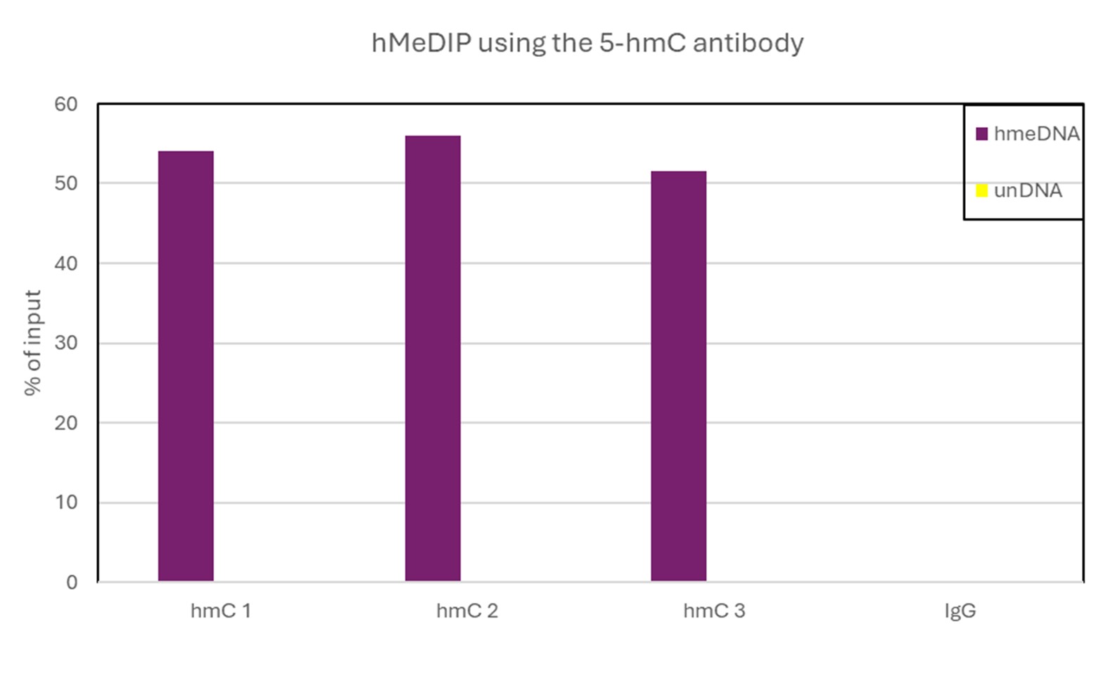
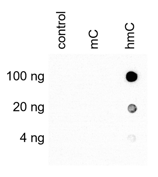
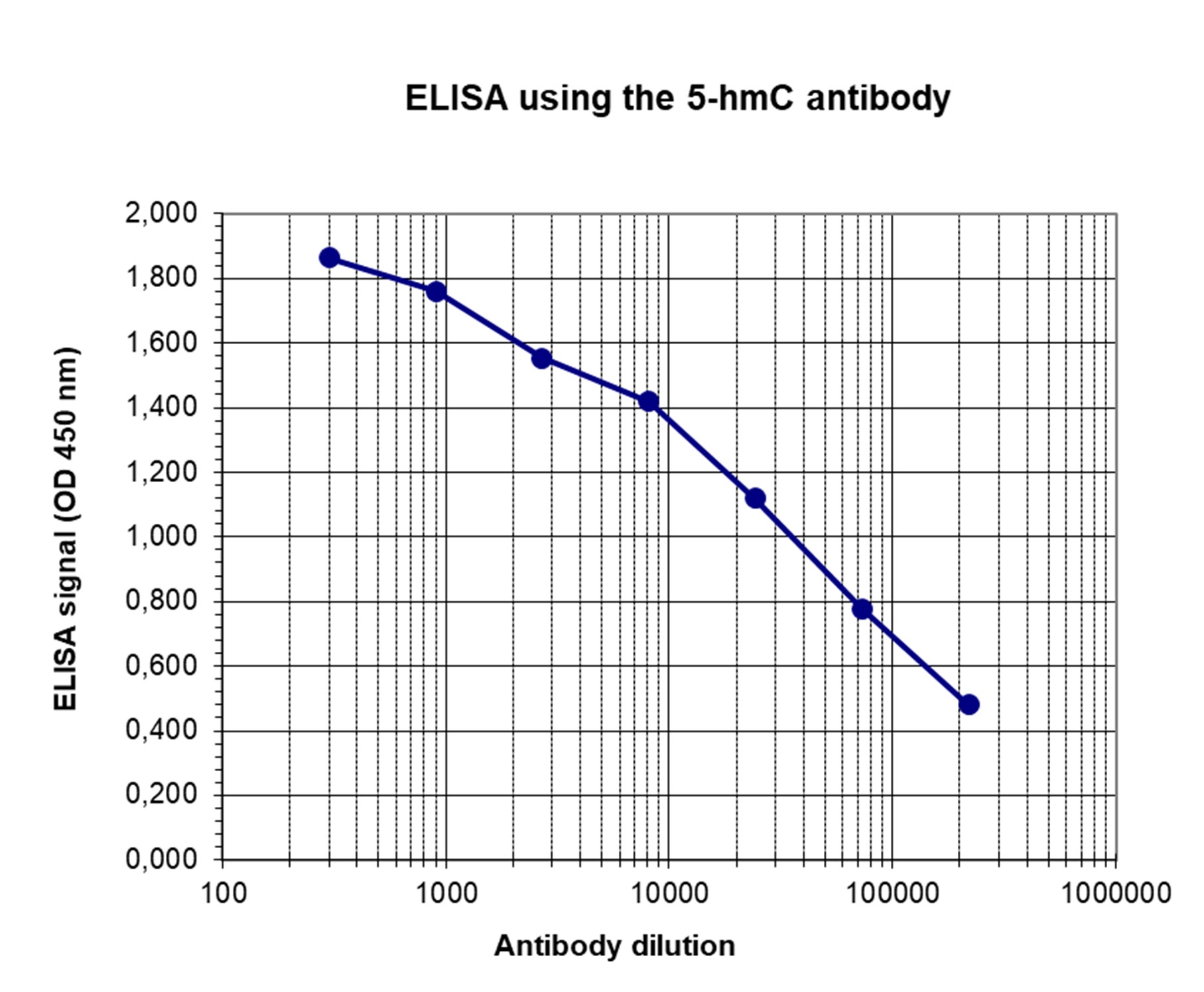
Notice (8): Undefined variable: solution_of_interest [APP/View/Products/view.ctp, line 755]Code Context<!-- BEGIN: REQUEST_FORM MODAL -->
<div id="request_formModal" class="reveal-modal medium" data-reveal aria-labelledby="modalTitle" aria-hidden="true" role="dialog">
<?= $this->element('Forms/simple_form', array('solution_of_interest' => $solution_of_interest, 'header' => $header, 'message' => $message, 'campaign_id' => $campaign_id)) ?>
$viewFile = '/home/website-server/www/app/View/Products/view.ctp'
$dataForView = array(
'language' => 'en',
'meta_keywords' => '',
'meta_description' => '5-hydroxymethylcytosine (5-hmC) Monoclonal Antibody (mouse) validated in hMeDIP, DB and ELISA. Batch-specific data available on the website. Sample size available.',
'meta_title' => '5-hmC Monoclonal Antibody (mouse) | Diagenode',
'product' => array(
'Product' => array(
'id' => '2010',
'antibody_id' => '48',
'name' => '5-hydroxymethylcytosine (5-hmC) Antibody (mouse) ',
'description' => '<p>One of the <strong>only two monoclonal antibodies raised against 5-hydroxymethylcytosine (5-hmC).</strong> 5-hmC is a recently discovered DNA modification which results from the enzymatic conversion of 5-methylcytosine into 5-hydroxymethylcytosine by the TET family of oxygenases. Preliminary results indicate that 5-hmC may have important roles distinct from 5-methylcytosine (5-mC). Although its precise role has still to be shown, early evidence suggests a few putative mechanisms that could have big implications in epigenetics.</p>
<p><strong></strong></p>',
'label1' => 'Validation Data',
'info1' => '<div class="row">
<div class="small-4 columns">
<p><img src="https://www.diagenode.com/img/product/antibodies/C15200200-fig1.png" alt="ChIP" width="180" height="315" caption="false" style="display: block; margin-left: auto; margin-right: auto;" /></p>
</div>
<div class="small-8 columns">
<p><small><strong> Figure 1. An hydroxymethylated DNA IP (hMeDIP) was performed using the Diagenode mouse monoclonal antibody directed against 5-hydroxymethylcytosine (Cat. No. MAb-31HMC-020, MAb-31HMC-050, MAb-31HMC-100).</strong> <br />The IgG isotype antibodies from mouse (Cat. No. kch-819-015) was used as negative control. The DNA was prepared with the GenDNA module of the hMeDIP kit and sonicated with our Bioruptor® (UCD-200/300 series) to have DNA fragments of 300-500 bp. 1 μg of human Hela cells DNA were spiked with non-methylated, methylated, and hydroxymethylated PCR fragments. The IP’d material has been analysed by qPCR using the primer pair specific for the 3 different control sequences. The obtained results show that the mouse monoclonal for 5-hmC is highly specific for this base modification (no IP with non-methylated or methylated C bases containing fragments). </small></p>
</div>
</div>
<div class="row">
<div class="small-4 columns">
<p><img src="https://www.diagenode.com/img/product/antibodies/C15200200-fig2.png" alt="ELISA" width="190" height="173" caption="false" style="display: block; margin-left: auto; margin-right: auto;" /></p>
</div>
<div class="small-8 columns">
<p><small><strong> Figure 2. Determination of the 5-hmC mouse monoclonal antibody titer </strong><br />To determine the titer, an ELISA was performed using a serial dilution of the Diagenode mouse monoclonal antibody directed against 5-hmC (Cat No. MAb-31HMC-050, MAb-31HMC-100) in antigen coated wells. The antigen used was KHL coupled to 5-hmC base. By plotting the absorbance against the antibody dilution, the titer of the antibody was estimated to be 1:40,000. </small></p>
</div>
</div>
<div class="row">
<div class="small-4 columns">
<p><img src="https://www.diagenode.com/img/product/antibodies/C15200200-fig3.png" alt="Dot Blot" width="100" caption="false" style="display: block; margin-left: auto; margin-right: auto;" /></p>
</div>
<div class="small-8 columns">
<p><small><strong> Figure 3. Dotblot analysis of the Diagenode 5-hmC mouse monoclonal antibody with the C, mC and hmC PCR controls </strong><br />200 to 2 ng (equivalent of 10 to 0.1 pmol of C-bases) of the hmC (1), mC (2) and C (3) PCR controls from the Diagenode “5-hmC, 5-mC & cytosine DNA Standard Pack” (Cat No. AF-101-0020) were spotted on a membrane (Amersham Hybond-N+). The membrane was incubated with 2 μg/ml of the mouse 5-hydroxymethylcytosine monoclonal antibody (dilution 1:500). The membranes were exposed for 30 seconds. </small></p>
</div>
</div>',
'label2' => '',
'info2' => '',
'label3' => '',
'info3' => '',
'format' => '100 µg/100 µl',
'catalog_number' => 'C15200200-100',
'old_catalog_number' => 'Mab-31HMC-100',
'sf_code' => 'C15200200-D001-000526',
'type' => 'FRE',
'search_order' => '03-Antibody',
'price_EUR' => '475',
'price_USD' => '475',
'price_GBP' => '425',
'price_JPY' => '69700',
'price_CNY' => '',
'price_AUD' => '1190',
'country' => 'ALL',
'except_countries' => 'None',
'quote' => false,
'in_stock' => false,
'featured' => false,
'no_promo' => false,
'online' => true,
'master' => false,
'last_datasheet_update' => '0000-00-00',
'slug' => '5-hmc-monoclonal-antibody-mouse-classic-100-ug-100-ul',
'meta_title' => '5-hmC Monoclonal Antibody (mouse) | Diagenode',
'meta_keywords' => '',
'meta_description' => '5-hydroxymethylcytosine (5-hmC) Monoclonal Antibody (mouse) validated in hMeDIP, DB and ELISA. Batch-specific data available on the website. Sample size available.',
'modified' => '2022-01-05 15:12:47',
'created' => '2015-06-29 14:08:20',
'locale' => 'eng'
),
'Antibody' => array(
'host' => '*****',
'id' => '48',
'name' => '5-hmC monoclonal antibody (mouse)',
'description' => '5-hydroxymethylcytosine (5-hmC) has been recently discovered in mammalian DNA. This results from the enzymatic conversion of 5-methylcytosine into 5-hydroxymethylcytosine by the TET family of oxygenases. So far, the 5-hmC bases have been identified in Purkinje neurons, in granule cells and embryonic stem cells where they are present at high levels (up to 0,6% of total nucleotides in Purkinje cells).
Preliminary results indicate that 5-hmC may have important roles distinct from 5-mC. Although its precise role has still to be shown, early evidence suggests a few putative mechanisms that could have big implications in epigenetics : 5-hydroxymethylcytosine may well represent a new pathway to demethylate DNA involving a repair mechanism converting 5-hmC to cytosine and, as such open up entirely new perspectives in epigenetic studies.
Due to the structural similarity between 5-mC and 5-hmC, these bases are experimentally almost indistinguishable. Recent articles demonstrated that the most common approaches (e.g. enzymatic approaches, bisulfite sequencing) do not account for 5-hmC. The development of the affinity-based technologies appears to be the most powerful way to differentially and specifically enrich 5-mC and 5-hmC sequences. The results shown here illustrate the use of this unique monoclonal antibody against 5-hydroxymethylcytosine that has been fully validated in various technologies.',
'clonality' => '',
'isotype' => '',
'lot' => '001',
'concentration' => '1.0 µg/µl',
'reactivity' => 'Human, mouse, other (wide range)',
'type' => 'Monoclonal',
'purity' => 'Protein G purified',
'classification' => 'Classic',
'application_table' => '<table>
<thead>
<tr>
<th>Applications</th>
<th>Suggested dilution</th>
<th>References</th>
</tr>
</thead>
<tbody>
<tr>
<td>hMeDIP</td>
<td>2.5 μg/IP</td>
<td>Fig 1</td>
</tr>
<tr>
<td>ELISA</td>
<td>1:500</td>
<td>Fig 2</td>
</tr>
<tr>
<td>Dot Blotting</td>
<td>2 μg / ml</td>
<td>Fig 3</td>
</tr>
</tbody>
</table>',
'storage_conditions' => '',
'storage_buffer' => '',
'precautions' => 'This product is for research use only. Not for use in diagnostic or therapeutic procedures.',
'uniprot_acc' => '',
'slug' => '',
'meta_keywords' => '',
'meta_description' => '',
'modified' => '2016-08-03 10:38:50',
'created' => '0000-00-00 00:00:00',
'select_label' => '48 - 5-hmC monoclonal antibody (mouse) (001 - 1.0 µg/µl - Human, mouse, other (wide range) - Protein G purified - Mouse)'
),
'Slave' => array(),
'Group' => array(
'Group' => array(
[maximum depth reached]
),
'Master' => array(
[maximum depth reached]
),
'Product' => array(
[maximum depth reached]
)
),
'Related' => array(
(int) 0 => array(
[maximum depth reached]
),
(int) 1 => array(
[maximum depth reached]
),
(int) 2 => array(
[maximum depth reached]
),
(int) 3 => array(
[maximum depth reached]
),
(int) 4 => array(
[maximum depth reached]
)
),
'Application' => array(
(int) 0 => array(
[maximum depth reached]
),
(int) 1 => array(
[maximum depth reached]
)
),
'Category' => array(
(int) 0 => array(
[maximum depth reached]
),
(int) 1 => array(
[maximum depth reached]
)
),
'Document' => array(
(int) 0 => array(
[maximum depth reached]
),
(int) 1 => array(
[maximum depth reached]
)
),
'Feature' => array(),
'Image' => array(
(int) 0 => array(
[maximum depth reached]
)
),
'Promotion' => array(),
'Protocol' => array(),
'Publication' => array(
(int) 0 => array(
[maximum depth reached]
),
(int) 1 => array(
[maximum depth reached]
),
(int) 2 => array(
[maximum depth reached]
),
(int) 3 => array(
[maximum depth reached]
),
(int) 4 => array(
[maximum depth reached]
),
(int) 5 => array(
[maximum depth reached]
),
(int) 6 => array(
[maximum depth reached]
),
(int) 7 => array(
[maximum depth reached]
),
(int) 8 => array(
[maximum depth reached]
),
(int) 9 => array(
[maximum depth reached]
),
(int) 10 => array(
[maximum depth reached]
)
),
'Testimonial' => array(),
'Area' => array(),
'SafetySheet' => array(
(int) 0 => array(
[maximum depth reached]
),
(int) 1 => array(
[maximum depth reached]
),
(int) 2 => array(
[maximum depth reached]
),
(int) 3 => array(
[maximum depth reached]
),
(int) 4 => array(
[maximum depth reached]
),
(int) 5 => array(
[maximum depth reached]
),
(int) 6 => array(
[maximum depth reached]
),
(int) 7 => array(
[maximum depth reached]
)
)
),
'meta_canonical' => 'https://www.diagenode.com/en/p/5-hmc-monoclonal-antibody-mouse-classic-50-ug-50-ul'
)
$language = 'en'
$meta_keywords = ''
$meta_description = '5-hydroxymethylcytosine (5-hmC) Monoclonal Antibody (mouse) validated in hMeDIP, DB and ELISA. Batch-specific data available on the website. Sample size available.'
$meta_title = '5-hmC Monoclonal Antibody (mouse) | Diagenode'
$product = array(
'Product' => array(
'id' => '2010',
'antibody_id' => '48',
'name' => '5-hydroxymethylcytosine (5-hmC) Antibody (mouse) ',
'description' => '<p>One of the <strong>only two monoclonal antibodies raised against 5-hydroxymethylcytosine (5-hmC).</strong> 5-hmC is a recently discovered DNA modification which results from the enzymatic conversion of 5-methylcytosine into 5-hydroxymethylcytosine by the TET family of oxygenases. Preliminary results indicate that 5-hmC may have important roles distinct from 5-methylcytosine (5-mC). Although its precise role has still to be shown, early evidence suggests a few putative mechanisms that could have big implications in epigenetics.</p>
<p><strong></strong></p>',
'label1' => 'Validation Data',
'info1' => '<div class="row">
<div class="small-4 columns">
<p><img src="https://www.diagenode.com/img/product/antibodies/C15200200-fig1.png" alt="ChIP" width="180" height="315" caption="false" style="display: block; margin-left: auto; margin-right: auto;" /></p>
</div>
<div class="small-8 columns">
<p><small><strong> Figure 1. An hydroxymethylated DNA IP (hMeDIP) was performed using the Diagenode mouse monoclonal antibody directed against 5-hydroxymethylcytosine (Cat. No. MAb-31HMC-020, MAb-31HMC-050, MAb-31HMC-100).</strong> <br />The IgG isotype antibodies from mouse (Cat. No. kch-819-015) was used as negative control. The DNA was prepared with the GenDNA module of the hMeDIP kit and sonicated with our Bioruptor® (UCD-200/300 series) to have DNA fragments of 300-500 bp. 1 μg of human Hela cells DNA were spiked with non-methylated, methylated, and hydroxymethylated PCR fragments. The IP’d material has been analysed by qPCR using the primer pair specific for the 3 different control sequences. The obtained results show that the mouse monoclonal for 5-hmC is highly specific for this base modification (no IP with non-methylated or methylated C bases containing fragments). </small></p>
</div>
</div>
<div class="row">
<div class="small-4 columns">
<p><img src="https://www.diagenode.com/img/product/antibodies/C15200200-fig2.png" alt="ELISA" width="190" height="173" caption="false" style="display: block; margin-left: auto; margin-right: auto;" /></p>
</div>
<div class="small-8 columns">
<p><small><strong> Figure 2. Determination of the 5-hmC mouse monoclonal antibody titer </strong><br />To determine the titer, an ELISA was performed using a serial dilution of the Diagenode mouse monoclonal antibody directed against 5-hmC (Cat No. MAb-31HMC-050, MAb-31HMC-100) in antigen coated wells. The antigen used was KHL coupled to 5-hmC base. By plotting the absorbance against the antibody dilution, the titer of the antibody was estimated to be 1:40,000. </small></p>
</div>
</div>
<div class="row">
<div class="small-4 columns">
<p><img src="https://www.diagenode.com/img/product/antibodies/C15200200-fig3.png" alt="Dot Blot" width="100" caption="false" style="display: block; margin-left: auto; margin-right: auto;" /></p>
</div>
<div class="small-8 columns">
<p><small><strong> Figure 3. Dotblot analysis of the Diagenode 5-hmC mouse monoclonal antibody with the C, mC and hmC PCR controls </strong><br />200 to 2 ng (equivalent of 10 to 0.1 pmol of C-bases) of the hmC (1), mC (2) and C (3) PCR controls from the Diagenode “5-hmC, 5-mC & cytosine DNA Standard Pack” (Cat No. AF-101-0020) were spotted on a membrane (Amersham Hybond-N+). The membrane was incubated with 2 μg/ml of the mouse 5-hydroxymethylcytosine monoclonal antibody (dilution 1:500). The membranes were exposed for 30 seconds. </small></p>
</div>
</div>',
'label2' => 'Target description',
'info2' => '<p>5-hydroxymethylcytosine (5-hmC) has been recently discovered in mammalian DNA. This results from the enzymatic conversion of 5-methylcytosine into 5-hydroxymethylcytosine by the TET family of oxygenases. So far, the 5-hmC bases have been identified in Purkinje neurons, in granule cells and embryonic stem cells where they are present at high levels (up to 0,6% of total nucleotides in Purkinje cells).</p>
<p>Preliminary results indicate that 5-hmC may have important roles distinct from 5-mC. Although its precise role has still to be shown, early evidence suggests a few putative mechanisms that could have big implications in epigenetics : 5-hydroxymethylcytosine may well represent a new pathway to demethylate DNA involving a repair mechanism converting 5-hmC to cytosine and, as such open up entirely new perspectives in epigenetic studies.</p>
<p>Due to the structural similarity between 5-mC and 5-hmC, these bases are experimentally almost indistinguishable. Recent articles demonstrated that the most common approaches (e.g. enzymatic approaches, bisulfite sequencing) do not account for 5-hmC. The development of the affinity-based technologies appears to be the most powerful way to differentially and specifically enrich 5-mC and 5-hmC sequences. The results shown here illustrate the use of this unique monoclonal antibody against 5-hydroxymethylcytosine that has been fully validated in various technologies.</p>',
'label3' => '',
'info3' => '',
'format' => '100 µg/100 µl',
'catalog_number' => 'C15200200-100',
'old_catalog_number' => 'Mab-31HMC-100',
'sf_code' => 'C15200200-D001-000526',
'type' => 'FRE',
'search_order' => '03-Antibody',
'price_EUR' => '475',
'price_USD' => '475',
'price_GBP' => '425',
'price_JPY' => '69700',
'price_CNY' => '',
'price_AUD' => '1190',
'country' => 'ALL',
'except_countries' => 'None',
'quote' => false,
'in_stock' => false,
'featured' => false,
'no_promo' => false,
'online' => true,
'master' => false,
'last_datasheet_update' => '0000-00-00',
'slug' => '5-hmc-monoclonal-antibody-mouse-classic-100-ug-100-ul',
'meta_title' => '5-hmC Monoclonal Antibody (mouse) | Diagenode',
'meta_keywords' => '',
'meta_description' => '5-hydroxymethylcytosine (5-hmC) Monoclonal Antibody (mouse) validated in hMeDIP, DB and ELISA. Batch-specific data available on the website. Sample size available.',
'modified' => '2022-01-05 15:12:47',
'created' => '2015-06-29 14:08:20',
'locale' => 'eng'
),
'Antibody' => array(
'host' => '*****',
'id' => '48',
'name' => '5-hmC monoclonal antibody (mouse)',
'description' => '5-hydroxymethylcytosine (5-hmC) has been recently discovered in mammalian DNA. This results from the enzymatic conversion of 5-methylcytosine into 5-hydroxymethylcytosine by the TET family of oxygenases. So far, the 5-hmC bases have been identified in Purkinje neurons, in granule cells and embryonic stem cells where they are present at high levels (up to 0,6% of total nucleotides in Purkinje cells).
Preliminary results indicate that 5-hmC may have important roles distinct from 5-mC. Although its precise role has still to be shown, early evidence suggests a few putative mechanisms that could have big implications in epigenetics : 5-hydroxymethylcytosine may well represent a new pathway to demethylate DNA involving a repair mechanism converting 5-hmC to cytosine and, as such open up entirely new perspectives in epigenetic studies.
Due to the structural similarity between 5-mC and 5-hmC, these bases are experimentally almost indistinguishable. Recent articles demonstrated that the most common approaches (e.g. enzymatic approaches, bisulfite sequencing) do not account for 5-hmC. The development of the affinity-based technologies appears to be the most powerful way to differentially and specifically enrich 5-mC and 5-hmC sequences. The results shown here illustrate the use of this unique monoclonal antibody against 5-hydroxymethylcytosine that has been fully validated in various technologies.',
'clonality' => '',
'isotype' => '',
'lot' => '001',
'concentration' => '1.0 µg/µl',
'reactivity' => 'Human, mouse, other (wide range)',
'type' => 'Monoclonal',
'purity' => 'Protein G purified',
'classification' => 'Classic',
'application_table' => '<table>
<thead>
<tr>
<th>Applications</th>
<th>Suggested dilution</th>
<th>References</th>
</tr>
</thead>
<tbody>
<tr>
<td>hMeDIP</td>
<td>2.5 μg/IP</td>
<td>Fig 1</td>
</tr>
<tr>
<td>ELISA</td>
<td>1:500</td>
<td>Fig 2</td>
</tr>
<tr>
<td>Dot Blotting</td>
<td>2 μg / ml</td>
<td>Fig 3</td>
</tr>
</tbody>
</table>',
'storage_conditions' => '',
'storage_buffer' => '',
'precautions' => 'This product is for research use only. Not for use in diagnostic or therapeutic procedures.',
'uniprot_acc' => '',
'slug' => '',
'meta_keywords' => '',
'meta_description' => '',
'modified' => '2016-08-03 10:38:50',
'created' => '0000-00-00 00:00:00',
'select_label' => '48 - 5-hmC monoclonal antibody (mouse) (001 - 1.0 µg/µl - Human, mouse, other (wide range) - Protein G purified - Mouse)'
),
'Slave' => array(),
'Group' => array(
'Group' => array(
'id' => '104',
'name' => 'C15200200',
'product_id' => '2009',
'modified' => '2016-02-19 16:52:05',
'created' => '2016-02-19 16:52:05'
),
'Master' => array(
'id' => '2009',
'antibody_id' => '47',
'name' => '5-hydroxymethylcytosine (5-hmC) Antibody (mouse) ',
'description' => '<p>One of the <strong>only two monoclonal antibodies raised against 5-hydroxymethylcytosine (5-hmC).</strong> 5-hmC is a recently discovered DNA modification which results from the enzymatic conversion of 5-methylcytosine into 5-hydroxymethylcytosine by the TET family of oxygenases. Preliminary results indicate that 5-hmC may have important roles distinct from 5-methylcytosine (5-mC). Although its precise role has still to be shown, early evidence suggests a few putative mechanisms that could have big implications in epigenetics.</p>
<p><strong></strong></p>',
'label1' => 'Validation Data',
'info1' => '<div class="row">
<div class="small-4 columns">
<p><img src="https://www.diagenode.com/img/product/antibodies/C15200200-fig1.png" alt="ChIP" width="160" caption="false" height="280" style="display: block; margin-left: auto; margin-right: auto;" /></p>
</div>
<div class="small-8 columns">
<p><small><strong> Figure 1. An hydroxymethylated DNA IP (hMeDIP) was performed using the Diagenode mouse monoclonal antibody directed against 5-hydroxymethylcytosine (Cat. No. MAb-31HMC-020, MAb-31HMC-050, MAb-31HMC-100).</strong> <br />The IgG isotype antibodies from mouse (Cat. No. kch-819-015) was used as negative control. The DNA was prepared with the GenDNA module of the hMeDIP kit and sonicated with our Bioruptor® (UCD-200/300 series) to have DNA fragments of 300-500 bp. 1 μg of human Hela cells DNA were spiked with non-methylated, methylated, and hydroxymethylated PCR fragments. The IP’d material has been analysed by qPCR using the primer pair specific for the 3 different control sequences. The obtained results show that the mouse monoclonal for 5-hmC is highly specific for this base modification (no IP with non-methylated or methylated C bases containing fragments). </small></p>
</div>
</div>
<div class="row">
<div class="small-4 columns">
<p><img src="https://www.diagenode.com/img/product/antibodies/C15200200-fig2.png" alt="ELISA" width="190" caption="false" style="display: block; margin-left: auto; margin-right: auto;" /></p>
</div>
<div class="small-8 columns">
<p><small><strong> Figure 2. Determination of the 5-hmC mouse monoclonal antibody titer </strong><br />To determine the titer, an ELISA was performed using a serial dilution of the Diagenode mouse monoclonal antibody directed against 5-hmC (Cat No. MAb-31HMC-050, MAb-31HMC-100) in antigen coated wells. The antigen used was KHL coupled to 5-hmC base. By plotting the absorbance against the antibody dilution, the titer of the antibody was estimated to be 1:40,000. </small></p>
</div>
</div>
<div class="row">
<div class="small-4 columns">
<p><img src="https://www.diagenode.com/img/product/antibodies/C15200200-fig3.png" alt="Dot Blot" width="100" caption="false" height="137" style="display: block; margin-left: auto; margin-right: auto;" /></p>
</div>
<div class="small-8 columns">
<p><small><strong> Figure 3. Dotblot analysis of the Diagenode 5-hmC mouse monoclonal antibody with the C, mC and hmC PCR controls </strong><br />200 to 2 ng (equivalent of 10 to 0.1 pmol of C-bases) of the hmC (1), mC (2) and C (3) PCR controls from the Diagenode “5-hmC, 5-mC & cytosine DNA Standard Pack” (Cat No. AF-101-0020) were spotted on a membrane (Amersham Hybond-N+). The membrane was incubated with 2 μg/ml of the mouse 5-hydroxymethylcytosine monoclonal antibody (dilution 1:500). The membranes were exposed for 30 seconds. </small></p>
</div>
</div>',
'label2' => 'Target description',
'info2' => '<p>5-hydroxymethylcytosine (5-hmC) has been recently discovered in mammalian DNA. This results from the enzymatic conversion of 5-methylcytosine into 5-hydroxymethylcytosine by the TET family of oxygenases. So far, the 5-hmC bases have been identified in Purkinje neurons, in granule cells and embryonic stem cells where they are present at high levels (up to 0,6% of total nucleotides in Purkinje cells).</p>
<p>Preliminary results indicate that 5-hmC may have important roles distinct from 5-mC. Although its precise role has still to be shown, early evidence suggests a few putative mechanisms that could have big implications in epigenetics : 5-hydroxymethylcytosine may well represent a new pathway to demethylate DNA involving a repair mechanism converting 5-hmC to cytosine and, as such open up entirely new perspectives in epigenetic studies.</p>
<p>Due to the structural similarity between 5-mC and 5-hmC, these bases are experimentally almost indistinguishable. Recent articles demonstrated that the most common approaches (e.g. enzymatic approaches, bisulfite sequencing) do not account for 5-hmC. The development of the affinity-based technologies appears to be the most powerful way to differentially and specifically enrich 5-mC and 5-hmC sequences. The results shown here illustrate the use of this unique monoclonal antibody against 5-hydroxymethylcytosine that has been fully validated in various technologies.</p>',
'label3' => '',
'info3' => '',
'format' => '50 µg/50 µl',
'catalog_number' => 'C15200200-50',
'old_catalog_number' => 'Mab-31HMC-050',
'sf_code' => 'C15200200-D001-000581',
'type' => 'FRE',
'search_order' => '03-Antibody',
'price_EUR' => '360',
'price_USD' => '360',
'price_GBP' => '320',
'price_JPY' => '52900',
'price_CNY' => '',
'price_AUD' => '900',
'country' => 'ALL',
'except_countries' => 'None',
'quote' => false,
'in_stock' => false,
'featured' => false,
'no_promo' => false,
'online' => true,
'master' => true,
'last_datasheet_update' => '0000-00-00',
'slug' => '5-hmc-monoclonal-antibody-mouse-classic-50-ug-50-ul',
'meta_title' => '5-hydroxymethylcytosine (5-hmC) Monoclonal Antibody (mouse) | Diagenode',
'meta_keywords' => '5-hydroxymethylcytosine,monoclonal antibody ,Diagenode',
'meta_description' => '5-hydroxymethylcytosine (5-hmC) Monoclonal Antibody (mouse) validated in hMeDIP, DB and ELISA. Batch-specific data available on the website. Sample size available.',
'modified' => '2022-01-05 15:12:18',
'created' => '2015-06-29 14:08:20'
),
'Product' => array(
(int) 0 => array(
[maximum depth reached]
),
(int) 1 => array(
[maximum depth reached]
)
)
),
'Related' => array(
(int) 0 => array(
'id' => '1882',
'antibody_id' => null,
'name' => 'hMeDIP kit x16 (monoclonal mouse antibody)',
'description' => '<p><a href="https://www.diagenode.com/files/products/kits/hMeDIP_kit_manual.pdf"><img src="https://www.diagenode.com/img/buttons/bt-manual.png" /></a></p>
<p><span>The hMeDIP kit is designed for enrichment of hydroxymethylated DNA from fragmented genomic DNA<span><span> </span>samples for use in genome-wide methylation analysis. It features</span></span><span> a highly specific monoclonal antibody against </span>5-hydroxymethylcytosine (5-hmC) for the immunoprecipitation of hydroxymethylated DNA<span>. It includes control DNA and primers to assess the effiency of the assay. </span>Performing hydroxymethylation profiling with the hMeDIP kit is fast, reliable and highly specific.</p>
<p><em>Looking for hMeDIP-seq protocol? <a href="https://go.diagenode.com/l/928883/2022-01-07/2m1ht" target="_blank" title="Contact us">Contact us</a></em></p>
<p><span></span></p>
<p><span></span></p>',
'label1' => 'Characteristics',
'info1' => '<ul style="list-style-type: disc;">
<li><span>Robust enrichment & immunoprecipitation of hydroxymethylated DNA</span></li>
<li>Highly specific monoclonal antibody against 5-hmC<span> for reliable, reproducible results</span></li>
<li>Including control DNA and primers to <span>monitor the efficiency of the assay</span>
<ul style="list-style-type: circle;">
<li>hmeDNA and unmethylated DNA sequences and primer pairs</li>
<li>Mouse primer pairs against Sfi1 targeting hydroxymethylated gene in mouse</li>
</ul>
</li>
<li>Improved single-tube, magnetic bead-based protocol</li>
</ul>',
'label2' => '',
'info2' => '',
'label3' => '',
'info3' => '',
'format' => '16 rxns',
'catalog_number' => 'C02010031',
'old_catalog_number' => 'AF-110-0016',
'sf_code' => 'C02010031-',
'type' => 'RFR',
'search_order' => '04-undefined',
'price_EUR' => '595',
'price_USD' => '665',
'price_GBP' => '550',
'price_JPY' => '104100',
'price_CNY' => '',
'price_AUD' => '1665',
'country' => 'ALL',
'except_countries' => 'None',
'quote' => false,
'in_stock' => false,
'featured' => true,
'no_promo' => false,
'online' => true,
'master' => true,
'last_datasheet_update' => '0000-00-00',
'slug' => 'hmedip-kit-x16-monoclonal-mouse-antibody-16-rxns',
'meta_title' => 'hMeDIP kit x16 (monoclonal mouse antibody)',
'meta_keywords' => '',
'meta_description' => 'hMeDIP kit x16 (monoclonal mouse antibody)',
'modified' => '2023-04-20 16:12:48',
'created' => '2015-06-29 14:08:20',
'ProductsRelated' => array(
[maximum depth reached]
),
'Image' => array(
[maximum depth reached]
)
),
(int) 1 => array(
'id' => '1885',
'antibody_id' => null,
'name' => 'Auto hMeDIP kit x16 (monoclonal mouse antibody)',
'description' => '<p><span>The Auro hMeDIP kit is designed for enrichment of hydroxymethylated DNA from fragmented genomic DNA samples for use in genome-wide methylation analysis. It features</span><span> a highly specific monoclonal antibody against </span><span>5-hydroxymethylcytosine (5-hmC) for the immunoprecipitation of hydroxymethylated DNA</span><span>. It includes control DNA and primers to assess the effiency of the assay. </span><span>Performing hydroxymethylation profiling with the hMeDIP kit is fast, reliable and highly specific.</span></p>',
'label1' => ' Characteristics',
'info1' => '<ul style="list-style-type: disc;">
<li><span>Robust enrichment & immunoprecipitation of hydroxymethylated DNA</span></li>
<li>Highly specific monoclonal antibody against 5-hmC<span> for reliable, reproducible results</span></li>
<li>Including control DNA and primers to <span>monitor the efficiency of the assay</span>
<ul style="list-style-type: circle;">
<li>5-hmC, 5-mC and unmethylated DNA sequences and primer pairs</li>
<li>Mouse primer pairs against Sfi1 targeting hydroxymethylated gene in mouse</li>
</ul>
</li>
</ul>
<ul style="list-style-type: disc;">
<li>Improved single-tube, magnetic bead-based protocol</li>
</ul>',
'label2' => '',
'info2' => '',
'label3' => '',
'info3' => '',
'format' => '16 rxns',
'catalog_number' => 'C02010034',
'old_catalog_number' => '',
'sf_code' => 'C02010034-',
'type' => 'RFR',
'search_order' => '04-undefined',
'price_EUR' => '595',
'price_USD' => '665',
'price_GBP' => '550',
'price_JPY' => '104100',
'price_CNY' => '',
'price_AUD' => '1665',
'country' => 'ALL',
'except_countries' => 'Japan',
'quote' => false,
'in_stock' => false,
'featured' => false,
'no_promo' => false,
'online' => true,
'master' => true,
'last_datasheet_update' => '0000-00-00',
'slug' => 'auto-hmedip-kit-x16-monoclonal-mouse-antibody-16-rxns',
'meta_title' => 'Auto hMeDIP kit x16 (monoclonal mouse antibody)',
'meta_keywords' => '',
'meta_description' => 'Auto hMeDIP kit x16 (monoclonal mouse antibody)',
'modified' => '2021-01-18 10:37:19',
'created' => '2015-06-29 14:08:20',
'ProductsRelated' => array(
[maximum depth reached]
),
'Image' => array(
[maximum depth reached]
)
),
(int) 2 => array(
'id' => '1887',
'antibody_id' => null,
'name' => 'MethylCap kit',
'description' => '<p>The MethylCap kit allows to specifically capture DNA fragments containing methylated CpGs. The assay is based on the affinity purification of methylated DNA using methyl-CpG-binding domain (MBD) of human MeCP2 protein. The procedure has been adapted to both manual process or <a href="https://www.diagenode.com/en/p/sx-8g-ip-star-compact-automated-system-1-unit">IP-Star® Compact Automated System</a>. Libraries of captured methylated DNA can be prepared for next-generation sequencing (NGS) by combining MBD technology with the <a href="https://www.diagenode.com/en/p/microplex-lib-prep-kit-v3-48-rxns">MicroPlex Library Preparation Kit v3</a>.</p>',
'label1' => 'Characteristics',
'info1' => '<ul style="list-style-type: circle;">
<li><strong>Fast & sensitive capture</strong> of methylated DNA</li>
<li><strong>High capture efficiency</strong></li>
<li><strong>Differential fractionation</strong> of methylated DNA by CpG density (3 eluted fractions)</li>
<li><strong>On-day protocol</strong></li>
<li><strong>NGS compatibility</strong></li>
</ul>
<h3>MBD-seq allows for detection of genomic regions with different CpG density</h3>
<p><img src="https://www.diagenode.com/img/product/kits/mbd_results1.png" alt="MBD-sequencing results have been validated by bisulfite sequencing" style="display: block; margin-left: auto; margin-right: auto;" /></p>
<p><strong></strong></p>
<p><strong></strong><strong>F</strong><strong>igure 1.</strong> Using the MBD approach, two methylated regions were detected in different elution fractions according to their methylated CpG density (A). Low, Medium and High refer to the sequenced DNA from different elution fractions with increasing salt concentration. Methylated patterns of these two different methylated regions were validated by bisulfite conversion assay (B).</p>',
'label2' => '',
'info2' => '',
'label3' => '',
'info3' => '',
'format' => '48 rxns',
'catalog_number' => 'C02020010',
'old_catalog_number' => 'AF-100-0048',
'sf_code' => 'C02020010-',
'type' => 'RFR',
'search_order' => '04-undefined',
'price_EUR' => '705',
'price_USD' => '665',
'price_GBP' => '645',
'price_JPY' => '123000',
'price_CNY' => '',
'price_AUD' => '1665',
'country' => 'ALL',
'except_countries' => 'None',
'quote' => false,
'in_stock' => false,
'featured' => true,
'no_promo' => false,
'online' => true,
'master' => true,
'last_datasheet_update' => '0000-00-00',
'slug' => 'methylcap-kit-x48-48-rxns',
'meta_title' => 'MethylCap kit x48',
'meta_keywords' => '',
'meta_description' => 'MethylCap kit x48',
'modified' => '2021-02-19 17:55:38',
'created' => '2015-06-29 14:08:20',
'ProductsRelated' => array(
[maximum depth reached]
),
'Image' => array(
[maximum depth reached]
)
),
(int) 3 => array(
'id' => '1888',
'antibody_id' => null,
'name' => 'Auto MethylCap kit',
'description' => '<p>The Auto MethylCap kit allows to specifically capture DNA fragments containing methylated CpGs. The assay is based on the affinity purification of methylated DNA using methyl-CpG-binding domain (MBD) of human MeCP2 protein. This procedure has been optimized to perform automated immunoprecipitation of chromatin using the <a href="https://www.diagenode.com/en/p/sx-8g-ip-star-compact-automated-system-1-unit">IP-Star® Compact Automated System</a> enabling highly reproducible results and allowing for high throughput.</p>',
'label1' => ' Characteristics',
'info1' => '<ul>
<li><strong>Fast & sensitive capture</strong> of methylated DNA</li>
<li><strong>High capture efficiency</strong></li>
<li><strong>Differential fractionation</strong> of methylated DNA by CpG density (3 eluted fractions)</li>
<li><strong>Automation compatibility</strong><strong></strong>
<h3>MBD-seq allows for detection of genomic regions with different CpG density</h3>
<p><img src="https://www.diagenode.com/img/product/kits/mbd_results1.png" alt="MBD-sequencing results have been validated by bisulfite sequencing" style="display: block; margin-left: auto; margin-right: auto;" /></p>
<p><strong>F</strong><strong>igure 1.</strong><span> </span>Using the MBD approach, two methylated regions were detected in different elution fractions according to their methylated CpG density (A). Low, Medium and High refer to the sequenced DNA from different elution fractions with increasing salt concentration. Methylated patterns of these two different methylated regions were validated by bisulfite conversion assay (B).<br /><strong></strong></p>
</li>
</ul>',
'label2' => '',
'info2' => '',
'label3' => '',
'info3' => '',
'format' => '48 rxns',
'catalog_number' => 'C02020011',
'old_catalog_number' => 'AF-Auto01-0048',
'sf_code' => 'C02020011-',
'type' => 'RFR',
'search_order' => '04-undefined',
'price_EUR' => '705',
'price_USD' => '665',
'price_GBP' => '645',
'price_JPY' => '123000',
'price_CNY' => '',
'price_AUD' => '1665',
'country' => 'ALL',
'except_countries' => 'Japan',
'quote' => false,
'in_stock' => false,
'featured' => false,
'no_promo' => false,
'online' => true,
'master' => true,
'last_datasheet_update' => '0000-00-00',
'slug' => 'auto-methylcap-kit-x48-48-rxns',
'meta_title' => 'Auto MethylCap kit x48',
'meta_keywords' => '',
'meta_description' => 'Auto MethylCap kit x48',
'modified' => '2020-09-17 13:33:12',
'created' => '2015-06-29 14:08:20',
'ProductsRelated' => array(
[maximum depth reached]
),
'Image' => array(
[maximum depth reached]
)
),
(int) 4 => array(
'id' => '1892',
'antibody_id' => null,
'name' => 'Premium Bisulfite kit',
'description' => '<p style="text-align: center;"><a href="https://www.diagenode.com/files/products/kits/Premium_Bisulfite_kit_manual.pdf"><img src="https://www.diagenode.com/img/buttons/bt-manual.png" /></a></p>
<p style="text-align: center;"><strong>Make your Bisulfite conversion now in only 60 minutes !</strong></p>
<p>Diagenode's Premium Bisulfite Kit rapidly converts DNA through bisulfite treatment. Our conversion reagent is added directly to DNA, requires no intermediate steps, and results in high yields of DNA ready for downstream analysis methods including PCR and Next-Generation Sequencing.</p>',
'label1' => '',
'info1' => '',
'label2' => '',
'info2' => '',
'label3' => '',
'info3' => '',
'format' => '50 rxns',
'catalog_number' => 'C02030030',
'old_catalog_number' => '',
'sf_code' => 'C02030030-',
'type' => 'REF',
'search_order' => '04-undefined',
'price_EUR' => '240',
'price_USD' => '225',
'price_GBP' => '220',
'price_JPY' => '41900',
'price_CNY' => '',
'price_AUD' => '565',
'country' => 'ALL',
'except_countries' => 'None',
'quote' => false,
'in_stock' => false,
'featured' => true,
'no_promo' => false,
'online' => true,
'master' => true,
'last_datasheet_update' => '0000-00-00',
'slug' => 'premium-bisulfite-kit-50-rxns',
'meta_title' => 'Premium Bisulfite kit',
'meta_keywords' => '',
'meta_description' => 'Premium Bisulfite kit',
'modified' => '2023-04-20 16:13:50',
'created' => '2015-06-29 14:08:20',
'ProductsRelated' => array(
[maximum depth reached]
),
'Image' => array([maximum depth reached])
)
),
'Application' => array(
(int) 0 => array(
'id' => '28',
'position' => '10',
'parent_id' => '40',
'name' => 'DB',
'description' => '<p>Dot blotting</p>',
'in_footer' => false,
'in_menu' => false,
'online' => true,
'tabular' => true,
'slug' => 'dot-blotting',
'meta_keywords' => 'Dot blotting,Monoclonal & Polyclonal antibody,',
'meta_description' => 'Diagenode offers Monoclonal & Polyclonal antibodies for Dot blotting applications',
'meta_title' => 'Dot blotting Antibodies - Monoclonal & Polyclonal antibody | Diagenode',
'modified' => '2016-01-13 14:40:49',
'created' => '2015-07-08 13:45:05',
'ProductsApplication' => array(
[maximum depth reached]
)
),
(int) 1 => array(
'id' => '20',
'position' => '10',
'parent_id' => '40',
'name' => 'ELISA',
'description' => '<div class="row">
<div class="small-12 medium-12 large-12 columns">Enzyme-linked immunosorbent assay.</div>
</div>',
'in_footer' => false,
'in_menu' => false,
'online' => true,
'tabular' => true,
'slug' => 'elisa-antibodies',
'meta_keywords' => ' ELISA Antibodies,Monoclonal antibody, Polyclonal antibody',
'meta_description' => 'Diagenode offers Monoclonal & Polyclonal antibodies for ELISA applications',
'meta_title' => 'ELISA Antibodies - Monoclonal & Polyclonal antibody | Diagenode',
'modified' => '2016-01-13 12:21:41',
'created' => '2014-07-08 08:13:28',
'ProductsApplication' => array(
[maximum depth reached]
)
)
),
'Category' => array(
(int) 0 => array(
'id' => '23',
'position' => '40',
'parent_id' => '4',
'name' => 'DNA modifications',
'description' => '<p><span style="font-weight: 400;">T</span><span style="font-weight: 400;">he pattern of <strong>DNA modifications</strong> is critical for genome stability and the control of gene expression in the cell. Methylation of 5-cytosine (5-mC), one of the best-studied epigenetic marks, is carried out by the <strong>DNA methyltransferases</strong> DNMT3A and B and DNMT1. DNMT3A and DNMT3B are responsible for </span><i><span style="font-weight: 400;">de novo</span></i><span style="font-weight: 400;"> DNA methylation, whereas DNMT1 maintains existing methylation. 5-mC undergoes active demethylation which is performed by the <strong>Ten-Eleven Translocation</strong> (TET) familly of DNA hydroxylases. The latter consists of 3 members TET1, 2 and 3. All 3 members catalyze the conversion of <strong>5-methylcytosine</strong> (5-mC) into <strong>5-hydroxymethylcytosine</strong> (5-hmC), and further into <strong>5-formylcytosine</strong> (5-fC) and <strong>5-carboxycytosine</strong> (5-caC). 5-fC and 5-caC can be converted to unmodified cytosine by <strong>Thymine DNA Glycosylase</strong> (TDG). It is not yet clear if 5-hmC, 5-fC and 5-caC have specific functions or are simply intermediates in the demethylation of 5-mC.</span></p>
<p><span style="font-weight: 400;">DNA methylation is generally considered as a repressive mark and is usually associated with gene silencing. It is essential that the balance between DNA methylation and demethylation is precisely maintained. Dysregulation of DNA methylation may lead to many different human diseases and is often observed in cancer cells.</span></p>
<p><span style="font-weight: 400;">Diagenode offers highly validated antibodies against different proteins involved in DNA modifications as well as against the modified bases allowing the study of all steps and intermediates in the DNA methylation/demethylation pathway:</span></p>
<p><img src="https://www.diagenode.com/img/categories/antibodies/dna-methylation.jpg" height="599" width="816" /></p>
<p><strong>Diagenode exclusively sources the original 5-methylcytosine monoclonal antibody (clone 33D3).</strong></p>
<p>Check out the list below to see all proposed antibodies for DNA modifications.</p>
<p>Diagenode’s highly validated antibodies:</p>
<ul>
<li>Highly sensitive and specific</li>
<li>Cost-effective (requires less antibody per reaction)</li>
<li>Batch-specific data is available on the website</li>
<li>Expert technical support</li>
<li>Sample sizes available</li>
<li>100% satisfaction guarantee</li>
</ul>',
'no_promo' => false,
'in_menu' => true,
'online' => true,
'tabular' => false,
'hide' => true,
'all_format' => false,
'is_antibody' => true,
'slug' => 'dna-methylation-antibody',
'cookies_tag_id' => null,
'meta_keywords' => 'Monoclonal Antibodies,Polyclonal antibodies,DNA methylation,Diagenode',
'meta_description' => 'Diagenode offers Monoclonal and Polyclonal antibodies for DNA Methylation. The pattern of DNA modifications is critical for genome stability and the control of gene expression in the cell. ',
'meta_title' => 'DNA modifications - Monoclonal and Polyclonal Antibodies for DNA Methylation | Diagenode',
'modified' => '2020-08-03 15:25:37',
'created' => '2014-07-31 03:51:53',
'ProductsCategory' => array(
[maximum depth reached]
),
'CookiesTag' => array([maximum depth reached])
),
(int) 1 => array(
'id' => '103',
'position' => '0',
'parent_id' => '4',
'name' => 'All antibodies',
'description' => '<p><span style="font-weight: 400;">All Diagenode’s antibodies are listed below. Please, use our Quick search field to find the antibody of interest by target name, application, purity.</span></p>
<p><span style="font-weight: 400;">Diagenode’s highly validated antibodies:</span></p>
<ul>
<li>Highly sensitive and specific</li>
<li>Cost-effective (requires less antibody per reaction)</li>
<li>Batch-specific data is available on the website</li>
<li>Expert technical support</li>
<li>Sample sizes available</li>
<li>100% satisfaction guarantee</li>
</ul>',
'no_promo' => false,
'in_menu' => true,
'online' => true,
'tabular' => false,
'hide' => true,
'all_format' => false,
'is_antibody' => true,
'slug' => 'all-antibodies',
'cookies_tag_id' => null,
'meta_keywords' => 'Antibodies,Premium Antibodies,Classic,Pioneer',
'meta_description' => 'Diagenode Offers Strict quality standards with Rigorous QC and validated Antibodies. Classified based on level of validation for flexibility of Application. Comprehensive selection of histone and non-histone Antibodies',
'meta_title' => 'Diagenode's selection of Antibodies is exclusively dedicated for Epigenetic Research | Diagenode',
'modified' => '2019-07-03 10:55:44',
'created' => '2015-11-02 14:49:22',
'ProductsCategory' => array(
[maximum depth reached]
),
'CookiesTag' => array([maximum depth reached])
)
),
'Document' => array(
(int) 0 => array(
'id' => '437',
'name' => 'Datasheet 5hmC MAb-31HMC-050',
'description' => '<p>Monoclonal antibody raised in mouse against 5-hydroxymethylcytosine conjugated to BSA.</p>',
'image_id' => null,
'type' => 'Datasheet',
'url' => 'files/products/antibodies/Datasheet_5hmC_MAb-31HMC-050.pdf',
'slug' => 'datasheet-5hmc-mab-31hmc-050',
'meta_keywords' => '',
'meta_description' => '',
'modified' => '2016-07-08 15:52:40',
'created' => '2015-07-07 11:47:44',
'ProductsDocument' => array(
[maximum depth reached]
)
),
(int) 1 => array(
'id' => '5',
'name' => 'Exclusive Highly Specific Kits Antibodies for DNA HydroxyMethylation Studies',
'description' => '<p>Cytosine hydroxymethylation was recently discovered as an important epigenetic mechanism. This cytosine base modification results from the enzymatic conversion of 5-methylcytosine (5-mC) into 5-hydroxymethylcytosine (5-hmC) by the TET family of oxygenases. Though the precise role of 5-hmC is the subject of intense research and debate, early studies strongly indicate that it is also involved in gene regulation and in numerous important biological processes including embryonic development, cellular differentiation, stem cell reprogramming and carcinogenesis.</p>
<p>The study of 5-hmC has long been limited due to the lack of high quality, validated tools and technologies that discriminate hydroxymethylation from methylation in regulating gene expression. The use of highly specific antibodies against 5-hmC for the immunoprecipitation of hydroxymethylated DNA offers a reliable solution for hydroxymethylation profiling.</p>
<p></p>',
'image_id' => null,
'type' => 'Poster',
'url' => 'files/posters/Exclusive_Highly_Specific_Kits_Antibodies_for_DNA_HydroxyMethylation_Studies_Poster.pdf',
'slug' => 'exclusive-highly-specific-kits-antibodies-for-dna-hydroxymethylation-studies-poster',
'meta_keywords' => '',
'meta_description' => '',
'modified' => '2020-11-23 17:39:14',
'created' => '2015-07-03 16:05:15',
'ProductsDocument' => array(
[maximum depth reached]
)
)
),
'Feature' => array(),
'Image' => array(
(int) 0 => array(
'id' => '250',
'name' => 'product/antibodies/antibody.png',
'alt' => 'Mouse IgG',
'modified' => '2020-11-27 07:00:09',
'created' => '2015-07-17 10:12:18',
'ProductsImage' => array(
[maximum depth reached]
)
)
),
'Promotion' => array(),
'Protocol' => array(),
'Publication' => array(
(int) 0 => array(
'id' => '3790',
'name' => 'Relationship between osteoporosis and osteoarthritis based on DNA methylation',
'authors' => 'Ying Li, Bing Xie, Zhiqiang Jiang, Binbin Yuan',
'description' => '<p>: The aim of this study was to investigate the relationship between osteoporosis and osteoarthritis by analyzing the DNA methylation in osteoporosis and osteoarthritis. The cancellous bone specimens were collected from a total of 12 hospitalized patients and divided into the osteoporosis group (OA), the osteoarthritis group (OP), the osteoporosis combined with osteoarthritis group (OA & OP), and the normal control group (N). The cancellous bone specimens of each group were detected and the differences in gene expression profiles by the MeDIP-chip technique were compared. Compared with Group OA & OP, the methylation levels in Group OA and Group OP were statistically higher, P < 0.05. In the microarray analysis, a total of 1,222 sites occurred hypermethylation. The analysis targeting the differentially expressed genes between Group OA & OP and Group N revealed that group OA and group OP had 4 common genes: PPIL3, NIF3L1, SMTN, and CALHM2. The level of genomic methylation is lower in the patients with osteoporosis and/or osteoarthritis. The common difference between osteoarthritis and osteoporosis is reflected in some specific promoters, which may participate in the processes of diseases through different pathways.</p>',
'date' => '2019-09-15',
'pmid' => 'http://www.ijcep.com/files/ijcep0098041.pdf',
'doi' => '/',
'modified' => '2019-12-05 11:53:16',
'created' => '2019-12-02 15:25:44',
'ProductsPublication' => array(
[maximum depth reached]
)
),
(int) 1 => array(
'id' => '3706',
'name' => 'TET3 prevents terminal differentiation of adult NSCs by a non-catalytic action at Snrpn.',
'authors' => 'Montalbán-Loro R, Lozano-Ureña A, Ito M, Krueger C, Reik W, Ferguson-Smith AC, Ferrón SR',
'description' => '<p>Ten-eleven-translocation (TET) proteins catalyze DNA hydroxylation, playing an important role in demethylation of DNA in mammals. Remarkably, although hydroxymethylation levels are high in the mouse brain, the potential role of TET proteins in adult neurogenesis is unknown. We show here that a non-catalytic action of TET3 is essentially required for the maintenance of the neural stem cell (NSC) pool in the adult subventricular zone (SVZ) niche by preventing premature differentiation of NSCs into non-neurogenic astrocytes. This occurs through direct binding of TET3 to the paternal transcribed allele of the imprinted gene Small nuclear ribonucleoprotein-associated polypeptide N (Snrpn), contributing to transcriptional repression of the gene. The study also identifies BMP2 as an effector of the astrocytic terminal differentiation mediated by SNRPN. Our work describes a novel mechanism of control of an imprinted gene in the regulation of adult neurogenesis through an unconventional role of TET3.</p>',
'date' => '2019-04-12',
'pmid' => 'http://www.pubmed.gov/30979904',
'doi' => '10.1038/s41467-019-09665-1',
'modified' => '2019-07-05 14:37:26',
'created' => '2019-07-04 10:42:34',
'ProductsPublication' => array(
[maximum depth reached]
)
),
(int) 2 => array(
'id' => '3171',
'name' => 'miR-30a as Potential Therapeutics by Targeting TET1 through Regulation of Drp-1 Promoter Hydroxymethylation in Idiopathic Pulmonary Fibrosis',
'authors' => 'Zhang S. et al.',
'description' => '<p>Several recent studies have indicated that miR-30a plays critical roles in various biological processes and diseases. However, the mechanism of miR-30a participation in idiopathic pulmonary fibrosis (IPF) regulation is ambiguous. Our previous study demonstrated that miR-30a may function as a novel therapeutic target for lung fibrosis by blocking mitochondrial fission, which is dependent on dynamin-related protein1 (Drp-1). However, the regulatory mechanism between miR-30a and Drp-1 is yet to be investigated. Additionally, whether miR-30a can act as a potential therapeutic has not been verified in vivo. In this study, the miR-30a expression in IPF patients was evaluated. Computational analysis and a dual-luciferase reporter assay system were used to identify the target gene of miR-30a, and cell transfection was utilized to confirm this relationship. Ten-eleven translocation 1 (TET1) was validated as a direct target of miR-30a, and miR-30a mimic and inhibitor transfection significantly reduced and increased the TET1 protein expression, respectively. Further experimentation verified that the TET1 siRNA interference could inhibit Drp-1 promoter hydroxymethylation. Finally, miR-30a agomir was designed and applied to identify and validate the therapeutic effect of miR-30a in vivo. Our study demonstrated that miR-30a could inhibit TET1 expression through base pairing with complementary sites in the 3'untranslated region to regulate Drp-1 promoter hydroxymethylation. Furthermore, miR-30a could act as a potential therapeutic target for IPF.</p>',
'date' => '2017-03-15',
'pmid' => 'https://www.ncbi.nlm.nih.gov/pubmed/28294974',
'doi' => '',
'modified' => '2017-05-10 16:15:16',
'created' => '2017-05-10 16:15:16',
'ProductsPublication' => array(
[maximum depth reached]
)
),
(int) 3 => array(
'id' => '2863',
'name' => 'Dynamic interplay between locus-specific DNA methylation and hydroxymethylation regulates distinct biological pathways in prostate carcinogenesis',
'authors' => 'Kamdar SN, Ho LT, Kron KJ, Isserlin R, van der Kwast T, Zlotta AR, Fleshner NE, Bader G, Bapat B',
'description' => '<div id="__sec1" class="sec sec-first">
<h3>Background</h3>
<p id="__p1" class="p p-first-last">Despite the significant global loss of DNA hydroxymethylation marks in prostate cancer tissues, the locus-specific role of hydroxymethylation in prostate tumorigenesis is unknown. We characterized hydroxymethylation and methylation marks by performing whole-genome next-generation sequencing in representative normal and prostate cancer-derived cell lines in order to determine functional pathways and key genes regulated by these epigenomic modifications in cancer.</p>
</div>
<div id="__sec2" class="sec">
<h3>Results</h3>
<p id="__p2" class="p p-first-last">Our cell line model shows disruption of hydroxymethylation distribution in cancer, with global loss and highly specific gain in promoter and CpG island regions. Significantly, we observed locus-specific retention of hydroxymethylation marks in specific intronic and intergenic regions which may play a novel role in the regulation of gene expression in critical functional pathways, such as BARD1 signaling and steroid hormone receptor signaling in cancer. We confirm a modest correlation of hydroxymethylation with expression in intragenic regions in prostate cancer, while identifying an original role for intergenic hydroxymethylation in differentially expressed regulatory pathways in cancer. We also demonstrate a successful strategy for the identification and validation of key candidate genes from differentially regulated biological pathways in prostate cancer.</p>
</div>
<div id="__sec3" class="sec">
<h3>Conclusions</h3>
<p id="__p3" class="p p-first-last">Our results indicate a distinct function for aberrant hydroxymethylation within each genomic feature in cancer, suggesting a specific and complex role for the deregulation of hydroxymethylation in tumorigenesis, similar to methylation. Subsequently, our characterization of key cellular pathways exhibiting dynamic enrichment patterns for methylation and hydroxymethylation marks may allow us to identify differentially epigenetically modified target genes implicated in prostate cancer tumorigenesis.</p>
</div>',
'date' => '2016-03-15',
'pmid' => 'http://www.ncbi.nlm.nih.gov/pmc/articles/PMC4791926/',
'doi' => '10.1186/s13148-016-0195-4',
'modified' => '2016-03-31 14:49:24',
'created' => '2016-03-21 10:18:12',
'ProductsPublication' => array(
[maximum depth reached]
)
),
(int) 4 => array(
'id' => '2837',
'name' => 'Hydroxymethylation of microRNA-365-3p Regulates Nociceptive Behaviors via Kcnh2',
'authors' => 'Pan Z, Zhang M, Ma T, Xue Z-Y, Li G-F, Hao L-Y, Zhu L-J, Li Y-Q, Ding H-L, Cao J-L',
'description' => '<p id="p-3">DNA 5-hydroxylmethylcytosine (5hmC) catalyzed by ten-eleven translocation methylcytosine dioxygenase (TET) occurs abundantly in neurons of mammals. However, the <em>in vivo</em> causal link between TET dysregulation and nociceptive modulation has not been established. Here, we found that spinal TET1 and TET3 were significantly increased in the model of formalin-induced acute inflammatory pain, which was accompanied with the augment of genome-wide 5hmC content in spinal cord. Knockdown of spinal TET1 or TET3 alleviated the formalin-induced nociceptive behavior and overexpression of spinal TET1 or TET3 in naive mice produced pain-like behavior as evidenced by decreased thermal pain threshold. Furthermore, we found that TET1 or TET3 regulated the nociceptive behavior by targeting microRNA-365-3p (miR-365-3p). Formalin increased 5hmC in the miR-365-3p promoter, which was inhibited by knockdown of TET1 or TET3 and mimicked by overexpression of TET1 or TET3 in naive mice. Nociceptive behavior induced by formalin or overexpression of spinal TET1 or TET3 could be prevented by downregulation of miR-365-3p, and mimicked by overexpression of spinal miR-365-3p. Finally, we demonstrated that a potassium channel, voltage-gated eag-related subfamily H member 2 (<em>Kcnh2</em>), validated as a target of miR-365-3p, played a critical role in nociceptive modulation by spinal TET or miR-365-3p. Together, we concluded that TET-mediated hydroxymethylation of miR-365-3p regulates nociceptive behavior via <em>Kcnh2</em>.</p>
<p id="p-4"><strong>SIGNIFICANCE STATEMENT</strong> Mounting evidence indicates that epigenetic modifications in the nociceptive pathway contribute to pain processes and analgesia response. Here, we found that the increase of 5hmC content mediated by TET1 or TET3 in miR-365-3p promoter in the spinal cord is involved in nociceptive modulation through targeting a potassium channel, <em>Kcnh2</em>. Our study reveals a new epigenetic mechanism underlying nociceptive information processing, which may be a novel target for development of antinociceptive drugs.</p>',
'date' => '2016-03-02',
'pmid' => 'http://www.jneurosci.org/content/36/9/2769.short',
'doi' => '10.1523/JNEUROSCI.3474-15.2016',
'modified' => '2016-03-08 10:40:48',
'created' => '2016-03-08 10:40:48',
'ProductsPublication' => array(
[maximum depth reached]
)
),
(int) 5 => array(
'id' => '2815',
'name' => 'The dual specificity phosphatase 2 gene is hypermethylated in human cancer and regulated by epigenetic mechanisms',
'authors' => 'Tanja Haag, Antje M. Richter, Martin B. Schneider, Adriana P. Jiménez and Reinhard H. Dammann',
'description' => '<p><span>Dual specificity phosphatases are a class of tumor-associated proteins involved in the negative regulation of the MAP kinase pathway. Downregulation of the </span><em xmlns="" class="EmphasisTypeItalic">dual specificity phosphatase 2</em><span> (</span><em xmlns="" class="EmphasisTypeItalic">DUSP2</em><span>) has been reported in cancer. Epigenetic silencing of tumor suppressor genes by abnormal promoter methylation is a frequent mechanism in oncogenesis. It has been shown that the epigenetic factor CTCF is involved in the regulation of tumor suppressor genes.</span></p>
<p><span>We analyzed the promoter hypermethylation of <em xmlns="" class="EmphasisTypeItalic">DUSP2</em><span> in human cancer, including primary Merkel cell carcinoma by bisulfite restriction analysis and pyrosequencing. Moreover we analyzed the impact of a DNA methyltransferase inhibitor (5-Aza-dC) and CTCF on the epigenetic regulation of </span><em xmlns="" class="EmphasisTypeItalic">DUSP2</em><span> by qRT-PCR, promoter assay, chromatin immuno-precipitation and methylation analysis.</span></span></p>
<p><span><span>Here we report a significant tumor-specific hypermethylation of <em xmlns="" class="EmphasisTypeItalic">DUSP2</em><span> in primary Merkel cell carcinoma (</span><em xmlns="" class="EmphasisTypeItalic">p</em><span> = 0.05). An increase in methylation of </span><em xmlns="" class="EmphasisTypeItalic">DUSP2</em><span> was also found in 17 out of 24 (71 %) cancer cell lines, including skin and lung cancer. Treatment of cancer cells with 5-Aza-dC induced </span><em xmlns="" class="EmphasisTypeItalic">DUSP2</em><span> expression by its promoter demethylation, Additionally we observed that CTCF induces </span><em xmlns="" class="EmphasisTypeItalic">DUSP2</em><span> expression in cell lines that exhibit silencing of </span><em xmlns="" class="EmphasisTypeItalic">DUSP2</em><span>. This reactivation was accompanied by increased CTCF binding and demethylation of the </span><em xmlns="" class="EmphasisTypeItalic">DUSP2</em><span> promoter.</span></span></span></p>
<p><span><span><span>Our data show that aberrant epigenetic inactivation of <em xmlns="" class="EmphasisTypeItalic">DUSP2</em><span> occurs in carcinogenesis and that CTCF is involved in the epigenetic regulation of </span><em xmlns="" class="EmphasisTypeItalic">DUSP2</em><span> expression.</span></span></span></span></p>',
'date' => '2016-02-01',
'pmid' => 'http://pubmed.gov/26833217',
'doi' => '10.1186/s12885-016-2087-6',
'modified' => '2016-03-09 16:34:51',
'created' => '2016-02-09 14:47:36',
'ProductsPublication' => array(
[maximum depth reached]
)
),
(int) 6 => array(
'id' => '2604',
'name' => 'Single-Base Resolution Analysis of 5-Formyl and 5-Carboxyl Cytosine Reveals Promoter DNA Methylation Dynamics.',
'authors' => 'Neri F, Incarnato D, Krepelova A, Rapelli S, Anselmi F, Parlato C, Medana C, Dal Bello F, Oliviero S',
'description' => '<p>Ten eleven translocation (Tet) proteins oxidize 5-methylcytosine (5mC) to 5-hydroxymethylcytosine (5hmC), 5-formylcytosine (5fC), and 5-carboxylcytosine (5caC). 5fC and 5caC can be further excised by thymine-DNA glycosylase (Tdg). Here, we present a genome-wide approach, named methylation-assisted bisulfite sequencing (MAB-seq), that enables single-base resolution mapping of 5fC and 5caC and measures their abundance. Application of this method to mouse embryonic stem cells (ESCs) shows the occurrence of 5fC and 5caC residues on the hypomethylated promoters of highly expressed genes, which is increased upon Tdg silencing, revealing active DNA demethylation on these promoters. Genome-wide mapping of Tdg reveals extensive colocalization with Tet1 on active promoters. These regions were found to be methylated by Dnmt1 and Dnmt3a and demethylated by a Tet-dependent mechanism. Our work demonstrates the DNA methylation dynamics that occurs on the promoters of the expressed genes and provides a genomic reference map of 5fC and 5caC in ESCs.</p>',
'date' => '2015-02-04',
'pmid' => 'https://www.ncbi.nlm.nih.gov/pubmed/25660018',
'doi' => '',
'modified' => '2016-04-04 10:37:14',
'created' => '2015-07-24 15:39:05',
'ProductsPublication' => array(
[maximum depth reached]
)
),
(int) 7 => array(
'id' => '1715',
'name' => 'Dynamic reprogramming of 5-hydroxymethylcytosine during early porcine embryogenesis.',
'authors' => 'Cao Z, Zhou N, Zhang Y, Zhang Y, Wu R, Li Y, Zhang Y, Li N',
'description' => 'DNA active demethylation is an important epigenetic phenomenon observed in porcine zygotes, yet its molecular origins are unknown. Our results show that 5-methylcytosine (5mC) converts into 5-hydroxymethylcytosine (5hmC) during the first cell cycle in porcine in vivo fertilization (IVV), IVF, and SCNT embryos, but not in parthenogenetically activated embryos. Expression of Ten-Eleven Translocation 1 (TET1) correlates with this conversion. Expression of 5mC gradually decreases until the morula stage; it is only expressed in the inner cell mass, but not trophectoderm regions of IVV and IVF blastocysts. Expression of 5mC in SCNT embryos is ectopically distinct from that observed in IVV and IVF embryos. In addition, 5hmC expression was similar to that of 5mC in IVV cleavage-stage embryos. Expression of 5hmC remained constant in IVF and SCNT embryos, and was evenly distributed among the inner cell mass and trophectoderm regions derived from IVV, IVF, and SCNT blastocysts. Ten-Eleven Translocation 3 was highly expressed in two-cell embryos, whereas TET1 and TET2 were highly expressed in blastocysts. These data suggest that TET1-catalyzed 5hmC may be involved in active DNA demethylation in porcine early embryos. In addition, 5mC, but not 5hmC, participates in the initial cell lineage specification in porcine IVV and IVF blastocysts. Last, SCNT embryos show aberrant 5mC and 5hmC expression during early porcine embryonic development.',
'date' => '2013-11-11',
'pmid' => 'https://www.ncbi.nlm.nih.gov/pubmed/24315686',
'doi' => '',
'modified' => '2015-07-24 15:39:01',
'created' => '2015-07-24 15:39:01',
'ProductsPublication' => array(
[maximum depth reached]
)
),
(int) 8 => array(
'id' => '1485',
'name' => 'Vitamin C induces Tet-dependent DNA demethylation and a blastocyst-like state in ES cells.',
'authors' => 'Blaschke K, Ebata KT, Karimi MM, Zepeda-Martínez JA, Goyal P, Mahapatra S, Tam A, Laird DJ, Hirst M, Rao A, Lorincz MC, Ramalho-Santos M',
'description' => 'DNA methylation is a heritable epigenetic modification involved in gene silencing, imprinting, and the suppression of retrotransposons. Global DNA demethylation occurs in the early embryo and the germ line, and may be mediated by Tet (ten eleven translocation) enzymes, which convert 5-methylcytosine (5mC) to 5-hydroxymethylcytosine (5hmC). Tet enzymes have been studied extensively in mouse embryonic stem (ES) cells, which are generally cultured in the absence of vitamin C, a potential cofactor for Fe(II) 2-oxoglutarate dioxygenase enzymes such as Tet enzymes. Here we report that addition of vitamin C to mouse ES cells promotes Tet activity, leading to a rapid and global increase in 5hmC. This is followed by DNA demethylation of many gene promoters and upregulation of demethylated germline genes. Tet1 binding is enriched near the transcription start site of genes affected by vitamin C treatment. Importantly, vitamin C, but not other antioxidants, enhances the activity of recombinant Tet1 in a biochemical assay, and the vitamin-C-induced changes in 5hmC and 5mC are entirely suppressed in Tet1 and Tet2 double knockout ES cells. Vitamin C has a stronger effect on regions that gain methylation in cultured ES cells compared to blastocysts, and in vivo are methylated only after implantation. In contrast, imprinted regions and intracisternal A particle retroelements, which are resistant to demethylation in the early embryo, are resistant to vitamin-C-induced DNA demethylation. Collectively, the results of this study establish vitamin C as a direct regulator of Tet activity and DNA methylation fidelity in ES cells.',
'date' => '2013-08-08',
'pmid' => 'https://www.ncbi.nlm.nih.gov/pubmed/23812591',
'doi' => '',
'modified' => '2015-07-24 15:39:00',
'created' => '2015-07-24 15:39:00',
'ProductsPublication' => array(
[maximum depth reached]
)
),
(int) 9 => array(
'id' => '546',
'name' => '5-Hydroxymethylcytosine is associated with enhancers and gene bodies in human embryonic stem cells.',
'authors' => 'Stroud H, Feng S, Morey Kinney S, Pradhan S, Jacobsen SE',
'description' => 'BACKGROUND: 5-Hydroxymethylcytosine (5hmC) was recently found to be abundantly present in certain cell types, including embryonic stem cells. There is growing evidence that TET proteins, which convert 5-methylcytosine (5mC) to 5hmC, play important biological roles. To further understand the function of 5hmC, an analysis of the genome-wide localization of this mark is required. RESULTS: Here, we have generated a genome-wide map of 5hmC in human embryonic stem cells by hmeDIP-seq, in which hydroxymethyl-DNA immunoprecipitation is followed by massively parallel sequencing. We found that 5hmC is enriched in enhancers as well as in gene bodies, suggesting a potential role for 5hmC in gene regulation. Consistent with localization of 5hmC at enhancers, 5hmC was significantly enriched in histone modifications associated with enhancers, such as H3K4me1 and H3K27ac. 5hmC was also enriched in other protein-DNA interaction sites, such as OCT4 and NANOG binding sites. Furthermore, we found that 5hmC regions tend to have an excess of G over C on one strand of DNA. CONCLUSIONS: Our findings suggest that 5hmC may be targeted to certain genomic regions based both on gene expression and sequence composition.',
'date' => '2011-06-20',
'pmid' => 'https://www.ncbi.nlm.nih.gov/pubmed/21689397',
'doi' => '',
'modified' => '2015-07-24 15:38:57',
'created' => '2015-07-24 15:38:57',
'ProductsPublication' => array(
[maximum depth reached]
)
),
(int) 10 => array(
'id' => '488',
'name' => '5-Hydroxymethylcytosine in the mammalian zygote is linked with epigenetic reprogramming.',
'authors' => 'Wossidlo M, Nakamura T, Lepikhov K, Marques CJ, Zakhartchenko V, Boiani M, Arand J, Nakano T, Reik W, Walter J',
'description' => 'The epigenomes of early mammalian embryos are extensively reprogrammed to acquire a totipotent developmental potential. A major initial event in this reprogramming is the active loss/demethylation of 5-methylcytosine (5mC) in the zygote. Here, we report on findings that link this active demethylation to molecular mechanisms. We detect 5-hydroxymethylcytosine (5hmC) as a novel modification in mouse, bovine and rabbit zygotes. On zygotic development 5hmC accumulates in the paternal pronucleus along with a reduction of 5mC. A knockdown of the 5hmC generating dioxygenase Tet3 simultaneously affects the patterns of 5hmC and 5mC in the paternal pronucleus. This finding links the loss of 5mC to its conversion into 5hmC. The maternal pronucleus seems to be largely protected against this mechanism by PGC7/Dppa3/Stella, as in PGC7 knockout zygotes 5mC also becomes accessible to oxidation into 5hmC. In summary, our data suggest an important role of 5hmC and Tet3 for DNA methylation reprogramming processes in the mammalian zygote.',
'date' => '2011-03-15',
'pmid' => 'https://www.ncbi.nlm.nih.gov/pubmed/21407207',
'doi' => '',
'modified' => '2015-07-24 15:38:57',
'created' => '2015-07-24 15:38:57',
'ProductsPublication' => array(
[maximum depth reached]
)
)
),
'Testimonial' => array(),
'Area' => array(),
'SafetySheet' => array(
(int) 0 => array(
'id' => '360',
'name' => '5-hmC antibody mouse SDS US en',
'language' => 'en',
'url' => 'files/SDS/5-hmC/SDS-C15200200-5-hydroxymethylcytosine_5-hmC_Antibody_mouse_-US-en-GHS_2_0.pdf',
'countries' => 'US',
'modified' => '2020-06-09 15:41:08',
'created' => '2020-06-09 15:41:08',
'ProductsSafetySheet' => array(
[maximum depth reached]
)
),
(int) 1 => array(
'id' => '358',
'name' => '5-hmC antibody mouse SDS GB en',
'language' => 'en',
'url' => 'files/SDS/5-hmC/SDS-C15200200-5-hydroxymethylcytosine_5-hmC_Antibody_mouse_-GB-en-GHS_2_0.pdf',
'countries' => 'GB',
'modified' => '2020-06-09 15:40:05',
'created' => '2020-06-09 15:40:05',
'ProductsSafetySheet' => array(
[maximum depth reached]
)
),
(int) 2 => array(
'id' => '356',
'name' => '5-hmC antibody mouse SDS ES es',
'language' => 'es',
'url' => 'files/SDS/5-hmC/SDS-C15200200-5-hydroxymethylcytosine_5-hmC_Antibody_mouse_-ES-es-GHS_2_0.pdf',
'countries' => 'ES',
'modified' => '2020-06-09 15:39:09',
'created' => '2020-06-09 15:39:09',
'ProductsSafetySheet' => array(
[maximum depth reached]
)
),
(int) 3 => array(
'id' => '355',
'name' => '5-hmC antibody mouse SDS DE de',
'language' => 'de',
'url' => 'files/SDS/5-hmC/SDS-C15200200-5-hydroxymethylcytosine_5-hmC_Antibody_mouse_-DE-de-GHS_2_0.pdf',
'countries' => 'DE',
'modified' => '2020-06-09 15:38:35',
'created' => '2020-06-09 15:38:35',
'ProductsSafetySheet' => array(
[maximum depth reached]
)
),
(int) 4 => array(
'id' => '359',
'name' => '5-hmC antibody mouse SDS JP ja',
'language' => 'ja',
'url' => 'files/SDS/5-hmC/SDS-C15200200-5-hydroxymethylcytosine_5-hmC_Antibody_mouse_-JP-ja-GHS_3_0.pdf',
'countries' => 'JP',
'modified' => '2020-06-09 15:40:36',
'created' => '2020-06-09 15:40:36',
'ProductsSafetySheet' => array(
[maximum depth reached]
)
),
(int) 5 => array(
'id' => '354',
'name' => '5-hmC antibody mouse SDS BE nl',
'language' => 'nl',
'url' => 'files/SDS/5-hmC/SDS-C15200200-5-hydroxymethylcytosine_5-hmC_Antibody_mouse_-BE-nl-GHS_2_0.pdf',
'countries' => 'BE',
'modified' => '2020-06-09 15:38:03',
'created' => '2020-06-09 15:38:03',
'ProductsSafetySheet' => array(
[maximum depth reached]
)
),
(int) 6 => array(
'id' => '353',
'name' => '5-hmC antibody mouse SDS BE fr',
'language' => 'fr',
'url' => 'files/SDS/5-hmC/SDS-C15200200-5-hydroxymethylcytosine_5-hmC_Antibody_mouse_-BE-fr-GHS_2_0.pdf',
'countries' => 'BE',
'modified' => '2020-06-09 15:37:34',
'created' => '2020-06-09 15:37:34',
'ProductsSafetySheet' => array(
[maximum depth reached]
)
),
(int) 7 => array(
'id' => '357',
'name' => '5-hmC antibody mouse SDS FR fr',
'language' => 'fr',
'url' => 'files/SDS/5-hmC/SDS-C15200200-5-hydroxymethylcytosine_5-hmC_Antibody_mouse_-FR-fr-GHS_2_0.pdf',
'countries' => 'FR',
'modified' => '2020-06-09 15:39:38',
'created' => '2020-06-09 15:39:38',
'ProductsSafetySheet' => array(
[maximum depth reached]
)
)
)
)
$meta_canonical = 'https://www.diagenode.com/en/p/5-hmc-monoclonal-antibody-mouse-classic-50-ug-50-ul'
$country = 'US'
$countries_allowed = array(
(int) 0 => 'CA',
(int) 1 => 'US',
(int) 2 => 'IE',
(int) 3 => 'GB',
(int) 4 => 'DK',
(int) 5 => 'NO',
(int) 6 => 'SE',
(int) 7 => 'FI',
(int) 8 => 'NL',
(int) 9 => 'BE',
(int) 10 => 'LU',
(int) 11 => 'FR',
(int) 12 => 'DE',
(int) 13 => 'CH',
(int) 14 => 'AT',
(int) 15 => 'ES',
(int) 16 => 'IT',
(int) 17 => 'PT'
)
$outsource = true
$other_formats = array(
(int) 0 => array(
'id' => '2009',
'antibody_id' => '47',
'name' => '5-hydroxymethylcytosine (5-hmC) Antibody (mouse) ',
'description' => '<p>One of the <strong>only two monoclonal antibodies raised against 5-hydroxymethylcytosine (5-hmC).</strong> 5-hmC is a recently discovered DNA modification which results from the enzymatic conversion of 5-methylcytosine into 5-hydroxymethylcytosine by the TET family of oxygenases. Preliminary results indicate that 5-hmC may have important roles distinct from 5-methylcytosine (5-mC). Although its precise role has still to be shown, early evidence suggests a few putative mechanisms that could have big implications in epigenetics.</p>
<p><strong></strong></p>',
'label1' => 'Validation Data',
'info1' => '<div class="row">
<div class="small-4 columns">
<p><img src="https://www.diagenode.com/img/product/antibodies/C15200200-fig1.png" alt="ChIP" width="160" caption="false" height="280" style="display: block; margin-left: auto; margin-right: auto;" /></p>
</div>
<div class="small-8 columns">
<p><small><strong> Figure 1. An hydroxymethylated DNA IP (hMeDIP) was performed using the Diagenode mouse monoclonal antibody directed against 5-hydroxymethylcytosine (Cat. No. MAb-31HMC-020, MAb-31HMC-050, MAb-31HMC-100).</strong> <br />The IgG isotype antibodies from mouse (Cat. No. kch-819-015) was used as negative control. The DNA was prepared with the GenDNA module of the hMeDIP kit and sonicated with our Bioruptor® (UCD-200/300 series) to have DNA fragments of 300-500 bp. 1 μg of human Hela cells DNA were spiked with non-methylated, methylated, and hydroxymethylated PCR fragments. The IP’d material has been analysed by qPCR using the primer pair specific for the 3 different control sequences. The obtained results show that the mouse monoclonal for 5-hmC is highly specific for this base modification (no IP with non-methylated or methylated C bases containing fragments). </small></p>
</div>
</div>
<div class="row">
<div class="small-4 columns">
<p><img src="https://www.diagenode.com/img/product/antibodies/C15200200-fig2.png" alt="ELISA" width="190" caption="false" style="display: block; margin-left: auto; margin-right: auto;" /></p>
</div>
<div class="small-8 columns">
<p><small><strong> Figure 2. Determination of the 5-hmC mouse monoclonal antibody titer </strong><br />To determine the titer, an ELISA was performed using a serial dilution of the Diagenode mouse monoclonal antibody directed against 5-hmC (Cat No. MAb-31HMC-050, MAb-31HMC-100) in antigen coated wells. The antigen used was KHL coupled to 5-hmC base. By plotting the absorbance against the antibody dilution, the titer of the antibody was estimated to be 1:40,000. </small></p>
</div>
</div>
<div class="row">
<div class="small-4 columns">
<p><img src="https://www.diagenode.com/img/product/antibodies/C15200200-fig3.png" alt="Dot Blot" width="100" caption="false" height="137" style="display: block; margin-left: auto; margin-right: auto;" /></p>
</div>
<div class="small-8 columns">
<p><small><strong> Figure 3. Dotblot analysis of the Diagenode 5-hmC mouse monoclonal antibody with the C, mC and hmC PCR controls </strong><br />200 to 2 ng (equivalent of 10 to 0.1 pmol of C-bases) of the hmC (1), mC (2) and C (3) PCR controls from the Diagenode “5-hmC, 5-mC & cytosine DNA Standard Pack” (Cat No. AF-101-0020) were spotted on a membrane (Amersham Hybond-N+). The membrane was incubated with 2 μg/ml of the mouse 5-hydroxymethylcytosine monoclonal antibody (dilution 1:500). The membranes were exposed for 30 seconds. </small></p>
</div>
</div>',
'label2' => 'Target description',
'info2' => '<p>5-hydroxymethylcytosine (5-hmC) has been recently discovered in mammalian DNA. This results from the enzymatic conversion of 5-methylcytosine into 5-hydroxymethylcytosine by the TET family of oxygenases. So far, the 5-hmC bases have been identified in Purkinje neurons, in granule cells and embryonic stem cells where they are present at high levels (up to 0,6% of total nucleotides in Purkinje cells).</p>
<p>Preliminary results indicate that 5-hmC may have important roles distinct from 5-mC. Although its precise role has still to be shown, early evidence suggests a few putative mechanisms that could have big implications in epigenetics : 5-hydroxymethylcytosine may well represent a new pathway to demethylate DNA involving a repair mechanism converting 5-hmC to cytosine and, as such open up entirely new perspectives in epigenetic studies.</p>
<p>Due to the structural similarity between 5-mC and 5-hmC, these bases are experimentally almost indistinguishable. Recent articles demonstrated that the most common approaches (e.g. enzymatic approaches, bisulfite sequencing) do not account for 5-hmC. The development of the affinity-based technologies appears to be the most powerful way to differentially and specifically enrich 5-mC and 5-hmC sequences. The results shown here illustrate the use of this unique monoclonal antibody against 5-hydroxymethylcytosine that has been fully validated in various technologies.</p>',
'label3' => '',
'info3' => '',
'format' => '50 µg/50 µl',
'catalog_number' => 'C15200200-50',
'old_catalog_number' => 'Mab-31HMC-050',
'sf_code' => 'C15200200-D001-000581',
'type' => 'FRE',
'search_order' => '03-Antibody',
'price_EUR' => '360',
'price_USD' => '360',
'price_GBP' => '320',
'price_JPY' => '52900',
'price_CNY' => '',
'price_AUD' => '900',
'country' => 'ALL',
'except_countries' => 'None',
'quote' => false,
'in_stock' => false,
'featured' => false,
'no_promo' => false,
'online' => true,
'master' => true,
'last_datasheet_update' => '0000-00-00',
'slug' => '5-hmc-monoclonal-antibody-mouse-classic-50-ug-50-ul',
'meta_title' => '5-hydroxymethylcytosine (5-hmC) Monoclonal Antibody (mouse) | Diagenode',
'meta_keywords' => '5-hydroxymethylcytosine,monoclonal antibody ,Diagenode',
'meta_description' => '5-hydroxymethylcytosine (5-hmC) Monoclonal Antibody (mouse) validated in hMeDIP, DB and ELISA. Batch-specific data available on the website. Sample size available.',
'modified' => '2022-01-05 15:12:18',
'created' => '2015-06-29 14:08:20'
),
(int) 1 => array(
'id' => '2008',
'antibody_id' => '46',
'name' => '5-hydroxymethylcytosine (5-hmC) Antibody (mouse) ',
'description' => '<p><span>One of the </span><strong>only two monoclonal antibodies raised against 5-hydroxymethylcytosine (5-hmC).</strong><span> 5-hmC is a recently discovered DNA modification which results from the enzymatic conversion of 5-methylcytosine into 5-hydroxymethylcytosine by the TET family of oxygenases. Preliminary results indicate that 5-hmC may have important roles distinct from 5-methylcytosine (5-mC). Although its precise role has still to be shown, early evidence suggests a few putative mechanisms that could have big implications in epigenetics.</span></p>
<p><strong></strong></p>',
'label1' => 'Validation Data',
'info1' => '<div class="row">
<div class="small-4 columns">
<p><img src="https://www.diagenode.com/img/product/antibodies/C15200200-fig1.png" alt="ChIP" caption="false" width="180" height="315" style="display: block; margin-left: auto; margin-right: auto;" /></p>
</div>
<div class="small-8 columns">
<p><small><strong> Figure 1. An hydroxymethylated DNA IP (hMeDIP) was performed using the Diagenode mouse monoclonal antibody directed against 5-hydroxymethylcytosine (Cat. No. MAb-31HMC-020, MAb-31HMC-050, MAb-31HMC-100).</strong> <br />The IgG isotype antibodies from mouse (Cat. No. kch-819-015) was used as negative control. The DNA was prepared with the GenDNA module of the hMeDIP kit and sonicated with our Bioruptor® (UCD-200/300 series) to have DNA fragments of 300-500 bp. 1 μg of human Hela cells DNA were spiked with non-methylated, methylated, and hydroxymethylated PCR fragments. The IP’d material has been analysed by qPCR using the primer pair specific for the 3 different control sequences. The obtained results show that the mouse monoclonal for 5-hmC is highly specific for this base modification (no IP with non-methylated or methylated C bases containing fragments). </small></p>
</div>
</div>
<div class="row">
<div class="small-4 columns">
<p><img src="https://www.diagenode.com/img/product/antibodies/C15200200-fig2.png" alt="ELISA" width="200" height="181" caption="false" style="display: block; margin-left: auto; margin-right: auto;" /></p>
</div>
<div class="small-8 columns">
<p><small><strong> Figure 2. Determination of the 5-hmC mouse monoclonal antibody titer </strong><br />To determine the titer, an ELISA was performed using a serial dilution of the Diagenode mouse monoclonal antibody directed against 5-hmC (Cat No. MAb-31HMC-050, MAb-31HMC-100) in antigen coated wells. The antigen used was KHL coupled to 5-hmC base. By plotting the absorbance against the antibody dilution, the titer of the antibody was estimated to be 1:40,000. </small></p>
</div>
</div>
<div class="row">
<div class="small-4 columns">
<p><img src="https://www.diagenode.com/img/product/antibodies/C15200200-fig3.png" alt="Dot Blot" caption="false" style="display: block; margin-left: auto; margin-right: auto;" width="110" height="150" /></p>
</div>
<div class="small-8 columns">
<p><small><strong> Figure 3. Dotblot analysis of the Diagenode 5-hmC mouse monoclonal antibody with the C, mC and hmC PCR controls </strong><br />200 to 2 ng (equivalent of 10 to 0.1 pmol of C-bases) of the hmC (1), mC (2) and C (3) PCR controls from the Diagenode “5-hmC, 5-mC & cytosine DNA Standard Pack” (Cat No. AF-101-0020) were spotted on a membrane (Amersham Hybond-N+). The membrane was incubated with 2 μg/ml of the mouse 5-hydroxymethylcytosine monoclonal antibody (dilution 1:500). The membranes were exposed for 30 seconds. </small></p>
</div>
</div>',
'label2' => '',
'info2' => '',
'label3' => '',
'info3' => '',
'format' => '20 µg/20 µl',
'catalog_number' => 'C15200200-20',
'old_catalog_number' => 'Mab-31HMC-020',
'sf_code' => 'C15200200-D001-000831',
'type' => 'FRE',
'search_order' => '03-Antibody',
'price_EUR' => '120',
'price_USD' => '120',
'price_GBP' => '110',
'price_JPY' => '17500',
'price_CNY' => '',
'price_AUD' => '300',
'country' => 'ALL',
'except_countries' => 'None',
'quote' => false,
'in_stock' => false,
'featured' => false,
'no_promo' => false,
'online' => true,
'master' => false,
'last_datasheet_update' => '0000-00-00',
'slug' => '5-hmc-monoclonal-antibody-mouse-classic-20-ug-20-ul',
'meta_title' => '5-hmC Monoclonal Antibody (mouse) | Diagenode',
'meta_keywords' => '',
'meta_description' => '5-hydroxymethylcytosine (5-hmC) Monoclonal Antibody (mouse) validated in hMeDIP, DB and ELISA. Batch-specific data available on the website. Sample size available.',
'modified' => '2022-01-05 15:12:32',
'created' => '2015-06-29 14:08:20',
'ProductsGroup' => array(
'id' => '114',
'product_id' => '2008',
'group_id' => '104'
)
)
)
$pro = array(
'id' => '2010',
'antibody_id' => '48',
'name' => '5-hydroxymethylcytosine (5-hmC) Antibody (mouse) ',
'description' => '<p>One of the <strong>only two monoclonal antibodies raised against 5-hydroxymethylcytosine (5-hmC).</strong> 5-hmC is a recently discovered DNA modification which results from the enzymatic conversion of 5-methylcytosine into 5-hydroxymethylcytosine by the TET family of oxygenases. Preliminary results indicate that 5-hmC may have important roles distinct from 5-methylcytosine (5-mC). Although its precise role has still to be shown, early evidence suggests a few putative mechanisms that could have big implications in epigenetics.</p>
<p><strong></strong></p>',
'label1' => 'Validation Data',
'info1' => '<div class="row">
<div class="small-4 columns">
<p><img src="https://www.diagenode.com/img/product/antibodies/C15200200-fig1.png" alt="ChIP" width="180" height="315" caption="false" style="display: block; margin-left: auto; margin-right: auto;" /></p>
</div>
<div class="small-8 columns">
<p><small><strong> Figure 1. An hydroxymethylated DNA IP (hMeDIP) was performed using the Diagenode mouse monoclonal antibody directed against 5-hydroxymethylcytosine (Cat. No. MAb-31HMC-020, MAb-31HMC-050, MAb-31HMC-100).</strong> <br />The IgG isotype antibodies from mouse (Cat. No. kch-819-015) was used as negative control. The DNA was prepared with the GenDNA module of the hMeDIP kit and sonicated with our Bioruptor® (UCD-200/300 series) to have DNA fragments of 300-500 bp. 1 μg of human Hela cells DNA were spiked with non-methylated, methylated, and hydroxymethylated PCR fragments. The IP’d material has been analysed by qPCR using the primer pair specific for the 3 different control sequences. The obtained results show that the mouse monoclonal for 5-hmC is highly specific for this base modification (no IP with non-methylated or methylated C bases containing fragments). </small></p>
</div>
</div>
<div class="row">
<div class="small-4 columns">
<p><img src="https://www.diagenode.com/img/product/antibodies/C15200200-fig2.png" alt="ELISA" width="190" height="173" caption="false" style="display: block; margin-left: auto; margin-right: auto;" /></p>
</div>
<div class="small-8 columns">
<p><small><strong> Figure 2. Determination of the 5-hmC mouse monoclonal antibody titer </strong><br />To determine the titer, an ELISA was performed using a serial dilution of the Diagenode mouse monoclonal antibody directed against 5-hmC (Cat No. MAb-31HMC-050, MAb-31HMC-100) in antigen coated wells. The antigen used was KHL coupled to 5-hmC base. By plotting the absorbance against the antibody dilution, the titer of the antibody was estimated to be 1:40,000. </small></p>
</div>
</div>
<div class="row">
<div class="small-4 columns">
<p><img src="https://www.diagenode.com/img/product/antibodies/C15200200-fig3.png" alt="Dot Blot" width="100" caption="false" style="display: block; margin-left: auto; margin-right: auto;" /></p>
</div>
<div class="small-8 columns">
<p><small><strong> Figure 3. Dotblot analysis of the Diagenode 5-hmC mouse monoclonal antibody with the C, mC and hmC PCR controls </strong><br />200 to 2 ng (equivalent of 10 to 0.1 pmol of C-bases) of the hmC (1), mC (2) and C (3) PCR controls from the Diagenode “5-hmC, 5-mC & cytosine DNA Standard Pack” (Cat No. AF-101-0020) were spotted on a membrane (Amersham Hybond-N+). The membrane was incubated with 2 μg/ml of the mouse 5-hydroxymethylcytosine monoclonal antibody (dilution 1:500). The membranes were exposed for 30 seconds. </small></p>
</div>
</div>',
'label2' => '',
'info2' => '',
'label3' => '',
'info3' => '',
'format' => '100 µg/100 µl',
'catalog_number' => 'C15200200-100',
'old_catalog_number' => 'Mab-31HMC-100',
'sf_code' => 'C15200200-D001-000526',
'type' => 'FRE',
'search_order' => '03-Antibody',
'price_EUR' => '475',
'price_USD' => '475',
'price_GBP' => '425',
'price_JPY' => '69700',
'price_CNY' => '',
'price_AUD' => '1190',
'country' => 'ALL',
'except_countries' => 'None',
'quote' => false,
'in_stock' => false,
'featured' => false,
'no_promo' => false,
'online' => true,
'master' => false,
'last_datasheet_update' => '0000-00-00',
'slug' => '5-hmc-monoclonal-antibody-mouse-classic-100-ug-100-ul',
'meta_title' => '5-hmC Monoclonal Antibody (mouse) | Diagenode',
'meta_keywords' => '',
'meta_description' => '5-hydroxymethylcytosine (5-hmC) Monoclonal Antibody (mouse) validated in hMeDIP, DB and ELISA. Batch-specific data available on the website. Sample size available.',
'modified' => '2022-01-05 15:12:47',
'created' => '2015-06-29 14:08:20',
'ProductsGroup' => array(
'id' => '115',
'product_id' => '2010',
'group_id' => '104'
)
)
$edit = ''
$testimonials = ''
$featured_testimonials = ''
$related_products = '<li>
<div class="row">
<div class="small-12 columns">
<a href="/en/p/hmedip-kit-x16-monoclonal-mouse-antibody-16-rxns"><img src="/img/product/kits/methyl-kit-icon.png" alt="Methylation kit icon" class="th"/></a> </div>
<div class="small-12 columns">
<div class="small-6 columns" style="padding-left:0px;padding-right:0px;margin-top:-6px;margin-left:-1px">
<span class="success label" style="">C02010031</span>
</div>
<div class="small-6 columns text-right" style="padding-left:0px;padding-right:0px;margin-top:-6px">
<!--a href="#" style="color:#B21329"><i class="fa fa-info-circle"></i></a-->
<!-- BEGIN: ADD TO CART MODAL --><div id="cartModal-1882" class="reveal-modal small" data-reveal aria-labelledby="modalTitle" aria-hidden="true" role="dialog">
<form action="/en/carts/add/1882" id="CartAdd/1882Form" method="post" accept-charset="utf-8"><div style="display:none;"><input type="hidden" name="_method" value="POST"/></div><input type="hidden" name="data[Cart][product_id]" value="1882" id="CartProductId"/>
<div class="row">
<div class="small-12 medium-12 large-12 columns">
<p>Add <input name="data[Cart][quantity]" placeholder="1" value="1" min="1" style="width:60px;display:inline" type="number" id="CartQuantity" required="required"/> <strong> hMeDIP kit x16 (monoclonal mouse antibody)</strong> to my shopping cart.</p>
<div class="row">
<div class="small-6 medium-6 large-6 columns">
<button class="alert small button expand" onclick="$(this).addToCart('hMeDIP kit x16 (monoclonal mouse antibody)',
'C02010031',
'665',
$('#CartQuantity').val());" name="checkout" id="checkout" value="checkout" type="submit">Checkout</button> </div>
<div class="small-6 medium-6 large-6 columns">
<button class="alert small button expand" onclick="$(this).addToCart('hMeDIP kit x16 (monoclonal mouse antibody)',
'C02010031',
'665',
$('#CartQuantity').val());" name="keepshop" id="keepshop" type="submit">Keep shopping</button> </div>
</div>
</div>
</div>
</form><a class="close-reveal-modal" aria-label="Close">×</a></div><!-- END: ADD TO CART MODAL --><a href="#" id="hmedip-kit-x16-monoclonal-mouse-antibody-16-rxns" data-reveal-id="cartModal-1882" class="" style="color:#B21329"><i class="fa fa-cart-plus"></i></a>
</div>
</div>
<div class="small-12 columns" >
<h6 style="height:60px">hMeDIP kit x16 (monoclonal mouse antibody)</h6>
</div>
</div>
</li>
<li>
<div class="row">
<div class="small-12 columns">
<a href="/en/p/auto-hmedip-kit-x16-monoclonal-mouse-antibody-16-rxns"><img src="/img/product/kits/methyl-kit-icon.png" alt="Methylation kit icon" class="th"/></a> </div>
<div class="small-12 columns">
<div class="small-6 columns" style="padding-left:0px;padding-right:0px;margin-top:-6px;margin-left:-1px">
<span class="success label" style="">C02010034</span>
</div>
<div class="small-6 columns text-right" style="padding-left:0px;padding-right:0px;margin-top:-6px">
<!--a href="#" style="color:#B21329"><i class="fa fa-info-circle"></i></a-->
<!-- BEGIN: ADD TO CART MODAL --><div id="cartModal-1885" class="reveal-modal small" data-reveal aria-labelledby="modalTitle" aria-hidden="true" role="dialog">
<form action="/en/carts/add/1885" id="CartAdd/1885Form" method="post" accept-charset="utf-8"><div style="display:none;"><input type="hidden" name="_method" value="POST"/></div><input type="hidden" name="data[Cart][product_id]" value="1885" id="CartProductId"/>
<div class="row">
<div class="small-12 medium-12 large-12 columns">
<p>Add <input name="data[Cart][quantity]" placeholder="1" value="1" min="1" style="width:60px;display:inline" type="number" id="CartQuantity" required="required"/> <strong> Auto hMeDIP kit x16 (monoclonal mouse antibody)</strong> to my shopping cart.</p>
<div class="row">
<div class="small-6 medium-6 large-6 columns">
<button class="alert small button expand" onclick="$(this).addToCart('Auto hMeDIP kit x16 (monoclonal mouse antibody)',
'C02010034',
'665',
$('#CartQuantity').val());" name="checkout" id="checkout" value="checkout" type="submit">Checkout</button> </div>
<div class="small-6 medium-6 large-6 columns">
<button class="alert small button expand" onclick="$(this).addToCart('Auto hMeDIP kit x16 (monoclonal mouse antibody)',
'C02010034',
'665',
$('#CartQuantity').val());" name="keepshop" id="keepshop" type="submit">Keep shopping</button> </div>
</div>
</div>
</div>
</form><a class="close-reveal-modal" aria-label="Close">×</a></div><!-- END: ADD TO CART MODAL --><a href="#" id="auto-hmedip-kit-x16-monoclonal-mouse-antibody-16-rxns" data-reveal-id="cartModal-1885" class="" style="color:#B21329"><i class="fa fa-cart-plus"></i></a>
</div>
</div>
<div class="small-12 columns" >
<h6 style="height:60px">Auto hMeDIP kit x16 (monoclonal mouse antibody)</h6>
</div>
</div>
</li>
<li>
<div class="row">
<div class="small-12 columns">
<a href="/en/p/methylcap-kit-x48-48-rxns"><img src="/img/product/kits/methyl-kit-icon.png" alt="Methylation kit icon" class="th"/></a> </div>
<div class="small-12 columns">
<div class="small-6 columns" style="padding-left:0px;padding-right:0px;margin-top:-6px;margin-left:-1px">
<span class="success label" style="">C02020010</span>
</div>
<div class="small-6 columns text-right" style="padding-left:0px;padding-right:0px;margin-top:-6px">
<!--a href="#" style="color:#B21329"><i class="fa fa-info-circle"></i></a-->
<!-- BEGIN: ADD TO CART MODAL --><div id="cartModal-1887" class="reveal-modal small" data-reveal aria-labelledby="modalTitle" aria-hidden="true" role="dialog">
<form action="/en/carts/add/1887" id="CartAdd/1887Form" method="post" accept-charset="utf-8"><div style="display:none;"><input type="hidden" name="_method" value="POST"/></div><input type="hidden" name="data[Cart][product_id]" value="1887" id="CartProductId"/>
<div class="row">
<div class="small-12 medium-12 large-12 columns">
<p>Add <input name="data[Cart][quantity]" placeholder="1" value="1" min="1" style="width:60px;display:inline" type="number" id="CartQuantity" required="required"/> <strong> MethylCap kit</strong> to my shopping cart.</p>
<div class="row">
<div class="small-6 medium-6 large-6 columns">
<button class="alert small button expand" onclick="$(this).addToCart('MethylCap kit',
'C02020010',
'665',
$('#CartQuantity').val());" name="checkout" id="checkout" value="checkout" type="submit">Checkout</button> </div>
<div class="small-6 medium-6 large-6 columns">
<button class="alert small button expand" onclick="$(this).addToCart('MethylCap kit',
'C02020010',
'665',
$('#CartQuantity').val());" name="keepshop" id="keepshop" type="submit">Keep shopping</button> </div>
</div>
</div>
</div>
</form><a class="close-reveal-modal" aria-label="Close">×</a></div><!-- END: ADD TO CART MODAL --><a href="#" id="methylcap-kit-x48-48-rxns" data-reveal-id="cartModal-1887" class="" style="color:#B21329"><i class="fa fa-cart-plus"></i></a>
</div>
</div>
<div class="small-12 columns" >
<h6 style="height:60px">MethylCap kit</h6>
</div>
</div>
</li>
<li>
<div class="row">
<div class="small-12 columns">
<a href="/en/p/auto-methylcap-kit-x48-48-rxns"><img src="/img/product/kits/methyl-kit-icon.png" alt="Methylation kit icon" class="th"/></a> </div>
<div class="small-12 columns">
<div class="small-6 columns" style="padding-left:0px;padding-right:0px;margin-top:-6px;margin-left:-1px">
<span class="success label" style="">C02020011</span>
</div>
<div class="small-6 columns text-right" style="padding-left:0px;padding-right:0px;margin-top:-6px">
<!--a href="#" style="color:#B21329"><i class="fa fa-info-circle"></i></a-->
<!-- BEGIN: ADD TO CART MODAL --><div id="cartModal-1888" class="reveal-modal small" data-reveal aria-labelledby="modalTitle" aria-hidden="true" role="dialog">
<form action="/en/carts/add/1888" id="CartAdd/1888Form" method="post" accept-charset="utf-8"><div style="display:none;"><input type="hidden" name="_method" value="POST"/></div><input type="hidden" name="data[Cart][product_id]" value="1888" id="CartProductId"/>
<div class="row">
<div class="small-12 medium-12 large-12 columns">
<p>Add <input name="data[Cart][quantity]" placeholder="1" value="1" min="1" style="width:60px;display:inline" type="number" id="CartQuantity" required="required"/> <strong> Auto MethylCap kit</strong> to my shopping cart.</p>
<div class="row">
<div class="small-6 medium-6 large-6 columns">
<button class="alert small button expand" onclick="$(this).addToCart('Auto MethylCap kit',
'C02020011',
'665',
$('#CartQuantity').val());" name="checkout" id="checkout" value="checkout" type="submit">Checkout</button> </div>
<div class="small-6 medium-6 large-6 columns">
<button class="alert small button expand" onclick="$(this).addToCart('Auto MethylCap kit',
'C02020011',
'665',
$('#CartQuantity').val());" name="keepshop" id="keepshop" type="submit">Keep shopping</button> </div>
</div>
</div>
</div>
</form><a class="close-reveal-modal" aria-label="Close">×</a></div><!-- END: ADD TO CART MODAL --><a href="#" id="auto-methylcap-kit-x48-48-rxns" data-reveal-id="cartModal-1888" class="" style="color:#B21329"><i class="fa fa-cart-plus"></i></a>
</div>
</div>
<div class="small-12 columns" >
<h6 style="height:60px">Auto MethylCap kit</h6>
</div>
</div>
</li>
<li>
<div class="row">
<div class="small-12 columns">
<a href="/en/p/premium-bisulfite-kit-50-rxns"><img src="/img/grey-logo.jpg" alt="default alt" class="th"/></a> </div>
<div class="small-12 columns">
<div class="small-6 columns" style="padding-left:0px;padding-right:0px;margin-top:-6px;margin-left:-1px">
<span class="success label" style="">C02030030</span>
</div>
<div class="small-6 columns text-right" style="padding-left:0px;padding-right:0px;margin-top:-6px">
<!--a href="#" style="color:#B21329"><i class="fa fa-info-circle"></i></a-->
<!-- BEGIN: ADD TO CART MODAL --><div id="cartModal-1892" class="reveal-modal small" data-reveal aria-labelledby="modalTitle" aria-hidden="true" role="dialog">
<form action="/en/carts/add/1892" id="CartAdd/1892Form" method="post" accept-charset="utf-8"><div style="display:none;"><input type="hidden" name="_method" value="POST"/></div><input type="hidden" name="data[Cart][product_id]" value="1892" id="CartProductId"/>
<div class="row">
<div class="small-12 medium-12 large-12 columns">
<p>Add <input name="data[Cart][quantity]" placeholder="1" value="1" min="1" style="width:60px;display:inline" type="number" id="CartQuantity" required="required"/> <strong> Premium Bisulfite kit</strong> to my shopping cart.</p>
<div class="row">
<div class="small-6 medium-6 large-6 columns">
<button class="alert small button expand" onclick="$(this).addToCart('Premium Bisulfite kit',
'C02030030',
'225',
$('#CartQuantity').val());" name="checkout" id="checkout" value="checkout" type="submit">Checkout</button> </div>
<div class="small-6 medium-6 large-6 columns">
<button class="alert small button expand" onclick="$(this).addToCart('Premium Bisulfite kit',
'C02030030',
'225',
$('#CartQuantity').val());" name="keepshop" id="keepshop" type="submit">Keep shopping</button> </div>
</div>
</div>
</div>
</form><a class="close-reveal-modal" aria-label="Close">×</a></div><!-- END: ADD TO CART MODAL --><a href="#" id="premium-bisulfite-kit-50-rxns" data-reveal-id="cartModal-1892" class="" style="color:#B21329"><i class="fa fa-cart-plus"></i></a>
</div>
</div>
<div class="small-12 columns" >
<h6 style="height:60px">Premium Bisulfite kit</h6>
</div>
</div>
</li>
'
$related = array(
'id' => '1892',
'antibody_id' => null,
'name' => 'Premium Bisulfite kit',
'description' => '<p style="text-align: center;"><a href="https://www.diagenode.com/files/products/kits/Premium_Bisulfite_kit_manual.pdf"><img src="https://www.diagenode.com/img/buttons/bt-manual.png" /></a></p>
<p style="text-align: center;"><strong>Make your Bisulfite conversion now in only 60 minutes !</strong></p>
<p>Diagenode's Premium Bisulfite Kit rapidly converts DNA through bisulfite treatment. Our conversion reagent is added directly to DNA, requires no intermediate steps, and results in high yields of DNA ready for downstream analysis methods including PCR and Next-Generation Sequencing.</p>',
'label1' => '',
'info1' => '',
'label2' => '',
'info2' => '',
'label3' => '',
'info3' => '',
'format' => '50 rxns',
'catalog_number' => 'C02030030',
'old_catalog_number' => '',
'sf_code' => 'C02030030-',
'type' => 'REF',
'search_order' => '04-undefined',
'price_EUR' => '240',
'price_USD' => '225',
'price_GBP' => '220',
'price_JPY' => '41900',
'price_CNY' => '',
'price_AUD' => '565',
'country' => 'ALL',
'except_countries' => 'None',
'quote' => false,
'in_stock' => false,
'featured' => true,
'no_promo' => false,
'online' => true,
'master' => true,
'last_datasheet_update' => '0000-00-00',
'slug' => 'premium-bisulfite-kit-50-rxns',
'meta_title' => 'Premium Bisulfite kit',
'meta_keywords' => '',
'meta_description' => 'Premium Bisulfite kit',
'modified' => '2023-04-20 16:13:50',
'created' => '2015-06-29 14:08:20',
'ProductsRelated' => array(
'id' => '155',
'product_id' => '2010',
'related_id' => '1892'
),
'Image' => array()
)
$rrbs_service = array(
(int) 0 => (int) 1894,
(int) 1 => (int) 1895
)
$chipseq_service = array(
(int) 0 => (int) 2683,
(int) 1 => (int) 1835,
(int) 2 => (int) 1836,
(int) 3 => (int) 2684,
(int) 4 => (int) 1838,
(int) 5 => (int) 1839,
(int) 6 => (int) 1856
)
$labelize = object(Closure) {
}
$old_catalog_number = ' <span style="color:#CCC">(Mab-31HMC-020)</span>'
$country_code = 'US'
$other_format = array(
'id' => '2008',
'antibody_id' => '46',
'name' => '5-hydroxymethylcytosine (5-hmC) Antibody (mouse) ',
'description' => '<p><span>One of the </span><strong>only two monoclonal antibodies raised against 5-hydroxymethylcytosine (5-hmC).</strong><span> 5-hmC is a recently discovered DNA modification which results from the enzymatic conversion of 5-methylcytosine into 5-hydroxymethylcytosine by the TET family of oxygenases. Preliminary results indicate that 5-hmC may have important roles distinct from 5-methylcytosine (5-mC). Although its precise role has still to be shown, early evidence suggests a few putative mechanisms that could have big implications in epigenetics.</span></p>
<p><strong></strong></p>',
'label1' => 'Validation Data',
'info1' => '<div class="row">
<div class="small-4 columns">
<p><img src="https://www.diagenode.com/img/product/antibodies/C15200200-fig1.png" alt="ChIP" caption="false" width="180" height="315" style="display: block; margin-left: auto; margin-right: auto;" /></p>
</div>
<div class="small-8 columns">
<p><small><strong> Figure 1. An hydroxymethylated DNA IP (hMeDIP) was performed using the Diagenode mouse monoclonal antibody directed against 5-hydroxymethylcytosine (Cat. No. MAb-31HMC-020, MAb-31HMC-050, MAb-31HMC-100).</strong> <br />The IgG isotype antibodies from mouse (Cat. No. kch-819-015) was used as negative control. The DNA was prepared with the GenDNA module of the hMeDIP kit and sonicated with our Bioruptor® (UCD-200/300 series) to have DNA fragments of 300-500 bp. 1 μg of human Hela cells DNA were spiked with non-methylated, methylated, and hydroxymethylated PCR fragments. The IP’d material has been analysed by qPCR using the primer pair specific for the 3 different control sequences. The obtained results show that the mouse monoclonal for 5-hmC is highly specific for this base modification (no IP with non-methylated or methylated C bases containing fragments). </small></p>
</div>
</div>
<div class="row">
<div class="small-4 columns">
<p><img src="https://www.diagenode.com/img/product/antibodies/C15200200-fig2.png" alt="ELISA" width="200" height="181" caption="false" style="display: block; margin-left: auto; margin-right: auto;" /></p>
</div>
<div class="small-8 columns">
<p><small><strong> Figure 2. Determination of the 5-hmC mouse monoclonal antibody titer </strong><br />To determine the titer, an ELISA was performed using a serial dilution of the Diagenode mouse monoclonal antibody directed against 5-hmC (Cat No. MAb-31HMC-050, MAb-31HMC-100) in antigen coated wells. The antigen used was KHL coupled to 5-hmC base. By plotting the absorbance against the antibody dilution, the titer of the antibody was estimated to be 1:40,000. </small></p>
</div>
</div>
<div class="row">
<div class="small-4 columns">
<p><img src="https://www.diagenode.com/img/product/antibodies/C15200200-fig3.png" alt="Dot Blot" caption="false" style="display: block; margin-left: auto; margin-right: auto;" width="110" height="150" /></p>
</div>
<div class="small-8 columns">
<p><small><strong> Figure 3. Dotblot analysis of the Diagenode 5-hmC mouse monoclonal antibody with the C, mC and hmC PCR controls </strong><br />200 to 2 ng (equivalent of 10 to 0.1 pmol of C-bases) of the hmC (1), mC (2) and C (3) PCR controls from the Diagenode “5-hmC, 5-mC & cytosine DNA Standard Pack” (Cat No. AF-101-0020) were spotted on a membrane (Amersham Hybond-N+). The membrane was incubated with 2 μg/ml of the mouse 5-hydroxymethylcytosine monoclonal antibody (dilution 1:500). The membranes were exposed for 30 seconds. </small></p>
</div>
</div>',
'label2' => '',
'info2' => '',
'label3' => '',
'info3' => '',
'format' => '20 µg/20 µl',
'catalog_number' => 'C15200200-20',
'old_catalog_number' => 'Mab-31HMC-020',
'sf_code' => 'C15200200-D001-000831',
'type' => 'FRE',
'search_order' => '03-Antibody',
'price_EUR' => '120',
'price_USD' => '120',
'price_GBP' => '110',
'price_JPY' => '17500',
'price_CNY' => '',
'price_AUD' => '300',
'country' => 'ALL',
'except_countries' => 'None',
'quote' => false,
'in_stock' => false,
'featured' => false,
'no_promo' => false,
'online' => true,
'master' => false,
'last_datasheet_update' => '0000-00-00',
'slug' => '5-hmc-monoclonal-antibody-mouse-classic-20-ug-20-ul',
'meta_title' => '5-hmC Monoclonal Antibody (mouse) | Diagenode',
'meta_keywords' => '',
'meta_description' => '5-hydroxymethylcytosine (5-hmC) Monoclonal Antibody (mouse) validated in hMeDIP, DB and ELISA. Batch-specific data available on the website. Sample size available.',
'modified' => '2022-01-05 15:12:32',
'created' => '2015-06-29 14:08:20',
'ProductsGroup' => array(
'id' => '114',
'product_id' => '2008',
'group_id' => '104'
)
)
$img = 'banners/banner-cut_tag-chipmentation-500.jpg'
$label = '<img src="/img/banners/banner-customizer-back.png" alt=""/>'
$application = array(
'id' => '20',
'position' => '10',
'parent_id' => '40',
'name' => 'ELISA',
'description' => '<div class="row">
<div class="small-12 medium-12 large-12 columns">Enzyme-linked immunosorbent assay.</div>
</div>',
'in_footer' => false,
'in_menu' => false,
'online' => true,
'tabular' => true,
'slug' => 'elisa-antibodies',
'meta_keywords' => ' ELISA Antibodies,Monoclonal antibody, Polyclonal antibody',
'meta_description' => 'Diagenode offers Monoclonal & Polyclonal antibodies for ELISA applications',
'meta_title' => 'ELISA Antibodies - Monoclonal & Polyclonal antibody | Diagenode',
'modified' => '2016-01-13 12:21:41',
'created' => '2014-07-08 08:13:28',
'ProductsApplication' => array(
'id' => '1883',
'product_id' => '2010',
'application_id' => '20'
)
)
$slugs = array(
(int) 0 => 'elisa-antibodies'
)
$applications = array(
'id' => '20',
'position' => '10',
'parent_id' => '40',
'name' => 'ELISA',
'description' => '<div class="row">
<div class="small-12 medium-12 large-12 columns">Enzyme-linked immunosorbent assay.</div>
</div>',
'in_footer' => false,
'in_menu' => false,
'online' => true,
'tabular' => true,
'slug' => 'elisa-antibodies',
'meta_keywords' => ' ELISA Antibodies,Monoclonal antibody, Polyclonal antibody',
'meta_description' => 'Diagenode offers Monoclonal & Polyclonal antibodies for ELISA applications',
'meta_title' => 'ELISA Antibodies - Monoclonal & Polyclonal antibody | Diagenode',
'modified' => '2016-01-13 12:21:41',
'created' => '2014-07-08 08:13:28',
'locale' => 'eng'
)
$description = '<div class="row">
<div class="small-12 medium-12 large-12 columns">Enzyme-linked immunosorbent assay.</div>
</div>'
$name = 'ELISA'
$document = array(
'id' => '5',
'name' => 'Exclusive Highly Specific Kits Antibodies for DNA HydroxyMethylation Studies',
'description' => '<p>Cytosine hydroxymethylation was recently discovered as an important epigenetic mechanism. This cytosine base modification results from the enzymatic conversion of 5-methylcytosine (5-mC) into 5-hydroxymethylcytosine (5-hmC) by the TET family of oxygenases. Though the precise role of 5-hmC is the subject of intense research and debate, early studies strongly indicate that it is also involved in gene regulation and in numerous important biological processes including embryonic development, cellular differentiation, stem cell reprogramming and carcinogenesis.</p>
<p>The study of 5-hmC has long been limited due to the lack of high quality, validated tools and technologies that discriminate hydroxymethylation from methylation in regulating gene expression. The use of highly specific antibodies against 5-hmC for the immunoprecipitation of hydroxymethylated DNA offers a reliable solution for hydroxymethylation profiling.</p>
<p></p>',
'image_id' => null,
'type' => 'Poster',
'url' => 'files/posters/Exclusive_Highly_Specific_Kits_Antibodies_for_DNA_HydroxyMethylation_Studies_Poster.pdf',
'slug' => 'exclusive-highly-specific-kits-antibodies-for-dna-hydroxymethylation-studies-poster',
'meta_keywords' => '',
'meta_description' => '',
'modified' => '2020-11-23 17:39:14',
'created' => '2015-07-03 16:05:15',
'ProductsDocument' => array(
'id' => '1209',
'product_id' => '2010',
'document_id' => '5'
)
)
$sds = array(
'id' => '357',
'name' => '5-hmC antibody mouse SDS FR fr',
'language' => 'fr',
'url' => 'files/SDS/5-hmC/SDS-C15200200-5-hydroxymethylcytosine_5-hmC_Antibody_mouse_-FR-fr-GHS_2_0.pdf',
'countries' => 'FR',
'modified' => '2020-06-09 15:39:38',
'created' => '2020-06-09 15:39:38',
'ProductsSafetySheet' => array(
'id' => '702',
'product_id' => '2010',
'safety_sheet_id' => '357'
)
)
$publication = array(
'id' => '488',
'name' => '5-Hydroxymethylcytosine in the mammalian zygote is linked with epigenetic reprogramming.',
'authors' => 'Wossidlo M, Nakamura T, Lepikhov K, Marques CJ, Zakhartchenko V, Boiani M, Arand J, Nakano T, Reik W, Walter J',
'description' => 'The epigenomes of early mammalian embryos are extensively reprogrammed to acquire a totipotent developmental potential. A major initial event in this reprogramming is the active loss/demethylation of 5-methylcytosine (5mC) in the zygote. Here, we report on findings that link this active demethylation to molecular mechanisms. We detect 5-hydroxymethylcytosine (5hmC) as a novel modification in mouse, bovine and rabbit zygotes. On zygotic development 5hmC accumulates in the paternal pronucleus along with a reduction of 5mC. A knockdown of the 5hmC generating dioxygenase Tet3 simultaneously affects the patterns of 5hmC and 5mC in the paternal pronucleus. This finding links the loss of 5mC to its conversion into 5hmC. The maternal pronucleus seems to be largely protected against this mechanism by PGC7/Dppa3/Stella, as in PGC7 knockout zygotes 5mC also becomes accessible to oxidation into 5hmC. In summary, our data suggest an important role of 5hmC and Tet3 for DNA methylation reprogramming processes in the mammalian zygote.',
'date' => '2011-03-15',
'pmid' => 'https://www.ncbi.nlm.nih.gov/pubmed/21407207',
'doi' => '',
'modified' => '2015-07-24 15:38:57',
'created' => '2015-07-24 15:38:57',
'ProductsPublication' => array(
'id' => '618',
'product_id' => '2010',
'publication_id' => '488'
)
)
$externalLink = ' <a href="https://www.ncbi.nlm.nih.gov/pubmed/21407207" target="_blank"><i class="fa fa-external-link"></i></a>'
include - APP/View/Products/view.ctp, line 755
View::_evaluate() - CORE/Cake/View/View.php, line 971
View::_render() - CORE/Cake/View/View.php, line 933
View::render() - CORE/Cake/View/View.php, line 473
Controller::render() - CORE/Cake/Controller/Controller.php, line 963
ProductsController::slug() - APP/Controller/ProductsController.php, line 1052
ReflectionMethod::invokeArgs() - [internal], line ??
Controller::invokeAction() - CORE/Cake/Controller/Controller.php, line 491
Dispatcher::_invoke() - CORE/Cake/Routing/Dispatcher.php, line 193
Dispatcher::dispatch() - CORE/Cake/Routing/Dispatcher.php, line 167
[main] - APP/webroot/index.php, line 118
Notice (8): Undefined variable: header [APP/View/Products/view.ctp, line 755]Code Context<!-- BEGIN: REQUEST_FORM MODAL -->
<div id="request_formModal" class="reveal-modal medium" data-reveal aria-labelledby="modalTitle" aria-hidden="true" role="dialog">
<?= $this->element('Forms/simple_form', array('solution_of_interest' => $solution_of_interest, 'header' => $header, 'message' => $message, 'campaign_id' => $campaign_id)) ?>
$viewFile = '/home/website-server/www/app/View/Products/view.ctp'
$dataForView = array(
'language' => 'en',
'meta_keywords' => '',
'meta_description' => '5-hydroxymethylcytosine (5-hmC) Monoclonal Antibody (mouse) validated in hMeDIP, DB and ELISA. Batch-specific data available on the website. Sample size available.',
'meta_title' => '5-hmC Monoclonal Antibody (mouse) | Diagenode',
'product' => array(
'Product' => array(
'id' => '2010',
'antibody_id' => '48',
'name' => '5-hydroxymethylcytosine (5-hmC) Antibody (mouse) ',
'description' => '<p>One of the <strong>only two monoclonal antibodies raised against 5-hydroxymethylcytosine (5-hmC).</strong> 5-hmC is a recently discovered DNA modification which results from the enzymatic conversion of 5-methylcytosine into 5-hydroxymethylcytosine by the TET family of oxygenases. Preliminary results indicate that 5-hmC may have important roles distinct from 5-methylcytosine (5-mC). Although its precise role has still to be shown, early evidence suggests a few putative mechanisms that could have big implications in epigenetics.</p>
<p><strong></strong></p>',
'label1' => 'Validation Data',
'info1' => '<div class="row">
<div class="small-4 columns">
<p><img src="https://www.diagenode.com/img/product/antibodies/C15200200-fig1.png" alt="ChIP" width="180" height="315" caption="false" style="display: block; margin-left: auto; margin-right: auto;" /></p>
</div>
<div class="small-8 columns">
<p><small><strong> Figure 1. An hydroxymethylated DNA IP (hMeDIP) was performed using the Diagenode mouse monoclonal antibody directed against 5-hydroxymethylcytosine (Cat. No. MAb-31HMC-020, MAb-31HMC-050, MAb-31HMC-100).</strong> <br />The IgG isotype antibodies from mouse (Cat. No. kch-819-015) was used as negative control. The DNA was prepared with the GenDNA module of the hMeDIP kit and sonicated with our Bioruptor® (UCD-200/300 series) to have DNA fragments of 300-500 bp. 1 μg of human Hela cells DNA were spiked with non-methylated, methylated, and hydroxymethylated PCR fragments. The IP’d material has been analysed by qPCR using the primer pair specific for the 3 different control sequences. The obtained results show that the mouse monoclonal for 5-hmC is highly specific for this base modification (no IP with non-methylated or methylated C bases containing fragments). </small></p>
</div>
</div>
<div class="row">
<div class="small-4 columns">
<p><img src="https://www.diagenode.com/img/product/antibodies/C15200200-fig2.png" alt="ELISA" width="190" height="173" caption="false" style="display: block; margin-left: auto; margin-right: auto;" /></p>
</div>
<div class="small-8 columns">
<p><small><strong> Figure 2. Determination of the 5-hmC mouse monoclonal antibody titer </strong><br />To determine the titer, an ELISA was performed using a serial dilution of the Diagenode mouse monoclonal antibody directed against 5-hmC (Cat No. MAb-31HMC-050, MAb-31HMC-100) in antigen coated wells. The antigen used was KHL coupled to 5-hmC base. By plotting the absorbance against the antibody dilution, the titer of the antibody was estimated to be 1:40,000. </small></p>
</div>
</div>
<div class="row">
<div class="small-4 columns">
<p><img src="https://www.diagenode.com/img/product/antibodies/C15200200-fig3.png" alt="Dot Blot" width="100" caption="false" style="display: block; margin-left: auto; margin-right: auto;" /></p>
</div>
<div class="small-8 columns">
<p><small><strong> Figure 3. Dotblot analysis of the Diagenode 5-hmC mouse monoclonal antibody with the C, mC and hmC PCR controls </strong><br />200 to 2 ng (equivalent of 10 to 0.1 pmol of C-bases) of the hmC (1), mC (2) and C (3) PCR controls from the Diagenode “5-hmC, 5-mC & cytosine DNA Standard Pack” (Cat No. AF-101-0020) were spotted on a membrane (Amersham Hybond-N+). The membrane was incubated with 2 μg/ml of the mouse 5-hydroxymethylcytosine monoclonal antibody (dilution 1:500). The membranes were exposed for 30 seconds. </small></p>
</div>
</div>',
'label2' => '',
'info2' => '',
'label3' => '',
'info3' => '',
'format' => '100 µg/100 µl',
'catalog_number' => 'C15200200-100',
'old_catalog_number' => 'Mab-31HMC-100',
'sf_code' => 'C15200200-D001-000526',
'type' => 'FRE',
'search_order' => '03-Antibody',
'price_EUR' => '475',
'price_USD' => '475',
'price_GBP' => '425',
'price_JPY' => '69700',
'price_CNY' => '',
'price_AUD' => '1190',
'country' => 'ALL',
'except_countries' => 'None',
'quote' => false,
'in_stock' => false,
'featured' => false,
'no_promo' => false,
'online' => true,
'master' => false,
'last_datasheet_update' => '0000-00-00',
'slug' => '5-hmc-monoclonal-antibody-mouse-classic-100-ug-100-ul',
'meta_title' => '5-hmC Monoclonal Antibody (mouse) | Diagenode',
'meta_keywords' => '',
'meta_description' => '5-hydroxymethylcytosine (5-hmC) Monoclonal Antibody (mouse) validated in hMeDIP, DB and ELISA. Batch-specific data available on the website. Sample size available.',
'modified' => '2022-01-05 15:12:47',
'created' => '2015-06-29 14:08:20',
'locale' => 'eng'
),
'Antibody' => array(
'host' => '*****',
'id' => '48',
'name' => '5-hmC monoclonal antibody (mouse)',
'description' => '5-hydroxymethylcytosine (5-hmC) has been recently discovered in mammalian DNA. This results from the enzymatic conversion of 5-methylcytosine into 5-hydroxymethylcytosine by the TET family of oxygenases. So far, the 5-hmC bases have been identified in Purkinje neurons, in granule cells and embryonic stem cells where they are present at high levels (up to 0,6% of total nucleotides in Purkinje cells).
Preliminary results indicate that 5-hmC may have important roles distinct from 5-mC. Although its precise role has still to be shown, early evidence suggests a few putative mechanisms that could have big implications in epigenetics : 5-hydroxymethylcytosine may well represent a new pathway to demethylate DNA involving a repair mechanism converting 5-hmC to cytosine and, as such open up entirely new perspectives in epigenetic studies.
Due to the structural similarity between 5-mC and 5-hmC, these bases are experimentally almost indistinguishable. Recent articles demonstrated that the most common approaches (e.g. enzymatic approaches, bisulfite sequencing) do not account for 5-hmC. The development of the affinity-based technologies appears to be the most powerful way to differentially and specifically enrich 5-mC and 5-hmC sequences. The results shown here illustrate the use of this unique monoclonal antibody against 5-hydroxymethylcytosine that has been fully validated in various technologies.',
'clonality' => '',
'isotype' => '',
'lot' => '001',
'concentration' => '1.0 µg/µl',
'reactivity' => 'Human, mouse, other (wide range)',
'type' => 'Monoclonal',
'purity' => 'Protein G purified',
'classification' => 'Classic',
'application_table' => '<table>
<thead>
<tr>
<th>Applications</th>
<th>Suggested dilution</th>
<th>References</th>
</tr>
</thead>
<tbody>
<tr>
<td>hMeDIP</td>
<td>2.5 μg/IP</td>
<td>Fig 1</td>
</tr>
<tr>
<td>ELISA</td>
<td>1:500</td>
<td>Fig 2</td>
</tr>
<tr>
<td>Dot Blotting</td>
<td>2 μg / ml</td>
<td>Fig 3</td>
</tr>
</tbody>
</table>',
'storage_conditions' => '',
'storage_buffer' => '',
'precautions' => 'This product is for research use only. Not for use in diagnostic or therapeutic procedures.',
'uniprot_acc' => '',
'slug' => '',
'meta_keywords' => '',
'meta_description' => '',
'modified' => '2016-08-03 10:38:50',
'created' => '0000-00-00 00:00:00',
'select_label' => '48 - 5-hmC monoclonal antibody (mouse) (001 - 1.0 µg/µl - Human, mouse, other (wide range) - Protein G purified - Mouse)'
),
'Slave' => array(),
'Group' => array(
'Group' => array(
[maximum depth reached]
),
'Master' => array(
[maximum depth reached]
),
'Product' => array(
[maximum depth reached]
)
),
'Related' => array(
(int) 0 => array(
[maximum depth reached]
),
(int) 1 => array(
[maximum depth reached]
),
(int) 2 => array(
[maximum depth reached]
),
(int) 3 => array(
[maximum depth reached]
),
(int) 4 => array(
[maximum depth reached]
)
),
'Application' => array(
(int) 0 => array(
[maximum depth reached]
),
(int) 1 => array(
[maximum depth reached]
)
),
'Category' => array(
(int) 0 => array(
[maximum depth reached]
),
(int) 1 => array(
[maximum depth reached]
)
),
'Document' => array(
(int) 0 => array(
[maximum depth reached]
),
(int) 1 => array(
[maximum depth reached]
)
),
'Feature' => array(),
'Image' => array(
(int) 0 => array(
[maximum depth reached]
)
),
'Promotion' => array(),
'Protocol' => array(),
'Publication' => array(
(int) 0 => array(
[maximum depth reached]
),
(int) 1 => array(
[maximum depth reached]
),
(int) 2 => array(
[maximum depth reached]
),
(int) 3 => array(
[maximum depth reached]
),
(int) 4 => array(
[maximum depth reached]
),
(int) 5 => array(
[maximum depth reached]
),
(int) 6 => array(
[maximum depth reached]
),
(int) 7 => array(
[maximum depth reached]
),
(int) 8 => array(
[maximum depth reached]
),
(int) 9 => array(
[maximum depth reached]
),
(int) 10 => array(
[maximum depth reached]
)
),
'Testimonial' => array(),
'Area' => array(),
'SafetySheet' => array(
(int) 0 => array(
[maximum depth reached]
),
(int) 1 => array(
[maximum depth reached]
),
(int) 2 => array(
[maximum depth reached]
),
(int) 3 => array(
[maximum depth reached]
),
(int) 4 => array(
[maximum depth reached]
),
(int) 5 => array(
[maximum depth reached]
),
(int) 6 => array(
[maximum depth reached]
),
(int) 7 => array(
[maximum depth reached]
)
)
),
'meta_canonical' => 'https://www.diagenode.com/en/p/5-hmc-monoclonal-antibody-mouse-classic-50-ug-50-ul'
)
$language = 'en'
$meta_keywords = ''
$meta_description = '5-hydroxymethylcytosine (5-hmC) Monoclonal Antibody (mouse) validated in hMeDIP, DB and ELISA. Batch-specific data available on the website. Sample size available.'
$meta_title = '5-hmC Monoclonal Antibody (mouse) | Diagenode'
$product = array(
'Product' => array(
'id' => '2010',
'antibody_id' => '48',
'name' => '5-hydroxymethylcytosine (5-hmC) Antibody (mouse) ',
'description' => '<p>One of the <strong>only two monoclonal antibodies raised against 5-hydroxymethylcytosine (5-hmC).</strong> 5-hmC is a recently discovered DNA modification which results from the enzymatic conversion of 5-methylcytosine into 5-hydroxymethylcytosine by the TET family of oxygenases. Preliminary results indicate that 5-hmC may have important roles distinct from 5-methylcytosine (5-mC). Although its precise role has still to be shown, early evidence suggests a few putative mechanisms that could have big implications in epigenetics.</p>
<p><strong></strong></p>',
'label1' => 'Validation Data',
'info1' => '<div class="row">
<div class="small-4 columns">
<p><img src="https://www.diagenode.com/img/product/antibodies/C15200200-fig1.png" alt="ChIP" width="180" height="315" caption="false" style="display: block; margin-left: auto; margin-right: auto;" /></p>
</div>
<div class="small-8 columns">
<p><small><strong> Figure 1. An hydroxymethylated DNA IP (hMeDIP) was performed using the Diagenode mouse monoclonal antibody directed against 5-hydroxymethylcytosine (Cat. No. MAb-31HMC-020, MAb-31HMC-050, MAb-31HMC-100).</strong> <br />The IgG isotype antibodies from mouse (Cat. No. kch-819-015) was used as negative control. The DNA was prepared with the GenDNA module of the hMeDIP kit and sonicated with our Bioruptor® (UCD-200/300 series) to have DNA fragments of 300-500 bp. 1 μg of human Hela cells DNA were spiked with non-methylated, methylated, and hydroxymethylated PCR fragments. The IP’d material has been analysed by qPCR using the primer pair specific for the 3 different control sequences. The obtained results show that the mouse monoclonal for 5-hmC is highly specific for this base modification (no IP with non-methylated or methylated C bases containing fragments). </small></p>
</div>
</div>
<div class="row">
<div class="small-4 columns">
<p><img src="https://www.diagenode.com/img/product/antibodies/C15200200-fig2.png" alt="ELISA" width="190" height="173" caption="false" style="display: block; margin-left: auto; margin-right: auto;" /></p>
</div>
<div class="small-8 columns">
<p><small><strong> Figure 2. Determination of the 5-hmC mouse monoclonal antibody titer </strong><br />To determine the titer, an ELISA was performed using a serial dilution of the Diagenode mouse monoclonal antibody directed against 5-hmC (Cat No. MAb-31HMC-050, MAb-31HMC-100) in antigen coated wells. The antigen used was KHL coupled to 5-hmC base. By plotting the absorbance against the antibody dilution, the titer of the antibody was estimated to be 1:40,000. </small></p>
</div>
</div>
<div class="row">
<div class="small-4 columns">
<p><img src="https://www.diagenode.com/img/product/antibodies/C15200200-fig3.png" alt="Dot Blot" width="100" caption="false" style="display: block; margin-left: auto; margin-right: auto;" /></p>
</div>
<div class="small-8 columns">
<p><small><strong> Figure 3. Dotblot analysis of the Diagenode 5-hmC mouse monoclonal antibody with the C, mC and hmC PCR controls </strong><br />200 to 2 ng (equivalent of 10 to 0.1 pmol of C-bases) of the hmC (1), mC (2) and C (3) PCR controls from the Diagenode “5-hmC, 5-mC & cytosine DNA Standard Pack” (Cat No. AF-101-0020) were spotted on a membrane (Amersham Hybond-N+). The membrane was incubated with 2 μg/ml of the mouse 5-hydroxymethylcytosine monoclonal antibody (dilution 1:500). The membranes were exposed for 30 seconds. </small></p>
</div>
</div>',
'label2' => 'Target description',
'info2' => '<p>5-hydroxymethylcytosine (5-hmC) has been recently discovered in mammalian DNA. This results from the enzymatic conversion of 5-methylcytosine into 5-hydroxymethylcytosine by the TET family of oxygenases. So far, the 5-hmC bases have been identified in Purkinje neurons, in granule cells and embryonic stem cells where they are present at high levels (up to 0,6% of total nucleotides in Purkinje cells).</p>
<p>Preliminary results indicate that 5-hmC may have important roles distinct from 5-mC. Although its precise role has still to be shown, early evidence suggests a few putative mechanisms that could have big implications in epigenetics : 5-hydroxymethylcytosine may well represent a new pathway to demethylate DNA involving a repair mechanism converting 5-hmC to cytosine and, as such open up entirely new perspectives in epigenetic studies.</p>
<p>Due to the structural similarity between 5-mC and 5-hmC, these bases are experimentally almost indistinguishable. Recent articles demonstrated that the most common approaches (e.g. enzymatic approaches, bisulfite sequencing) do not account for 5-hmC. The development of the affinity-based technologies appears to be the most powerful way to differentially and specifically enrich 5-mC and 5-hmC sequences. The results shown here illustrate the use of this unique monoclonal antibody against 5-hydroxymethylcytosine that has been fully validated in various technologies.</p>',
'label3' => '',
'info3' => '',
'format' => '100 µg/100 µl',
'catalog_number' => 'C15200200-100',
'old_catalog_number' => 'Mab-31HMC-100',
'sf_code' => 'C15200200-D001-000526',
'type' => 'FRE',
'search_order' => '03-Antibody',
'price_EUR' => '475',
'price_USD' => '475',
'price_GBP' => '425',
'price_JPY' => '69700',
'price_CNY' => '',
'price_AUD' => '1190',
'country' => 'ALL',
'except_countries' => 'None',
'quote' => false,
'in_stock' => false,
'featured' => false,
'no_promo' => false,
'online' => true,
'master' => false,
'last_datasheet_update' => '0000-00-00',
'slug' => '5-hmc-monoclonal-antibody-mouse-classic-100-ug-100-ul',
'meta_title' => '5-hmC Monoclonal Antibody (mouse) | Diagenode',
'meta_keywords' => '',
'meta_description' => '5-hydroxymethylcytosine (5-hmC) Monoclonal Antibody (mouse) validated in hMeDIP, DB and ELISA. Batch-specific data available on the website. Sample size available.',
'modified' => '2022-01-05 15:12:47',
'created' => '2015-06-29 14:08:20',
'locale' => 'eng'
),
'Antibody' => array(
'host' => '*****',
'id' => '48',
'name' => '5-hmC monoclonal antibody (mouse)',
'description' => '5-hydroxymethylcytosine (5-hmC) has been recently discovered in mammalian DNA. This results from the enzymatic conversion of 5-methylcytosine into 5-hydroxymethylcytosine by the TET family of oxygenases. So far, the 5-hmC bases have been identified in Purkinje neurons, in granule cells and embryonic stem cells where they are present at high levels (up to 0,6% of total nucleotides in Purkinje cells).
Preliminary results indicate that 5-hmC may have important roles distinct from 5-mC. Although its precise role has still to be shown, early evidence suggests a few putative mechanisms that could have big implications in epigenetics : 5-hydroxymethylcytosine may well represent a new pathway to demethylate DNA involving a repair mechanism converting 5-hmC to cytosine and, as such open up entirely new perspectives in epigenetic studies.
Due to the structural similarity between 5-mC and 5-hmC, these bases are experimentally almost indistinguishable. Recent articles demonstrated that the most common approaches (e.g. enzymatic approaches, bisulfite sequencing) do not account for 5-hmC. The development of the affinity-based technologies appears to be the most powerful way to differentially and specifically enrich 5-mC and 5-hmC sequences. The results shown here illustrate the use of this unique monoclonal antibody against 5-hydroxymethylcytosine that has been fully validated in various technologies.',
'clonality' => '',
'isotype' => '',
'lot' => '001',
'concentration' => '1.0 µg/µl',
'reactivity' => 'Human, mouse, other (wide range)',
'type' => 'Monoclonal',
'purity' => 'Protein G purified',
'classification' => 'Classic',
'application_table' => '<table>
<thead>
<tr>
<th>Applications</th>
<th>Suggested dilution</th>
<th>References</th>
</tr>
</thead>
<tbody>
<tr>
<td>hMeDIP</td>
<td>2.5 μg/IP</td>
<td>Fig 1</td>
</tr>
<tr>
<td>ELISA</td>
<td>1:500</td>
<td>Fig 2</td>
</tr>
<tr>
<td>Dot Blotting</td>
<td>2 μg / ml</td>
<td>Fig 3</td>
</tr>
</tbody>
</table>',
'storage_conditions' => '',
'storage_buffer' => '',
'precautions' => 'This product is for research use only. Not for use in diagnostic or therapeutic procedures.',
'uniprot_acc' => '',
'slug' => '',
'meta_keywords' => '',
'meta_description' => '',
'modified' => '2016-08-03 10:38:50',
'created' => '0000-00-00 00:00:00',
'select_label' => '48 - 5-hmC monoclonal antibody (mouse) (001 - 1.0 µg/µl - Human, mouse, other (wide range) - Protein G purified - Mouse)'
),
'Slave' => array(),
'Group' => array(
'Group' => array(
'id' => '104',
'name' => 'C15200200',
'product_id' => '2009',
'modified' => '2016-02-19 16:52:05',
'created' => '2016-02-19 16:52:05'
),
'Master' => array(
'id' => '2009',
'antibody_id' => '47',
'name' => '5-hydroxymethylcytosine (5-hmC) Antibody (mouse) ',
'description' => '<p>One of the <strong>only two monoclonal antibodies raised against 5-hydroxymethylcytosine (5-hmC).</strong> 5-hmC is a recently discovered DNA modification which results from the enzymatic conversion of 5-methylcytosine into 5-hydroxymethylcytosine by the TET family of oxygenases. Preliminary results indicate that 5-hmC may have important roles distinct from 5-methylcytosine (5-mC). Although its precise role has still to be shown, early evidence suggests a few putative mechanisms that could have big implications in epigenetics.</p>
<p><strong></strong></p>',
'label1' => 'Validation Data',
'info1' => '<div class="row">
<div class="small-4 columns">
<p><img src="https://www.diagenode.com/img/product/antibodies/C15200200-fig1.png" alt="ChIP" width="160" caption="false" height="280" style="display: block; margin-left: auto; margin-right: auto;" /></p>
</div>
<div class="small-8 columns">
<p><small><strong> Figure 1. An hydroxymethylated DNA IP (hMeDIP) was performed using the Diagenode mouse monoclonal antibody directed against 5-hydroxymethylcytosine (Cat. No. MAb-31HMC-020, MAb-31HMC-050, MAb-31HMC-100).</strong> <br />The IgG isotype antibodies from mouse (Cat. No. kch-819-015) was used as negative control. The DNA was prepared with the GenDNA module of the hMeDIP kit and sonicated with our Bioruptor® (UCD-200/300 series) to have DNA fragments of 300-500 bp. 1 μg of human Hela cells DNA were spiked with non-methylated, methylated, and hydroxymethylated PCR fragments. The IP’d material has been analysed by qPCR using the primer pair specific for the 3 different control sequences. The obtained results show that the mouse monoclonal for 5-hmC is highly specific for this base modification (no IP with non-methylated or methylated C bases containing fragments). </small></p>
</div>
</div>
<div class="row">
<div class="small-4 columns">
<p><img src="https://www.diagenode.com/img/product/antibodies/C15200200-fig2.png" alt="ELISA" width="190" caption="false" style="display: block; margin-left: auto; margin-right: auto;" /></p>
</div>
<div class="small-8 columns">
<p><small><strong> Figure 2. Determination of the 5-hmC mouse monoclonal antibody titer </strong><br />To determine the titer, an ELISA was performed using a serial dilution of the Diagenode mouse monoclonal antibody directed against 5-hmC (Cat No. MAb-31HMC-050, MAb-31HMC-100) in antigen coated wells. The antigen used was KHL coupled to 5-hmC base. By plotting the absorbance against the antibody dilution, the titer of the antibody was estimated to be 1:40,000. </small></p>
</div>
</div>
<div class="row">
<div class="small-4 columns">
<p><img src="https://www.diagenode.com/img/product/antibodies/C15200200-fig3.png" alt="Dot Blot" width="100" caption="false" height="137" style="display: block; margin-left: auto; margin-right: auto;" /></p>
</div>
<div class="small-8 columns">
<p><small><strong> Figure 3. Dotblot analysis of the Diagenode 5-hmC mouse monoclonal antibody with the C, mC and hmC PCR controls </strong><br />200 to 2 ng (equivalent of 10 to 0.1 pmol of C-bases) of the hmC (1), mC (2) and C (3) PCR controls from the Diagenode “5-hmC, 5-mC & cytosine DNA Standard Pack” (Cat No. AF-101-0020) were spotted on a membrane (Amersham Hybond-N+). The membrane was incubated with 2 μg/ml of the mouse 5-hydroxymethylcytosine monoclonal antibody (dilution 1:500). The membranes were exposed for 30 seconds. </small></p>
</div>
</div>',
'label2' => 'Target description',
'info2' => '<p>5-hydroxymethylcytosine (5-hmC) has been recently discovered in mammalian DNA. This results from the enzymatic conversion of 5-methylcytosine into 5-hydroxymethylcytosine by the TET family of oxygenases. So far, the 5-hmC bases have been identified in Purkinje neurons, in granule cells and embryonic stem cells where they are present at high levels (up to 0,6% of total nucleotides in Purkinje cells).</p>
<p>Preliminary results indicate that 5-hmC may have important roles distinct from 5-mC. Although its precise role has still to be shown, early evidence suggests a few putative mechanisms that could have big implications in epigenetics : 5-hydroxymethylcytosine may well represent a new pathway to demethylate DNA involving a repair mechanism converting 5-hmC to cytosine and, as such open up entirely new perspectives in epigenetic studies.</p>
<p>Due to the structural similarity between 5-mC and 5-hmC, these bases are experimentally almost indistinguishable. Recent articles demonstrated that the most common approaches (e.g. enzymatic approaches, bisulfite sequencing) do not account for 5-hmC. The development of the affinity-based technologies appears to be the most powerful way to differentially and specifically enrich 5-mC and 5-hmC sequences. The results shown here illustrate the use of this unique monoclonal antibody against 5-hydroxymethylcytosine that has been fully validated in various technologies.</p>',
'label3' => '',
'info3' => '',
'format' => '50 µg/50 µl',
'catalog_number' => 'C15200200-50',
'old_catalog_number' => 'Mab-31HMC-050',
'sf_code' => 'C15200200-D001-000581',
'type' => 'FRE',
'search_order' => '03-Antibody',
'price_EUR' => '360',
'price_USD' => '360',
'price_GBP' => '320',
'price_JPY' => '52900',
'price_CNY' => '',
'price_AUD' => '900',
'country' => 'ALL',
'except_countries' => 'None',
'quote' => false,
'in_stock' => false,
'featured' => false,
'no_promo' => false,
'online' => true,
'master' => true,
'last_datasheet_update' => '0000-00-00',
'slug' => '5-hmc-monoclonal-antibody-mouse-classic-50-ug-50-ul',
'meta_title' => '5-hydroxymethylcytosine (5-hmC) Monoclonal Antibody (mouse) | Diagenode',
'meta_keywords' => '5-hydroxymethylcytosine,monoclonal antibody ,Diagenode',
'meta_description' => '5-hydroxymethylcytosine (5-hmC) Monoclonal Antibody (mouse) validated in hMeDIP, DB and ELISA. Batch-specific data available on the website. Sample size available.',
'modified' => '2022-01-05 15:12:18',
'created' => '2015-06-29 14:08:20'
),
'Product' => array(
(int) 0 => array(
[maximum depth reached]
),
(int) 1 => array(
[maximum depth reached]
)
)
),
'Related' => array(
(int) 0 => array(
'id' => '1882',
'antibody_id' => null,
'name' => 'hMeDIP kit x16 (monoclonal mouse antibody)',
'description' => '<p><a href="https://www.diagenode.com/files/products/kits/hMeDIP_kit_manual.pdf"><img src="https://www.diagenode.com/img/buttons/bt-manual.png" /></a></p>
<p><span>The hMeDIP kit is designed for enrichment of hydroxymethylated DNA from fragmented genomic DNA<span><span> </span>samples for use in genome-wide methylation analysis. It features</span></span><span> a highly specific monoclonal antibody against </span>5-hydroxymethylcytosine (5-hmC) for the immunoprecipitation of hydroxymethylated DNA<span>. It includes control DNA and primers to assess the effiency of the assay. </span>Performing hydroxymethylation profiling with the hMeDIP kit is fast, reliable and highly specific.</p>
<p><em>Looking for hMeDIP-seq protocol? <a href="https://go.diagenode.com/l/928883/2022-01-07/2m1ht" target="_blank" title="Contact us">Contact us</a></em></p>
<p><span></span></p>
<p><span></span></p>',
'label1' => 'Characteristics',
'info1' => '<ul style="list-style-type: disc;">
<li><span>Robust enrichment & immunoprecipitation of hydroxymethylated DNA</span></li>
<li>Highly specific monoclonal antibody against 5-hmC<span> for reliable, reproducible results</span></li>
<li>Including control DNA and primers to <span>monitor the efficiency of the assay</span>
<ul style="list-style-type: circle;">
<li>hmeDNA and unmethylated DNA sequences and primer pairs</li>
<li>Mouse primer pairs against Sfi1 targeting hydroxymethylated gene in mouse</li>
</ul>
</li>
<li>Improved single-tube, magnetic bead-based protocol</li>
</ul>',
'label2' => '',
'info2' => '',
'label3' => '',
'info3' => '',
'format' => '16 rxns',
'catalog_number' => 'C02010031',
'old_catalog_number' => 'AF-110-0016',
'sf_code' => 'C02010031-',
'type' => 'RFR',
'search_order' => '04-undefined',
'price_EUR' => '595',
'price_USD' => '665',
'price_GBP' => '550',
'price_JPY' => '104100',
'price_CNY' => '',
'price_AUD' => '1665',
'country' => 'ALL',
'except_countries' => 'None',
'quote' => false,
'in_stock' => false,
'featured' => true,
'no_promo' => false,
'online' => true,
'master' => true,
'last_datasheet_update' => '0000-00-00',
'slug' => 'hmedip-kit-x16-monoclonal-mouse-antibody-16-rxns',
'meta_title' => 'hMeDIP kit x16 (monoclonal mouse antibody)',
'meta_keywords' => '',
'meta_description' => 'hMeDIP kit x16 (monoclonal mouse antibody)',
'modified' => '2023-04-20 16:12:48',
'created' => '2015-06-29 14:08:20',
'ProductsRelated' => array(
[maximum depth reached]
),
'Image' => array(
[maximum depth reached]
)
),
(int) 1 => array(
'id' => '1885',
'antibody_id' => null,
'name' => 'Auto hMeDIP kit x16 (monoclonal mouse antibody)',
'description' => '<p><span>The Auro hMeDIP kit is designed for enrichment of hydroxymethylated DNA from fragmented genomic DNA samples for use in genome-wide methylation analysis. It features</span><span> a highly specific monoclonal antibody against </span><span>5-hydroxymethylcytosine (5-hmC) for the immunoprecipitation of hydroxymethylated DNA</span><span>. It includes control DNA and primers to assess the effiency of the assay. </span><span>Performing hydroxymethylation profiling with the hMeDIP kit is fast, reliable and highly specific.</span></p>',
'label1' => ' Characteristics',
'info1' => '<ul style="list-style-type: disc;">
<li><span>Robust enrichment & immunoprecipitation of hydroxymethylated DNA</span></li>
<li>Highly specific monoclonal antibody against 5-hmC<span> for reliable, reproducible results</span></li>
<li>Including control DNA and primers to <span>monitor the efficiency of the assay</span>
<ul style="list-style-type: circle;">
<li>5-hmC, 5-mC and unmethylated DNA sequences and primer pairs</li>
<li>Mouse primer pairs against Sfi1 targeting hydroxymethylated gene in mouse</li>
</ul>
</li>
</ul>
<ul style="list-style-type: disc;">
<li>Improved single-tube, magnetic bead-based protocol</li>
</ul>',
'label2' => '',
'info2' => '',
'label3' => '',
'info3' => '',
'format' => '16 rxns',
'catalog_number' => 'C02010034',
'old_catalog_number' => '',
'sf_code' => 'C02010034-',
'type' => 'RFR',
'search_order' => '04-undefined',
'price_EUR' => '595',
'price_USD' => '665',
'price_GBP' => '550',
'price_JPY' => '104100',
'price_CNY' => '',
'price_AUD' => '1665',
'country' => 'ALL',
'except_countries' => 'Japan',
'quote' => false,
'in_stock' => false,
'featured' => false,
'no_promo' => false,
'online' => true,
'master' => true,
'last_datasheet_update' => '0000-00-00',
'slug' => 'auto-hmedip-kit-x16-monoclonal-mouse-antibody-16-rxns',
'meta_title' => 'Auto hMeDIP kit x16 (monoclonal mouse antibody)',
'meta_keywords' => '',
'meta_description' => 'Auto hMeDIP kit x16 (monoclonal mouse antibody)',
'modified' => '2021-01-18 10:37:19',
'created' => '2015-06-29 14:08:20',
'ProductsRelated' => array(
[maximum depth reached]
),
'Image' => array(
[maximum depth reached]
)
),
(int) 2 => array(
'id' => '1887',
'antibody_id' => null,
'name' => 'MethylCap kit',
'description' => '<p>The MethylCap kit allows to specifically capture DNA fragments containing methylated CpGs. The assay is based on the affinity purification of methylated DNA using methyl-CpG-binding domain (MBD) of human MeCP2 protein. The procedure has been adapted to both manual process or <a href="https://www.diagenode.com/en/p/sx-8g-ip-star-compact-automated-system-1-unit">IP-Star® Compact Automated System</a>. Libraries of captured methylated DNA can be prepared for next-generation sequencing (NGS) by combining MBD technology with the <a href="https://www.diagenode.com/en/p/microplex-lib-prep-kit-v3-48-rxns">MicroPlex Library Preparation Kit v3</a>.</p>',
'label1' => 'Characteristics',
'info1' => '<ul style="list-style-type: circle;">
<li><strong>Fast & sensitive capture</strong> of methylated DNA</li>
<li><strong>High capture efficiency</strong></li>
<li><strong>Differential fractionation</strong> of methylated DNA by CpG density (3 eluted fractions)</li>
<li><strong>On-day protocol</strong></li>
<li><strong>NGS compatibility</strong></li>
</ul>
<h3>MBD-seq allows for detection of genomic regions with different CpG density</h3>
<p><img src="https://www.diagenode.com/img/product/kits/mbd_results1.png" alt="MBD-sequencing results have been validated by bisulfite sequencing" style="display: block; margin-left: auto; margin-right: auto;" /></p>
<p><strong></strong></p>
<p><strong></strong><strong>F</strong><strong>igure 1.</strong> Using the MBD approach, two methylated regions were detected in different elution fractions according to their methylated CpG density (A). Low, Medium and High refer to the sequenced DNA from different elution fractions with increasing salt concentration. Methylated patterns of these two different methylated regions were validated by bisulfite conversion assay (B).</p>',
'label2' => '',
'info2' => '',
'label3' => '',
'info3' => '',
'format' => '48 rxns',
'catalog_number' => 'C02020010',
'old_catalog_number' => 'AF-100-0048',
'sf_code' => 'C02020010-',
'type' => 'RFR',
'search_order' => '04-undefined',
'price_EUR' => '705',
'price_USD' => '665',
'price_GBP' => '645',
'price_JPY' => '123000',
'price_CNY' => '',
'price_AUD' => '1665',
'country' => 'ALL',
'except_countries' => 'None',
'quote' => false,
'in_stock' => false,
'featured' => true,
'no_promo' => false,
'online' => true,
'master' => true,
'last_datasheet_update' => '0000-00-00',
'slug' => 'methylcap-kit-x48-48-rxns',
'meta_title' => 'MethylCap kit x48',
'meta_keywords' => '',
'meta_description' => 'MethylCap kit x48',
'modified' => '2021-02-19 17:55:38',
'created' => '2015-06-29 14:08:20',
'ProductsRelated' => array(
[maximum depth reached]
),
'Image' => array(
[maximum depth reached]
)
),
(int) 3 => array(
'id' => '1888',
'antibody_id' => null,
'name' => 'Auto MethylCap kit',
'description' => '<p>The Auto MethylCap kit allows to specifically capture DNA fragments containing methylated CpGs. The assay is based on the affinity purification of methylated DNA using methyl-CpG-binding domain (MBD) of human MeCP2 protein. This procedure has been optimized to perform automated immunoprecipitation of chromatin using the <a href="https://www.diagenode.com/en/p/sx-8g-ip-star-compact-automated-system-1-unit">IP-Star® Compact Automated System</a> enabling highly reproducible results and allowing for high throughput.</p>',
'label1' => ' Characteristics',
'info1' => '<ul>
<li><strong>Fast & sensitive capture</strong> of methylated DNA</li>
<li><strong>High capture efficiency</strong></li>
<li><strong>Differential fractionation</strong> of methylated DNA by CpG density (3 eluted fractions)</li>
<li><strong>Automation compatibility</strong><strong></strong>
<h3>MBD-seq allows for detection of genomic regions with different CpG density</h3>
<p><img src="https://www.diagenode.com/img/product/kits/mbd_results1.png" alt="MBD-sequencing results have been validated by bisulfite sequencing" style="display: block; margin-left: auto; margin-right: auto;" /></p>
<p><strong>F</strong><strong>igure 1.</strong><span> </span>Using the MBD approach, two methylated regions were detected in different elution fractions according to their methylated CpG density (A). Low, Medium and High refer to the sequenced DNA from different elution fractions with increasing salt concentration. Methylated patterns of these two different methylated regions were validated by bisulfite conversion assay (B).<br /><strong></strong></p>
</li>
</ul>',
'label2' => '',
'info2' => '',
'label3' => '',
'info3' => '',
'format' => '48 rxns',
'catalog_number' => 'C02020011',
'old_catalog_number' => 'AF-Auto01-0048',
'sf_code' => 'C02020011-',
'type' => 'RFR',
'search_order' => '04-undefined',
'price_EUR' => '705',
'price_USD' => '665',
'price_GBP' => '645',
'price_JPY' => '123000',
'price_CNY' => '',
'price_AUD' => '1665',
'country' => 'ALL',
'except_countries' => 'Japan',
'quote' => false,
'in_stock' => false,
'featured' => false,
'no_promo' => false,
'online' => true,
'master' => true,
'last_datasheet_update' => '0000-00-00',
'slug' => 'auto-methylcap-kit-x48-48-rxns',
'meta_title' => 'Auto MethylCap kit x48',
'meta_keywords' => '',
'meta_description' => 'Auto MethylCap kit x48',
'modified' => '2020-09-17 13:33:12',
'created' => '2015-06-29 14:08:20',
'ProductsRelated' => array(
[maximum depth reached]
),
'Image' => array(
[maximum depth reached]
)
),
(int) 4 => array(
'id' => '1892',
'antibody_id' => null,
'name' => 'Premium Bisulfite kit',
'description' => '<p style="text-align: center;"><a href="https://www.diagenode.com/files/products/kits/Premium_Bisulfite_kit_manual.pdf"><img src="https://www.diagenode.com/img/buttons/bt-manual.png" /></a></p>
<p style="text-align: center;"><strong>Make your Bisulfite conversion now in only 60 minutes !</strong></p>
<p>Diagenode's Premium Bisulfite Kit rapidly converts DNA through bisulfite treatment. Our conversion reagent is added directly to DNA, requires no intermediate steps, and results in high yields of DNA ready for downstream analysis methods including PCR and Next-Generation Sequencing.</p>',
'label1' => '',
'info1' => '',
'label2' => '',
'info2' => '',
'label3' => '',
'info3' => '',
'format' => '50 rxns',
'catalog_number' => 'C02030030',
'old_catalog_number' => '',
'sf_code' => 'C02030030-',
'type' => 'REF',
'search_order' => '04-undefined',
'price_EUR' => '240',
'price_USD' => '225',
'price_GBP' => '220',
'price_JPY' => '41900',
'price_CNY' => '',
'price_AUD' => '565',
'country' => 'ALL',
'except_countries' => 'None',
'quote' => false,
'in_stock' => false,
'featured' => true,
'no_promo' => false,
'online' => true,
'master' => true,
'last_datasheet_update' => '0000-00-00',
'slug' => 'premium-bisulfite-kit-50-rxns',
'meta_title' => 'Premium Bisulfite kit',
'meta_keywords' => '',
'meta_description' => 'Premium Bisulfite kit',
'modified' => '2023-04-20 16:13:50',
'created' => '2015-06-29 14:08:20',
'ProductsRelated' => array(
[maximum depth reached]
),
'Image' => array([maximum depth reached])
)
),
'Application' => array(
(int) 0 => array(
'id' => '28',
'position' => '10',
'parent_id' => '40',
'name' => 'DB',
'description' => '<p>Dot blotting</p>',
'in_footer' => false,
'in_menu' => false,
'online' => true,
'tabular' => true,
'slug' => 'dot-blotting',
'meta_keywords' => 'Dot blotting,Monoclonal & Polyclonal antibody,',
'meta_description' => 'Diagenode offers Monoclonal & Polyclonal antibodies for Dot blotting applications',
'meta_title' => 'Dot blotting Antibodies - Monoclonal & Polyclonal antibody | Diagenode',
'modified' => '2016-01-13 14:40:49',
'created' => '2015-07-08 13:45:05',
'ProductsApplication' => array(
[maximum depth reached]
)
),
(int) 1 => array(
'id' => '20',
'position' => '10',
'parent_id' => '40',
'name' => 'ELISA',
'description' => '<div class="row">
<div class="small-12 medium-12 large-12 columns">Enzyme-linked immunosorbent assay.</div>
</div>',
'in_footer' => false,
'in_menu' => false,
'online' => true,
'tabular' => true,
'slug' => 'elisa-antibodies',
'meta_keywords' => ' ELISA Antibodies,Monoclonal antibody, Polyclonal antibody',
'meta_description' => 'Diagenode offers Monoclonal & Polyclonal antibodies for ELISA applications',
'meta_title' => 'ELISA Antibodies - Monoclonal & Polyclonal antibody | Diagenode',
'modified' => '2016-01-13 12:21:41',
'created' => '2014-07-08 08:13:28',
'ProductsApplication' => array(
[maximum depth reached]
)
)
),
'Category' => array(
(int) 0 => array(
'id' => '23',
'position' => '40',
'parent_id' => '4',
'name' => 'DNA modifications',
'description' => '<p><span style="font-weight: 400;">T</span><span style="font-weight: 400;">he pattern of <strong>DNA modifications</strong> is critical for genome stability and the control of gene expression in the cell. Methylation of 5-cytosine (5-mC), one of the best-studied epigenetic marks, is carried out by the <strong>DNA methyltransferases</strong> DNMT3A and B and DNMT1. DNMT3A and DNMT3B are responsible for </span><i><span style="font-weight: 400;">de novo</span></i><span style="font-weight: 400;"> DNA methylation, whereas DNMT1 maintains existing methylation. 5-mC undergoes active demethylation which is performed by the <strong>Ten-Eleven Translocation</strong> (TET) familly of DNA hydroxylases. The latter consists of 3 members TET1, 2 and 3. All 3 members catalyze the conversion of <strong>5-methylcytosine</strong> (5-mC) into <strong>5-hydroxymethylcytosine</strong> (5-hmC), and further into <strong>5-formylcytosine</strong> (5-fC) and <strong>5-carboxycytosine</strong> (5-caC). 5-fC and 5-caC can be converted to unmodified cytosine by <strong>Thymine DNA Glycosylase</strong> (TDG). It is not yet clear if 5-hmC, 5-fC and 5-caC have specific functions or are simply intermediates in the demethylation of 5-mC.</span></p>
<p><span style="font-weight: 400;">DNA methylation is generally considered as a repressive mark and is usually associated with gene silencing. It is essential that the balance between DNA methylation and demethylation is precisely maintained. Dysregulation of DNA methylation may lead to many different human diseases and is often observed in cancer cells.</span></p>
<p><span style="font-weight: 400;">Diagenode offers highly validated antibodies against different proteins involved in DNA modifications as well as against the modified bases allowing the study of all steps and intermediates in the DNA methylation/demethylation pathway:</span></p>
<p><img src="https://www.diagenode.com/img/categories/antibodies/dna-methylation.jpg" height="599" width="816" /></p>
<p><strong>Diagenode exclusively sources the original 5-methylcytosine monoclonal antibody (clone 33D3).</strong></p>
<p>Check out the list below to see all proposed antibodies for DNA modifications.</p>
<p>Diagenode’s highly validated antibodies:</p>
<ul>
<li>Highly sensitive and specific</li>
<li>Cost-effective (requires less antibody per reaction)</li>
<li>Batch-specific data is available on the website</li>
<li>Expert technical support</li>
<li>Sample sizes available</li>
<li>100% satisfaction guarantee</li>
</ul>',
'no_promo' => false,
'in_menu' => true,
'online' => true,
'tabular' => false,
'hide' => true,
'all_format' => false,
'is_antibody' => true,
'slug' => 'dna-methylation-antibody',
'cookies_tag_id' => null,
'meta_keywords' => 'Monoclonal Antibodies,Polyclonal antibodies,DNA methylation,Diagenode',
'meta_description' => 'Diagenode offers Monoclonal and Polyclonal antibodies for DNA Methylation. The pattern of DNA modifications is critical for genome stability and the control of gene expression in the cell. ',
'meta_title' => 'DNA modifications - Monoclonal and Polyclonal Antibodies for DNA Methylation | Diagenode',
'modified' => '2020-08-03 15:25:37',
'created' => '2014-07-31 03:51:53',
'ProductsCategory' => array(
[maximum depth reached]
),
'CookiesTag' => array([maximum depth reached])
),
(int) 1 => array(
'id' => '103',
'position' => '0',
'parent_id' => '4',
'name' => 'All antibodies',
'description' => '<p><span style="font-weight: 400;">All Diagenode’s antibodies are listed below. Please, use our Quick search field to find the antibody of interest by target name, application, purity.</span></p>
<p><span style="font-weight: 400;">Diagenode’s highly validated antibodies:</span></p>
<ul>
<li>Highly sensitive and specific</li>
<li>Cost-effective (requires less antibody per reaction)</li>
<li>Batch-specific data is available on the website</li>
<li>Expert technical support</li>
<li>Sample sizes available</li>
<li>100% satisfaction guarantee</li>
</ul>',
'no_promo' => false,
'in_menu' => true,
'online' => true,
'tabular' => false,
'hide' => true,
'all_format' => false,
'is_antibody' => true,
'slug' => 'all-antibodies',
'cookies_tag_id' => null,
'meta_keywords' => 'Antibodies,Premium Antibodies,Classic,Pioneer',
'meta_description' => 'Diagenode Offers Strict quality standards with Rigorous QC and validated Antibodies. Classified based on level of validation for flexibility of Application. Comprehensive selection of histone and non-histone Antibodies',
'meta_title' => 'Diagenode's selection of Antibodies is exclusively dedicated for Epigenetic Research | Diagenode',
'modified' => '2019-07-03 10:55:44',
'created' => '2015-11-02 14:49:22',
'ProductsCategory' => array(
[maximum depth reached]
),
'CookiesTag' => array([maximum depth reached])
)
),
'Document' => array(
(int) 0 => array(
'id' => '437',
'name' => 'Datasheet 5hmC MAb-31HMC-050',
'description' => '<p>Monoclonal antibody raised in mouse against 5-hydroxymethylcytosine conjugated to BSA.</p>',
'image_id' => null,
'type' => 'Datasheet',
'url' => 'files/products/antibodies/Datasheet_5hmC_MAb-31HMC-050.pdf',
'slug' => 'datasheet-5hmc-mab-31hmc-050',
'meta_keywords' => '',
'meta_description' => '',
'modified' => '2016-07-08 15:52:40',
'created' => '2015-07-07 11:47:44',
'ProductsDocument' => array(
[maximum depth reached]
)
),
(int) 1 => array(
'id' => '5',
'name' => 'Exclusive Highly Specific Kits Antibodies for DNA HydroxyMethylation Studies',
'description' => '<p>Cytosine hydroxymethylation was recently discovered as an important epigenetic mechanism. This cytosine base modification results from the enzymatic conversion of 5-methylcytosine (5-mC) into 5-hydroxymethylcytosine (5-hmC) by the TET family of oxygenases. Though the precise role of 5-hmC is the subject of intense research and debate, early studies strongly indicate that it is also involved in gene regulation and in numerous important biological processes including embryonic development, cellular differentiation, stem cell reprogramming and carcinogenesis.</p>
<p>The study of 5-hmC has long been limited due to the lack of high quality, validated tools and technologies that discriminate hydroxymethylation from methylation in regulating gene expression. The use of highly specific antibodies against 5-hmC for the immunoprecipitation of hydroxymethylated DNA offers a reliable solution for hydroxymethylation profiling.</p>
<p></p>',
'image_id' => null,
'type' => 'Poster',
'url' => 'files/posters/Exclusive_Highly_Specific_Kits_Antibodies_for_DNA_HydroxyMethylation_Studies_Poster.pdf',
'slug' => 'exclusive-highly-specific-kits-antibodies-for-dna-hydroxymethylation-studies-poster',
'meta_keywords' => '',
'meta_description' => '',
'modified' => '2020-11-23 17:39:14',
'created' => '2015-07-03 16:05:15',
'ProductsDocument' => array(
[maximum depth reached]
)
)
),
'Feature' => array(),
'Image' => array(
(int) 0 => array(
'id' => '250',
'name' => 'product/antibodies/antibody.png',
'alt' => 'Mouse IgG',
'modified' => '2020-11-27 07:00:09',
'created' => '2015-07-17 10:12:18',
'ProductsImage' => array(
[maximum depth reached]
)
)
),
'Promotion' => array(),
'Protocol' => array(),
'Publication' => array(
(int) 0 => array(
'id' => '3790',
'name' => 'Relationship between osteoporosis and osteoarthritis based on DNA methylation',
'authors' => 'Ying Li, Bing Xie, Zhiqiang Jiang, Binbin Yuan',
'description' => '<p>: The aim of this study was to investigate the relationship between osteoporosis and osteoarthritis by analyzing the DNA methylation in osteoporosis and osteoarthritis. The cancellous bone specimens were collected from a total of 12 hospitalized patients and divided into the osteoporosis group (OA), the osteoarthritis group (OP), the osteoporosis combined with osteoarthritis group (OA & OP), and the normal control group (N). The cancellous bone specimens of each group were detected and the differences in gene expression profiles by the MeDIP-chip technique were compared. Compared with Group OA & OP, the methylation levels in Group OA and Group OP were statistically higher, P < 0.05. In the microarray analysis, a total of 1,222 sites occurred hypermethylation. The analysis targeting the differentially expressed genes between Group OA & OP and Group N revealed that group OA and group OP had 4 common genes: PPIL3, NIF3L1, SMTN, and CALHM2. The level of genomic methylation is lower in the patients with osteoporosis and/or osteoarthritis. The common difference between osteoarthritis and osteoporosis is reflected in some specific promoters, which may participate in the processes of diseases through different pathways.</p>',
'date' => '2019-09-15',
'pmid' => 'http://www.ijcep.com/files/ijcep0098041.pdf',
'doi' => '/',
'modified' => '2019-12-05 11:53:16',
'created' => '2019-12-02 15:25:44',
'ProductsPublication' => array(
[maximum depth reached]
)
),
(int) 1 => array(
'id' => '3706',
'name' => 'TET3 prevents terminal differentiation of adult NSCs by a non-catalytic action at Snrpn.',
'authors' => 'Montalbán-Loro R, Lozano-Ureña A, Ito M, Krueger C, Reik W, Ferguson-Smith AC, Ferrón SR',
'description' => '<p>Ten-eleven-translocation (TET) proteins catalyze DNA hydroxylation, playing an important role in demethylation of DNA in mammals. Remarkably, although hydroxymethylation levels are high in the mouse brain, the potential role of TET proteins in adult neurogenesis is unknown. We show here that a non-catalytic action of TET3 is essentially required for the maintenance of the neural stem cell (NSC) pool in the adult subventricular zone (SVZ) niche by preventing premature differentiation of NSCs into non-neurogenic astrocytes. This occurs through direct binding of TET3 to the paternal transcribed allele of the imprinted gene Small nuclear ribonucleoprotein-associated polypeptide N (Snrpn), contributing to transcriptional repression of the gene. The study also identifies BMP2 as an effector of the astrocytic terminal differentiation mediated by SNRPN. Our work describes a novel mechanism of control of an imprinted gene in the regulation of adult neurogenesis through an unconventional role of TET3.</p>',
'date' => '2019-04-12',
'pmid' => 'http://www.pubmed.gov/30979904',
'doi' => '10.1038/s41467-019-09665-1',
'modified' => '2019-07-05 14:37:26',
'created' => '2019-07-04 10:42:34',
'ProductsPublication' => array(
[maximum depth reached]
)
),
(int) 2 => array(
'id' => '3171',
'name' => 'miR-30a as Potential Therapeutics by Targeting TET1 through Regulation of Drp-1 Promoter Hydroxymethylation in Idiopathic Pulmonary Fibrosis',
'authors' => 'Zhang S. et al.',
'description' => '<p>Several recent studies have indicated that miR-30a plays critical roles in various biological processes and diseases. However, the mechanism of miR-30a participation in idiopathic pulmonary fibrosis (IPF) regulation is ambiguous. Our previous study demonstrated that miR-30a may function as a novel therapeutic target for lung fibrosis by blocking mitochondrial fission, which is dependent on dynamin-related protein1 (Drp-1). However, the regulatory mechanism between miR-30a and Drp-1 is yet to be investigated. Additionally, whether miR-30a can act as a potential therapeutic has not been verified in vivo. In this study, the miR-30a expression in IPF patients was evaluated. Computational analysis and a dual-luciferase reporter assay system were used to identify the target gene of miR-30a, and cell transfection was utilized to confirm this relationship. Ten-eleven translocation 1 (TET1) was validated as a direct target of miR-30a, and miR-30a mimic and inhibitor transfection significantly reduced and increased the TET1 protein expression, respectively. Further experimentation verified that the TET1 siRNA interference could inhibit Drp-1 promoter hydroxymethylation. Finally, miR-30a agomir was designed and applied to identify and validate the therapeutic effect of miR-30a in vivo. Our study demonstrated that miR-30a could inhibit TET1 expression through base pairing with complementary sites in the 3'untranslated region to regulate Drp-1 promoter hydroxymethylation. Furthermore, miR-30a could act as a potential therapeutic target for IPF.</p>',
'date' => '2017-03-15',
'pmid' => 'https://www.ncbi.nlm.nih.gov/pubmed/28294974',
'doi' => '',
'modified' => '2017-05-10 16:15:16',
'created' => '2017-05-10 16:15:16',
'ProductsPublication' => array(
[maximum depth reached]
)
),
(int) 3 => array(
'id' => '2863',
'name' => 'Dynamic interplay between locus-specific DNA methylation and hydroxymethylation regulates distinct biological pathways in prostate carcinogenesis',
'authors' => 'Kamdar SN, Ho LT, Kron KJ, Isserlin R, van der Kwast T, Zlotta AR, Fleshner NE, Bader G, Bapat B',
'description' => '<div id="__sec1" class="sec sec-first">
<h3>Background</h3>
<p id="__p1" class="p p-first-last">Despite the significant global loss of DNA hydroxymethylation marks in prostate cancer tissues, the locus-specific role of hydroxymethylation in prostate tumorigenesis is unknown. We characterized hydroxymethylation and methylation marks by performing whole-genome next-generation sequencing in representative normal and prostate cancer-derived cell lines in order to determine functional pathways and key genes regulated by these epigenomic modifications in cancer.</p>
</div>
<div id="__sec2" class="sec">
<h3>Results</h3>
<p id="__p2" class="p p-first-last">Our cell line model shows disruption of hydroxymethylation distribution in cancer, with global loss and highly specific gain in promoter and CpG island regions. Significantly, we observed locus-specific retention of hydroxymethylation marks in specific intronic and intergenic regions which may play a novel role in the regulation of gene expression in critical functional pathways, such as BARD1 signaling and steroid hormone receptor signaling in cancer. We confirm a modest correlation of hydroxymethylation with expression in intragenic regions in prostate cancer, while identifying an original role for intergenic hydroxymethylation in differentially expressed regulatory pathways in cancer. We also demonstrate a successful strategy for the identification and validation of key candidate genes from differentially regulated biological pathways in prostate cancer.</p>
</div>
<div id="__sec3" class="sec">
<h3>Conclusions</h3>
<p id="__p3" class="p p-first-last">Our results indicate a distinct function for aberrant hydroxymethylation within each genomic feature in cancer, suggesting a specific and complex role for the deregulation of hydroxymethylation in tumorigenesis, similar to methylation. Subsequently, our characterization of key cellular pathways exhibiting dynamic enrichment patterns for methylation and hydroxymethylation marks may allow us to identify differentially epigenetically modified target genes implicated in prostate cancer tumorigenesis.</p>
</div>',
'date' => '2016-03-15',
'pmid' => 'http://www.ncbi.nlm.nih.gov/pmc/articles/PMC4791926/',
'doi' => '10.1186/s13148-016-0195-4',
'modified' => '2016-03-31 14:49:24',
'created' => '2016-03-21 10:18:12',
'ProductsPublication' => array(
[maximum depth reached]
)
),
(int) 4 => array(
'id' => '2837',
'name' => 'Hydroxymethylation of microRNA-365-3p Regulates Nociceptive Behaviors via Kcnh2',
'authors' => 'Pan Z, Zhang M, Ma T, Xue Z-Y, Li G-F, Hao L-Y, Zhu L-J, Li Y-Q, Ding H-L, Cao J-L',
'description' => '<p id="p-3">DNA 5-hydroxylmethylcytosine (5hmC) catalyzed by ten-eleven translocation methylcytosine dioxygenase (TET) occurs abundantly in neurons of mammals. However, the <em>in vivo</em> causal link between TET dysregulation and nociceptive modulation has not been established. Here, we found that spinal TET1 and TET3 were significantly increased in the model of formalin-induced acute inflammatory pain, which was accompanied with the augment of genome-wide 5hmC content in spinal cord. Knockdown of spinal TET1 or TET3 alleviated the formalin-induced nociceptive behavior and overexpression of spinal TET1 or TET3 in naive mice produced pain-like behavior as evidenced by decreased thermal pain threshold. Furthermore, we found that TET1 or TET3 regulated the nociceptive behavior by targeting microRNA-365-3p (miR-365-3p). Formalin increased 5hmC in the miR-365-3p promoter, which was inhibited by knockdown of TET1 or TET3 and mimicked by overexpression of TET1 or TET3 in naive mice. Nociceptive behavior induced by formalin or overexpression of spinal TET1 or TET3 could be prevented by downregulation of miR-365-3p, and mimicked by overexpression of spinal miR-365-3p. Finally, we demonstrated that a potassium channel, voltage-gated eag-related subfamily H member 2 (<em>Kcnh2</em>), validated as a target of miR-365-3p, played a critical role in nociceptive modulation by spinal TET or miR-365-3p. Together, we concluded that TET-mediated hydroxymethylation of miR-365-3p regulates nociceptive behavior via <em>Kcnh2</em>.</p>
<p id="p-4"><strong>SIGNIFICANCE STATEMENT</strong> Mounting evidence indicates that epigenetic modifications in the nociceptive pathway contribute to pain processes and analgesia response. Here, we found that the increase of 5hmC content mediated by TET1 or TET3 in miR-365-3p promoter in the spinal cord is involved in nociceptive modulation through targeting a potassium channel, <em>Kcnh2</em>. Our study reveals a new epigenetic mechanism underlying nociceptive information processing, which may be a novel target for development of antinociceptive drugs.</p>',
'date' => '2016-03-02',
'pmid' => 'http://www.jneurosci.org/content/36/9/2769.short',
'doi' => '10.1523/JNEUROSCI.3474-15.2016',
'modified' => '2016-03-08 10:40:48',
'created' => '2016-03-08 10:40:48',
'ProductsPublication' => array(
[maximum depth reached]
)
),
(int) 5 => array(
'id' => '2815',
'name' => 'The dual specificity phosphatase 2 gene is hypermethylated in human cancer and regulated by epigenetic mechanisms',
'authors' => 'Tanja Haag, Antje M. Richter, Martin B. Schneider, Adriana P. Jiménez and Reinhard H. Dammann',
'description' => '<p><span>Dual specificity phosphatases are a class of tumor-associated proteins involved in the negative regulation of the MAP kinase pathway. Downregulation of the </span><em xmlns="" class="EmphasisTypeItalic">dual specificity phosphatase 2</em><span> (</span><em xmlns="" class="EmphasisTypeItalic">DUSP2</em><span>) has been reported in cancer. Epigenetic silencing of tumor suppressor genes by abnormal promoter methylation is a frequent mechanism in oncogenesis. It has been shown that the epigenetic factor CTCF is involved in the regulation of tumor suppressor genes.</span></p>
<p><span>We analyzed the promoter hypermethylation of <em xmlns="" class="EmphasisTypeItalic">DUSP2</em><span> in human cancer, including primary Merkel cell carcinoma by bisulfite restriction analysis and pyrosequencing. Moreover we analyzed the impact of a DNA methyltransferase inhibitor (5-Aza-dC) and CTCF on the epigenetic regulation of </span><em xmlns="" class="EmphasisTypeItalic">DUSP2</em><span> by qRT-PCR, promoter assay, chromatin immuno-precipitation and methylation analysis.</span></span></p>
<p><span><span>Here we report a significant tumor-specific hypermethylation of <em xmlns="" class="EmphasisTypeItalic">DUSP2</em><span> in primary Merkel cell carcinoma (</span><em xmlns="" class="EmphasisTypeItalic">p</em><span> = 0.05). An increase in methylation of </span><em xmlns="" class="EmphasisTypeItalic">DUSP2</em><span> was also found in 17 out of 24 (71 %) cancer cell lines, including skin and lung cancer. Treatment of cancer cells with 5-Aza-dC induced </span><em xmlns="" class="EmphasisTypeItalic">DUSP2</em><span> expression by its promoter demethylation, Additionally we observed that CTCF induces </span><em xmlns="" class="EmphasisTypeItalic">DUSP2</em><span> expression in cell lines that exhibit silencing of </span><em xmlns="" class="EmphasisTypeItalic">DUSP2</em><span>. This reactivation was accompanied by increased CTCF binding and demethylation of the </span><em xmlns="" class="EmphasisTypeItalic">DUSP2</em><span> promoter.</span></span></span></p>
<p><span><span><span>Our data show that aberrant epigenetic inactivation of <em xmlns="" class="EmphasisTypeItalic">DUSP2</em><span> occurs in carcinogenesis and that CTCF is involved in the epigenetic regulation of </span><em xmlns="" class="EmphasisTypeItalic">DUSP2</em><span> expression.</span></span></span></span></p>',
'date' => '2016-02-01',
'pmid' => 'http://pubmed.gov/26833217',
'doi' => '10.1186/s12885-016-2087-6',
'modified' => '2016-03-09 16:34:51',
'created' => '2016-02-09 14:47:36',
'ProductsPublication' => array(
[maximum depth reached]
)
),
(int) 6 => array(
'id' => '2604',
'name' => 'Single-Base Resolution Analysis of 5-Formyl and 5-Carboxyl Cytosine Reveals Promoter DNA Methylation Dynamics.',
'authors' => 'Neri F, Incarnato D, Krepelova A, Rapelli S, Anselmi F, Parlato C, Medana C, Dal Bello F, Oliviero S',
'description' => '<p>Ten eleven translocation (Tet) proteins oxidize 5-methylcytosine (5mC) to 5-hydroxymethylcytosine (5hmC), 5-formylcytosine (5fC), and 5-carboxylcytosine (5caC). 5fC and 5caC can be further excised by thymine-DNA glycosylase (Tdg). Here, we present a genome-wide approach, named methylation-assisted bisulfite sequencing (MAB-seq), that enables single-base resolution mapping of 5fC and 5caC and measures their abundance. Application of this method to mouse embryonic stem cells (ESCs) shows the occurrence of 5fC and 5caC residues on the hypomethylated promoters of highly expressed genes, which is increased upon Tdg silencing, revealing active DNA demethylation on these promoters. Genome-wide mapping of Tdg reveals extensive colocalization with Tet1 on active promoters. These regions were found to be methylated by Dnmt1 and Dnmt3a and demethylated by a Tet-dependent mechanism. Our work demonstrates the DNA methylation dynamics that occurs on the promoters of the expressed genes and provides a genomic reference map of 5fC and 5caC in ESCs.</p>',
'date' => '2015-02-04',
'pmid' => 'https://www.ncbi.nlm.nih.gov/pubmed/25660018',
'doi' => '',
'modified' => '2016-04-04 10:37:14',
'created' => '2015-07-24 15:39:05',
'ProductsPublication' => array(
[maximum depth reached]
)
),
(int) 7 => array(
'id' => '1715',
'name' => 'Dynamic reprogramming of 5-hydroxymethylcytosine during early porcine embryogenesis.',
'authors' => 'Cao Z, Zhou N, Zhang Y, Zhang Y, Wu R, Li Y, Zhang Y, Li N',
'description' => 'DNA active demethylation is an important epigenetic phenomenon observed in porcine zygotes, yet its molecular origins are unknown. Our results show that 5-methylcytosine (5mC) converts into 5-hydroxymethylcytosine (5hmC) during the first cell cycle in porcine in vivo fertilization (IVV), IVF, and SCNT embryos, but not in parthenogenetically activated embryos. Expression of Ten-Eleven Translocation 1 (TET1) correlates with this conversion. Expression of 5mC gradually decreases until the morula stage; it is only expressed in the inner cell mass, but not trophectoderm regions of IVV and IVF blastocysts. Expression of 5mC in SCNT embryos is ectopically distinct from that observed in IVV and IVF embryos. In addition, 5hmC expression was similar to that of 5mC in IVV cleavage-stage embryos. Expression of 5hmC remained constant in IVF and SCNT embryos, and was evenly distributed among the inner cell mass and trophectoderm regions derived from IVV, IVF, and SCNT blastocysts. Ten-Eleven Translocation 3 was highly expressed in two-cell embryos, whereas TET1 and TET2 were highly expressed in blastocysts. These data suggest that TET1-catalyzed 5hmC may be involved in active DNA demethylation in porcine early embryos. In addition, 5mC, but not 5hmC, participates in the initial cell lineage specification in porcine IVV and IVF blastocysts. Last, SCNT embryos show aberrant 5mC and 5hmC expression during early porcine embryonic development.',
'date' => '2013-11-11',
'pmid' => 'https://www.ncbi.nlm.nih.gov/pubmed/24315686',
'doi' => '',
'modified' => '2015-07-24 15:39:01',
'created' => '2015-07-24 15:39:01',
'ProductsPublication' => array(
[maximum depth reached]
)
),
(int) 8 => array(
'id' => '1485',
'name' => 'Vitamin C induces Tet-dependent DNA demethylation and a blastocyst-like state in ES cells.',
'authors' => 'Blaschke K, Ebata KT, Karimi MM, Zepeda-Martínez JA, Goyal P, Mahapatra S, Tam A, Laird DJ, Hirst M, Rao A, Lorincz MC, Ramalho-Santos M',
'description' => 'DNA methylation is a heritable epigenetic modification involved in gene silencing, imprinting, and the suppression of retrotransposons. Global DNA demethylation occurs in the early embryo and the germ line, and may be mediated by Tet (ten eleven translocation) enzymes, which convert 5-methylcytosine (5mC) to 5-hydroxymethylcytosine (5hmC). Tet enzymes have been studied extensively in mouse embryonic stem (ES) cells, which are generally cultured in the absence of vitamin C, a potential cofactor for Fe(II) 2-oxoglutarate dioxygenase enzymes such as Tet enzymes. Here we report that addition of vitamin C to mouse ES cells promotes Tet activity, leading to a rapid and global increase in 5hmC. This is followed by DNA demethylation of many gene promoters and upregulation of demethylated germline genes. Tet1 binding is enriched near the transcription start site of genes affected by vitamin C treatment. Importantly, vitamin C, but not other antioxidants, enhances the activity of recombinant Tet1 in a biochemical assay, and the vitamin-C-induced changes in 5hmC and 5mC are entirely suppressed in Tet1 and Tet2 double knockout ES cells. Vitamin C has a stronger effect on regions that gain methylation in cultured ES cells compared to blastocysts, and in vivo are methylated only after implantation. In contrast, imprinted regions and intracisternal A particle retroelements, which are resistant to demethylation in the early embryo, are resistant to vitamin-C-induced DNA demethylation. Collectively, the results of this study establish vitamin C as a direct regulator of Tet activity and DNA methylation fidelity in ES cells.',
'date' => '2013-08-08',
'pmid' => 'https://www.ncbi.nlm.nih.gov/pubmed/23812591',
'doi' => '',
'modified' => '2015-07-24 15:39:00',
'created' => '2015-07-24 15:39:00',
'ProductsPublication' => array(
[maximum depth reached]
)
),
(int) 9 => array(
'id' => '546',
'name' => '5-Hydroxymethylcytosine is associated with enhancers and gene bodies in human embryonic stem cells.',
'authors' => 'Stroud H, Feng S, Morey Kinney S, Pradhan S, Jacobsen SE',
'description' => 'BACKGROUND: 5-Hydroxymethylcytosine (5hmC) was recently found to be abundantly present in certain cell types, including embryonic stem cells. There is growing evidence that TET proteins, which convert 5-methylcytosine (5mC) to 5hmC, play important biological roles. To further understand the function of 5hmC, an analysis of the genome-wide localization of this mark is required. RESULTS: Here, we have generated a genome-wide map of 5hmC in human embryonic stem cells by hmeDIP-seq, in which hydroxymethyl-DNA immunoprecipitation is followed by massively parallel sequencing. We found that 5hmC is enriched in enhancers as well as in gene bodies, suggesting a potential role for 5hmC in gene regulation. Consistent with localization of 5hmC at enhancers, 5hmC was significantly enriched in histone modifications associated with enhancers, such as H3K4me1 and H3K27ac. 5hmC was also enriched in other protein-DNA interaction sites, such as OCT4 and NANOG binding sites. Furthermore, we found that 5hmC regions tend to have an excess of G over C on one strand of DNA. CONCLUSIONS: Our findings suggest that 5hmC may be targeted to certain genomic regions based both on gene expression and sequence composition.',
'date' => '2011-06-20',
'pmid' => 'https://www.ncbi.nlm.nih.gov/pubmed/21689397',
'doi' => '',
'modified' => '2015-07-24 15:38:57',
'created' => '2015-07-24 15:38:57',
'ProductsPublication' => array(
[maximum depth reached]
)
),
(int) 10 => array(
'id' => '488',
'name' => '5-Hydroxymethylcytosine in the mammalian zygote is linked with epigenetic reprogramming.',
'authors' => 'Wossidlo M, Nakamura T, Lepikhov K, Marques CJ, Zakhartchenko V, Boiani M, Arand J, Nakano T, Reik W, Walter J',
'description' => 'The epigenomes of early mammalian embryos are extensively reprogrammed to acquire a totipotent developmental potential. A major initial event in this reprogramming is the active loss/demethylation of 5-methylcytosine (5mC) in the zygote. Here, we report on findings that link this active demethylation to molecular mechanisms. We detect 5-hydroxymethylcytosine (5hmC) as a novel modification in mouse, bovine and rabbit zygotes. On zygotic development 5hmC accumulates in the paternal pronucleus along with a reduction of 5mC. A knockdown of the 5hmC generating dioxygenase Tet3 simultaneously affects the patterns of 5hmC and 5mC in the paternal pronucleus. This finding links the loss of 5mC to its conversion into 5hmC. The maternal pronucleus seems to be largely protected against this mechanism by PGC7/Dppa3/Stella, as in PGC7 knockout zygotes 5mC also becomes accessible to oxidation into 5hmC. In summary, our data suggest an important role of 5hmC and Tet3 for DNA methylation reprogramming processes in the mammalian zygote.',
'date' => '2011-03-15',
'pmid' => 'https://www.ncbi.nlm.nih.gov/pubmed/21407207',
'doi' => '',
'modified' => '2015-07-24 15:38:57',
'created' => '2015-07-24 15:38:57',
'ProductsPublication' => array(
[maximum depth reached]
)
)
),
'Testimonial' => array(),
'Area' => array(),
'SafetySheet' => array(
(int) 0 => array(
'id' => '360',
'name' => '5-hmC antibody mouse SDS US en',
'language' => 'en',
'url' => 'files/SDS/5-hmC/SDS-C15200200-5-hydroxymethylcytosine_5-hmC_Antibody_mouse_-US-en-GHS_2_0.pdf',
'countries' => 'US',
'modified' => '2020-06-09 15:41:08',
'created' => '2020-06-09 15:41:08',
'ProductsSafetySheet' => array(
[maximum depth reached]
)
),
(int) 1 => array(
'id' => '358',
'name' => '5-hmC antibody mouse SDS GB en',
'language' => 'en',
'url' => 'files/SDS/5-hmC/SDS-C15200200-5-hydroxymethylcytosine_5-hmC_Antibody_mouse_-GB-en-GHS_2_0.pdf',
'countries' => 'GB',
'modified' => '2020-06-09 15:40:05',
'created' => '2020-06-09 15:40:05',
'ProductsSafetySheet' => array(
[maximum depth reached]
)
),
(int) 2 => array(
'id' => '356',
'name' => '5-hmC antibody mouse SDS ES es',
'language' => 'es',
'url' => 'files/SDS/5-hmC/SDS-C15200200-5-hydroxymethylcytosine_5-hmC_Antibody_mouse_-ES-es-GHS_2_0.pdf',
'countries' => 'ES',
'modified' => '2020-06-09 15:39:09',
'created' => '2020-06-09 15:39:09',
'ProductsSafetySheet' => array(
[maximum depth reached]
)
),
(int) 3 => array(
'id' => '355',
'name' => '5-hmC antibody mouse SDS DE de',
'language' => 'de',
'url' => 'files/SDS/5-hmC/SDS-C15200200-5-hydroxymethylcytosine_5-hmC_Antibody_mouse_-DE-de-GHS_2_0.pdf',
'countries' => 'DE',
'modified' => '2020-06-09 15:38:35',
'created' => '2020-06-09 15:38:35',
'ProductsSafetySheet' => array(
[maximum depth reached]
)
),
(int) 4 => array(
'id' => '359',
'name' => '5-hmC antibody mouse SDS JP ja',
'language' => 'ja',
'url' => 'files/SDS/5-hmC/SDS-C15200200-5-hydroxymethylcytosine_5-hmC_Antibody_mouse_-JP-ja-GHS_3_0.pdf',
'countries' => 'JP',
'modified' => '2020-06-09 15:40:36',
'created' => '2020-06-09 15:40:36',
'ProductsSafetySheet' => array(
[maximum depth reached]
)
),
(int) 5 => array(
'id' => '354',
'name' => '5-hmC antibody mouse SDS BE nl',
'language' => 'nl',
'url' => 'files/SDS/5-hmC/SDS-C15200200-5-hydroxymethylcytosine_5-hmC_Antibody_mouse_-BE-nl-GHS_2_0.pdf',
'countries' => 'BE',
'modified' => '2020-06-09 15:38:03',
'created' => '2020-06-09 15:38:03',
'ProductsSafetySheet' => array(
[maximum depth reached]
)
),
(int) 6 => array(
'id' => '353',
'name' => '5-hmC antibody mouse SDS BE fr',
'language' => 'fr',
'url' => 'files/SDS/5-hmC/SDS-C15200200-5-hydroxymethylcytosine_5-hmC_Antibody_mouse_-BE-fr-GHS_2_0.pdf',
'countries' => 'BE',
'modified' => '2020-06-09 15:37:34',
'created' => '2020-06-09 15:37:34',
'ProductsSafetySheet' => array(
[maximum depth reached]
)
),
(int) 7 => array(
'id' => '357',
'name' => '5-hmC antibody mouse SDS FR fr',
'language' => 'fr',
'url' => 'files/SDS/5-hmC/SDS-C15200200-5-hydroxymethylcytosine_5-hmC_Antibody_mouse_-FR-fr-GHS_2_0.pdf',
'countries' => 'FR',
'modified' => '2020-06-09 15:39:38',
'created' => '2020-06-09 15:39:38',
'ProductsSafetySheet' => array(
[maximum depth reached]
)
)
)
)
$meta_canonical = 'https://www.diagenode.com/en/p/5-hmc-monoclonal-antibody-mouse-classic-50-ug-50-ul'
$country = 'US'
$countries_allowed = array(
(int) 0 => 'CA',
(int) 1 => 'US',
(int) 2 => 'IE',
(int) 3 => 'GB',
(int) 4 => 'DK',
(int) 5 => 'NO',
(int) 6 => 'SE',
(int) 7 => 'FI',
(int) 8 => 'NL',
(int) 9 => 'BE',
(int) 10 => 'LU',
(int) 11 => 'FR',
(int) 12 => 'DE',
(int) 13 => 'CH',
(int) 14 => 'AT',
(int) 15 => 'ES',
(int) 16 => 'IT',
(int) 17 => 'PT'
)
$outsource = true
$other_formats = array(
(int) 0 => array(
'id' => '2009',
'antibody_id' => '47',
'name' => '5-hydroxymethylcytosine (5-hmC) Antibody (mouse) ',
'description' => '<p>One of the <strong>only two monoclonal antibodies raised against 5-hydroxymethylcytosine (5-hmC).</strong> 5-hmC is a recently discovered DNA modification which results from the enzymatic conversion of 5-methylcytosine into 5-hydroxymethylcytosine by the TET family of oxygenases. Preliminary results indicate that 5-hmC may have important roles distinct from 5-methylcytosine (5-mC). Although its precise role has still to be shown, early evidence suggests a few putative mechanisms that could have big implications in epigenetics.</p>
<p><strong></strong></p>',
'label1' => 'Validation Data',
'info1' => '<div class="row">
<div class="small-4 columns">
<p><img src="https://www.diagenode.com/img/product/antibodies/C15200200-fig1.png" alt="ChIP" width="160" caption="false" height="280" style="display: block; margin-left: auto; margin-right: auto;" /></p>
</div>
<div class="small-8 columns">
<p><small><strong> Figure 1. An hydroxymethylated DNA IP (hMeDIP) was performed using the Diagenode mouse monoclonal antibody directed against 5-hydroxymethylcytosine (Cat. No. MAb-31HMC-020, MAb-31HMC-050, MAb-31HMC-100).</strong> <br />The IgG isotype antibodies from mouse (Cat. No. kch-819-015) was used as negative control. The DNA was prepared with the GenDNA module of the hMeDIP kit and sonicated with our Bioruptor® (UCD-200/300 series) to have DNA fragments of 300-500 bp. 1 μg of human Hela cells DNA were spiked with non-methylated, methylated, and hydroxymethylated PCR fragments. The IP’d material has been analysed by qPCR using the primer pair specific for the 3 different control sequences. The obtained results show that the mouse monoclonal for 5-hmC is highly specific for this base modification (no IP with non-methylated or methylated C bases containing fragments). </small></p>
</div>
</div>
<div class="row">
<div class="small-4 columns">
<p><img src="https://www.diagenode.com/img/product/antibodies/C15200200-fig2.png" alt="ELISA" width="190" caption="false" style="display: block; margin-left: auto; margin-right: auto;" /></p>
</div>
<div class="small-8 columns">
<p><small><strong> Figure 2. Determination of the 5-hmC mouse monoclonal antibody titer </strong><br />To determine the titer, an ELISA was performed using a serial dilution of the Diagenode mouse monoclonal antibody directed against 5-hmC (Cat No. MAb-31HMC-050, MAb-31HMC-100) in antigen coated wells. The antigen used was KHL coupled to 5-hmC base. By plotting the absorbance against the antibody dilution, the titer of the antibody was estimated to be 1:40,000. </small></p>
</div>
</div>
<div class="row">
<div class="small-4 columns">
<p><img src="https://www.diagenode.com/img/product/antibodies/C15200200-fig3.png" alt="Dot Blot" width="100" caption="false" height="137" style="display: block; margin-left: auto; margin-right: auto;" /></p>
</div>
<div class="small-8 columns">
<p><small><strong> Figure 3. Dotblot analysis of the Diagenode 5-hmC mouse monoclonal antibody with the C, mC and hmC PCR controls </strong><br />200 to 2 ng (equivalent of 10 to 0.1 pmol of C-bases) of the hmC (1), mC (2) and C (3) PCR controls from the Diagenode “5-hmC, 5-mC & cytosine DNA Standard Pack” (Cat No. AF-101-0020) were spotted on a membrane (Amersham Hybond-N+). The membrane was incubated with 2 μg/ml of the mouse 5-hydroxymethylcytosine monoclonal antibody (dilution 1:500). The membranes were exposed for 30 seconds. </small></p>
</div>
</div>',
'label2' => 'Target description',
'info2' => '<p>5-hydroxymethylcytosine (5-hmC) has been recently discovered in mammalian DNA. This results from the enzymatic conversion of 5-methylcytosine into 5-hydroxymethylcytosine by the TET family of oxygenases. So far, the 5-hmC bases have been identified in Purkinje neurons, in granule cells and embryonic stem cells where they are present at high levels (up to 0,6% of total nucleotides in Purkinje cells).</p>
<p>Preliminary results indicate that 5-hmC may have important roles distinct from 5-mC. Although its precise role has still to be shown, early evidence suggests a few putative mechanisms that could have big implications in epigenetics : 5-hydroxymethylcytosine may well represent a new pathway to demethylate DNA involving a repair mechanism converting 5-hmC to cytosine and, as such open up entirely new perspectives in epigenetic studies.</p>
<p>Due to the structural similarity between 5-mC and 5-hmC, these bases are experimentally almost indistinguishable. Recent articles demonstrated that the most common approaches (e.g. enzymatic approaches, bisulfite sequencing) do not account for 5-hmC. The development of the affinity-based technologies appears to be the most powerful way to differentially and specifically enrich 5-mC and 5-hmC sequences. The results shown here illustrate the use of this unique monoclonal antibody against 5-hydroxymethylcytosine that has been fully validated in various technologies.</p>',
'label3' => '',
'info3' => '',
'format' => '50 µg/50 µl',
'catalog_number' => 'C15200200-50',
'old_catalog_number' => 'Mab-31HMC-050',
'sf_code' => 'C15200200-D001-000581',
'type' => 'FRE',
'search_order' => '03-Antibody',
'price_EUR' => '360',
'price_USD' => '360',
'price_GBP' => '320',
'price_JPY' => '52900',
'price_CNY' => '',
'price_AUD' => '900',
'country' => 'ALL',
'except_countries' => 'None',
'quote' => false,
'in_stock' => false,
'featured' => false,
'no_promo' => false,
'online' => true,
'master' => true,
'last_datasheet_update' => '0000-00-00',
'slug' => '5-hmc-monoclonal-antibody-mouse-classic-50-ug-50-ul',
'meta_title' => '5-hydroxymethylcytosine (5-hmC) Monoclonal Antibody (mouse) | Diagenode',
'meta_keywords' => '5-hydroxymethylcytosine,monoclonal antibody ,Diagenode',
'meta_description' => '5-hydroxymethylcytosine (5-hmC) Monoclonal Antibody (mouse) validated in hMeDIP, DB and ELISA. Batch-specific data available on the website. Sample size available.',
'modified' => '2022-01-05 15:12:18',
'created' => '2015-06-29 14:08:20'
),
(int) 1 => array(
'id' => '2008',
'antibody_id' => '46',
'name' => '5-hydroxymethylcytosine (5-hmC) Antibody (mouse) ',
'description' => '<p><span>One of the </span><strong>only two monoclonal antibodies raised against 5-hydroxymethylcytosine (5-hmC).</strong><span> 5-hmC is a recently discovered DNA modification which results from the enzymatic conversion of 5-methylcytosine into 5-hydroxymethylcytosine by the TET family of oxygenases. Preliminary results indicate that 5-hmC may have important roles distinct from 5-methylcytosine (5-mC). Although its precise role has still to be shown, early evidence suggests a few putative mechanisms that could have big implications in epigenetics.</span></p>
<p><strong></strong></p>',
'label1' => 'Validation Data',
'info1' => '<div class="row">
<div class="small-4 columns">
<p><img src="https://www.diagenode.com/img/product/antibodies/C15200200-fig1.png" alt="ChIP" caption="false" width="180" height="315" style="display: block; margin-left: auto; margin-right: auto;" /></p>
</div>
<div class="small-8 columns">
<p><small><strong> Figure 1. An hydroxymethylated DNA IP (hMeDIP) was performed using the Diagenode mouse monoclonal antibody directed against 5-hydroxymethylcytosine (Cat. No. MAb-31HMC-020, MAb-31HMC-050, MAb-31HMC-100).</strong> <br />The IgG isotype antibodies from mouse (Cat. No. kch-819-015) was used as negative control. The DNA was prepared with the GenDNA module of the hMeDIP kit and sonicated with our Bioruptor® (UCD-200/300 series) to have DNA fragments of 300-500 bp. 1 μg of human Hela cells DNA were spiked with non-methylated, methylated, and hydroxymethylated PCR fragments. The IP’d material has been analysed by qPCR using the primer pair specific for the 3 different control sequences. The obtained results show that the mouse monoclonal for 5-hmC is highly specific for this base modification (no IP with non-methylated or methylated C bases containing fragments). </small></p>
</div>
</div>
<div class="row">
<div class="small-4 columns">
<p><img src="https://www.diagenode.com/img/product/antibodies/C15200200-fig2.png" alt="ELISA" width="200" height="181" caption="false" style="display: block; margin-left: auto; margin-right: auto;" /></p>
</div>
<div class="small-8 columns">
<p><small><strong> Figure 2. Determination of the 5-hmC mouse monoclonal antibody titer </strong><br />To determine the titer, an ELISA was performed using a serial dilution of the Diagenode mouse monoclonal antibody directed against 5-hmC (Cat No. MAb-31HMC-050, MAb-31HMC-100) in antigen coated wells. The antigen used was KHL coupled to 5-hmC base. By plotting the absorbance against the antibody dilution, the titer of the antibody was estimated to be 1:40,000. </small></p>
</div>
</div>
<div class="row">
<div class="small-4 columns">
<p><img src="https://www.diagenode.com/img/product/antibodies/C15200200-fig3.png" alt="Dot Blot" caption="false" style="display: block; margin-left: auto; margin-right: auto;" width="110" height="150" /></p>
</div>
<div class="small-8 columns">
<p><small><strong> Figure 3. Dotblot analysis of the Diagenode 5-hmC mouse monoclonal antibody with the C, mC and hmC PCR controls </strong><br />200 to 2 ng (equivalent of 10 to 0.1 pmol of C-bases) of the hmC (1), mC (2) and C (3) PCR controls from the Diagenode “5-hmC, 5-mC & cytosine DNA Standard Pack” (Cat No. AF-101-0020) were spotted on a membrane (Amersham Hybond-N+). The membrane was incubated with 2 μg/ml of the mouse 5-hydroxymethylcytosine monoclonal antibody (dilution 1:500). The membranes were exposed for 30 seconds. </small></p>
</div>
</div>',
'label2' => '',
'info2' => '',
'label3' => '',
'info3' => '',
'format' => '20 µg/20 µl',
'catalog_number' => 'C15200200-20',
'old_catalog_number' => 'Mab-31HMC-020',
'sf_code' => 'C15200200-D001-000831',
'type' => 'FRE',
'search_order' => '03-Antibody',
'price_EUR' => '120',
'price_USD' => '120',
'price_GBP' => '110',
'price_JPY' => '17500',
'price_CNY' => '',
'price_AUD' => '300',
'country' => 'ALL',
'except_countries' => 'None',
'quote' => false,
'in_stock' => false,
'featured' => false,
'no_promo' => false,
'online' => true,
'master' => false,
'last_datasheet_update' => '0000-00-00',
'slug' => '5-hmc-monoclonal-antibody-mouse-classic-20-ug-20-ul',
'meta_title' => '5-hmC Monoclonal Antibody (mouse) | Diagenode',
'meta_keywords' => '',
'meta_description' => '5-hydroxymethylcytosine (5-hmC) Monoclonal Antibody (mouse) validated in hMeDIP, DB and ELISA. Batch-specific data available on the website. Sample size available.',
'modified' => '2022-01-05 15:12:32',
'created' => '2015-06-29 14:08:20',
'ProductsGroup' => array(
'id' => '114',
'product_id' => '2008',
'group_id' => '104'
)
)
)
$pro = array(
'id' => '2010',
'antibody_id' => '48',
'name' => '5-hydroxymethylcytosine (5-hmC) Antibody (mouse) ',
'description' => '<p>One of the <strong>only two monoclonal antibodies raised against 5-hydroxymethylcytosine (5-hmC).</strong> 5-hmC is a recently discovered DNA modification which results from the enzymatic conversion of 5-methylcytosine into 5-hydroxymethylcytosine by the TET family of oxygenases. Preliminary results indicate that 5-hmC may have important roles distinct from 5-methylcytosine (5-mC). Although its precise role has still to be shown, early evidence suggests a few putative mechanisms that could have big implications in epigenetics.</p>
<p><strong></strong></p>',
'label1' => 'Validation Data',
'info1' => '<div class="row">
<div class="small-4 columns">
<p><img src="https://www.diagenode.com/img/product/antibodies/C15200200-fig1.png" alt="ChIP" width="180" height="315" caption="false" style="display: block; margin-left: auto; margin-right: auto;" /></p>
</div>
<div class="small-8 columns">
<p><small><strong> Figure 1. An hydroxymethylated DNA IP (hMeDIP) was performed using the Diagenode mouse monoclonal antibody directed against 5-hydroxymethylcytosine (Cat. No. MAb-31HMC-020, MAb-31HMC-050, MAb-31HMC-100).</strong> <br />The IgG isotype antibodies from mouse (Cat. No. kch-819-015) was used as negative control. The DNA was prepared with the GenDNA module of the hMeDIP kit and sonicated with our Bioruptor® (UCD-200/300 series) to have DNA fragments of 300-500 bp. 1 μg of human Hela cells DNA were spiked with non-methylated, methylated, and hydroxymethylated PCR fragments. The IP’d material has been analysed by qPCR using the primer pair specific for the 3 different control sequences. The obtained results show that the mouse monoclonal for 5-hmC is highly specific for this base modification (no IP with non-methylated or methylated C bases containing fragments). </small></p>
</div>
</div>
<div class="row">
<div class="small-4 columns">
<p><img src="https://www.diagenode.com/img/product/antibodies/C15200200-fig2.png" alt="ELISA" width="190" height="173" caption="false" style="display: block; margin-left: auto; margin-right: auto;" /></p>
</div>
<div class="small-8 columns">
<p><small><strong> Figure 2. Determination of the 5-hmC mouse monoclonal antibody titer </strong><br />To determine the titer, an ELISA was performed using a serial dilution of the Diagenode mouse monoclonal antibody directed against 5-hmC (Cat No. MAb-31HMC-050, MAb-31HMC-100) in antigen coated wells. The antigen used was KHL coupled to 5-hmC base. By plotting the absorbance against the antibody dilution, the titer of the antibody was estimated to be 1:40,000. </small></p>
</div>
</div>
<div class="row">
<div class="small-4 columns">
<p><img src="https://www.diagenode.com/img/product/antibodies/C15200200-fig3.png" alt="Dot Blot" width="100" caption="false" style="display: block; margin-left: auto; margin-right: auto;" /></p>
</div>
<div class="small-8 columns">
<p><small><strong> Figure 3. Dotblot analysis of the Diagenode 5-hmC mouse monoclonal antibody with the C, mC and hmC PCR controls </strong><br />200 to 2 ng (equivalent of 10 to 0.1 pmol of C-bases) of the hmC (1), mC (2) and C (3) PCR controls from the Diagenode “5-hmC, 5-mC & cytosine DNA Standard Pack” (Cat No. AF-101-0020) were spotted on a membrane (Amersham Hybond-N+). The membrane was incubated with 2 μg/ml of the mouse 5-hydroxymethylcytosine monoclonal antibody (dilution 1:500). The membranes were exposed for 30 seconds. </small></p>
</div>
</div>',
'label2' => '',
'info2' => '',
'label3' => '',
'info3' => '',
'format' => '100 µg/100 µl',
'catalog_number' => 'C15200200-100',
'old_catalog_number' => 'Mab-31HMC-100',
'sf_code' => 'C15200200-D001-000526',
'type' => 'FRE',
'search_order' => '03-Antibody',
'price_EUR' => '475',
'price_USD' => '475',
'price_GBP' => '425',
'price_JPY' => '69700',
'price_CNY' => '',
'price_AUD' => '1190',
'country' => 'ALL',
'except_countries' => 'None',
'quote' => false,
'in_stock' => false,
'featured' => false,
'no_promo' => false,
'online' => true,
'master' => false,
'last_datasheet_update' => '0000-00-00',
'slug' => '5-hmc-monoclonal-antibody-mouse-classic-100-ug-100-ul',
'meta_title' => '5-hmC Monoclonal Antibody (mouse) | Diagenode',
'meta_keywords' => '',
'meta_description' => '5-hydroxymethylcytosine (5-hmC) Monoclonal Antibody (mouse) validated in hMeDIP, DB and ELISA. Batch-specific data available on the website. Sample size available.',
'modified' => '2022-01-05 15:12:47',
'created' => '2015-06-29 14:08:20',
'ProductsGroup' => array(
'id' => '115',
'product_id' => '2010',
'group_id' => '104'
)
)
$edit = ''
$testimonials = ''
$featured_testimonials = ''
$related_products = '<li>
<div class="row">
<div class="small-12 columns">
<a href="/en/p/hmedip-kit-x16-monoclonal-mouse-antibody-16-rxns"><img src="/img/product/kits/methyl-kit-icon.png" alt="Methylation kit icon" class="th"/></a> </div>
<div class="small-12 columns">
<div class="small-6 columns" style="padding-left:0px;padding-right:0px;margin-top:-6px;margin-left:-1px">
<span class="success label" style="">C02010031</span>
</div>
<div class="small-6 columns text-right" style="padding-left:0px;padding-right:0px;margin-top:-6px">
<!--a href="#" style="color:#B21329"><i class="fa fa-info-circle"></i></a-->
<!-- BEGIN: ADD TO CART MODAL --><div id="cartModal-1882" class="reveal-modal small" data-reveal aria-labelledby="modalTitle" aria-hidden="true" role="dialog">
<form action="/en/carts/add/1882" id="CartAdd/1882Form" method="post" accept-charset="utf-8"><div style="display:none;"><input type="hidden" name="_method" value="POST"/></div><input type="hidden" name="data[Cart][product_id]" value="1882" id="CartProductId"/>
<div class="row">
<div class="small-12 medium-12 large-12 columns">
<p>Add <input name="data[Cart][quantity]" placeholder="1" value="1" min="1" style="width:60px;display:inline" type="number" id="CartQuantity" required="required"/> <strong> hMeDIP kit x16 (monoclonal mouse antibody)</strong> to my shopping cart.</p>
<div class="row">
<div class="small-6 medium-6 large-6 columns">
<button class="alert small button expand" onclick="$(this).addToCart('hMeDIP kit x16 (monoclonal mouse antibody)',
'C02010031',
'665',
$('#CartQuantity').val());" name="checkout" id="checkout" value="checkout" type="submit">Checkout</button> </div>
<div class="small-6 medium-6 large-6 columns">
<button class="alert small button expand" onclick="$(this).addToCart('hMeDIP kit x16 (monoclonal mouse antibody)',
'C02010031',
'665',
$('#CartQuantity').val());" name="keepshop" id="keepshop" type="submit">Keep shopping</button> </div>
</div>
</div>
</div>
</form><a class="close-reveal-modal" aria-label="Close">×</a></div><!-- END: ADD TO CART MODAL --><a href="#" id="hmedip-kit-x16-monoclonal-mouse-antibody-16-rxns" data-reveal-id="cartModal-1882" class="" style="color:#B21329"><i class="fa fa-cart-plus"></i></a>
</div>
</div>
<div class="small-12 columns" >
<h6 style="height:60px">hMeDIP kit x16 (monoclonal mouse antibody)</h6>
</div>
</div>
</li>
<li>
<div class="row">
<div class="small-12 columns">
<a href="/en/p/auto-hmedip-kit-x16-monoclonal-mouse-antibody-16-rxns"><img src="/img/product/kits/methyl-kit-icon.png" alt="Methylation kit icon" class="th"/></a> </div>
<div class="small-12 columns">
<div class="small-6 columns" style="padding-left:0px;padding-right:0px;margin-top:-6px;margin-left:-1px">
<span class="success label" style="">C02010034</span>
</div>
<div class="small-6 columns text-right" style="padding-left:0px;padding-right:0px;margin-top:-6px">
<!--a href="#" style="color:#B21329"><i class="fa fa-info-circle"></i></a-->
<!-- BEGIN: ADD TO CART MODAL --><div id="cartModal-1885" class="reveal-modal small" data-reveal aria-labelledby="modalTitle" aria-hidden="true" role="dialog">
<form action="/en/carts/add/1885" id="CartAdd/1885Form" method="post" accept-charset="utf-8"><div style="display:none;"><input type="hidden" name="_method" value="POST"/></div><input type="hidden" name="data[Cart][product_id]" value="1885" id="CartProductId"/>
<div class="row">
<div class="small-12 medium-12 large-12 columns">
<p>Add <input name="data[Cart][quantity]" placeholder="1" value="1" min="1" style="width:60px;display:inline" type="number" id="CartQuantity" required="required"/> <strong> Auto hMeDIP kit x16 (monoclonal mouse antibody)</strong> to my shopping cart.</p>
<div class="row">
<div class="small-6 medium-6 large-6 columns">
<button class="alert small button expand" onclick="$(this).addToCart('Auto hMeDIP kit x16 (monoclonal mouse antibody)',
'C02010034',
'665',
$('#CartQuantity').val());" name="checkout" id="checkout" value="checkout" type="submit">Checkout</button> </div>
<div class="small-6 medium-6 large-6 columns">
<button class="alert small button expand" onclick="$(this).addToCart('Auto hMeDIP kit x16 (monoclonal mouse antibody)',
'C02010034',
'665',
$('#CartQuantity').val());" name="keepshop" id="keepshop" type="submit">Keep shopping</button> </div>
</div>
</div>
</div>
</form><a class="close-reveal-modal" aria-label="Close">×</a></div><!-- END: ADD TO CART MODAL --><a href="#" id="auto-hmedip-kit-x16-monoclonal-mouse-antibody-16-rxns" data-reveal-id="cartModal-1885" class="" style="color:#B21329"><i class="fa fa-cart-plus"></i></a>
</div>
</div>
<div class="small-12 columns" >
<h6 style="height:60px">Auto hMeDIP kit x16 (monoclonal mouse antibody)</h6>
</div>
</div>
</li>
<li>
<div class="row">
<div class="small-12 columns">
<a href="/en/p/methylcap-kit-x48-48-rxns"><img src="/img/product/kits/methyl-kit-icon.png" alt="Methylation kit icon" class="th"/></a> </div>
<div class="small-12 columns">
<div class="small-6 columns" style="padding-left:0px;padding-right:0px;margin-top:-6px;margin-left:-1px">
<span class="success label" style="">C02020010</span>
</div>
<div class="small-6 columns text-right" style="padding-left:0px;padding-right:0px;margin-top:-6px">
<!--a href="#" style="color:#B21329"><i class="fa fa-info-circle"></i></a-->
<!-- BEGIN: ADD TO CART MODAL --><div id="cartModal-1887" class="reveal-modal small" data-reveal aria-labelledby="modalTitle" aria-hidden="true" role="dialog">
<form action="/en/carts/add/1887" id="CartAdd/1887Form" method="post" accept-charset="utf-8"><div style="display:none;"><input type="hidden" name="_method" value="POST"/></div><input type="hidden" name="data[Cart][product_id]" value="1887" id="CartProductId"/>
<div class="row">
<div class="small-12 medium-12 large-12 columns">
<p>Add <input name="data[Cart][quantity]" placeholder="1" value="1" min="1" style="width:60px;display:inline" type="number" id="CartQuantity" required="required"/> <strong> MethylCap kit</strong> to my shopping cart.</p>
<div class="row">
<div class="small-6 medium-6 large-6 columns">
<button class="alert small button expand" onclick="$(this).addToCart('MethylCap kit',
'C02020010',
'665',
$('#CartQuantity').val());" name="checkout" id="checkout" value="checkout" type="submit">Checkout</button> </div>
<div class="small-6 medium-6 large-6 columns">
<button class="alert small button expand" onclick="$(this).addToCart('MethylCap kit',
'C02020010',
'665',
$('#CartQuantity').val());" name="keepshop" id="keepshop" type="submit">Keep shopping</button> </div>
</div>
</div>
</div>
</form><a class="close-reveal-modal" aria-label="Close">×</a></div><!-- END: ADD TO CART MODAL --><a href="#" id="methylcap-kit-x48-48-rxns" data-reveal-id="cartModal-1887" class="" style="color:#B21329"><i class="fa fa-cart-plus"></i></a>
</div>
</div>
<div class="small-12 columns" >
<h6 style="height:60px">MethylCap kit</h6>
</div>
</div>
</li>
<li>
<div class="row">
<div class="small-12 columns">
<a href="/en/p/auto-methylcap-kit-x48-48-rxns"><img src="/img/product/kits/methyl-kit-icon.png" alt="Methylation kit icon" class="th"/></a> </div>
<div class="small-12 columns">
<div class="small-6 columns" style="padding-left:0px;padding-right:0px;margin-top:-6px;margin-left:-1px">
<span class="success label" style="">C02020011</span>
</div>
<div class="small-6 columns text-right" style="padding-left:0px;padding-right:0px;margin-top:-6px">
<!--a href="#" style="color:#B21329"><i class="fa fa-info-circle"></i></a-->
<!-- BEGIN: ADD TO CART MODAL --><div id="cartModal-1888" class="reveal-modal small" data-reveal aria-labelledby="modalTitle" aria-hidden="true" role="dialog">
<form action="/en/carts/add/1888" id="CartAdd/1888Form" method="post" accept-charset="utf-8"><div style="display:none;"><input type="hidden" name="_method" value="POST"/></div><input type="hidden" name="data[Cart][product_id]" value="1888" id="CartProductId"/>
<div class="row">
<div class="small-12 medium-12 large-12 columns">
<p>Add <input name="data[Cart][quantity]" placeholder="1" value="1" min="1" style="width:60px;display:inline" type="number" id="CartQuantity" required="required"/> <strong> Auto MethylCap kit</strong> to my shopping cart.</p>
<div class="row">
<div class="small-6 medium-6 large-6 columns">
<button class="alert small button expand" onclick="$(this).addToCart('Auto MethylCap kit',
'C02020011',
'665',
$('#CartQuantity').val());" name="checkout" id="checkout" value="checkout" type="submit">Checkout</button> </div>
<div class="small-6 medium-6 large-6 columns">
<button class="alert small button expand" onclick="$(this).addToCart('Auto MethylCap kit',
'C02020011',
'665',
$('#CartQuantity').val());" name="keepshop" id="keepshop" type="submit">Keep shopping</button> </div>
</div>
</div>
</div>
</form><a class="close-reveal-modal" aria-label="Close">×</a></div><!-- END: ADD TO CART MODAL --><a href="#" id="auto-methylcap-kit-x48-48-rxns" data-reveal-id="cartModal-1888" class="" style="color:#B21329"><i class="fa fa-cart-plus"></i></a>
</div>
</div>
<div class="small-12 columns" >
<h6 style="height:60px">Auto MethylCap kit</h6>
</div>
</div>
</li>
<li>
<div class="row">
<div class="small-12 columns">
<a href="/en/p/premium-bisulfite-kit-50-rxns"><img src="/img/grey-logo.jpg" alt="default alt" class="th"/></a> </div>
<div class="small-12 columns">
<div class="small-6 columns" style="padding-left:0px;padding-right:0px;margin-top:-6px;margin-left:-1px">
<span class="success label" style="">C02030030</span>
</div>
<div class="small-6 columns text-right" style="padding-left:0px;padding-right:0px;margin-top:-6px">
<!--a href="#" style="color:#B21329"><i class="fa fa-info-circle"></i></a-->
<!-- BEGIN: ADD TO CART MODAL --><div id="cartModal-1892" class="reveal-modal small" data-reveal aria-labelledby="modalTitle" aria-hidden="true" role="dialog">
<form action="/en/carts/add/1892" id="CartAdd/1892Form" method="post" accept-charset="utf-8"><div style="display:none;"><input type="hidden" name="_method" value="POST"/></div><input type="hidden" name="data[Cart][product_id]" value="1892" id="CartProductId"/>
<div class="row">
<div class="small-12 medium-12 large-12 columns">
<p>Add <input name="data[Cart][quantity]" placeholder="1" value="1" min="1" style="width:60px;display:inline" type="number" id="CartQuantity" required="required"/> <strong> Premium Bisulfite kit</strong> to my shopping cart.</p>
<div class="row">
<div class="small-6 medium-6 large-6 columns">
<button class="alert small button expand" onclick="$(this).addToCart('Premium Bisulfite kit',
'C02030030',
'225',
$('#CartQuantity').val());" name="checkout" id="checkout" value="checkout" type="submit">Checkout</button> </div>
<div class="small-6 medium-6 large-6 columns">
<button class="alert small button expand" onclick="$(this).addToCart('Premium Bisulfite kit',
'C02030030',
'225',
$('#CartQuantity').val());" name="keepshop" id="keepshop" type="submit">Keep shopping</button> </div>
</div>
</div>
</div>
</form><a class="close-reveal-modal" aria-label="Close">×</a></div><!-- END: ADD TO CART MODAL --><a href="#" id="premium-bisulfite-kit-50-rxns" data-reveal-id="cartModal-1892" class="" style="color:#B21329"><i class="fa fa-cart-plus"></i></a>
</div>
</div>
<div class="small-12 columns" >
<h6 style="height:60px">Premium Bisulfite kit</h6>
</div>
</div>
</li>
'
$related = array(
'id' => '1892',
'antibody_id' => null,
'name' => 'Premium Bisulfite kit',
'description' => '<p style="text-align: center;"><a href="https://www.diagenode.com/files/products/kits/Premium_Bisulfite_kit_manual.pdf"><img src="https://www.diagenode.com/img/buttons/bt-manual.png" /></a></p>
<p style="text-align: center;"><strong>Make your Bisulfite conversion now in only 60 minutes !</strong></p>
<p>Diagenode's Premium Bisulfite Kit rapidly converts DNA through bisulfite treatment. Our conversion reagent is added directly to DNA, requires no intermediate steps, and results in high yields of DNA ready for downstream analysis methods including PCR and Next-Generation Sequencing.</p>',
'label1' => '',
'info1' => '',
'label2' => '',
'info2' => '',
'label3' => '',
'info3' => '',
'format' => '50 rxns',
'catalog_number' => 'C02030030',
'old_catalog_number' => '',
'sf_code' => 'C02030030-',
'type' => 'REF',
'search_order' => '04-undefined',
'price_EUR' => '240',
'price_USD' => '225',
'price_GBP' => '220',
'price_JPY' => '41900',
'price_CNY' => '',
'price_AUD' => '565',
'country' => 'ALL',
'except_countries' => 'None',
'quote' => false,
'in_stock' => false,
'featured' => true,
'no_promo' => false,
'online' => true,
'master' => true,
'last_datasheet_update' => '0000-00-00',
'slug' => 'premium-bisulfite-kit-50-rxns',
'meta_title' => 'Premium Bisulfite kit',
'meta_keywords' => '',
'meta_description' => 'Premium Bisulfite kit',
'modified' => '2023-04-20 16:13:50',
'created' => '2015-06-29 14:08:20',
'ProductsRelated' => array(
'id' => '155',
'product_id' => '2010',
'related_id' => '1892'
),
'Image' => array()
)
$rrbs_service = array(
(int) 0 => (int) 1894,
(int) 1 => (int) 1895
)
$chipseq_service = array(
(int) 0 => (int) 2683,
(int) 1 => (int) 1835,
(int) 2 => (int) 1836,
(int) 3 => (int) 2684,
(int) 4 => (int) 1838,
(int) 5 => (int) 1839,
(int) 6 => (int) 1856
)
$labelize = object(Closure) {
}
$old_catalog_number = ' <span style="color:#CCC">(Mab-31HMC-020)</span>'
$country_code = 'US'
$other_format = array(
'id' => '2008',
'antibody_id' => '46',
'name' => '5-hydroxymethylcytosine (5-hmC) Antibody (mouse) ',
'description' => '<p><span>One of the </span><strong>only two monoclonal antibodies raised against 5-hydroxymethylcytosine (5-hmC).</strong><span> 5-hmC is a recently discovered DNA modification which results from the enzymatic conversion of 5-methylcytosine into 5-hydroxymethylcytosine by the TET family of oxygenases. Preliminary results indicate that 5-hmC may have important roles distinct from 5-methylcytosine (5-mC). Although its precise role has still to be shown, early evidence suggests a few putative mechanisms that could have big implications in epigenetics.</span></p>
<p><strong></strong></p>',
'label1' => 'Validation Data',
'info1' => '<div class="row">
<div class="small-4 columns">
<p><img src="https://www.diagenode.com/img/product/antibodies/C15200200-fig1.png" alt="ChIP" caption="false" width="180" height="315" style="display: block; margin-left: auto; margin-right: auto;" /></p>
</div>
<div class="small-8 columns">
<p><small><strong> Figure 1. An hydroxymethylated DNA IP (hMeDIP) was performed using the Diagenode mouse monoclonal antibody directed against 5-hydroxymethylcytosine (Cat. No. MAb-31HMC-020, MAb-31HMC-050, MAb-31HMC-100).</strong> <br />The IgG isotype antibodies from mouse (Cat. No. kch-819-015) was used as negative control. The DNA was prepared with the GenDNA module of the hMeDIP kit and sonicated with our Bioruptor® (UCD-200/300 series) to have DNA fragments of 300-500 bp. 1 μg of human Hela cells DNA were spiked with non-methylated, methylated, and hydroxymethylated PCR fragments. The IP’d material has been analysed by qPCR using the primer pair specific for the 3 different control sequences. The obtained results show that the mouse monoclonal for 5-hmC is highly specific for this base modification (no IP with non-methylated or methylated C bases containing fragments). </small></p>
</div>
</div>
<div class="row">
<div class="small-4 columns">
<p><img src="https://www.diagenode.com/img/product/antibodies/C15200200-fig2.png" alt="ELISA" width="200" height="181" caption="false" style="display: block; margin-left: auto; margin-right: auto;" /></p>
</div>
<div class="small-8 columns">
<p><small><strong> Figure 2. Determination of the 5-hmC mouse monoclonal antibody titer </strong><br />To determine the titer, an ELISA was performed using a serial dilution of the Diagenode mouse monoclonal antibody directed against 5-hmC (Cat No. MAb-31HMC-050, MAb-31HMC-100) in antigen coated wells. The antigen used was KHL coupled to 5-hmC base. By plotting the absorbance against the antibody dilution, the titer of the antibody was estimated to be 1:40,000. </small></p>
</div>
</div>
<div class="row">
<div class="small-4 columns">
<p><img src="https://www.diagenode.com/img/product/antibodies/C15200200-fig3.png" alt="Dot Blot" caption="false" style="display: block; margin-left: auto; margin-right: auto;" width="110" height="150" /></p>
</div>
<div class="small-8 columns">
<p><small><strong> Figure 3. Dotblot analysis of the Diagenode 5-hmC mouse monoclonal antibody with the C, mC and hmC PCR controls </strong><br />200 to 2 ng (equivalent of 10 to 0.1 pmol of C-bases) of the hmC (1), mC (2) and C (3) PCR controls from the Diagenode “5-hmC, 5-mC & cytosine DNA Standard Pack” (Cat No. AF-101-0020) were spotted on a membrane (Amersham Hybond-N+). The membrane was incubated with 2 μg/ml of the mouse 5-hydroxymethylcytosine monoclonal antibody (dilution 1:500). The membranes were exposed for 30 seconds. </small></p>
</div>
</div>',
'label2' => '',
'info2' => '',
'label3' => '',
'info3' => '',
'format' => '20 µg/20 µl',
'catalog_number' => 'C15200200-20',
'old_catalog_number' => 'Mab-31HMC-020',
'sf_code' => 'C15200200-D001-000831',
'type' => 'FRE',
'search_order' => '03-Antibody',
'price_EUR' => '120',
'price_USD' => '120',
'price_GBP' => '110',
'price_JPY' => '17500',
'price_CNY' => '',
'price_AUD' => '300',
'country' => 'ALL',
'except_countries' => 'None',
'quote' => false,
'in_stock' => false,
'featured' => false,
'no_promo' => false,
'online' => true,
'master' => false,
'last_datasheet_update' => '0000-00-00',
'slug' => '5-hmc-monoclonal-antibody-mouse-classic-20-ug-20-ul',
'meta_title' => '5-hmC Monoclonal Antibody (mouse) | Diagenode',
'meta_keywords' => '',
'meta_description' => '5-hydroxymethylcytosine (5-hmC) Monoclonal Antibody (mouse) validated in hMeDIP, DB and ELISA. Batch-specific data available on the website. Sample size available.',
'modified' => '2022-01-05 15:12:32',
'created' => '2015-06-29 14:08:20',
'ProductsGroup' => array(
'id' => '114',
'product_id' => '2008',
'group_id' => '104'
)
)
$img = 'banners/banner-cut_tag-chipmentation-500.jpg'
$label = '<img src="/img/banners/banner-customizer-back.png" alt=""/>'
$application = array(
'id' => '20',
'position' => '10',
'parent_id' => '40',
'name' => 'ELISA',
'description' => '<div class="row">
<div class="small-12 medium-12 large-12 columns">Enzyme-linked immunosorbent assay.</div>
</div>',
'in_footer' => false,
'in_menu' => false,
'online' => true,
'tabular' => true,
'slug' => 'elisa-antibodies',
'meta_keywords' => ' ELISA Antibodies,Monoclonal antibody, Polyclonal antibody',
'meta_description' => 'Diagenode offers Monoclonal & Polyclonal antibodies for ELISA applications',
'meta_title' => 'ELISA Antibodies - Monoclonal & Polyclonal antibody | Diagenode',
'modified' => '2016-01-13 12:21:41',
'created' => '2014-07-08 08:13:28',
'ProductsApplication' => array(
'id' => '1883',
'product_id' => '2010',
'application_id' => '20'
)
)
$slugs = array(
(int) 0 => 'elisa-antibodies'
)
$applications = array(
'id' => '20',
'position' => '10',
'parent_id' => '40',
'name' => 'ELISA',
'description' => '<div class="row">
<div class="small-12 medium-12 large-12 columns">Enzyme-linked immunosorbent assay.</div>
</div>',
'in_footer' => false,
'in_menu' => false,
'online' => true,
'tabular' => true,
'slug' => 'elisa-antibodies',
'meta_keywords' => ' ELISA Antibodies,Monoclonal antibody, Polyclonal antibody',
'meta_description' => 'Diagenode offers Monoclonal & Polyclonal antibodies for ELISA applications',
'meta_title' => 'ELISA Antibodies - Monoclonal & Polyclonal antibody | Diagenode',
'modified' => '2016-01-13 12:21:41',
'created' => '2014-07-08 08:13:28',
'locale' => 'eng'
)
$description = '<div class="row">
<div class="small-12 medium-12 large-12 columns">Enzyme-linked immunosorbent assay.</div>
</div>'
$name = 'ELISA'
$document = array(
'id' => '5',
'name' => 'Exclusive Highly Specific Kits Antibodies for DNA HydroxyMethylation Studies',
'description' => '<p>Cytosine hydroxymethylation was recently discovered as an important epigenetic mechanism. This cytosine base modification results from the enzymatic conversion of 5-methylcytosine (5-mC) into 5-hydroxymethylcytosine (5-hmC) by the TET family of oxygenases. Though the precise role of 5-hmC is the subject of intense research and debate, early studies strongly indicate that it is also involved in gene regulation and in numerous important biological processes including embryonic development, cellular differentiation, stem cell reprogramming and carcinogenesis.</p>
<p>The study of 5-hmC has long been limited due to the lack of high quality, validated tools and technologies that discriminate hydroxymethylation from methylation in regulating gene expression. The use of highly specific antibodies against 5-hmC for the immunoprecipitation of hydroxymethylated DNA offers a reliable solution for hydroxymethylation profiling.</p>
<p></p>',
'image_id' => null,
'type' => 'Poster',
'url' => 'files/posters/Exclusive_Highly_Specific_Kits_Antibodies_for_DNA_HydroxyMethylation_Studies_Poster.pdf',
'slug' => 'exclusive-highly-specific-kits-antibodies-for-dna-hydroxymethylation-studies-poster',
'meta_keywords' => '',
'meta_description' => '',
'modified' => '2020-11-23 17:39:14',
'created' => '2015-07-03 16:05:15',
'ProductsDocument' => array(
'id' => '1209',
'product_id' => '2010',
'document_id' => '5'
)
)
$sds = array(
'id' => '357',
'name' => '5-hmC antibody mouse SDS FR fr',
'language' => 'fr',
'url' => 'files/SDS/5-hmC/SDS-C15200200-5-hydroxymethylcytosine_5-hmC_Antibody_mouse_-FR-fr-GHS_2_0.pdf',
'countries' => 'FR',
'modified' => '2020-06-09 15:39:38',
'created' => '2020-06-09 15:39:38',
'ProductsSafetySheet' => array(
'id' => '702',
'product_id' => '2010',
'safety_sheet_id' => '357'
)
)
$publication = array(
'id' => '488',
'name' => '5-Hydroxymethylcytosine in the mammalian zygote is linked with epigenetic reprogramming.',
'authors' => 'Wossidlo M, Nakamura T, Lepikhov K, Marques CJ, Zakhartchenko V, Boiani M, Arand J, Nakano T, Reik W, Walter J',
'description' => 'The epigenomes of early mammalian embryos are extensively reprogrammed to acquire a totipotent developmental potential. A major initial event in this reprogramming is the active loss/demethylation of 5-methylcytosine (5mC) in the zygote. Here, we report on findings that link this active demethylation to molecular mechanisms. We detect 5-hydroxymethylcytosine (5hmC) as a novel modification in mouse, bovine and rabbit zygotes. On zygotic development 5hmC accumulates in the paternal pronucleus along with a reduction of 5mC. A knockdown of the 5hmC generating dioxygenase Tet3 simultaneously affects the patterns of 5hmC and 5mC in the paternal pronucleus. This finding links the loss of 5mC to its conversion into 5hmC. The maternal pronucleus seems to be largely protected against this mechanism by PGC7/Dppa3/Stella, as in PGC7 knockout zygotes 5mC also becomes accessible to oxidation into 5hmC. In summary, our data suggest an important role of 5hmC and Tet3 for DNA methylation reprogramming processes in the mammalian zygote.',
'date' => '2011-03-15',
'pmid' => 'https://www.ncbi.nlm.nih.gov/pubmed/21407207',
'doi' => '',
'modified' => '2015-07-24 15:38:57',
'created' => '2015-07-24 15:38:57',
'ProductsPublication' => array(
'id' => '618',
'product_id' => '2010',
'publication_id' => '488'
)
)
$externalLink = ' <a href="https://www.ncbi.nlm.nih.gov/pubmed/21407207" target="_blank"><i class="fa fa-external-link"></i></a>'
include - APP/View/Products/view.ctp, line 755
View::_evaluate() - CORE/Cake/View/View.php, line 971
View::_render() - CORE/Cake/View/View.php, line 933
View::render() - CORE/Cake/View/View.php, line 473
Controller::render() - CORE/Cake/Controller/Controller.php, line 963
ProductsController::slug() - APP/Controller/ProductsController.php, line 1052
ReflectionMethod::invokeArgs() - [internal], line ??
Controller::invokeAction() - CORE/Cake/Controller/Controller.php, line 491
Dispatcher::_invoke() - CORE/Cake/Routing/Dispatcher.php, line 193
Dispatcher::dispatch() - CORE/Cake/Routing/Dispatcher.php, line 167
[main] - APP/webroot/index.php, line 118
Notice (8): Undefined variable: message [APP/View/Products/view.ctp, line 755]Code Context<!-- BEGIN: REQUEST_FORM MODAL -->
<div id="request_formModal" class="reveal-modal medium" data-reveal aria-labelledby="modalTitle" aria-hidden="true" role="dialog">
<?= $this->element('Forms/simple_form', array('solution_of_interest' => $solution_of_interest, 'header' => $header, 'message' => $message, 'campaign_id' => $campaign_id)) ?>
$viewFile = '/home/website-server/www/app/View/Products/view.ctp'
$dataForView = array(
'language' => 'en',
'meta_keywords' => '',
'meta_description' => '5-hydroxymethylcytosine (5-hmC) Monoclonal Antibody (mouse) validated in hMeDIP, DB and ELISA. Batch-specific data available on the website. Sample size available.',
'meta_title' => '5-hmC Monoclonal Antibody (mouse) | Diagenode',
'product' => array(
'Product' => array(
'id' => '2010',
'antibody_id' => '48',
'name' => '5-hydroxymethylcytosine (5-hmC) Antibody (mouse) ',
'description' => '<p>One of the <strong>only two monoclonal antibodies raised against 5-hydroxymethylcytosine (5-hmC).</strong> 5-hmC is a recently discovered DNA modification which results from the enzymatic conversion of 5-methylcytosine into 5-hydroxymethylcytosine by the TET family of oxygenases. Preliminary results indicate that 5-hmC may have important roles distinct from 5-methylcytosine (5-mC). Although its precise role has still to be shown, early evidence suggests a few putative mechanisms that could have big implications in epigenetics.</p>
<p><strong></strong></p>',
'label1' => 'Validation Data',
'info1' => '<div class="row">
<div class="small-4 columns">
<p><img src="https://www.diagenode.com/img/product/antibodies/C15200200-fig1.png" alt="ChIP" width="180" height="315" caption="false" style="display: block; margin-left: auto; margin-right: auto;" /></p>
</div>
<div class="small-8 columns">
<p><small><strong> Figure 1. An hydroxymethylated DNA IP (hMeDIP) was performed using the Diagenode mouse monoclonal antibody directed against 5-hydroxymethylcytosine (Cat. No. MAb-31HMC-020, MAb-31HMC-050, MAb-31HMC-100).</strong> <br />The IgG isotype antibodies from mouse (Cat. No. kch-819-015) was used as negative control. The DNA was prepared with the GenDNA module of the hMeDIP kit and sonicated with our Bioruptor® (UCD-200/300 series) to have DNA fragments of 300-500 bp. 1 μg of human Hela cells DNA were spiked with non-methylated, methylated, and hydroxymethylated PCR fragments. The IP’d material has been analysed by qPCR using the primer pair specific for the 3 different control sequences. The obtained results show that the mouse monoclonal for 5-hmC is highly specific for this base modification (no IP with non-methylated or methylated C bases containing fragments). </small></p>
</div>
</div>
<div class="row">
<div class="small-4 columns">
<p><img src="https://www.diagenode.com/img/product/antibodies/C15200200-fig2.png" alt="ELISA" width="190" height="173" caption="false" style="display: block; margin-left: auto; margin-right: auto;" /></p>
</div>
<div class="small-8 columns">
<p><small><strong> Figure 2. Determination of the 5-hmC mouse monoclonal antibody titer </strong><br />To determine the titer, an ELISA was performed using a serial dilution of the Diagenode mouse monoclonal antibody directed against 5-hmC (Cat No. MAb-31HMC-050, MAb-31HMC-100) in antigen coated wells. The antigen used was KHL coupled to 5-hmC base. By plotting the absorbance against the antibody dilution, the titer of the antibody was estimated to be 1:40,000. </small></p>
</div>
</div>
<div class="row">
<div class="small-4 columns">
<p><img src="https://www.diagenode.com/img/product/antibodies/C15200200-fig3.png" alt="Dot Blot" width="100" caption="false" style="display: block; margin-left: auto; margin-right: auto;" /></p>
</div>
<div class="small-8 columns">
<p><small><strong> Figure 3. Dotblot analysis of the Diagenode 5-hmC mouse monoclonal antibody with the C, mC and hmC PCR controls </strong><br />200 to 2 ng (equivalent of 10 to 0.1 pmol of C-bases) of the hmC (1), mC (2) and C (3) PCR controls from the Diagenode “5-hmC, 5-mC & cytosine DNA Standard Pack” (Cat No. AF-101-0020) were spotted on a membrane (Amersham Hybond-N+). The membrane was incubated with 2 μg/ml of the mouse 5-hydroxymethylcytosine monoclonal antibody (dilution 1:500). The membranes were exposed for 30 seconds. </small></p>
</div>
</div>',
'label2' => '',
'info2' => '',
'label3' => '',
'info3' => '',
'format' => '100 µg/100 µl',
'catalog_number' => 'C15200200-100',
'old_catalog_number' => 'Mab-31HMC-100',
'sf_code' => 'C15200200-D001-000526',
'type' => 'FRE',
'search_order' => '03-Antibody',
'price_EUR' => '475',
'price_USD' => '475',
'price_GBP' => '425',
'price_JPY' => '69700',
'price_CNY' => '',
'price_AUD' => '1190',
'country' => 'ALL',
'except_countries' => 'None',
'quote' => false,
'in_stock' => false,
'featured' => false,
'no_promo' => false,
'online' => true,
'master' => false,
'last_datasheet_update' => '0000-00-00',
'slug' => '5-hmc-monoclonal-antibody-mouse-classic-100-ug-100-ul',
'meta_title' => '5-hmC Monoclonal Antibody (mouse) | Diagenode',
'meta_keywords' => '',
'meta_description' => '5-hydroxymethylcytosine (5-hmC) Monoclonal Antibody (mouse) validated in hMeDIP, DB and ELISA. Batch-specific data available on the website. Sample size available.',
'modified' => '2022-01-05 15:12:47',
'created' => '2015-06-29 14:08:20',
'locale' => 'eng'
),
'Antibody' => array(
'host' => '*****',
'id' => '48',
'name' => '5-hmC monoclonal antibody (mouse)',
'description' => '5-hydroxymethylcytosine (5-hmC) has been recently discovered in mammalian DNA. This results from the enzymatic conversion of 5-methylcytosine into 5-hydroxymethylcytosine by the TET family of oxygenases. So far, the 5-hmC bases have been identified in Purkinje neurons, in granule cells and embryonic stem cells where they are present at high levels (up to 0,6% of total nucleotides in Purkinje cells).
Preliminary results indicate that 5-hmC may have important roles distinct from 5-mC. Although its precise role has still to be shown, early evidence suggests a few putative mechanisms that could have big implications in epigenetics : 5-hydroxymethylcytosine may well represent a new pathway to demethylate DNA involving a repair mechanism converting 5-hmC to cytosine and, as such open up entirely new perspectives in epigenetic studies.
Due to the structural similarity between 5-mC and 5-hmC, these bases are experimentally almost indistinguishable. Recent articles demonstrated that the most common approaches (e.g. enzymatic approaches, bisulfite sequencing) do not account for 5-hmC. The development of the affinity-based technologies appears to be the most powerful way to differentially and specifically enrich 5-mC and 5-hmC sequences. The results shown here illustrate the use of this unique monoclonal antibody against 5-hydroxymethylcytosine that has been fully validated in various technologies.',
'clonality' => '',
'isotype' => '',
'lot' => '001',
'concentration' => '1.0 µg/µl',
'reactivity' => 'Human, mouse, other (wide range)',
'type' => 'Monoclonal',
'purity' => 'Protein G purified',
'classification' => 'Classic',
'application_table' => '<table>
<thead>
<tr>
<th>Applications</th>
<th>Suggested dilution</th>
<th>References</th>
</tr>
</thead>
<tbody>
<tr>
<td>hMeDIP</td>
<td>2.5 μg/IP</td>
<td>Fig 1</td>
</tr>
<tr>
<td>ELISA</td>
<td>1:500</td>
<td>Fig 2</td>
</tr>
<tr>
<td>Dot Blotting</td>
<td>2 μg / ml</td>
<td>Fig 3</td>
</tr>
</tbody>
</table>',
'storage_conditions' => '',
'storage_buffer' => '',
'precautions' => 'This product is for research use only. Not for use in diagnostic or therapeutic procedures.',
'uniprot_acc' => '',
'slug' => '',
'meta_keywords' => '',
'meta_description' => '',
'modified' => '2016-08-03 10:38:50',
'created' => '0000-00-00 00:00:00',
'select_label' => '48 - 5-hmC monoclonal antibody (mouse) (001 - 1.0 µg/µl - Human, mouse, other (wide range) - Protein G purified - Mouse)'
),
'Slave' => array(),
'Group' => array(
'Group' => array(
[maximum depth reached]
),
'Master' => array(
[maximum depth reached]
),
'Product' => array(
[maximum depth reached]
)
),
'Related' => array(
(int) 0 => array(
[maximum depth reached]
),
(int) 1 => array(
[maximum depth reached]
),
(int) 2 => array(
[maximum depth reached]
),
(int) 3 => array(
[maximum depth reached]
),
(int) 4 => array(
[maximum depth reached]
)
),
'Application' => array(
(int) 0 => array(
[maximum depth reached]
),
(int) 1 => array(
[maximum depth reached]
)
),
'Category' => array(
(int) 0 => array(
[maximum depth reached]
),
(int) 1 => array(
[maximum depth reached]
)
),
'Document' => array(
(int) 0 => array(
[maximum depth reached]
),
(int) 1 => array(
[maximum depth reached]
)
),
'Feature' => array(),
'Image' => array(
(int) 0 => array(
[maximum depth reached]
)
),
'Promotion' => array(),
'Protocol' => array(),
'Publication' => array(
(int) 0 => array(
[maximum depth reached]
),
(int) 1 => array(
[maximum depth reached]
),
(int) 2 => array(
[maximum depth reached]
),
(int) 3 => array(
[maximum depth reached]
),
(int) 4 => array(
[maximum depth reached]
),
(int) 5 => array(
[maximum depth reached]
),
(int) 6 => array(
[maximum depth reached]
),
(int) 7 => array(
[maximum depth reached]
),
(int) 8 => array(
[maximum depth reached]
),
(int) 9 => array(
[maximum depth reached]
),
(int) 10 => array(
[maximum depth reached]
)
),
'Testimonial' => array(),
'Area' => array(),
'SafetySheet' => array(
(int) 0 => array(
[maximum depth reached]
),
(int) 1 => array(
[maximum depth reached]
),
(int) 2 => array(
[maximum depth reached]
),
(int) 3 => array(
[maximum depth reached]
),
(int) 4 => array(
[maximum depth reached]
),
(int) 5 => array(
[maximum depth reached]
),
(int) 6 => array(
[maximum depth reached]
),
(int) 7 => array(
[maximum depth reached]
)
)
),
'meta_canonical' => 'https://www.diagenode.com/en/p/5-hmc-monoclonal-antibody-mouse-classic-50-ug-50-ul'
)
$language = 'en'
$meta_keywords = ''
$meta_description = '5-hydroxymethylcytosine (5-hmC) Monoclonal Antibody (mouse) validated in hMeDIP, DB and ELISA. Batch-specific data available on the website. Sample size available.'
$meta_title = '5-hmC Monoclonal Antibody (mouse) | Diagenode'
$product = array(
'Product' => array(
'id' => '2010',
'antibody_id' => '48',
'name' => '5-hydroxymethylcytosine (5-hmC) Antibody (mouse) ',
'description' => '<p>One of the <strong>only two monoclonal antibodies raised against 5-hydroxymethylcytosine (5-hmC).</strong> 5-hmC is a recently discovered DNA modification which results from the enzymatic conversion of 5-methylcytosine into 5-hydroxymethylcytosine by the TET family of oxygenases. Preliminary results indicate that 5-hmC may have important roles distinct from 5-methylcytosine (5-mC). Although its precise role has still to be shown, early evidence suggests a few putative mechanisms that could have big implications in epigenetics.</p>
<p><strong></strong></p>',
'label1' => 'Validation Data',
'info1' => '<div class="row">
<div class="small-4 columns">
<p><img src="https://www.diagenode.com/img/product/antibodies/C15200200-fig1.png" alt="ChIP" width="180" height="315" caption="false" style="display: block; margin-left: auto; margin-right: auto;" /></p>
</div>
<div class="small-8 columns">
<p><small><strong> Figure 1. An hydroxymethylated DNA IP (hMeDIP) was performed using the Diagenode mouse monoclonal antibody directed against 5-hydroxymethylcytosine (Cat. No. MAb-31HMC-020, MAb-31HMC-050, MAb-31HMC-100).</strong> <br />The IgG isotype antibodies from mouse (Cat. No. kch-819-015) was used as negative control. The DNA was prepared with the GenDNA module of the hMeDIP kit and sonicated with our Bioruptor® (UCD-200/300 series) to have DNA fragments of 300-500 bp. 1 μg of human Hela cells DNA were spiked with non-methylated, methylated, and hydroxymethylated PCR fragments. The IP’d material has been analysed by qPCR using the primer pair specific for the 3 different control sequences. The obtained results show that the mouse monoclonal for 5-hmC is highly specific for this base modification (no IP with non-methylated or methylated C bases containing fragments). </small></p>
</div>
</div>
<div class="row">
<div class="small-4 columns">
<p><img src="https://www.diagenode.com/img/product/antibodies/C15200200-fig2.png" alt="ELISA" width="190" height="173" caption="false" style="display: block; margin-left: auto; margin-right: auto;" /></p>
</div>
<div class="small-8 columns">
<p><small><strong> Figure 2. Determination of the 5-hmC mouse monoclonal antibody titer </strong><br />To determine the titer, an ELISA was performed using a serial dilution of the Diagenode mouse monoclonal antibody directed against 5-hmC (Cat No. MAb-31HMC-050, MAb-31HMC-100) in antigen coated wells. The antigen used was KHL coupled to 5-hmC base. By plotting the absorbance against the antibody dilution, the titer of the antibody was estimated to be 1:40,000. </small></p>
</div>
</div>
<div class="row">
<div class="small-4 columns">
<p><img src="https://www.diagenode.com/img/product/antibodies/C15200200-fig3.png" alt="Dot Blot" width="100" caption="false" style="display: block; margin-left: auto; margin-right: auto;" /></p>
</div>
<div class="small-8 columns">
<p><small><strong> Figure 3. Dotblot analysis of the Diagenode 5-hmC mouse monoclonal antibody with the C, mC and hmC PCR controls </strong><br />200 to 2 ng (equivalent of 10 to 0.1 pmol of C-bases) of the hmC (1), mC (2) and C (3) PCR controls from the Diagenode “5-hmC, 5-mC & cytosine DNA Standard Pack” (Cat No. AF-101-0020) were spotted on a membrane (Amersham Hybond-N+). The membrane was incubated with 2 μg/ml of the mouse 5-hydroxymethylcytosine monoclonal antibody (dilution 1:500). The membranes were exposed for 30 seconds. </small></p>
</div>
</div>',
'label2' => 'Target description',
'info2' => '<p>5-hydroxymethylcytosine (5-hmC) has been recently discovered in mammalian DNA. This results from the enzymatic conversion of 5-methylcytosine into 5-hydroxymethylcytosine by the TET family of oxygenases. So far, the 5-hmC bases have been identified in Purkinje neurons, in granule cells and embryonic stem cells where they are present at high levels (up to 0,6% of total nucleotides in Purkinje cells).</p>
<p>Preliminary results indicate that 5-hmC may have important roles distinct from 5-mC. Although its precise role has still to be shown, early evidence suggests a few putative mechanisms that could have big implications in epigenetics : 5-hydroxymethylcytosine may well represent a new pathway to demethylate DNA involving a repair mechanism converting 5-hmC to cytosine and, as such open up entirely new perspectives in epigenetic studies.</p>
<p>Due to the structural similarity between 5-mC and 5-hmC, these bases are experimentally almost indistinguishable. Recent articles demonstrated that the most common approaches (e.g. enzymatic approaches, bisulfite sequencing) do not account for 5-hmC. The development of the affinity-based technologies appears to be the most powerful way to differentially and specifically enrich 5-mC and 5-hmC sequences. The results shown here illustrate the use of this unique monoclonal antibody against 5-hydroxymethylcytosine that has been fully validated in various technologies.</p>',
'label3' => '',
'info3' => '',
'format' => '100 µg/100 µl',
'catalog_number' => 'C15200200-100',
'old_catalog_number' => 'Mab-31HMC-100',
'sf_code' => 'C15200200-D001-000526',
'type' => 'FRE',
'search_order' => '03-Antibody',
'price_EUR' => '475',
'price_USD' => '475',
'price_GBP' => '425',
'price_JPY' => '69700',
'price_CNY' => '',
'price_AUD' => '1190',
'country' => 'ALL',
'except_countries' => 'None',
'quote' => false,
'in_stock' => false,
'featured' => false,
'no_promo' => false,
'online' => true,
'master' => false,
'last_datasheet_update' => '0000-00-00',
'slug' => '5-hmc-monoclonal-antibody-mouse-classic-100-ug-100-ul',
'meta_title' => '5-hmC Monoclonal Antibody (mouse) | Diagenode',
'meta_keywords' => '',
'meta_description' => '5-hydroxymethylcytosine (5-hmC) Monoclonal Antibody (mouse) validated in hMeDIP, DB and ELISA. Batch-specific data available on the website. Sample size available.',
'modified' => '2022-01-05 15:12:47',
'created' => '2015-06-29 14:08:20',
'locale' => 'eng'
),
'Antibody' => array(
'host' => '*****',
'id' => '48',
'name' => '5-hmC monoclonal antibody (mouse)',
'description' => '5-hydroxymethylcytosine (5-hmC) has been recently discovered in mammalian DNA. This results from the enzymatic conversion of 5-methylcytosine into 5-hydroxymethylcytosine by the TET family of oxygenases. So far, the 5-hmC bases have been identified in Purkinje neurons, in granule cells and embryonic stem cells where they are present at high levels (up to 0,6% of total nucleotides in Purkinje cells).
Preliminary results indicate that 5-hmC may have important roles distinct from 5-mC. Although its precise role has still to be shown, early evidence suggests a few putative mechanisms that could have big implications in epigenetics : 5-hydroxymethylcytosine may well represent a new pathway to demethylate DNA involving a repair mechanism converting 5-hmC to cytosine and, as such open up entirely new perspectives in epigenetic studies.
Due to the structural similarity between 5-mC and 5-hmC, these bases are experimentally almost indistinguishable. Recent articles demonstrated that the most common approaches (e.g. enzymatic approaches, bisulfite sequencing) do not account for 5-hmC. The development of the affinity-based technologies appears to be the most powerful way to differentially and specifically enrich 5-mC and 5-hmC sequences. The results shown here illustrate the use of this unique monoclonal antibody against 5-hydroxymethylcytosine that has been fully validated in various technologies.',
'clonality' => '',
'isotype' => '',
'lot' => '001',
'concentration' => '1.0 µg/µl',
'reactivity' => 'Human, mouse, other (wide range)',
'type' => 'Monoclonal',
'purity' => 'Protein G purified',
'classification' => 'Classic',
'application_table' => '<table>
<thead>
<tr>
<th>Applications</th>
<th>Suggested dilution</th>
<th>References</th>
</tr>
</thead>
<tbody>
<tr>
<td>hMeDIP</td>
<td>2.5 μg/IP</td>
<td>Fig 1</td>
</tr>
<tr>
<td>ELISA</td>
<td>1:500</td>
<td>Fig 2</td>
</tr>
<tr>
<td>Dot Blotting</td>
<td>2 μg / ml</td>
<td>Fig 3</td>
</tr>
</tbody>
</table>',
'storage_conditions' => '',
'storage_buffer' => '',
'precautions' => 'This product is for research use only. Not for use in diagnostic or therapeutic procedures.',
'uniprot_acc' => '',
'slug' => '',
'meta_keywords' => '',
'meta_description' => '',
'modified' => '2016-08-03 10:38:50',
'created' => '0000-00-00 00:00:00',
'select_label' => '48 - 5-hmC monoclonal antibody (mouse) (001 - 1.0 µg/µl - Human, mouse, other (wide range) - Protein G purified - Mouse)'
),
'Slave' => array(),
'Group' => array(
'Group' => array(
'id' => '104',
'name' => 'C15200200',
'product_id' => '2009',
'modified' => '2016-02-19 16:52:05',
'created' => '2016-02-19 16:52:05'
),
'Master' => array(
'id' => '2009',
'antibody_id' => '47',
'name' => '5-hydroxymethylcytosine (5-hmC) Antibody (mouse) ',
'description' => '<p>One of the <strong>only two monoclonal antibodies raised against 5-hydroxymethylcytosine (5-hmC).</strong> 5-hmC is a recently discovered DNA modification which results from the enzymatic conversion of 5-methylcytosine into 5-hydroxymethylcytosine by the TET family of oxygenases. Preliminary results indicate that 5-hmC may have important roles distinct from 5-methylcytosine (5-mC). Although its precise role has still to be shown, early evidence suggests a few putative mechanisms that could have big implications in epigenetics.</p>
<p><strong></strong></p>',
'label1' => 'Validation Data',
'info1' => '<div class="row">
<div class="small-4 columns">
<p><img src="https://www.diagenode.com/img/product/antibodies/C15200200-fig1.png" alt="ChIP" width="160" caption="false" height="280" style="display: block; margin-left: auto; margin-right: auto;" /></p>
</div>
<div class="small-8 columns">
<p><small><strong> Figure 1. An hydroxymethylated DNA IP (hMeDIP) was performed using the Diagenode mouse monoclonal antibody directed against 5-hydroxymethylcytosine (Cat. No. MAb-31HMC-020, MAb-31HMC-050, MAb-31HMC-100).</strong> <br />The IgG isotype antibodies from mouse (Cat. No. kch-819-015) was used as negative control. The DNA was prepared with the GenDNA module of the hMeDIP kit and sonicated with our Bioruptor® (UCD-200/300 series) to have DNA fragments of 300-500 bp. 1 μg of human Hela cells DNA were spiked with non-methylated, methylated, and hydroxymethylated PCR fragments. The IP’d material has been analysed by qPCR using the primer pair specific for the 3 different control sequences. The obtained results show that the mouse monoclonal for 5-hmC is highly specific for this base modification (no IP with non-methylated or methylated C bases containing fragments). </small></p>
</div>
</div>
<div class="row">
<div class="small-4 columns">
<p><img src="https://www.diagenode.com/img/product/antibodies/C15200200-fig2.png" alt="ELISA" width="190" caption="false" style="display: block; margin-left: auto; margin-right: auto;" /></p>
</div>
<div class="small-8 columns">
<p><small><strong> Figure 2. Determination of the 5-hmC mouse monoclonal antibody titer </strong><br />To determine the titer, an ELISA was performed using a serial dilution of the Diagenode mouse monoclonal antibody directed against 5-hmC (Cat No. MAb-31HMC-050, MAb-31HMC-100) in antigen coated wells. The antigen used was KHL coupled to 5-hmC base. By plotting the absorbance against the antibody dilution, the titer of the antibody was estimated to be 1:40,000. </small></p>
</div>
</div>
<div class="row">
<div class="small-4 columns">
<p><img src="https://www.diagenode.com/img/product/antibodies/C15200200-fig3.png" alt="Dot Blot" width="100" caption="false" height="137" style="display: block; margin-left: auto; margin-right: auto;" /></p>
</div>
<div class="small-8 columns">
<p><small><strong> Figure 3. Dotblot analysis of the Diagenode 5-hmC mouse monoclonal antibody with the C, mC and hmC PCR controls </strong><br />200 to 2 ng (equivalent of 10 to 0.1 pmol of C-bases) of the hmC (1), mC (2) and C (3) PCR controls from the Diagenode “5-hmC, 5-mC & cytosine DNA Standard Pack” (Cat No. AF-101-0020) were spotted on a membrane (Amersham Hybond-N+). The membrane was incubated with 2 μg/ml of the mouse 5-hydroxymethylcytosine monoclonal antibody (dilution 1:500). The membranes were exposed for 30 seconds. </small></p>
</div>
</div>',
'label2' => 'Target description',
'info2' => '<p>5-hydroxymethylcytosine (5-hmC) has been recently discovered in mammalian DNA. This results from the enzymatic conversion of 5-methylcytosine into 5-hydroxymethylcytosine by the TET family of oxygenases. So far, the 5-hmC bases have been identified in Purkinje neurons, in granule cells and embryonic stem cells where they are present at high levels (up to 0,6% of total nucleotides in Purkinje cells).</p>
<p>Preliminary results indicate that 5-hmC may have important roles distinct from 5-mC. Although its precise role has still to be shown, early evidence suggests a few putative mechanisms that could have big implications in epigenetics : 5-hydroxymethylcytosine may well represent a new pathway to demethylate DNA involving a repair mechanism converting 5-hmC to cytosine and, as such open up entirely new perspectives in epigenetic studies.</p>
<p>Due to the structural similarity between 5-mC and 5-hmC, these bases are experimentally almost indistinguishable. Recent articles demonstrated that the most common approaches (e.g. enzymatic approaches, bisulfite sequencing) do not account for 5-hmC. The development of the affinity-based technologies appears to be the most powerful way to differentially and specifically enrich 5-mC and 5-hmC sequences. The results shown here illustrate the use of this unique monoclonal antibody against 5-hydroxymethylcytosine that has been fully validated in various technologies.</p>',
'label3' => '',
'info3' => '',
'format' => '50 µg/50 µl',
'catalog_number' => 'C15200200-50',
'old_catalog_number' => 'Mab-31HMC-050',
'sf_code' => 'C15200200-D001-000581',
'type' => 'FRE',
'search_order' => '03-Antibody',
'price_EUR' => '360',
'price_USD' => '360',
'price_GBP' => '320',
'price_JPY' => '52900',
'price_CNY' => '',
'price_AUD' => '900',
'country' => 'ALL',
'except_countries' => 'None',
'quote' => false,
'in_stock' => false,
'featured' => false,
'no_promo' => false,
'online' => true,
'master' => true,
'last_datasheet_update' => '0000-00-00',
'slug' => '5-hmc-monoclonal-antibody-mouse-classic-50-ug-50-ul',
'meta_title' => '5-hydroxymethylcytosine (5-hmC) Monoclonal Antibody (mouse) | Diagenode',
'meta_keywords' => '5-hydroxymethylcytosine,monoclonal antibody ,Diagenode',
'meta_description' => '5-hydroxymethylcytosine (5-hmC) Monoclonal Antibody (mouse) validated in hMeDIP, DB and ELISA. Batch-specific data available on the website. Sample size available.',
'modified' => '2022-01-05 15:12:18',
'created' => '2015-06-29 14:08:20'
),
'Product' => array(
(int) 0 => array(
[maximum depth reached]
),
(int) 1 => array(
[maximum depth reached]
)
)
),
'Related' => array(
(int) 0 => array(
'id' => '1882',
'antibody_id' => null,
'name' => 'hMeDIP kit x16 (monoclonal mouse antibody)',
'description' => '<p><a href="https://www.diagenode.com/files/products/kits/hMeDIP_kit_manual.pdf"><img src="https://www.diagenode.com/img/buttons/bt-manual.png" /></a></p>
<p><span>The hMeDIP kit is designed for enrichment of hydroxymethylated DNA from fragmented genomic DNA<span><span> </span>samples for use in genome-wide methylation analysis. It features</span></span><span> a highly specific monoclonal antibody against </span>5-hydroxymethylcytosine (5-hmC) for the immunoprecipitation of hydroxymethylated DNA<span>. It includes control DNA and primers to assess the effiency of the assay. </span>Performing hydroxymethylation profiling with the hMeDIP kit is fast, reliable and highly specific.</p>
<p><em>Looking for hMeDIP-seq protocol? <a href="https://go.diagenode.com/l/928883/2022-01-07/2m1ht" target="_blank" title="Contact us">Contact us</a></em></p>
<p><span></span></p>
<p><span></span></p>',
'label1' => 'Characteristics',
'info1' => '<ul style="list-style-type: disc;">
<li><span>Robust enrichment & immunoprecipitation of hydroxymethylated DNA</span></li>
<li>Highly specific monoclonal antibody against 5-hmC<span> for reliable, reproducible results</span></li>
<li>Including control DNA and primers to <span>monitor the efficiency of the assay</span>
<ul style="list-style-type: circle;">
<li>hmeDNA and unmethylated DNA sequences and primer pairs</li>
<li>Mouse primer pairs against Sfi1 targeting hydroxymethylated gene in mouse</li>
</ul>
</li>
<li>Improved single-tube, magnetic bead-based protocol</li>
</ul>',
'label2' => '',
'info2' => '',
'label3' => '',
'info3' => '',
'format' => '16 rxns',
'catalog_number' => 'C02010031',
'old_catalog_number' => 'AF-110-0016',
'sf_code' => 'C02010031-',
'type' => 'RFR',
'search_order' => '04-undefined',
'price_EUR' => '595',
'price_USD' => '665',
'price_GBP' => '550',
'price_JPY' => '104100',
'price_CNY' => '',
'price_AUD' => '1665',
'country' => 'ALL',
'except_countries' => 'None',
'quote' => false,
'in_stock' => false,
'featured' => true,
'no_promo' => false,
'online' => true,
'master' => true,
'last_datasheet_update' => '0000-00-00',
'slug' => 'hmedip-kit-x16-monoclonal-mouse-antibody-16-rxns',
'meta_title' => 'hMeDIP kit x16 (monoclonal mouse antibody)',
'meta_keywords' => '',
'meta_description' => 'hMeDIP kit x16 (monoclonal mouse antibody)',
'modified' => '2023-04-20 16:12:48',
'created' => '2015-06-29 14:08:20',
'ProductsRelated' => array(
[maximum depth reached]
),
'Image' => array(
[maximum depth reached]
)
),
(int) 1 => array(
'id' => '1885',
'antibody_id' => null,
'name' => 'Auto hMeDIP kit x16 (monoclonal mouse antibody)',
'description' => '<p><span>The Auro hMeDIP kit is designed for enrichment of hydroxymethylated DNA from fragmented genomic DNA samples for use in genome-wide methylation analysis. It features</span><span> a highly specific monoclonal antibody against </span><span>5-hydroxymethylcytosine (5-hmC) for the immunoprecipitation of hydroxymethylated DNA</span><span>. It includes control DNA and primers to assess the effiency of the assay. </span><span>Performing hydroxymethylation profiling with the hMeDIP kit is fast, reliable and highly specific.</span></p>',
'label1' => ' Characteristics',
'info1' => '<ul style="list-style-type: disc;">
<li><span>Robust enrichment & immunoprecipitation of hydroxymethylated DNA</span></li>
<li>Highly specific monoclonal antibody against 5-hmC<span> for reliable, reproducible results</span></li>
<li>Including control DNA and primers to <span>monitor the efficiency of the assay</span>
<ul style="list-style-type: circle;">
<li>5-hmC, 5-mC and unmethylated DNA sequences and primer pairs</li>
<li>Mouse primer pairs against Sfi1 targeting hydroxymethylated gene in mouse</li>
</ul>
</li>
</ul>
<ul style="list-style-type: disc;">
<li>Improved single-tube, magnetic bead-based protocol</li>
</ul>',
'label2' => '',
'info2' => '',
'label3' => '',
'info3' => '',
'format' => '16 rxns',
'catalog_number' => 'C02010034',
'old_catalog_number' => '',
'sf_code' => 'C02010034-',
'type' => 'RFR',
'search_order' => '04-undefined',
'price_EUR' => '595',
'price_USD' => '665',
'price_GBP' => '550',
'price_JPY' => '104100',
'price_CNY' => '',
'price_AUD' => '1665',
'country' => 'ALL',
'except_countries' => 'Japan',
'quote' => false,
'in_stock' => false,
'featured' => false,
'no_promo' => false,
'online' => true,
'master' => true,
'last_datasheet_update' => '0000-00-00',
'slug' => 'auto-hmedip-kit-x16-monoclonal-mouse-antibody-16-rxns',
'meta_title' => 'Auto hMeDIP kit x16 (monoclonal mouse antibody)',
'meta_keywords' => '',
'meta_description' => 'Auto hMeDIP kit x16 (monoclonal mouse antibody)',
'modified' => '2021-01-18 10:37:19',
'created' => '2015-06-29 14:08:20',
'ProductsRelated' => array(
[maximum depth reached]
),
'Image' => array(
[maximum depth reached]
)
),
(int) 2 => array(
'id' => '1887',
'antibody_id' => null,
'name' => 'MethylCap kit',
'description' => '<p>The MethylCap kit allows to specifically capture DNA fragments containing methylated CpGs. The assay is based on the affinity purification of methylated DNA using methyl-CpG-binding domain (MBD) of human MeCP2 protein. The procedure has been adapted to both manual process or <a href="https://www.diagenode.com/en/p/sx-8g-ip-star-compact-automated-system-1-unit">IP-Star® Compact Automated System</a>. Libraries of captured methylated DNA can be prepared for next-generation sequencing (NGS) by combining MBD technology with the <a href="https://www.diagenode.com/en/p/microplex-lib-prep-kit-v3-48-rxns">MicroPlex Library Preparation Kit v3</a>.</p>',
'label1' => 'Characteristics',
'info1' => '<ul style="list-style-type: circle;">
<li><strong>Fast & sensitive capture</strong> of methylated DNA</li>
<li><strong>High capture efficiency</strong></li>
<li><strong>Differential fractionation</strong> of methylated DNA by CpG density (3 eluted fractions)</li>
<li><strong>On-day protocol</strong></li>
<li><strong>NGS compatibility</strong></li>
</ul>
<h3>MBD-seq allows for detection of genomic regions with different CpG density</h3>
<p><img src="https://www.diagenode.com/img/product/kits/mbd_results1.png" alt="MBD-sequencing results have been validated by bisulfite sequencing" style="display: block; margin-left: auto; margin-right: auto;" /></p>
<p><strong></strong></p>
<p><strong></strong><strong>F</strong><strong>igure 1.</strong> Using the MBD approach, two methylated regions were detected in different elution fractions according to their methylated CpG density (A). Low, Medium and High refer to the sequenced DNA from different elution fractions with increasing salt concentration. Methylated patterns of these two different methylated regions were validated by bisulfite conversion assay (B).</p>',
'label2' => '',
'info2' => '',
'label3' => '',
'info3' => '',
'format' => '48 rxns',
'catalog_number' => 'C02020010',
'old_catalog_number' => 'AF-100-0048',
'sf_code' => 'C02020010-',
'type' => 'RFR',
'search_order' => '04-undefined',
'price_EUR' => '705',
'price_USD' => '665',
'price_GBP' => '645',
'price_JPY' => '123000',
'price_CNY' => '',
'price_AUD' => '1665',
'country' => 'ALL',
'except_countries' => 'None',
'quote' => false,
'in_stock' => false,
'featured' => true,
'no_promo' => false,
'online' => true,
'master' => true,
'last_datasheet_update' => '0000-00-00',
'slug' => 'methylcap-kit-x48-48-rxns',
'meta_title' => 'MethylCap kit x48',
'meta_keywords' => '',
'meta_description' => 'MethylCap kit x48',
'modified' => '2021-02-19 17:55:38',
'created' => '2015-06-29 14:08:20',
'ProductsRelated' => array(
[maximum depth reached]
),
'Image' => array(
[maximum depth reached]
)
),
(int) 3 => array(
'id' => '1888',
'antibody_id' => null,
'name' => 'Auto MethylCap kit',
'description' => '<p>The Auto MethylCap kit allows to specifically capture DNA fragments containing methylated CpGs. The assay is based on the affinity purification of methylated DNA using methyl-CpG-binding domain (MBD) of human MeCP2 protein. This procedure has been optimized to perform automated immunoprecipitation of chromatin using the <a href="https://www.diagenode.com/en/p/sx-8g-ip-star-compact-automated-system-1-unit">IP-Star® Compact Automated System</a> enabling highly reproducible results and allowing for high throughput.</p>',
'label1' => ' Characteristics',
'info1' => '<ul>
<li><strong>Fast & sensitive capture</strong> of methylated DNA</li>
<li><strong>High capture efficiency</strong></li>
<li><strong>Differential fractionation</strong> of methylated DNA by CpG density (3 eluted fractions)</li>
<li><strong>Automation compatibility</strong><strong></strong>
<h3>MBD-seq allows for detection of genomic regions with different CpG density</h3>
<p><img src="https://www.diagenode.com/img/product/kits/mbd_results1.png" alt="MBD-sequencing results have been validated by bisulfite sequencing" style="display: block; margin-left: auto; margin-right: auto;" /></p>
<p><strong>F</strong><strong>igure 1.</strong><span> </span>Using the MBD approach, two methylated regions were detected in different elution fractions according to their methylated CpG density (A). Low, Medium and High refer to the sequenced DNA from different elution fractions with increasing salt concentration. Methylated patterns of these two different methylated regions were validated by bisulfite conversion assay (B).<br /><strong></strong></p>
</li>
</ul>',
'label2' => '',
'info2' => '',
'label3' => '',
'info3' => '',
'format' => '48 rxns',
'catalog_number' => 'C02020011',
'old_catalog_number' => 'AF-Auto01-0048',
'sf_code' => 'C02020011-',
'type' => 'RFR',
'search_order' => '04-undefined',
'price_EUR' => '705',
'price_USD' => '665',
'price_GBP' => '645',
'price_JPY' => '123000',
'price_CNY' => '',
'price_AUD' => '1665',
'country' => 'ALL',
'except_countries' => 'Japan',
'quote' => false,
'in_stock' => false,
'featured' => false,
'no_promo' => false,
'online' => true,
'master' => true,
'last_datasheet_update' => '0000-00-00',
'slug' => 'auto-methylcap-kit-x48-48-rxns',
'meta_title' => 'Auto MethylCap kit x48',
'meta_keywords' => '',
'meta_description' => 'Auto MethylCap kit x48',
'modified' => '2020-09-17 13:33:12',
'created' => '2015-06-29 14:08:20',
'ProductsRelated' => array(
[maximum depth reached]
),
'Image' => array(
[maximum depth reached]
)
),
(int) 4 => array(
'id' => '1892',
'antibody_id' => null,
'name' => 'Premium Bisulfite kit',
'description' => '<p style="text-align: center;"><a href="https://www.diagenode.com/files/products/kits/Premium_Bisulfite_kit_manual.pdf"><img src="https://www.diagenode.com/img/buttons/bt-manual.png" /></a></p>
<p style="text-align: center;"><strong>Make your Bisulfite conversion now in only 60 minutes !</strong></p>
<p>Diagenode's Premium Bisulfite Kit rapidly converts DNA through bisulfite treatment. Our conversion reagent is added directly to DNA, requires no intermediate steps, and results in high yields of DNA ready for downstream analysis methods including PCR and Next-Generation Sequencing.</p>',
'label1' => '',
'info1' => '',
'label2' => '',
'info2' => '',
'label3' => '',
'info3' => '',
'format' => '50 rxns',
'catalog_number' => 'C02030030',
'old_catalog_number' => '',
'sf_code' => 'C02030030-',
'type' => 'REF',
'search_order' => '04-undefined',
'price_EUR' => '240',
'price_USD' => '225',
'price_GBP' => '220',
'price_JPY' => '41900',
'price_CNY' => '',
'price_AUD' => '565',
'country' => 'ALL',
'except_countries' => 'None',
'quote' => false,
'in_stock' => false,
'featured' => true,
'no_promo' => false,
'online' => true,
'master' => true,
'last_datasheet_update' => '0000-00-00',
'slug' => 'premium-bisulfite-kit-50-rxns',
'meta_title' => 'Premium Bisulfite kit',
'meta_keywords' => '',
'meta_description' => 'Premium Bisulfite kit',
'modified' => '2023-04-20 16:13:50',
'created' => '2015-06-29 14:08:20',
'ProductsRelated' => array(
[maximum depth reached]
),
'Image' => array([maximum depth reached])
)
),
'Application' => array(
(int) 0 => array(
'id' => '28',
'position' => '10',
'parent_id' => '40',
'name' => 'DB',
'description' => '<p>Dot blotting</p>',
'in_footer' => false,
'in_menu' => false,
'online' => true,
'tabular' => true,
'slug' => 'dot-blotting',
'meta_keywords' => 'Dot blotting,Monoclonal & Polyclonal antibody,',
'meta_description' => 'Diagenode offers Monoclonal & Polyclonal antibodies for Dot blotting applications',
'meta_title' => 'Dot blotting Antibodies - Monoclonal & Polyclonal antibody | Diagenode',
'modified' => '2016-01-13 14:40:49',
'created' => '2015-07-08 13:45:05',
'ProductsApplication' => array(
[maximum depth reached]
)
),
(int) 1 => array(
'id' => '20',
'position' => '10',
'parent_id' => '40',
'name' => 'ELISA',
'description' => '<div class="row">
<div class="small-12 medium-12 large-12 columns">Enzyme-linked immunosorbent assay.</div>
</div>',
'in_footer' => false,
'in_menu' => false,
'online' => true,
'tabular' => true,
'slug' => 'elisa-antibodies',
'meta_keywords' => ' ELISA Antibodies,Monoclonal antibody, Polyclonal antibody',
'meta_description' => 'Diagenode offers Monoclonal & Polyclonal antibodies for ELISA applications',
'meta_title' => 'ELISA Antibodies - Monoclonal & Polyclonal antibody | Diagenode',
'modified' => '2016-01-13 12:21:41',
'created' => '2014-07-08 08:13:28',
'ProductsApplication' => array(
[maximum depth reached]
)
)
),
'Category' => array(
(int) 0 => array(
'id' => '23',
'position' => '40',
'parent_id' => '4',
'name' => 'DNA modifications',
'description' => '<p><span style="font-weight: 400;">T</span><span style="font-weight: 400;">he pattern of <strong>DNA modifications</strong> is critical for genome stability and the control of gene expression in the cell. Methylation of 5-cytosine (5-mC), one of the best-studied epigenetic marks, is carried out by the <strong>DNA methyltransferases</strong> DNMT3A and B and DNMT1. DNMT3A and DNMT3B are responsible for </span><i><span style="font-weight: 400;">de novo</span></i><span style="font-weight: 400;"> DNA methylation, whereas DNMT1 maintains existing methylation. 5-mC undergoes active demethylation which is performed by the <strong>Ten-Eleven Translocation</strong> (TET) familly of DNA hydroxylases. The latter consists of 3 members TET1, 2 and 3. All 3 members catalyze the conversion of <strong>5-methylcytosine</strong> (5-mC) into <strong>5-hydroxymethylcytosine</strong> (5-hmC), and further into <strong>5-formylcytosine</strong> (5-fC) and <strong>5-carboxycytosine</strong> (5-caC). 5-fC and 5-caC can be converted to unmodified cytosine by <strong>Thymine DNA Glycosylase</strong> (TDG). It is not yet clear if 5-hmC, 5-fC and 5-caC have specific functions or are simply intermediates in the demethylation of 5-mC.</span></p>
<p><span style="font-weight: 400;">DNA methylation is generally considered as a repressive mark and is usually associated with gene silencing. It is essential that the balance between DNA methylation and demethylation is precisely maintained. Dysregulation of DNA methylation may lead to many different human diseases and is often observed in cancer cells.</span></p>
<p><span style="font-weight: 400;">Diagenode offers highly validated antibodies against different proteins involved in DNA modifications as well as against the modified bases allowing the study of all steps and intermediates in the DNA methylation/demethylation pathway:</span></p>
<p><img src="https://www.diagenode.com/img/categories/antibodies/dna-methylation.jpg" height="599" width="816" /></p>
<p><strong>Diagenode exclusively sources the original 5-methylcytosine monoclonal antibody (clone 33D3).</strong></p>
<p>Check out the list below to see all proposed antibodies for DNA modifications.</p>
<p>Diagenode’s highly validated antibodies:</p>
<ul>
<li>Highly sensitive and specific</li>
<li>Cost-effective (requires less antibody per reaction)</li>
<li>Batch-specific data is available on the website</li>
<li>Expert technical support</li>
<li>Sample sizes available</li>
<li>100% satisfaction guarantee</li>
</ul>',
'no_promo' => false,
'in_menu' => true,
'online' => true,
'tabular' => false,
'hide' => true,
'all_format' => false,
'is_antibody' => true,
'slug' => 'dna-methylation-antibody',
'cookies_tag_id' => null,
'meta_keywords' => 'Monoclonal Antibodies,Polyclonal antibodies,DNA methylation,Diagenode',
'meta_description' => 'Diagenode offers Monoclonal and Polyclonal antibodies for DNA Methylation. The pattern of DNA modifications is critical for genome stability and the control of gene expression in the cell. ',
'meta_title' => 'DNA modifications - Monoclonal and Polyclonal Antibodies for DNA Methylation | Diagenode',
'modified' => '2020-08-03 15:25:37',
'created' => '2014-07-31 03:51:53',
'ProductsCategory' => array(
[maximum depth reached]
),
'CookiesTag' => array([maximum depth reached])
),
(int) 1 => array(
'id' => '103',
'position' => '0',
'parent_id' => '4',
'name' => 'All antibodies',
'description' => '<p><span style="font-weight: 400;">All Diagenode’s antibodies are listed below. Please, use our Quick search field to find the antibody of interest by target name, application, purity.</span></p>
<p><span style="font-weight: 400;">Diagenode’s highly validated antibodies:</span></p>
<ul>
<li>Highly sensitive and specific</li>
<li>Cost-effective (requires less antibody per reaction)</li>
<li>Batch-specific data is available on the website</li>
<li>Expert technical support</li>
<li>Sample sizes available</li>
<li>100% satisfaction guarantee</li>
</ul>',
'no_promo' => false,
'in_menu' => true,
'online' => true,
'tabular' => false,
'hide' => true,
'all_format' => false,
'is_antibody' => true,
'slug' => 'all-antibodies',
'cookies_tag_id' => null,
'meta_keywords' => 'Antibodies,Premium Antibodies,Classic,Pioneer',
'meta_description' => 'Diagenode Offers Strict quality standards with Rigorous QC and validated Antibodies. Classified based on level of validation for flexibility of Application. Comprehensive selection of histone and non-histone Antibodies',
'meta_title' => 'Diagenode's selection of Antibodies is exclusively dedicated for Epigenetic Research | Diagenode',
'modified' => '2019-07-03 10:55:44',
'created' => '2015-11-02 14:49:22',
'ProductsCategory' => array(
[maximum depth reached]
),
'CookiesTag' => array([maximum depth reached])
)
),
'Document' => array(
(int) 0 => array(
'id' => '437',
'name' => 'Datasheet 5hmC MAb-31HMC-050',
'description' => '<p>Monoclonal antibody raised in mouse against 5-hydroxymethylcytosine conjugated to BSA.</p>',
'image_id' => null,
'type' => 'Datasheet',
'url' => 'files/products/antibodies/Datasheet_5hmC_MAb-31HMC-050.pdf',
'slug' => 'datasheet-5hmc-mab-31hmc-050',
'meta_keywords' => '',
'meta_description' => '',
'modified' => '2016-07-08 15:52:40',
'created' => '2015-07-07 11:47:44',
'ProductsDocument' => array(
[maximum depth reached]
)
),
(int) 1 => array(
'id' => '5',
'name' => 'Exclusive Highly Specific Kits Antibodies for DNA HydroxyMethylation Studies',
'description' => '<p>Cytosine hydroxymethylation was recently discovered as an important epigenetic mechanism. This cytosine base modification results from the enzymatic conversion of 5-methylcytosine (5-mC) into 5-hydroxymethylcytosine (5-hmC) by the TET family of oxygenases. Though the precise role of 5-hmC is the subject of intense research and debate, early studies strongly indicate that it is also involved in gene regulation and in numerous important biological processes including embryonic development, cellular differentiation, stem cell reprogramming and carcinogenesis.</p>
<p>The study of 5-hmC has long been limited due to the lack of high quality, validated tools and technologies that discriminate hydroxymethylation from methylation in regulating gene expression. The use of highly specific antibodies against 5-hmC for the immunoprecipitation of hydroxymethylated DNA offers a reliable solution for hydroxymethylation profiling.</p>
<p></p>',
'image_id' => null,
'type' => 'Poster',
'url' => 'files/posters/Exclusive_Highly_Specific_Kits_Antibodies_for_DNA_HydroxyMethylation_Studies_Poster.pdf',
'slug' => 'exclusive-highly-specific-kits-antibodies-for-dna-hydroxymethylation-studies-poster',
'meta_keywords' => '',
'meta_description' => '',
'modified' => '2020-11-23 17:39:14',
'created' => '2015-07-03 16:05:15',
'ProductsDocument' => array(
[maximum depth reached]
)
)
),
'Feature' => array(),
'Image' => array(
(int) 0 => array(
'id' => '250',
'name' => 'product/antibodies/antibody.png',
'alt' => 'Mouse IgG',
'modified' => '2020-11-27 07:00:09',
'created' => '2015-07-17 10:12:18',
'ProductsImage' => array(
[maximum depth reached]
)
)
),
'Promotion' => array(),
'Protocol' => array(),
'Publication' => array(
(int) 0 => array(
'id' => '3790',
'name' => 'Relationship between osteoporosis and osteoarthritis based on DNA methylation',
'authors' => 'Ying Li, Bing Xie, Zhiqiang Jiang, Binbin Yuan',
'description' => '<p>: The aim of this study was to investigate the relationship between osteoporosis and osteoarthritis by analyzing the DNA methylation in osteoporosis and osteoarthritis. The cancellous bone specimens were collected from a total of 12 hospitalized patients and divided into the osteoporosis group (OA), the osteoarthritis group (OP), the osteoporosis combined with osteoarthritis group (OA & OP), and the normal control group (N). The cancellous bone specimens of each group were detected and the differences in gene expression profiles by the MeDIP-chip technique were compared. Compared with Group OA & OP, the methylation levels in Group OA and Group OP were statistically higher, P < 0.05. In the microarray analysis, a total of 1,222 sites occurred hypermethylation. The analysis targeting the differentially expressed genes between Group OA & OP and Group N revealed that group OA and group OP had 4 common genes: PPIL3, NIF3L1, SMTN, and CALHM2. The level of genomic methylation is lower in the patients with osteoporosis and/or osteoarthritis. The common difference between osteoarthritis and osteoporosis is reflected in some specific promoters, which may participate in the processes of diseases through different pathways.</p>',
'date' => '2019-09-15',
'pmid' => 'http://www.ijcep.com/files/ijcep0098041.pdf',
'doi' => '/',
'modified' => '2019-12-05 11:53:16',
'created' => '2019-12-02 15:25:44',
'ProductsPublication' => array(
[maximum depth reached]
)
),
(int) 1 => array(
'id' => '3706',
'name' => 'TET3 prevents terminal differentiation of adult NSCs by a non-catalytic action at Snrpn.',
'authors' => 'Montalbán-Loro R, Lozano-Ureña A, Ito M, Krueger C, Reik W, Ferguson-Smith AC, Ferrón SR',
'description' => '<p>Ten-eleven-translocation (TET) proteins catalyze DNA hydroxylation, playing an important role in demethylation of DNA in mammals. Remarkably, although hydroxymethylation levels are high in the mouse brain, the potential role of TET proteins in adult neurogenesis is unknown. We show here that a non-catalytic action of TET3 is essentially required for the maintenance of the neural stem cell (NSC) pool in the adult subventricular zone (SVZ) niche by preventing premature differentiation of NSCs into non-neurogenic astrocytes. This occurs through direct binding of TET3 to the paternal transcribed allele of the imprinted gene Small nuclear ribonucleoprotein-associated polypeptide N (Snrpn), contributing to transcriptional repression of the gene. The study also identifies BMP2 as an effector of the astrocytic terminal differentiation mediated by SNRPN. Our work describes a novel mechanism of control of an imprinted gene in the regulation of adult neurogenesis through an unconventional role of TET3.</p>',
'date' => '2019-04-12',
'pmid' => 'http://www.pubmed.gov/30979904',
'doi' => '10.1038/s41467-019-09665-1',
'modified' => '2019-07-05 14:37:26',
'created' => '2019-07-04 10:42:34',
'ProductsPublication' => array(
[maximum depth reached]
)
),
(int) 2 => array(
'id' => '3171',
'name' => 'miR-30a as Potential Therapeutics by Targeting TET1 through Regulation of Drp-1 Promoter Hydroxymethylation in Idiopathic Pulmonary Fibrosis',
'authors' => 'Zhang S. et al.',
'description' => '<p>Several recent studies have indicated that miR-30a plays critical roles in various biological processes and diseases. However, the mechanism of miR-30a participation in idiopathic pulmonary fibrosis (IPF) regulation is ambiguous. Our previous study demonstrated that miR-30a may function as a novel therapeutic target for lung fibrosis by blocking mitochondrial fission, which is dependent on dynamin-related protein1 (Drp-1). However, the regulatory mechanism between miR-30a and Drp-1 is yet to be investigated. Additionally, whether miR-30a can act as a potential therapeutic has not been verified in vivo. In this study, the miR-30a expression in IPF patients was evaluated. Computational analysis and a dual-luciferase reporter assay system were used to identify the target gene of miR-30a, and cell transfection was utilized to confirm this relationship. Ten-eleven translocation 1 (TET1) was validated as a direct target of miR-30a, and miR-30a mimic and inhibitor transfection significantly reduced and increased the TET1 protein expression, respectively. Further experimentation verified that the TET1 siRNA interference could inhibit Drp-1 promoter hydroxymethylation. Finally, miR-30a agomir was designed and applied to identify and validate the therapeutic effect of miR-30a in vivo. Our study demonstrated that miR-30a could inhibit TET1 expression through base pairing with complementary sites in the 3'untranslated region to regulate Drp-1 promoter hydroxymethylation. Furthermore, miR-30a could act as a potential therapeutic target for IPF.</p>',
'date' => '2017-03-15',
'pmid' => 'https://www.ncbi.nlm.nih.gov/pubmed/28294974',
'doi' => '',
'modified' => '2017-05-10 16:15:16',
'created' => '2017-05-10 16:15:16',
'ProductsPublication' => array(
[maximum depth reached]
)
),
(int) 3 => array(
'id' => '2863',
'name' => 'Dynamic interplay between locus-specific DNA methylation and hydroxymethylation regulates distinct biological pathways in prostate carcinogenesis',
'authors' => 'Kamdar SN, Ho LT, Kron KJ, Isserlin R, van der Kwast T, Zlotta AR, Fleshner NE, Bader G, Bapat B',
'description' => '<div id="__sec1" class="sec sec-first">
<h3>Background</h3>
<p id="__p1" class="p p-first-last">Despite the significant global loss of DNA hydroxymethylation marks in prostate cancer tissues, the locus-specific role of hydroxymethylation in prostate tumorigenesis is unknown. We characterized hydroxymethylation and methylation marks by performing whole-genome next-generation sequencing in representative normal and prostate cancer-derived cell lines in order to determine functional pathways and key genes regulated by these epigenomic modifications in cancer.</p>
</div>
<div id="__sec2" class="sec">
<h3>Results</h3>
<p id="__p2" class="p p-first-last">Our cell line model shows disruption of hydroxymethylation distribution in cancer, with global loss and highly specific gain in promoter and CpG island regions. Significantly, we observed locus-specific retention of hydroxymethylation marks in specific intronic and intergenic regions which may play a novel role in the regulation of gene expression in critical functional pathways, such as BARD1 signaling and steroid hormone receptor signaling in cancer. We confirm a modest correlation of hydroxymethylation with expression in intragenic regions in prostate cancer, while identifying an original role for intergenic hydroxymethylation in differentially expressed regulatory pathways in cancer. We also demonstrate a successful strategy for the identification and validation of key candidate genes from differentially regulated biological pathways in prostate cancer.</p>
</div>
<div id="__sec3" class="sec">
<h3>Conclusions</h3>
<p id="__p3" class="p p-first-last">Our results indicate a distinct function for aberrant hydroxymethylation within each genomic feature in cancer, suggesting a specific and complex role for the deregulation of hydroxymethylation in tumorigenesis, similar to methylation. Subsequently, our characterization of key cellular pathways exhibiting dynamic enrichment patterns for methylation and hydroxymethylation marks may allow us to identify differentially epigenetically modified target genes implicated in prostate cancer tumorigenesis.</p>
</div>',
'date' => '2016-03-15',
'pmid' => 'http://www.ncbi.nlm.nih.gov/pmc/articles/PMC4791926/',
'doi' => '10.1186/s13148-016-0195-4',
'modified' => '2016-03-31 14:49:24',
'created' => '2016-03-21 10:18:12',
'ProductsPublication' => array(
[maximum depth reached]
)
),
(int) 4 => array(
'id' => '2837',
'name' => 'Hydroxymethylation of microRNA-365-3p Regulates Nociceptive Behaviors via Kcnh2',
'authors' => 'Pan Z, Zhang M, Ma T, Xue Z-Y, Li G-F, Hao L-Y, Zhu L-J, Li Y-Q, Ding H-L, Cao J-L',
'description' => '<p id="p-3">DNA 5-hydroxylmethylcytosine (5hmC) catalyzed by ten-eleven translocation methylcytosine dioxygenase (TET) occurs abundantly in neurons of mammals. However, the <em>in vivo</em> causal link between TET dysregulation and nociceptive modulation has not been established. Here, we found that spinal TET1 and TET3 were significantly increased in the model of formalin-induced acute inflammatory pain, which was accompanied with the augment of genome-wide 5hmC content in spinal cord. Knockdown of spinal TET1 or TET3 alleviated the formalin-induced nociceptive behavior and overexpression of spinal TET1 or TET3 in naive mice produced pain-like behavior as evidenced by decreased thermal pain threshold. Furthermore, we found that TET1 or TET3 regulated the nociceptive behavior by targeting microRNA-365-3p (miR-365-3p). Formalin increased 5hmC in the miR-365-3p promoter, which was inhibited by knockdown of TET1 or TET3 and mimicked by overexpression of TET1 or TET3 in naive mice. Nociceptive behavior induced by formalin or overexpression of spinal TET1 or TET3 could be prevented by downregulation of miR-365-3p, and mimicked by overexpression of spinal miR-365-3p. Finally, we demonstrated that a potassium channel, voltage-gated eag-related subfamily H member 2 (<em>Kcnh2</em>), validated as a target of miR-365-3p, played a critical role in nociceptive modulation by spinal TET or miR-365-3p. Together, we concluded that TET-mediated hydroxymethylation of miR-365-3p regulates nociceptive behavior via <em>Kcnh2</em>.</p>
<p id="p-4"><strong>SIGNIFICANCE STATEMENT</strong> Mounting evidence indicates that epigenetic modifications in the nociceptive pathway contribute to pain processes and analgesia response. Here, we found that the increase of 5hmC content mediated by TET1 or TET3 in miR-365-3p promoter in the spinal cord is involved in nociceptive modulation through targeting a potassium channel, <em>Kcnh2</em>. Our study reveals a new epigenetic mechanism underlying nociceptive information processing, which may be a novel target for development of antinociceptive drugs.</p>',
'date' => '2016-03-02',
'pmid' => 'http://www.jneurosci.org/content/36/9/2769.short',
'doi' => '10.1523/JNEUROSCI.3474-15.2016',
'modified' => '2016-03-08 10:40:48',
'created' => '2016-03-08 10:40:48',
'ProductsPublication' => array(
[maximum depth reached]
)
),
(int) 5 => array(
'id' => '2815',
'name' => 'The dual specificity phosphatase 2 gene is hypermethylated in human cancer and regulated by epigenetic mechanisms',
'authors' => 'Tanja Haag, Antje M. Richter, Martin B. Schneider, Adriana P. Jiménez and Reinhard H. Dammann',
'description' => '<p><span>Dual specificity phosphatases are a class of tumor-associated proteins involved in the negative regulation of the MAP kinase pathway. Downregulation of the </span><em xmlns="" class="EmphasisTypeItalic">dual specificity phosphatase 2</em><span> (</span><em xmlns="" class="EmphasisTypeItalic">DUSP2</em><span>) has been reported in cancer. Epigenetic silencing of tumor suppressor genes by abnormal promoter methylation is a frequent mechanism in oncogenesis. It has been shown that the epigenetic factor CTCF is involved in the regulation of tumor suppressor genes.</span></p>
<p><span>We analyzed the promoter hypermethylation of <em xmlns="" class="EmphasisTypeItalic">DUSP2</em><span> in human cancer, including primary Merkel cell carcinoma by bisulfite restriction analysis and pyrosequencing. Moreover we analyzed the impact of a DNA methyltransferase inhibitor (5-Aza-dC) and CTCF on the epigenetic regulation of </span><em xmlns="" class="EmphasisTypeItalic">DUSP2</em><span> by qRT-PCR, promoter assay, chromatin immuno-precipitation and methylation analysis.</span></span></p>
<p><span><span>Here we report a significant tumor-specific hypermethylation of <em xmlns="" class="EmphasisTypeItalic">DUSP2</em><span> in primary Merkel cell carcinoma (</span><em xmlns="" class="EmphasisTypeItalic">p</em><span> = 0.05). An increase in methylation of </span><em xmlns="" class="EmphasisTypeItalic">DUSP2</em><span> was also found in 17 out of 24 (71 %) cancer cell lines, including skin and lung cancer. Treatment of cancer cells with 5-Aza-dC induced </span><em xmlns="" class="EmphasisTypeItalic">DUSP2</em><span> expression by its promoter demethylation, Additionally we observed that CTCF induces </span><em xmlns="" class="EmphasisTypeItalic">DUSP2</em><span> expression in cell lines that exhibit silencing of </span><em xmlns="" class="EmphasisTypeItalic">DUSP2</em><span>. This reactivation was accompanied by increased CTCF binding and demethylation of the </span><em xmlns="" class="EmphasisTypeItalic">DUSP2</em><span> promoter.</span></span></span></p>
<p><span><span><span>Our data show that aberrant epigenetic inactivation of <em xmlns="" class="EmphasisTypeItalic">DUSP2</em><span> occurs in carcinogenesis and that CTCF is involved in the epigenetic regulation of </span><em xmlns="" class="EmphasisTypeItalic">DUSP2</em><span> expression.</span></span></span></span></p>',
'date' => '2016-02-01',
'pmid' => 'http://pubmed.gov/26833217',
'doi' => '10.1186/s12885-016-2087-6',
'modified' => '2016-03-09 16:34:51',
'created' => '2016-02-09 14:47:36',
'ProductsPublication' => array(
[maximum depth reached]
)
),
(int) 6 => array(
'id' => '2604',
'name' => 'Single-Base Resolution Analysis of 5-Formyl and 5-Carboxyl Cytosine Reveals Promoter DNA Methylation Dynamics.',
'authors' => 'Neri F, Incarnato D, Krepelova A, Rapelli S, Anselmi F, Parlato C, Medana C, Dal Bello F, Oliviero S',
'description' => '<p>Ten eleven translocation (Tet) proteins oxidize 5-methylcytosine (5mC) to 5-hydroxymethylcytosine (5hmC), 5-formylcytosine (5fC), and 5-carboxylcytosine (5caC). 5fC and 5caC can be further excised by thymine-DNA glycosylase (Tdg). Here, we present a genome-wide approach, named methylation-assisted bisulfite sequencing (MAB-seq), that enables single-base resolution mapping of 5fC and 5caC and measures their abundance. Application of this method to mouse embryonic stem cells (ESCs) shows the occurrence of 5fC and 5caC residues on the hypomethylated promoters of highly expressed genes, which is increased upon Tdg silencing, revealing active DNA demethylation on these promoters. Genome-wide mapping of Tdg reveals extensive colocalization with Tet1 on active promoters. These regions were found to be methylated by Dnmt1 and Dnmt3a and demethylated by a Tet-dependent mechanism. Our work demonstrates the DNA methylation dynamics that occurs on the promoters of the expressed genes and provides a genomic reference map of 5fC and 5caC in ESCs.</p>',
'date' => '2015-02-04',
'pmid' => 'https://www.ncbi.nlm.nih.gov/pubmed/25660018',
'doi' => '',
'modified' => '2016-04-04 10:37:14',
'created' => '2015-07-24 15:39:05',
'ProductsPublication' => array(
[maximum depth reached]
)
),
(int) 7 => array(
'id' => '1715',
'name' => 'Dynamic reprogramming of 5-hydroxymethylcytosine during early porcine embryogenesis.',
'authors' => 'Cao Z, Zhou N, Zhang Y, Zhang Y, Wu R, Li Y, Zhang Y, Li N',
'description' => 'DNA active demethylation is an important epigenetic phenomenon observed in porcine zygotes, yet its molecular origins are unknown. Our results show that 5-methylcytosine (5mC) converts into 5-hydroxymethylcytosine (5hmC) during the first cell cycle in porcine in vivo fertilization (IVV), IVF, and SCNT embryos, but not in parthenogenetically activated embryos. Expression of Ten-Eleven Translocation 1 (TET1) correlates with this conversion. Expression of 5mC gradually decreases until the morula stage; it is only expressed in the inner cell mass, but not trophectoderm regions of IVV and IVF blastocysts. Expression of 5mC in SCNT embryos is ectopically distinct from that observed in IVV and IVF embryos. In addition, 5hmC expression was similar to that of 5mC in IVV cleavage-stage embryos. Expression of 5hmC remained constant in IVF and SCNT embryos, and was evenly distributed among the inner cell mass and trophectoderm regions derived from IVV, IVF, and SCNT blastocysts. Ten-Eleven Translocation 3 was highly expressed in two-cell embryos, whereas TET1 and TET2 were highly expressed in blastocysts. These data suggest that TET1-catalyzed 5hmC may be involved in active DNA demethylation in porcine early embryos. In addition, 5mC, but not 5hmC, participates in the initial cell lineage specification in porcine IVV and IVF blastocysts. Last, SCNT embryos show aberrant 5mC and 5hmC expression during early porcine embryonic development.',
'date' => '2013-11-11',
'pmid' => 'https://www.ncbi.nlm.nih.gov/pubmed/24315686',
'doi' => '',
'modified' => '2015-07-24 15:39:01',
'created' => '2015-07-24 15:39:01',
'ProductsPublication' => array(
[maximum depth reached]
)
),
(int) 8 => array(
'id' => '1485',
'name' => 'Vitamin C induces Tet-dependent DNA demethylation and a blastocyst-like state in ES cells.',
'authors' => 'Blaschke K, Ebata KT, Karimi MM, Zepeda-Martínez JA, Goyal P, Mahapatra S, Tam A, Laird DJ, Hirst M, Rao A, Lorincz MC, Ramalho-Santos M',
'description' => 'DNA methylation is a heritable epigenetic modification involved in gene silencing, imprinting, and the suppression of retrotransposons. Global DNA demethylation occurs in the early embryo and the germ line, and may be mediated by Tet (ten eleven translocation) enzymes, which convert 5-methylcytosine (5mC) to 5-hydroxymethylcytosine (5hmC). Tet enzymes have been studied extensively in mouse embryonic stem (ES) cells, which are generally cultured in the absence of vitamin C, a potential cofactor for Fe(II) 2-oxoglutarate dioxygenase enzymes such as Tet enzymes. Here we report that addition of vitamin C to mouse ES cells promotes Tet activity, leading to a rapid and global increase in 5hmC. This is followed by DNA demethylation of many gene promoters and upregulation of demethylated germline genes. Tet1 binding is enriched near the transcription start site of genes affected by vitamin C treatment. Importantly, vitamin C, but not other antioxidants, enhances the activity of recombinant Tet1 in a biochemical assay, and the vitamin-C-induced changes in 5hmC and 5mC are entirely suppressed in Tet1 and Tet2 double knockout ES cells. Vitamin C has a stronger effect on regions that gain methylation in cultured ES cells compared to blastocysts, and in vivo are methylated only after implantation. In contrast, imprinted regions and intracisternal A particle retroelements, which are resistant to demethylation in the early embryo, are resistant to vitamin-C-induced DNA demethylation. Collectively, the results of this study establish vitamin C as a direct regulator of Tet activity and DNA methylation fidelity in ES cells.',
'date' => '2013-08-08',
'pmid' => 'https://www.ncbi.nlm.nih.gov/pubmed/23812591',
'doi' => '',
'modified' => '2015-07-24 15:39:00',
'created' => '2015-07-24 15:39:00',
'ProductsPublication' => array(
[maximum depth reached]
)
),
(int) 9 => array(
'id' => '546',
'name' => '5-Hydroxymethylcytosine is associated with enhancers and gene bodies in human embryonic stem cells.',
'authors' => 'Stroud H, Feng S, Morey Kinney S, Pradhan S, Jacobsen SE',
'description' => 'BACKGROUND: 5-Hydroxymethylcytosine (5hmC) was recently found to be abundantly present in certain cell types, including embryonic stem cells. There is growing evidence that TET proteins, which convert 5-methylcytosine (5mC) to 5hmC, play important biological roles. To further understand the function of 5hmC, an analysis of the genome-wide localization of this mark is required. RESULTS: Here, we have generated a genome-wide map of 5hmC in human embryonic stem cells by hmeDIP-seq, in which hydroxymethyl-DNA immunoprecipitation is followed by massively parallel sequencing. We found that 5hmC is enriched in enhancers as well as in gene bodies, suggesting a potential role for 5hmC in gene regulation. Consistent with localization of 5hmC at enhancers, 5hmC was significantly enriched in histone modifications associated with enhancers, such as H3K4me1 and H3K27ac. 5hmC was also enriched in other protein-DNA interaction sites, such as OCT4 and NANOG binding sites. Furthermore, we found that 5hmC regions tend to have an excess of G over C on one strand of DNA. CONCLUSIONS: Our findings suggest that 5hmC may be targeted to certain genomic regions based both on gene expression and sequence composition.',
'date' => '2011-06-20',
'pmid' => 'https://www.ncbi.nlm.nih.gov/pubmed/21689397',
'doi' => '',
'modified' => '2015-07-24 15:38:57',
'created' => '2015-07-24 15:38:57',
'ProductsPublication' => array(
[maximum depth reached]
)
),
(int) 10 => array(
'id' => '488',
'name' => '5-Hydroxymethylcytosine in the mammalian zygote is linked with epigenetic reprogramming.',
'authors' => 'Wossidlo M, Nakamura T, Lepikhov K, Marques CJ, Zakhartchenko V, Boiani M, Arand J, Nakano T, Reik W, Walter J',
'description' => 'The epigenomes of early mammalian embryos are extensively reprogrammed to acquire a totipotent developmental potential. A major initial event in this reprogramming is the active loss/demethylation of 5-methylcytosine (5mC) in the zygote. Here, we report on findings that link this active demethylation to molecular mechanisms. We detect 5-hydroxymethylcytosine (5hmC) as a novel modification in mouse, bovine and rabbit zygotes. On zygotic development 5hmC accumulates in the paternal pronucleus along with a reduction of 5mC. A knockdown of the 5hmC generating dioxygenase Tet3 simultaneously affects the patterns of 5hmC and 5mC in the paternal pronucleus. This finding links the loss of 5mC to its conversion into 5hmC. The maternal pronucleus seems to be largely protected against this mechanism by PGC7/Dppa3/Stella, as in PGC7 knockout zygotes 5mC also becomes accessible to oxidation into 5hmC. In summary, our data suggest an important role of 5hmC and Tet3 for DNA methylation reprogramming processes in the mammalian zygote.',
'date' => '2011-03-15',
'pmid' => 'https://www.ncbi.nlm.nih.gov/pubmed/21407207',
'doi' => '',
'modified' => '2015-07-24 15:38:57',
'created' => '2015-07-24 15:38:57',
'ProductsPublication' => array(
[maximum depth reached]
)
)
),
'Testimonial' => array(),
'Area' => array(),
'SafetySheet' => array(
(int) 0 => array(
'id' => '360',
'name' => '5-hmC antibody mouse SDS US en',
'language' => 'en',
'url' => 'files/SDS/5-hmC/SDS-C15200200-5-hydroxymethylcytosine_5-hmC_Antibody_mouse_-US-en-GHS_2_0.pdf',
'countries' => 'US',
'modified' => '2020-06-09 15:41:08',
'created' => '2020-06-09 15:41:08',
'ProductsSafetySheet' => array(
[maximum depth reached]
)
),
(int) 1 => array(
'id' => '358',
'name' => '5-hmC antibody mouse SDS GB en',
'language' => 'en',
'url' => 'files/SDS/5-hmC/SDS-C15200200-5-hydroxymethylcytosine_5-hmC_Antibody_mouse_-GB-en-GHS_2_0.pdf',
'countries' => 'GB',
'modified' => '2020-06-09 15:40:05',
'created' => '2020-06-09 15:40:05',
'ProductsSafetySheet' => array(
[maximum depth reached]
)
),
(int) 2 => array(
'id' => '356',
'name' => '5-hmC antibody mouse SDS ES es',
'language' => 'es',
'url' => 'files/SDS/5-hmC/SDS-C15200200-5-hydroxymethylcytosine_5-hmC_Antibody_mouse_-ES-es-GHS_2_0.pdf',
'countries' => 'ES',
'modified' => '2020-06-09 15:39:09',
'created' => '2020-06-09 15:39:09',
'ProductsSafetySheet' => array(
[maximum depth reached]
)
),
(int) 3 => array(
'id' => '355',
'name' => '5-hmC antibody mouse SDS DE de',
'language' => 'de',
'url' => 'files/SDS/5-hmC/SDS-C15200200-5-hydroxymethylcytosine_5-hmC_Antibody_mouse_-DE-de-GHS_2_0.pdf',
'countries' => 'DE',
'modified' => '2020-06-09 15:38:35',
'created' => '2020-06-09 15:38:35',
'ProductsSafetySheet' => array(
[maximum depth reached]
)
),
(int) 4 => array(
'id' => '359',
'name' => '5-hmC antibody mouse SDS JP ja',
'language' => 'ja',
'url' => 'files/SDS/5-hmC/SDS-C15200200-5-hydroxymethylcytosine_5-hmC_Antibody_mouse_-JP-ja-GHS_3_0.pdf',
'countries' => 'JP',
'modified' => '2020-06-09 15:40:36',
'created' => '2020-06-09 15:40:36',
'ProductsSafetySheet' => array(
[maximum depth reached]
)
),
(int) 5 => array(
'id' => '354',
'name' => '5-hmC antibody mouse SDS BE nl',
'language' => 'nl',
'url' => 'files/SDS/5-hmC/SDS-C15200200-5-hydroxymethylcytosine_5-hmC_Antibody_mouse_-BE-nl-GHS_2_0.pdf',
'countries' => 'BE',
'modified' => '2020-06-09 15:38:03',
'created' => '2020-06-09 15:38:03',
'ProductsSafetySheet' => array(
[maximum depth reached]
)
),
(int) 6 => array(
'id' => '353',
'name' => '5-hmC antibody mouse SDS BE fr',
'language' => 'fr',
'url' => 'files/SDS/5-hmC/SDS-C15200200-5-hydroxymethylcytosine_5-hmC_Antibody_mouse_-BE-fr-GHS_2_0.pdf',
'countries' => 'BE',
'modified' => '2020-06-09 15:37:34',
'created' => '2020-06-09 15:37:34',
'ProductsSafetySheet' => array(
[maximum depth reached]
)
),
(int) 7 => array(
'id' => '357',
'name' => '5-hmC antibody mouse SDS FR fr',
'language' => 'fr',
'url' => 'files/SDS/5-hmC/SDS-C15200200-5-hydroxymethylcytosine_5-hmC_Antibody_mouse_-FR-fr-GHS_2_0.pdf',
'countries' => 'FR',
'modified' => '2020-06-09 15:39:38',
'created' => '2020-06-09 15:39:38',
'ProductsSafetySheet' => array(
[maximum depth reached]
)
)
)
)
$meta_canonical = 'https://www.diagenode.com/en/p/5-hmc-monoclonal-antibody-mouse-classic-50-ug-50-ul'
$country = 'US'
$countries_allowed = array(
(int) 0 => 'CA',
(int) 1 => 'US',
(int) 2 => 'IE',
(int) 3 => 'GB',
(int) 4 => 'DK',
(int) 5 => 'NO',
(int) 6 => 'SE',
(int) 7 => 'FI',
(int) 8 => 'NL',
(int) 9 => 'BE',
(int) 10 => 'LU',
(int) 11 => 'FR',
(int) 12 => 'DE',
(int) 13 => 'CH',
(int) 14 => 'AT',
(int) 15 => 'ES',
(int) 16 => 'IT',
(int) 17 => 'PT'
)
$outsource = true
$other_formats = array(
(int) 0 => array(
'id' => '2009',
'antibody_id' => '47',
'name' => '5-hydroxymethylcytosine (5-hmC) Antibody (mouse) ',
'description' => '<p>One of the <strong>only two monoclonal antibodies raised against 5-hydroxymethylcytosine (5-hmC).</strong> 5-hmC is a recently discovered DNA modification which results from the enzymatic conversion of 5-methylcytosine into 5-hydroxymethylcytosine by the TET family of oxygenases. Preliminary results indicate that 5-hmC may have important roles distinct from 5-methylcytosine (5-mC). Although its precise role has still to be shown, early evidence suggests a few putative mechanisms that could have big implications in epigenetics.</p>
<p><strong></strong></p>',
'label1' => 'Validation Data',
'info1' => '<div class="row">
<div class="small-4 columns">
<p><img src="https://www.diagenode.com/img/product/antibodies/C15200200-fig1.png" alt="ChIP" width="160" caption="false" height="280" style="display: block; margin-left: auto; margin-right: auto;" /></p>
</div>
<div class="small-8 columns">
<p><small><strong> Figure 1. An hydroxymethylated DNA IP (hMeDIP) was performed using the Diagenode mouse monoclonal antibody directed against 5-hydroxymethylcytosine (Cat. No. MAb-31HMC-020, MAb-31HMC-050, MAb-31HMC-100).</strong> <br />The IgG isotype antibodies from mouse (Cat. No. kch-819-015) was used as negative control. The DNA was prepared with the GenDNA module of the hMeDIP kit and sonicated with our Bioruptor® (UCD-200/300 series) to have DNA fragments of 300-500 bp. 1 μg of human Hela cells DNA were spiked with non-methylated, methylated, and hydroxymethylated PCR fragments. The IP’d material has been analysed by qPCR using the primer pair specific for the 3 different control sequences. The obtained results show that the mouse monoclonal for 5-hmC is highly specific for this base modification (no IP with non-methylated or methylated C bases containing fragments). </small></p>
</div>
</div>
<div class="row">
<div class="small-4 columns">
<p><img src="https://www.diagenode.com/img/product/antibodies/C15200200-fig2.png" alt="ELISA" width="190" caption="false" style="display: block; margin-left: auto; margin-right: auto;" /></p>
</div>
<div class="small-8 columns">
<p><small><strong> Figure 2. Determination of the 5-hmC mouse monoclonal antibody titer </strong><br />To determine the titer, an ELISA was performed using a serial dilution of the Diagenode mouse monoclonal antibody directed against 5-hmC (Cat No. MAb-31HMC-050, MAb-31HMC-100) in antigen coated wells. The antigen used was KHL coupled to 5-hmC base. By plotting the absorbance against the antibody dilution, the titer of the antibody was estimated to be 1:40,000. </small></p>
</div>
</div>
<div class="row">
<div class="small-4 columns">
<p><img src="https://www.diagenode.com/img/product/antibodies/C15200200-fig3.png" alt="Dot Blot" width="100" caption="false" height="137" style="display: block; margin-left: auto; margin-right: auto;" /></p>
</div>
<div class="small-8 columns">
<p><small><strong> Figure 3. Dotblot analysis of the Diagenode 5-hmC mouse monoclonal antibody with the C, mC and hmC PCR controls </strong><br />200 to 2 ng (equivalent of 10 to 0.1 pmol of C-bases) of the hmC (1), mC (2) and C (3) PCR controls from the Diagenode “5-hmC, 5-mC & cytosine DNA Standard Pack” (Cat No. AF-101-0020) were spotted on a membrane (Amersham Hybond-N+). The membrane was incubated with 2 μg/ml of the mouse 5-hydroxymethylcytosine monoclonal antibody (dilution 1:500). The membranes were exposed for 30 seconds. </small></p>
</div>
</div>',
'label2' => 'Target description',
'info2' => '<p>5-hydroxymethylcytosine (5-hmC) has been recently discovered in mammalian DNA. This results from the enzymatic conversion of 5-methylcytosine into 5-hydroxymethylcytosine by the TET family of oxygenases. So far, the 5-hmC bases have been identified in Purkinje neurons, in granule cells and embryonic stem cells where they are present at high levels (up to 0,6% of total nucleotides in Purkinje cells).</p>
<p>Preliminary results indicate that 5-hmC may have important roles distinct from 5-mC. Although its precise role has still to be shown, early evidence suggests a few putative mechanisms that could have big implications in epigenetics : 5-hydroxymethylcytosine may well represent a new pathway to demethylate DNA involving a repair mechanism converting 5-hmC to cytosine and, as such open up entirely new perspectives in epigenetic studies.</p>
<p>Due to the structural similarity between 5-mC and 5-hmC, these bases are experimentally almost indistinguishable. Recent articles demonstrated that the most common approaches (e.g. enzymatic approaches, bisulfite sequencing) do not account for 5-hmC. The development of the affinity-based technologies appears to be the most powerful way to differentially and specifically enrich 5-mC and 5-hmC sequences. The results shown here illustrate the use of this unique monoclonal antibody against 5-hydroxymethylcytosine that has been fully validated in various technologies.</p>',
'label3' => '',
'info3' => '',
'format' => '50 µg/50 µl',
'catalog_number' => 'C15200200-50',
'old_catalog_number' => 'Mab-31HMC-050',
'sf_code' => 'C15200200-D001-000581',
'type' => 'FRE',
'search_order' => '03-Antibody',
'price_EUR' => '360',
'price_USD' => '360',
'price_GBP' => '320',
'price_JPY' => '52900',
'price_CNY' => '',
'price_AUD' => '900',
'country' => 'ALL',
'except_countries' => 'None',
'quote' => false,
'in_stock' => false,
'featured' => false,
'no_promo' => false,
'online' => true,
'master' => true,
'last_datasheet_update' => '0000-00-00',
'slug' => '5-hmc-monoclonal-antibody-mouse-classic-50-ug-50-ul',
'meta_title' => '5-hydroxymethylcytosine (5-hmC) Monoclonal Antibody (mouse) | Diagenode',
'meta_keywords' => '5-hydroxymethylcytosine,monoclonal antibody ,Diagenode',
'meta_description' => '5-hydroxymethylcytosine (5-hmC) Monoclonal Antibody (mouse) validated in hMeDIP, DB and ELISA. Batch-specific data available on the website. Sample size available.',
'modified' => '2022-01-05 15:12:18',
'created' => '2015-06-29 14:08:20'
),
(int) 1 => array(
'id' => '2008',
'antibody_id' => '46',
'name' => '5-hydroxymethylcytosine (5-hmC) Antibody (mouse) ',
'description' => '<p><span>One of the </span><strong>only two monoclonal antibodies raised against 5-hydroxymethylcytosine (5-hmC).</strong><span> 5-hmC is a recently discovered DNA modification which results from the enzymatic conversion of 5-methylcytosine into 5-hydroxymethylcytosine by the TET family of oxygenases. Preliminary results indicate that 5-hmC may have important roles distinct from 5-methylcytosine (5-mC). Although its precise role has still to be shown, early evidence suggests a few putative mechanisms that could have big implications in epigenetics.</span></p>
<p><strong></strong></p>',
'label1' => 'Validation Data',
'info1' => '<div class="row">
<div class="small-4 columns">
<p><img src="https://www.diagenode.com/img/product/antibodies/C15200200-fig1.png" alt="ChIP" caption="false" width="180" height="315" style="display: block; margin-left: auto; margin-right: auto;" /></p>
</div>
<div class="small-8 columns">
<p><small><strong> Figure 1. An hydroxymethylated DNA IP (hMeDIP) was performed using the Diagenode mouse monoclonal antibody directed against 5-hydroxymethylcytosine (Cat. No. MAb-31HMC-020, MAb-31HMC-050, MAb-31HMC-100).</strong> <br />The IgG isotype antibodies from mouse (Cat. No. kch-819-015) was used as negative control. The DNA was prepared with the GenDNA module of the hMeDIP kit and sonicated with our Bioruptor® (UCD-200/300 series) to have DNA fragments of 300-500 bp. 1 μg of human Hela cells DNA were spiked with non-methylated, methylated, and hydroxymethylated PCR fragments. The IP’d material has been analysed by qPCR using the primer pair specific for the 3 different control sequences. The obtained results show that the mouse monoclonal for 5-hmC is highly specific for this base modification (no IP with non-methylated or methylated C bases containing fragments). </small></p>
</div>
</div>
<div class="row">
<div class="small-4 columns">
<p><img src="https://www.diagenode.com/img/product/antibodies/C15200200-fig2.png" alt="ELISA" width="200" height="181" caption="false" style="display: block; margin-left: auto; margin-right: auto;" /></p>
</div>
<div class="small-8 columns">
<p><small><strong> Figure 2. Determination of the 5-hmC mouse monoclonal antibody titer </strong><br />To determine the titer, an ELISA was performed using a serial dilution of the Diagenode mouse monoclonal antibody directed against 5-hmC (Cat No. MAb-31HMC-050, MAb-31HMC-100) in antigen coated wells. The antigen used was KHL coupled to 5-hmC base. By plotting the absorbance against the antibody dilution, the titer of the antibody was estimated to be 1:40,000. </small></p>
</div>
</div>
<div class="row">
<div class="small-4 columns">
<p><img src="https://www.diagenode.com/img/product/antibodies/C15200200-fig3.png" alt="Dot Blot" caption="false" style="display: block; margin-left: auto; margin-right: auto;" width="110" height="150" /></p>
</div>
<div class="small-8 columns">
<p><small><strong> Figure 3. Dotblot analysis of the Diagenode 5-hmC mouse monoclonal antibody with the C, mC and hmC PCR controls </strong><br />200 to 2 ng (equivalent of 10 to 0.1 pmol of C-bases) of the hmC (1), mC (2) and C (3) PCR controls from the Diagenode “5-hmC, 5-mC & cytosine DNA Standard Pack” (Cat No. AF-101-0020) were spotted on a membrane (Amersham Hybond-N+). The membrane was incubated with 2 μg/ml of the mouse 5-hydroxymethylcytosine monoclonal antibody (dilution 1:500). The membranes were exposed for 30 seconds. </small></p>
</div>
</div>',
'label2' => '',
'info2' => '',
'label3' => '',
'info3' => '',
'format' => '20 µg/20 µl',
'catalog_number' => 'C15200200-20',
'old_catalog_number' => 'Mab-31HMC-020',
'sf_code' => 'C15200200-D001-000831',
'type' => 'FRE',
'search_order' => '03-Antibody',
'price_EUR' => '120',
'price_USD' => '120',
'price_GBP' => '110',
'price_JPY' => '17500',
'price_CNY' => '',
'price_AUD' => '300',
'country' => 'ALL',
'except_countries' => 'None',
'quote' => false,
'in_stock' => false,
'featured' => false,
'no_promo' => false,
'online' => true,
'master' => false,
'last_datasheet_update' => '0000-00-00',
'slug' => '5-hmc-monoclonal-antibody-mouse-classic-20-ug-20-ul',
'meta_title' => '5-hmC Monoclonal Antibody (mouse) | Diagenode',
'meta_keywords' => '',
'meta_description' => '5-hydroxymethylcytosine (5-hmC) Monoclonal Antibody (mouse) validated in hMeDIP, DB and ELISA. Batch-specific data available on the website. Sample size available.',
'modified' => '2022-01-05 15:12:32',
'created' => '2015-06-29 14:08:20',
'ProductsGroup' => array(
'id' => '114',
'product_id' => '2008',
'group_id' => '104'
)
)
)
$pro = array(
'id' => '2010',
'antibody_id' => '48',
'name' => '5-hydroxymethylcytosine (5-hmC) Antibody (mouse) ',
'description' => '<p>One of the <strong>only two monoclonal antibodies raised against 5-hydroxymethylcytosine (5-hmC).</strong> 5-hmC is a recently discovered DNA modification which results from the enzymatic conversion of 5-methylcytosine into 5-hydroxymethylcytosine by the TET family of oxygenases. Preliminary results indicate that 5-hmC may have important roles distinct from 5-methylcytosine (5-mC). Although its precise role has still to be shown, early evidence suggests a few putative mechanisms that could have big implications in epigenetics.</p>
<p><strong></strong></p>',
'label1' => 'Validation Data',
'info1' => '<div class="row">
<div class="small-4 columns">
<p><img src="https://www.diagenode.com/img/product/antibodies/C15200200-fig1.png" alt="ChIP" width="180" height="315" caption="false" style="display: block; margin-left: auto; margin-right: auto;" /></p>
</div>
<div class="small-8 columns">
<p><small><strong> Figure 1. An hydroxymethylated DNA IP (hMeDIP) was performed using the Diagenode mouse monoclonal antibody directed against 5-hydroxymethylcytosine (Cat. No. MAb-31HMC-020, MAb-31HMC-050, MAb-31HMC-100).</strong> <br />The IgG isotype antibodies from mouse (Cat. No. kch-819-015) was used as negative control. The DNA was prepared with the GenDNA module of the hMeDIP kit and sonicated with our Bioruptor® (UCD-200/300 series) to have DNA fragments of 300-500 bp. 1 μg of human Hela cells DNA were spiked with non-methylated, methylated, and hydroxymethylated PCR fragments. The IP’d material has been analysed by qPCR using the primer pair specific for the 3 different control sequences. The obtained results show that the mouse monoclonal for 5-hmC is highly specific for this base modification (no IP with non-methylated or methylated C bases containing fragments). </small></p>
</div>
</div>
<div class="row">
<div class="small-4 columns">
<p><img src="https://www.diagenode.com/img/product/antibodies/C15200200-fig2.png" alt="ELISA" width="190" height="173" caption="false" style="display: block; margin-left: auto; margin-right: auto;" /></p>
</div>
<div class="small-8 columns">
<p><small><strong> Figure 2. Determination of the 5-hmC mouse monoclonal antibody titer </strong><br />To determine the titer, an ELISA was performed using a serial dilution of the Diagenode mouse monoclonal antibody directed against 5-hmC (Cat No. MAb-31HMC-050, MAb-31HMC-100) in antigen coated wells. The antigen used was KHL coupled to 5-hmC base. By plotting the absorbance against the antibody dilution, the titer of the antibody was estimated to be 1:40,000. </small></p>
</div>
</div>
<div class="row">
<div class="small-4 columns">
<p><img src="https://www.diagenode.com/img/product/antibodies/C15200200-fig3.png" alt="Dot Blot" width="100" caption="false" style="display: block; margin-left: auto; margin-right: auto;" /></p>
</div>
<div class="small-8 columns">
<p><small><strong> Figure 3. Dotblot analysis of the Diagenode 5-hmC mouse monoclonal antibody with the C, mC and hmC PCR controls </strong><br />200 to 2 ng (equivalent of 10 to 0.1 pmol of C-bases) of the hmC (1), mC (2) and C (3) PCR controls from the Diagenode “5-hmC, 5-mC & cytosine DNA Standard Pack” (Cat No. AF-101-0020) were spotted on a membrane (Amersham Hybond-N+). The membrane was incubated with 2 μg/ml of the mouse 5-hydroxymethylcytosine monoclonal antibody (dilution 1:500). The membranes were exposed for 30 seconds. </small></p>
</div>
</div>',
'label2' => '',
'info2' => '',
'label3' => '',
'info3' => '',
'format' => '100 µg/100 µl',
'catalog_number' => 'C15200200-100',
'old_catalog_number' => 'Mab-31HMC-100',
'sf_code' => 'C15200200-D001-000526',
'type' => 'FRE',
'search_order' => '03-Antibody',
'price_EUR' => '475',
'price_USD' => '475',
'price_GBP' => '425',
'price_JPY' => '69700',
'price_CNY' => '',
'price_AUD' => '1190',
'country' => 'ALL',
'except_countries' => 'None',
'quote' => false,
'in_stock' => false,
'featured' => false,
'no_promo' => false,
'online' => true,
'master' => false,
'last_datasheet_update' => '0000-00-00',
'slug' => '5-hmc-monoclonal-antibody-mouse-classic-100-ug-100-ul',
'meta_title' => '5-hmC Monoclonal Antibody (mouse) | Diagenode',
'meta_keywords' => '',
'meta_description' => '5-hydroxymethylcytosine (5-hmC) Monoclonal Antibody (mouse) validated in hMeDIP, DB and ELISA. Batch-specific data available on the website. Sample size available.',
'modified' => '2022-01-05 15:12:47',
'created' => '2015-06-29 14:08:20',
'ProductsGroup' => array(
'id' => '115',
'product_id' => '2010',
'group_id' => '104'
)
)
$edit = ''
$testimonials = ''
$featured_testimonials = ''
$related_products = '<li>
<div class="row">
<div class="small-12 columns">
<a href="/en/p/hmedip-kit-x16-monoclonal-mouse-antibody-16-rxns"><img src="/img/product/kits/methyl-kit-icon.png" alt="Methylation kit icon" class="th"/></a> </div>
<div class="small-12 columns">
<div class="small-6 columns" style="padding-left:0px;padding-right:0px;margin-top:-6px;margin-left:-1px">
<span class="success label" style="">C02010031</span>
</div>
<div class="small-6 columns text-right" style="padding-left:0px;padding-right:0px;margin-top:-6px">
<!--a href="#" style="color:#B21329"><i class="fa fa-info-circle"></i></a-->
<!-- BEGIN: ADD TO CART MODAL --><div id="cartModal-1882" class="reveal-modal small" data-reveal aria-labelledby="modalTitle" aria-hidden="true" role="dialog">
<form action="/en/carts/add/1882" id="CartAdd/1882Form" method="post" accept-charset="utf-8"><div style="display:none;"><input type="hidden" name="_method" value="POST"/></div><input type="hidden" name="data[Cart][product_id]" value="1882" id="CartProductId"/>
<div class="row">
<div class="small-12 medium-12 large-12 columns">
<p>Add <input name="data[Cart][quantity]" placeholder="1" value="1" min="1" style="width:60px;display:inline" type="number" id="CartQuantity" required="required"/> <strong> hMeDIP kit x16 (monoclonal mouse antibody)</strong> to my shopping cart.</p>
<div class="row">
<div class="small-6 medium-6 large-6 columns">
<button class="alert small button expand" onclick="$(this).addToCart('hMeDIP kit x16 (monoclonal mouse antibody)',
'C02010031',
'665',
$('#CartQuantity').val());" name="checkout" id="checkout" value="checkout" type="submit">Checkout</button> </div>
<div class="small-6 medium-6 large-6 columns">
<button class="alert small button expand" onclick="$(this).addToCart('hMeDIP kit x16 (monoclonal mouse antibody)',
'C02010031',
'665',
$('#CartQuantity').val());" name="keepshop" id="keepshop" type="submit">Keep shopping</button> </div>
</div>
</div>
</div>
</form><a class="close-reveal-modal" aria-label="Close">×</a></div><!-- END: ADD TO CART MODAL --><a href="#" id="hmedip-kit-x16-monoclonal-mouse-antibody-16-rxns" data-reveal-id="cartModal-1882" class="" style="color:#B21329"><i class="fa fa-cart-plus"></i></a>
</div>
</div>
<div class="small-12 columns" >
<h6 style="height:60px">hMeDIP kit x16 (monoclonal mouse antibody)</h6>
</div>
</div>
</li>
<li>
<div class="row">
<div class="small-12 columns">
<a href="/en/p/auto-hmedip-kit-x16-monoclonal-mouse-antibody-16-rxns"><img src="/img/product/kits/methyl-kit-icon.png" alt="Methylation kit icon" class="th"/></a> </div>
<div class="small-12 columns">
<div class="small-6 columns" style="padding-left:0px;padding-right:0px;margin-top:-6px;margin-left:-1px">
<span class="success label" style="">C02010034</span>
</div>
<div class="small-6 columns text-right" style="padding-left:0px;padding-right:0px;margin-top:-6px">
<!--a href="#" style="color:#B21329"><i class="fa fa-info-circle"></i></a-->
<!-- BEGIN: ADD TO CART MODAL --><div id="cartModal-1885" class="reveal-modal small" data-reveal aria-labelledby="modalTitle" aria-hidden="true" role="dialog">
<form action="/en/carts/add/1885" id="CartAdd/1885Form" method="post" accept-charset="utf-8"><div style="display:none;"><input type="hidden" name="_method" value="POST"/></div><input type="hidden" name="data[Cart][product_id]" value="1885" id="CartProductId"/>
<div class="row">
<div class="small-12 medium-12 large-12 columns">
<p>Add <input name="data[Cart][quantity]" placeholder="1" value="1" min="1" style="width:60px;display:inline" type="number" id="CartQuantity" required="required"/> <strong> Auto hMeDIP kit x16 (monoclonal mouse antibody)</strong> to my shopping cart.</p>
<div class="row">
<div class="small-6 medium-6 large-6 columns">
<button class="alert small button expand" onclick="$(this).addToCart('Auto hMeDIP kit x16 (monoclonal mouse antibody)',
'C02010034',
'665',
$('#CartQuantity').val());" name="checkout" id="checkout" value="checkout" type="submit">Checkout</button> </div>
<div class="small-6 medium-6 large-6 columns">
<button class="alert small button expand" onclick="$(this).addToCart('Auto hMeDIP kit x16 (monoclonal mouse antibody)',
'C02010034',
'665',
$('#CartQuantity').val());" name="keepshop" id="keepshop" type="submit">Keep shopping</button> </div>
</div>
</div>
</div>
</form><a class="close-reveal-modal" aria-label="Close">×</a></div><!-- END: ADD TO CART MODAL --><a href="#" id="auto-hmedip-kit-x16-monoclonal-mouse-antibody-16-rxns" data-reveal-id="cartModal-1885" class="" style="color:#B21329"><i class="fa fa-cart-plus"></i></a>
</div>
</div>
<div class="small-12 columns" >
<h6 style="height:60px">Auto hMeDIP kit x16 (monoclonal mouse antibody)</h6>
</div>
</div>
</li>
<li>
<div class="row">
<div class="small-12 columns">
<a href="/en/p/methylcap-kit-x48-48-rxns"><img src="/img/product/kits/methyl-kit-icon.png" alt="Methylation kit icon" class="th"/></a> </div>
<div class="small-12 columns">
<div class="small-6 columns" style="padding-left:0px;padding-right:0px;margin-top:-6px;margin-left:-1px">
<span class="success label" style="">C02020010</span>
</div>
<div class="small-6 columns text-right" style="padding-left:0px;padding-right:0px;margin-top:-6px">
<!--a href="#" style="color:#B21329"><i class="fa fa-info-circle"></i></a-->
<!-- BEGIN: ADD TO CART MODAL --><div id="cartModal-1887" class="reveal-modal small" data-reveal aria-labelledby="modalTitle" aria-hidden="true" role="dialog">
<form action="/en/carts/add/1887" id="CartAdd/1887Form" method="post" accept-charset="utf-8"><div style="display:none;"><input type="hidden" name="_method" value="POST"/></div><input type="hidden" name="data[Cart][product_id]" value="1887" id="CartProductId"/>
<div class="row">
<div class="small-12 medium-12 large-12 columns">
<p>Add <input name="data[Cart][quantity]" placeholder="1" value="1" min="1" style="width:60px;display:inline" type="number" id="CartQuantity" required="required"/> <strong> MethylCap kit</strong> to my shopping cart.</p>
<div class="row">
<div class="small-6 medium-6 large-6 columns">
<button class="alert small button expand" onclick="$(this).addToCart('MethylCap kit',
'C02020010',
'665',
$('#CartQuantity').val());" name="checkout" id="checkout" value="checkout" type="submit">Checkout</button> </div>
<div class="small-6 medium-6 large-6 columns">
<button class="alert small button expand" onclick="$(this).addToCart('MethylCap kit',
'C02020010',
'665',
$('#CartQuantity').val());" name="keepshop" id="keepshop" type="submit">Keep shopping</button> </div>
</div>
</div>
</div>
</form><a class="close-reveal-modal" aria-label="Close">×</a></div><!-- END: ADD TO CART MODAL --><a href="#" id="methylcap-kit-x48-48-rxns" data-reveal-id="cartModal-1887" class="" style="color:#B21329"><i class="fa fa-cart-plus"></i></a>
</div>
</div>
<div class="small-12 columns" >
<h6 style="height:60px">MethylCap kit</h6>
</div>
</div>
</li>
<li>
<div class="row">
<div class="small-12 columns">
<a href="/en/p/auto-methylcap-kit-x48-48-rxns"><img src="/img/product/kits/methyl-kit-icon.png" alt="Methylation kit icon" class="th"/></a> </div>
<div class="small-12 columns">
<div class="small-6 columns" style="padding-left:0px;padding-right:0px;margin-top:-6px;margin-left:-1px">
<span class="success label" style="">C02020011</span>
</div>
<div class="small-6 columns text-right" style="padding-left:0px;padding-right:0px;margin-top:-6px">
<!--a href="#" style="color:#B21329"><i class="fa fa-info-circle"></i></a-->
<!-- BEGIN: ADD TO CART MODAL --><div id="cartModal-1888" class="reveal-modal small" data-reveal aria-labelledby="modalTitle" aria-hidden="true" role="dialog">
<form action="/en/carts/add/1888" id="CartAdd/1888Form" method="post" accept-charset="utf-8"><div style="display:none;"><input type="hidden" name="_method" value="POST"/></div><input type="hidden" name="data[Cart][product_id]" value="1888" id="CartProductId"/>
<div class="row">
<div class="small-12 medium-12 large-12 columns">
<p>Add <input name="data[Cart][quantity]" placeholder="1" value="1" min="1" style="width:60px;display:inline" type="number" id="CartQuantity" required="required"/> <strong> Auto MethylCap kit</strong> to my shopping cart.</p>
<div class="row">
<div class="small-6 medium-6 large-6 columns">
<button class="alert small button expand" onclick="$(this).addToCart('Auto MethylCap kit',
'C02020011',
'665',
$('#CartQuantity').val());" name="checkout" id="checkout" value="checkout" type="submit">Checkout</button> </div>
<div class="small-6 medium-6 large-6 columns">
<button class="alert small button expand" onclick="$(this).addToCart('Auto MethylCap kit',
'C02020011',
'665',
$('#CartQuantity').val());" name="keepshop" id="keepshop" type="submit">Keep shopping</button> </div>
</div>
</div>
</div>
</form><a class="close-reveal-modal" aria-label="Close">×</a></div><!-- END: ADD TO CART MODAL --><a href="#" id="auto-methylcap-kit-x48-48-rxns" data-reveal-id="cartModal-1888" class="" style="color:#B21329"><i class="fa fa-cart-plus"></i></a>
</div>
</div>
<div class="small-12 columns" >
<h6 style="height:60px">Auto MethylCap kit</h6>
</div>
</div>
</li>
<li>
<div class="row">
<div class="small-12 columns">
<a href="/en/p/premium-bisulfite-kit-50-rxns"><img src="/img/grey-logo.jpg" alt="default alt" class="th"/></a> </div>
<div class="small-12 columns">
<div class="small-6 columns" style="padding-left:0px;padding-right:0px;margin-top:-6px;margin-left:-1px">
<span class="success label" style="">C02030030</span>
</div>
<div class="small-6 columns text-right" style="padding-left:0px;padding-right:0px;margin-top:-6px">
<!--a href="#" style="color:#B21329"><i class="fa fa-info-circle"></i></a-->
<!-- BEGIN: ADD TO CART MODAL --><div id="cartModal-1892" class="reveal-modal small" data-reveal aria-labelledby="modalTitle" aria-hidden="true" role="dialog">
<form action="/en/carts/add/1892" id="CartAdd/1892Form" method="post" accept-charset="utf-8"><div style="display:none;"><input type="hidden" name="_method" value="POST"/></div><input type="hidden" name="data[Cart][product_id]" value="1892" id="CartProductId"/>
<div class="row">
<div class="small-12 medium-12 large-12 columns">
<p>Add <input name="data[Cart][quantity]" placeholder="1" value="1" min="1" style="width:60px;display:inline" type="number" id="CartQuantity" required="required"/> <strong> Premium Bisulfite kit</strong> to my shopping cart.</p>
<div class="row">
<div class="small-6 medium-6 large-6 columns">
<button class="alert small button expand" onclick="$(this).addToCart('Premium Bisulfite kit',
'C02030030',
'225',
$('#CartQuantity').val());" name="checkout" id="checkout" value="checkout" type="submit">Checkout</button> </div>
<div class="small-6 medium-6 large-6 columns">
<button class="alert small button expand" onclick="$(this).addToCart('Premium Bisulfite kit',
'C02030030',
'225',
$('#CartQuantity').val());" name="keepshop" id="keepshop" type="submit">Keep shopping</button> </div>
</div>
</div>
</div>
</form><a class="close-reveal-modal" aria-label="Close">×</a></div><!-- END: ADD TO CART MODAL --><a href="#" id="premium-bisulfite-kit-50-rxns" data-reveal-id="cartModal-1892" class="" style="color:#B21329"><i class="fa fa-cart-plus"></i></a>
</div>
</div>
<div class="small-12 columns" >
<h6 style="height:60px">Premium Bisulfite kit</h6>
</div>
</div>
</li>
'
$related = array(
'id' => '1892',
'antibody_id' => null,
'name' => 'Premium Bisulfite kit',
'description' => '<p style="text-align: center;"><a href="https://www.diagenode.com/files/products/kits/Premium_Bisulfite_kit_manual.pdf"><img src="https://www.diagenode.com/img/buttons/bt-manual.png" /></a></p>
<p style="text-align: center;"><strong>Make your Bisulfite conversion now in only 60 minutes !</strong></p>
<p>Diagenode's Premium Bisulfite Kit rapidly converts DNA through bisulfite treatment. Our conversion reagent is added directly to DNA, requires no intermediate steps, and results in high yields of DNA ready for downstream analysis methods including PCR and Next-Generation Sequencing.</p>',
'label1' => '',
'info1' => '',
'label2' => '',
'info2' => '',
'label3' => '',
'info3' => '',
'format' => '50 rxns',
'catalog_number' => 'C02030030',
'old_catalog_number' => '',
'sf_code' => 'C02030030-',
'type' => 'REF',
'search_order' => '04-undefined',
'price_EUR' => '240',
'price_USD' => '225',
'price_GBP' => '220',
'price_JPY' => '41900',
'price_CNY' => '',
'price_AUD' => '565',
'country' => 'ALL',
'except_countries' => 'None',
'quote' => false,
'in_stock' => false,
'featured' => true,
'no_promo' => false,
'online' => true,
'master' => true,
'last_datasheet_update' => '0000-00-00',
'slug' => 'premium-bisulfite-kit-50-rxns',
'meta_title' => 'Premium Bisulfite kit',
'meta_keywords' => '',
'meta_description' => 'Premium Bisulfite kit',
'modified' => '2023-04-20 16:13:50',
'created' => '2015-06-29 14:08:20',
'ProductsRelated' => array(
'id' => '155',
'product_id' => '2010',
'related_id' => '1892'
),
'Image' => array()
)
$rrbs_service = array(
(int) 0 => (int) 1894,
(int) 1 => (int) 1895
)
$chipseq_service = array(
(int) 0 => (int) 2683,
(int) 1 => (int) 1835,
(int) 2 => (int) 1836,
(int) 3 => (int) 2684,
(int) 4 => (int) 1838,
(int) 5 => (int) 1839,
(int) 6 => (int) 1856
)
$labelize = object(Closure) {
}
$old_catalog_number = ' <span style="color:#CCC">(Mab-31HMC-020)</span>'
$country_code = 'US'
$other_format = array(
'id' => '2008',
'antibody_id' => '46',
'name' => '5-hydroxymethylcytosine (5-hmC) Antibody (mouse) ',
'description' => '<p><span>One of the </span><strong>only two monoclonal antibodies raised against 5-hydroxymethylcytosine (5-hmC).</strong><span> 5-hmC is a recently discovered DNA modification which results from the enzymatic conversion of 5-methylcytosine into 5-hydroxymethylcytosine by the TET family of oxygenases. Preliminary results indicate that 5-hmC may have important roles distinct from 5-methylcytosine (5-mC). Although its precise role has still to be shown, early evidence suggests a few putative mechanisms that could have big implications in epigenetics.</span></p>
<p><strong></strong></p>',
'label1' => 'Validation Data',
'info1' => '<div class="row">
<div class="small-4 columns">
<p><img src="https://www.diagenode.com/img/product/antibodies/C15200200-fig1.png" alt="ChIP" caption="false" width="180" height="315" style="display: block; margin-left: auto; margin-right: auto;" /></p>
</div>
<div class="small-8 columns">
<p><small><strong> Figure 1. An hydroxymethylated DNA IP (hMeDIP) was performed using the Diagenode mouse monoclonal antibody directed against 5-hydroxymethylcytosine (Cat. No. MAb-31HMC-020, MAb-31HMC-050, MAb-31HMC-100).</strong> <br />The IgG isotype antibodies from mouse (Cat. No. kch-819-015) was used as negative control. The DNA was prepared with the GenDNA module of the hMeDIP kit and sonicated with our Bioruptor® (UCD-200/300 series) to have DNA fragments of 300-500 bp. 1 μg of human Hela cells DNA were spiked with non-methylated, methylated, and hydroxymethylated PCR fragments. The IP’d material has been analysed by qPCR using the primer pair specific for the 3 different control sequences. The obtained results show that the mouse monoclonal for 5-hmC is highly specific for this base modification (no IP with non-methylated or methylated C bases containing fragments). </small></p>
</div>
</div>
<div class="row">
<div class="small-4 columns">
<p><img src="https://www.diagenode.com/img/product/antibodies/C15200200-fig2.png" alt="ELISA" width="200" height="181" caption="false" style="display: block; margin-left: auto; margin-right: auto;" /></p>
</div>
<div class="small-8 columns">
<p><small><strong> Figure 2. Determination of the 5-hmC mouse monoclonal antibody titer </strong><br />To determine the titer, an ELISA was performed using a serial dilution of the Diagenode mouse monoclonal antibody directed against 5-hmC (Cat No. MAb-31HMC-050, MAb-31HMC-100) in antigen coated wells. The antigen used was KHL coupled to 5-hmC base. By plotting the absorbance against the antibody dilution, the titer of the antibody was estimated to be 1:40,000. </small></p>
</div>
</div>
<div class="row">
<div class="small-4 columns">
<p><img src="https://www.diagenode.com/img/product/antibodies/C15200200-fig3.png" alt="Dot Blot" caption="false" style="display: block; margin-left: auto; margin-right: auto;" width="110" height="150" /></p>
</div>
<div class="small-8 columns">
<p><small><strong> Figure 3. Dotblot analysis of the Diagenode 5-hmC mouse monoclonal antibody with the C, mC and hmC PCR controls </strong><br />200 to 2 ng (equivalent of 10 to 0.1 pmol of C-bases) of the hmC (1), mC (2) and C (3) PCR controls from the Diagenode “5-hmC, 5-mC & cytosine DNA Standard Pack” (Cat No. AF-101-0020) were spotted on a membrane (Amersham Hybond-N+). The membrane was incubated with 2 μg/ml of the mouse 5-hydroxymethylcytosine monoclonal antibody (dilution 1:500). The membranes were exposed for 30 seconds. </small></p>
</div>
</div>',
'label2' => '',
'info2' => '',
'label3' => '',
'info3' => '',
'format' => '20 µg/20 µl',
'catalog_number' => 'C15200200-20',
'old_catalog_number' => 'Mab-31HMC-020',
'sf_code' => 'C15200200-D001-000831',
'type' => 'FRE',
'search_order' => '03-Antibody',
'price_EUR' => '120',
'price_USD' => '120',
'price_GBP' => '110',
'price_JPY' => '17500',
'price_CNY' => '',
'price_AUD' => '300',
'country' => 'ALL',
'except_countries' => 'None',
'quote' => false,
'in_stock' => false,
'featured' => false,
'no_promo' => false,
'online' => true,
'master' => false,
'last_datasheet_update' => '0000-00-00',
'slug' => '5-hmc-monoclonal-antibody-mouse-classic-20-ug-20-ul',
'meta_title' => '5-hmC Monoclonal Antibody (mouse) | Diagenode',
'meta_keywords' => '',
'meta_description' => '5-hydroxymethylcytosine (5-hmC) Monoclonal Antibody (mouse) validated in hMeDIP, DB and ELISA. Batch-specific data available on the website. Sample size available.',
'modified' => '2022-01-05 15:12:32',
'created' => '2015-06-29 14:08:20',
'ProductsGroup' => array(
'id' => '114',
'product_id' => '2008',
'group_id' => '104'
)
)
$img = 'banners/banner-cut_tag-chipmentation-500.jpg'
$label = '<img src="/img/banners/banner-customizer-back.png" alt=""/>'
$application = array(
'id' => '20',
'position' => '10',
'parent_id' => '40',
'name' => 'ELISA',
'description' => '<div class="row">
<div class="small-12 medium-12 large-12 columns">Enzyme-linked immunosorbent assay.</div>
</div>',
'in_footer' => false,
'in_menu' => false,
'online' => true,
'tabular' => true,
'slug' => 'elisa-antibodies',
'meta_keywords' => ' ELISA Antibodies,Monoclonal antibody, Polyclonal antibody',
'meta_description' => 'Diagenode offers Monoclonal & Polyclonal antibodies for ELISA applications',
'meta_title' => 'ELISA Antibodies - Monoclonal & Polyclonal antibody | Diagenode',
'modified' => '2016-01-13 12:21:41',
'created' => '2014-07-08 08:13:28',
'ProductsApplication' => array(
'id' => '1883',
'product_id' => '2010',
'application_id' => '20'
)
)
$slugs = array(
(int) 0 => 'elisa-antibodies'
)
$applications = array(
'id' => '20',
'position' => '10',
'parent_id' => '40',
'name' => 'ELISA',
'description' => '<div class="row">
<div class="small-12 medium-12 large-12 columns">Enzyme-linked immunosorbent assay.</div>
</div>',
'in_footer' => false,
'in_menu' => false,
'online' => true,
'tabular' => true,
'slug' => 'elisa-antibodies',
'meta_keywords' => ' ELISA Antibodies,Monoclonal antibody, Polyclonal antibody',
'meta_description' => 'Diagenode offers Monoclonal & Polyclonal antibodies for ELISA applications',
'meta_title' => 'ELISA Antibodies - Monoclonal & Polyclonal antibody | Diagenode',
'modified' => '2016-01-13 12:21:41',
'created' => '2014-07-08 08:13:28',
'locale' => 'eng'
)
$description = '<div class="row">
<div class="small-12 medium-12 large-12 columns">Enzyme-linked immunosorbent assay.</div>
</div>'
$name = 'ELISA'
$document = array(
'id' => '5',
'name' => 'Exclusive Highly Specific Kits Antibodies for DNA HydroxyMethylation Studies',
'description' => '<p>Cytosine hydroxymethylation was recently discovered as an important epigenetic mechanism. This cytosine base modification results from the enzymatic conversion of 5-methylcytosine (5-mC) into 5-hydroxymethylcytosine (5-hmC) by the TET family of oxygenases. Though the precise role of 5-hmC is the subject of intense research and debate, early studies strongly indicate that it is also involved in gene regulation and in numerous important biological processes including embryonic development, cellular differentiation, stem cell reprogramming and carcinogenesis.</p>
<p>The study of 5-hmC has long been limited due to the lack of high quality, validated tools and technologies that discriminate hydroxymethylation from methylation in regulating gene expression. The use of highly specific antibodies against 5-hmC for the immunoprecipitation of hydroxymethylated DNA offers a reliable solution for hydroxymethylation profiling.</p>
<p></p>',
'image_id' => null,
'type' => 'Poster',
'url' => 'files/posters/Exclusive_Highly_Specific_Kits_Antibodies_for_DNA_HydroxyMethylation_Studies_Poster.pdf',
'slug' => 'exclusive-highly-specific-kits-antibodies-for-dna-hydroxymethylation-studies-poster',
'meta_keywords' => '',
'meta_description' => '',
'modified' => '2020-11-23 17:39:14',
'created' => '2015-07-03 16:05:15',
'ProductsDocument' => array(
'id' => '1209',
'product_id' => '2010',
'document_id' => '5'
)
)
$sds = array(
'id' => '357',
'name' => '5-hmC antibody mouse SDS FR fr',
'language' => 'fr',
'url' => 'files/SDS/5-hmC/SDS-C15200200-5-hydroxymethylcytosine_5-hmC_Antibody_mouse_-FR-fr-GHS_2_0.pdf',
'countries' => 'FR',
'modified' => '2020-06-09 15:39:38',
'created' => '2020-06-09 15:39:38',
'ProductsSafetySheet' => array(
'id' => '702',
'product_id' => '2010',
'safety_sheet_id' => '357'
)
)
$publication = array(
'id' => '488',
'name' => '5-Hydroxymethylcytosine in the mammalian zygote is linked with epigenetic reprogramming.',
'authors' => 'Wossidlo M, Nakamura T, Lepikhov K, Marques CJ, Zakhartchenko V, Boiani M, Arand J, Nakano T, Reik W, Walter J',
'description' => 'The epigenomes of early mammalian embryos are extensively reprogrammed to acquire a totipotent developmental potential. A major initial event in this reprogramming is the active loss/demethylation of 5-methylcytosine (5mC) in the zygote. Here, we report on findings that link this active demethylation to molecular mechanisms. We detect 5-hydroxymethylcytosine (5hmC) as a novel modification in mouse, bovine and rabbit zygotes. On zygotic development 5hmC accumulates in the paternal pronucleus along with a reduction of 5mC. A knockdown of the 5hmC generating dioxygenase Tet3 simultaneously affects the patterns of 5hmC and 5mC in the paternal pronucleus. This finding links the loss of 5mC to its conversion into 5hmC. The maternal pronucleus seems to be largely protected against this mechanism by PGC7/Dppa3/Stella, as in PGC7 knockout zygotes 5mC also becomes accessible to oxidation into 5hmC. In summary, our data suggest an important role of 5hmC and Tet3 for DNA methylation reprogramming processes in the mammalian zygote.',
'date' => '2011-03-15',
'pmid' => 'https://www.ncbi.nlm.nih.gov/pubmed/21407207',
'doi' => '',
'modified' => '2015-07-24 15:38:57',
'created' => '2015-07-24 15:38:57',
'ProductsPublication' => array(
'id' => '618',
'product_id' => '2010',
'publication_id' => '488'
)
)
$externalLink = ' <a href="https://www.ncbi.nlm.nih.gov/pubmed/21407207" target="_blank"><i class="fa fa-external-link"></i></a>'
include - APP/View/Products/view.ctp, line 755
View::_evaluate() - CORE/Cake/View/View.php, line 971
View::_render() - CORE/Cake/View/View.php, line 933
View::render() - CORE/Cake/View/View.php, line 473
Controller::render() - CORE/Cake/Controller/Controller.php, line 963
ProductsController::slug() - APP/Controller/ProductsController.php, line 1052
ReflectionMethod::invokeArgs() - [internal], line ??
Controller::invokeAction() - CORE/Cake/Controller/Controller.php, line 491
Dispatcher::_invoke() - CORE/Cake/Routing/Dispatcher.php, line 193
Dispatcher::dispatch() - CORE/Cake/Routing/Dispatcher.php, line 167
[main] - APP/webroot/index.php, line 118
Notice (8): Undefined variable: campaign_id [APP/View/Products/view.ctp, line 755]Code Context<!-- BEGIN: REQUEST_FORM MODAL -->
<div id="request_formModal" class="reveal-modal medium" data-reveal aria-labelledby="modalTitle" aria-hidden="true" role="dialog">
<?= $this->element('Forms/simple_form', array('solution_of_interest' => $solution_of_interest, 'header' => $header, 'message' => $message, 'campaign_id' => $campaign_id)) ?>
$viewFile = '/home/website-server/www/app/View/Products/view.ctp'
$dataForView = array(
'language' => 'en',
'meta_keywords' => '',
'meta_description' => '5-hydroxymethylcytosine (5-hmC) Monoclonal Antibody (mouse) validated in hMeDIP, DB and ELISA. Batch-specific data available on the website. Sample size available.',
'meta_title' => '5-hmC Monoclonal Antibody (mouse) | Diagenode',
'product' => array(
'Product' => array(
'id' => '2010',
'antibody_id' => '48',
'name' => '5-hydroxymethylcytosine (5-hmC) Antibody (mouse) ',
'description' => '<p>One of the <strong>only two monoclonal antibodies raised against 5-hydroxymethylcytosine (5-hmC).</strong> 5-hmC is a recently discovered DNA modification which results from the enzymatic conversion of 5-methylcytosine into 5-hydroxymethylcytosine by the TET family of oxygenases. Preliminary results indicate that 5-hmC may have important roles distinct from 5-methylcytosine (5-mC). Although its precise role has still to be shown, early evidence suggests a few putative mechanisms that could have big implications in epigenetics.</p>
<p><strong></strong></p>',
'label1' => 'Validation Data',
'info1' => '<div class="row">
<div class="small-4 columns">
<p><img src="https://www.diagenode.com/img/product/antibodies/C15200200-fig1.png" alt="ChIP" width="180" height="315" caption="false" style="display: block; margin-left: auto; margin-right: auto;" /></p>
</div>
<div class="small-8 columns">
<p><small><strong> Figure 1. An hydroxymethylated DNA IP (hMeDIP) was performed using the Diagenode mouse monoclonal antibody directed against 5-hydroxymethylcytosine (Cat. No. MAb-31HMC-020, MAb-31HMC-050, MAb-31HMC-100).</strong> <br />The IgG isotype antibodies from mouse (Cat. No. kch-819-015) was used as negative control. The DNA was prepared with the GenDNA module of the hMeDIP kit and sonicated with our Bioruptor® (UCD-200/300 series) to have DNA fragments of 300-500 bp. 1 μg of human Hela cells DNA were spiked with non-methylated, methylated, and hydroxymethylated PCR fragments. The IP’d material has been analysed by qPCR using the primer pair specific for the 3 different control sequences. The obtained results show that the mouse monoclonal for 5-hmC is highly specific for this base modification (no IP with non-methylated or methylated C bases containing fragments). </small></p>
</div>
</div>
<div class="row">
<div class="small-4 columns">
<p><img src="https://www.diagenode.com/img/product/antibodies/C15200200-fig2.png" alt="ELISA" width="190" height="173" caption="false" style="display: block; margin-left: auto; margin-right: auto;" /></p>
</div>
<div class="small-8 columns">
<p><small><strong> Figure 2. Determination of the 5-hmC mouse monoclonal antibody titer </strong><br />To determine the titer, an ELISA was performed using a serial dilution of the Diagenode mouse monoclonal antibody directed against 5-hmC (Cat No. MAb-31HMC-050, MAb-31HMC-100) in antigen coated wells. The antigen used was KHL coupled to 5-hmC base. By plotting the absorbance against the antibody dilution, the titer of the antibody was estimated to be 1:40,000. </small></p>
</div>
</div>
<div class="row">
<div class="small-4 columns">
<p><img src="https://www.diagenode.com/img/product/antibodies/C15200200-fig3.png" alt="Dot Blot" width="100" caption="false" style="display: block; margin-left: auto; margin-right: auto;" /></p>
</div>
<div class="small-8 columns">
<p><small><strong> Figure 3. Dotblot analysis of the Diagenode 5-hmC mouse monoclonal antibody with the C, mC and hmC PCR controls </strong><br />200 to 2 ng (equivalent of 10 to 0.1 pmol of C-bases) of the hmC (1), mC (2) and C (3) PCR controls from the Diagenode “5-hmC, 5-mC & cytosine DNA Standard Pack” (Cat No. AF-101-0020) were spotted on a membrane (Amersham Hybond-N+). The membrane was incubated with 2 μg/ml of the mouse 5-hydroxymethylcytosine monoclonal antibody (dilution 1:500). The membranes were exposed for 30 seconds. </small></p>
</div>
</div>',
'label2' => '',
'info2' => '',
'label3' => '',
'info3' => '',
'format' => '100 µg/100 µl',
'catalog_number' => 'C15200200-100',
'old_catalog_number' => 'Mab-31HMC-100',
'sf_code' => 'C15200200-D001-000526',
'type' => 'FRE',
'search_order' => '03-Antibody',
'price_EUR' => '475',
'price_USD' => '475',
'price_GBP' => '425',
'price_JPY' => '69700',
'price_CNY' => '',
'price_AUD' => '1190',
'country' => 'ALL',
'except_countries' => 'None',
'quote' => false,
'in_stock' => false,
'featured' => false,
'no_promo' => false,
'online' => true,
'master' => false,
'last_datasheet_update' => '0000-00-00',
'slug' => '5-hmc-monoclonal-antibody-mouse-classic-100-ug-100-ul',
'meta_title' => '5-hmC Monoclonal Antibody (mouse) | Diagenode',
'meta_keywords' => '',
'meta_description' => '5-hydroxymethylcytosine (5-hmC) Monoclonal Antibody (mouse) validated in hMeDIP, DB and ELISA. Batch-specific data available on the website. Sample size available.',
'modified' => '2022-01-05 15:12:47',
'created' => '2015-06-29 14:08:20',
'locale' => 'eng'
),
'Antibody' => array(
'host' => '*****',
'id' => '48',
'name' => '5-hmC monoclonal antibody (mouse)',
'description' => '5-hydroxymethylcytosine (5-hmC) has been recently discovered in mammalian DNA. This results from the enzymatic conversion of 5-methylcytosine into 5-hydroxymethylcytosine by the TET family of oxygenases. So far, the 5-hmC bases have been identified in Purkinje neurons, in granule cells and embryonic stem cells where they are present at high levels (up to 0,6% of total nucleotides in Purkinje cells).
Preliminary results indicate that 5-hmC may have important roles distinct from 5-mC. Although its precise role has still to be shown, early evidence suggests a few putative mechanisms that could have big implications in epigenetics : 5-hydroxymethylcytosine may well represent a new pathway to demethylate DNA involving a repair mechanism converting 5-hmC to cytosine and, as such open up entirely new perspectives in epigenetic studies.
Due to the structural similarity between 5-mC and 5-hmC, these bases are experimentally almost indistinguishable. Recent articles demonstrated that the most common approaches (e.g. enzymatic approaches, bisulfite sequencing) do not account for 5-hmC. The development of the affinity-based technologies appears to be the most powerful way to differentially and specifically enrich 5-mC and 5-hmC sequences. The results shown here illustrate the use of this unique monoclonal antibody against 5-hydroxymethylcytosine that has been fully validated in various technologies.',
'clonality' => '',
'isotype' => '',
'lot' => '001',
'concentration' => '1.0 µg/µl',
'reactivity' => 'Human, mouse, other (wide range)',
'type' => 'Monoclonal',
'purity' => 'Protein G purified',
'classification' => 'Classic',
'application_table' => '<table>
<thead>
<tr>
<th>Applications</th>
<th>Suggested dilution</th>
<th>References</th>
</tr>
</thead>
<tbody>
<tr>
<td>hMeDIP</td>
<td>2.5 μg/IP</td>
<td>Fig 1</td>
</tr>
<tr>
<td>ELISA</td>
<td>1:500</td>
<td>Fig 2</td>
</tr>
<tr>
<td>Dot Blotting</td>
<td>2 μg / ml</td>
<td>Fig 3</td>
</tr>
</tbody>
</table>',
'storage_conditions' => '',
'storage_buffer' => '',
'precautions' => 'This product is for research use only. Not for use in diagnostic or therapeutic procedures.',
'uniprot_acc' => '',
'slug' => '',
'meta_keywords' => '',
'meta_description' => '',
'modified' => '2016-08-03 10:38:50',
'created' => '0000-00-00 00:00:00',
'select_label' => '48 - 5-hmC monoclonal antibody (mouse) (001 - 1.0 µg/µl - Human, mouse, other (wide range) - Protein G purified - Mouse)'
),
'Slave' => array(),
'Group' => array(
'Group' => array(
[maximum depth reached]
),
'Master' => array(
[maximum depth reached]
),
'Product' => array(
[maximum depth reached]
)
),
'Related' => array(
(int) 0 => array(
[maximum depth reached]
),
(int) 1 => array(
[maximum depth reached]
),
(int) 2 => array(
[maximum depth reached]
),
(int) 3 => array(
[maximum depth reached]
),
(int) 4 => array(
[maximum depth reached]
)
),
'Application' => array(
(int) 0 => array(
[maximum depth reached]
),
(int) 1 => array(
[maximum depth reached]
)
),
'Category' => array(
(int) 0 => array(
[maximum depth reached]
),
(int) 1 => array(
[maximum depth reached]
)
),
'Document' => array(
(int) 0 => array(
[maximum depth reached]
),
(int) 1 => array(
[maximum depth reached]
)
),
'Feature' => array(),
'Image' => array(
(int) 0 => array(
[maximum depth reached]
)
),
'Promotion' => array(),
'Protocol' => array(),
'Publication' => array(
(int) 0 => array(
[maximum depth reached]
),
(int) 1 => array(
[maximum depth reached]
),
(int) 2 => array(
[maximum depth reached]
),
(int) 3 => array(
[maximum depth reached]
),
(int) 4 => array(
[maximum depth reached]
),
(int) 5 => array(
[maximum depth reached]
),
(int) 6 => array(
[maximum depth reached]
),
(int) 7 => array(
[maximum depth reached]
),
(int) 8 => array(
[maximum depth reached]
),
(int) 9 => array(
[maximum depth reached]
),
(int) 10 => array(
[maximum depth reached]
)
),
'Testimonial' => array(),
'Area' => array(),
'SafetySheet' => array(
(int) 0 => array(
[maximum depth reached]
),
(int) 1 => array(
[maximum depth reached]
),
(int) 2 => array(
[maximum depth reached]
),
(int) 3 => array(
[maximum depth reached]
),
(int) 4 => array(
[maximum depth reached]
),
(int) 5 => array(
[maximum depth reached]
),
(int) 6 => array(
[maximum depth reached]
),
(int) 7 => array(
[maximum depth reached]
)
)
),
'meta_canonical' => 'https://www.diagenode.com/en/p/5-hmc-monoclonal-antibody-mouse-classic-50-ug-50-ul'
)
$language = 'en'
$meta_keywords = ''
$meta_description = '5-hydroxymethylcytosine (5-hmC) Monoclonal Antibody (mouse) validated in hMeDIP, DB and ELISA. Batch-specific data available on the website. Sample size available.'
$meta_title = '5-hmC Monoclonal Antibody (mouse) | Diagenode'
$product = array(
'Product' => array(
'id' => '2010',
'antibody_id' => '48',
'name' => '5-hydroxymethylcytosine (5-hmC) Antibody (mouse) ',
'description' => '<p>One of the <strong>only two monoclonal antibodies raised against 5-hydroxymethylcytosine (5-hmC).</strong> 5-hmC is a recently discovered DNA modification which results from the enzymatic conversion of 5-methylcytosine into 5-hydroxymethylcytosine by the TET family of oxygenases. Preliminary results indicate that 5-hmC may have important roles distinct from 5-methylcytosine (5-mC). Although its precise role has still to be shown, early evidence suggests a few putative mechanisms that could have big implications in epigenetics.</p>
<p><strong></strong></p>',
'label1' => 'Validation Data',
'info1' => '<div class="row">
<div class="small-4 columns">
<p><img src="https://www.diagenode.com/img/product/antibodies/C15200200-fig1.png" alt="ChIP" width="180" height="315" caption="false" style="display: block; margin-left: auto; margin-right: auto;" /></p>
</div>
<div class="small-8 columns">
<p><small><strong> Figure 1. An hydroxymethylated DNA IP (hMeDIP) was performed using the Diagenode mouse monoclonal antibody directed against 5-hydroxymethylcytosine (Cat. No. MAb-31HMC-020, MAb-31HMC-050, MAb-31HMC-100).</strong> <br />The IgG isotype antibodies from mouse (Cat. No. kch-819-015) was used as negative control. The DNA was prepared with the GenDNA module of the hMeDIP kit and sonicated with our Bioruptor® (UCD-200/300 series) to have DNA fragments of 300-500 bp. 1 μg of human Hela cells DNA were spiked with non-methylated, methylated, and hydroxymethylated PCR fragments. The IP’d material has been analysed by qPCR using the primer pair specific for the 3 different control sequences. The obtained results show that the mouse monoclonal for 5-hmC is highly specific for this base modification (no IP with non-methylated or methylated C bases containing fragments). </small></p>
</div>
</div>
<div class="row">
<div class="small-4 columns">
<p><img src="https://www.diagenode.com/img/product/antibodies/C15200200-fig2.png" alt="ELISA" width="190" height="173" caption="false" style="display: block; margin-left: auto; margin-right: auto;" /></p>
</div>
<div class="small-8 columns">
<p><small><strong> Figure 2. Determination of the 5-hmC mouse monoclonal antibody titer </strong><br />To determine the titer, an ELISA was performed using a serial dilution of the Diagenode mouse monoclonal antibody directed against 5-hmC (Cat No. MAb-31HMC-050, MAb-31HMC-100) in antigen coated wells. The antigen used was KHL coupled to 5-hmC base. By plotting the absorbance against the antibody dilution, the titer of the antibody was estimated to be 1:40,000. </small></p>
</div>
</div>
<div class="row">
<div class="small-4 columns">
<p><img src="https://www.diagenode.com/img/product/antibodies/C15200200-fig3.png" alt="Dot Blot" width="100" caption="false" style="display: block; margin-left: auto; margin-right: auto;" /></p>
</div>
<div class="small-8 columns">
<p><small><strong> Figure 3. Dotblot analysis of the Diagenode 5-hmC mouse monoclonal antibody with the C, mC and hmC PCR controls </strong><br />200 to 2 ng (equivalent of 10 to 0.1 pmol of C-bases) of the hmC (1), mC (2) and C (3) PCR controls from the Diagenode “5-hmC, 5-mC & cytosine DNA Standard Pack” (Cat No. AF-101-0020) were spotted on a membrane (Amersham Hybond-N+). The membrane was incubated with 2 μg/ml of the mouse 5-hydroxymethylcytosine monoclonal antibody (dilution 1:500). The membranes were exposed for 30 seconds. </small></p>
</div>
</div>',
'label2' => 'Target description',
'info2' => '<p>5-hydroxymethylcytosine (5-hmC) has been recently discovered in mammalian DNA. This results from the enzymatic conversion of 5-methylcytosine into 5-hydroxymethylcytosine by the TET family of oxygenases. So far, the 5-hmC bases have been identified in Purkinje neurons, in granule cells and embryonic stem cells where they are present at high levels (up to 0,6% of total nucleotides in Purkinje cells).</p>
<p>Preliminary results indicate that 5-hmC may have important roles distinct from 5-mC. Although its precise role has still to be shown, early evidence suggests a few putative mechanisms that could have big implications in epigenetics : 5-hydroxymethylcytosine may well represent a new pathway to demethylate DNA involving a repair mechanism converting 5-hmC to cytosine and, as such open up entirely new perspectives in epigenetic studies.</p>
<p>Due to the structural similarity between 5-mC and 5-hmC, these bases are experimentally almost indistinguishable. Recent articles demonstrated that the most common approaches (e.g. enzymatic approaches, bisulfite sequencing) do not account for 5-hmC. The development of the affinity-based technologies appears to be the most powerful way to differentially and specifically enrich 5-mC and 5-hmC sequences. The results shown here illustrate the use of this unique monoclonal antibody against 5-hydroxymethylcytosine that has been fully validated in various technologies.</p>',
'label3' => '',
'info3' => '',
'format' => '100 µg/100 µl',
'catalog_number' => 'C15200200-100',
'old_catalog_number' => 'Mab-31HMC-100',
'sf_code' => 'C15200200-D001-000526',
'type' => 'FRE',
'search_order' => '03-Antibody',
'price_EUR' => '475',
'price_USD' => '475',
'price_GBP' => '425',
'price_JPY' => '69700',
'price_CNY' => '',
'price_AUD' => '1190',
'country' => 'ALL',
'except_countries' => 'None',
'quote' => false,
'in_stock' => false,
'featured' => false,
'no_promo' => false,
'online' => true,
'master' => false,
'last_datasheet_update' => '0000-00-00',
'slug' => '5-hmc-monoclonal-antibody-mouse-classic-100-ug-100-ul',
'meta_title' => '5-hmC Monoclonal Antibody (mouse) | Diagenode',
'meta_keywords' => '',
'meta_description' => '5-hydroxymethylcytosine (5-hmC) Monoclonal Antibody (mouse) validated in hMeDIP, DB and ELISA. Batch-specific data available on the website. Sample size available.',
'modified' => '2022-01-05 15:12:47',
'created' => '2015-06-29 14:08:20',
'locale' => 'eng'
),
'Antibody' => array(
'host' => '*****',
'id' => '48',
'name' => '5-hmC monoclonal antibody (mouse)',
'description' => '5-hydroxymethylcytosine (5-hmC) has been recently discovered in mammalian DNA. This results from the enzymatic conversion of 5-methylcytosine into 5-hydroxymethylcytosine by the TET family of oxygenases. So far, the 5-hmC bases have been identified in Purkinje neurons, in granule cells and embryonic stem cells where they are present at high levels (up to 0,6% of total nucleotides in Purkinje cells).
Preliminary results indicate that 5-hmC may have important roles distinct from 5-mC. Although its precise role has still to be shown, early evidence suggests a few putative mechanisms that could have big implications in epigenetics : 5-hydroxymethylcytosine may well represent a new pathway to demethylate DNA involving a repair mechanism converting 5-hmC to cytosine and, as such open up entirely new perspectives in epigenetic studies.
Due to the structural similarity between 5-mC and 5-hmC, these bases are experimentally almost indistinguishable. Recent articles demonstrated that the most common approaches (e.g. enzymatic approaches, bisulfite sequencing) do not account for 5-hmC. The development of the affinity-based technologies appears to be the most powerful way to differentially and specifically enrich 5-mC and 5-hmC sequences. The results shown here illustrate the use of this unique monoclonal antibody against 5-hydroxymethylcytosine that has been fully validated in various technologies.',
'clonality' => '',
'isotype' => '',
'lot' => '001',
'concentration' => '1.0 µg/µl',
'reactivity' => 'Human, mouse, other (wide range)',
'type' => 'Monoclonal',
'purity' => 'Protein G purified',
'classification' => 'Classic',
'application_table' => '<table>
<thead>
<tr>
<th>Applications</th>
<th>Suggested dilution</th>
<th>References</th>
</tr>
</thead>
<tbody>
<tr>
<td>hMeDIP</td>
<td>2.5 μg/IP</td>
<td>Fig 1</td>
</tr>
<tr>
<td>ELISA</td>
<td>1:500</td>
<td>Fig 2</td>
</tr>
<tr>
<td>Dot Blotting</td>
<td>2 μg / ml</td>
<td>Fig 3</td>
</tr>
</tbody>
</table>',
'storage_conditions' => '',
'storage_buffer' => '',
'precautions' => 'This product is for research use only. Not for use in diagnostic or therapeutic procedures.',
'uniprot_acc' => '',
'slug' => '',
'meta_keywords' => '',
'meta_description' => '',
'modified' => '2016-08-03 10:38:50',
'created' => '0000-00-00 00:00:00',
'select_label' => '48 - 5-hmC monoclonal antibody (mouse) (001 - 1.0 µg/µl - Human, mouse, other (wide range) - Protein G purified - Mouse)'
),
'Slave' => array(),
'Group' => array(
'Group' => array(
'id' => '104',
'name' => 'C15200200',
'product_id' => '2009',
'modified' => '2016-02-19 16:52:05',
'created' => '2016-02-19 16:52:05'
),
'Master' => array(
'id' => '2009',
'antibody_id' => '47',
'name' => '5-hydroxymethylcytosine (5-hmC) Antibody (mouse) ',
'description' => '<p>One of the <strong>only two monoclonal antibodies raised against 5-hydroxymethylcytosine (5-hmC).</strong> 5-hmC is a recently discovered DNA modification which results from the enzymatic conversion of 5-methylcytosine into 5-hydroxymethylcytosine by the TET family of oxygenases. Preliminary results indicate that 5-hmC may have important roles distinct from 5-methylcytosine (5-mC). Although its precise role has still to be shown, early evidence suggests a few putative mechanisms that could have big implications in epigenetics.</p>
<p><strong></strong></p>',
'label1' => 'Validation Data',
'info1' => '<div class="row">
<div class="small-4 columns">
<p><img src="https://www.diagenode.com/img/product/antibodies/C15200200-fig1.png" alt="ChIP" width="160" caption="false" height="280" style="display: block; margin-left: auto; margin-right: auto;" /></p>
</div>
<div class="small-8 columns">
<p><small><strong> Figure 1. An hydroxymethylated DNA IP (hMeDIP) was performed using the Diagenode mouse monoclonal antibody directed against 5-hydroxymethylcytosine (Cat. No. MAb-31HMC-020, MAb-31HMC-050, MAb-31HMC-100).</strong> <br />The IgG isotype antibodies from mouse (Cat. No. kch-819-015) was used as negative control. The DNA was prepared with the GenDNA module of the hMeDIP kit and sonicated with our Bioruptor® (UCD-200/300 series) to have DNA fragments of 300-500 bp. 1 μg of human Hela cells DNA were spiked with non-methylated, methylated, and hydroxymethylated PCR fragments. The IP’d material has been analysed by qPCR using the primer pair specific for the 3 different control sequences. The obtained results show that the mouse monoclonal for 5-hmC is highly specific for this base modification (no IP with non-methylated or methylated C bases containing fragments). </small></p>
</div>
</div>
<div class="row">
<div class="small-4 columns">
<p><img src="https://www.diagenode.com/img/product/antibodies/C15200200-fig2.png" alt="ELISA" width="190" caption="false" style="display: block; margin-left: auto; margin-right: auto;" /></p>
</div>
<div class="small-8 columns">
<p><small><strong> Figure 2. Determination of the 5-hmC mouse monoclonal antibody titer </strong><br />To determine the titer, an ELISA was performed using a serial dilution of the Diagenode mouse monoclonal antibody directed against 5-hmC (Cat No. MAb-31HMC-050, MAb-31HMC-100) in antigen coated wells. The antigen used was KHL coupled to 5-hmC base. By plotting the absorbance against the antibody dilution, the titer of the antibody was estimated to be 1:40,000. </small></p>
</div>
</div>
<div class="row">
<div class="small-4 columns">
<p><img src="https://www.diagenode.com/img/product/antibodies/C15200200-fig3.png" alt="Dot Blot" width="100" caption="false" height="137" style="display: block; margin-left: auto; margin-right: auto;" /></p>
</div>
<div class="small-8 columns">
<p><small><strong> Figure 3. Dotblot analysis of the Diagenode 5-hmC mouse monoclonal antibody with the C, mC and hmC PCR controls </strong><br />200 to 2 ng (equivalent of 10 to 0.1 pmol of C-bases) of the hmC (1), mC (2) and C (3) PCR controls from the Diagenode “5-hmC, 5-mC & cytosine DNA Standard Pack” (Cat No. AF-101-0020) were spotted on a membrane (Amersham Hybond-N+). The membrane was incubated with 2 μg/ml of the mouse 5-hydroxymethylcytosine monoclonal antibody (dilution 1:500). The membranes were exposed for 30 seconds. </small></p>
</div>
</div>',
'label2' => 'Target description',
'info2' => '<p>5-hydroxymethylcytosine (5-hmC) has been recently discovered in mammalian DNA. This results from the enzymatic conversion of 5-methylcytosine into 5-hydroxymethylcytosine by the TET family of oxygenases. So far, the 5-hmC bases have been identified in Purkinje neurons, in granule cells and embryonic stem cells where they are present at high levels (up to 0,6% of total nucleotides in Purkinje cells).</p>
<p>Preliminary results indicate that 5-hmC may have important roles distinct from 5-mC. Although its precise role has still to be shown, early evidence suggests a few putative mechanisms that could have big implications in epigenetics : 5-hydroxymethylcytosine may well represent a new pathway to demethylate DNA involving a repair mechanism converting 5-hmC to cytosine and, as such open up entirely new perspectives in epigenetic studies.</p>
<p>Due to the structural similarity between 5-mC and 5-hmC, these bases are experimentally almost indistinguishable. Recent articles demonstrated that the most common approaches (e.g. enzymatic approaches, bisulfite sequencing) do not account for 5-hmC. The development of the affinity-based technologies appears to be the most powerful way to differentially and specifically enrich 5-mC and 5-hmC sequences. The results shown here illustrate the use of this unique monoclonal antibody against 5-hydroxymethylcytosine that has been fully validated in various technologies.</p>',
'label3' => '',
'info3' => '',
'format' => '50 µg/50 µl',
'catalog_number' => 'C15200200-50',
'old_catalog_number' => 'Mab-31HMC-050',
'sf_code' => 'C15200200-D001-000581',
'type' => 'FRE',
'search_order' => '03-Antibody',
'price_EUR' => '360',
'price_USD' => '360',
'price_GBP' => '320',
'price_JPY' => '52900',
'price_CNY' => '',
'price_AUD' => '900',
'country' => 'ALL',
'except_countries' => 'None',
'quote' => false,
'in_stock' => false,
'featured' => false,
'no_promo' => false,
'online' => true,
'master' => true,
'last_datasheet_update' => '0000-00-00',
'slug' => '5-hmc-monoclonal-antibody-mouse-classic-50-ug-50-ul',
'meta_title' => '5-hydroxymethylcytosine (5-hmC) Monoclonal Antibody (mouse) | Diagenode',
'meta_keywords' => '5-hydroxymethylcytosine,monoclonal antibody ,Diagenode',
'meta_description' => '5-hydroxymethylcytosine (5-hmC) Monoclonal Antibody (mouse) validated in hMeDIP, DB and ELISA. Batch-specific data available on the website. Sample size available.',
'modified' => '2022-01-05 15:12:18',
'created' => '2015-06-29 14:08:20'
),
'Product' => array(
(int) 0 => array(
[maximum depth reached]
),
(int) 1 => array(
[maximum depth reached]
)
)
),
'Related' => array(
(int) 0 => array(
'id' => '1882',
'antibody_id' => null,
'name' => 'hMeDIP kit x16 (monoclonal mouse antibody)',
'description' => '<p><a href="https://www.diagenode.com/files/products/kits/hMeDIP_kit_manual.pdf"><img src="https://www.diagenode.com/img/buttons/bt-manual.png" /></a></p>
<p><span>The hMeDIP kit is designed for enrichment of hydroxymethylated DNA from fragmented genomic DNA<span><span> </span>samples for use in genome-wide methylation analysis. It features</span></span><span> a highly specific monoclonal antibody against </span>5-hydroxymethylcytosine (5-hmC) for the immunoprecipitation of hydroxymethylated DNA<span>. It includes control DNA and primers to assess the effiency of the assay. </span>Performing hydroxymethylation profiling with the hMeDIP kit is fast, reliable and highly specific.</p>
<p><em>Looking for hMeDIP-seq protocol? <a href="https://go.diagenode.com/l/928883/2022-01-07/2m1ht" target="_blank" title="Contact us">Contact us</a></em></p>
<p><span></span></p>
<p><span></span></p>',
'label1' => 'Characteristics',
'info1' => '<ul style="list-style-type: disc;">
<li><span>Robust enrichment & immunoprecipitation of hydroxymethylated DNA</span></li>
<li>Highly specific monoclonal antibody against 5-hmC<span> for reliable, reproducible results</span></li>
<li>Including control DNA and primers to <span>monitor the efficiency of the assay</span>
<ul style="list-style-type: circle;">
<li>hmeDNA and unmethylated DNA sequences and primer pairs</li>
<li>Mouse primer pairs against Sfi1 targeting hydroxymethylated gene in mouse</li>
</ul>
</li>
<li>Improved single-tube, magnetic bead-based protocol</li>
</ul>',
'label2' => '',
'info2' => '',
'label3' => '',
'info3' => '',
'format' => '16 rxns',
'catalog_number' => 'C02010031',
'old_catalog_number' => 'AF-110-0016',
'sf_code' => 'C02010031-',
'type' => 'RFR',
'search_order' => '04-undefined',
'price_EUR' => '595',
'price_USD' => '665',
'price_GBP' => '550',
'price_JPY' => '104100',
'price_CNY' => '',
'price_AUD' => '1665',
'country' => 'ALL',
'except_countries' => 'None',
'quote' => false,
'in_stock' => false,
'featured' => true,
'no_promo' => false,
'online' => true,
'master' => true,
'last_datasheet_update' => '0000-00-00',
'slug' => 'hmedip-kit-x16-monoclonal-mouse-antibody-16-rxns',
'meta_title' => 'hMeDIP kit x16 (monoclonal mouse antibody)',
'meta_keywords' => '',
'meta_description' => 'hMeDIP kit x16 (monoclonal mouse antibody)',
'modified' => '2023-04-20 16:12:48',
'created' => '2015-06-29 14:08:20',
'ProductsRelated' => array(
[maximum depth reached]
),
'Image' => array(
[maximum depth reached]
)
),
(int) 1 => array(
'id' => '1885',
'antibody_id' => null,
'name' => 'Auto hMeDIP kit x16 (monoclonal mouse antibody)',
'description' => '<p><span>The Auro hMeDIP kit is designed for enrichment of hydroxymethylated DNA from fragmented genomic DNA samples for use in genome-wide methylation analysis. It features</span><span> a highly specific monoclonal antibody against </span><span>5-hydroxymethylcytosine (5-hmC) for the immunoprecipitation of hydroxymethylated DNA</span><span>. It includes control DNA and primers to assess the effiency of the assay. </span><span>Performing hydroxymethylation profiling with the hMeDIP kit is fast, reliable and highly specific.</span></p>',
'label1' => ' Characteristics',
'info1' => '<ul style="list-style-type: disc;">
<li><span>Robust enrichment & immunoprecipitation of hydroxymethylated DNA</span></li>
<li>Highly specific monoclonal antibody against 5-hmC<span> for reliable, reproducible results</span></li>
<li>Including control DNA and primers to <span>monitor the efficiency of the assay</span>
<ul style="list-style-type: circle;">
<li>5-hmC, 5-mC and unmethylated DNA sequences and primer pairs</li>
<li>Mouse primer pairs against Sfi1 targeting hydroxymethylated gene in mouse</li>
</ul>
</li>
</ul>
<ul style="list-style-type: disc;">
<li>Improved single-tube, magnetic bead-based protocol</li>
</ul>',
'label2' => '',
'info2' => '',
'label3' => '',
'info3' => '',
'format' => '16 rxns',
'catalog_number' => 'C02010034',
'old_catalog_number' => '',
'sf_code' => 'C02010034-',
'type' => 'RFR',
'search_order' => '04-undefined',
'price_EUR' => '595',
'price_USD' => '665',
'price_GBP' => '550',
'price_JPY' => '104100',
'price_CNY' => '',
'price_AUD' => '1665',
'country' => 'ALL',
'except_countries' => 'Japan',
'quote' => false,
'in_stock' => false,
'featured' => false,
'no_promo' => false,
'online' => true,
'master' => true,
'last_datasheet_update' => '0000-00-00',
'slug' => 'auto-hmedip-kit-x16-monoclonal-mouse-antibody-16-rxns',
'meta_title' => 'Auto hMeDIP kit x16 (monoclonal mouse antibody)',
'meta_keywords' => '',
'meta_description' => 'Auto hMeDIP kit x16 (monoclonal mouse antibody)',
'modified' => '2021-01-18 10:37:19',
'created' => '2015-06-29 14:08:20',
'ProductsRelated' => array(
[maximum depth reached]
),
'Image' => array(
[maximum depth reached]
)
),
(int) 2 => array(
'id' => '1887',
'antibody_id' => null,
'name' => 'MethylCap kit',
'description' => '<p>The MethylCap kit allows to specifically capture DNA fragments containing methylated CpGs. The assay is based on the affinity purification of methylated DNA using methyl-CpG-binding domain (MBD) of human MeCP2 protein. The procedure has been adapted to both manual process or <a href="https://www.diagenode.com/en/p/sx-8g-ip-star-compact-automated-system-1-unit">IP-Star® Compact Automated System</a>. Libraries of captured methylated DNA can be prepared for next-generation sequencing (NGS) by combining MBD technology with the <a href="https://www.diagenode.com/en/p/microplex-lib-prep-kit-v3-48-rxns">MicroPlex Library Preparation Kit v3</a>.</p>',
'label1' => 'Characteristics',
'info1' => '<ul style="list-style-type: circle;">
<li><strong>Fast & sensitive capture</strong> of methylated DNA</li>
<li><strong>High capture efficiency</strong></li>
<li><strong>Differential fractionation</strong> of methylated DNA by CpG density (3 eluted fractions)</li>
<li><strong>On-day protocol</strong></li>
<li><strong>NGS compatibility</strong></li>
</ul>
<h3>MBD-seq allows for detection of genomic regions with different CpG density</h3>
<p><img src="https://www.diagenode.com/img/product/kits/mbd_results1.png" alt="MBD-sequencing results have been validated by bisulfite sequencing" style="display: block; margin-left: auto; margin-right: auto;" /></p>
<p><strong></strong></p>
<p><strong></strong><strong>F</strong><strong>igure 1.</strong> Using the MBD approach, two methylated regions were detected in different elution fractions according to their methylated CpG density (A). Low, Medium and High refer to the sequenced DNA from different elution fractions with increasing salt concentration. Methylated patterns of these two different methylated regions were validated by bisulfite conversion assay (B).</p>',
'label2' => '',
'info2' => '',
'label3' => '',
'info3' => '',
'format' => '48 rxns',
'catalog_number' => 'C02020010',
'old_catalog_number' => 'AF-100-0048',
'sf_code' => 'C02020010-',
'type' => 'RFR',
'search_order' => '04-undefined',
'price_EUR' => '705',
'price_USD' => '665',
'price_GBP' => '645',
'price_JPY' => '123000',
'price_CNY' => '',
'price_AUD' => '1665',
'country' => 'ALL',
'except_countries' => 'None',
'quote' => false,
'in_stock' => false,
'featured' => true,
'no_promo' => false,
'online' => true,
'master' => true,
'last_datasheet_update' => '0000-00-00',
'slug' => 'methylcap-kit-x48-48-rxns',
'meta_title' => 'MethylCap kit x48',
'meta_keywords' => '',
'meta_description' => 'MethylCap kit x48',
'modified' => '2021-02-19 17:55:38',
'created' => '2015-06-29 14:08:20',
'ProductsRelated' => array(
[maximum depth reached]
),
'Image' => array(
[maximum depth reached]
)
),
(int) 3 => array(
'id' => '1888',
'antibody_id' => null,
'name' => 'Auto MethylCap kit',
'description' => '<p>The Auto MethylCap kit allows to specifically capture DNA fragments containing methylated CpGs. The assay is based on the affinity purification of methylated DNA using methyl-CpG-binding domain (MBD) of human MeCP2 protein. This procedure has been optimized to perform automated immunoprecipitation of chromatin using the <a href="https://www.diagenode.com/en/p/sx-8g-ip-star-compact-automated-system-1-unit">IP-Star® Compact Automated System</a> enabling highly reproducible results and allowing for high throughput.</p>',
'label1' => ' Characteristics',
'info1' => '<ul>
<li><strong>Fast & sensitive capture</strong> of methylated DNA</li>
<li><strong>High capture efficiency</strong></li>
<li><strong>Differential fractionation</strong> of methylated DNA by CpG density (3 eluted fractions)</li>
<li><strong>Automation compatibility</strong><strong></strong>
<h3>MBD-seq allows for detection of genomic regions with different CpG density</h3>
<p><img src="https://www.diagenode.com/img/product/kits/mbd_results1.png" alt="MBD-sequencing results have been validated by bisulfite sequencing" style="display: block; margin-left: auto; margin-right: auto;" /></p>
<p><strong>F</strong><strong>igure 1.</strong><span> </span>Using the MBD approach, two methylated regions were detected in different elution fractions according to their methylated CpG density (A). Low, Medium and High refer to the sequenced DNA from different elution fractions with increasing salt concentration. Methylated patterns of these two different methylated regions were validated by bisulfite conversion assay (B).<br /><strong></strong></p>
</li>
</ul>',
'label2' => '',
'info2' => '',
'label3' => '',
'info3' => '',
'format' => '48 rxns',
'catalog_number' => 'C02020011',
'old_catalog_number' => 'AF-Auto01-0048',
'sf_code' => 'C02020011-',
'type' => 'RFR',
'search_order' => '04-undefined',
'price_EUR' => '705',
'price_USD' => '665',
'price_GBP' => '645',
'price_JPY' => '123000',
'price_CNY' => '',
'price_AUD' => '1665',
'country' => 'ALL',
'except_countries' => 'Japan',
'quote' => false,
'in_stock' => false,
'featured' => false,
'no_promo' => false,
'online' => true,
'master' => true,
'last_datasheet_update' => '0000-00-00',
'slug' => 'auto-methylcap-kit-x48-48-rxns',
'meta_title' => 'Auto MethylCap kit x48',
'meta_keywords' => '',
'meta_description' => 'Auto MethylCap kit x48',
'modified' => '2020-09-17 13:33:12',
'created' => '2015-06-29 14:08:20',
'ProductsRelated' => array(
[maximum depth reached]
),
'Image' => array(
[maximum depth reached]
)
),
(int) 4 => array(
'id' => '1892',
'antibody_id' => null,
'name' => 'Premium Bisulfite kit',
'description' => '<p style="text-align: center;"><a href="https://www.diagenode.com/files/products/kits/Premium_Bisulfite_kit_manual.pdf"><img src="https://www.diagenode.com/img/buttons/bt-manual.png" /></a></p>
<p style="text-align: center;"><strong>Make your Bisulfite conversion now in only 60 minutes !</strong></p>
<p>Diagenode's Premium Bisulfite Kit rapidly converts DNA through bisulfite treatment. Our conversion reagent is added directly to DNA, requires no intermediate steps, and results in high yields of DNA ready for downstream analysis methods including PCR and Next-Generation Sequencing.</p>',
'label1' => '',
'info1' => '',
'label2' => '',
'info2' => '',
'label3' => '',
'info3' => '',
'format' => '50 rxns',
'catalog_number' => 'C02030030',
'old_catalog_number' => '',
'sf_code' => 'C02030030-',
'type' => 'REF',
'search_order' => '04-undefined',
'price_EUR' => '240',
'price_USD' => '225',
'price_GBP' => '220',
'price_JPY' => '41900',
'price_CNY' => '',
'price_AUD' => '565',
'country' => 'ALL',
'except_countries' => 'None',
'quote' => false,
'in_stock' => false,
'featured' => true,
'no_promo' => false,
'online' => true,
'master' => true,
'last_datasheet_update' => '0000-00-00',
'slug' => 'premium-bisulfite-kit-50-rxns',
'meta_title' => 'Premium Bisulfite kit',
'meta_keywords' => '',
'meta_description' => 'Premium Bisulfite kit',
'modified' => '2023-04-20 16:13:50',
'created' => '2015-06-29 14:08:20',
'ProductsRelated' => array(
[maximum depth reached]
),
'Image' => array([maximum depth reached])
)
),
'Application' => array(
(int) 0 => array(
'id' => '28',
'position' => '10',
'parent_id' => '40',
'name' => 'DB',
'description' => '<p>Dot blotting</p>',
'in_footer' => false,
'in_menu' => false,
'online' => true,
'tabular' => true,
'slug' => 'dot-blotting',
'meta_keywords' => 'Dot blotting,Monoclonal & Polyclonal antibody,',
'meta_description' => 'Diagenode offers Monoclonal & Polyclonal antibodies for Dot blotting applications',
'meta_title' => 'Dot blotting Antibodies - Monoclonal & Polyclonal antibody | Diagenode',
'modified' => '2016-01-13 14:40:49',
'created' => '2015-07-08 13:45:05',
'ProductsApplication' => array(
[maximum depth reached]
)
),
(int) 1 => array(
'id' => '20',
'position' => '10',
'parent_id' => '40',
'name' => 'ELISA',
'description' => '<div class="row">
<div class="small-12 medium-12 large-12 columns">Enzyme-linked immunosorbent assay.</div>
</div>',
'in_footer' => false,
'in_menu' => false,
'online' => true,
'tabular' => true,
'slug' => 'elisa-antibodies',
'meta_keywords' => ' ELISA Antibodies,Monoclonal antibody, Polyclonal antibody',
'meta_description' => 'Diagenode offers Monoclonal & Polyclonal antibodies for ELISA applications',
'meta_title' => 'ELISA Antibodies - Monoclonal & Polyclonal antibody | Diagenode',
'modified' => '2016-01-13 12:21:41',
'created' => '2014-07-08 08:13:28',
'ProductsApplication' => array(
[maximum depth reached]
)
)
),
'Category' => array(
(int) 0 => array(
'id' => '23',
'position' => '40',
'parent_id' => '4',
'name' => 'DNA modifications',
'description' => '<p><span style="font-weight: 400;">T</span><span style="font-weight: 400;">he pattern of <strong>DNA modifications</strong> is critical for genome stability and the control of gene expression in the cell. Methylation of 5-cytosine (5-mC), one of the best-studied epigenetic marks, is carried out by the <strong>DNA methyltransferases</strong> DNMT3A and B and DNMT1. DNMT3A and DNMT3B are responsible for </span><i><span style="font-weight: 400;">de novo</span></i><span style="font-weight: 400;"> DNA methylation, whereas DNMT1 maintains existing methylation. 5-mC undergoes active demethylation which is performed by the <strong>Ten-Eleven Translocation</strong> (TET) familly of DNA hydroxylases. The latter consists of 3 members TET1, 2 and 3. All 3 members catalyze the conversion of <strong>5-methylcytosine</strong> (5-mC) into <strong>5-hydroxymethylcytosine</strong> (5-hmC), and further into <strong>5-formylcytosine</strong> (5-fC) and <strong>5-carboxycytosine</strong> (5-caC). 5-fC and 5-caC can be converted to unmodified cytosine by <strong>Thymine DNA Glycosylase</strong> (TDG). It is not yet clear if 5-hmC, 5-fC and 5-caC have specific functions or are simply intermediates in the demethylation of 5-mC.</span></p>
<p><span style="font-weight: 400;">DNA methylation is generally considered as a repressive mark and is usually associated with gene silencing. It is essential that the balance between DNA methylation and demethylation is precisely maintained. Dysregulation of DNA methylation may lead to many different human diseases and is often observed in cancer cells.</span></p>
<p><span style="font-weight: 400;">Diagenode offers highly validated antibodies against different proteins involved in DNA modifications as well as against the modified bases allowing the study of all steps and intermediates in the DNA methylation/demethylation pathway:</span></p>
<p><img src="https://www.diagenode.com/img/categories/antibodies/dna-methylation.jpg" height="599" width="816" /></p>
<p><strong>Diagenode exclusively sources the original 5-methylcytosine monoclonal antibody (clone 33D3).</strong></p>
<p>Check out the list below to see all proposed antibodies for DNA modifications.</p>
<p>Diagenode’s highly validated antibodies:</p>
<ul>
<li>Highly sensitive and specific</li>
<li>Cost-effective (requires less antibody per reaction)</li>
<li>Batch-specific data is available on the website</li>
<li>Expert technical support</li>
<li>Sample sizes available</li>
<li>100% satisfaction guarantee</li>
</ul>',
'no_promo' => false,
'in_menu' => true,
'online' => true,
'tabular' => false,
'hide' => true,
'all_format' => false,
'is_antibody' => true,
'slug' => 'dna-methylation-antibody',
'cookies_tag_id' => null,
'meta_keywords' => 'Monoclonal Antibodies,Polyclonal antibodies,DNA methylation,Diagenode',
'meta_description' => 'Diagenode offers Monoclonal and Polyclonal antibodies for DNA Methylation. The pattern of DNA modifications is critical for genome stability and the control of gene expression in the cell. ',
'meta_title' => 'DNA modifications - Monoclonal and Polyclonal Antibodies for DNA Methylation | Diagenode',
'modified' => '2020-08-03 15:25:37',
'created' => '2014-07-31 03:51:53',
'ProductsCategory' => array(
[maximum depth reached]
),
'CookiesTag' => array([maximum depth reached])
),
(int) 1 => array(
'id' => '103',
'position' => '0',
'parent_id' => '4',
'name' => 'All antibodies',
'description' => '<p><span style="font-weight: 400;">All Diagenode’s antibodies are listed below. Please, use our Quick search field to find the antibody of interest by target name, application, purity.</span></p>
<p><span style="font-weight: 400;">Diagenode’s highly validated antibodies:</span></p>
<ul>
<li>Highly sensitive and specific</li>
<li>Cost-effective (requires less antibody per reaction)</li>
<li>Batch-specific data is available on the website</li>
<li>Expert technical support</li>
<li>Sample sizes available</li>
<li>100% satisfaction guarantee</li>
</ul>',
'no_promo' => false,
'in_menu' => true,
'online' => true,
'tabular' => false,
'hide' => true,
'all_format' => false,
'is_antibody' => true,
'slug' => 'all-antibodies',
'cookies_tag_id' => null,
'meta_keywords' => 'Antibodies,Premium Antibodies,Classic,Pioneer',
'meta_description' => 'Diagenode Offers Strict quality standards with Rigorous QC and validated Antibodies. Classified based on level of validation for flexibility of Application. Comprehensive selection of histone and non-histone Antibodies',
'meta_title' => 'Diagenode's selection of Antibodies is exclusively dedicated for Epigenetic Research | Diagenode',
'modified' => '2019-07-03 10:55:44',
'created' => '2015-11-02 14:49:22',
'ProductsCategory' => array(
[maximum depth reached]
),
'CookiesTag' => array([maximum depth reached])
)
),
'Document' => array(
(int) 0 => array(
'id' => '437',
'name' => 'Datasheet 5hmC MAb-31HMC-050',
'description' => '<p>Monoclonal antibody raised in mouse against 5-hydroxymethylcytosine conjugated to BSA.</p>',
'image_id' => null,
'type' => 'Datasheet',
'url' => 'files/products/antibodies/Datasheet_5hmC_MAb-31HMC-050.pdf',
'slug' => 'datasheet-5hmc-mab-31hmc-050',
'meta_keywords' => '',
'meta_description' => '',
'modified' => '2016-07-08 15:52:40',
'created' => '2015-07-07 11:47:44',
'ProductsDocument' => array(
[maximum depth reached]
)
),
(int) 1 => array(
'id' => '5',
'name' => 'Exclusive Highly Specific Kits Antibodies for DNA HydroxyMethylation Studies',
'description' => '<p>Cytosine hydroxymethylation was recently discovered as an important epigenetic mechanism. This cytosine base modification results from the enzymatic conversion of 5-methylcytosine (5-mC) into 5-hydroxymethylcytosine (5-hmC) by the TET family of oxygenases. Though the precise role of 5-hmC is the subject of intense research and debate, early studies strongly indicate that it is also involved in gene regulation and in numerous important biological processes including embryonic development, cellular differentiation, stem cell reprogramming and carcinogenesis.</p>
<p>The study of 5-hmC has long been limited due to the lack of high quality, validated tools and technologies that discriminate hydroxymethylation from methylation in regulating gene expression. The use of highly specific antibodies against 5-hmC for the immunoprecipitation of hydroxymethylated DNA offers a reliable solution for hydroxymethylation profiling.</p>
<p></p>',
'image_id' => null,
'type' => 'Poster',
'url' => 'files/posters/Exclusive_Highly_Specific_Kits_Antibodies_for_DNA_HydroxyMethylation_Studies_Poster.pdf',
'slug' => 'exclusive-highly-specific-kits-antibodies-for-dna-hydroxymethylation-studies-poster',
'meta_keywords' => '',
'meta_description' => '',
'modified' => '2020-11-23 17:39:14',
'created' => '2015-07-03 16:05:15',
'ProductsDocument' => array(
[maximum depth reached]
)
)
),
'Feature' => array(),
'Image' => array(
(int) 0 => array(
'id' => '250',
'name' => 'product/antibodies/antibody.png',
'alt' => 'Mouse IgG',
'modified' => '2020-11-27 07:00:09',
'created' => '2015-07-17 10:12:18',
'ProductsImage' => array(
[maximum depth reached]
)
)
),
'Promotion' => array(),
'Protocol' => array(),
'Publication' => array(
(int) 0 => array(
'id' => '3790',
'name' => 'Relationship between osteoporosis and osteoarthritis based on DNA methylation',
'authors' => 'Ying Li, Bing Xie, Zhiqiang Jiang, Binbin Yuan',
'description' => '<p>: The aim of this study was to investigate the relationship between osteoporosis and osteoarthritis by analyzing the DNA methylation in osteoporosis and osteoarthritis. The cancellous bone specimens were collected from a total of 12 hospitalized patients and divided into the osteoporosis group (OA), the osteoarthritis group (OP), the osteoporosis combined with osteoarthritis group (OA & OP), and the normal control group (N). The cancellous bone specimens of each group were detected and the differences in gene expression profiles by the MeDIP-chip technique were compared. Compared with Group OA & OP, the methylation levels in Group OA and Group OP were statistically higher, P < 0.05. In the microarray analysis, a total of 1,222 sites occurred hypermethylation. The analysis targeting the differentially expressed genes between Group OA & OP and Group N revealed that group OA and group OP had 4 common genes: PPIL3, NIF3L1, SMTN, and CALHM2. The level of genomic methylation is lower in the patients with osteoporosis and/or osteoarthritis. The common difference between osteoarthritis and osteoporosis is reflected in some specific promoters, which may participate in the processes of diseases through different pathways.</p>',
'date' => '2019-09-15',
'pmid' => 'http://www.ijcep.com/files/ijcep0098041.pdf',
'doi' => '/',
'modified' => '2019-12-05 11:53:16',
'created' => '2019-12-02 15:25:44',
'ProductsPublication' => array(
[maximum depth reached]
)
),
(int) 1 => array(
'id' => '3706',
'name' => 'TET3 prevents terminal differentiation of adult NSCs by a non-catalytic action at Snrpn.',
'authors' => 'Montalbán-Loro R, Lozano-Ureña A, Ito M, Krueger C, Reik W, Ferguson-Smith AC, Ferrón SR',
'description' => '<p>Ten-eleven-translocation (TET) proteins catalyze DNA hydroxylation, playing an important role in demethylation of DNA in mammals. Remarkably, although hydroxymethylation levels are high in the mouse brain, the potential role of TET proteins in adult neurogenesis is unknown. We show here that a non-catalytic action of TET3 is essentially required for the maintenance of the neural stem cell (NSC) pool in the adult subventricular zone (SVZ) niche by preventing premature differentiation of NSCs into non-neurogenic astrocytes. This occurs through direct binding of TET3 to the paternal transcribed allele of the imprinted gene Small nuclear ribonucleoprotein-associated polypeptide N (Snrpn), contributing to transcriptional repression of the gene. The study also identifies BMP2 as an effector of the astrocytic terminal differentiation mediated by SNRPN. Our work describes a novel mechanism of control of an imprinted gene in the regulation of adult neurogenesis through an unconventional role of TET3.</p>',
'date' => '2019-04-12',
'pmid' => 'http://www.pubmed.gov/30979904',
'doi' => '10.1038/s41467-019-09665-1',
'modified' => '2019-07-05 14:37:26',
'created' => '2019-07-04 10:42:34',
'ProductsPublication' => array(
[maximum depth reached]
)
),
(int) 2 => array(
'id' => '3171',
'name' => 'miR-30a as Potential Therapeutics by Targeting TET1 through Regulation of Drp-1 Promoter Hydroxymethylation in Idiopathic Pulmonary Fibrosis',
'authors' => 'Zhang S. et al.',
'description' => '<p>Several recent studies have indicated that miR-30a plays critical roles in various biological processes and diseases. However, the mechanism of miR-30a participation in idiopathic pulmonary fibrosis (IPF) regulation is ambiguous. Our previous study demonstrated that miR-30a may function as a novel therapeutic target for lung fibrosis by blocking mitochondrial fission, which is dependent on dynamin-related protein1 (Drp-1). However, the regulatory mechanism between miR-30a and Drp-1 is yet to be investigated. Additionally, whether miR-30a can act as a potential therapeutic has not been verified in vivo. In this study, the miR-30a expression in IPF patients was evaluated. Computational analysis and a dual-luciferase reporter assay system were used to identify the target gene of miR-30a, and cell transfection was utilized to confirm this relationship. Ten-eleven translocation 1 (TET1) was validated as a direct target of miR-30a, and miR-30a mimic and inhibitor transfection significantly reduced and increased the TET1 protein expression, respectively. Further experimentation verified that the TET1 siRNA interference could inhibit Drp-1 promoter hydroxymethylation. Finally, miR-30a agomir was designed and applied to identify and validate the therapeutic effect of miR-30a in vivo. Our study demonstrated that miR-30a could inhibit TET1 expression through base pairing with complementary sites in the 3'untranslated region to regulate Drp-1 promoter hydroxymethylation. Furthermore, miR-30a could act as a potential therapeutic target for IPF.</p>',
'date' => '2017-03-15',
'pmid' => 'https://www.ncbi.nlm.nih.gov/pubmed/28294974',
'doi' => '',
'modified' => '2017-05-10 16:15:16',
'created' => '2017-05-10 16:15:16',
'ProductsPublication' => array(
[maximum depth reached]
)
),
(int) 3 => array(
'id' => '2863',
'name' => 'Dynamic interplay between locus-specific DNA methylation and hydroxymethylation regulates distinct biological pathways in prostate carcinogenesis',
'authors' => 'Kamdar SN, Ho LT, Kron KJ, Isserlin R, van der Kwast T, Zlotta AR, Fleshner NE, Bader G, Bapat B',
'description' => '<div id="__sec1" class="sec sec-first">
<h3>Background</h3>
<p id="__p1" class="p p-first-last">Despite the significant global loss of DNA hydroxymethylation marks in prostate cancer tissues, the locus-specific role of hydroxymethylation in prostate tumorigenesis is unknown. We characterized hydroxymethylation and methylation marks by performing whole-genome next-generation sequencing in representative normal and prostate cancer-derived cell lines in order to determine functional pathways and key genes regulated by these epigenomic modifications in cancer.</p>
</div>
<div id="__sec2" class="sec">
<h3>Results</h3>
<p id="__p2" class="p p-first-last">Our cell line model shows disruption of hydroxymethylation distribution in cancer, with global loss and highly specific gain in promoter and CpG island regions. Significantly, we observed locus-specific retention of hydroxymethylation marks in specific intronic and intergenic regions which may play a novel role in the regulation of gene expression in critical functional pathways, such as BARD1 signaling and steroid hormone receptor signaling in cancer. We confirm a modest correlation of hydroxymethylation with expression in intragenic regions in prostate cancer, while identifying an original role for intergenic hydroxymethylation in differentially expressed regulatory pathways in cancer. We also demonstrate a successful strategy for the identification and validation of key candidate genes from differentially regulated biological pathways in prostate cancer.</p>
</div>
<div id="__sec3" class="sec">
<h3>Conclusions</h3>
<p id="__p3" class="p p-first-last">Our results indicate a distinct function for aberrant hydroxymethylation within each genomic feature in cancer, suggesting a specific and complex role for the deregulation of hydroxymethylation in tumorigenesis, similar to methylation. Subsequently, our characterization of key cellular pathways exhibiting dynamic enrichment patterns for methylation and hydroxymethylation marks may allow us to identify differentially epigenetically modified target genes implicated in prostate cancer tumorigenesis.</p>
</div>',
'date' => '2016-03-15',
'pmid' => 'http://www.ncbi.nlm.nih.gov/pmc/articles/PMC4791926/',
'doi' => '10.1186/s13148-016-0195-4',
'modified' => '2016-03-31 14:49:24',
'created' => '2016-03-21 10:18:12',
'ProductsPublication' => array(
[maximum depth reached]
)
),
(int) 4 => array(
'id' => '2837',
'name' => 'Hydroxymethylation of microRNA-365-3p Regulates Nociceptive Behaviors via Kcnh2',
'authors' => 'Pan Z, Zhang M, Ma T, Xue Z-Y, Li G-F, Hao L-Y, Zhu L-J, Li Y-Q, Ding H-L, Cao J-L',
'description' => '<p id="p-3">DNA 5-hydroxylmethylcytosine (5hmC) catalyzed by ten-eleven translocation methylcytosine dioxygenase (TET) occurs abundantly in neurons of mammals. However, the <em>in vivo</em> causal link between TET dysregulation and nociceptive modulation has not been established. Here, we found that spinal TET1 and TET3 were significantly increased in the model of formalin-induced acute inflammatory pain, which was accompanied with the augment of genome-wide 5hmC content in spinal cord. Knockdown of spinal TET1 or TET3 alleviated the formalin-induced nociceptive behavior and overexpression of spinal TET1 or TET3 in naive mice produced pain-like behavior as evidenced by decreased thermal pain threshold. Furthermore, we found that TET1 or TET3 regulated the nociceptive behavior by targeting microRNA-365-3p (miR-365-3p). Formalin increased 5hmC in the miR-365-3p promoter, which was inhibited by knockdown of TET1 or TET3 and mimicked by overexpression of TET1 or TET3 in naive mice. Nociceptive behavior induced by formalin or overexpression of spinal TET1 or TET3 could be prevented by downregulation of miR-365-3p, and mimicked by overexpression of spinal miR-365-3p. Finally, we demonstrated that a potassium channel, voltage-gated eag-related subfamily H member 2 (<em>Kcnh2</em>), validated as a target of miR-365-3p, played a critical role in nociceptive modulation by spinal TET or miR-365-3p. Together, we concluded that TET-mediated hydroxymethylation of miR-365-3p regulates nociceptive behavior via <em>Kcnh2</em>.</p>
<p id="p-4"><strong>SIGNIFICANCE STATEMENT</strong> Mounting evidence indicates that epigenetic modifications in the nociceptive pathway contribute to pain processes and analgesia response. Here, we found that the increase of 5hmC content mediated by TET1 or TET3 in miR-365-3p promoter in the spinal cord is involved in nociceptive modulation through targeting a potassium channel, <em>Kcnh2</em>. Our study reveals a new epigenetic mechanism underlying nociceptive information processing, which may be a novel target for development of antinociceptive drugs.</p>',
'date' => '2016-03-02',
'pmid' => 'http://www.jneurosci.org/content/36/9/2769.short',
'doi' => '10.1523/JNEUROSCI.3474-15.2016',
'modified' => '2016-03-08 10:40:48',
'created' => '2016-03-08 10:40:48',
'ProductsPublication' => array(
[maximum depth reached]
)
),
(int) 5 => array(
'id' => '2815',
'name' => 'The dual specificity phosphatase 2 gene is hypermethylated in human cancer and regulated by epigenetic mechanisms',
'authors' => 'Tanja Haag, Antje M. Richter, Martin B. Schneider, Adriana P. Jiménez and Reinhard H. Dammann',
'description' => '<p><span>Dual specificity phosphatases are a class of tumor-associated proteins involved in the negative regulation of the MAP kinase pathway. Downregulation of the </span><em xmlns="" class="EmphasisTypeItalic">dual specificity phosphatase 2</em><span> (</span><em xmlns="" class="EmphasisTypeItalic">DUSP2</em><span>) has been reported in cancer. Epigenetic silencing of tumor suppressor genes by abnormal promoter methylation is a frequent mechanism in oncogenesis. It has been shown that the epigenetic factor CTCF is involved in the regulation of tumor suppressor genes.</span></p>
<p><span>We analyzed the promoter hypermethylation of <em xmlns="" class="EmphasisTypeItalic">DUSP2</em><span> in human cancer, including primary Merkel cell carcinoma by bisulfite restriction analysis and pyrosequencing. Moreover we analyzed the impact of a DNA methyltransferase inhibitor (5-Aza-dC) and CTCF on the epigenetic regulation of </span><em xmlns="" class="EmphasisTypeItalic">DUSP2</em><span> by qRT-PCR, promoter assay, chromatin immuno-precipitation and methylation analysis.</span></span></p>
<p><span><span>Here we report a significant tumor-specific hypermethylation of <em xmlns="" class="EmphasisTypeItalic">DUSP2</em><span> in primary Merkel cell carcinoma (</span><em xmlns="" class="EmphasisTypeItalic">p</em><span> = 0.05). An increase in methylation of </span><em xmlns="" class="EmphasisTypeItalic">DUSP2</em><span> was also found in 17 out of 24 (71 %) cancer cell lines, including skin and lung cancer. Treatment of cancer cells with 5-Aza-dC induced </span><em xmlns="" class="EmphasisTypeItalic">DUSP2</em><span> expression by its promoter demethylation, Additionally we observed that CTCF induces </span><em xmlns="" class="EmphasisTypeItalic">DUSP2</em><span> expression in cell lines that exhibit silencing of </span><em xmlns="" class="EmphasisTypeItalic">DUSP2</em><span>. This reactivation was accompanied by increased CTCF binding and demethylation of the </span><em xmlns="" class="EmphasisTypeItalic">DUSP2</em><span> promoter.</span></span></span></p>
<p><span><span><span>Our data show that aberrant epigenetic inactivation of <em xmlns="" class="EmphasisTypeItalic">DUSP2</em><span> occurs in carcinogenesis and that CTCF is involved in the epigenetic regulation of </span><em xmlns="" class="EmphasisTypeItalic">DUSP2</em><span> expression.</span></span></span></span></p>',
'date' => '2016-02-01',
'pmid' => 'http://pubmed.gov/26833217',
'doi' => '10.1186/s12885-016-2087-6',
'modified' => '2016-03-09 16:34:51',
'created' => '2016-02-09 14:47:36',
'ProductsPublication' => array(
[maximum depth reached]
)
),
(int) 6 => array(
'id' => '2604',
'name' => 'Single-Base Resolution Analysis of 5-Formyl and 5-Carboxyl Cytosine Reveals Promoter DNA Methylation Dynamics.',
'authors' => 'Neri F, Incarnato D, Krepelova A, Rapelli S, Anselmi F, Parlato C, Medana C, Dal Bello F, Oliviero S',
'description' => '<p>Ten eleven translocation (Tet) proteins oxidize 5-methylcytosine (5mC) to 5-hydroxymethylcytosine (5hmC), 5-formylcytosine (5fC), and 5-carboxylcytosine (5caC). 5fC and 5caC can be further excised by thymine-DNA glycosylase (Tdg). Here, we present a genome-wide approach, named methylation-assisted bisulfite sequencing (MAB-seq), that enables single-base resolution mapping of 5fC and 5caC and measures their abundance. Application of this method to mouse embryonic stem cells (ESCs) shows the occurrence of 5fC and 5caC residues on the hypomethylated promoters of highly expressed genes, which is increased upon Tdg silencing, revealing active DNA demethylation on these promoters. Genome-wide mapping of Tdg reveals extensive colocalization with Tet1 on active promoters. These regions were found to be methylated by Dnmt1 and Dnmt3a and demethylated by a Tet-dependent mechanism. Our work demonstrates the DNA methylation dynamics that occurs on the promoters of the expressed genes and provides a genomic reference map of 5fC and 5caC in ESCs.</p>',
'date' => '2015-02-04',
'pmid' => 'https://www.ncbi.nlm.nih.gov/pubmed/25660018',
'doi' => '',
'modified' => '2016-04-04 10:37:14',
'created' => '2015-07-24 15:39:05',
'ProductsPublication' => array(
[maximum depth reached]
)
),
(int) 7 => array(
'id' => '1715',
'name' => 'Dynamic reprogramming of 5-hydroxymethylcytosine during early porcine embryogenesis.',
'authors' => 'Cao Z, Zhou N, Zhang Y, Zhang Y, Wu R, Li Y, Zhang Y, Li N',
'description' => 'DNA active demethylation is an important epigenetic phenomenon observed in porcine zygotes, yet its molecular origins are unknown. Our results show that 5-methylcytosine (5mC) converts into 5-hydroxymethylcytosine (5hmC) during the first cell cycle in porcine in vivo fertilization (IVV), IVF, and SCNT embryos, but not in parthenogenetically activated embryos. Expression of Ten-Eleven Translocation 1 (TET1) correlates with this conversion. Expression of 5mC gradually decreases until the morula stage; it is only expressed in the inner cell mass, but not trophectoderm regions of IVV and IVF blastocysts. Expression of 5mC in SCNT embryos is ectopically distinct from that observed in IVV and IVF embryos. In addition, 5hmC expression was similar to that of 5mC in IVV cleavage-stage embryos. Expression of 5hmC remained constant in IVF and SCNT embryos, and was evenly distributed among the inner cell mass and trophectoderm regions derived from IVV, IVF, and SCNT blastocysts. Ten-Eleven Translocation 3 was highly expressed in two-cell embryos, whereas TET1 and TET2 were highly expressed in blastocysts. These data suggest that TET1-catalyzed 5hmC may be involved in active DNA demethylation in porcine early embryos. In addition, 5mC, but not 5hmC, participates in the initial cell lineage specification in porcine IVV and IVF blastocysts. Last, SCNT embryos show aberrant 5mC and 5hmC expression during early porcine embryonic development.',
'date' => '2013-11-11',
'pmid' => 'https://www.ncbi.nlm.nih.gov/pubmed/24315686',
'doi' => '',
'modified' => '2015-07-24 15:39:01',
'created' => '2015-07-24 15:39:01',
'ProductsPublication' => array(
[maximum depth reached]
)
),
(int) 8 => array(
'id' => '1485',
'name' => 'Vitamin C induces Tet-dependent DNA demethylation and a blastocyst-like state in ES cells.',
'authors' => 'Blaschke K, Ebata KT, Karimi MM, Zepeda-Martínez JA, Goyal P, Mahapatra S, Tam A, Laird DJ, Hirst M, Rao A, Lorincz MC, Ramalho-Santos M',
'description' => 'DNA methylation is a heritable epigenetic modification involved in gene silencing, imprinting, and the suppression of retrotransposons. Global DNA demethylation occurs in the early embryo and the germ line, and may be mediated by Tet (ten eleven translocation) enzymes, which convert 5-methylcytosine (5mC) to 5-hydroxymethylcytosine (5hmC). Tet enzymes have been studied extensively in mouse embryonic stem (ES) cells, which are generally cultured in the absence of vitamin C, a potential cofactor for Fe(II) 2-oxoglutarate dioxygenase enzymes such as Tet enzymes. Here we report that addition of vitamin C to mouse ES cells promotes Tet activity, leading to a rapid and global increase in 5hmC. This is followed by DNA demethylation of many gene promoters and upregulation of demethylated germline genes. Tet1 binding is enriched near the transcription start site of genes affected by vitamin C treatment. Importantly, vitamin C, but not other antioxidants, enhances the activity of recombinant Tet1 in a biochemical assay, and the vitamin-C-induced changes in 5hmC and 5mC are entirely suppressed in Tet1 and Tet2 double knockout ES cells. Vitamin C has a stronger effect on regions that gain methylation in cultured ES cells compared to blastocysts, and in vivo are methylated only after implantation. In contrast, imprinted regions and intracisternal A particle retroelements, which are resistant to demethylation in the early embryo, are resistant to vitamin-C-induced DNA demethylation. Collectively, the results of this study establish vitamin C as a direct regulator of Tet activity and DNA methylation fidelity in ES cells.',
'date' => '2013-08-08',
'pmid' => 'https://www.ncbi.nlm.nih.gov/pubmed/23812591',
'doi' => '',
'modified' => '2015-07-24 15:39:00',
'created' => '2015-07-24 15:39:00',
'ProductsPublication' => array(
[maximum depth reached]
)
),
(int) 9 => array(
'id' => '546',
'name' => '5-Hydroxymethylcytosine is associated with enhancers and gene bodies in human embryonic stem cells.',
'authors' => 'Stroud H, Feng S, Morey Kinney S, Pradhan S, Jacobsen SE',
'description' => 'BACKGROUND: 5-Hydroxymethylcytosine (5hmC) was recently found to be abundantly present in certain cell types, including embryonic stem cells. There is growing evidence that TET proteins, which convert 5-methylcytosine (5mC) to 5hmC, play important biological roles. To further understand the function of 5hmC, an analysis of the genome-wide localization of this mark is required. RESULTS: Here, we have generated a genome-wide map of 5hmC in human embryonic stem cells by hmeDIP-seq, in which hydroxymethyl-DNA immunoprecipitation is followed by massively parallel sequencing. We found that 5hmC is enriched in enhancers as well as in gene bodies, suggesting a potential role for 5hmC in gene regulation. Consistent with localization of 5hmC at enhancers, 5hmC was significantly enriched in histone modifications associated with enhancers, such as H3K4me1 and H3K27ac. 5hmC was also enriched in other protein-DNA interaction sites, such as OCT4 and NANOG binding sites. Furthermore, we found that 5hmC regions tend to have an excess of G over C on one strand of DNA. CONCLUSIONS: Our findings suggest that 5hmC may be targeted to certain genomic regions based both on gene expression and sequence composition.',
'date' => '2011-06-20',
'pmid' => 'https://www.ncbi.nlm.nih.gov/pubmed/21689397',
'doi' => '',
'modified' => '2015-07-24 15:38:57',
'created' => '2015-07-24 15:38:57',
'ProductsPublication' => array(
[maximum depth reached]
)
),
(int) 10 => array(
'id' => '488',
'name' => '5-Hydroxymethylcytosine in the mammalian zygote is linked with epigenetic reprogramming.',
'authors' => 'Wossidlo M, Nakamura T, Lepikhov K, Marques CJ, Zakhartchenko V, Boiani M, Arand J, Nakano T, Reik W, Walter J',
'description' => 'The epigenomes of early mammalian embryos are extensively reprogrammed to acquire a totipotent developmental potential. A major initial event in this reprogramming is the active loss/demethylation of 5-methylcytosine (5mC) in the zygote. Here, we report on findings that link this active demethylation to molecular mechanisms. We detect 5-hydroxymethylcytosine (5hmC) as a novel modification in mouse, bovine and rabbit zygotes. On zygotic development 5hmC accumulates in the paternal pronucleus along with a reduction of 5mC. A knockdown of the 5hmC generating dioxygenase Tet3 simultaneously affects the patterns of 5hmC and 5mC in the paternal pronucleus. This finding links the loss of 5mC to its conversion into 5hmC. The maternal pronucleus seems to be largely protected against this mechanism by PGC7/Dppa3/Stella, as in PGC7 knockout zygotes 5mC also becomes accessible to oxidation into 5hmC. In summary, our data suggest an important role of 5hmC and Tet3 for DNA methylation reprogramming processes in the mammalian zygote.',
'date' => '2011-03-15',
'pmid' => 'https://www.ncbi.nlm.nih.gov/pubmed/21407207',
'doi' => '',
'modified' => '2015-07-24 15:38:57',
'created' => '2015-07-24 15:38:57',
'ProductsPublication' => array(
[maximum depth reached]
)
)
),
'Testimonial' => array(),
'Area' => array(),
'SafetySheet' => array(
(int) 0 => array(
'id' => '360',
'name' => '5-hmC antibody mouse SDS US en',
'language' => 'en',
'url' => 'files/SDS/5-hmC/SDS-C15200200-5-hydroxymethylcytosine_5-hmC_Antibody_mouse_-US-en-GHS_2_0.pdf',
'countries' => 'US',
'modified' => '2020-06-09 15:41:08',
'created' => '2020-06-09 15:41:08',
'ProductsSafetySheet' => array(
[maximum depth reached]
)
),
(int) 1 => array(
'id' => '358',
'name' => '5-hmC antibody mouse SDS GB en',
'language' => 'en',
'url' => 'files/SDS/5-hmC/SDS-C15200200-5-hydroxymethylcytosine_5-hmC_Antibody_mouse_-GB-en-GHS_2_0.pdf',
'countries' => 'GB',
'modified' => '2020-06-09 15:40:05',
'created' => '2020-06-09 15:40:05',
'ProductsSafetySheet' => array(
[maximum depth reached]
)
),
(int) 2 => array(
'id' => '356',
'name' => '5-hmC antibody mouse SDS ES es',
'language' => 'es',
'url' => 'files/SDS/5-hmC/SDS-C15200200-5-hydroxymethylcytosine_5-hmC_Antibody_mouse_-ES-es-GHS_2_0.pdf',
'countries' => 'ES',
'modified' => '2020-06-09 15:39:09',
'created' => '2020-06-09 15:39:09',
'ProductsSafetySheet' => array(
[maximum depth reached]
)
),
(int) 3 => array(
'id' => '355',
'name' => '5-hmC antibody mouse SDS DE de',
'language' => 'de',
'url' => 'files/SDS/5-hmC/SDS-C15200200-5-hydroxymethylcytosine_5-hmC_Antibody_mouse_-DE-de-GHS_2_0.pdf',
'countries' => 'DE',
'modified' => '2020-06-09 15:38:35',
'created' => '2020-06-09 15:38:35',
'ProductsSafetySheet' => array(
[maximum depth reached]
)
),
(int) 4 => array(
'id' => '359',
'name' => '5-hmC antibody mouse SDS JP ja',
'language' => 'ja',
'url' => 'files/SDS/5-hmC/SDS-C15200200-5-hydroxymethylcytosine_5-hmC_Antibody_mouse_-JP-ja-GHS_3_0.pdf',
'countries' => 'JP',
'modified' => '2020-06-09 15:40:36',
'created' => '2020-06-09 15:40:36',
'ProductsSafetySheet' => array(
[maximum depth reached]
)
),
(int) 5 => array(
'id' => '354',
'name' => '5-hmC antibody mouse SDS BE nl',
'language' => 'nl',
'url' => 'files/SDS/5-hmC/SDS-C15200200-5-hydroxymethylcytosine_5-hmC_Antibody_mouse_-BE-nl-GHS_2_0.pdf',
'countries' => 'BE',
'modified' => '2020-06-09 15:38:03',
'created' => '2020-06-09 15:38:03',
'ProductsSafetySheet' => array(
[maximum depth reached]
)
),
(int) 6 => array(
'id' => '353',
'name' => '5-hmC antibody mouse SDS BE fr',
'language' => 'fr',
'url' => 'files/SDS/5-hmC/SDS-C15200200-5-hydroxymethylcytosine_5-hmC_Antibody_mouse_-BE-fr-GHS_2_0.pdf',
'countries' => 'BE',
'modified' => '2020-06-09 15:37:34',
'created' => '2020-06-09 15:37:34',
'ProductsSafetySheet' => array(
[maximum depth reached]
)
),
(int) 7 => array(
'id' => '357',
'name' => '5-hmC antibody mouse SDS FR fr',
'language' => 'fr',
'url' => 'files/SDS/5-hmC/SDS-C15200200-5-hydroxymethylcytosine_5-hmC_Antibody_mouse_-FR-fr-GHS_2_0.pdf',
'countries' => 'FR',
'modified' => '2020-06-09 15:39:38',
'created' => '2020-06-09 15:39:38',
'ProductsSafetySheet' => array(
[maximum depth reached]
)
)
)
)
$meta_canonical = 'https://www.diagenode.com/en/p/5-hmc-monoclonal-antibody-mouse-classic-50-ug-50-ul'
$country = 'US'
$countries_allowed = array(
(int) 0 => 'CA',
(int) 1 => 'US',
(int) 2 => 'IE',
(int) 3 => 'GB',
(int) 4 => 'DK',
(int) 5 => 'NO',
(int) 6 => 'SE',
(int) 7 => 'FI',
(int) 8 => 'NL',
(int) 9 => 'BE',
(int) 10 => 'LU',
(int) 11 => 'FR',
(int) 12 => 'DE',
(int) 13 => 'CH',
(int) 14 => 'AT',
(int) 15 => 'ES',
(int) 16 => 'IT',
(int) 17 => 'PT'
)
$outsource = true
$other_formats = array(
(int) 0 => array(
'id' => '2009',
'antibody_id' => '47',
'name' => '5-hydroxymethylcytosine (5-hmC) Antibody (mouse) ',
'description' => '<p>One of the <strong>only two monoclonal antibodies raised against 5-hydroxymethylcytosine (5-hmC).</strong> 5-hmC is a recently discovered DNA modification which results from the enzymatic conversion of 5-methylcytosine into 5-hydroxymethylcytosine by the TET family of oxygenases. Preliminary results indicate that 5-hmC may have important roles distinct from 5-methylcytosine (5-mC). Although its precise role has still to be shown, early evidence suggests a few putative mechanisms that could have big implications in epigenetics.</p>
<p><strong></strong></p>',
'label1' => 'Validation Data',
'info1' => '<div class="row">
<div class="small-4 columns">
<p><img src="https://www.diagenode.com/img/product/antibodies/C15200200-fig1.png" alt="ChIP" width="160" caption="false" height="280" style="display: block; margin-left: auto; margin-right: auto;" /></p>
</div>
<div class="small-8 columns">
<p><small><strong> Figure 1. An hydroxymethylated DNA IP (hMeDIP) was performed using the Diagenode mouse monoclonal antibody directed against 5-hydroxymethylcytosine (Cat. No. MAb-31HMC-020, MAb-31HMC-050, MAb-31HMC-100).</strong> <br />The IgG isotype antibodies from mouse (Cat. No. kch-819-015) was used as negative control. The DNA was prepared with the GenDNA module of the hMeDIP kit and sonicated with our Bioruptor® (UCD-200/300 series) to have DNA fragments of 300-500 bp. 1 μg of human Hela cells DNA were spiked with non-methylated, methylated, and hydroxymethylated PCR fragments. The IP’d material has been analysed by qPCR using the primer pair specific for the 3 different control sequences. The obtained results show that the mouse monoclonal for 5-hmC is highly specific for this base modification (no IP with non-methylated or methylated C bases containing fragments). </small></p>
</div>
</div>
<div class="row">
<div class="small-4 columns">
<p><img src="https://www.diagenode.com/img/product/antibodies/C15200200-fig2.png" alt="ELISA" width="190" caption="false" style="display: block; margin-left: auto; margin-right: auto;" /></p>
</div>
<div class="small-8 columns">
<p><small><strong> Figure 2. Determination of the 5-hmC mouse monoclonal antibody titer </strong><br />To determine the titer, an ELISA was performed using a serial dilution of the Diagenode mouse monoclonal antibody directed against 5-hmC (Cat No. MAb-31HMC-050, MAb-31HMC-100) in antigen coated wells. The antigen used was KHL coupled to 5-hmC base. By plotting the absorbance against the antibody dilution, the titer of the antibody was estimated to be 1:40,000. </small></p>
</div>
</div>
<div class="row">
<div class="small-4 columns">
<p><img src="https://www.diagenode.com/img/product/antibodies/C15200200-fig3.png" alt="Dot Blot" width="100" caption="false" height="137" style="display: block; margin-left: auto; margin-right: auto;" /></p>
</div>
<div class="small-8 columns">
<p><small><strong> Figure 3. Dotblot analysis of the Diagenode 5-hmC mouse monoclonal antibody with the C, mC and hmC PCR controls </strong><br />200 to 2 ng (equivalent of 10 to 0.1 pmol of C-bases) of the hmC (1), mC (2) and C (3) PCR controls from the Diagenode “5-hmC, 5-mC & cytosine DNA Standard Pack” (Cat No. AF-101-0020) were spotted on a membrane (Amersham Hybond-N+). The membrane was incubated with 2 μg/ml of the mouse 5-hydroxymethylcytosine monoclonal antibody (dilution 1:500). The membranes were exposed for 30 seconds. </small></p>
</div>
</div>',
'label2' => 'Target description',
'info2' => '<p>5-hydroxymethylcytosine (5-hmC) has been recently discovered in mammalian DNA. This results from the enzymatic conversion of 5-methylcytosine into 5-hydroxymethylcytosine by the TET family of oxygenases. So far, the 5-hmC bases have been identified in Purkinje neurons, in granule cells and embryonic stem cells where they are present at high levels (up to 0,6% of total nucleotides in Purkinje cells).</p>
<p>Preliminary results indicate that 5-hmC may have important roles distinct from 5-mC. Although its precise role has still to be shown, early evidence suggests a few putative mechanisms that could have big implications in epigenetics : 5-hydroxymethylcytosine may well represent a new pathway to demethylate DNA involving a repair mechanism converting 5-hmC to cytosine and, as such open up entirely new perspectives in epigenetic studies.</p>
<p>Due to the structural similarity between 5-mC and 5-hmC, these bases are experimentally almost indistinguishable. Recent articles demonstrated that the most common approaches (e.g. enzymatic approaches, bisulfite sequencing) do not account for 5-hmC. The development of the affinity-based technologies appears to be the most powerful way to differentially and specifically enrich 5-mC and 5-hmC sequences. The results shown here illustrate the use of this unique monoclonal antibody against 5-hydroxymethylcytosine that has been fully validated in various technologies.</p>',
'label3' => '',
'info3' => '',
'format' => '50 µg/50 µl',
'catalog_number' => 'C15200200-50',
'old_catalog_number' => 'Mab-31HMC-050',
'sf_code' => 'C15200200-D001-000581',
'type' => 'FRE',
'search_order' => '03-Antibody',
'price_EUR' => '360',
'price_USD' => '360',
'price_GBP' => '320',
'price_JPY' => '52900',
'price_CNY' => '',
'price_AUD' => '900',
'country' => 'ALL',
'except_countries' => 'None',
'quote' => false,
'in_stock' => false,
'featured' => false,
'no_promo' => false,
'online' => true,
'master' => true,
'last_datasheet_update' => '0000-00-00',
'slug' => '5-hmc-monoclonal-antibody-mouse-classic-50-ug-50-ul',
'meta_title' => '5-hydroxymethylcytosine (5-hmC) Monoclonal Antibody (mouse) | Diagenode',
'meta_keywords' => '5-hydroxymethylcytosine,monoclonal antibody ,Diagenode',
'meta_description' => '5-hydroxymethylcytosine (5-hmC) Monoclonal Antibody (mouse) validated in hMeDIP, DB and ELISA. Batch-specific data available on the website. Sample size available.',
'modified' => '2022-01-05 15:12:18',
'created' => '2015-06-29 14:08:20'
),
(int) 1 => array(
'id' => '2008',
'antibody_id' => '46',
'name' => '5-hydroxymethylcytosine (5-hmC) Antibody (mouse) ',
'description' => '<p><span>One of the </span><strong>only two monoclonal antibodies raised against 5-hydroxymethylcytosine (5-hmC).</strong><span> 5-hmC is a recently discovered DNA modification which results from the enzymatic conversion of 5-methylcytosine into 5-hydroxymethylcytosine by the TET family of oxygenases. Preliminary results indicate that 5-hmC may have important roles distinct from 5-methylcytosine (5-mC). Although its precise role has still to be shown, early evidence suggests a few putative mechanisms that could have big implications in epigenetics.</span></p>
<p><strong></strong></p>',
'label1' => 'Validation Data',
'info1' => '<div class="row">
<div class="small-4 columns">
<p><img src="https://www.diagenode.com/img/product/antibodies/C15200200-fig1.png" alt="ChIP" caption="false" width="180" height="315" style="display: block; margin-left: auto; margin-right: auto;" /></p>
</div>
<div class="small-8 columns">
<p><small><strong> Figure 1. An hydroxymethylated DNA IP (hMeDIP) was performed using the Diagenode mouse monoclonal antibody directed against 5-hydroxymethylcytosine (Cat. No. MAb-31HMC-020, MAb-31HMC-050, MAb-31HMC-100).</strong> <br />The IgG isotype antibodies from mouse (Cat. No. kch-819-015) was used as negative control. The DNA was prepared with the GenDNA module of the hMeDIP kit and sonicated with our Bioruptor® (UCD-200/300 series) to have DNA fragments of 300-500 bp. 1 μg of human Hela cells DNA were spiked with non-methylated, methylated, and hydroxymethylated PCR fragments. The IP’d material has been analysed by qPCR using the primer pair specific for the 3 different control sequences. The obtained results show that the mouse monoclonal for 5-hmC is highly specific for this base modification (no IP with non-methylated or methylated C bases containing fragments). </small></p>
</div>
</div>
<div class="row">
<div class="small-4 columns">
<p><img src="https://www.diagenode.com/img/product/antibodies/C15200200-fig2.png" alt="ELISA" width="200" height="181" caption="false" style="display: block; margin-left: auto; margin-right: auto;" /></p>
</div>
<div class="small-8 columns">
<p><small><strong> Figure 2. Determination of the 5-hmC mouse monoclonal antibody titer </strong><br />To determine the titer, an ELISA was performed using a serial dilution of the Diagenode mouse monoclonal antibody directed against 5-hmC (Cat No. MAb-31HMC-050, MAb-31HMC-100) in antigen coated wells. The antigen used was KHL coupled to 5-hmC base. By plotting the absorbance against the antibody dilution, the titer of the antibody was estimated to be 1:40,000. </small></p>
</div>
</div>
<div class="row">
<div class="small-4 columns">
<p><img src="https://www.diagenode.com/img/product/antibodies/C15200200-fig3.png" alt="Dot Blot" caption="false" style="display: block; margin-left: auto; margin-right: auto;" width="110" height="150" /></p>
</div>
<div class="small-8 columns">
<p><small><strong> Figure 3. Dotblot analysis of the Diagenode 5-hmC mouse monoclonal antibody with the C, mC and hmC PCR controls </strong><br />200 to 2 ng (equivalent of 10 to 0.1 pmol of C-bases) of the hmC (1), mC (2) and C (3) PCR controls from the Diagenode “5-hmC, 5-mC & cytosine DNA Standard Pack” (Cat No. AF-101-0020) were spotted on a membrane (Amersham Hybond-N+). The membrane was incubated with 2 μg/ml of the mouse 5-hydroxymethylcytosine monoclonal antibody (dilution 1:500). The membranes were exposed for 30 seconds. </small></p>
</div>
</div>',
'label2' => '',
'info2' => '',
'label3' => '',
'info3' => '',
'format' => '20 µg/20 µl',
'catalog_number' => 'C15200200-20',
'old_catalog_number' => 'Mab-31HMC-020',
'sf_code' => 'C15200200-D001-000831',
'type' => 'FRE',
'search_order' => '03-Antibody',
'price_EUR' => '120',
'price_USD' => '120',
'price_GBP' => '110',
'price_JPY' => '17500',
'price_CNY' => '',
'price_AUD' => '300',
'country' => 'ALL',
'except_countries' => 'None',
'quote' => false,
'in_stock' => false,
'featured' => false,
'no_promo' => false,
'online' => true,
'master' => false,
'last_datasheet_update' => '0000-00-00',
'slug' => '5-hmc-monoclonal-antibody-mouse-classic-20-ug-20-ul',
'meta_title' => '5-hmC Monoclonal Antibody (mouse) | Diagenode',
'meta_keywords' => '',
'meta_description' => '5-hydroxymethylcytosine (5-hmC) Monoclonal Antibody (mouse) validated in hMeDIP, DB and ELISA. Batch-specific data available on the website. Sample size available.',
'modified' => '2022-01-05 15:12:32',
'created' => '2015-06-29 14:08:20',
'ProductsGroup' => array(
'id' => '114',
'product_id' => '2008',
'group_id' => '104'
)
)
)
$pro = array(
'id' => '2010',
'antibody_id' => '48',
'name' => '5-hydroxymethylcytosine (5-hmC) Antibody (mouse) ',
'description' => '<p>One of the <strong>only two monoclonal antibodies raised against 5-hydroxymethylcytosine (5-hmC).</strong> 5-hmC is a recently discovered DNA modification which results from the enzymatic conversion of 5-methylcytosine into 5-hydroxymethylcytosine by the TET family of oxygenases. Preliminary results indicate that 5-hmC may have important roles distinct from 5-methylcytosine (5-mC). Although its precise role has still to be shown, early evidence suggests a few putative mechanisms that could have big implications in epigenetics.</p>
<p><strong></strong></p>',
'label1' => 'Validation Data',
'info1' => '<div class="row">
<div class="small-4 columns">
<p><img src="https://www.diagenode.com/img/product/antibodies/C15200200-fig1.png" alt="ChIP" width="180" height="315" caption="false" style="display: block; margin-left: auto; margin-right: auto;" /></p>
</div>
<div class="small-8 columns">
<p><small><strong> Figure 1. An hydroxymethylated DNA IP (hMeDIP) was performed using the Diagenode mouse monoclonal antibody directed against 5-hydroxymethylcytosine (Cat. No. MAb-31HMC-020, MAb-31HMC-050, MAb-31HMC-100).</strong> <br />The IgG isotype antibodies from mouse (Cat. No. kch-819-015) was used as negative control. The DNA was prepared with the GenDNA module of the hMeDIP kit and sonicated with our Bioruptor® (UCD-200/300 series) to have DNA fragments of 300-500 bp. 1 μg of human Hela cells DNA were spiked with non-methylated, methylated, and hydroxymethylated PCR fragments. The IP’d material has been analysed by qPCR using the primer pair specific for the 3 different control sequences. The obtained results show that the mouse monoclonal for 5-hmC is highly specific for this base modification (no IP with non-methylated or methylated C bases containing fragments). </small></p>
</div>
</div>
<div class="row">
<div class="small-4 columns">
<p><img src="https://www.diagenode.com/img/product/antibodies/C15200200-fig2.png" alt="ELISA" width="190" height="173" caption="false" style="display: block; margin-left: auto; margin-right: auto;" /></p>
</div>
<div class="small-8 columns">
<p><small><strong> Figure 2. Determination of the 5-hmC mouse monoclonal antibody titer </strong><br />To determine the titer, an ELISA was performed using a serial dilution of the Diagenode mouse monoclonal antibody directed against 5-hmC (Cat No. MAb-31HMC-050, MAb-31HMC-100) in antigen coated wells. The antigen used was KHL coupled to 5-hmC base. By plotting the absorbance against the antibody dilution, the titer of the antibody was estimated to be 1:40,000. </small></p>
</div>
</div>
<div class="row">
<div class="small-4 columns">
<p><img src="https://www.diagenode.com/img/product/antibodies/C15200200-fig3.png" alt="Dot Blot" width="100" caption="false" style="display: block; margin-left: auto; margin-right: auto;" /></p>
</div>
<div class="small-8 columns">
<p><small><strong> Figure 3. Dotblot analysis of the Diagenode 5-hmC mouse monoclonal antibody with the C, mC and hmC PCR controls </strong><br />200 to 2 ng (equivalent of 10 to 0.1 pmol of C-bases) of the hmC (1), mC (2) and C (3) PCR controls from the Diagenode “5-hmC, 5-mC & cytosine DNA Standard Pack” (Cat No. AF-101-0020) were spotted on a membrane (Amersham Hybond-N+). The membrane was incubated with 2 μg/ml of the mouse 5-hydroxymethylcytosine monoclonal antibody (dilution 1:500). The membranes were exposed for 30 seconds. </small></p>
</div>
</div>',
'label2' => '',
'info2' => '',
'label3' => '',
'info3' => '',
'format' => '100 µg/100 µl',
'catalog_number' => 'C15200200-100',
'old_catalog_number' => 'Mab-31HMC-100',
'sf_code' => 'C15200200-D001-000526',
'type' => 'FRE',
'search_order' => '03-Antibody',
'price_EUR' => '475',
'price_USD' => '475',
'price_GBP' => '425',
'price_JPY' => '69700',
'price_CNY' => '',
'price_AUD' => '1190',
'country' => 'ALL',
'except_countries' => 'None',
'quote' => false,
'in_stock' => false,
'featured' => false,
'no_promo' => false,
'online' => true,
'master' => false,
'last_datasheet_update' => '0000-00-00',
'slug' => '5-hmc-monoclonal-antibody-mouse-classic-100-ug-100-ul',
'meta_title' => '5-hmC Monoclonal Antibody (mouse) | Diagenode',
'meta_keywords' => '',
'meta_description' => '5-hydroxymethylcytosine (5-hmC) Monoclonal Antibody (mouse) validated in hMeDIP, DB and ELISA. Batch-specific data available on the website. Sample size available.',
'modified' => '2022-01-05 15:12:47',
'created' => '2015-06-29 14:08:20',
'ProductsGroup' => array(
'id' => '115',
'product_id' => '2010',
'group_id' => '104'
)
)
$edit = ''
$testimonials = ''
$featured_testimonials = ''
$related_products = '<li>
<div class="row">
<div class="small-12 columns">
<a href="/en/p/hmedip-kit-x16-monoclonal-mouse-antibody-16-rxns"><img src="/img/product/kits/methyl-kit-icon.png" alt="Methylation kit icon" class="th"/></a> </div>
<div class="small-12 columns">
<div class="small-6 columns" style="padding-left:0px;padding-right:0px;margin-top:-6px;margin-left:-1px">
<span class="success label" style="">C02010031</span>
</div>
<div class="small-6 columns text-right" style="padding-left:0px;padding-right:0px;margin-top:-6px">
<!--a href="#" style="color:#B21329"><i class="fa fa-info-circle"></i></a-->
<!-- BEGIN: ADD TO CART MODAL --><div id="cartModal-1882" class="reveal-modal small" data-reveal aria-labelledby="modalTitle" aria-hidden="true" role="dialog">
<form action="/en/carts/add/1882" id="CartAdd/1882Form" method="post" accept-charset="utf-8"><div style="display:none;"><input type="hidden" name="_method" value="POST"/></div><input type="hidden" name="data[Cart][product_id]" value="1882" id="CartProductId"/>
<div class="row">
<div class="small-12 medium-12 large-12 columns">
<p>Add <input name="data[Cart][quantity]" placeholder="1" value="1" min="1" style="width:60px;display:inline" type="number" id="CartQuantity" required="required"/> <strong> hMeDIP kit x16 (monoclonal mouse antibody)</strong> to my shopping cart.</p>
<div class="row">
<div class="small-6 medium-6 large-6 columns">
<button class="alert small button expand" onclick="$(this).addToCart('hMeDIP kit x16 (monoclonal mouse antibody)',
'C02010031',
'665',
$('#CartQuantity').val());" name="checkout" id="checkout" value="checkout" type="submit">Checkout</button> </div>
<div class="small-6 medium-6 large-6 columns">
<button class="alert small button expand" onclick="$(this).addToCart('hMeDIP kit x16 (monoclonal mouse antibody)',
'C02010031',
'665',
$('#CartQuantity').val());" name="keepshop" id="keepshop" type="submit">Keep shopping</button> </div>
</div>
</div>
</div>
</form><a class="close-reveal-modal" aria-label="Close">×</a></div><!-- END: ADD TO CART MODAL --><a href="#" id="hmedip-kit-x16-monoclonal-mouse-antibody-16-rxns" data-reveal-id="cartModal-1882" class="" style="color:#B21329"><i class="fa fa-cart-plus"></i></a>
</div>
</div>
<div class="small-12 columns" >
<h6 style="height:60px">hMeDIP kit x16 (monoclonal mouse antibody)</h6>
</div>
</div>
</li>
<li>
<div class="row">
<div class="small-12 columns">
<a href="/en/p/auto-hmedip-kit-x16-monoclonal-mouse-antibody-16-rxns"><img src="/img/product/kits/methyl-kit-icon.png" alt="Methylation kit icon" class="th"/></a> </div>
<div class="small-12 columns">
<div class="small-6 columns" style="padding-left:0px;padding-right:0px;margin-top:-6px;margin-left:-1px">
<span class="success label" style="">C02010034</span>
</div>
<div class="small-6 columns text-right" style="padding-left:0px;padding-right:0px;margin-top:-6px">
<!--a href="#" style="color:#B21329"><i class="fa fa-info-circle"></i></a-->
<!-- BEGIN: ADD TO CART MODAL --><div id="cartModal-1885" class="reveal-modal small" data-reveal aria-labelledby="modalTitle" aria-hidden="true" role="dialog">
<form action="/en/carts/add/1885" id="CartAdd/1885Form" method="post" accept-charset="utf-8"><div style="display:none;"><input type="hidden" name="_method" value="POST"/></div><input type="hidden" name="data[Cart][product_id]" value="1885" id="CartProductId"/>
<div class="row">
<div class="small-12 medium-12 large-12 columns">
<p>Add <input name="data[Cart][quantity]" placeholder="1" value="1" min="1" style="width:60px;display:inline" type="number" id="CartQuantity" required="required"/> <strong> Auto hMeDIP kit x16 (monoclonal mouse antibody)</strong> to my shopping cart.</p>
<div class="row">
<div class="small-6 medium-6 large-6 columns">
<button class="alert small button expand" onclick="$(this).addToCart('Auto hMeDIP kit x16 (monoclonal mouse antibody)',
'C02010034',
'665',
$('#CartQuantity').val());" name="checkout" id="checkout" value="checkout" type="submit">Checkout</button> </div>
<div class="small-6 medium-6 large-6 columns">
<button class="alert small button expand" onclick="$(this).addToCart('Auto hMeDIP kit x16 (monoclonal mouse antibody)',
'C02010034',
'665',
$('#CartQuantity').val());" name="keepshop" id="keepshop" type="submit">Keep shopping</button> </div>
</div>
</div>
</div>
</form><a class="close-reveal-modal" aria-label="Close">×</a></div><!-- END: ADD TO CART MODAL --><a href="#" id="auto-hmedip-kit-x16-monoclonal-mouse-antibody-16-rxns" data-reveal-id="cartModal-1885" class="" style="color:#B21329"><i class="fa fa-cart-plus"></i></a>
</div>
</div>
<div class="small-12 columns" >
<h6 style="height:60px">Auto hMeDIP kit x16 (monoclonal mouse antibody)</h6>
</div>
</div>
</li>
<li>
<div class="row">
<div class="small-12 columns">
<a href="/en/p/methylcap-kit-x48-48-rxns"><img src="/img/product/kits/methyl-kit-icon.png" alt="Methylation kit icon" class="th"/></a> </div>
<div class="small-12 columns">
<div class="small-6 columns" style="padding-left:0px;padding-right:0px;margin-top:-6px;margin-left:-1px">
<span class="success label" style="">C02020010</span>
</div>
<div class="small-6 columns text-right" style="padding-left:0px;padding-right:0px;margin-top:-6px">
<!--a href="#" style="color:#B21329"><i class="fa fa-info-circle"></i></a-->
<!-- BEGIN: ADD TO CART MODAL --><div id="cartModal-1887" class="reveal-modal small" data-reveal aria-labelledby="modalTitle" aria-hidden="true" role="dialog">
<form action="/en/carts/add/1887" id="CartAdd/1887Form" method="post" accept-charset="utf-8"><div style="display:none;"><input type="hidden" name="_method" value="POST"/></div><input type="hidden" name="data[Cart][product_id]" value="1887" id="CartProductId"/>
<div class="row">
<div class="small-12 medium-12 large-12 columns">
<p>Add <input name="data[Cart][quantity]" placeholder="1" value="1" min="1" style="width:60px;display:inline" type="number" id="CartQuantity" required="required"/> <strong> MethylCap kit</strong> to my shopping cart.</p>
<div class="row">
<div class="small-6 medium-6 large-6 columns">
<button class="alert small button expand" onclick="$(this).addToCart('MethylCap kit',
'C02020010',
'665',
$('#CartQuantity').val());" name="checkout" id="checkout" value="checkout" type="submit">Checkout</button> </div>
<div class="small-6 medium-6 large-6 columns">
<button class="alert small button expand" onclick="$(this).addToCart('MethylCap kit',
'C02020010',
'665',
$('#CartQuantity').val());" name="keepshop" id="keepshop" type="submit">Keep shopping</button> </div>
</div>
</div>
</div>
</form><a class="close-reveal-modal" aria-label="Close">×</a></div><!-- END: ADD TO CART MODAL --><a href="#" id="methylcap-kit-x48-48-rxns" data-reveal-id="cartModal-1887" class="" style="color:#B21329"><i class="fa fa-cart-plus"></i></a>
</div>
</div>
<div class="small-12 columns" >
<h6 style="height:60px">MethylCap kit</h6>
</div>
</div>
</li>
<li>
<div class="row">
<div class="small-12 columns">
<a href="/en/p/auto-methylcap-kit-x48-48-rxns"><img src="/img/product/kits/methyl-kit-icon.png" alt="Methylation kit icon" class="th"/></a> </div>
<div class="small-12 columns">
<div class="small-6 columns" style="padding-left:0px;padding-right:0px;margin-top:-6px;margin-left:-1px">
<span class="success label" style="">C02020011</span>
</div>
<div class="small-6 columns text-right" style="padding-left:0px;padding-right:0px;margin-top:-6px">
<!--a href="#" style="color:#B21329"><i class="fa fa-info-circle"></i></a-->
<!-- BEGIN: ADD TO CART MODAL --><div id="cartModal-1888" class="reveal-modal small" data-reveal aria-labelledby="modalTitle" aria-hidden="true" role="dialog">
<form action="/en/carts/add/1888" id="CartAdd/1888Form" method="post" accept-charset="utf-8"><div style="display:none;"><input type="hidden" name="_method" value="POST"/></div><input type="hidden" name="data[Cart][product_id]" value="1888" id="CartProductId"/>
<div class="row">
<div class="small-12 medium-12 large-12 columns">
<p>Add <input name="data[Cart][quantity]" placeholder="1" value="1" min="1" style="width:60px;display:inline" type="number" id="CartQuantity" required="required"/> <strong> Auto MethylCap kit</strong> to my shopping cart.</p>
<div class="row">
<div class="small-6 medium-6 large-6 columns">
<button class="alert small button expand" onclick="$(this).addToCart('Auto MethylCap kit',
'C02020011',
'665',
$('#CartQuantity').val());" name="checkout" id="checkout" value="checkout" type="submit">Checkout</button> </div>
<div class="small-6 medium-6 large-6 columns">
<button class="alert small button expand" onclick="$(this).addToCart('Auto MethylCap kit',
'C02020011',
'665',
$('#CartQuantity').val());" name="keepshop" id="keepshop" type="submit">Keep shopping</button> </div>
</div>
</div>
</div>
</form><a class="close-reveal-modal" aria-label="Close">×</a></div><!-- END: ADD TO CART MODAL --><a href="#" id="auto-methylcap-kit-x48-48-rxns" data-reveal-id="cartModal-1888" class="" style="color:#B21329"><i class="fa fa-cart-plus"></i></a>
</div>
</div>
<div class="small-12 columns" >
<h6 style="height:60px">Auto MethylCap kit</h6>
</div>
</div>
</li>
<li>
<div class="row">
<div class="small-12 columns">
<a href="/en/p/premium-bisulfite-kit-50-rxns"><img src="/img/grey-logo.jpg" alt="default alt" class="th"/></a> </div>
<div class="small-12 columns">
<div class="small-6 columns" style="padding-left:0px;padding-right:0px;margin-top:-6px;margin-left:-1px">
<span class="success label" style="">C02030030</span>
</div>
<div class="small-6 columns text-right" style="padding-left:0px;padding-right:0px;margin-top:-6px">
<!--a href="#" style="color:#B21329"><i class="fa fa-info-circle"></i></a-->
<!-- BEGIN: ADD TO CART MODAL --><div id="cartModal-1892" class="reveal-modal small" data-reveal aria-labelledby="modalTitle" aria-hidden="true" role="dialog">
<form action="/en/carts/add/1892" id="CartAdd/1892Form" method="post" accept-charset="utf-8"><div style="display:none;"><input type="hidden" name="_method" value="POST"/></div><input type="hidden" name="data[Cart][product_id]" value="1892" id="CartProductId"/>
<div class="row">
<div class="small-12 medium-12 large-12 columns">
<p>Add <input name="data[Cart][quantity]" placeholder="1" value="1" min="1" style="width:60px;display:inline" type="number" id="CartQuantity" required="required"/> <strong> Premium Bisulfite kit</strong> to my shopping cart.</p>
<div class="row">
<div class="small-6 medium-6 large-6 columns">
<button class="alert small button expand" onclick="$(this).addToCart('Premium Bisulfite kit',
'C02030030',
'225',
$('#CartQuantity').val());" name="checkout" id="checkout" value="checkout" type="submit">Checkout</button> </div>
<div class="small-6 medium-6 large-6 columns">
<button class="alert small button expand" onclick="$(this).addToCart('Premium Bisulfite kit',
'C02030030',
'225',
$('#CartQuantity').val());" name="keepshop" id="keepshop" type="submit">Keep shopping</button> </div>
</div>
</div>
</div>
</form><a class="close-reveal-modal" aria-label="Close">×</a></div><!-- END: ADD TO CART MODAL --><a href="#" id="premium-bisulfite-kit-50-rxns" data-reveal-id="cartModal-1892" class="" style="color:#B21329"><i class="fa fa-cart-plus"></i></a>
</div>
</div>
<div class="small-12 columns" >
<h6 style="height:60px">Premium Bisulfite kit</h6>
</div>
</div>
</li>
'
$related = array(
'id' => '1892',
'antibody_id' => null,
'name' => 'Premium Bisulfite kit',
'description' => '<p style="text-align: center;"><a href="https://www.diagenode.com/files/products/kits/Premium_Bisulfite_kit_manual.pdf"><img src="https://www.diagenode.com/img/buttons/bt-manual.png" /></a></p>
<p style="text-align: center;"><strong>Make your Bisulfite conversion now in only 60 minutes !</strong></p>
<p>Diagenode's Premium Bisulfite Kit rapidly converts DNA through bisulfite treatment. Our conversion reagent is added directly to DNA, requires no intermediate steps, and results in high yields of DNA ready for downstream analysis methods including PCR and Next-Generation Sequencing.</p>',
'label1' => '',
'info1' => '',
'label2' => '',
'info2' => '',
'label3' => '',
'info3' => '',
'format' => '50 rxns',
'catalog_number' => 'C02030030',
'old_catalog_number' => '',
'sf_code' => 'C02030030-',
'type' => 'REF',
'search_order' => '04-undefined',
'price_EUR' => '240',
'price_USD' => '225',
'price_GBP' => '220',
'price_JPY' => '41900',
'price_CNY' => '',
'price_AUD' => '565',
'country' => 'ALL',
'except_countries' => 'None',
'quote' => false,
'in_stock' => false,
'featured' => true,
'no_promo' => false,
'online' => true,
'master' => true,
'last_datasheet_update' => '0000-00-00',
'slug' => 'premium-bisulfite-kit-50-rxns',
'meta_title' => 'Premium Bisulfite kit',
'meta_keywords' => '',
'meta_description' => 'Premium Bisulfite kit',
'modified' => '2023-04-20 16:13:50',
'created' => '2015-06-29 14:08:20',
'ProductsRelated' => array(
'id' => '155',
'product_id' => '2010',
'related_id' => '1892'
),
'Image' => array()
)
$rrbs_service = array(
(int) 0 => (int) 1894,
(int) 1 => (int) 1895
)
$chipseq_service = array(
(int) 0 => (int) 2683,
(int) 1 => (int) 1835,
(int) 2 => (int) 1836,
(int) 3 => (int) 2684,
(int) 4 => (int) 1838,
(int) 5 => (int) 1839,
(int) 6 => (int) 1856
)
$labelize = object(Closure) {
}
$old_catalog_number = ' <span style="color:#CCC">(Mab-31HMC-020)</span>'
$country_code = 'US'
$other_format = array(
'id' => '2008',
'antibody_id' => '46',
'name' => '5-hydroxymethylcytosine (5-hmC) Antibody (mouse) ',
'description' => '<p><span>One of the </span><strong>only two monoclonal antibodies raised against 5-hydroxymethylcytosine (5-hmC).</strong><span> 5-hmC is a recently discovered DNA modification which results from the enzymatic conversion of 5-methylcytosine into 5-hydroxymethylcytosine by the TET family of oxygenases. Preliminary results indicate that 5-hmC may have important roles distinct from 5-methylcytosine (5-mC). Although its precise role has still to be shown, early evidence suggests a few putative mechanisms that could have big implications in epigenetics.</span></p>
<p><strong></strong></p>',
'label1' => 'Validation Data',
'info1' => '<div class="row">
<div class="small-4 columns">
<p><img src="https://www.diagenode.com/img/product/antibodies/C15200200-fig1.png" alt="ChIP" caption="false" width="180" height="315" style="display: block; margin-left: auto; margin-right: auto;" /></p>
</div>
<div class="small-8 columns">
<p><small><strong> Figure 1. An hydroxymethylated DNA IP (hMeDIP) was performed using the Diagenode mouse monoclonal antibody directed against 5-hydroxymethylcytosine (Cat. No. MAb-31HMC-020, MAb-31HMC-050, MAb-31HMC-100).</strong> <br />The IgG isotype antibodies from mouse (Cat. No. kch-819-015) was used as negative control. The DNA was prepared with the GenDNA module of the hMeDIP kit and sonicated with our Bioruptor® (UCD-200/300 series) to have DNA fragments of 300-500 bp. 1 μg of human Hela cells DNA were spiked with non-methylated, methylated, and hydroxymethylated PCR fragments. The IP’d material has been analysed by qPCR using the primer pair specific for the 3 different control sequences. The obtained results show that the mouse monoclonal for 5-hmC is highly specific for this base modification (no IP with non-methylated or methylated C bases containing fragments). </small></p>
</div>
</div>
<div class="row">
<div class="small-4 columns">
<p><img src="https://www.diagenode.com/img/product/antibodies/C15200200-fig2.png" alt="ELISA" width="200" height="181" caption="false" style="display: block; margin-left: auto; margin-right: auto;" /></p>
</div>
<div class="small-8 columns">
<p><small><strong> Figure 2. Determination of the 5-hmC mouse monoclonal antibody titer </strong><br />To determine the titer, an ELISA was performed using a serial dilution of the Diagenode mouse monoclonal antibody directed against 5-hmC (Cat No. MAb-31HMC-050, MAb-31HMC-100) in antigen coated wells. The antigen used was KHL coupled to 5-hmC base. By plotting the absorbance against the antibody dilution, the titer of the antibody was estimated to be 1:40,000. </small></p>
</div>
</div>
<div class="row">
<div class="small-4 columns">
<p><img src="https://www.diagenode.com/img/product/antibodies/C15200200-fig3.png" alt="Dot Blot" caption="false" style="display: block; margin-left: auto; margin-right: auto;" width="110" height="150" /></p>
</div>
<div class="small-8 columns">
<p><small><strong> Figure 3. Dotblot analysis of the Diagenode 5-hmC mouse monoclonal antibody with the C, mC and hmC PCR controls </strong><br />200 to 2 ng (equivalent of 10 to 0.1 pmol of C-bases) of the hmC (1), mC (2) and C (3) PCR controls from the Diagenode “5-hmC, 5-mC & cytosine DNA Standard Pack” (Cat No. AF-101-0020) were spotted on a membrane (Amersham Hybond-N+). The membrane was incubated with 2 μg/ml of the mouse 5-hydroxymethylcytosine monoclonal antibody (dilution 1:500). The membranes were exposed for 30 seconds. </small></p>
</div>
</div>',
'label2' => '',
'info2' => '',
'label3' => '',
'info3' => '',
'format' => '20 µg/20 µl',
'catalog_number' => 'C15200200-20',
'old_catalog_number' => 'Mab-31HMC-020',
'sf_code' => 'C15200200-D001-000831',
'type' => 'FRE',
'search_order' => '03-Antibody',
'price_EUR' => '120',
'price_USD' => '120',
'price_GBP' => '110',
'price_JPY' => '17500',
'price_CNY' => '',
'price_AUD' => '300',
'country' => 'ALL',
'except_countries' => 'None',
'quote' => false,
'in_stock' => false,
'featured' => false,
'no_promo' => false,
'online' => true,
'master' => false,
'last_datasheet_update' => '0000-00-00',
'slug' => '5-hmc-monoclonal-antibody-mouse-classic-20-ug-20-ul',
'meta_title' => '5-hmC Monoclonal Antibody (mouse) | Diagenode',
'meta_keywords' => '',
'meta_description' => '5-hydroxymethylcytosine (5-hmC) Monoclonal Antibody (mouse) validated in hMeDIP, DB and ELISA. Batch-specific data available on the website. Sample size available.',
'modified' => '2022-01-05 15:12:32',
'created' => '2015-06-29 14:08:20',
'ProductsGroup' => array(
'id' => '114',
'product_id' => '2008',
'group_id' => '104'
)
)
$img = 'banners/banner-cut_tag-chipmentation-500.jpg'
$label = '<img src="/img/banners/banner-customizer-back.png" alt=""/>'
$application = array(
'id' => '20',
'position' => '10',
'parent_id' => '40',
'name' => 'ELISA',
'description' => '<div class="row">
<div class="small-12 medium-12 large-12 columns">Enzyme-linked immunosorbent assay.</div>
</div>',
'in_footer' => false,
'in_menu' => false,
'online' => true,
'tabular' => true,
'slug' => 'elisa-antibodies',
'meta_keywords' => ' ELISA Antibodies,Monoclonal antibody, Polyclonal antibody',
'meta_description' => 'Diagenode offers Monoclonal & Polyclonal antibodies for ELISA applications',
'meta_title' => 'ELISA Antibodies - Monoclonal & Polyclonal antibody | Diagenode',
'modified' => '2016-01-13 12:21:41',
'created' => '2014-07-08 08:13:28',
'ProductsApplication' => array(
'id' => '1883',
'product_id' => '2010',
'application_id' => '20'
)
)
$slugs = array(
(int) 0 => 'elisa-antibodies'
)
$applications = array(
'id' => '20',
'position' => '10',
'parent_id' => '40',
'name' => 'ELISA',
'description' => '<div class="row">
<div class="small-12 medium-12 large-12 columns">Enzyme-linked immunosorbent assay.</div>
</div>',
'in_footer' => false,
'in_menu' => false,
'online' => true,
'tabular' => true,
'slug' => 'elisa-antibodies',
'meta_keywords' => ' ELISA Antibodies,Monoclonal antibody, Polyclonal antibody',
'meta_description' => 'Diagenode offers Monoclonal & Polyclonal antibodies for ELISA applications',
'meta_title' => 'ELISA Antibodies - Monoclonal & Polyclonal antibody | Diagenode',
'modified' => '2016-01-13 12:21:41',
'created' => '2014-07-08 08:13:28',
'locale' => 'eng'
)
$description = '<div class="row">
<div class="small-12 medium-12 large-12 columns">Enzyme-linked immunosorbent assay.</div>
</div>'
$name = 'ELISA'
$document = array(
'id' => '5',
'name' => 'Exclusive Highly Specific Kits Antibodies for DNA HydroxyMethylation Studies',
'description' => '<p>Cytosine hydroxymethylation was recently discovered as an important epigenetic mechanism. This cytosine base modification results from the enzymatic conversion of 5-methylcytosine (5-mC) into 5-hydroxymethylcytosine (5-hmC) by the TET family of oxygenases. Though the precise role of 5-hmC is the subject of intense research and debate, early studies strongly indicate that it is also involved in gene regulation and in numerous important biological processes including embryonic development, cellular differentiation, stem cell reprogramming and carcinogenesis.</p>
<p>The study of 5-hmC has long been limited due to the lack of high quality, validated tools and technologies that discriminate hydroxymethylation from methylation in regulating gene expression. The use of highly specific antibodies against 5-hmC for the immunoprecipitation of hydroxymethylated DNA offers a reliable solution for hydroxymethylation profiling.</p>
<p></p>',
'image_id' => null,
'type' => 'Poster',
'url' => 'files/posters/Exclusive_Highly_Specific_Kits_Antibodies_for_DNA_HydroxyMethylation_Studies_Poster.pdf',
'slug' => 'exclusive-highly-specific-kits-antibodies-for-dna-hydroxymethylation-studies-poster',
'meta_keywords' => '',
'meta_description' => '',
'modified' => '2020-11-23 17:39:14',
'created' => '2015-07-03 16:05:15',
'ProductsDocument' => array(
'id' => '1209',
'product_id' => '2010',
'document_id' => '5'
)
)
$sds = array(
'id' => '357',
'name' => '5-hmC antibody mouse SDS FR fr',
'language' => 'fr',
'url' => 'files/SDS/5-hmC/SDS-C15200200-5-hydroxymethylcytosine_5-hmC_Antibody_mouse_-FR-fr-GHS_2_0.pdf',
'countries' => 'FR',
'modified' => '2020-06-09 15:39:38',
'created' => '2020-06-09 15:39:38',
'ProductsSafetySheet' => array(
'id' => '702',
'product_id' => '2010',
'safety_sheet_id' => '357'
)
)
$publication = array(
'id' => '488',
'name' => '5-Hydroxymethylcytosine in the mammalian zygote is linked with epigenetic reprogramming.',
'authors' => 'Wossidlo M, Nakamura T, Lepikhov K, Marques CJ, Zakhartchenko V, Boiani M, Arand J, Nakano T, Reik W, Walter J',
'description' => 'The epigenomes of early mammalian embryos are extensively reprogrammed to acquire a totipotent developmental potential. A major initial event in this reprogramming is the active loss/demethylation of 5-methylcytosine (5mC) in the zygote. Here, we report on findings that link this active demethylation to molecular mechanisms. We detect 5-hydroxymethylcytosine (5hmC) as a novel modification in mouse, bovine and rabbit zygotes. On zygotic development 5hmC accumulates in the paternal pronucleus along with a reduction of 5mC. A knockdown of the 5hmC generating dioxygenase Tet3 simultaneously affects the patterns of 5hmC and 5mC in the paternal pronucleus. This finding links the loss of 5mC to its conversion into 5hmC. The maternal pronucleus seems to be largely protected against this mechanism by PGC7/Dppa3/Stella, as in PGC7 knockout zygotes 5mC also becomes accessible to oxidation into 5hmC. In summary, our data suggest an important role of 5hmC and Tet3 for DNA methylation reprogramming processes in the mammalian zygote.',
'date' => '2011-03-15',
'pmid' => 'https://www.ncbi.nlm.nih.gov/pubmed/21407207',
'doi' => '',
'modified' => '2015-07-24 15:38:57',
'created' => '2015-07-24 15:38:57',
'ProductsPublication' => array(
'id' => '618',
'product_id' => '2010',
'publication_id' => '488'
)
)
$externalLink = ' <a href="https://www.ncbi.nlm.nih.gov/pubmed/21407207" target="_blank"><i class="fa fa-external-link"></i></a>'
include - APP/View/Products/view.ctp, line 755
View::_evaluate() - CORE/Cake/View/View.php, line 971
View::_render() - CORE/Cake/View/View.php, line 933
View::render() - CORE/Cake/View/View.php, line 473
Controller::render() - CORE/Cake/Controller/Controller.php, line 963
ProductsController::slug() - APP/Controller/ProductsController.php, line 1052
ReflectionMethod::invokeArgs() - [internal], line ??
Controller::invokeAction() - CORE/Cake/Controller/Controller.php, line 491
Dispatcher::_invoke() - CORE/Cake/Routing/Dispatcher.php, line 193
Dispatcher::dispatch() - CORE/Cake/Routing/Dispatcher.php, line 167
[main] - APP/webroot/index.php, line 118
×