Code Context<!-- BEGIN: REQUEST_FORM MODAL -->
<div id="request_formModal" class="reveal-modal medium" data-reveal aria-labelledby="modalTitle" aria-hidden="true" role="dialog">
<?= $this->element('Forms/simple_form', array('solution_of_interest' => $solution_of_interest, 'header' => $header, 'message' => $message, 'campaign_id' => $campaign_id)) ?>
$viewFile = '/home/website-server/www/app/View/Products/view.ctp'
$dataForView = array(
'language' => 'jp',
'meta_keywords' => '5-methylcytosine (5-mC),monoclonal antibody,Methylated DNA Immunoprecipitation',
'meta_description' => ' Methylated DNA immunoprecipitation (MeDIP) results obtained with the Diagenode monoclonal antibody directed against 5-methyl Cytidine.',
'meta_title' => '5-methylcytosine (5-mC) monoclonal antibody cl. b - Classic | Diagenode',
'product' => array(
'Product' => array(
'id' => '1965',
'antibody_id' => '302',
'name' => '5-methylcytosine (5-mC) monoclonal antibody cl. b ',
'description' => '<p><span>Monoclonal antibody raised in mouse against 5-mC (5-methylcytosine) conjugated to ovalbumine.</span></p>',
'label1' => 'Validation Data',
'info1' => '<div class="row">
<div class="small-4 columns">
<p><img src="https://www.diagenode.com/img/product/antibodies/C15200006_MeDIP_1.png" alt="5-methylcytosine (5-mC) Antibody validated in MeDIP" caption="false" width="278" height="292" /></p>
</div>
<div class="small-8 columns">
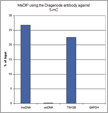
<p><small><strong>Figure 1. Methylated DNA immunoprecipitation (MeDIP) results obtained with the Diagenode monoclonal antibody directed against 5-mC</strong><br />MeDIP (Methylated DNA immunoprecipitation) was performed on 1 µg fragmented human genomic DNA using 0.2 µg of the Diagenode monoclonal antibody against 5-mC (Cat. No. C15200006) and the MagMeDIP Kit (Cat. No. C02010021). The fragmented DNA was spiked with the internal controls present in the kit (methylated DNA (meDNA) as a positive and unmethylated DNA (unDNA) as a negative control) prior to performing the IP. QPCR was performed with optimized primer sets, included in the kit, specific for the methylated and unmethylated DNA controls, and for a known methylated (TSH2B) and unmethylated (GAPDH) genomic region. Figure 2 shows the recovery expressed as a % of input (the relative amount of immunoprecipitated DNA compared to input DNA after qPCR analysis).</small></p>
</div>
</div>
<div class="row">
<div class="small-4 columns">
<p><img src="https://www.diagenode.com/img/product/antibodies/C15200006_MeDIP_2.png" alt="5-methylcytosine (5-mC) Antibody validated method" caption="false" width="278" height="333" /></p>
</div>
<div class="small-8 columns">
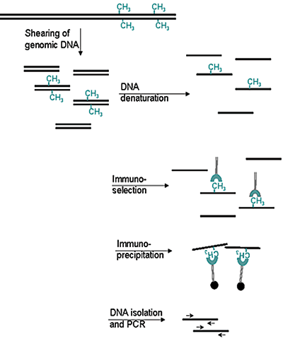
<p><small><strong>Figure 2. Methylated DNA immunoprecipitation (MeDIP) method</strong></small></p>
<ul>
<li><small>Prepare genomic DNA from cultured cells</small></li>
<li><small>Shear genomic DNA</small></li>
<li><small>Denature the sheared genomic DNA</small></li>
<li><small>Immunoprecipitate with the antibody against 5-meC</small></li>
<li><small>Isolate DNA and perform PCR</small></li>
</ul>
</div>
</div>
<div class="row">
<div class="small-4 columns">
<p><img src="https://www.diagenode.com/img/product/antibodies/C15200006_if.jpg" alt="5-methylcytosine (5-mC) Antibody for IF" caption="false" width="278" height="91" /></p>
</div>
<div class="small-8 columns">
<p><small><strong>Figure 3. Immunofluorescence using the Diagenode monoclonal antibody directed against 5-mC</strong> <br />HeLa cells were stained with the Diagenode antibody against 5-mC (Cat. No. C15200006) and with DAPI. Cells were fixed with 4% formaldehyde for 10’ and blocked with PBS/TX-100 containing 1% BSA. The cells were immunofluorescently labelled with the 5-mC antibody (left) diluted 1:1,000 in blocking solution followed by an anti-mouse antibody conjugated to Alexa594. The middle panel shows staining of the nuclei with DAPI. A merge of the two stainings is shown on the right.</small></p>
</div>
</div>',
'label2' => '',
'info2' => '',
'label3' => '',
'info3' => '',
'format' => '100 µg',
'catalog_number' => 'C15200006-100',
'old_catalog_number' => 'MAb-006-100',
'sf_code' => 'C15200006-D001-000526',
'type' => 'FRE',
'search_order' => '03-Antibody',
'price_EUR' => '385',
'price_USD' => '450',
'price_GBP' => '350',
'price_JPY' => '60310',
'price_CNY' => '',
'price_AUD' => '1125',
'country' => 'ALL',
'except_countries' => 'None',
'quote' => false,
'in_stock' => false,
'featured' => false,
'no_promo' => false,
'online' => true,
'master' => true,
'last_datasheet_update' => 'February 11, 2019',
'slug' => '5-mc-monoclonal-antibody-cl-b-classic-100-ug-50-ul',
'meta_title' => '5-methylcytosine (5-mC) monoclonal antibody cl. b - Classic | Diagenode',
'meta_keywords' => '5-methylcytosine (5-mC),monoclonal antibody,Methylated DNA Immunoprecipitation',
'meta_description' => ' Methylated DNA immunoprecipitation (MeDIP) results obtained with the Diagenode monoclonal antibody directed against 5-methyl Cytidine.',
'modified' => '2023-08-03 10:25:19',
'created' => '2015-06-29 14:08:20',
'locale' => 'jpn'
),
'Antibody' => array(
'host' => '*****',
'id' => '302',
'name' => '5-mC monoclonal antibody (100µg)',
'description' => '',
'clonality' => '',
'isotype' => 'IgG1',
'lot' => 'GF-005 (100µg - lot 006)',
'concentration' => '2.1 µg/µl',
'reactivity' => 'Human, mouse, rat, cow, alligator, zebrafish, plants, finch, wide range expected.',
'type' => 'Monoclonal <strong>MEDIP-grade</strong>',
'purity' => 'Monoclonal antibody purified by gel filtration.',
'classification' => 'Classic',
'application_table' => '<table>
<thead>
<tr>
<th>Applications</th>
<th>Suggested dilution</th>
<th>References</th>
</tr>
</thead>
<tbody>
<tr>
<td>MeDIP</td>
<td>0.5 - 1 µg/IP</td>
<td>Fig 1, 2</td>
</tr>
<tr>
<td>Immunofluorescence</td>
<td>1:1,000</td>
<td>Fig 3</td>
</tr>
</tbody>
</table>
<p></p>
<p><small><sup>*</sup> Please note that of the optimal antibody amount per IP should be determined by the end-user. We recommend testing 0.5-5 µg per IP.</small></p>',
'storage_conditions' => 'Store at -20°C; for long storage, store at -80°C. Avoid multiple freeze-thaw cycles.',
'storage_buffer' => 'PBS containing 0.05% azide.',
'precautions' => 'This product is for research use only. Not for use in diagnostic or therapeutic procedures.',
'uniprot_acc' => '',
'slug' => '',
'meta_keywords' => '',
'meta_description' => '',
'modified' => '2023-08-03 10:26:04',
'created' => '0000-00-00 00:00:00',
'select_label' => '302 - 5-mC monoclonal antibody (100µg) (GF-005 (100µg - lot 006) - 2.1 µg/µl - Human, mouse, rat, cow, alligator, zebrafish, plants, finch, wide range expected. - Monoclonal antibody purified by gel filtration. - Mouse)'
),
'Slave' => array(
(int) 0 => array(
[maximum depth reached]
)
),
'Group' => array(
'Group' => array(
[maximum depth reached]
),
'Master' => array(
[maximum depth reached]
),
'Product' => array(
[maximum depth reached]
)
),
'Related' => array(
(int) 0 => array(
[maximum depth reached]
),
(int) 1 => array(
[maximum depth reached]
),
(int) 2 => array(
[maximum depth reached]
),
(int) 3 => array(
[maximum depth reached]
),
(int) 4 => array(
[maximum depth reached]
),
(int) 5 => array(
[maximum depth reached]
),
(int) 6 => array(
[maximum depth reached]
),
(int) 7 => array(
[maximum depth reached]
),
(int) 8 => array(
[maximum depth reached]
),
(int) 9 => array(
[maximum depth reached]
),
(int) 10 => array(
[maximum depth reached]
),
(int) 11 => array(
[maximum depth reached]
),
(int) 12 => array(
[maximum depth reached]
),
(int) 13 => array(
[maximum depth reached]
),
(int) 14 => array(
[maximum depth reached]
),
(int) 15 => array(
[maximum depth reached]
),
(int) 16 => array(
[maximum depth reached]
)
),
'Application' => array(
(int) 0 => array(
[maximum depth reached]
),
(int) 1 => array(
[maximum depth reached]
)
),
'Category' => array(
(int) 0 => array(
[maximum depth reached]
),
(int) 1 => array(
[maximum depth reached]
)
),
'Document' => array(
(int) 0 => array(
[maximum depth reached]
),
(int) 1 => array(
[maximum depth reached]
),
(int) 2 => array(
[maximum depth reached]
)
),
'Feature' => array(),
'Image' => array(
(int) 0 => array(
[maximum depth reached]
)
),
'Promotion' => array(),
'Protocol' => array(),
'Publication' => array(
(int) 0 => array(
[maximum depth reached]
),
(int) 1 => array(
[maximum depth reached]
),
(int) 2 => array(
[maximum depth reached]
),
(int) 3 => array(
[maximum depth reached]
),
(int) 4 => array(
[maximum depth reached]
),
(int) 5 => array(
[maximum depth reached]
),
(int) 6 => array(
[maximum depth reached]
),
(int) 7 => array(
[maximum depth reached]
),
(int) 8 => array(
[maximum depth reached]
),
(int) 9 => array(
[maximum depth reached]
),
(int) 10 => array(
[maximum depth reached]
),
(int) 11 => array(
[maximum depth reached]
),
(int) 12 => array(
[maximum depth reached]
),
(int) 13 => array(
[maximum depth reached]
),
(int) 14 => array(
[maximum depth reached]
),
(int) 15 => array(
[maximum depth reached]
),
(int) 16 => array(
[maximum depth reached]
),
(int) 17 => array(
[maximum depth reached]
),
(int) 18 => array(
[maximum depth reached]
),
(int) 19 => array(
[maximum depth reached]
),
(int) 20 => array(
[maximum depth reached]
),
(int) 21 => array(
[maximum depth reached]
),
(int) 22 => array(
[maximum depth reached]
),
(int) 23 => array(
[maximum depth reached]
),
(int) 24 => array(
[maximum depth reached]
),
(int) 25 => array(
[maximum depth reached]
),
(int) 26 => array(
[maximum depth reached]
),
(int) 27 => array(
[maximum depth reached]
),
(int) 28 => array(
[maximum depth reached]
),
(int) 29 => array(
[maximum depth reached]
),
(int) 30 => array(
[maximum depth reached]
),
(int) 31 => array(
[maximum depth reached]
),
(int) 32 => array(
[maximum depth reached]
),
(int) 33 => array(
[maximum depth reached]
),
(int) 34 => array(
[maximum depth reached]
),
(int) 35 => array(
[maximum depth reached]
),
(int) 36 => array(
[maximum depth reached]
),
(int) 37 => array(
[maximum depth reached]
),
(int) 38 => array(
[maximum depth reached]
),
(int) 39 => array(
[maximum depth reached]
),
(int) 40 => array(
[maximum depth reached]
),
(int) 41 => array(
[maximum depth reached]
),
(int) 42 => array(
[maximum depth reached]
),
(int) 43 => array(
[maximum depth reached]
),
(int) 44 => array(
[maximum depth reached]
),
(int) 45 => array(
[maximum depth reached]
),
(int) 46 => array(
[maximum depth reached]
),
(int) 47 => array(
[maximum depth reached]
),
(int) 48 => array(
[maximum depth reached]
),
(int) 49 => array(
[maximum depth reached]
),
(int) 50 => array(
[maximum depth reached]
),
(int) 51 => array(
[maximum depth reached]
)
),
'Testimonial' => array(),
'Area' => array(),
'SafetySheet' => array(
(int) 0 => array(
[maximum depth reached]
),
(int) 1 => array(
[maximum depth reached]
),
(int) 2 => array(
[maximum depth reached]
),
(int) 3 => array(
[maximum depth reached]
),
(int) 4 => array(
[maximum depth reached]
),
(int) 5 => array(
[maximum depth reached]
),
(int) 6 => array(
[maximum depth reached]
),
(int) 7 => array(
[maximum depth reached]
)
)
),
'meta_canonical' => 'https://www.diagenode.com/jp/p/5-mc-monoclonal-antibody-cl-b-classic-100-ug-50-ul'
)
$language = 'jp'
$meta_keywords = '5-methylcytosine (5-mC),monoclonal antibody,Methylated DNA Immunoprecipitation'
$meta_description = ' Methylated DNA immunoprecipitation (MeDIP) results obtained with the Diagenode monoclonal antibody directed against 5-methyl Cytidine.'
$meta_title = '5-methylcytosine (5-mC) monoclonal antibody cl. b - Classic | Diagenode'
$product = array(
'Product' => array(
'id' => '1965',
'antibody_id' => '302',
'name' => '5-methylcytosine (5-mC) monoclonal antibody cl. b ',
'description' => '<p><span>Monoclonal antibody raised in mouse against 5-mC (5-methylcytosine) conjugated to ovalbumine.</span></p>',
'label1' => 'Validation Data',
'info1' => '<div class="row">
<div class="small-4 columns">
<p><img src="https://www.diagenode.com/img/product/antibodies/C15200006_MeDIP_1.png" alt="5-methylcytosine (5-mC) Antibody validated in MeDIP" caption="false" width="278" height="292" /></p>
</div>
<div class="small-8 columns">
<p><small><strong>Figure 1. Methylated DNA immunoprecipitation (MeDIP) results obtained with the Diagenode monoclonal antibody directed against 5-mC</strong><br />MeDIP (Methylated DNA immunoprecipitation) was performed on 1 µg fragmented human genomic DNA using 0.2 µg of the Diagenode monoclonal antibody against 5-mC (Cat. No. C15200006) and the MagMeDIP Kit (Cat. No. C02010021). The fragmented DNA was spiked with the internal controls present in the kit (methylated DNA (meDNA) as a positive and unmethylated DNA (unDNA) as a negative control) prior to performing the IP. QPCR was performed with optimized primer sets, included in the kit, specific for the methylated and unmethylated DNA controls, and for a known methylated (TSH2B) and unmethylated (GAPDH) genomic region. Figure 2 shows the recovery expressed as a % of input (the relative amount of immunoprecipitated DNA compared to input DNA after qPCR analysis).</small></p>
</div>
</div>
<div class="row">
<div class="small-4 columns">
<p><img src="https://www.diagenode.com/img/product/antibodies/C15200006_MeDIP_2.png" alt="5-methylcytosine (5-mC) Antibody validated method" caption="false" width="278" height="333" /></p>
</div>
<div class="small-8 columns">
<p><small><strong>Figure 2. Methylated DNA immunoprecipitation (MeDIP) method</strong></small></p>
<ul>
<li><small>Prepare genomic DNA from cultured cells</small></li>
<li><small>Shear genomic DNA</small></li>
<li><small>Denature the sheared genomic DNA</small></li>
<li><small>Immunoprecipitate with the antibody against 5-meC</small></li>
<li><small>Isolate DNA and perform PCR</small></li>
</ul>
</div>
</div>
<div class="row">
<div class="small-4 columns">
<p><img src="https://www.diagenode.com/img/product/antibodies/C15200006_if.jpg" alt="5-methylcytosine (5-mC) Antibody for IF" caption="false" width="278" height="91" /></p>
</div>
<div class="small-8 columns">
<p><small><strong>Figure 3. Immunofluorescence using the Diagenode monoclonal antibody directed against 5-mC</strong> <br />HeLa cells were stained with the Diagenode antibody against 5-mC (Cat. No. C15200006) and with DAPI. Cells were fixed with 4% formaldehyde for 10’ and blocked with PBS/TX-100 containing 1% BSA. The cells were immunofluorescently labelled with the 5-mC antibody (left) diluted 1:1,000 in blocking solution followed by an anti-mouse antibody conjugated to Alexa594. The middle panel shows staining of the nuclei with DAPI. A merge of the two stainings is shown on the right.</small></p>
</div>
</div>',
'label2' => '',
'info2' => '',
'label3' => '',
'info3' => '',
'format' => '100 µg',
'catalog_number' => 'C15200006-100',
'old_catalog_number' => 'MAb-006-100',
'sf_code' => 'C15200006-D001-000526',
'type' => 'FRE',
'search_order' => '03-Antibody',
'price_EUR' => '385',
'price_USD' => '450',
'price_GBP' => '350',
'price_JPY' => '60310',
'price_CNY' => '',
'price_AUD' => '1125',
'country' => 'ALL',
'except_countries' => 'None',
'quote' => false,
'in_stock' => false,
'featured' => false,
'no_promo' => false,
'online' => true,
'master' => true,
'last_datasheet_update' => 'February 11, 2019',
'slug' => '5-mc-monoclonal-antibody-cl-b-classic-100-ug-50-ul',
'meta_title' => '5-methylcytosine (5-mC) monoclonal antibody cl. b - Classic | Diagenode',
'meta_keywords' => '5-methylcytosine (5-mC),monoclonal antibody,Methylated DNA Immunoprecipitation',
'meta_description' => ' Methylated DNA immunoprecipitation (MeDIP) results obtained with the Diagenode monoclonal antibody directed against 5-methyl Cytidine.',
'modified' => '2023-08-03 10:25:19',
'created' => '2015-06-29 14:08:20',
'locale' => 'jpn'
),
'Antibody' => array(
'host' => '*****',
'id' => '302',
'name' => '5-mC monoclonal antibody (100µg)',
'description' => '',
'clonality' => '',
'isotype' => 'IgG1',
'lot' => 'GF-005 (100µg - lot 006)',
'concentration' => '2.1 µg/µl',
'reactivity' => 'Human, mouse, rat, cow, alligator, zebrafish, plants, finch, wide range expected.',
'type' => 'Monoclonal <strong>MEDIP-grade</strong>',
'purity' => 'Monoclonal antibody purified by gel filtration.',
'classification' => 'Classic',
'application_table' => '<table>
<thead>
<tr>
<th>Applications</th>
<th>Suggested dilution</th>
<th>References</th>
</tr>
</thead>
<tbody>
<tr>
<td>MeDIP</td>
<td>0.5 - 1 µg/IP</td>
<td>Fig 1, 2</td>
</tr>
<tr>
<td>Immunofluorescence</td>
<td>1:1,000</td>
<td>Fig 3</td>
</tr>
</tbody>
</table>
<p></p>
<p><small><sup>*</sup> Please note that of the optimal antibody amount per IP should be determined by the end-user. We recommend testing 0.5-5 µg per IP.</small></p>',
'storage_conditions' => 'Store at -20°C; for long storage, store at -80°C. Avoid multiple freeze-thaw cycles.',
'storage_buffer' => 'PBS containing 0.05% azide.',
'precautions' => 'This product is for research use only. Not for use in diagnostic or therapeutic procedures.',
'uniprot_acc' => '',
'slug' => '',
'meta_keywords' => '',
'meta_description' => '',
'modified' => '2023-08-03 10:26:04',
'created' => '0000-00-00 00:00:00',
'select_label' => '302 - 5-mC monoclonal antibody (100µg) (GF-005 (100µg - lot 006) - 2.1 µg/µl - Human, mouse, rat, cow, alligator, zebrafish, plants, finch, wide range expected. - Monoclonal antibody purified by gel filtration. - Mouse)'
),
'Slave' => array(
(int) 0 => array(
'id' => '166',
'name' => 'C15200006',
'product_id' => '1965',
'modified' => '2016-02-23 20:46:06',
'created' => '2016-02-23 20:46:06'
)
),
'Group' => array(
'Group' => array(
'id' => '166',
'name' => 'C15200006',
'product_id' => '1965',
'modified' => '2016-02-23 20:46:06',
'created' => '2016-02-23 20:46:06'
),
'Master' => array(
'id' => '1965',
'antibody_id' => '302',
'name' => '5-methylcytosine (5-mC) Antibody - cl. b ',
'description' => '<p><span>Monoclonal antibody raised in mouse against <strong>5-mC</strong> (<strong>5-methylcytosine</strong>) conjugated to ovalbumine.</span></p>',
'label1' => 'Validation Data',
'info1' => '<div class="row">
<div class="small-4 columns">
<p><img src="https://www.diagenode.com/img/product/antibodies/C15200006_MeDIP_1.png" alt="5-methylcytosine (5-mC) Antibody validated in MeDIP" caption="false" width="278" height="292" /></p>
</div>
<div class="small-8 columns">
<p><small><strong>Figure 1. Methylated DNA immunoprecipitation (MeDIP) results obtained with the Diagenode monoclonal antibody directed against 5-mC</strong><br />MeDIP (Methylated DNA immunoprecipitation) was performed on 1 µg fragmented human genomic DNA using 0.2 µg of the Diagenode monoclonal antibody against 5-mC (Cat. No. C15200006) and the MagMeDIP Kit (Cat. No. C02010021). The fragmented DNA was spiked with the internal controls present in the kit (methylated DNA (meDNA) as a positive and unmethylated DNA (unDNA) as a negative control) prior to performing the IP. QPCR was performed with optimized primer sets, included in the kit, specific for the methylated and unmethylated DNA controls, and for a known methylated (TSH2B) and unmethylated (GAPDH) genomic region. Figure 2 shows the recovery expressed as a % of input (the relative amount of immunoprecipitated DNA compared to input DNA after qPCR analysis).</small></p>
</div>
</div>
<div class="row">
<div class="small-4 columns">
<p><img src="https://www.diagenode.com/img/product/antibodies/C15200006_MeDIP_2.png" alt="5-methylcytosine (5-mC) Antibody validated method" caption="false" width="278" height="333" /></p>
</div>
<div class="small-8 columns">
<p><small><strong>Figure 2. Methylated DNA immunoprecipitation (MeDIP) method</strong></small></p>
<ul>
<li><small>Prepare genomic DNA from cultured cells</small></li>
<li><small>Shear genomic DNA</small></li>
<li><small>Denature the sheared genomic DNA</small></li>
<li><small>Immunoprecipitate with the antibody against 5-meC</small></li>
<li><small>Isolate DNA and perform PCR</small></li>
</ul>
</div>
</div>
<div class="row">
<div class="small-4 columns">
<p><img src="https://www.diagenode.com/img/product/antibodies/C15200006_if.jpg" alt="5-methylcytosine (5-mC) Antibody for IF" caption="false" width="278" height="91" /></p>
</div>
<div class="small-8 columns">
<p><small><strong>Figure 3. Immunofluorescence using the Diagenode monoclonal antibody directed against 5-mC</strong> <br />HeLa cells were stained with the Diagenode antibody against 5-mC (Cat. No. C15200006) and with DAPI. Cells were fixed with 4% formaldehyde for 10’ and blocked with PBS/TX-100 containing 1% BSA. The cells were immunofluorescently labelled with the 5-mC antibody (left) diluted 1:1,000 in blocking solution followed by an anti-mouse antibody conjugated to Alexa594. The middle panel shows staining of the nuclei with DAPI. A merge of the two stainings is shown on the right.</small></p>
</div>
</div>',
'label2' => '',
'info2' => '',
'label3' => '',
'info3' => '',
'format' => '100 µg',
'catalog_number' => 'C15200006-100',
'old_catalog_number' => 'MAb-006-100',
'sf_code' => 'C15200006-D001-000526',
'type' => 'FRE',
'search_order' => '03-Antibody',
'price_EUR' => '385',
'price_USD' => '450',
'price_GBP' => '350',
'price_JPY' => '60310',
'price_CNY' => '',
'price_AUD' => '1125',
'country' => 'ALL',
'except_countries' => 'None',
'quote' => false,
'in_stock' => false,
'featured' => false,
'no_promo' => false,
'online' => true,
'master' => true,
'last_datasheet_update' => 'February 11, 2019',
'slug' => '5-mc-monoclonal-antibody-cl-b-classic-100-ug-50-ul',
'meta_title' => '5-methylcytosine (5-mC) Antibody - cl. b (C15200006) | Diagenode',
'meta_keywords' => '5-methylcytosine (5-mC),monoclonal antibody,Methylated DNA Immunoprecipitation',
'meta_description' => '5-methylcytosine (5-mC) Monoclonal Antibody cl. b validated in MeDIP and IF. Batch-specific data available on the website. Sample size available.',
'modified' => '2023-08-03 10:25:19',
'created' => '2015-06-29 14:08:20'
),
'Product' => array(
(int) 0 => array(
[maximum depth reached]
),
(int) 1 => array(
[maximum depth reached]
)
)
),
'Related' => array(
(int) 0 => array(
'id' => '1882',
'antibody_id' => null,
'name' => 'hMeDIP kit x16 (monoclonal mouse antibody)',
'description' => '<p><a href="https://www.diagenode.com/files/products/kits/hMeDIP_kit_manual.pdf"><img src="https://www.diagenode.com/img/buttons/bt-manual.png" /></a></p>
<p><span>The hMeDIP kit is designed for enrichment of hydroxymethylated DNA from fragmented genomic DNA<span><span> </span>samples for use in genome-wide methylation analysis. It features</span></span><span> a highly specific monoclonal antibody against </span>5-hydroxymethylcytosine (5-hmC) for the immunoprecipitation of hydroxymethylated DNA<span>. It includes control DNA and primers to assess the effiency of the assay. </span>Performing hydroxymethylation profiling with the hMeDIP kit is fast, reliable and highly specific.</p>
<p><em>Looking for hMeDIP-seq protocol? <a href="https://go.diagenode.com/l/928883/2022-01-07/2m1ht" target="_blank" title="Contact us">Contact us</a></em></p>
<p><span></span></p>
<p><span></span></p>',
'label1' => 'Characteristics',
'info1' => '<ul style="list-style-type: disc;">
<li><span>Robust enrichment & immunoprecipitation of hydroxymethylated DNA</span></li>
<li>Highly specific monoclonal antibody against 5-hmC<span> for reliable, reproducible results</span></li>
<li>Including control DNA and primers to <span>monitor the efficiency of the assay</span>
<ul style="list-style-type: circle;">
<li>hmeDNA and unmethylated DNA sequences and primer pairs</li>
<li>Mouse primer pairs against Sfi1 targeting hydroxymethylated gene in mouse</li>
</ul>
</li>
<li>Improved single-tube, magnetic bead-based protocol</li>
</ul>',
'label2' => '',
'info2' => '',
'label3' => '',
'info3' => '',
'format' => '16 rxns',
'catalog_number' => 'C02010031',
'old_catalog_number' => 'AF-110-0016',
'sf_code' => 'C02010031-',
'type' => 'RFR',
'search_order' => '04-undefined',
'price_EUR' => '630',
'price_USD' => '690',
'price_GBP' => '580',
'price_JPY' => '98690',
'price_CNY' => '',
'price_AUD' => '1725',
'country' => 'ALL',
'except_countries' => 'None',
'quote' => false,
'in_stock' => false,
'featured' => true,
'no_promo' => false,
'online' => true,
'master' => true,
'last_datasheet_update' => '0000-00-00',
'slug' => 'hmedip-kit-x16-monoclonal-mouse-antibody-16-rxns',
'meta_title' => 'hMeDIP kit x16 (monoclonal mouse antibody)',
'meta_keywords' => '',
'meta_description' => 'hMeDIP kit x16 (monoclonal mouse antibody)',
'modified' => '2023-04-20 16:12:48',
'created' => '2015-06-29 14:08:20',
'ProductsRelated' => array(
[maximum depth reached]
),
'Image' => array(
[maximum depth reached]
)
),
(int) 1 => array(
'id' => '1880',
'antibody_id' => null,
'name' => 'MagMeDIP Kit',
'description' => '<p><a href="https://www.diagenode.com/files/products/kits/magmedip-kit-manual-C02010020-21.pdf"><img src="https://www.diagenode.com/img/buttons/bt-manual.png" /></a></p>
<p> </p>
<div class="small-12 medium-4 large-4 columns"><center></center><center></center><center></center><center><a href="https://www.ncbi.nlm.nih.gov/pubmed/30429608" target="_blank"><img src="https://www.diagenode.com/img/banners/banner-nature-publication-580.png" alt="Click here to read more about MeDIP " caption="false" width="80%" /></a></center></div>
<div class="small-12 medium-8 large-8 columns">
<h3 style="text-align: justify;">Sensitive tumour detection and classification using plasma cell-free DNA methylomes<br /><a href="https://www.ncbi.nlm.nih.gov/pubmed/30429608" target="_blank">Read the publication</a></h3>
<h3 class="c-article-title u-h1" data-test="article-title" itemprop="name headline" style="text-align: justify;">Preparation of cfMeDIP-seq libraries for methylome profiling of plasma cell-free DNA<br /><a href="https://www.nature.com/articles/s41596-019-0202-2" target="_blank" title="cfMeDIP-seq Nature Method">Read the method</a></h3>
</div>
<p></p>
<p></p>
<p></p>
<div class="row">
<div class="small-12 medium-4 large-4 columns"><center>
<script>// <![CDATA[
var date = new Date(); var heure = date.getHours(); var jour = date.getDay(); var semaine = Math.floor(date.getDate() / 7) + 1; if (jour === 2 && ( (heure >= 9 && heure < 9.5) || (heure >= 18 && heure < 18.5) )) { document.write('<a href="https://us02web.zoom.us/j/85467619762"><img src="https://www.diagenode.com/img/epicafe-ON.gif"></a>'); } else { document.write('<a href="https://go.diagenode.com/l/928883/2023-04-26/3kq1v"><img src="https://www.diagenode.com/img/epicafe-OFF.png"></a>'); }
// ]]></script>
</center></div>
<div class="small-12 medium-8 large-8 columns"><br />
<p>Perform <strong>MeDIP</strong> (<strong>Me</strong>thylated <strong>D</strong>NA <strong>I</strong>mmuno<strong>p</strong>recipitation) followed by qPCR or NGS to estimate DNA methylation status of your sample using a highly sensitive 5-methylcytosine antibody. Our MagMeDIP kit contains high quality reagents to get the highest enrichment of methylated DNA with an optimized user-friendly protocol.</p>
</div>
</div>
<h3><span>Features</span></h3>
<ul>
<li>Starting DNA amount: <strong>10 ng – 1 µg</strong></li>
<li>Content: <strong>all reagents included</strong> for DNA extraction, immunoprecipitation (including the 5-mC antibody, spike-in controls and their corresponding qPCR primer pairs) as well as DNA isolation after IP.</li>
<li>Application: <strong>qPCR</strong> and <strong>NGS</strong></li>
<li>Robust method, <strong>superior enrichment</strong>, and easy-to-use protocol</li>
<li><strong>High reproducibility</strong> between replicates and repetitive experiments</li>
<li>Compatible with <strong>all species </strong></li>
</ul>',
'label1' => 'MagMeDIP workflow',
'info1' => '<p>DNA methylation occurs primarily as 5-methylcytosine (5-mC), and the Diagenode MagMeDIP Kit takes advantage of a specific antibody targeting this 5-mC to immunoprecipitate methylated DNA, which can be thereafter directly analyzed by qPCR or Next-Generation Sequencing (NGS).</p>
<h3><span>How it works</span></h3>
<p>In brief, after the cell collection and lysis, the genomic DNA is extracted, sheared, and then denatured. In the next step the antibody directed against 5 methylcytosine and antibody binding beads are used for immunoselection and immunoprecipitation of methylated DNA fragments. Then, the IP’d methylated DNA is isolated and can be used for any subsequent analysis as qPCR, amplification, hybridization on microarrays or next generation sequencing.</p>
<center><img src="https://www.diagenode.com/img/product/kits/MagMeDIP-workflow.png" width="70%" alt="5-methylcytosine" caption="false" /></center>
<script async="" src="https://edge.fullstory.com/s/fs.js" crossorigin="anonymous"></script>
<script src="chrome-extension://hhojmcideegachlhfgfdhailpfhgknjm/web_accessible_resources/index.js"></script>',
'label2' => 'MeDIP-qPCR',
'info2' => '<p>The kit MagMeDIP contains all reagents necessary for a complete MeDIP-qPCR workflow. Two MagMeDIP protocols have been validated: for manual processing as well as for automated processing, using the Diagenode’s IP-Star Compact Automated System (please refer to the kit manual).</p>
<ul>
<li><strong>Complete kit</strong> including DNA extraction module, IP antibody and reagents, DNA isolation buffer</li>
<li><strong>Quality control of the IP:</strong> due to methylated and unmethylated DNA spike-in controls and their associated qPCR primers</li>
<li><strong>Easy to use</strong> with user-friendly magnetic beads and rack</li>
<li><strong>Highly validated protocol</strong></li>
<li>Automated protocol supplied</li>
</ul>
<center><img src="https://www.diagenode.com/img/product/kits/fig1-magmedipkit.png" width="85%" alt="Methylated DNA Immunoprecipitation" caption="false" /></center>
<p style="font-size: 0.9em;"><em><strong>Figure 1.</strong> Immunoprecipitation results obtained with Diagenode MagMeDIP Kit</em></p>
<p style="font-size: 0.9em;">MeDIP assays were performed manually using 1 µg or 50 ng gDNA from blood cells with the MagMeDIP kit (Diagenode). The IP was performed with the Methylated and Unmethylated spike-in controls included in the kit, together with the human DNA samples. The DNA was isolated/purified using DIB. Afterwards, qPCR was performed using the primer pairs included in this kit.</p>
<script async="" src="https://edge.fullstory.com/s/fs.js" crossorigin="anonymous"></script>
<script src="chrome-extension://hhojmcideegachlhfgfdhailpfhgknjm/web_accessible_resources/index.js"></script>',
'label3' => 'MeDIP-seq',
'info3' => '<p>For DNA methylation analysis on the whole genome, MagMeDIP kit can be coupled with Next-Generation Sequencing. To perform MeDIP-sequencing we recommend the following strategy:</p>
<ul style="list-style-type: circle;">
<li>Choose a library preparation solution which is compatible with the starting amount of DNA you are planning to use (from 10 ng to 1 μg). It can be a home-made solution or a commercial one.</li>
<li>Choose the indexing system that fits your needs considering the following features:</li>
<ul>
<ul>
<ul>
<li>Single-indexing, combinatorial dual-indexing or unique dual-indexing</li>
<li>Number of barcodes</li>
<li>Full-length adaptors containing the barcodes or barcoding at the final amplification step</li>
<li>Presence / absence of Unique Molecular Identifiers (for PCR duplicates removal)</li>
</ul>
</ul>
</ul>
<li>Standard library preparation protocols are compatible with double-stranded DNA only, therefore the first steps of the library preparation (end repair, A-tailing, adaptor ligation and clean-up) will have to be performed on sheared DNA, before the IP.</li>
</ul>
<p style="padding-left: 30px;"><strong>CAUTION:</strong> As the immunoprecipitation step occurs at the middle of the library preparation workflow, single-tube solutions for library preparation are usually not compatible with MeDIP-sequencing.</p>
<ul style="list-style-type: circle;">
<li>For DNA isolation after the IP, we recommend using the <a href="https://www.diagenode.com/en/p/ipure-kit-v2-x24" title="IPure kit v2">IPure kit v2</a> (available separately, Cat. No. C03010014) instead of DNA isolation Buffer.</li>
</ul>
<ul style="list-style-type: circle;">
<li>Perform library amplification after the DNA isolation following the standard protocol of the chosen library preparation solution.</li>
</ul>
<h3><span>MeDIP-seq workflow</span></h3>
<center><img src="https://www.diagenode.com/img/product/kits/MeDIP-seq-workflow.png" width="110%" alt="MagMeDIP qPCR Kit x10 workflow" caption="false" /></center>
<h3><span>Example of results</span></h3>
<center><img src="https://www.diagenode.com/img/product/kits/medip-specificity.png" alt="MagMeDIP qPCR Kit Result" caption="false" width="951" height="488" /></center>
<p></p>
<p style="font-size: 0.9em;"><strong>Figure 1. qPCR analysis of external spike-in DNA controls (methylated and unmethylated) after IP.</strong> Samples were prepared using 1μg – 100ng -10ng sheared human gDNA with the MagMeDIP kit (Diagenode) and a commercially available library prep kit. DNA isolation after IP has been performed with IPure kit V2 (Diagenode).</p>
<p></p>
<p></p>
<center><img src="https://www.diagenode.com/img/product/kits/medip-saturation-analysis.png" alt=" MagMeDIP kit " caption="false" width="951" height="461" /></center>
<p></p>
<p style="font-size: 0.9em;"><strong>Figure 2. Saturation analysis.</strong> Clean reads were aligned to the human genome (hg19) using Burrows-Wheeler aligner (BWA) algorithm after which duplicated and unmapped reads were removed resulting in a mapping efficiency >98% for all samples. Quality and validity check of the mapped MeDIP-seq data was performed using MEDIPS R package. Saturation plots show that all sets of reads have sufficient complexity and depth to saturate the coverage profile of the reference genome and that this is reproducible between replicates and repetitive experiments (data shown for 50 ng gDNA input: left panel = replicate a, right panel = replicate b).</p>
<p></p>
<p></p>
<center><img src="https://www.diagenode.com/img/product/kits/medip-libraries-prep.png" alt="MagMeDIP x10 " caption="false" width="951" height="708" /></center>
<p></p>
<p style="font-size: 0.9em;"><strong>Figure 3. Sequencing profiles of MeDIP-seq libraries prepared from different starting amounts of sheared gDNA on the positive and negative methylated control regions.</strong> MeDIP-seq libraries were prepared from decreasing starting amounts of gDNA (1 μg (green), 50 ng (red), and 10ng (blue)) originating from human blood with the MagMeDIP kit (Diagenode) and a commercially available library prep kit. DNA isolation after IP has been performed with IPure kit V2 (Diagenode). IP and corresponding INPUT samples were sequenced on Illumina NovaSeq SP with 2x50 PE reads. The reads were mapped to the human genome (hg19) with bwa and the alignments were loaded into IGV (the tracks use an identical scale). The top IGV figure shows the TSH2B (also known as H2BC1) gene (marked by blue boxes in the bottom track) and its surroundings. The TSH2B gene is coding for a histone variant that does not occur in blood cells, and it is known to be silenced by methylation. Accordingly, we see a high coverage in the vicinity of this gene. The bottom IGV figure shows the GADPH locus (marked by blue boxes in the bottom track) and its surroundings. The GADPH gene is a highly active transcription region and should not be methylated, resulting in no reads accumulation following MeDIP-seq experiment.</p>
<p></p>
<ul>
<ul>
<script async="" src="https://edge.fullstory.com/s/fs.js" crossorigin="anonymous"></script>
<script src="chrome-extension://hhojmcideegachlhfgfdhailpfhgknjm/web_accessible_resources/index.js"></script>
</ul>
</ul>',
'format' => '48 rxns (IP)',
'catalog_number' => 'C02010021',
'old_catalog_number' => 'mc-magme-048',
'sf_code' => 'C02010021-',
'type' => 'RFR',
'search_order' => '04-undefined',
'price_EUR' => '745',
'price_USD' => '750',
'price_GBP' => '680',
'price_JPY' => '116705',
'price_CNY' => '',
'price_AUD' => '1875',
'country' => 'ALL',
'except_countries' => 'None',
'quote' => false,
'in_stock' => false,
'featured' => true,
'no_promo' => false,
'online' => true,
'master' => true,
'last_datasheet_update' => '0000-00-00',
'slug' => 'magmedip-kit-x48-48-rxns',
'meta_title' => 'MagMeDIP Kit for efficient immunoprecipitation of methylated DNA | Diagenode',
'meta_keywords' => '',
'meta_description' => 'Perform Methylated DNA Immunoprecipitation (MeDIP) to estimate DNA methylation status of your sample using highly specific 5-mC antibody. This kit allows the preparation of cfMeDIP-seq libraries.',
'modified' => '2023-07-06 14:15:10',
'created' => '2015-06-29 14:08:20',
'ProductsRelated' => array(
[maximum depth reached]
),
'Image' => array(
[maximum depth reached]
)
),
(int) 2 => array(
'id' => '1887',
'antibody_id' => null,
'name' => 'MethylCap kit',
'description' => '<p>The MethylCap kit allows to specifically capture DNA fragments containing methylated CpGs. The assay is based on the affinity purification of methylated DNA using methyl-CpG-binding domain (MBD) of human MeCP2 protein. The procedure has been adapted to both manual process or <a href="https://www.diagenode.com/en/p/sx-8g-ip-star-compact-automated-system-1-unit">IP-Star® Compact Automated System</a>. Libraries of captured methylated DNA can be prepared for next-generation sequencing (NGS) by combining MBD technology with the <a href="https://www.diagenode.com/en/p/microplex-lib-prep-kit-v3-48-rxns">MicroPlex Library Preparation Kit v3</a>.</p>',
'label1' => 'Characteristics',
'info1' => '<ul style="list-style-type: circle;">
<li><strong>Fast & sensitive capture</strong> of methylated DNA</li>
<li><strong>High capture efficiency</strong></li>
<li><strong>Differential fractionation</strong> of methylated DNA by CpG density (3 eluted fractions)</li>
<li><strong>On-day protocol</strong></li>
<li><strong>NGS compatibility</strong></li>
</ul>
<h3>MBD-seq allows for detection of genomic regions with different CpG density</h3>
<p><img src="https://www.diagenode.com/img/product/kits/mbd_results1.png" alt="MBD-sequencing results have been validated by bisulfite sequencing" style="display: block; margin-left: auto; margin-right: auto;" /></p>
<p><strong></strong></p>
<p><strong></strong><strong>F</strong><strong>igure 1.</strong> Using the MBD approach, two methylated regions were detected in different elution fractions according to their methylated CpG density (A). Low, Medium and High refer to the sequenced DNA from different elution fractions with increasing salt concentration. Methylated patterns of these two different methylated regions were validated by bisulfite conversion assay (B).</p>',
'label2' => '',
'info2' => '',
'label3' => '',
'info3' => '',
'format' => '48 rxns',
'catalog_number' => 'C02020010',
'old_catalog_number' => 'AF-100-0048',
'sf_code' => 'C02020010-',
'type' => 'RFR',
'search_order' => '04-undefined',
'price_EUR' => '740',
'price_USD' => '695',
'price_GBP' => '675',
'price_JPY' => '115920',
'price_CNY' => '',
'price_AUD' => '1738',
'country' => 'ALL',
'except_countries' => 'None',
'quote' => false,
'in_stock' => false,
'featured' => true,
'no_promo' => false,
'online' => true,
'master' => true,
'last_datasheet_update' => '0000-00-00',
'slug' => 'methylcap-kit-x48-48-rxns',
'meta_title' => 'MethylCap kit x48',
'meta_keywords' => '',
'meta_description' => 'MethylCap kit x48',
'modified' => '2021-02-19 17:55:38',
'created' => '2015-06-29 14:08:20',
'ProductsRelated' => array(
[maximum depth reached]
),
'Image' => array(
[maximum depth reached]
)
),
(int) 3 => array(
'id' => '1888',
'antibody_id' => null,
'name' => 'Auto MethylCap kit',
'description' => '<p>The Auto MethylCap kit allows to specifically capture DNA fragments containing methylated CpGs. The assay is based on the affinity purification of methylated DNA using methyl-CpG-binding domain (MBD) of human MeCP2 protein. This procedure has been optimized to perform automated immunoprecipitation of chromatin using the <a href="https://www.diagenode.com/en/p/sx-8g-ip-star-compact-automated-system-1-unit">IP-Star® Compact Automated System</a> enabling highly reproducible results and allowing for high throughput.</p>',
'label1' => ' Characteristics',
'info1' => '<ul>
<li><strong>Fast & sensitive capture</strong> of methylated DNA</li>
<li><strong>High capture efficiency</strong></li>
<li><strong>Differential fractionation</strong> of methylated DNA by CpG density (3 eluted fractions)</li>
<li><strong>Automation compatibility</strong><strong></strong>
<h3>MBD-seq allows for detection of genomic regions with different CpG density</h3>
<p><img src="https://www.diagenode.com/img/product/kits/mbd_results1.png" alt="MBD-sequencing results have been validated by bisulfite sequencing" style="display: block; margin-left: auto; margin-right: auto;" /></p>
<p><strong>F</strong><strong>igure 1.</strong><span> </span>Using the MBD approach, two methylated regions were detected in different elution fractions according to their methylated CpG density (A). Low, Medium and High refer to the sequenced DNA from different elution fractions with increasing salt concentration. Methylated patterns of these two different methylated regions were validated by bisulfite conversion assay (B).<br /><strong></strong></p>
</li>
</ul>',
'label2' => '',
'info2' => '',
'label3' => '',
'info3' => '',
'format' => '48 rxns',
'catalog_number' => 'C02020011',
'old_catalog_number' => 'AF-Auto01-0048',
'sf_code' => 'C02020011-',
'type' => 'RFR',
'search_order' => '04-undefined',
'price_EUR' => '740',
'price_USD' => '695',
'price_GBP' => '675',
'price_JPY' => '115920',
'price_CNY' => '',
'price_AUD' => '1738',
'country' => 'ALL',
'except_countries' => 'Japan',
'quote' => false,
'in_stock' => false,
'featured' => false,
'no_promo' => false,
'online' => true,
'master' => true,
'last_datasheet_update' => '0000-00-00',
'slug' => 'auto-methylcap-kit-x48-48-rxns',
'meta_title' => 'Auto MethylCap kit x48',
'meta_keywords' => '',
'meta_description' => 'Auto MethylCap kit x48',
'modified' => '2020-09-17 13:33:12',
'created' => '2015-06-29 14:08:20',
'ProductsRelated' => array(
[maximum depth reached]
),
'Image' => array(
[maximum depth reached]
)
),
(int) 4 => array(
'id' => '1892',
'antibody_id' => null,
'name' => 'Premium Bisulfite kit',
'description' => '<p style="text-align: center;"><a href="https://www.diagenode.com/files/products/kits/Premium_Bisulfite_kit_manual.pdf"><img src="https://www.diagenode.com/img/buttons/bt-manual.png" /></a></p>
<p style="text-align: center;"><strong>Make your Bisulfite conversion now in only 60 minutes !</strong></p>
<p>Diagenode's Premium Bisulfite Kit rapidly converts DNA through bisulfite treatment. Our conversion reagent is added directly to DNA, requires no intermediate steps, and results in high yields of DNA ready for downstream analysis methods including PCR and Next-Generation Sequencing.</p>',
'label1' => '',
'info1' => '',
'label2' => '',
'info2' => '',
'label3' => '',
'info3' => '',
'format' => '50 rxns',
'catalog_number' => 'C02030030',
'old_catalog_number' => '',
'sf_code' => 'C02030030-',
'type' => 'REF',
'search_order' => '04-undefined',
'price_EUR' => '255',
'price_USD' => '240',
'price_GBP' => '230',
'price_JPY' => '39945',
'price_CNY' => '',
'price_AUD' => '600',
'country' => 'ALL',
'except_countries' => 'None',
'quote' => false,
'in_stock' => false,
'featured' => true,
'no_promo' => false,
'online' => true,
'master' => true,
'last_datasheet_update' => '0000-00-00',
'slug' => 'premium-bisulfite-kit-50-rxns',
'meta_title' => 'Premium Bisulfite kit',
'meta_keywords' => '',
'meta_description' => 'Premium Bisulfite kit',
'modified' => '2023-04-20 16:13:50',
'created' => '2015-06-29 14:08:20',
'ProductsRelated' => array(
[maximum depth reached]
),
'Image' => array([maximum depth reached])
),
(int) 5 => array(
'id' => '1980',
'antibody_id' => '630',
'name' => '5-methylcytosine (5-mC) Antibody - clone 33D3',
'description' => '<p><span>Monoclonal antibody raised in mouse against </span><b>5-mC</b><span><span> </span>(</span><b>5-methylcytosine</b><span>) conjugated to ovalbumine (</span><b>33D3 clone</b><span>).</span></p>',
'label1' => 'Validation Data',
'info1' => '<div class="row">
<div class="small-12 columns">
<p class="text-center"><img src="https://www.diagenode.com/img/product/antibodies/C15200081_ChIPSeq-A.png" alt="5-mC (5-methylcytosine) Antibody validated in MeDIP-seq" caption="false" width="886" height="173" /></p>
<p class="text-center"><img src="https://www.diagenode.com/img/product/antibodies/C15200081_ChIPSeq-B.png" alt="5-mC (5-methylcytosine) Antibody validated in MeDIP-seq" caption="false" width="886" height="184" /></p>
</div>
</div>
<div class="row">
<div class="small-12 columns">
<p><small><strong>Figure 1. MeDIP-seq with the Diagenode monoclonal antibody directed against 5-mC</strong><br /> Genomic DNA from E14 ES cells was sheared with the Bioruptor® to generate random fragments (size range 300 to 700 bp). One µg of the fragmented DNA was ligated to Illumina adapters and the resulting DNA was used for a standard MeDIP assay, using 2 µg of the Diagenode monoclonal against 5-mC (Cat. No. C15200081). After recovery of the methylated DNA, Illumina sequencing libraries were generated and sequenced on an Illumina Genome Analyzer according to the manufacturer’s instructions. Figure 1A and 1B show Genome browser views of CA simple repeat elements with read distributions specific for 5-mC at 2 gene locations (SigleC15 and Mfsd4). Visual inspection of the peak profiles in a genome browser reveals high enrichment of CA simple repeats in affinity-enriched methylated fragments after MeDIP with the Diagenode 5-mC monoclonal antibody.</small></p>
</div>
</div>
<div class="row">
<div class="small-5 columns">
<p><img src="https://www.diagenode.com/img/product/antibodies/C15200081_medip.png" alt="5-mC (5-methylcytosine) Antibody validated in MeDIP" caption="false" width="355" height="372" /></p>
</div>
<div class="small-7 columns">
<p><small><strong>Figure 2. MeDIP results obtained with the Diagenode monoclonal antibody directed against 5-mC</strong><br /> MeDIP (Methylated DNA immunoprecipitation) was performed on 1 µg fragmented human genomic DNA using 0.2 µg of the Diagenode monoclonal antibody against 5-mC (cat. No. C15200081) and the MagMeDIP Kit (cat. No. C02010021). The fragmented DNA was spiked with the internal controls present in the kit (methylated DNA (meDNA) as a positive and unmethylated DNA (unDNA) as a negative control) prior to performing the IP. QPCR was performed with optimized primer sets, included in the kit, specific for the methylated and unmethylated DNA controls, and for a known methylated (TSH2B) and unmethylated (GAPDH) genomic region. Figure 2 shows the recovery expressed as a % of input (the relative amount of immunoprecipitated DNA compared to input DNA after qPCR analysis).</small></p>
</div>
</div>
<div class="row">
<div class="small-3 columns">
<p><img src="https://www.diagenode.com/img/product/antibodies/C15200081_Dotblot.png" alt=" 5-mC (5-methylcytosine) Antibody validated in dot blot" caption="false" width="201" height="196" /></p>
</div>
<div class="small-9 columns">
<p><small><strong>Figure 3. Dot blot analysis using the Diagenode monoclonal antibody directed against 5-mC</strong><br />To demonstrate the specificity of the Diagenode antibody against 5-mC (cat. No. C15200081), a Dot blot analysis was performed using the hmC, mC and C controls from the Diagenode “5-hmC, 5-mC & cytosine DNA Standard Pack” (cat. No. C02040010). One hundred to 4 ng (equivalent of 5 to 0.2 pmol of C-bases) of the controls were spotted on a membrane. Figure 3 shows a high specificity of the antibody for the methylated control.</small></p>
</div>
</div>
<div class="row">
<div class="small-12 columns"><center><img src="https://www.diagenode.com/img/product/antibodies/C15200081_IF1.png" alt="5-mC (5-methylcytosine) Antibody for immunofluorescence" height="121" width="500" caption="false" /></center></div>
</div>
<div class="row">
<div class="small-12 columns">
<p><small><strong>Figure 4. Immunofluorescence using the Diagenode monoclonal antibody directed against 5-mC</strong><br />HeLa cells were stained with the Diagenode antibody against 5-mC (Cat. No. C15200081) and with DAPI. Cells were fixed with 4% formaldehyde for 10’ and blocked with PBS/TX-100 containing 1% BSA. The cells were immunofluorescently labelled with the 5-mC antibody (middle) diluted 1:500 in blocking solution followed by an anti-mouse antibody conjugated to Alexa594. The left panel shows staining of the nuclei with DAPI. A merge of the two stainings is shown on the right.</small></p>
</div>
</div>
<!--
<div class="row">
<div class="small-12 columns"><center><img src="https://www.diagenode.com/img/product/antibodies/C15200081_SPR.png" alt="5-methylcytosine (5-mC) Antibody" surface="" plasmon="" resonance="" caption="false" width="700" height="372" /></center></div>
</div>
<div class="row">
<div class="small-12 columns">
<p><small><strong>Figure 5. Surface plasmon resonance (SPR) analysis of the the Diagenode monoclonal antibody directed against 5-mC</strong><br />A synthesized biotin-labeled 5-mC conjugate was immobilized on a CM4 BIAcore sensorchip (GE Healthcare, France). Briefly, two flowcells were prepared by sequential injections of EDC/NHS, streptavidin, and ethanolamine. One of these flowcells served as negative control (biotinylated spacer without 5-mC), while biotinylated 5-mC conjugate was injected in the other one, to get an immobilization level of 55 response units (RU). All SPR experiments were performed, using HBS-N buffer (10 mM HEPES,150 mM NaCl, pH 7.4), at a flow rate of 5 µl/min. Interaction assays involved injections of 2 different dilutions of the Diagenode 5-mC monoclonal antibody (Cat. No. C15200081) over the biotinylated 5-mC conjugate and negative control surfaces, followed by a 3 min washing step with HBS-N buffer to allow dissociation of the complexes formed. At the end of each cycle, the streptavidin surface was regenerated by injection of 0.1M citric acid (pH=3).</small></p>
<p><small>The sensorgrams correspond to the biotinylated 5-mC conjugate surface signal subtracted with the negative control. Data from the sensorgrams that reached binding equilibrium were used for Scatchard analysis. The value of the dissociation constant (kd) obtained by global fitting and 1:1 Langmuir model is 65 nM.</small></p>
</div>
</div>-->',
'label2' => '',
'info2' => '',
'label3' => '',
'info3' => '',
'format' => '100 µg',
'catalog_number' => 'C15200081-100',
'old_catalog_number' => 'MAb-081-100',
'sf_code' => 'C15200081-D001-000526',
'type' => 'FRE',
'search_order' => '03-Antibody',
'price_EUR' => '505',
'price_USD' => '575',
'price_GBP' => '450',
'price_JPY' => '79110',
'price_CNY' => '0',
'price_AUD' => '1438',
'country' => 'ALL',
'except_countries' => 'None',
'quote' => false,
'in_stock' => false,
'featured' => false,
'no_promo' => false,
'online' => true,
'master' => true,
'last_datasheet_update' => 'October 27, 2020',
'slug' => '5-mc-monoclonal-antibody-33d3-premium-100-ug-50-ul',
'meta_title' => '5-methylcytosine (5-mC) Antibody - clone 33D3 (C15200081) | Diagenode',
'meta_keywords' => '5-methylcytosine (5-mC),monoclonal antibody,Methylated DNA Immunoprecipitation',
'meta_description' => '5-methylcytosine (5-mC) Monoclonal Antibody, clone 33D3 validated in MeDIP-seq, MeDIP, DB and IF. Batch-specific data available on the website. Sample size available.',
'modified' => '2023-05-17 10:08:33',
'created' => '2015-06-29 14:08:20',
'ProductsRelated' => array(
[maximum depth reached]
),
'Image' => array(
[maximum depth reached]
)
),
(int) 6 => array(
'id' => '1885',
'antibody_id' => null,
'name' => 'Auto hMeDIP kit x16 (monoclonal mouse antibody)',
'description' => '<p><span>The Auro hMeDIP kit is designed for enrichment of hydroxymethylated DNA from fragmented genomic DNA samples for use in genome-wide methylation analysis. It features</span><span> a highly specific monoclonal antibody against </span><span>5-hydroxymethylcytosine (5-hmC) for the immunoprecipitation of hydroxymethylated DNA</span><span>. It includes control DNA and primers to assess the effiency of the assay. </span><span>Performing hydroxymethylation profiling with the hMeDIP kit is fast, reliable and highly specific.</span></p>',
'label1' => ' Characteristics',
'info1' => '<ul style="list-style-type: disc;">
<li><span>Robust enrichment & immunoprecipitation of hydroxymethylated DNA</span></li>
<li>Highly specific monoclonal antibody against 5-hmC<span> for reliable, reproducible results</span></li>
<li>Including control DNA and primers to <span>monitor the efficiency of the assay</span>
<ul style="list-style-type: circle;">
<li>5-hmC, 5-mC and unmethylated DNA sequences and primer pairs</li>
<li>Mouse primer pairs against Sfi1 targeting hydroxymethylated gene in mouse</li>
</ul>
</li>
</ul>
<ul style="list-style-type: disc;">
<li>Improved single-tube, magnetic bead-based protocol</li>
</ul>',
'label2' => '',
'info2' => '',
'label3' => '',
'info3' => '',
'format' => '16 rxns',
'catalog_number' => 'C02010034',
'old_catalog_number' => '',
'sf_code' => 'C02010034-',
'type' => 'RFR',
'search_order' => '04-undefined',
'price_EUR' => '630',
'price_USD' => '690',
'price_GBP' => '580',
'price_JPY' => '98690',
'price_CNY' => '',
'price_AUD' => '1725',
'country' => 'ALL',
'except_countries' => 'Japan',
'quote' => false,
'in_stock' => false,
'featured' => false,
'no_promo' => false,
'online' => true,
'master' => true,
'last_datasheet_update' => '0000-00-00',
'slug' => 'auto-hmedip-kit-x16-monoclonal-mouse-antibody-16-rxns',
'meta_title' => 'Auto hMeDIP kit x16 (monoclonal mouse antibody)',
'meta_keywords' => '',
'meta_description' => 'Auto hMeDIP kit x16 (monoclonal mouse antibody)',
'modified' => '2021-01-18 10:37:19',
'created' => '2015-06-29 14:08:20',
'ProductsRelated' => array(
[maximum depth reached]
),
'Image' => array(
[maximum depth reached]
)
),
(int) 7 => array(
'id' => '2241',
'antibody_id' => '152',
'name' => 'DNMT3A Antibody ',
'description' => '<p><span>Alternative names: <strong>DNMT3A2</strong>, <strong>TBRS</strong></span></p>
<p><span>Polyclonal antibody raised in rabbit against human DNMT3A, (DNA methyltransferase 3A), using a KLH-conjugated synthetic peptide corresponding to amino acids 44-58.</span></p>',
'label1' => 'Validation Data',
'info1' => '<div class="row">
<div class="small-4 columns">
<p><img src="https://www.diagenode.com/img/product/antibodies/C15410084_fig1.jpg" alt="ELISA" style="display: block; margin-left: auto; margin-right: auto;" /></p>
</div>
<div class="small-8 columns">
<p><small><strong> Figure 1 Determination of the antibody titer</strong><br /> To determine the titer of the antibody, an ELISA was performed using a serial dilution of Diagenode antibody directed against human DNMT3A (Cat. No. pAb-084-050), crude serum and Flow Through, in antigen coated wells. By plotting the absorbance against the antibody dilution (Figure 1), the titer of the antibody was estimated to be 1:500. </small></p>
</div>
</div>
<div class="row">
<div class="small-4 columns">
<p><img src="https://www.diagenode.com/img/product/antibodies/C15410084_fig2.jpg" alt="Western blot" style="display: block; margin-left: auto; margin-right: auto;" /></p>
</div>
<div class="small-8 columns">
<p><small><strong> Figure 2 Western blot and Immunoprecipitation using the Diagenode antibody directed against DNMT3A</strong><br /> Human embryonic kidney cells (HEK293T) were transiently transfected with an expression vector for GAL4- tagged DNMT3A. Whole cell extracts were immunoprecipitated with 2 μg of antibody against DNMT3A (Cat. No. pAb-084-050). The presence of GAL4-DNMT3A in the non-treated cell extracts and in the immunoprecipitates was demonstrated by western blot analysis with anti-GAL4 antibody (lane 1 and 2 respectively). Alternatively, GAL4-DNMT3A was immunoprecipitated and western blot analysis was performed with the DNMT3A antibody (diluted 1:2,000) (lane 3). </small></p>
</div>
</div>',
'label2' => 'Target description',
'info2' => '<p>DNMT3A (UniProtKB/Swiss-Prot entry Q9Y6K1) catalyses the genome wide de novo methylation of CpG residues. DNA methylation on CpG residues by DNMT3A regulates gene expression and is essential for development. DNMT3A is strongly expressed in embryonic stem cells, but low in adult somatic cells. DNA methylation is coordinated with methylation of histones. DNMT3A binds to SETDB1 and HDAC1, and is involved in the repression of transcription from promoters containing an E2F binding site.</p>',
'label3' => '',
'info3' => '',
'format' => '50 µg/67 µl',
'catalog_number' => 'C15410084',
'old_catalog_number' => 'pAb-084-050',
'sf_code' => 'C15410084-D001-000581',
'type' => 'FRE',
'search_order' => '03-Antibody',
'price_EUR' => '380',
'price_USD' => '380',
'price_GBP' => '340',
'price_JPY' => '59525',
'price_CNY' => '',
'price_AUD' => '950',
'country' => 'ALL',
'except_countries' => 'None',
'quote' => false,
'in_stock' => false,
'featured' => false,
'no_promo' => false,
'online' => true,
'master' => true,
'last_datasheet_update' => '0000-00-00',
'slug' => 'dnmt3a-polyclonal-antibody-classic-50-ug-67-ul',
'meta_title' => 'DNMT3A Polyclonal Antibody | Diagenode',
'meta_keywords' => '',
'meta_description' => 'DNMT3A (DNA methyltransferase 3A) Polyclonal Antibody validated in WB, IP and ELISA. Batch-specific data available on the website. ',
'modified' => '2022-01-05 15:30:56',
'created' => '2015-06-29 14:08:20',
'ProductsRelated' => array(
[maximum depth reached]
),
'Image' => array(
[maximum depth reached]
)
),
(int) 8 => array(
'id' => '2242',
'antibody_id' => '153',
'name' => 'DNMT3A Antibody ',
'description' => '<p><span>Alternative names: <strong>DNMT3A2</strong>, <strong>TBRS</strong></span></p>
<p><span>Polyclonal antibody raised in rabbit against human DNMT3A, (DNA methyltransferase 3A), using a KLH-conjugated synthetic peptide corresponding to amino acids 92-106.</span></p>',
'label1' => 'Validation Data',
'info1' => '<div class="row">
<div class="small-4 columns">
<p><img src="https://www.diagenode.com/img/product/antibodies/C15410085_fig1.jpg" alt="ELISA" style="display: block; margin-left: auto; margin-right: auto;" /></p>
</div>
<div class="small-8 columns">
<p><small><strong> Figure 1 Determination of the antibody titer</strong><br /> To determine the titer of the antibody, an ELISA was performed using a serial dilution of the Diagenode antibody directed against human DNMT3A (Cat. No. pAb-085-050), crude serum and Flow Through in antigen coated wells. By plotting the absorbance against the antibody dilution (Figure 1), the titer of the antibody was estimated to be 1:2,000. </small></p>
</div>
</div>
<div class="row">
<div class="small-4 columns">
<p><img src="https://www.diagenode.com/img/product/antibodies/C15410085_fig2.jpg" alt="Western blot" style="display: block; margin-left: auto; margin-right: auto;" /></p>
</div>
<div class="small-8 columns">
<p><small><strong> Figure 2 Western blot and Immunoprecipitation using the Diagenode antibody directed against DNMT3A</strong><br /> Human embryonic kidney cells (HEK293T) were transiently transfected with an expression vector for GAL4- tagged DNMT3A. Whole cell extracts were immunoprecipitated with 2 μg of antibody against DNMT3A (Cat. No. pAb-085-050). The presence of GAL4-DNMT3A in the non-treated cell extracts and in the immunoprecipitates was demonstrated by western blot analysis with anti-GAL4 antibody (lane 1 and 2 respectively). Alternatively, GAL4-DNMT3A was immunoprecipitated and western blot analysis was performed with the DNMT3A antibody (diluted 1:2,500) (lane 3). </small></p>
</div>
</div>',
'label2' => 'Target description',
'info2' => '<p>DNMT3A (UniProtKB/Swiss-Prot entry Q9Y6K1) catalyses the genome wide de novo methylation of CpG residues. DNA methylation on CpG residues by DNMT3A regulates gene expression and is essential for development. DNMT3A is strongly expressed in embryonic stem cells, but low in adult somatic cells. DNA methylation is coordinated with methylation of histones. DNMT3A binds to SETDB1 and HDAC1, and is involved in the repression of transcription from promoters containing an E2F binding site.</p>',
'label3' => '',
'info3' => '',
'format' => '50 µg/54 µl',
'catalog_number' => 'C15410085',
'old_catalog_number' => 'pAb-085-050',
'sf_code' => 'C15410085-D001-000581',
'type' => 'FRE',
'search_order' => '03-Antibody',
'price_EUR' => '380',
'price_USD' => '380',
'price_GBP' => '340',
'price_JPY' => '59525',
'price_CNY' => '',
'price_AUD' => '950',
'country' => 'ALL',
'except_countries' => 'None',
'quote' => false,
'in_stock' => false,
'featured' => false,
'no_promo' => false,
'online' => true,
'master' => true,
'last_datasheet_update' => '0000-00-00',
'slug' => 'dnmt3a-polyclonal-antibody-classic-50-ug-54-ul',
'meta_title' => 'DNMT3A Polyclonal Antibody | Diagenode',
'meta_keywords' => '',
'meta_description' => 'DNMT3A (DNA methyltransferase 3A) Polyclonal Antibody validated in IP, WB and ELISA. Batch-specific data available on the website. Alternative names: DNMT3A2, TBRS',
'modified' => '2022-01-05 15:33:31',
'created' => '2015-06-29 14:08:20',
'ProductsRelated' => array(
[maximum depth reached]
),
'Image' => array(
[maximum depth reached]
)
),
(int) 9 => array(
'id' => '2243',
'antibody_id' => '154',
'name' => 'DNMT3A Antibody ',
'description' => '<p><span>Alternative names: <strong>DNMT3A2</strong>, <strong>TBRS</strong></span></p>
<p><span>Polyclonal antibody raised in rabbit against human DNMT3A, (DNA methyltransferase 3A), using a KLH-conjugated synthetic peptide corresponding to amino acids 107-121.</span></p>',
'label1' => 'Validation Data',
'info1' => '<div class="row">
<div class="small-4 columns">
<p><img src="https://www.diagenode.com/img/product/antibodies/C15410086_fig1.jpg" alt="ELISA" style="display: block; margin-left: auto; margin-right: auto;" /></p>
</div>
<div class="small-8 columns">
<p><small><strong> Figure 1 Determination of the antibody titer</strong><br /> To determine the titer of the antibody, an ELISA was performed using a serial dilution of the Diagenode antibody directed against human DNMT3A (Cat. No. pAb-086-050), crude serum and Flow Through in antigen coated wells. By plotting the absorbance against the antibody dilution (Figure 1), the titer of the antibody was estimated to be 1:400. </small></p>
</div>
</div>
<div class="row">
<div class="small-4 columns">
<p><img src="https://www.diagenode.com/img/product/antibodies/C15410086_fig2.jpg" alt="Western blot" style="display: block; margin-left: auto; margin-right: auto;" /></p>
</div>
<div class="small-8 columns">
<p><small><strong> Figure 2 Western blot and Immunoprecipitation using the Diagenode antibody directed against DNMT3A</strong><br /> Human embryonic kidney cells (HEK293T) were transiently transfected with an expression vector for GAL4- tagged DNMT3A. Whole cell extracts were immunoprecipitated with 2 μg of antibody against DNMT3A (Cat. No. pAb-086-050). The presence of GAL4-DNMT3A in the non-treated cell extracts and in the immunoprecipitates was demonstrated by western blot analysis with anti-GAL4 antibody (lane 1 and 2 respectively). Alternatively, GAL4-DNMT3A was immunoprecipitated and western blot analysis was performed with the DNMT3A antibody (diluted 1:2,000) (lane 3). </small></p>
</div>
</div>',
'label2' => 'Target description',
'info2' => '<p>DNMT3A (UniProtKB/Swiss-Prot entry Q9Y6K1) catalyses the genome wide de novo methylation of CpG residues. DNA methylation on CpG residues by DNMT3A regulates gene expression and is essential for development. DNMT3A is strongly expressed in embryonic stem cells, but low in adult somatic cells. DNA methylation is coordinated with methylation of histones. DNMT3A binds to SETDB1 and HDAC1, and is involved in the repression of transcription from promoters containing an E2F binding site.</p>',
'label3' => '',
'info3' => '',
'format' => '50 µg/64 µl',
'catalog_number' => 'C15410086',
'old_catalog_number' => 'pAb-086-050',
'sf_code' => 'C15410086-D001-000581',
'type' => 'FRE',
'search_order' => '03-Antibody',
'price_EUR' => '380',
'price_USD' => '380',
'price_GBP' => '340',
'price_JPY' => '59525',
'price_CNY' => '',
'price_AUD' => '950',
'country' => 'ALL',
'except_countries' => 'None',
'quote' => false,
'in_stock' => false,
'featured' => false,
'no_promo' => false,
'online' => true,
'master' => true,
'last_datasheet_update' => '0000-00-00',
'slug' => 'dnmt3a-polyclonal-antibody-classic-50-ug-64-ul',
'meta_title' => 'DNMT3A Polyclonal Antibody | Diagenode',
'meta_keywords' => '',
'meta_description' => 'DNMT3A (DNA methyltransferase 3A) Polyclonal Antibody validated in WB, IP and ELISA. Batch-specific data available on the website. Alternative names: DNMT3A2, TBRS',
'modified' => '2022-01-05 15:31:07',
'created' => '2015-06-29 14:08:20',
'ProductsRelated' => array(
[maximum depth reached]
),
'Image' => array(
[maximum depth reached]
)
),
(int) 10 => array(
'id' => '2294',
'antibody_id' => '157',
'name' => 'DNMT3B Antibody ',
'description' => '<p>Alternative names: <strong>Dnmt3b</strong>, <strong>DNA MTase HsaIIIB</strong>, <strong>M.HsaIIIB</strong></p>
<p>Polyclonal antibody raised in rabbit against mouse DNMT3B (DNA methyltransferase 3B), using 3 KLH-conjugated synthetic peptides containing sequences from different parts of the protein.</p>',
'label1' => 'Validation Data',
'info1' => '<div class="row">
<div class="small-4 columns">
<p><img src="https://www.diagenode.com/img/product/antibodies/C15410218_ELISA.jpg" alt="ELISA" style="display: block; margin-left: auto; margin-right: auto;" /></p>
</div>
<div class="small-8 columns">
<p><small><strong> Figure 1. Determination of the antibody titer</strong><br />To determine the titer of the antibody, an ELISA was performed using a serial dilution of Diagenode antibody directed against DNMT3B (Cat. No. C15410218). The plates were coated with the peptides used for immunization of the rabbit. By plotting the absorbance against the antibody dilution (Figure 1), the titer of the antibody was estimated to be 1:220,000. </small></p>
</div>
</div>
<div class="row">
<div class="small-4 columns">
<p><img src="https://www.diagenode.com/img/product/antibodies/C15410218_WB.jpg" alt="Western blot" style="display: block; margin-left: auto; margin-right: auto;" /></p>
</div>
<div class="small-8 columns">
<p><small><strong> Figure 2. Western blot analysis using the Diagenode antibody directed against DNMT3B</strong><br /> Whole cell extracts (25 μg) from HeLa cells were analysed by Western blot using the Diagenode antibody against DNMT3B (Cat. No. C15410218) diluted 1:1,000 in TBS-Tween containing 5% skimmed milk. The position of the protein of interest is indicated on the right; the marker (in kDa) is shown on the left. </small></p>
</div>
</div>
<div class="row">
<div class="small-5 columns">
<p><img src="https://www.diagenode.com/img/product/antibodies/C15410218_IF.jpg" alt="Immunofluorescence" style="display: block; margin-left: auto; margin-right: auto;" /></p>
</div>
<div class="small-7 columns">
<p><small><strong> Figure 3. Immunofluorescence using the Diagenode antibody directed against DNMT3B</strong><br /> Human HeLa cells were stained with the Diagenode antibody against DNMT3B (Cat. No. C15410218) and with DAPI. Cells were fixed with 4% formaldehyde for 10’ and blocked with PBS/TX-100 containing 5% normal goat serum and 1% BSA. The cells were immunofluorescently labelled with the DNMT3B antibody (left) diluted 1:1,000 in blocking solution followed by an anti-rabbit antibody conjugated to Alexa488. The middle panel shows staining of the nuclei with DAPI. A merge of the two stainings is shown on the right. </small></p>
</div>
</div>',
'label2' => 'Target description',
'info2' => '<p>DNMT3B (UniProtKB/Swiss-Prot entry Q9UBC3) catalyses the genome wide de novo methylation of CpG residues, which regulates gene expression. DNMT3B is essential for development. DNA methylation on CpG residues is coordinated with methylation of histones. Six different isoforms of DNMT3B, produced by alternative splicing, exist although isoforms 4 and 5 may not be functional due to the absence of two conserved methyltransferase motifs.</p>
<p> </p>',
'label3' => '',
'info3' => '',
'format' => '50 μg/ 16 μl',
'catalog_number' => 'C15410218',
'old_catalog_number' => '',
'sf_code' => 'C15410218-D001-000581',
'type' => 'FRE',
'search_order' => '03-Antibody',
'price_EUR' => '380',
'price_USD' => '380',
'price_GBP' => '340',
'price_JPY' => '59525',
'price_CNY' => '',
'price_AUD' => '950',
'country' => 'ALL',
'except_countries' => 'None',
'quote' => false,
'in_stock' => false,
'featured' => false,
'no_promo' => false,
'online' => true,
'master' => true,
'last_datasheet_update' => '0000-00-00',
'slug' => 'dnmt3b-polyclonal-antibody-classic-50-mg-16-ml',
'meta_title' => 'DNMT3B Polyclonal Antibody | Diagenode',
'meta_keywords' => '',
'meta_description' => 'DNMT3B (DNA methyltransferase 3B) Polyclonal Antibody validated in IF, WB and ELISA. Batch-specific data available on the website. Alternative names: Dnmt3b, DNA MTase HsaIIIB, M.HsaIIIB',
'modified' => '2024-01-17 17:55:24',
'created' => '2015-06-29 14:08:20',
'ProductsRelated' => array(
[maximum depth reached]
),
'Image' => array(
[maximum depth reached]
)
),
(int) 11 => array(
'id' => '2033',
'antibody_id' => '59',
'name' => '5-hydroxymethylcytosine (5-hmC) Antibody (rat) ',
'description' => '<p>5<span>-hmC is a DNA modification which results from the enzymatic conversion of 5-methylcytosine into 5-hydroxymethylcytosine by the TET family of oxygenases. Preliminary results indicate that 5-hmC may have important roles distinct from 5-methylcytosine (5-mC). Although its precise role has still to be shown, early evidence suggests a few putative mechanisms that could have big implications in epigenetics.</span></p>',
'label1' => 'Validation Data',
'info1' => '<div class="row">
<div class="small-6 columns">
<p><img src="https://www.diagenode.com/img/product/antibodies/C15220001-fig1.png" alt="hMeDIP" style="display: block; margin-left: auto; margin-right: auto;" /></p>
</div>
<div class="small-6 columns">
<p><small><strong>Figure 1. Hydroxymethylated DNA IP results obtained with our hMeDIP kit (Cat. No. AF-104-0016)</strong><br /> Hydroxymethylated DNA IP (hMeDIP) assays were performed using the Diagenode hMeDIP kit. This kit includes: the monoclonal antibody against 5-hydroxymethylcytosine (Cat. No. MAb-633HMC-050), 5-hmC, 5-mC & cytosine DNA standards & Rat IgG (Cat. No. AF-105-0025). The DNA was prepared with the GenDNA module and sonicated with our Bioruptor® (UCD-200/300 series) to obtain DNA fragments of 300-500 bp. 1 μg of mouse ES cells DNA was spiked with 0.025 ng of each DNA standard. The IP’d material has been analysed by qPCR using the primer pairs specific to the control sequences. The obtained results are as follows: - hMeDIP on unmethylated control • with Rat IgG as negative control (0.06%, almost no recovery) • with 5-hmC antibody (0.61%, almost no recovery) - hMeDIP on methylated control • with Rat IgG as negative control (0.03%, almost no recovery) • with 5-hmC antibody (0.62%, almost no recovery) - hMeDIP on hydroxymethylated control • with Rat IgG as negative control (0.04%, almost no recovery) • with 5-hmC (97.60% recovery, almost full recovery) These results clearly demonstrate the high specificity and efficiency of the 5-hydroxymethylcytosine monoclonal antibody.</small></p>
</div>
</div>
<div class="row">
<div class="small-6 columns">
<p><img src="https://www.diagenode.com/img/product/antibodies/C15220001-fig2.png" alt="ELISA" style="display: block; margin-left: auto; margin-right: auto;" width="375" height="274" /></p>
</div>
<div class="small-6 columns">
<p><small><strong>Figure 2. Determination of the 5-hmC rat monoclonal antibody titer</strong><br />To determine the titer, an ELISA was performed using a serial dilution of the Diagenode monoclonal antibody directed against 5-hmC (Cat No. MAb-633HMC-050, MAb-633HMC-100) in antigen coated wells. The antigen used was a 5-hmC base coupled to KHL. By plotting the absorbance against the antibody dilution, the titer of the antibody was estimated to be 1:25,000.</small></p>
</div>
</div>
<div class="row">
<div class="small-6 columns">
<p><img src="https://www.diagenode.com/img/product/antibodies/C15220001-fig3.png" alt="Dot blot" style="display: block; margin-left: auto; margin-right: auto;" width="190" height="192" /></p>
</div>
<div class="small-6 columns">
<p><small><strong>Figure 3. Dot blot analysis of the Diagenode 5-hmC and 5-mC monoclonal antibodies with the C, mC and hmC PCR controls</strong><br />Figure 3A: Approximately 200 ng, equivalent 10 pmol of C-bases, of the hmC (1), mC (2) and C (3) PCR controls from the Diagenode “5-hmC, 5-mC & cytosine DNA Standard Pack” (Cat. No. AF-101-0002) were spotted on a membrane (Amersham Hybond-N+). The membrane was incubated with 5-hydroxymethylcytosine rat monoclonal antibody (dilution 1:500 ; 4 μg/ml final concentration), followed by an HRP conjugated anti-rat secondary antibody. The membrane was exposed during 30 seconds. Figure 3B: Incubation of the same membrane with the 5-methylcytosine mouse monoclonal antibody (Cat. No. MAb-335MEC-100/500) (dilution 1:250). Note that the membrane was not stripped after the 5-hmC incubation. The left spot represents the remaining hmC signal. This result confirms that an equal amount of mC bases was spotted at position 2.</small></p>
</div>
</div>
<div class="row">
<div class="small-6 columns">
<p><img src="https://www.diagenode.com/img/product/antibodies/C15220001-fig4.png" style="display: block; margin-left: auto; margin-right: auto;" width="115" height="232" /></p>
</div>
<div class="small-6 columns">
<p><small><strong>Figure 4. Dot blot analysis of the Diagenode 5-hmC rat monoclonal antibody with the C, mC and hmC PCR controls</strong><br />200 to 2 ng (equivalent of 10 to 0.1 pmol of C-base) of the hmC (1), mC (2) and C (3) PCR controls from the Diagenode « 5-hmC, 5-mC & cytosine DNA Standard Pack” (Cat. No. AF-101-0020) were spotted on a membrane (Amersham Hybond-N+). The membrane was incubated with 4 μg/ml (dilution 1:500) of the 5-hydroxymethylcytosine rat monoclonal antibody, followed by an HRP conjugated anti-rat secondary antibody. The membrane was exposed for 30 seconds.</small></p>
</div>
</div>',
'label2' => 'Target description',
'info2' => '<p>5-hydroxymethylcytosine (5-hmC) has been recently discovered in mammalian DNA. This results from the enzymatic conversion of 5-methylcytosine into 5-hydroxymethylcytosine by the TET family of oxygenases. So far, the 5-hmC bases have been identified in Purkinje neurons, in granule cells and embryonic stem cells where they are present at high levels (up to 0,6% of total nucleotides in Purkinje cells).</p>
<p>Preliminary results indicate that 5-hmC may have important roles distinct from 5-mC. Although its precise role has still to be shown, early evidence suggests a few putative mechanisms that could have big implications in epigenetics : 5-hydroxymethylcytosine may well represent a new pathway to demethylate DNA involving a repair mechanism converting 5-hmC to cytosine and, as such open up entirely new perspectives in epigenetic studies.</p>
<p>Due to the structural similarity between 5-mC and 5-hmC, these bases are experimentally almost indistinguishable. Recent articles demonstrated that the most common approaches (e.g. enzymatic approaches, bisulfite sequencing) do not account for 5-hmC. The development of the affinity-based technologies appears to be the most powerful way to differentially and specifically enrich 5-mC and 5-hmC sequences. The results shown here illustrate the use of this unique monoclonal antibody against 5-hydroxymethylcytosine that has been fully validated in various technologies.</p>',
'label3' => '',
'info3' => '',
'format' => '50 µg',
'catalog_number' => 'C15220001-50',
'old_catalog_number' => 'MAb-633HMC-050',
'sf_code' => 'C15220001-D001-000581',
'type' => 'FRE',
'search_order' => '03-Antibody',
'price_EUR' => '380',
'price_USD' => '380',
'price_GBP' => '340',
'price_JPY' => '59525',
'price_CNY' => '',
'price_AUD' => '950',
'country' => 'ALL',
'except_countries' => 'None',
'quote' => false,
'in_stock' => false,
'featured' => false,
'no_promo' => false,
'online' => true,
'master' => true,
'last_datasheet_update' => '0000-00-00',
'slug' => '5-hmc-monoclonal-antibody-rat-classic-50-ug-32-ul',
'meta_title' => '5-hydroxymethylcytosine (5-hmC) Monoclonal Antibody (rat) | Diagenode',
'meta_keywords' => '5-hydroxymethylcytosine,5-hmC, 5-mC,monoclonal antibody ,Diagenode',
'meta_description' => '5-hydroxymethylcytosine (5-hmC) Monoclonal Antibody (rat) validated in hMeDIP, DB and ELISA. Batch-specific data available on the website. Sample size available',
'modified' => '2022-01-05 15:32:51',
'created' => '2015-06-29 14:08:20',
'ProductsRelated' => array(
[maximum depth reached]
),
'Image' => array(
[maximum depth reached]
)
),
(int) 12 => array(
'id' => '2009',
'antibody_id' => '47',
'name' => '5-hydroxymethylcytosine (5-hmC) Antibody (mouse) ',
'description' => '<p>One of the <strong>only two monoclonal antibodies raised against 5-hydroxymethylcytosine (5-hmC).</strong> 5-hmC is a recently discovered DNA modification which results from the enzymatic conversion of 5-methylcytosine into 5-hydroxymethylcytosine by the TET family of oxygenases. Preliminary results indicate that 5-hmC may have important roles distinct from 5-methylcytosine (5-mC). Although its precise role has still to be shown, early evidence suggests a few putative mechanisms that could have big implications in epigenetics.</p>
<p><strong></strong></p>',
'label1' => 'Validation Data',
'info1' => '<div class="row">
<div class="small-4 columns">
<p><img src="https://www.diagenode.com/img/product/antibodies/C15200200-fig1.png" alt="ChIP" width="160" caption="false" height="280" style="display: block; margin-left: auto; margin-right: auto;" /></p>
</div>
<div class="small-8 columns">
<p><small><strong> Figure 1. An hydroxymethylated DNA IP (hMeDIP) was performed using the Diagenode mouse monoclonal antibody directed against 5-hydroxymethylcytosine (Cat. No. MAb-31HMC-020, MAb-31HMC-050, MAb-31HMC-100).</strong> <br />The IgG isotype antibodies from mouse (Cat. No. kch-819-015) was used as negative control. The DNA was prepared with the GenDNA module of the hMeDIP kit and sonicated with our Bioruptor® (UCD-200/300 series) to have DNA fragments of 300-500 bp. 1 μg of human Hela cells DNA were spiked with non-methylated, methylated, and hydroxymethylated PCR fragments. The IP’d material has been analysed by qPCR using the primer pair specific for the 3 different control sequences. The obtained results show that the mouse monoclonal for 5-hmC is highly specific for this base modification (no IP with non-methylated or methylated C bases containing fragments). </small></p>
</div>
</div>
<div class="row">
<div class="small-4 columns">
<p><img src="https://www.diagenode.com/img/product/antibodies/C15200200-fig2.png" alt="ELISA" width="190" caption="false" style="display: block; margin-left: auto; margin-right: auto;" /></p>
</div>
<div class="small-8 columns">
<p><small><strong> Figure 2. Determination of the 5-hmC mouse monoclonal antibody titer </strong><br />To determine the titer, an ELISA was performed using a serial dilution of the Diagenode mouse monoclonal antibody directed against 5-hmC (Cat No. MAb-31HMC-050, MAb-31HMC-100) in antigen coated wells. The antigen used was KHL coupled to 5-hmC base. By plotting the absorbance against the antibody dilution, the titer of the antibody was estimated to be 1:40,000. </small></p>
</div>
</div>
<div class="row">
<div class="small-4 columns">
<p><img src="https://www.diagenode.com/img/product/antibodies/C15200200-fig3.png" alt="Dot Blot" width="100" caption="false" height="137" style="display: block; margin-left: auto; margin-right: auto;" /></p>
</div>
<div class="small-8 columns">
<p><small><strong> Figure 3. Dotblot analysis of the Diagenode 5-hmC mouse monoclonal antibody with the C, mC and hmC PCR controls </strong><br />200 to 2 ng (equivalent of 10 to 0.1 pmol of C-bases) of the hmC (1), mC (2) and C (3) PCR controls from the Diagenode “5-hmC, 5-mC & cytosine DNA Standard Pack” (Cat No. AF-101-0020) were spotted on a membrane (Amersham Hybond-N+). The membrane was incubated with 2 μg/ml of the mouse 5-hydroxymethylcytosine monoclonal antibody (dilution 1:500). The membranes were exposed for 30 seconds. </small></p>
</div>
</div>',
'label2' => 'Target description',
'info2' => '<p>5-hydroxymethylcytosine (5-hmC) has been recently discovered in mammalian DNA. This results from the enzymatic conversion of 5-methylcytosine into 5-hydroxymethylcytosine by the TET family of oxygenases. So far, the 5-hmC bases have been identified in Purkinje neurons, in granule cells and embryonic stem cells where they are present at high levels (up to 0,6% of total nucleotides in Purkinje cells).</p>
<p>Preliminary results indicate that 5-hmC may have important roles distinct from 5-mC. Although its precise role has still to be shown, early evidence suggests a few putative mechanisms that could have big implications in epigenetics : 5-hydroxymethylcytosine may well represent a new pathway to demethylate DNA involving a repair mechanism converting 5-hmC to cytosine and, as such open up entirely new perspectives in epigenetic studies.</p>
<p>Due to the structural similarity between 5-mC and 5-hmC, these bases are experimentally almost indistinguishable. Recent articles demonstrated that the most common approaches (e.g. enzymatic approaches, bisulfite sequencing) do not account for 5-hmC. The development of the affinity-based technologies appears to be the most powerful way to differentially and specifically enrich 5-mC and 5-hmC sequences. The results shown here illustrate the use of this unique monoclonal antibody against 5-hydroxymethylcytosine that has been fully validated in various technologies.</p>',
'label3' => '',
'info3' => '',
'format' => '50 µg/50 µl',
'catalog_number' => 'C15200200-50',
'old_catalog_number' => 'Mab-31HMC-050',
'sf_code' => 'C15200200-D001-000581',
'type' => 'FRE',
'search_order' => '03-Antibody',
'price_EUR' => '380',
'price_USD' => '380',
'price_GBP' => '340',
'price_JPY' => '59525',
'price_CNY' => '',
'price_AUD' => '950',
'country' => 'ALL',
'except_countries' => 'None',
'quote' => false,
'in_stock' => false,
'featured' => false,
'no_promo' => false,
'online' => true,
'master' => true,
'last_datasheet_update' => '0000-00-00',
'slug' => '5-hmc-monoclonal-antibody-mouse-classic-50-ug-50-ul',
'meta_title' => '5-hydroxymethylcytosine (5-hmC) Monoclonal Antibody (mouse) | Diagenode',
'meta_keywords' => '5-hydroxymethylcytosine,monoclonal antibody ,Diagenode',
'meta_description' => '5-hydroxymethylcytosine (5-hmC) Monoclonal Antibody (mouse) validated in hMeDIP, DB and ELISA. Batch-specific data available on the website. Sample size available.',
'modified' => '2022-01-05 15:12:18',
'created' => '2015-06-29 14:08:20',
'ProductsRelated' => array(
[maximum depth reached]
),
'Image' => array(
[maximum depth reached]
)
),
(int) 13 => array(
'id' => '2138',
'antibody_id' => '37',
'name' => '5-hydroxymethylcytosine (5-hmC) Antibody (rabbit) ',
'description' => '<p><span>Polyclonal antibody raised against 5-hydroxymethylcytosine (5-hmC). 5-hmC is a recently discovered DNA modification which results from the enzymatic conversion of 5-methylcytosine into 5-hydroxymethylcytosine by the TET family of oxygenases. Preliminary results indicate that 5-hmC may have important roles distinct from 5-methylcytosine (5-mC). Although its precise role has still to be shown, early evidence suggests a few putative mechanisms that could have big implications in epigenetics.</span></p>',
'label1' => 'Validation Data',
'info1' => '<div class="row">
<div class="small-6 columns">
<p class="text-center"><img src="https://www.diagenode.com/img/product/antibodies/C15310210-elisa.png" alt="ELISA" width="342" height="266" /></p>
</div>
<div class="small-6 columns">
<p><small><strong>Figure 1. Determination of the 5-hmC rabbit polyclonal antibody titer</strong><br />To determine the titer, an ELISA was performed using a serial dilution of the Diagenode rabbit polyclonal antibody directed against 5-hmC in antigen coated wells. The antigen used was BSA coupled to the 5-hmC base. By plotting the absorbance against the antibody dilution, the titer of the antibody was estimated to be 1: 3,500. </small></p>
</div>
</div>
<div class="row">
<div class="small-6 columns">
<p class="text-center"><img src="https://www.diagenode.com/img/product/antibodies/C15310210-fig2.png" alt="" width="161" height="399" /></p>
</div>
<div class="small-6 columns">
<p><small><strong>Figure 2. An hydroxymethylated DNA IP (hMeDIP) was performed using the Diagenode rabbit polyclonal antibody directed against 5-hydroxymethylcytosine (Cat. No. CS-HMC-100).</strong><br />The IgG isotype antibodies from rabbit (Cat. No. kch-504-250) was used as negative control. The DNA was prepared with the GenDNA module of the hMeDIP kit and sonicated with our Bioruptor® (UCD-200/300 series) to have DNA fragments of 300-500 bp. 1 μg of human Hela cells DNA were spiked with non-methylated, methylated, and hydroxymethylated fragments. The IP’d material has been analysed by qPCR using the primer pair specific for the 3 different control sequences. The obtained results show that the Diagenode rabbit polyclonal for 5-hmC is highly specific for this base modification (no IP with non-methylated or methylated C bases containing fragments). </small></p>
</div>
</div>
<div class="row">
<div class="small-6 columns">
<p class="text-center"><img src="https://www.diagenode.com/img/product/antibodies/C15310210-fig3.png" alt="Dot Blot" width="135" height="119" /></p>
</div>
<div class="small-6 columns">
<p><small><strong>Figure 3. Dotblot analysis of the Diagenode 5-hmC rabbit polyclonal antibody with the C, mC and hmC PCR controls</strong><br />100 to 4 ng (equivalent of 5 to 0.2 pmol of C-bases) of the hmC, mC and C PCR controls from the Diagenode “5-hmC, 5-mC & cytosine DNA Standard Pack” (Cat No. AF-101-0002) were spotted on a membrane (Amersham Hybond-N+). The membrane was incubated with the rabbit 5-hydroxymethylcytosine polyclonal antibody (dilution 1:200). The membranes were exposed for 30 seconds. </small></p>
</div>
</div>',
'label2' => 'Target description',
'info2' => '<p>5-hydroxymethylcytosine (5-hmC) has been recently discovered in mammalian DNA. This results from the enzymatic conversion of 5-methylcytosine into 5-hydroxymethylcytosine by the TET family of oxygenases. So far, the 5-hmC bases have been identified in Purkinje neurons, in granule cells and embryonic stem cells where they are present at high levels (up to 0,6% of total nucleotides in Purkinje cells).</p>
<p>Preliminary results indicate that 5-hmC may have important roles distinct from 5-mC. Although its precise role has still to be shown, early evidence suggests a few putative mechanisms that could have big implications in epigenetics : 5-hydroxymethylcytosine may well represent a new pathway to demethylate DNA involving a repair mechanism converting 5-hmC to cytosine and, as such open up entirely new perspectives in epigenetic studies.</p>
<p>Due to the structural similarity between 5-mC and 5-hmC, these bases are experimentally almost indistinguishable. Recent articles demonstrated that the most common approaches (e.g. enzymatic approaches, bisulfite sequencing) do not account for 5-hmC. The development of the affinity-based technologies appears to be the most powerful way to differentially and specifically enrich 5-mC and 5-hmC sequences. The results shown here illustrate the use of this unique monoclonal antibody against 5-hydroxymethylcytosine that has been fully validated in various technologies.</p>',
'label3' => '',
'info3' => '',
'format' => '100 µl',
'catalog_number' => 'C15310210-100',
'old_catalog_number' => 'CS-HMC-100',
'sf_code' => 'C15310210-D001-001161',
'type' => 'FRE',
'search_order' => '03-Antibody',
'price_EUR' => '380',
'price_USD' => '380',
'price_GBP' => '340',
'price_JPY' => '59525',
'price_CNY' => '',
'price_AUD' => '950',
'country' => 'ALL',
'except_countries' => 'Japan',
'quote' => false,
'in_stock' =>