
| DB Dot blotting Read more |
| IF Immunofluorescence: Diagenode offers huge selection of highly sensitive antibodies validated in IF. Immunofluorescence using the Diagenode monoclonal antibody directed against CRISPR/Cas9 HeLa cells transfected with a Cas9 expression vector (... Read more |
| FISH FISH Read more |
5-methylcytosine (5-mC) Antibody for ICC/IF
Catalog Number
Format
Price
Other format

Monoclonal antibody raised in mouse against 5-mC (5-methylcytosine) conjugated to BSA.
| Lot | 003 |
|---|---|
| Concentration | 1.3 µg/µl |
| Species reactivity | Human, mouse, drosophila, other (wide range): positive. |
| Type | Monoclonal |
| Purity | Protein A purified monoclonal antibody. |
| Host | Mouse |
| Storage Conditions | Store at -20°C; for long storage, store at -80°C. Avoid multiple freeze-thaw cycles. |
| Storage Buffer | PBS containing 0.05% azide. |
| Precautions | This product is for research use only. Not for use in diagnostic or therapeutic procedures. |
| Applications | Suggested dilution | References |
|---|---|---|
| MeDIP * | 1-2 µg per IP | |
| Dot Blotting ** | 1:600 | Fig 1 |
| Immunofluorescence | 1:1,000 | Fig 2, 3 |
| FISH | 1:200 - 1:1,000 | Fig 4, 5 |
* Please note that the optimal antibody amount per IP should be determined by the end-user. We recommend testing 1-5 µg per IP.
** Dot blot was only performed to demonstrate the specificity. This antibody is not recommended for dot blot on biological samples
- Validation Data
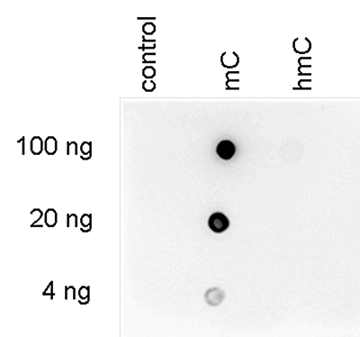
Figure 1. Dot blot analysis using the Diagenode monoclonal antibody directed against 5-mC
To demonstrate the specificity of the Diagenode antibody against 5-mC (Cat. No. C15200003), a Dot blot analysis was performed using the hmC, mC and C controls from the Diagenode “5-hmC, 5-mC & cytosine DNA Standard Pack” (Cat. No. AF-101-0002). One hundred to 4 ng (equivalent of 5 to 0.2 pmol of C-bases) of the controls were spotted on a membrane (Amersham Hybond-N+). The antibody was used at a dilution of 1:600. Figure 1 shows a high specificity of the antibody for the methylated control.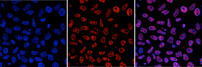
Figure 2. Immunofluorescence results obtained with the Diagenode monoclonal antibody directed against 5-mC
HeLa cells were stained with the Diagenode antibody against 5-mC (Cat. No. C15200003) and with DAPI. Cells were fixed with 4% formaldehyde for 10’ and blocked with PBS/TX-100 containing 1% BSA. The cells were immunofluorescently labelled with the 5-mC antibody (middle) diluted 1:1,000 in blocking solution followed by an anti-mouse antibody conjugated to Alexa594. The left panel shows staining of the nuclei with DAPI. A merge of the two stainings is shown on the right.A.
B.
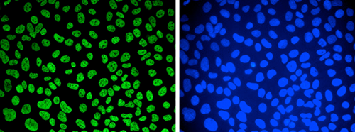
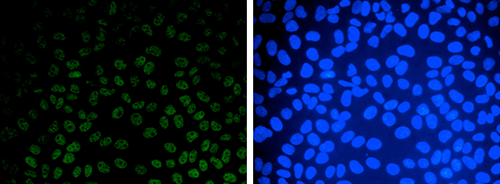
Figure 3. Immunofluorescence results obtained with the Diagenode monoclonal antibody directed against 5-mC
Human osteosarcoma (U2OS) cells were stained with the Diagenode monoclonal antibody against 5-mC (Cat. No. C15200003). Cells were fixed with 2.5% PFA in PBS for 30’, permeabilised with 0.5% Triton X-100 for 1 hour and treated with 2N HCl for 1 hour followed by 2 x 5 minutes with 0.1 M borate buffer to depurinate the DNA. After blocking with PBS containing 0.1% Triton X-100 and 1% BSA, the cells were immunofluorescently labelled with the 5-mC antibody diluted 1:500 in blocking solution, followed by a goat anti-mouse antibody conjugated to Alexa488. Figure 3A: cells were immunofluorescently labelled with the 5-mC antibody after incubation of the antibody with 50 µM mCTP (left) or with DAPI (right). Figure 3C: staining of the cells with the 5-mC antibody after incubation of the antibody with 50 µM hmCTP and with DAPI.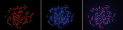
Figure 4. FISH using the Diagenode monoclonal antibody directed against 5-mC
To detect methylated chromosomal regions, FISH was performed on metaphase chromosomes from HeLa cells using the Diagenode monoclonal antibody against 5-mC (Cat. No. C15200003). The cells were blocked in metaphase by treatment with colcemid (0.1 µg/ml) for 1 - 2 hours, fixed overnight at -20°C with ethanol/glacial acetic acid and treated with 2N HCl for 30’ at room temperature. Subsequently, the cells were blocked with PBS containing 1% BSA and 0.1% Triton X-100 and stained with the 5-mC antibody (left) diluted 1:1,000 in blocking solution, followed by an anti-mouse antibody conjugated to Alexa594. The middle panel shows staining of the chromosomes with DAPI. A merge of the two stainings is shown on the right.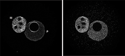
Figure 5. FISH using the Diagenode monoclonal antibody directed against 5-mC
To detect methylated chromosomal regions, FISH was performed on metaphase chromosomes from HeLa cells using the Diagenode monoclonal antibody against 5-mC (Cat. No. C15200003). The cells were blocked in metaphase by treatment with colcemid (0.1 µg/ml) for 1 - 2 hours, fixed overnight at -20°C with ethanol/glacial acetic acid and treated with 2N HCl for 30’ at room temperature. Subsequently, the cells were blocked with PBS containing 1% BSA and 0.1% Triton X-100 and stained with the 5-mC antibody (left) diluted 1:1,000 in blocking solution, followed by an anti-mouse antibody conjugated to Alexa594. The middle panel shows staining of the chromosomes with DAPI. A merge of the two stainings is shown on the right. - Publications

How to properly cite our product/service in your work
We strongly recommend using this: 5-methylcytosine (5-mC) Antibody for ICC/IF (Hologic Diagenode Cat# C15200003 Lot# 003). Click here to copy to clipboard.
Using our products or services in your publication? Let us know!
Developmental mRNA mC landscape and regulatory innovations of massivemC modification of maternal mRNAs in animals.
Liu J. et al.
mC is one of the longest-known RNA modifications, however, its developmental dynamics, functions, and evolution in mRNAs remain largely unknown. Here, we generate quantitative mRNA mC maps at different stages of development in 6 vertebrate and invertebrate species and find convergent and unexpected massive methylati...Detection of genetic variation and base modifications at base-pairresolution on both DNA and RNA.
Wang, Zhen et al.
Accurate decoding of nucleic acid variation is critical to understand the complexity and regulation of genome function. Here we use a single-molecule magnetic tweezer (MT) platform to identify sequence variation and map a range of important epigenetic base modifications with high sensitivity, specificity, and precis...Transcriptome-wide distribution and function of RNA hydroxymethylcytosine
Delatte B, Wang F, Ngoc LV, Collignon E, Bonvin E, Deplus R, Calonne E, Hassabi B, Putmans P, Awe S, Wetzel C, Kreher J, Soin R, Creppe C, Limbach PA, Gueydan C, Kruys V, Brehm A, Minakhina S, Defrance M, Steward R, Fuks F.
Hydroxymethylcytosine, well described in DNA, occurs also in RNA. Here, we show that hydroxymethylcytosine preferentially marks polyadenylated RNAs and is deposited by Tet in Drosophila. We map the transcriptome-wide hydroxymethylation landscape, revealing hydroxymethylcytosine in the transcripts of many genes, nota...RNA biochemistry. Transcriptome-wide distribution and function of RNA hydroxymethylcytosine.
Delatte B, Wang F, Ngoc LV, Collignon E, Bonvin E, Deplus R, Calonne E, Hassabi B, Putmans P, Awe S, Wetzel C, Kreher J, Soin R, Creppe C, Limbach PA, Gueydan C, Kruys V, Brehm A, Minakhina S, Defrance M, Steward R, Fuks F
Hydroxymethylcytosine, well described in DNA, occurs also in RNA. Here, we show that hydroxymethylcytosine preferentially marks polyadenylated RNAs and is deposited by Tet in Drosophila. We map the transcriptome-wide hydroxymethylation landscape, revealing hydroxymethylcytosine in the transcripts of many genes, nota... - Related products