Histones are the main constituents of the protein part of chromosomes of eukaryotic cells. They are rich in the amino acids arginine and lysine and have been greatly conserved during evolution. Histones pack the DNA into tight masses of chromatin. Two core histones of each class H2A, H2B, H3 and H4 assemble and are wrapped by 146 base pairs of DNA to form one octameric nucleosome. Histone tails undergo numerous post-translational modifications, which either directly or indirectly alter chromatin structure to facilitate transcriptional activation or repression or other nuclear processes. In addition to the genetic code, combinations of the different histone modifications reveal the so-called “histone code”. Histone methylation and demethylation is dynamically regulated by respectively histone methyl transferases and histone demethylases. Acetylation of histone H3K18 is associated with gene activation.
H3K18ac Antibody

Polyclonal antibody raised in rabbit against the region of histone H3 containing the acetylated lysine 18 (H3K18ac), using a KLH-conjugated synthetic peptide.
| Lot | A1460D |
|---|---|
| Concentration | 0.81 µg/µl |
| Species reactivity | Human, mouse, wide range expected |
| Type | Polyclonal |
| Purity | Affinity purified |
| Host | Rabbit |
| Precautions | This product is for research use only. Not for use in diagnostic or therapeutic procedures. |
| Applications | Suggested dilution | References |
|---|---|---|
| ChIP/ChIP-seq * | 1 μg/IP | Fig 1, 2 |
| CUT&Tag | 0.5 µg | Fig 3 |
| ELISA | 1:100 | Fig 4 |
| Dot Blotting | 1:5,000 | Fig 5 |
| Western Blotting | 1:500 | Fig 6 |
| Immunofluorescence | 1:200 | Fig 7 |
* Please note that the optimal antibody amount per ChIP should be determined by the end-user. We recommend testing 0.5-5 μg per IP.
- Validation Data
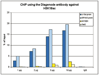
Figure 1. ChIP results obtained with the Diagenode antibody directed against H3K18ac
ChIP assays were performed using human HeLa cells, treated with TSA, the Diagenode antibody against H3K18ac (cat. No. C15410139) and optimized PCR primer pairs for qPCR. ChIP was performed with the “iDeal ChIP-seq” kit (cat. No. C15410139), using sheared chromatin from 1,000,000 cells. A titration consisting of 1, 2, 5 and 10 μg of antibody per ChIP experiment was analyzed. IgG (2 μg/IP) was used as a negative IP control. Quantitative PCR was performed with primers for the promoters of the active EIF4A2 and c-fos genes, used as positive controls, and for the inactive MYOD1 gene and the Sat2 satellite repeat, used as negative controls. Figure 1 shows the recovery, expressed as a % of input (the relative amount of immunoprecipitated DNA compared to input DNA after qPCR analysis).A.
B.
C.

D.
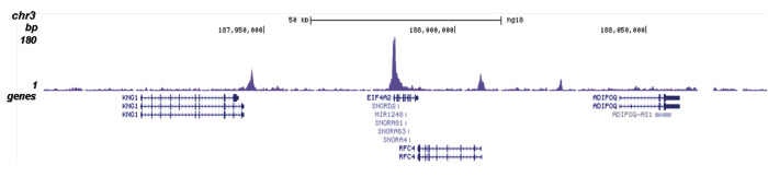
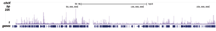
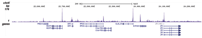
Figure 2. ChIP-seq results obtained with the Diagenode antibody directed against H3K18ac
ChIP was performed as described above using 1 μg of the Diagenode antibody against H3K18ac (cat. No. C15410139). The IP’d DNA was subsequently analysed on an Illumina Genome Analyzer. Library preparation, cluster generation and sequencing were performed according to the manufacturer’s instructions. The 36 bp tags were aligned to the human genome using the ELAND algorithm. Figure 2 shows the peak distribution along the complete human X-chromosome and a zoomin to a 600 kb region (figure 2A and B), and in two regions on chromosome 14 and 3 surrounding the c-fos and EIF4A2 positive control genes (figure 2C and D, respectively).A.
B.
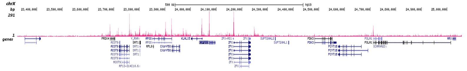
Figure 3. Cut&Tag results obtained with the Diagenode antibody directed against H3K18ac
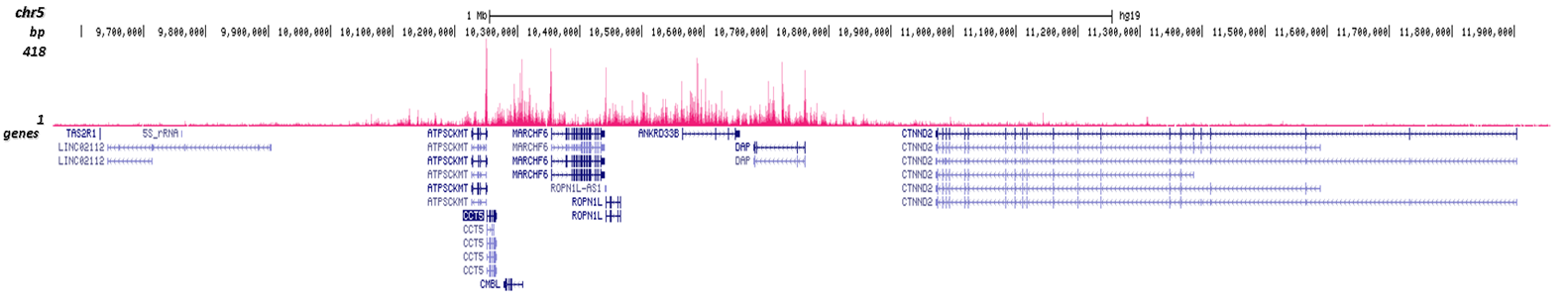
CUT&TAG (Kaya-Okur, H.S., Nat Commun 10, 1930, 2019) was performed on 50,000 K562 cells using 0.5 µg of the Diagenode antibody against H3K18ac (cat. No. C15410139) and the Diagenode pA-Tn5 transposase (C01070001). The libraries were subsequently analysed on an Illumina NextSeq 500 sequencer (2x75 paired-end reads) according to the manufacturer's instructions. The tags were aligned to the human genome (hg19) using the BWA algorithm. Figure 3 shows the peak distribution in 2 genomic regions surrounding the CCT5 gene on chromosome 5 and the EIF2S3 gene on the X-chromosome (figure 3A and B, respectively).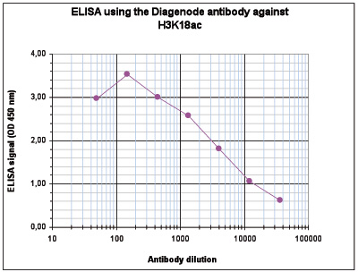
Figure 4. Determination of the antibody titer
To determine the titer of the antibody, an ELISA was performed using a serial dilution of the Diagenode antibody against H3K18ac (cat. No. C15410139). The antigen used was a peptide containing the histone modification of interest. By plotting the absorbance against the antibody dilution (Figure 4), the titer of the antibody was estimated to be 1:4,300.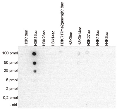
Figure 5. Cross reactivity tests using the Diagenode antibody directed against H3K18ac
To test the cross reactivity of the Diagenode antibody against H3K18ac (cat. No. C15410139), a Dot Blot analysis was performed with peptides containing other histone modifications and the unmodified H3K18. One hundred to 0.2 pmol of the respective peptides were spotted on a membrane. The antibody was used at a dilution of 1:5,000. Figure 5 shows a high specificity of the antibody for the modification of interest.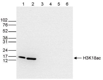
Figure 6. Western blot analysis using the Diagenode antibody directed against H3K18ac
Western blot was performed on whole cell (25 μg, lane 1) and histone extracts (15 μg, lane 2) from HeLa cells, and on 1 μg of recombinant histone H2A, H2B, H3 and H4 (lane 3, 4, 5 and 6, respectively) using the Diagenode antibody against H3K18ac (cat. No. C15410139). The antibody was diluted 1:500 in TBS-Tween containing 5% skimmed milk. The marker (in kDa) is shown on the left.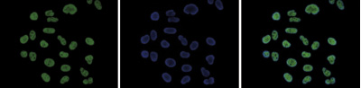
Figure 7. Immunofluorescence using the Diagenode antibody directed against H3K18ac
HeLa cells were stained with the Diagenode antibody against H3K18ac (cat. No. C15410139) and with DAPI. Cells were fixed with 4% formaldehyde for 10’ and blocked with PBS/TX-100 containing 5% normal goat serum and 1% BSA. The cells were immunofluorescently labeled with the H3K18ac antibody (left) diluted 1:200 in blocking solution followed by an anti-rabbit antibody conjugated to Alexa488. The middle panel shows staining of the nuclei with DAPI. A merge of the two stainings is shown on the right. - Publications

How to properly cite our product/service in your work
We strongly recommend using this: H3K18ac Antibody (Hologic Diagenode Cat# C15410139 Lot# A1460D). Click here to copy to clipboard.
Using our products or services in your publication? Let us know!
Disease-specific epigenetic deregulation of enhancers, transposons, and polycomb targets in acute promyelocytic leukemia
Zhong, Xiangfu et al.
Background: Acute promyelocytic leukemia (APL) is a subtype of acute myeloid leukemia (AML), characterized by a fusion between the PML and RARA genes and by a block in the myeloid maturation at the promyelocytic stage. Methods: This study investigates the epigenetic landscape of APL by integrating ChIP-se...Accelerated epigenetic aging in Huntington’s disease involves polycomb repressive complex 1
Baptiste Brulé et al.
Loss of epigenetic information during physiological aging compromises cellular identity, leading to de-repression of developmental genes. Here, we assessed the epigenomic landscape of vulnerable neurons in two reference mouse models of Huntington neurodegenerative disease (HD), using cell-type-specific multi-omics, ...Myc Regulates Chromatin Decompaction and Nuclear Architecture during B Cell Activation
Kieffer-Kwon K.R. et al.
50 years ago, Vincent Allfrey and colleagues discovered that lymphocyte activation triggers massive acetylation of chromatin. However, the molecular mechanisms driving epigenetic accessibility are still unknown. We here show that stimulated lymphocytes decondense chromatin by three differentially regulated steps. Fi...