Histones are the main constituents of the protein part of chromosomes of eukaryotic cells. They are rich in the amino acids arginine and lysine and have been greatly conserved during evolution. Histones pack the DNA into tight masses of chromatin. Two core histones of each class H2A, H2B, H3 and H4 assemble and are wrapped by 146 base pairs of DNA to form one octameric nucleosome. Histone tails undergo numerous post-translational modifications, which either directly or indirectly alter chromatin structure to facilitate transcriptional activation or repression or other nuclear processes. In addition to the genetic code, combinations of the different histone modifications reveal the so-called “histone code”. Histone methylation and demethylation is dynamically regulated by respectively histone methyl transferases and histone demethylases.
H3K79me1 Antibody

Polyclonal antibody raised in rabbit against histone H3 containing the monomethylated lysine 79 (H3K79me1), using a KLH-conjugated synthetic peptide.
| Lot | A823-001D |
|---|---|
| Concentration | 1.66 µg/µl |
| Species reactivity | Human, yeast |
| Type | Polyclonal |
| Purity | Affinity purified |
| Host | Rabbit |
| Precautions | This product is for research use only. Not for use in diagnostic or therapeutic procedures. |
| Applications | Suggested dilution | References |
|---|---|---|
| ChIP/ ChIP-seq * | 1-2 μg/ChIP | Fig 1, 2 |
| CUT&TAG | 1 μg | Fig 3 |
| ELISA | 1:500 | Fig 4 |
| Dot Blotting | 1:20,000 | Fig 5 |
| Western Blotting | 1:200 | Fig 6 |
* Please note that the optimal antibody amount per IP should be determined by the end-user. We recommend testing 1-5 μg per IP.
- Validation Data
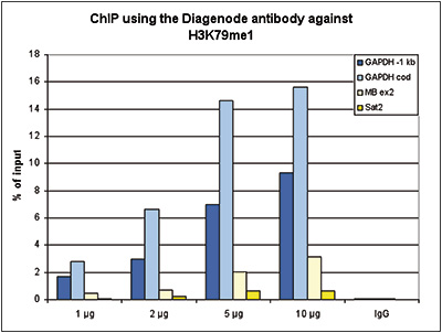
Figure 1. ChIP results obtained with the Diagenode antibody directed against H3K79me1
ChIP assays were performed using human HeLa cells, the Diagenode antibody against H3K79me1 (Cat. No. pAb-082-050) and optimized PCR primer pairs for qPCR. ChIP was performed with the “iDeal ChIP-seq” kit (cat. No. AB-001-0024), using sheared chromatin from 1 million cells. A titration consisting of 1, 2, 5 and 10 μg of antibody per ChIP experiment was analyzed. IgG (2 μg/IP) was used as a negative IP control. Quantitative PCR was performed with primers specific for a region 1 kb upstream of the promoter and the coding region of the active GAPDH gene, used as positive controls, and for exon 2 of the inactive myoglobin (MB) gene and the Sat2 satellite repeat, used as negative controls. Figure 1 shows the recovery, expressed as a % of input (the relative amount of immunoprecipitated DNA compared to input DNA after qPCR analysis).A. 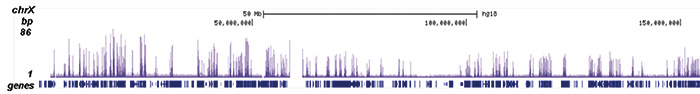
B. 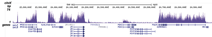
C. 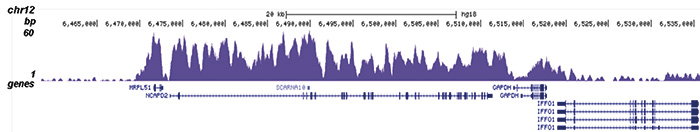
D. 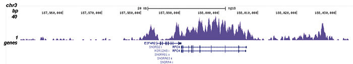
Figure 2. ChIP-seq results obtained with the Diagenode antibody directed against H3K79me1
ChIP was performed with 1 μg of the Diagenode antibody against H3K79me1 (Cat. No. pAb-082-050) as described above and the IP’d DNA was subsequently analysed on an Illumina Genome Analyzer. Library preparation, cluster generation and sequencing were performed according to the manufacturer’s instructions. The 36 bp tags were aligned to the human genome using the ELAND algorithm. Figure 2 shows the peak distribution along the complete sequence and a 1 Mb region of the X-chromosome (figure 2A and B), in 100 kb regions surrounding the GAPDH positive control and EIF4A2 genes (figure 2C and D).A. 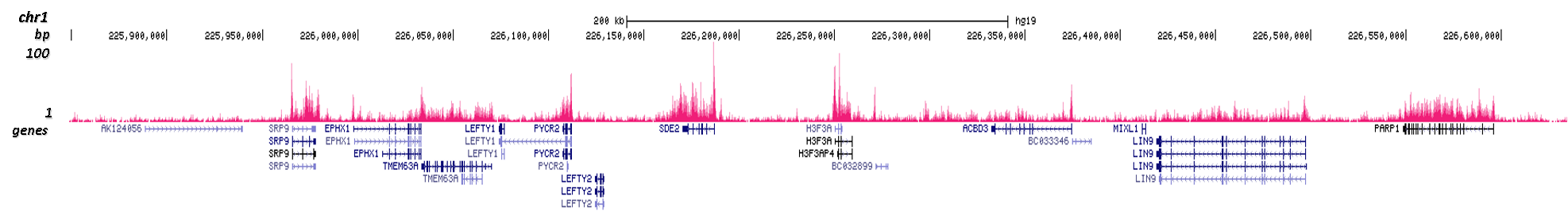
B. 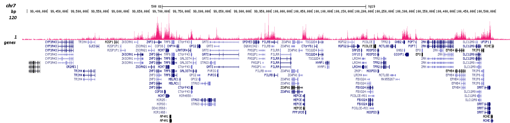
Figure 3. Cut&Tag results obtained with the Diagenode antibody directed against H3K79me1
CUT&TAG (Kaya-Okur, H.S., Nat Commun 10, 1930, 2019) was performed on 50,000 K562 cells using 1 µg of the Diagenode antibody against H3K79me1 (cat. No. C1541082) and the Diagenode pA-Tn5 transposase (C01070001). The libraries were subsequently analysed on an Illumina NextSeq 500 sequencer (2x75 paired-end reads) according to the manufacturer's instructions. The tags were aligned to the human genome (hg19) using the BWA algorithm. Figure 3 shows the peak distribution in 2 genomic regions on chromosome 1 and 7 (figure 3A and B, respectively).
Figure 4. Determination of the antibody titer
To determine the titer of the antibody, an ELISA was performed using a serial dilution of the Diagenode antibody directed against H3K79me1 (Cat. No. pAb-082-050). The antigen used was a peptide containing the histone modification of interest. By plotting the absorbance against the antibody dilution (Figure 4), the titer of the antibody was estimated to be 1:11,500.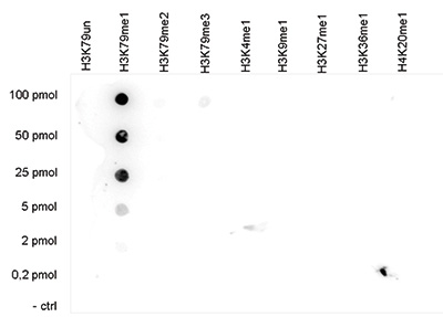
Figure 5. Cross reactivity test using the Diagenode antibody directed against H3K79me1
A Dot Blot analysis was performed to test the cross reactivity of the Diagenode antibody against H3K79me1 (Cat. No. pAb-082-050) with peptides containing other histone modifications and the unmodified H3K79. One hundred to 0.2 pmol of the respective peptides were spotted on a membrane. The antibody was used at a dilution of 1:20,000. Figure 5 shows a high specificity of the antibody for the modification of interest.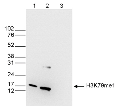
Figure 6. Western blot analysis using the Diagenode antibody directed against H3K79me1
Western blot was performed on whole cell (25 μg, lane 1) and histone extracts (15 μg, lane 2) from HeLa cells, and on 1 μg of recombinant histone H3 (lane 3) using the Diagenode antibody against H3K79me1 (Cat. No. pAb-082-050). The antibody was diluted 1:200 in TBS-Tween containing 5% skimmed milk. The position of the protein of interest is indicated on the right; the marker (in kDa) is shown on the left. - Publications

How to properly cite our product/service in your work
We strongly recommend using this: H3K79me1 Antibody (Hologic Diagenode Cat# C15410082 Lot# A823-001D). Click here to copy to clipboard.
Using our products or services in your publication? Let us know!
Myc Regulates Chromatin Decompaction and Nuclear Architecture during B Cell Activation
Kieffer-Kwon K.R. et al.
50 years ago, Vincent Allfrey and colleagues discovered that lymphocyte activation triggers massive acetylation of chromatin. However, the molecular mechanisms driving epigenetic accessibility are still unknown. We here show that stimulated lymphocytes decondense chromatin by three differentially regulated steps. Fi...Anticheckpoint pathways at telomeres in yeast
Ribeyre Cyril, Shore David
Telomeres hide (or ‘cap’) chromosome ends from DNA-damage surveillance mechanisms that arrest the cell cycle and promote repair, but the checkpoint status of telomeres is not well understood. Here we characterize the response in Saccharomyces cerevisiae to DNA double-strand breaks (DSBs) flanked by varying amounts o...Chromatin states of core pluripotency-associated genes in pluripotent, multipotent and differentiated cells.
Barrand S, Collas P
Oct4, Nanog and Sox2 constitute a core of transcription factors controlling pluripotency. Differentiation and reprogramming studies have unraveled a few epigenetic modifications associated in relation to the expression state of OCT4, NANOG and SOX2. There is, however, no comprehensive map of chromatin states on thes...