Histones are the main constituents of the protein part of chromosomes of eukaryotic cells. They are rich in the amino acids arginine and lysine and have been greatly conserved during evolution. Histones pack the DNA into tight masses of chromatin. Two core histones of each class H2A, H2B, H3 and H4 assemble and are wrapped by 146 base pairs of DNA to form one octameric nucleosome. Histone tails undergo numerous post-translational modifications, which either directly or indirectly alter chromatin structure to facilitate transcriptional activation or repression or other nuclear processes. In addition to the genetic code, combinations of the different histone modifications reveal the so-called “histone code”. Histone methylation and demethylation is dynamically regulated by respectively histone methyl transferases and histone demethylases. Acetylation of the histone H2A variant H2A.Z is associated with the promoters of active genes.
H2A.Zac Antibody

Polyclonal antibody raised in rabbit against the region of histone H2A.Z containing the acetylated lysines 4, 7 and 11, using a KLH-conjugated synthetic peptide.
| Lot | A1738P |
|---|---|
| Concentration | 1,09 µg/µl |
| Species reactivity | Human, mouse, wide range expected. |
| Type | Polyclonal, ChIP-grade, ChIP-seq grade, CUT&Tag-grade |
| Purity | Affinity purified |
| Host | Rabbit |
| Storage Conditions | Store at -20°C; for long storage, store at -80°C. Avoid multiple freeze-thaw cycles |
| Storage Buffer | PBS containing 0.05% azide and 0.05% ProClin 300 |
| Precautions | This product is for research use only. Not for use in diagnostic or therapeutic procedures. |
| Applications | Suggested dilution | References |
|---|---|---|
| ChIP/ChIP-seq * | 1 μg/IP | Fig 1, 2 |
| CUT&Tag | 1 µg | Fig 3 |
| ELISA | 1:500 | Fig 4 |
| Dot Blotting | 1:20,000 | Fig 5 |
| Western Blotting | 1:1,000 | Fig 6 |
| Immunofluorescence | 1:500 | Fig 7 |
* Please note that the optimal antibody amount per IP should be determined by the end-user. We recommend testing 0.5-5 μg per IP.
- Validation DataA
 B
B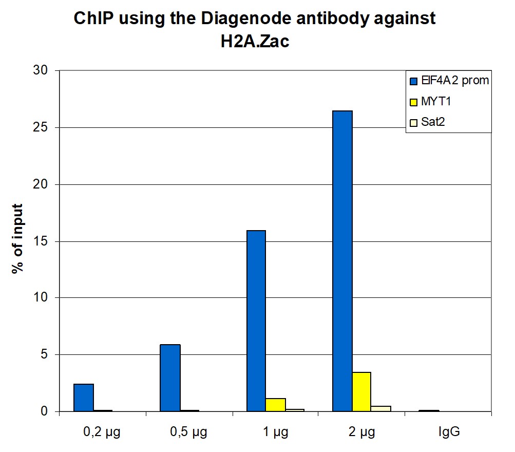
Figure 1. ChIP results obtained with the antibody directed against H2A.Zac
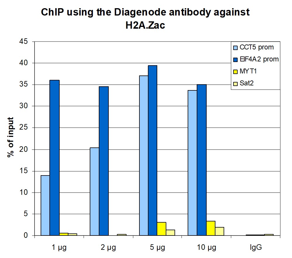
Figure 1A ChIP assays were performed using human HeLa cells, the antibody against H2A.Zac (cat. No. C15410202) and optimized PCR primer pairs for qPCR. ChIP was performed with the iDeal ChIP-seq kit (cat. No. C01010051), using sheared chromatin from 1,000,000 cells. A titration consisting of 1, 2, 5 and 10 µg of antibody per ChIP experiment was analyzed. IgG (2 µg/IP) was used as a negative IP control. Quantitative PCR was performed with primers specific for the promoter of the active genes CCT5 and EIF4A2, used as positive controls, and for the coding region of the inactive MYT1 gene and the Sat2 satellite repeat, used as negative controls. Figure 1 shows the recovery, expressed as a % of input (the relative amount of immunoprecipitated DNA compared to input DNA after qPCR analysis).
Figure 1B ChIP assays were performed as described above on 100,000 K562 cells. A titration of the antibody consisting of 0.2, 0.5, 1 and 2 µg per ChIP experiment was analysed. IgG (1 µg/IP) was used as negative IP control. Quantitative PCR was performed with primers specific for the EIF4A2, used as positive control, and for the coding region of the inactive MYT1 gene and the Sat2 satellite repeat, used as negative controls. Figure 1 shows the recovery, expressed as a % of input (the relative amount of immunoprecipitated DNA compared to input DNA after qPCR analysis).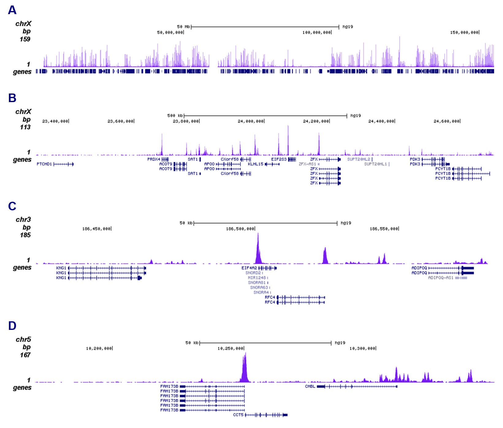
Figure 2. ChIP-seq results obtained with the antibody directed against H2A.Zac
ChIP was performed on sheared chromatin from 100,000 K562 cells using 1 µg the antibody against H2A.Zac (cat. No. C15410202) with the iDeal ChIP-seq kit (cat. No. C01010051) as described above. The IP’d DNA was subsequently analysed with an Illumina Genome Analyzer. Library preparation, cluster generation and sequencing were performed according to the manufacturer’s instructions. The 36 bp tags were aligned to the human genome using the ELAND algorithm. Figure 2 shows the peak distribution along the complete sequence and a 1.5 Mb region of the X-chromosome (figure 2A and B) and in two regions surrounding the EIF4A2 and CCT5 positive control genes, respectively (figure 2C and D).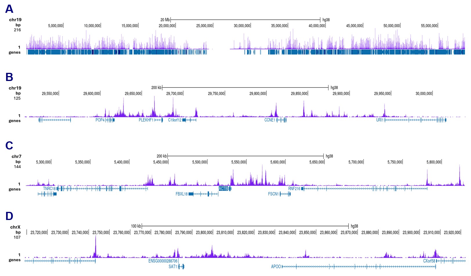
Figure 3. CUT&Tag results obtained with the antibody directed against H2A.Zac
CUT&Tag was performed on 50,000 K562 cells using 1 µg of the antibody against H2A.Zac (cat. No. C15410202) and the Universal CUT&Tag kit (cat. No. C01070024). The libraries were subsequently analysed on an Illumina NovaSeqX sequencer (2x150 bp paired-end reads) according to the manufacturer’s instructions. The tags were aligned to the human genome (hg38) using the BWA algorithm. Figure 2 shows the peak distribution along the complete sequence and a 600 kb region of chromosome 19 (figure 3A and B) and in 2 genomic regions on chromosome 7 and X (figure 3C and D, respectively).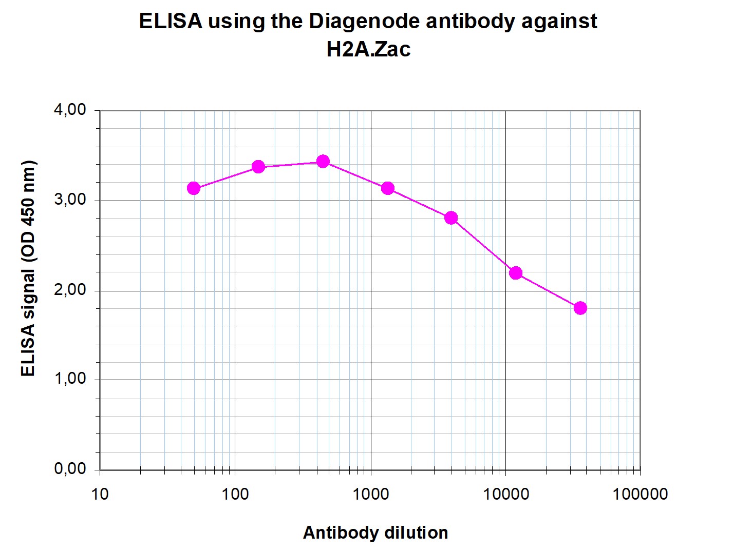
Figure 4. Determination of the antibody titer
To determine the titer of the antibody, an ELISA was performed using a serial dilution of the antibody against H2A.Zac (cat. No. C15410202). The antigen used was a peptide containing the histone modifications of interest. By plotting the absorbance against the antibody dilution (Figure 4), the titer of the antibody was estimated to be 1:56,600.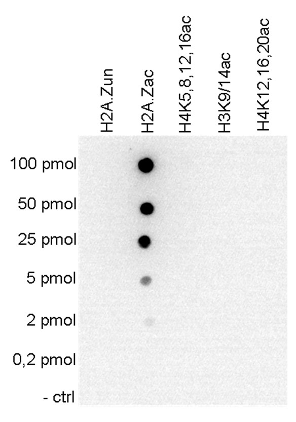
Figure 5. Cross reactivity tests using the antibody directed against H2A.Zac
To test the cross reactivity of the antibody against H2A.Zac (cat. No. C15410202), a Dot Blot analysis was performed with peptides containing different multiple acetylations and the unmodified H2A.Z. One hundred to 0.2 pmol of the respective peptides were spotted on a membrane. The antibody was used at a dilution of 1:20,000. Figure 5 shows a high specificity of the antibody for the modification of interest.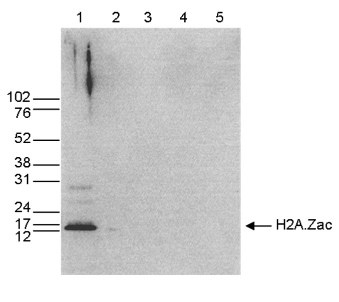
Figure 6. Western blot analysis using the antibody directed against H2A.Zac
Western blot was performed on whole cell extracts (25 µg, lane 1) from HeLa cells, and on 1 µg of recombinant histone H2A, H2B, H3 and H4 (lane 2, 3, 4 and 5, respectively) using the antibody against H2A.Zac (cat. No. C15410202). The antibody was diluted 1:1,000 in TBS-Tween containing 5% skimmed milk. The position of the protein of interest is indicated on the right, the marker (in kDa) is shown on the left.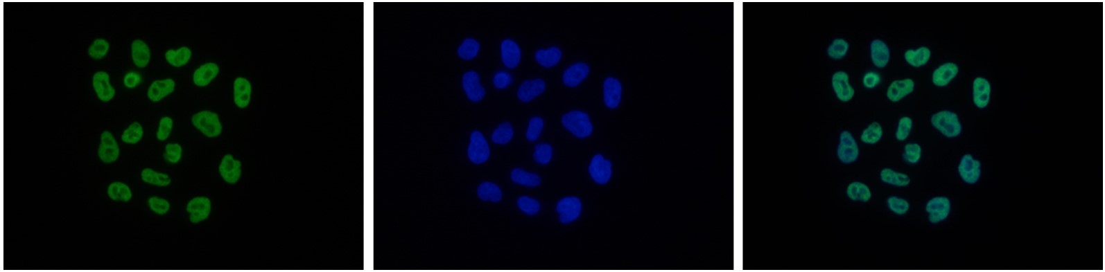
Figure 6. Immunofluorescence using the antibody directed against H2A.Zac
HeLa cells were stained with the antibody against H2A.Zac (cat. No. C15410202) and with DAPI. Cells were fixed with 4% formaldehyde for 10’ and blocked with PBS/TX-100 containing 5% normal goat serum and 1% BSA. The cells were immunofluorescently labelled with the H2A.Zac antibody (left) diluted 1:500 in blocking solution followed by an anti-rabbit antibody conjugated to Alexa488. The middle panel shows staining of the nuclei with DAPI. A merge of the two stains is shown on the right. - Publications

How to properly cite our product/service in your work
We strongly recommend using this: H2A.Zac Antibody (Hologic Diagenode Cat# C15410202 Lot# A1738P). Click here to copy to clipboard.
Using our products or services in your publication? Let us know!
PRC1-mediated H2A.Zub promotes gene expression by preventing H3.1K27me1 incorporation in Arabidopsis
Baile, Fernando et al.
Background: PcG complexes are pivotal in orchestrating the transition from embryonic to vegetative development in plants. However, the mechanisms underlying the gene expression reprogramming that takes place during this developmental transition are still not fully understood. Several studies suggest that incorp...Ep400 deficiency in Schwann cells causes persistent expression of early developmental regulators and peripheral neuropathy.
Fröb F, Sock E, Tamm ER, Saur AL, Hillgärtner S, Williams TJ, Fujii T, Fukunaga R, Wegner M
Schwann cells ensure efficient nerve impulse conduction in the peripheral nervous system. Their development is accompanied by defined chromatin changes, including variant histone deposition and redistribution. To study the importance of variant histones for Schwann cell development, we altered their genomic distribu...PWWP2A binds distinct chromatin moieties and interacts with an MTA1-specific core NuRD complex.
Link S, Spitzer RMM, Sana M, Torrado M, Völker-Albert MC, Keilhauer EC, Burgold T, Pünzeler S, Low JKK, Lindström I, Nist A, Regnard C, Stiewe T, Hendrich B, Imhof A, Mann M, Mackay JP, Bartkuhn M, Hake SB
Chromatin structure and function is regulated by reader proteins recognizing histone modifications and/or histone variants. We recently identified that PWWP2A tightly binds to H2A.Z-containing nucleosomes and is involved in mitotic progression and cranial-facial development. Here, using in vitro assays, we show that...