Code Context<!-- BEGIN: REQUEST_FORM MODAL -->
<div id="request_formModal" class="reveal-modal medium" data-reveal aria-labelledby="modalTitle" aria-hidden="true" role="dialog">
<?= $this->element('Forms/simple_form', array('solution_of_interest' => $solution_of_interest, 'header' => $header, 'message' => $message, 'campaign_id' => $campaign_id)) ?>
$viewFile = '/home/website-server/www/app/View/Products/view.ctp'
$dataForView = array(
'language' => 'cn',
'meta_keywords' => '',
'meta_description' => 'H3K9me3 (Histone H3 trimethylated at lysine 9) Polyclonal Antibody validated in ChIP-seq, ChIP-qPCR, ELISA, DB, WB and IF. Batch-specific data available on the website. Sample size available',
'meta_title' => 'H3K9me3 Antibody - ChIP-seq Grade (C15410056) | Diagenode',
'product' => array(
'Product' => array(
'id' => '2217',
'antibody_id' => '120',
'name' => 'H3K9me3 Antibody - ChIP-seq Grade (sample size)',
'description' => '<p><span>Polyclonal antibody raised in rabbit against the region of histone H3 containing the trimethylated lysine 9 (H3K9me3 Antibody validated in Western blot), using a KLH-conjugated synthetic peptide.</span></p>',
'label1' => 'Validation Data',
'info1' => '<div class="row">
<div class="small-4 columns">
<p><img src="https://www.diagenode.com/img/product/antibodies/C15410056_ChIP.jpg" alt="H3K9me3 Antibody ChIP Grade" caption="false" width="278" height="189" /></p>
</div>
<div class="small-8 columns">
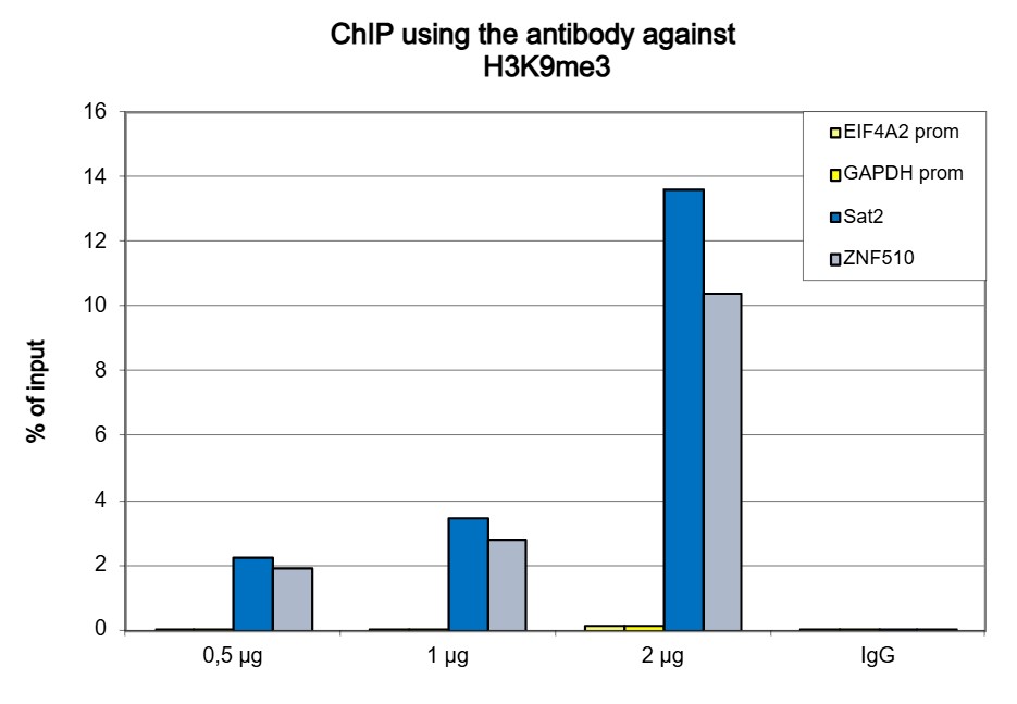
<p><small> <strong>Figure 1. ChIP results obtained with the Diagenode antibody directed against H3K9me3</strong><br /> ChIP assays were performed using human HeLa cells, the Diagenode antibody against H3K9me3 (Cat. No. C15410056) and optimized PCR primer sets for qPCR. ChIP was performed on sheared chromatin from 1 million HeLaS3 cells using the “Auto Histone ChIP-seq” kit (Cat. No. C01010022 ) on the SX-8G IP-Star automated system. A titration of the antibody consisting of 1, 2, 5, and 10 μg per ChIP experiment was analysed. IgG (2 μg/IP) was used as negative IP control. QPCR was performed with primers for the heterochromatin marker Sat2 and for the ZNF510 gene, used as positive controls, and for the promoters of the active c-fos and GAPDH genes, used as negative controls. Figure 1 shows the recovery, expressed as a % of input (the relative amount of immunoprecipitated DNA compared to input DNA after qPCR analysis). </small></p>
</div>
</div>
<div class="row">
<div class="small-6 columns">
<p><img src="https://www.diagenode.com/img/product/antibodies/C15410056-Fig2A.jpg" alt="H3K9me3 Antibody ChIP-seq assay" caption="false" width="400" height="358" /></p>
<p><img src="https://www.diagenode.com/img/product/antibodies/C15410056-Fig2B.jpg" alt="H3K9me3 Antibody for ChIP-seq " caption="false" width="432" height="214" /></p>
<p><img src="https://www.diagenode.com/img/product/antibodies/C15410056-Fig2C.jpg" alt="H3K9me3 Antibody for ChIP-seq assay" caption="false" width="432" height="61" /> <img src="https://www.diagenode.com/img/product/antibodies/C15410056-FigD.jpg" alt="H3K9me3 Antibody validated in ChIP-seq " caption="false" width="700" height="112" /></p>
</div>
<div class="small-6 columns">
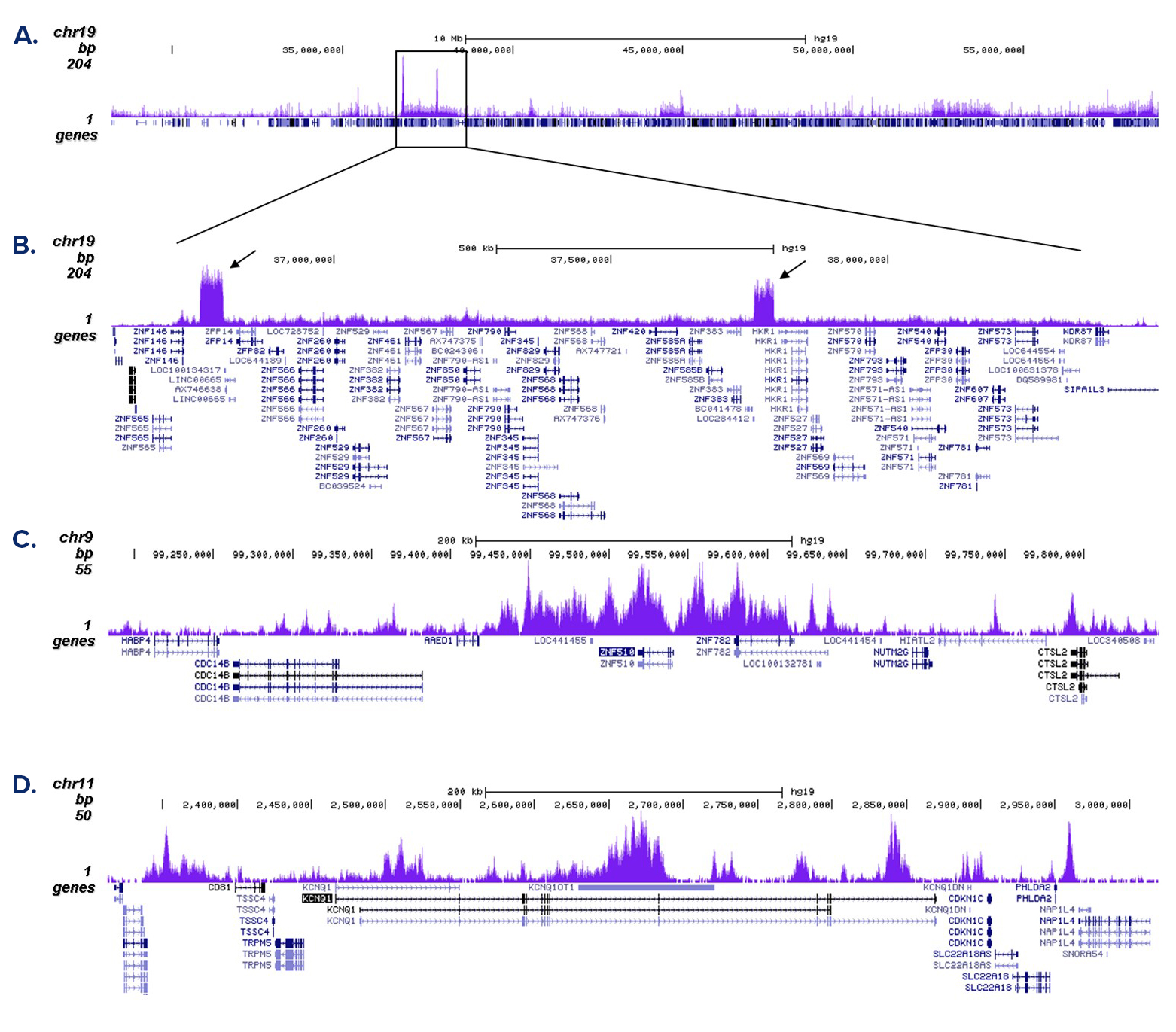
<p><small> <strong>Figure 2. ChIP-seq results obtained with the Diagenode antibody directed against H3K9me3</strong><br /> ChIP was performed with 0.5 μg of the Diagenode antibody against H3K9me3 (Cat. No. C15410056) on sheared chromatin from 1 million HeLa cells using the “iDeal ChIP-seq” kit (Cat. No. C01010051). The IP’d DNA was analysed by QPCR with optimized PCR primer pairs for the promoter regions of the active GAPDH and EIF4A2 genes, for the coding region of the ZNF510 gene and for the Sat2 satellite repeat (figure 2A). The IP’d DNA was subsequently analysed on an Illumina Genome Analyzer. Library preparation, cluster generation and sequencing were performed according to the manufacturer’s instructions. The 36 bp tags were aligned to the human genome using the BWA algorithm. Figure 2B shows the signal distribution along the long arm of chromosome 19 and a zoomin to an enriched region containing several ZNF repeat genes. Figure 2C and D show the enrichment at ZNF510 and ZNF12 on chromosome 9 and 7, respectively. These results clearly show an enrichment of H3K9me3 at ZNF repeat genes. </small></p>
</div>
</div>
<div class="row">
<div class="small-4 columns">
<p><img src="https://www.diagenode.com/img/product/antibodies/C15410056-Elisa.jpg" alt="H3K9me3 Antibody ELISA validation" caption="false" width="278" height="212" /></p>
</div>
<div class="small-8 columns">
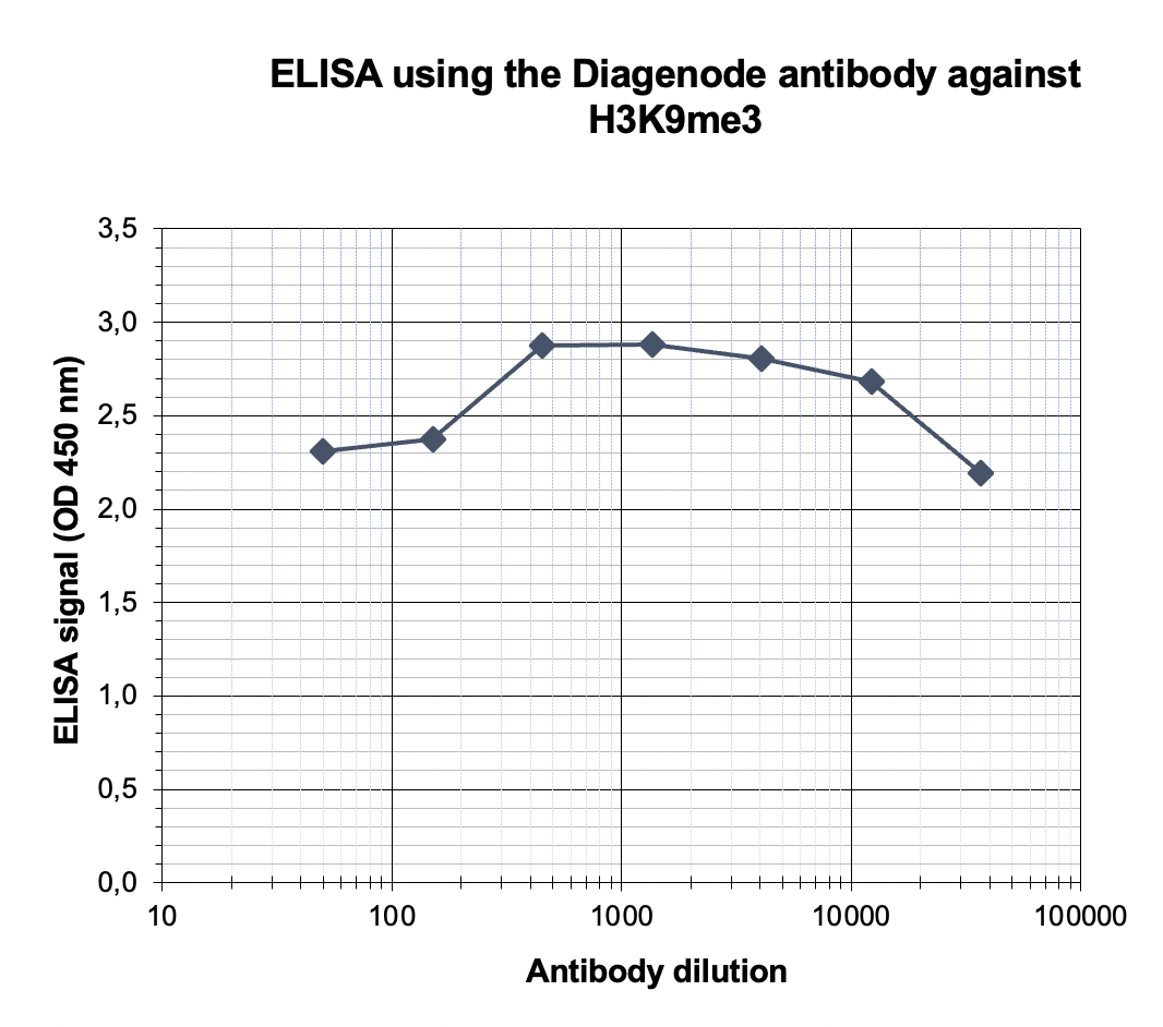
<p><small> <strong>Figure 3. Determination of the antibody titer</strong><br /> To determine the titer of the antibody, an ELISA was performed using a serial dilution of the antibody directed against human H3K9me3 (Cat. No. C15410056), crude serum and flow through in antigen coated wells. The antigen used was a peptide containing the histone modification of interest. By plotting the absorbance against the antibody dilution (Figure 3), the titer of the antibody was estimated to be 1:60,000. </small></p>
</div>
</div>
<div class="row">
<div class="small-4 columns">
<p><img src="https://www.diagenode.com/img/product/antibodies/C15410056-Fig4.jpg" alt="H3K9me3 Antibody validated in Dot Blot" caption="false" width="400" height="304" /></p>
</div>
<div class="small-8 columns">
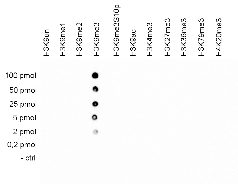
<p><small> <strong>Figure 4. Cross reactivity tests using the Diagenode antibody directed against H3K9me3</strong><br /> A Dot Blot analysis was performed to test the cross reactivity of the Diagenode antibody against H3K9me3 (Cat. No. C15410056) with peptides containing other modifications and unmodified sequences of histone H3 and H4. One hundred to 0.2 pmol of the peptide containing the respective histone modification were spotted on a membrane. The antibody was used at a dilution of 1:20,000. Figure 4 shows a high specificity of the antibody for the modification of interest. </small></p>
</div>
</div>
<div class="row">
<div class="small-4 columns">
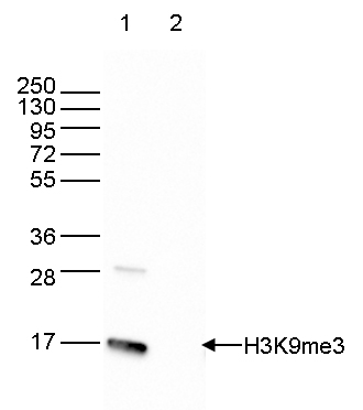
<p><img src="https://www.diagenode.com/img/product/antibodies/C15410056_WB.jpg" alt="H3K9me3 Antibody validated in Western Blot" caption="false" width="200" height="213" /></p>
</div>
<div class="small-8 columns">
<p><small> <strong>Figure 5. Western blot analysis using the Diagenode antibody directed against H3K9me3</strong><br /> Western blot analysis was performed on histone extracts from HeLa cells (15 μg, lane 1) and on recombinant H3 (1 μg, lane2) using the Diagenode antibody against H3K9me3 (Cat. No. C15410056). The antibody was diluted 1:1,000 in TBS-Tween containing 5% skimmed milk. The position of the protein of interest is indicated on the right; the marker (in kDa) is shown on the left. </small></p>
</div>
</div>
<div class="row">
<div class="small-5 columns">
<p><img src="https://www.diagenode.com/img/product/antibodies/C15410056-IF.jpg" alt="H3K9me3 Antibody validated in Immunofluorescence " caption="false" width="354" height="117" /></p>
</div>
<div class="small-7 columns">
<p><small> <strong>Figure 6. Immunofluorescence using the Diagenode antibody directed against H3K9me3</strong><br /> HeLa cells were stained with the Diagenode antibody against H3K9me3 (Cat. No. C15410056) and with DAPI. Cells were fixed with 4% formaldehyde for 10’ and blocked with PBS/TX-100 containing 5% normal goat serum and 1% BSA. The cells were immunofluorescently labelled with the H3K9me3 antibody (left) diluted 1:500 in blocking solution followed by an anti-rabbit antibody conjugated to Alexa488. The middle panel shows staining of the nuclei with DAPI. A merge of the two stainings is shown on the right. </small></p>
</div>
</div>',
'label2' => '',
'info2' => '',
'label3' => '',
'info3' => '',
'format' => '10 µg',
'catalog_number' => 'C15410056-10',
'old_catalog_number' => 'pAb-056-050',
'sf_code' => 'C15410056-D001-000582',
'type' => 'FRE',
'search_order' => '03-Antibody',
'price_EUR' => '105',
'price_USD' => '115',
'price_GBP' => '100',
'price_JPY' => '16450',
'price_CNY' => '',
'price_AUD' => '288',
'country' => 'ALL',
'except_countries' => 'None',
'quote' => false,
'in_stock' => false,
'featured' => false,
'no_promo' => false,
'online' => true,
'master' => false,
'last_datasheet_update' => '0000-00-00',
'slug' => 'h3k9me3-polyclonal-antibody-classic-sample-size-10-ug',
'meta_title' => 'H3K9me3 Antibody - ChIP-seq Grade (C15410056) | Diagenode',
'meta_keywords' => '',
'meta_description' => 'H3K9me3 (Histone H3 trimethylated at lysine 9) Polyclonal Antibody validated in ChIP-seq, ChIP-qPCR, ELISA, DB, WB and IF. Batch-specific data available on the website. Sample size available',
'modified' => '2021-10-20 09:34:17',
'created' => '2015-06-29 14:08:20',
'locale' => 'zho'
),
'Antibody' => array(
'host' => '*****',
'id' => '120',
'name' => 'H3K9me3 polyclonal antibody',
'description' => 'Histones are the main constituents of the protein part of chromosomes of eukaryotic cells. They are rich in the amino acids arginine and lysine and have been greatly conserved during evolution. Histones pack the DNA into tight masses of chromatin. Two core histones of each class H2A, H2B, H3 and H4 assemble and are wrapped by 146 base pairs of DNA to form one octameric nucleosome. Histone tails undergo numerous post-translational modifications, which either directly or indirectly alter chromatin structure to facilitate transcriptional activation or repression or other nuclear processes. In addition to the genetic code, combinations of the different histone modifications reveal the so-called “histone code”. Histone methylation and demethylation is dynamically regulated by respectively histone methyl transferases and histone demethylases. Trimethylation of histone H3K9 is associated with satellite repeat regions and ZNF repeat genes.',
'clonality' => '',
'isotype' => '',
'lot' => 'A2810P',
'concentration' => '1.85 µg/µl',
'reactivity' => 'Human, mouse, zebrafish, trout, Daphnia, Arabidopsis, Drosophila, silena latifolia: positive. Other species: not tested.',
'type' => 'Polyclonal ChIP grade, ChIP-seq grade',
'purity' => 'Affinity purified polyclonal antibody.',
'classification' => 'Classic',
'application_table' => '<table>
<thead>
<tr>
<th>Applications</th>
<th>Suggested dilution</th>
<th>References</th>
</tr>
</thead>
<tbody>
<tr>
<td>ChIP/ChIP-seq <sup>*</sup></td>
<td>0.5 - 1 µg per ChIP</td>
<td>Fig 1, 2</td>
</tr>
<tr>
<td>ELISA</td>
<td>1:1,000 - 1:10,000</td>
<td>Fig 3</td>
</tr>
<tr>
<td>Dot Blotting</td>
<td>1:2,000</td>
<td>Fig 4</td>
</tr>
<tr>
<td>Western Blotting</td>
<td>1:2,000</td>
<td>Fig 5</td>
</tr>
</tbody>
</table>
<p><small><sup>*</sup> Please note that the optimal antibody amount per IP should be determined by the end-user. We recommend testing 0.5-5 µg per IP.</small></p>',
'storage_conditions' => 'Store at -20°C; for long storage, store at -80°C. Avoid multiple freeze-thaw cycles.',
'storage_buffer' => 'PBS containing 0.05% azide.',
'precautions' => 'This product is for research use only. Not for use in diagnostic or therapeutic procedures.',
'uniprot_acc' => '',
'slug' => '',
'meta_keywords' => '',
'meta_description' => '',
'modified' => '2021-07-28 12:12:46',
'created' => '0000-00-00 00:00:00',
'select_label' => '120 - H3K9me3 polyclonal antibody (A2810P - 1.85 µg/µl - Human, mouse, zebrafish, trout, Daphnia, Arabidopsis, Drosophila, silena latifolia: positive. Other species: not tested. - Affinity purified polyclonal antibody. - Rabbit)'
),
'Slave' => array(),
'Group' => array(
'Group' => array(
[maximum depth reached]
),
'Master' => array(
[maximum depth reached]
),
'Product' => array(
[maximum depth reached]
)
),
'Related' => array(
(int) 0 => array(
[maximum depth reached]
),
(int) 1 => array(
[maximum depth reached]
),
(int) 2 => array(
[maximum depth reached]
),
(int) 3 => array(
[maximum depth reached]
),
(int) 4 => array(
[maximum depth reached]
),
(int) 5 => array(
[maximum depth reached]
)
),
'Application' => array(
(int) 0 => array(
[maximum depth reached]
),
(int) 1 => array(
[maximum depth reached]
),
(int) 2 => array(
[maximum depth reached]
),
(int) 3 => array(
[maximum depth reached]
),
(int) 4 => array(
[maximum depth reached]
),
(int) 5 => array(
[maximum depth reached]
)
),
'Category' => array(
(int) 0 => array(
[maximum depth reached]
),
(int) 1 => array(
[maximum depth reached]
),
(int) 2 => array(
[maximum depth reached]
),
(int) 3 => array(
[maximum depth reached]
),
(int) 4 => array(
[maximum depth reached]
)
),
'Document' => array(
(int) 0 => array(
[maximum depth reached]
),
(int) 1 => array(
[maximum depth reached]
),
(int) 2 => array(
[maximum depth reached]
)
),
'Feature' => array(),
'Image' => array(
(int) 0 => array(
[maximum depth reached]
)
),
'Promotion' => array(),
'Protocol' => array(),
'Publication' => array(
(int) 0 => array(
[maximum depth reached]
),
(int) 1 => array(
[maximum depth reached]
),
(int) 2 => array(
[maximum depth reached]
),
(int) 3 => array(
[maximum depth reached]
),
(int) 4 => array(
[maximum depth reached]
),
(int) 5 => array(
[maximum depth reached]
),
(int) 6 => array(
[maximum depth reached]
),
(int) 7 => array(
[maximum depth reached]
),
(int) 8 => array(
[maximum depth reached]
),
(int) 9 => array(
[maximum depth reached]
),
(int) 10 => array(
[maximum depth reached]
),
(int) 11 => array(
[maximum depth reached]
),
(int) 12 => array(
[maximum depth reached]
),
(int) 13 => array(
[maximum depth reached]
),
(int) 14 => array(
[maximum depth reached]
),
(int) 15 => array(
[maximum depth reached]
),
(int) 16 => array(
[maximum depth reached]
),
(int) 17 => array(
[maximum depth reached]
),
(int) 18 => array(
[maximum depth reached]
),
(int) 19 => array(
[maximum depth reached]
),
(int) 20 => array(
[maximum depth reached]
),
(int) 21 => array(
[maximum depth reached]
),
(int) 22 => array(
[maximum depth reached]
),
(int) 23 => array(
[maximum depth reached]
),
(int) 24 => array(
[maximum depth reached]
),
(int) 25 => array(
[maximum depth reached]
),
(int) 26 => array(
[maximum depth reached]
),
(int) 27 => array(
[maximum depth reached]
),
(int) 28 => array(
[maximum depth reached]
),
(int) 29 => array(
[maximum depth reached]
),
(int) 30 => array(
[maximum depth reached]
),
(int) 31 => array(
[maximum depth reached]
),
(int) 32 => array(
[maximum depth reached]
),
(int) 33 => array(
[maximum depth reached]
),
(int) 34 => array(
[maximum depth reached]
),
(int) 35 => array(
[maximum depth reached]
),
(int) 36 => array(
[maximum depth reached]
),
(int) 37 => array(
[maximum depth reached]
),
(int) 38 => array(
[maximum depth reached]
),
(int) 39 => array(
[maximum depth reached]
),
(int) 40 => array(
[maximum depth reached]
),
(int) 41 => array(
[maximum depth reached]
),
(int) 42 => array(
[maximum depth reached]
),
(int) 43 => array(
[maximum depth reached]
),
(int) 44 => array(
[maximum depth reached]
),
(int) 45 => array(
[maximum depth reached]
),
(int) 46 => array(
[maximum depth reached]
),
(int) 47 => array(
[maximum depth reached]
),
(int) 48 => array(
[maximum depth reached]
),
(int) 49 => array(
[maximum depth reached]
),
(int) 50 => array(
[maximum depth reached]
),
(int) 51 => array(
[maximum depth reached]
),
(int) 52 => array(
[maximum depth reached]
),
(int) 53 => array(
[maximum depth reached]
),
(int) 54 => array(
[maximum depth reached]
),
(int) 55 => array(
[maximum depth reached]
),
(int) 56 => array(
[maximum depth reached]
),
(int) 57 => array(
[maximum depth reached]
),
(int) 58 => array(
[maximum depth reached]
),
(int) 59 => array(
[maximum depth reached]
),
(int) 60 => array(
[maximum depth reached]
),
(int) 61 => array(
[maximum depth reached]
),
(int) 62 => array(
[maximum depth reached]
)
),
'Testimonial' => array(),
'Area' => array(),
'SafetySheet' => array(
(int) 0 => array(
[maximum depth reached]
),
(int) 1 => array(
[maximum depth reached]
),
(int) 2 => array(
[maximum depth reached]
),
(int) 3 => array(
[maximum depth reached]
),
(int) 4 => array(
[maximum depth reached]
),
(int) 5 => array(
[maximum depth reached]
),
(int) 6 => array(
[maximum depth reached]
),
(int) 7 => array(
[maximum depth reached]
),
(int) 8 => array(
[maximum depth reached]
),
(int) 9 => array(
[maximum depth reached]
),
(int) 10 => array(
[maximum depth reached]
),
(int) 11 => array(
[maximum depth reached]
),
(int) 12 => array(
[maximum depth reached]
),
(int) 13 => array(
[maximum depth reached]
),
(int) 14 => array(
[maximum depth reached]
),
(int) 15 => array(
[maximum depth reached]
)
)
),
'meta_canonical' => 'https://www.diagenode.com/cn/p/h3k9me3-polyclonal-antibody-classic-50-ug'
)
$language = 'cn'
$meta_keywords = ''
$meta_description = 'H3K9me3 (Histone H3 trimethylated at lysine 9) Polyclonal Antibody validated in ChIP-seq, ChIP-qPCR, ELISA, DB, WB and IF. Batch-specific data available on the website. Sample size available'
$meta_title = 'H3K9me3 Antibody - ChIP-seq Grade (C15410056) | Diagenode'
$product = array(
'Product' => array(
'id' => '2217',
'antibody_id' => '120',
'name' => 'H3K9me3 Antibody - ChIP-seq Grade (sample size)',
'description' => '<p><span>Polyclonal antibody raised in rabbit against the region of histone H3 containing the trimethylated lysine 9 (H3K9me3 Antibody validated in Western blot), using a KLH-conjugated synthetic peptide.</span></p>',
'label1' => 'Validation Data',
'info1' => '<div class="row">
<div class="small-4 columns">
<p><img src="https://www.diagenode.com/img/product/antibodies/C15410056_ChIP.jpg" alt="H3K9me3 Antibody ChIP Grade" caption="false" width="278" height="189" /></p>
</div>
<div class="small-8 columns">
<p><small> <strong>Figure 1. ChIP results obtained with the Diagenode antibody directed against H3K9me3</strong><br /> ChIP assays were performed using human HeLa cells, the Diagenode antibody against H3K9me3 (Cat. No. C15410056) and optimized PCR primer sets for qPCR. ChIP was performed on sheared chromatin from 1 million HeLaS3 cells using the “Auto Histone ChIP-seq” kit (Cat. No. C01010022 ) on the SX-8G IP-Star automated system. A titration of the antibody consisting of 1, 2, 5, and 10 μg per ChIP experiment was analysed. IgG (2 μg/IP) was used as negative IP control. QPCR was performed with primers for the heterochromatin marker Sat2 and for the ZNF510 gene, used as positive controls, and for the promoters of the active c-fos and GAPDH genes, used as negative controls. Figure 1 shows the recovery, expressed as a % of input (the relative amount of immunoprecipitated DNA compared to input DNA after qPCR analysis). </small></p>
</div>
</div>
<div class="row">
<div class="small-6 columns">
<p><img src="https://www.diagenode.com/img/product/antibodies/C15410056-Fig2A.jpg" alt="H3K9me3 Antibody ChIP-seq assay" caption="false" width="400" height="358" /></p>
<p><img src="https://www.diagenode.com/img/product/antibodies/C15410056-Fig2B.jpg" alt="H3K9me3 Antibody for ChIP-seq " caption="false" width="432" height="214" /></p>
<p><img src="https://www.diagenode.com/img/product/antibodies/C15410056-Fig2C.jpg" alt="H3K9me3 Antibody for ChIP-seq assay" caption="false" width="432" height="61" /> <img src="https://www.diagenode.com/img/product/antibodies/C15410056-FigD.jpg" alt="H3K9me3 Antibody validated in ChIP-seq " caption="false" width="700" height="112" /></p>
</div>
<div class="small-6 columns">
<p><small> <strong>Figure 2. ChIP-seq results obtained with the Diagenode antibody directed against H3K9me3</strong><br /> ChIP was performed with 0.5 μg of the Diagenode antibody against H3K9me3 (Cat. No. C15410056) on sheared chromatin from 1 million HeLa cells using the “iDeal ChIP-seq” kit (Cat. No. C01010051). The IP’d DNA was analysed by QPCR with optimized PCR primer pairs for the promoter regions of the active GAPDH and EIF4A2 genes, for the coding region of the ZNF510 gene and for the Sat2 satellite repeat (figure 2A). The IP’d DNA was subsequently analysed on an Illumina Genome Analyzer. Library preparation, cluster generation and sequencing were performed according to the manufacturer’s instructions. The 36 bp tags were aligned to the human genome using the BWA algorithm. Figure 2B shows the signal distribution along the long arm of chromosome 19 and a zoomin to an enriched region containing several ZNF repeat genes. Figure 2C and D show the enrichment at ZNF510 and ZNF12 on chromosome 9 and 7, respectively. These results clearly show an enrichment of H3K9me3 at ZNF repeat genes. </small></p>
</div>
</div>
<div class="row">
<div class="small-4 columns">
<p><img src="https://www.diagenode.com/img/product/antibodies/C15410056-Elisa.jpg" alt="H3K9me3 Antibody ELISA validation" caption="false" width="278" height="212" /></p>
</div>
<div class="small-8 columns">
<p><small> <strong>Figure 3. Determination of the antibody titer</strong><br /> To determine the titer of the antibody, an ELISA was performed using a serial dilution of the antibody directed against human H3K9me3 (Cat. No. C15410056), crude serum and flow through in antigen coated wells. The antigen used was a peptide containing the histone modification of interest. By plotting the absorbance against the antibody dilution (Figure 3), the titer of the antibody was estimated to be 1:60,000. </small></p>
</div>
</div>
<div class="row">
<div class="small-4 columns">
<p><img src="https://www.diagenode.com/img/product/antibodies/C15410056-Fig4.jpg" alt="H3K9me3 Antibody validated in Dot Blot" caption="false" width="400" height="304" /></p>
</div>
<div class="small-8 columns">
<p><small> <strong>Figure 4. Cross reactivity tests using the Diagenode antibody directed against H3K9me3</strong><br /> A Dot Blot analysis was performed to test the cross reactivity of the Diagenode antibody against H3K9me3 (Cat. No. C15410056) with peptides containing other modifications and unmodified sequences of histone H3 and H4. One hundred to 0.2 pmol of the peptide containing the respective histone modification were spotted on a membrane. The antibody was used at a dilution of 1:20,000. Figure 4 shows a high specificity of the antibody for the modification of interest. </small></p>
</div>
</div>
<div class="row">
<div class="small-4 columns">
<p><img src="https://www.diagenode.com/img/product/antibodies/C15410056_WB.jpg" alt="H3K9me3 Antibody validated in Western Blot" caption="false" width="200" height="213" /></p>
</div>
<div class="small-8 columns">
<p><small> <strong>Figure 5. Western blot analysis using the Diagenode antibody directed against H3K9me3</strong><br /> Western blot analysis was performed on histone extracts from HeLa cells (15 μg, lane 1) and on recombinant H3 (1 μg, lane2) using the Diagenode antibody against H3K9me3 (Cat. No. C15410056). The antibody was diluted 1:1,000 in TBS-Tween containing 5% skimmed milk. The position of the protein of interest is indicated on the right; the marker (in kDa) is shown on the left. </small></p>
</div>
</div>
<div class="row">
<div class="small-5 columns">
<p><img src="https://www.diagenode.com/img/product/antibodies/C15410056-IF.jpg" alt="H3K9me3 Antibody validated in Immunofluorescence " caption="false" width="354" height="117" /></p>
</div>
<div class="small-7 columns">
<p><small> <strong>Figure 6. Immunofluorescence using the Diagenode antibody directed against H3K9me3</strong><br /> HeLa cells were stained with the Diagenode antibody against H3K9me3 (Cat. No. C15410056) and with DAPI. Cells were fixed with 4% formaldehyde for 10’ and blocked with PBS/TX-100 containing 5% normal goat serum and 1% BSA. The cells were immunofluorescently labelled with the H3K9me3 antibody (left) diluted 1:500 in blocking solution followed by an anti-rabbit antibody conjugated to Alexa488. The middle panel shows staining of the nuclei with DAPI. A merge of the two stainings is shown on the right. </small></p>
</div>
</div>',
'label2' => '',
'info2' => '',
'label3' => '',
'info3' => '',
'format' => '10 µg',
'catalog_number' => 'C15410056-10',
'old_catalog_number' => 'pAb-056-050',
'sf_code' => 'C15410056-D001-000582',
'type' => 'FRE',
'search_order' => '03-Antibody',
'price_EUR' => '105',
'price_USD' => '115',
'price_GBP' => '100',
'price_JPY' => '16450',
'price_CNY' => '',
'price_AUD' => '288',
'country' => 'ALL',
'except_countries' => 'None',
'quote' => false,
'in_stock' => false,
'featured' => false,
'no_promo' => false,
'online' => true,
'master' => false,
'last_datasheet_update' => '0000-00-00',
'slug' => 'h3k9me3-polyclonal-antibody-classic-sample-size-10-ug',
'meta_title' => 'H3K9me3 Antibody - ChIP-seq Grade (C15410056) | Diagenode',
'meta_keywords' => '',
'meta_description' => 'H3K9me3 (Histone H3 trimethylated at lysine 9) Polyclonal Antibody validated in ChIP-seq, ChIP-qPCR, ELISA, DB, WB and IF. Batch-specific data available on the website. Sample size available',
'modified' => '2021-10-20 09:34:17',
'created' => '2015-06-29 14:08:20',
'locale' => 'zho'
),
'Antibody' => array(
'host' => '*****',
'id' => '120',
'name' => 'H3K9me3 polyclonal antibody',
'description' => 'Histones are the main constituents of the protein part of chromosomes of eukaryotic cells. They are rich in the amino acids arginine and lysine and have been greatly conserved during evolution. Histones pack the DNA into tight masses of chromatin. Two core histones of each class H2A, H2B, H3 and H4 assemble and are wrapped by 146 base pairs of DNA to form one octameric nucleosome. Histone tails undergo numerous post-translational modifications, which either directly or indirectly alter chromatin structure to facilitate transcriptional activation or repression or other nuclear processes. In addition to the genetic code, combinations of the different histone modifications reveal the so-called “histone code”. Histone methylation and demethylation is dynamically regulated by respectively histone methyl transferases and histone demethylases. Trimethylation of histone H3K9 is associated with satellite repeat regions and ZNF repeat genes.',
'clonality' => '',
'isotype' => '',
'lot' => 'A2810P',
'concentration' => '1.85 µg/µl',
'reactivity' => 'Human, mouse, zebrafish, trout, Daphnia, Arabidopsis, Drosophila, silena latifolia: positive. Other species: not tested.',
'type' => 'Polyclonal ChIP grade, ChIP-seq grade',
'purity' => 'Affinity purified polyclonal antibody.',
'classification' => 'Classic',
'application_table' => '<table>
<thead>
<tr>
<th>Applications</th>
<th>Suggested dilution</th>
<th>References</th>
</tr>
</thead>
<tbody>
<tr>
<td>ChIP/ChIP-seq <sup>*</sup></td>
<td>0.5 - 1 µg per ChIP</td>
<td>Fig 1, 2</td>
</tr>
<tr>
<td>ELISA</td>
<td>1:1,000 - 1:10,000</td>
<td>Fig 3</td>
</tr>
<tr>
<td>Dot Blotting</td>
<td>1:2,000</td>
<td>Fig 4</td>
</tr>
<tr>
<td>Western Blotting</td>
<td>1:2,000</td>
<td>Fig 5</td>
</tr>
</tbody>
</table>
<p><small><sup>*</sup> Please note that the optimal antibody amount per IP should be determined by the end-user. We recommend testing 0.5-5 µg per IP.</small></p>',
'storage_conditions' => 'Store at -20°C; for long storage, store at -80°C. Avoid multiple freeze-thaw cycles.',
'storage_buffer' => 'PBS containing 0.05% azide.',
'precautions' => 'This product is for research use only. Not for use in diagnostic or therapeutic procedures.',
'uniprot_acc' => '',
'slug' => '',
'meta_keywords' => '',
'meta_description' => '',
'modified' => '2021-07-28 12:12:46',
'created' => '0000-00-00 00:00:00',
'select_label' => '120 - H3K9me3 polyclonal antibody (A2810P - 1.85 µg/µl - Human, mouse, zebrafish, trout, Daphnia, Arabidopsis, Drosophila, silena latifolia: positive. Other species: not tested. - Affinity purified polyclonal antibody. - Rabbit)'
),
'Slave' => array(),
'Group' => array(
'Group' => array(
'id' => '53',
'name' => 'C15410056',
'product_id' => '2216',
'modified' => '2016-02-18 20:52:54',
'created' => '2016-02-18 20:52:54'
),
'Master' => array(
'id' => '2216',
'antibody_id' => '120',
'name' => 'H3K9me3 Antibody',
'description' => '<p><span>Polyclonal antibody raised in rabbit against the region of histone <strong>H3 containing the trimethylated lysine 9</strong> (<strong>H3K9me3</strong>), using a KLH-conjugated synthetic peptide.</span></p>',
'label1' => 'Validation data',
'info1' => '<div class="row">
<div class="small-6 columns">
<p><img src="https://www.diagenode.com/img/product/antibodies/C15410056_ChIP.jpg" alt="H3K9me3 Antibody ChIP Grade" /></p>
</div>
<div class="small-6 columns">
<p><small><strong>Figure 1. ChIP results obtained with the Diagenode antibody directed against H3K9me3</strong><br />ChIP was performed with the Diagenode antibody against H3K9me3 (cat. No. C15410056) on sheared chromatin from 500,000 K562 cells using the “iDeal ChIP-seq” kit (cat. No. C01010051). The chromatin was spiked with a panel of in vitro assembled nucleosomes, each containing a specific lysine methylation (SNAP-ChIP K-MetStat Panel, Epicypher). A titration of the antibody consisting of 0.5, 1 and 2 µg per ChIP experiment was analysed. IgG (1 µg/IP) was used as negative IP control.<br /> <strong>Figure 1A.</strong> Quantitative PCR was performed with primers for the ZNF510 gene and the Sat2 satellite repeat, used as positive controls, and for the EIF4A2 and GAPDH promoters, used as negative controls. The graph shows the recovery, expressed as a % of input (the relative amount of immunoprecipitated DNA compared to input DNA after qPCR analysis).<br /> <strong>Figure 1B.</strong> Recovery of the nucleosomes carrying the H3K9me1, H3K9me2, H3K9me3, H3K4me3, H3K27me3, H3K36me3, H4K20me3 modifications and the unmodified H3K9 as determined by qPCR. The figure clearly shows the antibody is very specific in ChIP for the H3K9me3 modification when using 0.5 or 1 µg. With 2 µg of antibody, some recovery of the H3K27me3 nucleosome is observed.</small></p>
</div>
</div>
<div class="extra-spaced"></div>
<div class="row">
<div class="small-12 columns">
<p>A.<img src="https://www.diagenode.com/img/product/antibodies/C15410056-Fig2A.jpg" alt="H3K9me3 Antibody ChIP-seq Grade" /></p>
</div>
</div>
<div class="row">
<div class="small-12 columns">
<p>B.<img src="https://www.diagenode.com/img/product/antibodies/C15410056-Fig2B.jpg" alt="H3K9me3 Antibody for ChIP-seq " /></p>
<p>C.<img src="https://www.diagenode.com/img/product/antibodies/C15410056-Fig2C.jpg" alt="H3K9me3 Antibody for ChIP-seq assay" /></p>
</div>
</div>
<div class="row">
<div class="small-12 columns">
<p><small><strong>Figure 2. ChIP-seq results obtained with the Diagenode antibody directed against H3K9me3</strong><br /> ChIP was performed with 1 µg of the Diagenode antibody against H3K9me3 (cat. No. C15410056) on sheared chromatin from 500,000 K562 cells using the “iDeal ChIP-seq” kit as described above. The IP'd DNA was subsequently analysed on an Illumina HiSeq4000. Library preparation, cluster generation and sequencing were performed according to the manufacturer's instructions. The 50 bp tags were aligned to the human genome using the BWA algorithm. Figure 2A shows the signal distribution along the long arm of chromosome 19 and a zoom-in to an enriched region containing several ZNF repeat genes. The arrows indicate two satellite repeat regions which exhibit a stronger signal. Figures 2B and 2C show the enrichment along the ZNF510 positive control target and at the KCNQ1 imprinted gene.</small></p>
<p></p>
</div>
</div>
<div class="extra-spaced"></div>
<div class="row">
<div class="small-6 columns">
<p><img src="https://www.diagenode.com/img/product/antibodies/C15410056-Elisa.jpg" alt="H3K9me3 Antibody ELISA validation" /></p>
</div>
<div class="small-6 columns">
<p><strong></strong></p>
<p><small><strong>Figure 3. Determination of the antibody titer</strong><br />To determine the titer of the antibody, an ELISA was performed using a serial dilution of the antibody directed against human H3K9me3 (cat. No. C15410056) in antigen coated wells. The antigen used was a peptide containing the histone modification of interest. By plotting the absorbance against the antibody dilution (Figure 3), the titer of the antibody was estimated to be 1:198,000.</small></p>
<p></p>
<p></p>
<p></p>
<p></p>
<p></p>
</div>
</div>
<div class="row">
<div class="small-4 columns">
<p><img src="https://www.diagenode.com/img/product/antibodies/C15410056-Fig4.jpg" alt="H3K9me3 Antibody validated in Dot Blot" /></p>
</div>
<div class="small-8 columns">
<p><small><strong>Figure 4. Cross reactivity tests using the Diagenode antibody directed against H3K9me3</strong><br />A Dot Blot analysis was performed to test the cross reactivity of the Diagenode antibody against H3K9me3 (cat. No. C15410056) with peptides containing other modifications of histone H3 and H4 and the unmodified sequence of histone H3. One hundred to 0.2 pmol of the peptide containing the respective histone modification were spotted on a membrane. The antibody was used at a dilution of 1:2,000. Figure 4 shows a high specificity of the antibody for the modification of interest.</small></p>
<p></p>
<p></p>
</div>
</div>
<div class="row">
<div class="small-4 columns">
<p><img src="https://www.diagenode.com/img/product/antibodies/C15410056_WB.jpg" alt="H3K9me3 Antibody validated in Western blot" /></p>
</div>
<div class="small-8 columns">
<p><small><strong>Figure 5. Western blot analysis using the Diagenode antibody directed against H3K9me3</strong><br /> Western blot was performed on 40 µg whole cell extracts from HeLa cells (lane 1) and on 1 µg of recombinant histone H3 (lane 2) using the Diagenode antibody against H3K9me3 (cat. No. C15410056). The antibody was diluted 1:2,000 in TBS-Tween containing 5% skimmed milk. The position of the protein of interest is indicated on the right, the marker (in kDa) is shown on the left.</small></p>
</div>
</div>',
'label2' => '',
'info2' => '',
'label3' => '',
'info3' => '',
'format' => '50 µg',
'catalog_number' => 'C15410056',
'old_catalog_number' => 'pAb-056-050',
'sf_code' => 'C15410056-D001-000581',
'type' => 'FRE',
'search_order' => '03-Antibody',
'price_EUR' => '380',
'price_USD' => '380',
'price_GBP' => '340',
'price_JPY' => '59525',
'price_CNY' => '',
'price_AUD' => '950',
'country' => 'ALL',
'except_countries' => 'None',
'quote' => false,
'in_stock' => false,
'featured' => false,
'no_promo' => false,
'online' => true,
'master' => true,
'last_datasheet_update' => 'December 17, 2020',
'slug' => 'h3k9me3-polyclonal-antibody-classic-50-ug',
'meta_title' => 'H3K9me3 Antibody - ChIP-seq Grade (C15410056) | Diagenode',
'meta_keywords' => '',
'meta_description' => 'H3K9me3 (Histone H3 trimethylated at lysine 9) Polyclonal Antibody validated in ChIP-seq, ChIP-qPCR, ELISA, DB, WB and IF. Batch specific data available on the website.',
'modified' => '2021-10-20 09:33:57',
'created' => '2015-06-29 14:08:20'
),
'Product' => array(
(int) 0 => array(
[maximum depth reached]
)
)
),
'Related' => array(
(int) 0 => array(
'id' => '1836',
'antibody_id' => null,
'name' => 'iDeal ChIP-seq kit for Histones',
'description' => '<p><a href="https://www.diagenode.com/files/products/kits/ideal-chipseq-for-histones-complete-manual.pdf"><img src="https://www.diagenode.com/img/buttons/bt-manual.png" /></a></p>
<p>Don’t risk wasting your precious sequencing samples. Diagenode’s validated <strong>iDeal ChIP-seq kit for Histones</strong> has everything you need for a successful start-to-finish <strong>ChIP of histones prior to Next-Generation Sequencing</strong>. The complete kit contains all buffers and reagents for cell lysis, chromatin shearing, immunoprecipitation and DNA purification. In addition, unlike competing solutions, the kit contains positive and negative control antibodies (H3K4me3 and IgG, respectively) as well as positive and negative control PCR primers pairs (GAPDH TSS and Myoglobin exon 2, respectively) for your convenience and a guarantee of optimal results. The kit has been validated on multiple histone marks.</p>
<p> The iDeal ChIP-seq kit for Histones<strong> </strong>is perfect for <strong>cells</strong> (<strong>100,000 cells</strong> to <strong>1,000,000 cells</strong> per IP) and has been validated for <strong>tissues</strong> (<strong>1.5 mg</strong> to <strong>5 mg</strong> of tissue per IP).</p>
<p> The iDeal ChIP-seq kit is the only kit on the market validated for the major sequencing systems. Our expertise in ChIP-seq tools allows reproducible and efficient results every time.</p>
<p></p>
<p> <strong></strong></p>
<p></p>',
'label1' => 'Characteristics',
'info1' => '<ul style="list-style-type: disc;">
<li>Highly <strong>optimized</strong> protocol for ChIP-seq from cells and tissues</li>
<li><strong>Validated</strong> for ChIP-seq with multiple histones marks</li>
<li>Most <strong>complete</strong> kit available (covers all steps, including the control antibodies and primers)</li>
<li>Optimized chromatin preparation in combination with the Bioruptor ensuring the best <strong>epitope integrity</strong></li>
<li>Magnetic beads make ChIP easy, fast and more <strong>reproducible</strong></li>
<li>Combination with Diagenode ChIP-seq antibodies provides high yields with excellent <strong>specificity</strong> and <strong>sensitivity</strong></li>
<li>Purified DNA suitable for any downstream application</li>
<li>Easy-to-follow protocol</li>
</ul>
<p>Note: to obtain optimal results, this kit should be used in combination with the DiaMag1.5 - magnetic rack.</p>
<h3>ChIP-seq on cells</h3>
<p><img src="https://www.diagenode.com/img/product/kits/iDeal-kit-C01010053-figure-1.jpg" alt="Figure 1A" style="display: block; margin-left: auto; margin-right: auto;" /></p>
<p><strong>Figure 1A. The high consistency of the iDeal ChIP-seq kit on the Ion Torrent™ PGM™ (Life Technologies) and GAIIx (Illumina<sup>®</sup>)</strong><br /> ChIP was performed on sheared chromatin from 1 million HelaS3 cells using the iDeal ChIP-seq kit and 1 µg of H3K4me3 positive control antibody. Two different biological samples have been analyzed using two different sequencers - GAIIx (Illumina<sup>®</sup>) and PGM™ (Ion Torrent™). The expected ChIP-seq profile for H3K4me3 on the GAPDH promoter region has been obtained.<br /> Image A shows a several hundred bp along chr12 with high similarity of read distribution despite the radically different sequencers. Image B is a close capture focusing on the GAPDH that shows that even the peak structure is similar.</p>
<p class="text-center"><strong>Perfect match between ChIP-seq data obtained with the iDeal ChIP-seq workflow and reference dataset</strong></p>
<p><img src="https://www.diagenode.com/img/product/kits/perfect-match-between-chipseq-data.png" alt="Figure 1B" style="display: block; margin-left: auto; margin-right: auto;" /></p>
<p><strong>Figure 1B.</strong> The ChIP-seq dataset from this experiment has been compared with a reference dataset from the Broad Institute. We observed a perfect match between the top 40% of Diagenode peaks and the reference dataset. Based on the NIH Encode project criterion, ChIP-seq results are considered reproducible between an original and reproduced dataset if the top 40% of peaks have at least an 80% overlap ratio with the compared dataset.</p>
<p><img src="https://www.diagenode.com/img/product/kits/iDeal-kit-C01010053-figure-2.jpg" alt="Figure 2" style="display: block; margin-left: auto; margin-right: auto;" /></p>
<p><strong>Figure 2. Efficient and easy chromatin shearing using the Bioruptor<sup>®</sup> and Shearing buffer iS1 from the iDeal ChIP-seq kit</strong><br /> Chromatin from 1 million of Hela cells was sheared using the Bioruptor<sup>®</sup> combined with the Bioruptor<sup>®</sup> Water cooler (Cat No. BioAcc-cool) during 3 rounds of 10 cycles of 30 seconds “ON” / 30 seconds “OFF” at HIGH power setting (position H). Diagenode 1.5 ml TPX tubes (Cat No. M-50001) were used for chromatin shearing. Samples were gently vortexed before and after performing each sonication round (rounds of 10 cycles), followed by a short centrifugation at 4°C to recover the sample volume at the bottom of the tube. The sheared chromatin was then decross-linked as described in the kit manual and analyzed by agarose gel electrophoresis.</p>
<p><img src="https://www.diagenode.com/img/product/kits/iDeal-kit-C01010053-figure-3.jpg" alt="Figure 3" style="display: block; margin-left: auto; margin-right: auto;" width="264" height="320" /></p>
<p><strong>Figure 3. Validation of ChIP by qPCR: reliable results using Diagenode’s ChIP-seq grade H3K4me3 antibody, isotype control and sets of validated primers</strong><br /> Specific enrichment on positive loci (GAPDH, EIF4A2, c-fos promoter regions) comparing to no enrichment on negative loci (TSH2B promoter region and Myoglobin exon 2) was detected by qPCR. Samples were prepared using the Diagenode iDeal ChIP-seq kit. Diagenode ChIP-seq grade antibody against H3K4me3 and the corresponding isotype control IgG were used for immunoprecipitation. qPCR amplification was performed with sets of validated primers.</p>
<h3>ChIP-seq on tissue</h3>
<p><img src="https://www.diagenode.com/img/product/kits/ideal-figure-h3k4me3.jpg" alt="Figure 4A" style="display: block; margin-left: auto; margin-right: auto;" /></p>
<p><strong>Figure 4A.</strong> Chromatin Immunoprecipitation has been performed using chromatin from mouse liver tissue, the iDeal ChIP-seq kit for Histones and the Diagenode ChIP-seq-grade H3K4me3 (Cat. No. C15410003) antibody. The IP'd DNA was subsequently analysed on an Illumina® HiSeq. Library preparation, cluster generation and sequencing were performed according to the manufacturer's instructions. This figure shows the peak distribution in a region surrounding the GAPDH positive control gene.</p>
<p><img src="https://www.diagenode.com/img/product/kits/match-of-the-top40-peaks-2.png" alt="Figure 4B" caption="false" style="display: block; margin-left: auto; margin-right: auto;" width="700" height="280" /></p>
<p><strong>Figure 4B.</strong> The ChIP-seq dataset from this experiment has been compared with a reference dataset from the Broad Institute. We observed a perfect match between the top 40% of Diagenode peaks and the reference dataset. Based on the NIH Encode project criterion, ChIP-seq results are considered reproducible between an original and reproduced dataset if the top 40% of peaks have at least an 80% overlap ratio with the compared dataset.</p>',
'label2' => 'Species, cell lines, tissues tested',
'info2' => '<p>The iDeal ChIP-seq Kit for Histones is compatible with a broad variety of cell lines, tissues and species - some examples are shown below. Other species / cell lines / tissues can be used with this kit.</p>
<p><u>Cell lines:</u></p>
<p>Human: A549, A673, CD8+ T, Blood vascular endothelial cells, Lymphatic endothelial cells, fibroblasts, K562, MDA-MB231</p>
<p>Pig: Alveolar macrophages</p>
<p>Mouse: C2C12, primary HSPC, synovial fibroblasts, HeLa-S3, FACS sorted cells from embryonic kidneys, macrophages, mesodermal cells, myoblasts, NPC, salivary glands, spermatids, spermatocytes, skeletal muscle stem cells, stem cells, Th2</p>
<p>Hamster: CHO</p>
<p>Other cell lines / species: compatible, not tested</p>
<p><u>Tissues</u></p>
<p>Bee – brain</p>
<p>Daphnia – whole animal</p>
<p>Horse – brain, heart, lamina, liver, lung, skeletal muscles, ovary</p>
<p>Human – Erwing sarcoma tumor samples</p>
<p>Other tissues: compatible, not tested</p>
<p>Did you use the iDeal ChIP-seq for Histones Kit on other cell line / tissue / species? <a href="mailto:agnieszka.zelisko@diagenode.com?subject=Species, cell lines, tissues tested with the iDeal ChIP-seq Kit for TF&body=Dear Customer,%0D%0A%0D%0APlease, leave below your feedback about the iDeal ChIP-seq for Transcription Factors (cell / tissue type, species, other information...).%0D%0A%0D%0AThank you for sharing with us your experience !%0D%0A%0D%0ABest regards,%0D%0A%0D%0AAgnieszka Zelisko-Schmidt, PhD">Let us know!</a></p>',
'label3' => ' Additional solutions compatible with iDeal ChIP-seq Kit for Histones',
'info3' => '<p><a href="../p/chromatin-shearing-optimization-kit-low-sds-100-million-cells">Chromatin EasyShear Kit - Ultra Low SDS </a>optimizes chromatin shearing, a critical step for ChIP.</p>
<p> The <a href="https://www.diagenode.com/en/categories/library-preparation-for-ChIP-seq">MicroPlex Library Preparation Kit </a>provides easy and optimal library preparation of ChIPed samples.</p>
<p><a href="../categories/chip-seq-grade-antibodies">ChIP-seq grade anti-histone antibodies</a> provide high yields with excellent specificity and sensitivity.</p>
<p> Plus, for our IP-Star Automation users for automated ChIP, check out our <a href="../p/auto-ideal-chip-seq-kit-for-histones-x24-24-rxns">automated</a> version of this kit.</p>',
'format' => '4 chrom. prep./24 IPs',
'catalog_number' => 'C01010051',
'old_catalog_number' => 'AB-001-0024',
'sf_code' => 'C01010051-',
'type' => 'RFR',
'search_order' => '04-undefined',
'price_EUR' => '915',
'price_USD' => '1130',
'price_GBP' => '840',
'price_JPY' => '143335',
'price_CNY' => '',
'price_AUD' => '2825',
'country' => 'ALL',
'except_countries' => 'None',
'quote' => false,
'in_stock' => false,
'featured' => true,
'no_promo' => false,
'online' => true,
'master' => true,
'last_datasheet_update' => '0000-00-00',
'slug' => 'ideal-chip-seq-kit-x24-24-rxns',
'meta_title' => 'iDeal ChIP-seq kit x24',
'meta_keywords' => '',
'meta_description' => 'iDeal ChIP-seq kit x24',
'modified' => '2023-04-20 16:00:20',
'created' => '2015-06-29 14:08:20',
'ProductsRelated' => array(
[maximum depth reached]
),
'Image' => array(
[maximum depth reached]
)
),
(int) 1 => array(
'id' => '1856',
'antibody_id' => null,
'name' => 'True MicroChIP-seq Kit',
'description' => '<p><a href="https://www.diagenode.com/files/products/kits/truemicrochipseq-kit-manual.pdf"><img src="https://www.diagenode.com/img/buttons/bt-manual.png" /></a></p>
<p>The <b>True </b><b>MicroChIP-seq</b><b> kit </b>provides a robust ChIP protocol suitable for the investigation of histone modifications within chromatin from as few as <b>10 000 cells</b>, including <b>FACS sorted cells</b>. The kit can be used for chromatin preparation for downstream ChIP-qPCR or ChIP-seq analysis. The <b>complete kit</b> contains everything you need for start-to-finish ChIP including all validated buffers and reagents for chromatin shearing, immunoprecipitation and DNA purification for exceptional <strong>ChIP-qPCR</strong> or <strong>ChIP-seq</strong> results. In addition, positive control antibodies and negative control PCR primers are included for your convenience and assurance of result sensitivity and specificity.</p>
<p>The True MicroChIP-seq kit offers unique benefits:</p>
<ul>
<li>An <b>optimized chromatin preparation </b>protocol compatible with low number of cells (<b>10.000</b>) in combination with the Bioruptor™ shearing device</li>
<li>Most <b>complete kit </b>available (covers all steps and includes control antibodies and primers)</li>
<li><b>Magnetic beads </b>make ChIP easy, fast, and more reproducible</li>
<li>MicroChIP DiaPure columns (included in the kit) enable the <b>maximum recovery </b>of immunoprecipitation DNA suitable for any downstream application</li>
<li><b>Excellent </b><b>ChIP</b><b>-seq </b>result when combined with <a href="https://www.diagenode.com/en/categories/library-preparation-for-ChIP-seq">MicroPlex</a><a href="https://www.diagenode.com/en/categories/library-preparation-for-ChIP-seq"> Library Preparation kit </a>adapted for low input</li>
</ul>
<p>For fast ChIP-seq on low input – check out Diagenode’s <a href="https://www.diagenode.com/en/p/uchipmentation-for-histones-24-rxns">µ</a><a href="https://www.diagenode.com/en/p/uchipmentation-for-histones-24-rxns">ChIPmentation</a><a href="https://www.diagenode.com/en/p/uchipmentation-for-histones-24-rxns"> for histones</a>.</p>
<p><sub>The True MicroChIP-seq kit, Cat. No. C01010132 is an upgraded version of the kit True MicroChIP, Cat. No. C01010130, with the new validated protocols (e.g. FACS sorted cells) and MicroChIP DiaPure columns included in the kit.</sub></p>',
'label1' => 'Characteristics',
'info1' => '<ul>
<li><b>Revolutionary:</b> Only 10,000 cells needed for complete ChIP-seq procedure</li>
<li><b>Validated on</b> studies for histone marks</li>
<li><b>Automated protocol </b>for the IP-Star<sup>®</sup> Compact Automated Platform available</li>
</ul>
<p></p>
<p>The True MicroChIP-seq kit protocol has been optimized for the use of 10,000 - 100,000 cells per immunoprecipitation reaction. Regarding chromatin immunoprecipitation, three protocol variants have been optimized:<br />starting with a batch, starting with an individual sample and starting with the FACS-sorted cells.</p>
<div><button id="readmorebtn" style="background-color: #b02736; color: white; border-radius: 5px; border: none; padding: 5px;">Show Workflow</button></div>
<p><br /> <img src="https://www.diagenode.com/img/product/kits/workflow-microchip.png" id="workflowchip" class="hidden" width="600px" /></p>
<p>
<script type="text/javascript">// <![CDATA[
const bouton = document.querySelector('#readmorebtn');
const workflow = document.getElementById('workflowchip');
bouton.addEventListener('click', () => workflow.classList.toggle('hidden'))
// ]]></script>
</p>
<div class="extra-spaced" align="center"></div>
<div class="row">
<div class="carrousel" style="background-position: center;">
<div class="container">
<div class="row" style="background: rgba(255,255,255,0.1);">
<div class="large-12 columns truemicro-slider" id="truemicro-slider">
<div>
<h3>High efficiency ChIP on 10,000 cells</h3>
<div class="large-10 small-12 medium-10 large-centered medium-centered small-centered columns"><img src="https://www.diagenode.com/img/product/kits/true-micro-chip-histone-results.png" width="800px" /></div>
<div class="large-10 small-12 medium-10 large-centered medium-centered small-centered columns"><center>
<p><small><strong>Figure 1. </strong>ChIP efficiency on 10,000 cells. ChIP was performed on human Hela cells using the Diagenode antibodies <a href="https://www.diagenode.com/en/p/h3k4me3-polyclonal-antibody-premium-50-ug-50-ul">H3K4me3</a> (Cat. No. C15410003), <a href="https://www.diagenode.com/en/p/h3k27ac-polyclonal-antibody-classic-50-mg-42-ml">H3K27ac</a> (C15410174), <a href="https://www.diagenode.com/en/p/h3k9me3-polyclonal-antibody-classic-50-ug">H3K9me3</a> (C15410056) and <a href="https://www.diagenode.com/en/p/h3k27me3-polyclonal-antibody-classic-50-mg-34-ml">H3K27me3</a> (C15410069). Sheared chromatin from 10,000 cells and 0.1 µg (H3K27ac), 0.25 µg (H3K4me3 and H3K27me3) or 0.5 µg (H3K9me3) of the antibody were used per IP. Corresponding amount of IgG was used as control. Quantitative PCR was performed with primers for corresponding positive and negative loci. Figure shows the recovery, expressed as a % of input (the relative amount of immunoprecipitated DNA compared to input DNA after qPCR analysis).</small></p>
</center></div>
</div>
<div>
<h3>True MicroChIP-seq protocol in a combination with MicroPlex library preparation kit results in reliable and accurate sequencing data</h3>
<div class="large-10 small-12 medium-10 large-centered medium-centered small-centered columns"><img src="https://www.diagenode.com/img/product/kits/fig2-truemicro.jpg" alt="True MicroChip results" width="800px" /></div>
<div class="large-10 small-12 medium-10 large-centered medium-centered small-centered columns"><center>
<p><small><strong>Figure 2.</strong> Integrative genomics viewer (IGV) visualization of ChIP-seq experiments using 50.000 of K562 cells. ChIP has been performed accordingly to True MicroChIP protocol followed by the library preparation using MicroPlex Library Preparation Kit (C05010001). The above figure shows the peaks from ChIP-seq experiments using the following antibodies: H3K4me1 (C15410194), H3K9/14ac (C15410200), H3K27ac (C15410196) and H3K36me3 (C15410192).</small></p>
</center></div>
</div>
<div>
<h3>Successful chromatin profiling from 10.000 of FACS-sorted cells</h3>
<div class="large-10 small-12 medium-10 large-centered medium-centered small-centered columns"><img src="https://www.diagenode.com/img/product/kits/fig3ab-truemicro.jpg" alt="small non coding RNA" width="800px" /></div>
<div class="large-10 small-12 medium-10 large-centered medium-centered small-centered columns"><center>
<p><small><strong>Figure 3.</strong> (A) Integrative genomics viewer (IGV) visualization of ChIP-seq experiments and heatmap 3kb upstream and downstream of the TSS (B) for H3K4me3. ChIP has been performed using 10.000 of FACS-sorted cells (K562) and H3K4me3 antibody (C15410003) accordingly to True MicroChIP protocol followed by the library preparation using MicroPlex Library Preparation Kit (C05010001). Data were compared to ENCODE standards.</small></p>
</center></div>
</div>
</div>
</div>
</div>
</div>
</div>
<p>
<script type="text/javascript">// <![CDATA[
$('.truemicro-slider').slick({
arrows: true,
dots: true,
autoplay:true,
autoplaySpeed: 3000
});
// ]]></script>
</p>',
'label2' => 'Additional solutions compatible with the True MicroChIP-seq Kit',
'info2' => '<p><span style="font-weight: 400;">The <a href="https://www.diagenode.com/en/p/chromatin-shearing-optimization-kit-high-sds-100-million-cells">Chromatin EasyShear Kit – High SDS</a></span><span style="font-weight: 400;"> Recommended for the optimizing chromatin shearing.</span></p>
<p><a href="https://www.diagenode.com/en/categories/chip-seq-grade-antibodies"><span style="font-weight: 400;">ChIP-seq grade antibodies</span></a><span style="font-weight: 400;"> for high yields, specificity, and sensitivity.</span></p>
<p><span style="font-weight: 400;">Check the list of available </span><a href="https://www.diagenode.com/en/categories/primer-pairs"><span style="font-weight: 400;">primer pairs</span></a><span style="font-weight: 400;"> designed for high specificity to specific genomic regions.</span></p>
<p><span style="font-weight: 400;">For library preparation of immunoprecipitated samples we recommend to use the </span><b> </b><a href="https://www.diagenode.com/en/categories/library-preparation-for-ChIP-seq"><span style="font-weight: 400;">MicroPlex Library Preparation Kit</span></a><span style="font-weight: 400;"> - validated for library preparation from picogram inputs.</span></p>
<p><span style="font-weight: 400;">For IP-Star Automation users, check out the </span><a href="https://www.diagenode.com/en/p/auto-true-microchip-kit-16-rxns"><span style="font-weight: 400;">automated version</span></a><span style="font-weight: 400;"> of this kit.</span></p>
<p><span style="font-weight: 400;">Application note: </span><a href="https://www.diagenode.com/files/application_notes/Diagenode_AATI_Joint.pdf"><span style="font-weight: 400;">Best Workflow Practices for ChIP-seq Analysis with Small Samples</span></a></p>
<p></p>',
'label3' => 'Species, cell lines, tissues tested',
'info3' => '<p>The True MicroChIP-seq kit is compatible with a broad variety of cell lines, tissues and species - some examples are shown below. Other species / cell lines / tissues can be used with this kit.</p>
<p><strong>Cell lines:</strong></p>
<p>Bovine: blastocysts,<br />Drosophila: embryos, salivary glands<br />Human: EndoC-ẞH1 cells, HeLa cells, PBMC, urothelial cells<br />Mouse: adipocytes, B cells, blastocysts, pre-B cells, BMDM cells, chondrocytes, embryonic stem cells, KH2 cells, LSK cells, macrophages, MEP cells, microglia, NK cells, oocytes, pancreatic cells, P19Cl6 cells, RPE cells,</p>
<p>Other cell lines / species: compatible, not tested</p>
<p><strong>Tissues:</strong></p>
<p>Horse: adipose tissue</p>
<p>Mice: intestine tissue</p>
<p>Other tissues: not tested</p>',
'format' => '20 rxns',
'catalog_number' => 'C01010132',
'old_catalog_number' => 'C01010130',
'sf_code' => 'C01010132-',
'type' => 'RFR',
'search_order' => '04-undefined',
'price_EUR' => '625',
'price_USD' => '680',
'price_GBP' => '575',
'price_JPY' => '97905',
'price_CNY' => '',
'price_AUD' => '1700',
'country' => 'ALL',
'except_countries' => 'None',
'quote' => false,
'in_stock' => false,
'featured' => true,
'no_promo' => false,
'online' => true,
'master' => true,
'last_datasheet_update' => '0000-00-00',
'slug' => 'true-microchip-kit-x16-16-rxns',
'meta_title' => 'True MicroChIP-seq Kit | Diagenode C01010132',
'meta_keywords' => '',
'meta_description' => 'True MicroChIP-seq Kit provides a robust ChIP protocol suitable for the investigation of histone modifications within chromatin from as few as 10 000 cells, including FACS sorted cells. Compatible with ChIP-qPCR as well as ChIP-seq.',
'modified' => '2023-04-20 16:06:10',
'created' => '2015-06-29 14:08:20',
'ProductsRelated' => array(
[maximum depth reached]
),
'Image' => array(
[maximum depth reached]
)
),
(int) 2 => array(
'id' => '1927',
'antibody_id' => null,
'name' => 'MicroPlex Library Preparation Kit v2 (12 indexes)',
'description' => '<p><a href="https://www.diagenode.com/files/products/kits/MicroPlex-Libary-Prep-Kit-v2-manual.pdf"><img src="https://www.diagenode.com/img/buttons/bt-manual.png" /></a></p>
<p><span><strong>Specifically optimized for ChIP-seq</strong></span><br /><br /><span>The MicroPlex Library Preparation™ kit is the only kit on the market which is validated for ChIP-seq and which allows the preparation of indexed libraries from just picogram inputs. In combination with the </span><a href="./true-microchip-kit-x16-16-rxns">True MicroChIP kit</a><span>, it allows for performing ChIP-seq on as few as 10,000 cells. Less input, fewer steps, fewer supplies, faster time to results! </span></p>
<p>The MicroPlex v2 kit (Cat. No. C05010012) contains all necessary reagents including single indexes for multiplexing up to 12 samples using single barcoding. For higher multiplexing (using dual indexes) check <a href="https://www.diagenode.com/en/p/microplex-lib-prep-kit-v3-48-rxns">MicroPlex Library Preparation Kits v3</a>.</p>',
'label1' => 'Characteristics',
'info1' => '<ul>
<li><strong>1 tube, 2 hours, 3 steps</strong> protocol</li>
<li><strong>Input: </strong>50 pg – 50 ng</li>
<li><strong>Reduce potential bias</strong> - few PCR amplification cycles needed</li>
<li><strong>High sensitivity ChIP-seq</strong> - low PCR duplication rate</li>
<li><strong>Great multiplexing flexibility</strong> with 12 barcodes (8 nt) included</li>
<li><strong>Validated with the <a href="https://www.diagenode.com/p/sx-8g-ip-star-compact-automated-system-1-unit" title="IP-Star Automated System">IP-Star<sup>®</sup> Automated Platform</a></strong></li>
</ul>
<h3>How it works</h3>
<center><img src="https://www.diagenode.com/img/product/kits/microplex-method-overview-v2.png" /></center>
<p style="margin-bottom: 0;"><small><strong>Microplex workflow - protocol with single indexes</strong><br />An input of 50 pg to 50 ng of fragmented dsDNA is converted into sequencing-ready libraries for Illumina® NGS platforms using a fast and simple 3-step protocol</small></p>
<ul class="accordion" data-accordion="" id="readmore" style="margin-left: 0;">
<li class="accordion-navigation"><a href="#first" style="background: #ffffff; padding: 0rem; margin: 0rem; color: #13b2a2;"><small>Read more about MicroPlex workflow</small></a>
<div id="first" class="content">
<p><small><strong>Step 1. Template Preparation</strong> provides efficient repair of the fragmented double-stranded DNA input.</small></p>
<p><small>In this step, the DNA is repaired and yields molecules with blunt ends.</small></p>
<p><small><strong>Step 2. Library Synthesis.</strong> enables ligation of MicroPlex patented stem- loop adapters.</small></p>
<p><small>In the next step, stem-loop adaptors with blocked 5’ ends are ligated with high efficiency to the 5’ end of the genomic DNA, leaving a nick at the 3’ end. The adaptors cannot ligate to each other and do not have single- strand tails, both of which contribute to non-specific background found with many other NGS preparations.</small></p>
<p><small><strong>Step 3. Library Amplification</strong> enables extension of the template, cleavage of the stem-loop adaptors, and amplification of the library. Illumina- compatible indexes are also introduced using a high-fidelity, highly- processive, low-bias DNA polymerase.</small></p>
<p><small>In the final step, the 3’ ends of the genomic DNA are extended to complete library synthesis and Illumina-compatible indexes are added through a high-fidelity amplification. Any remaining free adaptors are destroyed. Hands-on time and the risk of contamination are minimized by using a single tube and eliminating intermediate purifications.</small></p>
<p><small>Obtained libraries are purified, quantified and sized. The libraries pooling can be performed as well before sequencing.</small></p>
</div>
</li>
</ul>
<p></p>
<h3>Reliable detection of enrichments in ChIP-seq</h3>
<p><img src="https://www.diagenode.com/img/product/kits/microplex-library-prep-kit-figure-a.png" alt="Reliable detection of enrichments in ChIP-seq figure 1" style="display: block; margin-left: auto; margin-right: auto;" /></p>
<p><strong>Figure A.</strong> ChIP has been peformed with H3K4me3 antibody, amplification of 17 pg of DNA ChIP'd from 10.000 cells and amplification of 35 pg of DNA ChIP'd from 100.000 cells (control experiment). The IP'd DNA was amplified and transformed into a sequencing-ready preparation for the Illumina plateform with the MicroPlex Library Preparation kit. The library was then analysed on an Illumina<sup>®</sup> Genome Analyzer. Cluster generation and sequencing were performed according to the manufacturer's instructions.</p>
<p><img src="https://www.diagenode.com/img/product/kits/microplex-library-prep-kit-figure-b.png" alt="Reliable detection of enrichments in ChIP-seq figure 2" style="display: block; margin-left: auto; margin-right: auto;" /></p>
<p><strong>Figure B.</strong> We observed a perfect match between the top 40% of True MicroChIP peaks and the reference dataset. Based on the NIH Encode project criterion, ChIP-seq results are considered reproducible between an original and reproduced dataset if the top 40% of peaks have at least an 80% overlap ratio with the compared dataset.</p>',
'label2' => '',
'info2' => '',
'label3' => '',
'info3' => '',
'format' => '12 rxns',
'catalog_number' => 'C05010012',
'old_catalog_number' => 'C05010010',
'sf_code' => 'C05010012-',
'type' => 'FRE',
'search_order' => '04-undefined',
'price_EUR' => '935',
'price_USD' => '1215',
'price_GBP' => '835',
'price_JPY' => '146470',
'price_CNY' => '',
'price_AUD' => '3038',
'country' => 'ALL',
'except_countries' => 'None',
'quote' => false,
'in_stock' => false,
'featured' => false,
'no_promo' => false,
'online' => true,
'master' => true,
'last_datasheet_update' => '0000-00-00',
'slug' => 'microplex-library-preparation-kit-v2-x12-12-indices-12-rxns',
'meta_title' => 'MicroPlex Library Preparation Kit v2 x12 (12 indices)',
'meta_keywords' => '',
'meta_description' => 'MicroPlex Library Preparation Kit v2 x12 (12 indices)',
'modified' => '2023-04-20 15:01:16',
'created' => '2015-06-29 14:08:20',
'ProductsRelated' => array(
[maximum depth reached]
),
'Image' => array(
[maximum depth reached]
)
),
(int) 3 => array(
'id' => '2264',
'antibody_id' => '121',
'name' => 'H3K9me3 Antibody',
'description' => '<p><span>Polyclonal antibody raised in rabbit against the region of histone<strong> H3 containing the trimethylated lysine 9</strong> (<strong>H3K9me3</strong>), using a KLH-conjugated synthetic peptide.</span></p>',
'label1' => 'Validation Data',
'info1' => '<div class="row">
<div class="small-6 columns"><center><img src="https://www.diagenode.com/img/product/antibodies/C15410193-ChIP-Fig1.png" /></center></div>
<div class="small-6 columns">
<p><small><strong>Figure 1. ChIP results obtained with the Diagenode antibody directed against H3K9me3</strong><br />ChIP assays were performed using human HeLa cells, the Diagenode antibody against H3K9me3 (cat. No. C15410193) and optimized PCR primer sets for qPCR. ChIP was performed on sheared chromatin from 1 million HeLaS3 cells using the “iDeal ChIP-seq” kit (cat. No. C01010051). A titration of the antibody consisting of 0.5, 1, 2, and 5 µg per ChIP experiment was analysed. IgG (1 µg/IP) was used as negative IP control. QPCR was performed with primers for the heterochromatin marker Sat2 and for the ZNF510 gene, used as positive controls, and for the promoters of the active EIF4A2 and GAPDH genes, used as negative controls. Figure 1 shows the recovery, expressed as a % of input (the relative amount of immunoprecipitated DNA compared to input DNA after qPCR analysis).</small></p>
</div>
</div>
<div class="row">
<div class="small-12 columns"><center>A.<img src="https://www.diagenode.com/img/product/antibodies/C15410193-ChIP-Fig2a.png" width="700" /></center><center>B.<img src="https://www.diagenode.com/img/product/antibodies/C15410193-ChIP-Fig2b.png" width="700" /></center><center>C.<img src="https://www.diagenode.com/img/product/antibodies/C15410193-ChIP-Fig2c.png" width="700" /></center><center>D.<img src="https://www.diagenode.com/img/product/antibodies/C15410193-ChIP-Fig2d.png" width="700" /></center></div>
</div>
<div class="row">
<div class="small-12 columns">
<p><small><strong>Figure 2. ChIP-seq results obtained with the Diagenode antibody directed against H3K9me3</strong><br />ChIP was performed with 0.5 µg of the Diagenode antibody against H3K9me3 (cat. No. C15410193) on sheared chromatin from 1,000,000 HeLa cells using the “iDeal ChIP-seq” kit as described above. The IP'd DNA was subsequently analysed on an Illumina HiSeq 2000. Library preparation, cluster generation and sequencing were performed according to the manufacturer's instructions. The 50 bp tags were aligned to the human genome using the BWA algorithm. Figure 2A shows the signal distribution along the long arm of chromosome 19 and a zoomin to an enriched region containing several ZNF repeat genes. The arrows indicate two satellite repeat regions which exhibit a stronger signal. Figures 2B, 2C and 2D show the enrichment along the ZNF510 positive control target and at the H19 and KCNQ1 imprinted genes.</small></p>
</div>
</div>
<div class="row">
<div class="small-12 columns"><center>A.<img src="https://www.diagenode.com/img/product/antibodies/C15410193-CT-Fig3a.png" width="700" /></center><center>B.<img src="https://www.diagenode.com/img/product/antibodies/C15410193-CT-Fig3b.png" width="700" /></center></div>
</div>
<div class="row">
<div class="small-12 columns">
<p><small><strong>Figure 3. Cut&Tag results obtained with the Diagenode antibody directed against H3K9me3</strong><br />CUT&TAG (Kaya-Okur, H.S., Nat Commun 10, 1930, 2019) was performed on 50,000 K562 cells using 1 µg of the Diagenode antibody against H3K9me3 (cat. No. C15410193) and the Diagenode pA-Tn5 transposase (C01070001). The libraries were subsequently analysed on an Illumina NextSeq 500 sequencer (2x75 paired-end reads) according to the manufacturer's instructions. The tags were aligned to the human genome (hg19) using the BWA algorithm. Figure 3 shows the peak distribution in a genomic regions on chromosome 1 containing several ZNF repeat genes and in a genomic region surrounding the KCNQ1 imprinting control gene on chromosome 11 (figure 3A and B, respectively).</small></p>
</div>
</div>
<div class="row">
<div class="small-6 columns"><center><img src="https://www.diagenode.com/img/product/antibodies/C15410193-Elisa-Fig4.png" /></center></div>
<div class="small-6 columns">
<p><small><strong>Figure 4. Determination of the antibody titer</strong><br />To determine the titer of the antibody, an ELISA was performed using a serial dilution of the antibody directed against human H3K9me3 (cat. No. C15410193) in antigen coated wells. The antigen used was a peptide containing the histone modification of interest. By plotting the absorbance against the antibody dilution (Figure 4), the titer of the antibody was estimated to be 1:87,000.</small></p>
</div>
</div>
<div class="row">
<div class="small-4 columns"><center><img src="https://www.diagenode.com/img/product/antibodies/C15410193-DB-Fig5.png" /></center></div>
<div class="small-8 columns">
<p><small><strong>Figure 5. Cross reactivity tests using the Diagenode antibody directed against H3K9me3</strong><br />A Dot Blot analysis was performed to test the cross reactivity of the Diagenode antibody against H3K9me3 (cat. No. C15410193) with peptides containing other modifications and unmodified sequences of histone H3 and H4. One hundred to 0.2 pmol of the peptide containing the respective histone modification were spotted on a membrane. The antibody was used at a dilution of 1:20,000. Figure 5 shows a high specificity of the antibody for the modification of interest.</small></p>
</div>
</div>
<div class="row">
<div class="small-4 columns"><center><img src="https://www.diagenode.com/img/product/antibodies/C15410193-WB-Fig6.png" /></center></div>
<div class="small-8 columns">
<p><small><strong>Figure 6. Western blot analysis using the Diagenode antibody directed against H3K9me3</strong><br />Western blot was performed on whole cell (25 µg, lane 1) and histone extracts (15 µg, lane 2) from HeLa cells, and on 1 µg of recombinant histone H2A, H2B, H3 and H4 (lane 3, 4, 5 and 6, respectively) using the Diagenode antibody against H3K9me3 (cat. No. C15410193). The antibody was diluted 1:1,000 in TBS-Tween containing 5% skimmed milk. The position of the protein of interest is indicated on the right; the marker (in kDa) is shown on the left.</small></p>
</div>
</div>
<div class="row">
<div class="small-12 columns"><center><img src="https://www.diagenode.com/img/product/antibodies/C15410193-IF-Fig7.png" /></center></div>
</div>
<div class="row">
<div class="small-12 columns">
<p><small><strong>Figure 7. Immunofluorescence using the Diagenode antibody directed against H3K9me3</strong><br />HeLa cells were stained with the Diagenode antibody against H3K9me3 (cat. No. C15410193) and with DAPI. Cells were fixed with 4% formaldehyde for 10’ and blocked with PBS/TX-100 containing 5% normal goat serum and 1% BSA. The cells were immunofluorescently labelled with the H3K9me3 antibody (middle) diluted 1:500 in blocking solution followed by an anti-rabbit antibody conjugated to Alexa488. The left panel shows staining of the nuclei with DAPI. A merge of both stainings is shown on the right.</small></p>
</div>
</div>',
'label2' => 'Target Description',
'info2' => '<p>Histones are the main constituents of the protein part of chromosomes of eukaryotic cells. They are rich in the amino acids arginine and lysine and have been greatly conserved during evolution. Histones pack the DNA into tight masses of chromatin. Two core histones of each class H2A, H2B, H3 and H4 assemble and are wrapped by 146 base pairs of DNA to form one octameric nucleosome. Histone tails undergo numerous post-translational modifications, which either directly or indirectly alter chromatin structure to facilitate transcriptional activation or repression or other nuclear processes. In addition to the genetic code, combinations of the different histone modifications reveal the so-called “histone code”. Histone methylation and demethylation is dynamically regulated by respectively histone methyl transferases and histone demethylases. Trimethylation of histone H3K9 is associated with inactive genomic regions, satellite repeats and ZNF gene repeats.</p>',
'label3' => '',
'info3' => '',
'format' => '50 μg',
'catalog_number' => 'C15410193',
'old_catalog_number' => 'pAb-193-050',
'sf_code' => 'C15410193-D001-000581',
'type' => 'FRE',
'search_order' => '03-Antibody',
'price_EUR' => '480',
'price_USD' => '470',
'price_GBP' => '430',
'price_JPY' => '75190',
'price_CNY' => '0',
'price_AUD' => '1175',
'country' => 'ALL',
'except_countries' => 'None',
'quote' => false,
'in_stock' => false,
'featured' => false,
'no_promo' => false,
'online' => true,
'master' => true,
'last_datasheet_update' => 'December 12, 2017',
'slug' => 'h3k9me3-polyclonal-antibody-premium-50-mg',
'meta_title' => 'H3K9me3 Antibody - ChIP-seq Grade (C15410193) | Diagenode',
'meta_keywords' => '',
'meta_description' => 'H3K9me3 (Histone H3 trimethylated at lysine 9) Polyclonal Antibody validated in ChIP-seq, ChIP-qPCR, CUT&Tag, ELISA, DB, WB and IF. Specificity confirmed by Peptide array assay. Batch-specific data available on the website. Sample size available.',
'modified' => '2021-10-20 09:55:53',
'created' => '2015-06-29 14:08:20',
'ProductsRelated' => array(
[maximum depth reached]
),
'Image' => array(
[maximum depth reached]
)
),
(int) 4 => array(
'id' => '2268',
'antibody_id' => '70',
'name' => 'H3K27me3 Antibody',
'description' => '<p>Polyclonal antibody raised in rabbit against the region of histone <strong>H3 containing the trimethylated lysine 27</strong> (<strong>H3K27me3</strong>), using a KLH-conjugated synthetic peptide.</p>',
'label1' => 'Validation Data',
'info1' => '<div class="row">
<div class="small-6 columns">
<p>A. <img src="https://www.diagenode.com/img/product/antibodies/C15410195-ChIP-Fig1.png" alt="H3K27me3 Antibody ChIP Grade" /></p>
<p>B. <img src="https://www.diagenode.com/img/product/antibodies/C15410195-ChIP-Fig2.png" alt="H3K27me3 Antibody for ChIP" /></p>
</div>
<div class="small-6 columns">
<p><small><strong>Figure 1. ChIP results obtained with the Diagenode antibody directed against H3K27me3</strong><br />ChIP assays were performed using human HeLa cells, the Diagenode antibody against H3K27me3 (Cat. No. C15410195) and optimized PCR primer pairs for qPCR. ChIP was performed with the “iDeal ChIP-seq” kit (Cat. No. C01010051), using sheared chromatin from 1 million cells. The chromatin was spiked with a panel of in vitro assembled nucleosomes, each containing a specific lysine methylation. A titration consisting of 0.5, 1, 2 and 5 µg of antibody per ChIP experiment was analyzed. IgG (1 µg/IP) was used as a negative IP control.</small></p>
<p><small><strong>Figure 1A.</strong> Quantitative PCR was performed with primers specific for the promoter of the active GAPDH and EIF4A2 genes, used as negative controls, and for the inactive TSH2B and MYT1 genes, used as positive controls. The graph shows the recovery, expressed as a % of input (the relative amount of immunoprecipitated DNA compared to input DNA after qPCR analysis).</small></p>
<p><small><strong>Figure 1B.</strong> Recovery of the nucleosomes carrying the H3K27me1, H3K27me2, H3K27me3, H3K4me3, H3K9me3 and H3K36me3 modifications and the unmodified H3K27 as determined by qPCR. The figure clearly shows the antibody is very specific in ChIP for the H3K27me3 modification.</small></p>
</div>
</div>
<div class="row">
<div class="small-12 columns">
<p>A. <img src="https://www.diagenode.com/img/product/antibodies/C15410195-ChIP-Fig2a.png" alt="H3K27me3 Antibody ChIP-seq Grade" /></p>
</div>
</div>
<div class="extra-spaced"></div>
<div class="row">
<div class="small-12 columns">
<p>B. <img src="https://www.diagenode.com/img/product/antibodies/C15410195-ChIP-Fig2b.png" alt="H3K27me3 Antibody for ChIP-seq" /></p>
<p>C. <img src="https://www.diagenode.com/img/product/antibodies/C15410195-ChIP-Fig2c.png" alt="H3K27me3 Antibody for ChIP-seq assay" /></p>
<p>D. <img src="https://www.diagenode.com/img/product/antibodies/C15410195-ChIP-Fig2d.png" alt="H3K27me3 Antibody validated in ChIP-seq" /></p>
</div>
</div>
<div class="row">
<div class="small-12 columns">
<p><small><strong>Figure 2. ChIP-seq results obtained with the Diagenode antibody directed against H3K27me3</strong><br />ChIP was performed on sheared chromatin from 1 million HeLa cells using 1 µg of the Diagenode antibody against H3K27me3 (Cat. No. C15410195) as described above. The IP'd DNA was subsequently analysed on an Illumina HiSeq. Library preparation, cluster generation and sequencing were performed according to the manufacturer's instructions. The 50 bp tags were aligned to the human genome using the BWA algorithm. Figure 2 shows the enrichment in genomic regions of chromosome 6 and 20, surrounding the TSH2B and MYT1 positive control genes (fig 2A and 2B, respectively), and in two genomic regions of chromosome 1 and X (figure 2C and D).</small></p>
</div>
</div>
<div class="extra-spaced"></div>
<div class="row">
<div class="small-12 columns">
<p>A. <img src="https://www.diagenode.com/img/product/antibodies/C15410195-CUTTAG-Fig3A.png" /></p>
<p>B. <img src="https://www.diagenode.com/img/product/antibodies/C15410195-CUTTAG-Fig3B.png" /></p>
</div>
</div>
<div class="row">
<div class="small-12 columns">
<p><small><strong>Figure 3. Cut&Tag results obtained with the Diagenode antibody directed against H3K27me3</strong><br />CUT&TAG (Kaya-Okur, H.S., Nat Commun 10, 1930, 2019) was performed on 50,000 K562 cells using 1 µg of the Diagenode antibody against H3K27me3 (cat. No. C15410195) and the Diagenode pA-Tn5 transposase (C01070001). The libraries were subsequently analysed on an Illumina NextSeq 500 sequencer (2x75 paired-end reads) according to the manufacturer's instructions. The tags were aligned to the human genome (hg19) using the BWA algorithm. Figure 3 shows the peak distribution in 2 genomic regions on chromosome and 13 and 20 (figure 3A and B, respectively).</small></p>
</div>
</div>
<div class="extra-spaced"></div>
<div class="row">
<div class="small-6 columns">
<p><img src="https://www.diagenode.com/img/product/antibodies/C15410195-ELISA-Fig4.png" alt="H3K27me3 Antibody ELISA Validation " /></p>
</div>
<div class="small-6 columns">
<p><small><strong>Figure 4. Determination of the antibody titer</strong><br />To determine the titer of the antibody, an ELISA was performed using a serial dilution of the Diagenode antibody directed against H3K27me3 (Cat. No. C15410195). The antigen used was a peptide containing the histone modification of interest. By plotting the absorbance against the antibody dilution (Figure 4), the titer of the antibody was estimated to be 1:3,000.</small></p>
</div>
</div>
<div class="extra-spaced"></div>
<div class="row">
<div class="small-6 columns">
<p><img src="https://www.diagenode.com/img/product/antibodies/C15410195-DB-Fig5a.png" alt="H3K27me3 Antibody Dot Blot Validation " /></p>
</div>
<div class="small-6 columns">
<p><small><strong>Figure 5. Cross reactivity tests using the Diagenode antibody directed against H3K27me3</strong><br />A Dot Blot analysis was performed to test the cross reactivity of the Diagenode antibody against H3K27me3 (Cat. No. C15410195) with peptides containing other modifications of histone H3 and H4 and the unmodified H3K27 sequence. One hundred to 0.2 pmol of the peptide containing the respective histone modification were spotted on a membrane. The antibody was used at a dilution of 1:5,000. Figure 5 shows a high specificity of the antibody for the modification of interest. Please note that the antibody also recognizes the modification if S28 is phosphorylated.</small></p>
</div>
</div>
<div class="row">
<div class="small-6 columns">
<p><img src="https://www.diagenode.com/img/product/antibodies/C15410195-WB-Fig6.png" alt="H3K27me3 Antibody validated in Western Blot" /></p>
</div>
<div class="small-6 columns">
<p><small><strong>Figure 6. Western blot analysis using the Diagenode antibody directed against H3K27me3</strong><br />Western blot was performed on whole cell (25 µg, lane 1) and histone extracts (15 µg, lane 2) from HeLa cells, and on 1 µg of recombinant histone H2A, H2B, H3 and H4 (lane 3, 4, 5 and 6, respectively) using the Diagenode antibody against H3K27me3 (cat. No. C15410195) diluted 1:500 in TBS-Tween containing 5% skimmed milk. The position of the protein of interest is indicated on the right; the marker (in kDa) is shown on the left.</small></p>
</div>
</div>
<div class="row">
<div class="small-12 columns">
<p><img src="https://www.diagenode.com/img/product/antibodies/C15410195-IF-Fig7.png" alt="H3K27me3 Antibody validated for Immunofluorescence" /></p>
</div>
</div>
<div class="row">
<div class="small-12 columns">
<p><small><strong>Figure 7. Immunofluorescence using the Diagenode antibody directed against H3K27me3</strong><br />Human HeLa cells were stained with the Diagenode antibody against H3K27me3 (Cat. No. C15410195) and with DAPI. Cells were fixed with 4% formaldehyde for 10’ and blocked with PBS/TX-100 containing 5% normal goat serum and 1% BSA. The cells were immunofluorescently labelled with the H3K27me3 antibody (left) diluted 1:200 in blocking solution followed by an anti-rabbit antibody conjugated to Alexa488. The middle panel shows staining of the nuclei with DAPI. A merge of the two stainings is shown on the right.</small></p>
</div>
</div>',
'label2' => 'Target Description',
'info2' => '<p><small>Histones are the main constituents of the protein part of chromosomes of eukaryotic cells. They are rich in the amino acids arginine and lysine and have been greatly conserved during evolution. Histones pack the DNA into tight masses of chromatin. Two core histones of each class H2A, H2B, H3 and H4 assemble and are wrapped by 146 base pairs of DNA to form one octameric nucleosome. Histone tails undergo numerous post-translational modifications, which alter chromatin structure to facilitate transcriptional activation, repression or other nuclear processes. In addition to the genetic code, combinations of the different histone modifications reveal the so-called “histone code”. Histone methylation and demethylation is regulated by histone methyl transferases and histone demethylases. Methylation of histone H3K27 is associated with inactive genomic regions.</small></p>',
'label3' => '',
'info3' => '',
'format' => '50 μg',
'catalog_number' => 'C15410195',
'old_catalog_number' => 'pAb-195-050',
'sf_code' => 'C15410195-D001-000581',
'type' => 'FRE',
'search_order' => '03-Antibody',
'price_EUR' => '480',
'price_USD' => '470',
'price_GBP' => '430',
'price_JPY' => '75190',
'price_CNY' => '',
'price_AUD' => '1175',
'country' => 'ALL',
'except_countries' => 'None',
'quote' => false,
'in_stock' => false,
'featured' => false,
'no_promo' => false,
'online' => true,
'master' => true,
'last_datasheet_update' => 'January 14, 2021',
'slug' => 'h3k27me3-polyclonal-antibody-premium-50-mg-27-ml',
'meta_title' => 'H3K27me3 Antibody - ChIP-seq Grade (C15410195) | Diagenode',
'meta_keywords' => '',
'meta_description' => 'H3K27me3 (Histone H3 trimethylated at lysine 27) Polyclonal Antibody validated in ChIP-seq, ChIP-qPCR, CUT&Tag, ELISA, DB, WB and IF. Specificity confirmed by Peptide array assay. Batch-specific data available on the website. Sample size available.',
'modified' => '2024-01-17 13:55:58',
'created' => '2015-06-29 14:08:20',
'ProductsRelated' => array(
[maximum depth reached]
),
'Image' => array(
[maximum depth reached]
)
),
(int) 5 => array(
'id' => '2270',
'antibody_id' => '109',
'name' => 'H3K27ac Antibody',
'description' => '<p><span>Polyclonal antibody raised in rabbit against the region of histone <strong>H3 containing the acetylated lysine 27</strong> (<strong>H3K27ac</strong>), using a KLH-conjugated synthetic peptide.</span></p>',
'label1' => 'Validation Data',
'info1' => '<div class="row">
<div class="small-6 columns">A.<img src="https://www.diagenode.com/img/product/antibodies/C15410196-ChIP-Fig1a.png" width="356" /><br /> B.<img src="https://www.diagenode.com/img/product/antibodies/C15410196-ChIP-Fig1b.png" width="356" /></div>
<div class="small-6 columns">
<p><strong>Figure 1. ChIP results obtained with the Diagenode antibody directed against H3K27ac</strong></p>
<p>Figure 1A ChIP assays were performed using human HeLa cells, the Diagenode antibody against H3K27ac (Cat. No. C15410196) and optimized PCR primer pairs for qPCR. ChIP was performed with the “Auto Histone ChIP-seq” kit on the IP-Star automated system, using sheared chromatin from 1,000,000 cells. A titration consisting of 1, 2, 5 and 10 µg of antibody per ChIP experiment was analyzed. IgG (2 µg/IP) was used as a negative IP control. Quantitative PCR was performed with primers for the promoters of the active EIF4A2 and ACTB genes, used as positive controls, and for the inactive TSH2B and MYT1 genes, used as negative controls.</p>
<p>Figure 1B ChIP assays were performed using human K562 cells, the Diagenode antibody against H3K27ac (Cat. No. C15410196)and optimized PCR primer pairs for qPCR. ChIP was performed with the “iDeal ChIP-seq” kit (Cat. No. C01010051), using sheared chromatin from 100,000 cells. A titration consisting of 0.2, 0.5, 1 and 2 µg of antibody per ChIP experiment was analyzed. IgG (1 µg/IP) was used as a negative IP control. Quantitative PCR was performed with primers for the promoters of the active GAPDH and EIF4A2 genes, used as positive controls, and for the coding regions of the inactive MB and MYT1 genes, used as negative controls. Figure 1 shows the recovery, expressed as a % of input (the relative amount of immunoprecipitated DNA compared to input DNA after qPCR analysis)</p>
</div>
</div>
<div class="extra-spaced"></div>
<div class="row">
<div class="small-12 columns"><center>
<p>A.<img src="https://www.diagenode.com/img/product/antibodies/C15410196-ChIP-Fig2a.png" /></p>
</center><center>
<p>B.<img src="https://www.diagenode.com/img/product/antibodies/C15410196-ChIP-Fig2b.png" /></p>
</center><center>
<p>C.<img src="https://www.diagenode.com/img/product/antibodies/C15410196-ChIP-Fig2c.png" /></p>
</center></div>
</div>
<div class="row">
<div class="small-12 columns">
<p><strong>Figure 2. ChIP-seq results obtained with the Diagenode antibody directed against H3K27ac</strong></p>
<p>ChIP was performed on sheared chromatin from 100,000 K562 cells using 1 µg of the Diagenode antibody against H3K27ac (Cat. No. C15410196<span class="label-primary"></span>) as described above. The IP’d DNA was subsequently analysed on an Illumina Genome Analyzer. Library preparation, cluster generation and sequencing were performed according to the manufacturer’s instructions. The 36 bp tags were aligned to the human genome using the ELAND algorithm. Figure 2A shows the peak distribution along the complete human X-chromosome. Figure 2 B and C show the peak distribution in two regions surrounding the EIF4A2 and GAPDH positive control genes, respectively. The position of the PCR amplicon, used for validating the ChIP assay is indicated with an arrow.</p>
</div>
</div>
<div class="row">
<div class="small-12 columns"><center><img src="https://www.diagenode.com/img/product/antibodies/C15410196-fig3.jpg" /></center></div>
</div>
<div class="row">
<div class="small-12 columns">
<p><strong>Figure 3. Cut&Tag results obtained with the Diagenode antibody directed against H3K27ac</strong></p>
<p>CUT&TAG (Kaya-Okur, H.S., Nat Commun 10, 1930, 2019) was performed on 50,000 K562 cells using 1 µg of the Diagenode antibody against H3K27ac (cat. No. C15410196) and the Diagenode pA-Tn5 transposase (C01070001). The libraries were subsequently analysed on an Illumina NextSeq 500 sequencer (2x75 paired-end reads) according to the manufacturer's instructions. The tags were aligned to the human genome (hg19) using the BWA algorithm. Figure 3 shows the peak distribution in 2 genomic regions surrounding the EIF2S3 gene on the X-chromosome and the CCT5 gene on chromosome 5 (figure 3A and B, respectively).</p>
</div>
</div>
<div class="row">
<div class="small-6 columns"><img src="https://www.diagenode.com/img/product/antibodies/C15410196-ELISA-Fig3.png" /></div>
<div class="small-6 columns">
<p><strong>Figure 4. Determination of the antibody titer</strong></p>
<p>To determine the titer of the antibody, an ELISA was performed using a serial dilution of the Diagenode antibody against H3K27ac (Cat. No. C15410196<span class="label-primary"></span>). The antigen used was a peptide containing the histone modification of interest. By plotting the absorbance against the antibody dilution (Figure 4), the titer of the antibody was estimated to be 1:8,300.</p>
</div>
</div>
<div class="row">
<div class="small-4 columns"><center><img src="https://www.diagenode.com/img/product/antibodies/C15410196-DB-Fig4.png" /></center></div>
<div class="small-8 columns">
<p><strong>Figure 5. Cross reactivity tests using the Diagenode antibody directed against H3K27ac</strong><br />To test the cross reactivity of the Diagenode antibody against H3K27ac (Cat. No. C15410196<span class="label-primary"></span>), a Dot Blot analysis was performed with peptides containing other histone modifications and the unmodified H3K27. One hundred to 0.2 pmol of the respective peptides were spotted on a membrane. The antibody was used at a dilution of 1:20,000. Figure 5 shows a high specificity of the antibody for the modification of interest.</p>
</div>
</div>
<div class="row">
<div class="small-4 columns"><center><img src="https://www.diagenode.com/img/product/antibodies/C15410196-WB-Fig5.png" /></center></div>
<div class="small-8 columns">
<p><strong>Figure 6. Western blot analysis using the Diagenode antibody directed against H3K27ac</strong><br />Western blot was performed on whole cell (25 µg, lane 1) and histone extracts (15 µg, lane 2) from HeLa cells, and on 1 µg of recombinant histone H2A, H2B, H3 and H4 (lane 3, 4, 5 and 6, respectively) using the Diagenode antibody against H3K27ac (Cat. No. C1541196). The antibody was diluted 1:1,000 in TBS-Tween containing 5% skimmed milk. The marker (in kDa) is shown on the left.</p>
</div>
</div>
<div class="row">
<div class="small-4 columns"><img src="https://www.diagenode.com/img/product/antibodies/C15410196-IF-Fig6.png" /></div>
<div class="small-8 columns">
<p><strong>Figure 7. Immunofluorescence using the Diagenode antibody directed against H3K27ac</strong></p>
<p>HeLa cells were stained with the Diagenode antibody against H3K27ac (Cat. No. C15410196<span class="label-primary"></span>) and with DAPI. Cells were fixed with 4% formaldehyde for 10’ and blocked with PBS/ TX-100 containing 5% normal goat serum and 1% BSA. The cells were immunofluorescently labeled with the H3K27ac antibody (top) diluted 1:500 in blocking solution followed by an anti-rabbit antibody conjugated to Alexa488. The middle panel shows staining of the nuclei with DAPI. A merge of the two stainings is shown at the bottom.</p>
</div>
</div>',
'label2' => 'Target Description',
'info2' => '<p style="text-align: justify;">Histones are the main constituents of the protein part of chromosomes of eukaryotic cells. They are rich in the amino acids arginine and lysine and have been greatly conserved during evolution. Histones pack the DNA into tight masses of chromatin. Two core histones of each class H2A, H2B, H3 and H4 assemble and are wrapped by 146 base pairs of DNA to form one octameric nucleosome. Histone tails undergo numerous post-translational modifications, which either directly or indirectly alter chromatin structure to facilitate transcriptional activation or repression or other nuclear processes. In addition to the genetic code, combinations of the different histone modifications reveal the so-called “histone code”. Histone methylation and demethylation is dynamically regulated by respectively histone methyl transferases and histone demethylases. Acetylation of histone H3K27 is associated with active promoters and enhancers.</p>',
'label3' => '',
'info3' => '',
'format' => '50 μg',
'catalog_number' => 'C15410196',
'old_catalog_number' => 'pAb-196-050',
'sf_code' => 'C15410196-D001-000581',
'type' => 'FRE',
'search_order' => '03-Antibody',
'price_EUR' => '480',
'price_USD' => '470',
'price_GBP' => '430',
'price_JPY' => '75190',
'price_CNY' => '',
'price_AUD' => '1175',
'country' => 'ALL',
'except_countries' => 'None',
'quote' => false,
'in_stock' => false,
'featured' => false,
'no_promo' => false,
'online' => true,
'master' => true,
'last_datasheet_update' => 'January 11, 2021',
'slug' => 'h3k27ac-polyclonal-antibody-premium-50-mg-18-ml',
'meta_title' => 'H3K27ac Antibody - ChIP-seq Grade (C15410196) | Diagenode',
'meta_keywords' => '',
'meta_description' => 'H3K27ac (Histone H3 acetylated at lysine 27) Polyclonal Antibody validated in ChIP-seq, ChIP-qPCR, CUT&Tag, ELISA, DB, WB and IF. Batch-specific data available on the website. Sample size available. ',
'modified' => '2021-10-20 10:28:57',
'created' => '2015-06-29 14:08:20',
'ProductsRelated' => array(
[maximum depth reached]
),
'Image' => array(
[maximum depth reached]
)
)
),
'Application' => array(
(int) 0 => array(
'id' => '20',
'position' => '10',
'parent_id' => '40',
'name' => 'ELISA',
'description' => '<div class="row">
<div class="small-12 medium-12 large-12 columns">Enzyme-linked immunosorbent assay.</div>
</div>',
'in_footer' => false,
'in_menu' => false,
'online' => true,
'tabular' => true,
'slug' => 'elisa-antibodies',
'meta_keywords' => ' ELISA Antibodies,Monoclonal antibody, Polyclonal antibody',
'meta_description' => 'Diagenode offers Monoclonal & Polyclonal antibodies for ELISA applications',
'meta_title' => 'ELISA Antibodies - Monoclonal & Polyclonal antibody | Diagenode',
'modified' => '2016-01-13 12:21:41',
'created' => '2014-07-08 08:13:28',
'ProductsApplication' => array(
[maximum depth reached]
)
),
(int) 1 => array(
'id' => '28',
'position' => '10',
'parent_id' => '40',
'name' => 'DB',
'description' => '<p>Dot blotting</p>',
'in_footer' => false,
'in_menu' => false,
'online' => true,
'tabular' => true,
'slug' => 'dot-blotting',
'meta_keywords' => 'Dot blotting,Monoclonal & Polyclonal antibody,',
'meta_description' => 'Diagenode offers Monoclonal & Polyclonal antibodies for Dot blotting applications',
'meta_title' => 'Dot blotting Antibodies - Monoclonal & Polyclonal antibody | Diagenode',
'modified' => '2016-01-13 14:40:49',
'created' => '2015-07-08 13:45:05',
'ProductsApplication' => array(
[maximum depth reached]
)
),
(int) 2 => array(
'id' => '19',
'position' => '10',
'parent_id' => '40',
'name' => 'WB',
'description' => '<p><strong>Western blot</strong> : The quality of antibodies used in this technique is crucial for correct and specific protein identification. Diagenode offers huge selection of highly sensitive and specific western blot-validated antibodies.</p>
<p>Learn more about: <a href="https://www.diagenode.com/applications/western-blot">Loading control, MW marker visualization</a><em>. <br /></em></p>
<p><em></em>Check our selection of antibodies validated in Western blot.</p>',
'in_footer' => false,
'in_menu' => false,
'online' => true,
'tabular' => true,
'slug' => 'western-blot-antibodies',
'meta_keywords' => ' Western Blot Antibodies ,western blot protocol,Western Blotting Products,Polyclonal antibodies ,monoclonal antibodies ',
'meta_description' => 'Diagenode offers a wide range of antibodies and technical support for western blot applications',
'meta_title' => ' Western Blot - Monoclonal antibody - Polyclonal antibody | Diagenode',
'modified' => '2016-04-26 12:44:51',
'created' => '2015-01-07 09:20:00',
'ProductsApplication' => array(
[maximum depth reached]
)
),
(int) 3 => array(
'id' => '29',
'position' => '10',
'parent_id' => '40',
'name' => 'IF',
'description' => '<p><strong>Immunofluorescence</strong>:</p>
<p>Diagenode offers huge selection of highly sensitive antibodies validated in IF.</p>
<p><img src="https://www.diagenode.com/img/product/antibodies/C15200229-IF.jpg" alt="" height="245" width="256" /></p>
<p><sup><strong>Immunofluorescence using the Diagenode monoclonal antibody directed against CRISPR/Cas9</strong></sup></p>
<p><sup>HeLa cells transfected with a Cas9 expression vector (left) or untransfected cells (right) were fixed in methanol at -20°C, permeabilized with acetone at -20°C and blocked with PBS containing 2% BSA. The cells were stained with the Cas9 C-terminal antibody (Cat. No. C15200229) diluted 1:400, followed by incubation with an anti-mouse secondary antibody coupled to AF488. The bottom images show counter-staining of the nuclei with Hoechst 33342.</sup></p>
<h5><sup>Check our selection of antibodies validated in IF.</sup></h5>',
'in_footer' => false,
'in_menu' => false,
'online' => true,
'tabular' => true,
'slug' => 'immunofluorescence',
'meta_keywords' => 'Immunofluorescence,Monoclonal antibody,Polyclonal antibody',
'meta_description' => 'Diagenode offers a wide range of antibodies and technical support for Immunofluorescence applications',
'meta_title' => 'Immunofluorescence - Monoclonal antibody - Polyclonal antibody | Diagenode',
'modified' => '2016-04-27 16:23:10',
'created' => '2015-07-08 13:46:02',
'ProductsApplication' => array(
[maximum depth reached]
)
),
(int) 4 => array(
'id' => '42',
'position' => '10',
'parent_id' => '40',
'name' => 'ChIP-seq (ab)',
'description' => '',
'in_footer' => false,
'in_menu' => false,
'online' => true,
'tabular' => true,
'slug' => 'chip-seq-antibodies',
'meta_keywords' => 'Chromatin Immunoprecipitation Sequencing,ChIP-Seq,ChIP-seq grade antibodies,DNA purification,qPCR,Shearing of chromatin',
'meta_description' => 'Diagenode offers a wide range of antibodies and technical support for ChIP Sequencing applications',
'meta_title' => 'ChIP Sequencing Antibodies (ChIP-Seq) | Diagenode',
'modified' => '2016-01-20 11:06:19',
'created' => '2015-10-20 11:44:45',
'ProductsApplication' => array(
[maximum depth reached]
)
),
(int) 5 => array(
'id' => '43',
'position' => '10',
'parent_id' => '40',
'name' => 'ChIP-qPCR (ab)',
'description' => '',
'in_footer' => false,
'in_menu' => false,
'online' => true,
'tabular' => true,
'slug' => 'chip-qpcr-antibodies',
'meta_keywords' => 'Chromatin Immunoprecipitation Sequencing,ChIP-Seq,ChIP-seq grade antibodies,DNA purification,qPCR,Shearing of chromatin',
'meta_description' => 'Diagenode offers a wide range of antibodies and technical support for ChIP-qPCR applications',
'meta_title' => 'ChIP Quantitative PCR Antibodies (ChIP-qPCR) | Diagenode',
'modified' => '2016-01-20 11:30:24',
'created' => '2015-10-20 11:45:36',
'ProductsApplication' => array(
[maximum depth reached]
)
)
),
'Category' => array(
(int) 0 => array(
'id' => '17',
'position' => '10',
'parent_id' => '4',
'name' => 'ChIP-seq grade antibodies',
'description' => '<p><b>Unparalleled ChIP-Seq results with the most rigorously validated antibodies</b></p>
<p><span style="font-weight: 400;">Diagenode provides leading solutions for epigenetic research. Because ChIP-seq is a widely-used technique, we validate our antibodies in ChIP and ChIP-seq experiments (in addition to conventional methods like Western blot, Dot blot, ELISA, and immunofluorescence) to provide the highest quality antibody. We standardize our validation and production to guarantee high product quality without technical bias. Diagenode guarantees ChIP-seq grade antibody performance under our suggested conditions.</span></p>
<div class="row">
<div class="small-12 medium-9 large-9 columns">
<p><strong>ChIP-seq profile</strong> of active (H3K4me3 and H3K36me3) and inactive (H3K27me3) marks using Diagenode antibodies.</p>
<img src="https://www.diagenode.com/img/categories/antibodies/chip-seq-grade-antibodies.png" /></div>
<div class="small-12 medium-3 large-3 columns">
<p><small> ChIP was performed on sheared chromatin from 100,000 K562 cells using iDeal ChIP-seq kit for Histones (cat. No. C01010051) with 1 µg of the Diagenode antibodies against H3K27me3 (cat. No. C15410195) and H3K4me3 (cat. No. C15410003), and 0.5 µg of the antibody against H3K36me3 (cat. No. C15410192). The IP'd DNA was subsequently analysed on an Illumina Genome Analyzer. Library preparation, cluster generation and sequencing were performed according to the manufacturer's instructions. The 36 bp tags were aligned to the human genome using the ELAND algorithm. The figure shows the signal distribution along the complete sequence of human chromosome 3, a zoomin to a 10 Mb region and a further zoomin to a 1.5 Mb region. </small></p>
</div>
</div>
<p>Diagenode’s highly validated antibodies:</p>
<ul>
<li>Highly sensitive and specific</li>
<li>Cost-effective (requires less antibody per reaction)</li>
<li>Batch-specific data is available on the website</li>
<li>Expert technical support</li>
<li>Sample sizes available</li>
<li>100% satisfaction guarantee</li>
</ul>',
'no_promo' => false,
'in_menu' => true,
'online' => true,
'tabular' => false,
'hide' => true,
'all_format' => false,
'is_antibody' => true,
'slug' => 'chip-seq-grade-antibodies',
'cookies_tag_id' => null,
'meta_keywords' => 'ChIP-seq grade antibodies,polyclonal antibody,WB, ELISA, ChIP-seq (ab), ChIP-qPCR (ab)',
'meta_description' => 'Diagenode Offers Wide Range of Validated ChIP-Seq Grade Antibodies for Unparalleled ChIP-Seq Results',
'meta_title' => 'Chromatin Immunoprecipitation ChIP-Seq Grade Antibodies | Diagenode',
'modified' => '2019-07-03 10:57:22',
'created' => '2015-02-16 02:24:01',
'ProductsCategory' => array(
[maximum depth reached]
),
'CookiesTag' => array([maximum depth reached])
),
(int) 1 => array(
'id' => '111',
'position' => '40',
'parent_id' => '4',
'name' => 'Histone antibodies',
'description' => '<p>Histones are the main protein components of chromatin involved in the compaction of DNA into nucleosomes, the basic units of chromatin. A <strong>nucleosome</strong> consists of one pair of each of the core histones (<strong>H2A</strong>, <strong>H2B</strong>, <strong>H3</strong> and <strong>H4</strong>) forming an octameric structure wrapped by 146 base pairs of DNA. The different nucleosomes are linked by the linker histone<strong> H1, </strong>allowing for further condensation of chromatin.</p>
<p>The core histones have a globular structure with large unstructured N-terminal tails protruding from the nucleosome. They can undergo to multiple post-translational modifications (PTM), mainly at the N-terminal tails. These <strong>post-translational modifications </strong>include methylation, acetylation, phosphorylation, ubiquitinylation, citrullination, sumoylation, deamination and crotonylation. The most well characterized PTMs are <strong>methylation,</strong> <strong>acetylation and phosphorylation</strong>. Histone methylation occurs mainly on lysine (K) residues, which can be mono-, di- or tri-methylated, and on arginines (R), which can be mono-methylated and symmetrically or asymmetrically di-methylated. Histone acetylation occurs on lysines and histone phosphorylation mainly on serines (S), threonines (T) and tyrosines (Y).</p>
<p>The PTMs of the different residues are involved in numerous processes such as DNA repair, DNA replication and chromosome condensation. They influence the chromatin organization and can be positively or negatively associated with gene expression. Trimethylation of H3K4, H3K36 and H3K79, and lysine acetylation generally result in an open chromatin configuration (figure below) and are therefore associated with <strong>euchromatin</strong> and gene activation. Trimethylation of H3K9, K3K27 and H4K20, on the other hand, is enriched in <strong>heterochromatin </strong>and associated with gene silencing. The combination of different histone modifications is called the "<strong>histone code</strong>”, analogous to the genetic code.</p>
<p><img src="https://www.diagenode.com/img/categories/antibodies/histone-marks-illustration.png" /></p>
<p>Diagenode is proud to offer a large range of antibodies against histones and histone modifications. Our antibodies are highly specific and have been validated in many applications, including <strong>ChIP</strong> and <strong>ChIP-seq</strong>.</p>
<p>Diagenode’s collection includes antibodies recognizing:</p>
<ul>
<li><strong>Histone H1 variants</strong></li>
<li><strong>Histone H2A, H2A variants and histone H2A</strong> <strong>modifications</strong> (serine phosphorylation, lysine acetylation, lysine ubiquitinylation)</li>
<li><strong>Histone H2B and H2B</strong> <strong>modifications </strong>(serine phosphorylation, lysine acetylation)</li>
<li><strong>Histone H3 and H3 modifications </strong>(lysine methylation (mono-, di- and tri-methylated), lysine acetylation, serine phosphorylation, threonine phosphorylation, arginine methylation (mono-methylated, symmetrically and asymmetrically di-methylated))</li>
<li><strong>Histone H4 and H4 modifications (</strong>lysine methylation (mono-, di- and tri-methylated), lysine acetylation, arginine methylation (mono-methylated and symmetrically di-methylated), serine phosphorylation )</li>
</ul>
<p><span style="font-weight: 400;"><strong>HDAC's HAT's, HMT's and other</strong> <strong>enzymes</strong> which modify histones can be found in the category <a href="../categories/chromatin-modifying-proteins-histone-transferase">Histone modifying enzymes</a><br /></span></p>
<p><span style="font-weight: 400;"> Diagenode’s highly validated antibodies:</span></p>
<ul>
<li><span style="font-weight: 400;"> Highly sensitive and specific</span></li>
<li><span style="font-weight: 400;"> Cost-effective (requires less antibody per reaction)</span></li>
<li><span style="font-weight: 400;"> Batch-specific data is available on the website</span></li>
<li><span style="font-weight: 400;"> Expert technical support</span></li>
<li><span style="font-weight: 400;"> Sample sizes available</span></li>
<li><span style="font-weight: 400;"> 100% satisfaction guarantee</span></li>
</ul>',
'no_promo' => false,
'in_menu' => false,
'online' => true,
'tabular' => false,
'hide' => true,
'all_format' => false,
'is_antibody' => true,
'slug' => 'histone-antibodies',
'cookies_tag_id' => null,
'meta_keywords' => 'Histone, antibody, histone h1, histone h2, histone h3, histone h4',
'meta_description' => 'Polyclonal and Monoclonal Antibodies against Histones and their modifications validated for many applications, including Chromatin Immunoprecipitation (ChIP) and ChIP-Sequencing (ChIP-seq)',
'meta_title' => 'Histone and Modified Histone Antibodies | Diagenode',
'modified' => '2020-09-17 13:34:56',
'created' => '2016-04-01 16:01:32',
'ProductsCategory' => array(
[maximum depth reached]
),
'CookiesTag' => array([maximum depth reached])
),
(int) 2 => array(
'id' => '102',
'position' => '1',
'parent_id' => '4',
'name' => 'Sample size antibodies',
'description' => '<h1><strong>Validated epigenetics antibodies</strong> – care for a sample?<br /> </h1>
<p>Diagenode has partnered with leading epigenetics experts and numerous epigenetics consortiums to bring to you a validated and comprehensive collection of epigenetic antibodies. As an expert in epigenetics, we are committed to offering highly-specific antibodies validated for ChIP/ChIP-seq and many other applications. All batch-specific validation data is available on our website.<br /><a href="../categories/antibodies">Read about our expertise in antibody production</a>.</p>
<ul>
<li><strong>Focused</strong> - Diagenode's selection of antibodies is exclusively dedicated for epigenetic research. <a title="See the full collection." href="../categories/all-antibodies">See the full collection.</a></li>
<li><strong>Strict quality standards</strong> with rigorous QC and validation</li>
<li><strong>Classified</strong> based on level of validation for flexibility of application</li>
</ul>
<p>Existing sample sizes are listed below. We will soon expand our collection. Are you looking for a sample size of another antibody? Just <a href="mailto:agnieszka.zelisko@diagenode.com?Subject=Sample%20Size%20Request" target="_top">Contact us</a>.</p>',
'no_promo' => false,
'in_menu' => true,
'online' => true,
'tabular' => false,
'hide' => true,
'all_format' => true,
'is_antibody' => true,
'slug' => 'sample-size-antibodies',
'cookies_tag_id' => null,
'meta_keywords' => '5-hmC monoclonal antibody,CRISPR/Cas9 polyclonal antibody ,H3K36me3 polyclonal antibody,diagenode',
'meta_description' => 'Diagenode offers sample volume on selected antibodies for researchers to test, validate and provide confidence and flexibility in choosing from our wide range of antibodies ',
'meta_title' => 'Sample-size Antibodies | Diagenode',
'modified' => '2019-07-03 10:57:05',
'created' => '2015-10-27 12:13:34',
'ProductsCategory' => array(
[maximum depth reached]
),
'CookiesTag' => array([maximum depth reached])
),
(int) 3 => array(
'id' => '103',
'position' => '0',
'parent_id' => '4',
'name' => 'All antibodies',
'description' => '<p><span style="font-weight: 400;">All Diagenode’s antibodies are listed below. Please, use our Quick search field to find the antibody of interest by target name, application, purity.</span></p>
<p><span style="font-weight: 400;">Diagenode’s highly validated antibodies:</span></p>
<ul>
<li>Highly sensitive and specific</li>
<li>Cost-effective (requires less antibody per reaction)</li>
<li>Batch-specific data is available on the website</li>
<li>Expert technical support</li>
<li>Sample sizes available</li>
<li>100% satisfaction guarantee</li>
</ul>',
'no_promo' => false,
'in_menu' => true,
'online' => true,
'tabular' => false,
'hide' => true,
'all_format' => false,
'is_antibody' => true,
'slug' => 'all-antibodies',
'cookies_tag_id' => null,
'meta_keywords' => 'Antibodies,Premium Antibodies,Classic,Pioneer',
'meta_description' => 'Diagenode Offers Strict quality standards with Rigorous QC and validated Antibodies. Classified based on level of validation for flexibility of Application. Comprehensive selection of histone and non-histone Antibodies',
'meta_title' => 'Diagenode's selection of Antibodies is exclusively dedicated for Epigenetic Research | Diagenode',
'modified' => '2019-07-03 10:55:44',
'created' => '2015-11-02 14:49:22',
'ProductsCategory' => array(
[maximum depth reached]
),
'CookiesTag' => array([maximum depth reached])
),
(int) 4 => array(
'id' => '127',
'position' => '10',
'parent_id' => '4',
'name' => 'ChIP-grade antibodies',
'description' => '<div class="row">
<div class="small-12 columns"><center></center>
<p><br />Chromatin immunoprecipitation (<b>ChIP</b>) is a technique to study the associations of proteins with the specific genomic regions in intact cells. One of the most important steps of this protocol is the immunoprecipitation of targeted protein using the antibody specifically recognizing it. The quality of antibodies used in ChIP is essential for the success of the experiment. Diagenode offers extensively validated ChIP-grade antibodies, confirmed for their specificity, and high level of performance in ChIP. Each batch is validated, and batch-specific data are available on the website.</p>
<p></p>
</div>
</div>
<p><strong>ChIP results</strong> obtained with the antibody directed against H3K4me3 (Cat. No. <a href="../p/h3k4me3-polyclonal-antibody-premium-50-ug-50-ul">C15410003</a>). </p>
<div class="row">
<div class="small-12 medium-6 large-6 columns"><img src="https://www.diagenode.com/img/product/antibodies/C15410003-fig1-ChIP.jpg" alt="" width="400" height="315" /> </div>
<div class="small-12 medium-6 large-6 columns">
<p></p>
<p></p>
<p></p>
</div>
</div>
<p></p>
<p>Our aim at Diagenode is to offer the largest collection of highly specific <strong>ChIP-grade antibodies</strong>. We add new antibodies monthly. Find your ChIP-grade antibody in the list below and check more information about tested applications, extensive validation data, and product information.</p>',
'no_promo' => false,
'in_menu' => true,
'online' => true,
'tabular' => false,
'hide' => true,
'all_format' => false,
'is_antibody' => true,
'slug' => 'chip-grade-antibodies',
'cookies_tag_id' => null,
'meta_keywords' => 'ChIP-grade antibodies, polyclonal antibody, monoclonal antibody, Diagenode',
'meta_description' => 'Diagenode Offers Extensively Validated ChIP-Grade Antibodies, Confirmed for their Specificity, and high level of Performance in Chromatin Immunoprecipitation ChIP',
'meta_title' => 'Chromatin immunoprecipitation ChIP-grade antibodies | Diagenode',
'modified' => '2024-11-19 17:27:07',
'created' => '2017-02-14 11:16:04',
'ProductsCategory' => array(
[maximum depth reached]
),
'CookiesTag' => array([maximum depth reached])
)
),
'Document' => array(
(int) 0 => array(
'id' => '404',
'name' => 'Datasheet H3K9me3 C15410056',
'description' => '<p><span>Polyclonal antibody raised in rabbit against the region of histone H3 containing the trimethylated lysine 9 (H3K9me3), using a KLH-conjugated synthetic peptide.</span></p>',
'image_id' => null,
'type' => 'Datasheet',
'url' => 'files/products/antibodies/Datasheet_H3K9me3_C15410056.pdf',
'slug' => 'datasheet-h3k9me3-C15410056',
'meta_keywords' => '',
'meta_description' => '',
'modified' => '2015-11-23 17:45:49',
'created' => '2015-07-07 11:47:44',
'ProductsDocument' => array(
[maximum depth reached]
)
),
(int) 1 => array(
'id' => '11',
'name' => 'Antibodies you can trust',
'description' => '<p style="text-align: justify;"><span>Epigenetic research tools have evolved over time from endpoint PCR to qPCR to the analyses of large sets of genome-wide sequencing data. ChIP sequencing (ChIP-seq) has now become the gold standard method for chromatin studies, given the accuracy and coverage scale of the approach over other methods. Successful ChIP-seq, however, requires a higher level of experimental accuracy and consistency in all steps of ChIP than ever before. Particularly crucial is the quality of ChIP antibodies. </span></p>',
'image_id' => null,
'type' => 'Poster',
'url' => 'files/posters/Antibodies_you_can_trust_Poster.pdf',
'slug' => 'antibodies-you-can-trust-poster',
'meta_keywords' => '',
'meta_description' => '',
'modified' => '2015-10-01 20:18:31',
'created' => '2015-07-03 16:05:15',
'ProductsDocument' => array(
[maximum depth reached]
)
),
(int) 2 => array(
'id' => '38',
'name' => 'Epigenetic Antibodies Brochure',
'description' => '<p>More than in any other immuoprecipitation assays, quality antibodies are critical tools in many epigenetics experiments. Since 10 years, Diagenode has developed the most stringent quality production available on the market for antibodies exclusively focused on epigenetic uses. All our antibodies have been qualified to work in epigenetic applications.</p>',
'image_id' => null,
'type' => 'Brochure',
'url' => 'files/brochures/Epigenetic_Antibodies_Brochure.pdf',
'slug' => 'epigenetic-antibodies-brochure',
'meta_keywords' => '',
'meta_description' => '',
'modified' => '2016-06-15 11:24:06',
'created' => '2015-07-03 16:05:27',
'ProductsDocument' => array(
[maximum depth reached]
)
)
),
'Feature' => array(),
'Image' => array(
(int) 0 => array(
'id' => '1783',
'name' => 'product/antibodies/chipseq-grade-ab-icon.png',
'alt' => 'ChIP-seq Grade',
'modified' => '2020-11-27 07:04:40',
'created' => '2018-03-15 15:54:09',
'ProductsImage' => array(
[maximum depth reached]
)
)
),
'Promotion' => array(),
'Protocol' => array(),
'Publication' => array(
(int) 0 => array(
'id' => '4844',
'name' => 'SMYD3 represses tumor-intrinsic interferon response in HPV-negativesquamous cell carcinoma of the head and neck.',
'authors' => 'Nigam N. et al.',
'description' => '<p>Cancers often display immune escape, but the mechanisms are incompletely understood. Herein, we identify SMYD3 as a mediator of immune escape in human papilloma virus (HPV)-negative head and neck squamous cell carcinoma (HNSCC), an aggressive disease with poor response to immunotherapy with pembrolizumab. SMYD3 depletion induces upregulation of multiple type I interferon (IFN) response and antigen presentation machinery genes in HNSCC cells. Mechanistically, SMYD3 binds to and regulates the transcription of UHRF1, encoding for a reader of H3K9me3, which binds to H3K9me3-enriched promoters of key immune-related genes, recruits DNMT1, and silences their expression. SMYD3 further maintains the repression of immune-related genes through intragenic deposition of H4K20me3. In vivo, Smyd3 depletion induces influx of CD8 T cells and increases sensitivity to anti-programmed death 1 (PD-1) therapy. SMYD3 overexpression is associated with decreased CD8 T cell infiltration and poor response to neoadjuvant pembrolizumab. These data support combining SMYD3 depletion strategies with checkpoint blockade to overcome anti-PD-1 resistance in HPV-negative HNSCC.</p>',
'date' => '2023-07-01',
'pmid' => 'https://www.ncbi.nlm.nih.gov/pubmed/37463106',
'doi' => '10.1016/j.celrep.2023.112823',
'modified' => '2023-08-01 14:15:28',
'created' => '2023-08-01 15:59:38',
'ProductsPublication' => array(
[maximum depth reached]
)
),
(int) 1 => array(
'id' => '4822',
'name' => 'RUNX1 colludes with NOTCH1 to reprogram chromatin in T-cell acutelymphoblastic leukemia',
'authors' => 'Islam R. et al.',
'description' => '<p><span>Runt-related transcription factor 1 (RUNX1) is oncogenic in diverse types of leukemia and epithelial cancers where its expression is associated with poor prognosis. Current models suggest that RUNX1 cooperates with other oncogenic factors (e.g., NOTCH1, TAL1) to drive the expression of proto-oncogenes in T cell acute lymphoblastic leukemia (T-ALL) but the molecular mechanisms controlled by RUNX1 and its cooperation with other factors remain unclear. Integrative chromatin and transcriptional analysis following inhibition of RUNX1 and NOTCH1 revealed a surprisingly widespread role of RUNX1 in the establishment of global H3K27ac levels and that RUNX1 is required by NOTCH1 for cooperative transcription activation of key NOTCH1 target genes including </span><em>MYC, DTX1, HES4, IL7R,</em><span><span> </span>and<span> </span></span><em>NOTCH3</em><span>. Super-enhancers were preferentially sensitive to RUNX1 knockdown and RUNX1-dependent super-enhancers were disrupted following the treatment of a pan-BET inhibitor, I-BET151.</span></p>',
'date' => '2023-05-01',
'pmid' => 'https://doi.org/10.1016%2Fj.isci.2023.106795',
'doi' => '10.1016/j.isci.2023.106795',
'modified' => '2023-06-19 10:14:27',
'created' => '2023-06-13 21:11:31',
'ProductsPublication' => array(
[maximum depth reached]
)
),
(int) 2 => array(
'id' => '4605',
'name' => 'Gene Regulatory Interactions at Lamina-Associated Domains',
'authors' => 'Madsen-Østerbye J. et al.',
'description' => '<p>The nuclear lamina provides a repressive chromatin environment at the nuclear periphery. However, whereas most genes in lamina-associated domains (LADs) are inactive, over ten percent reside in local euchromatic contexts and are expressed. How these genes are regulated and whether they are able to interact with regulatory elements remain unclear. Here, we integrate publicly available enhancer-capture Hi-C data with our own chromatin state and transcriptomic datasets to show that inferred enhancers of active genes in LADs are able to form connections with other enhancers within LADs and outside LADs. Fluorescence in situ hybridization analyses show proximity changes between differentially expressed genes in LADs and distant enhancers upon the induction of adipogenic differentiation. We also provide evidence of involvement of lamin A/C, but not lamin B1, in repressing genes at the border of an in-LAD active region within a topological domain. Our data favor a model where the spatial topology of chromatin at the nuclear lamina is compatible with gene expression in this dynamic nuclear compartment.</p>',
'date' => '2023-01-01',
'pmid' => 'https://doi.org/10.3390%2Fgenes14020334',
'doi' => '10.3390/genes14020334',
'modified' => '2023-04-04 08:57:32',
'created' => '2023-02-21 09:59:46',
'ProductsPublication' => array(
[maximum depth reached]
)
),
(int) 3 => array(
'id' => '4566',
'name' => 'Determinants of heritable gene silencing for KRAB-dCas9 + DNMT3and Ezh2-dCas9 + DNMT3 hit-and-run epigenome editing.',
'authors' => 'O'Geen H.et al.',
'description' => '<p>Precision epigenome editing has gained significant attention as a method to modulate gene expression without altering genetic information. However, a major limiting factor has been that the gene expression changes are often transient, unlike the life-long epigenetic changes that occur frequently in nature. Here, we systematically interrogate the ability of CRISPR/dCas9-based epigenome editors (Epi-dCas9) to engineer persistent epigenetic silencing. We elucidated cis regulatory features that contribute to the differential stability of epigenetic reprogramming, such as the active transcription histone marks H3K36me3 and H3K27ac strongly correlating with resistance to short-term repression and resistance to long-term silencing, respectively. H3K27ac inversely correlates with increased DNA methylation. Interestingly, the dependance on H3K27ac was only observed when a combination of KRAB-dCas9 and targetable DNA methyltransferases (DNMT3A-dCas9 + DNMT3L) was used, but not when KRAB was replaced with the targetable H3K27 histone methyltransferase Ezh2. In addition, programmable Ezh2/DNMT3A + L treatment demonstrated enhanced engineering of localized DNA methylation and was not sensitive to a divergent chromatin state. Our results highlight the importance of local chromatin features for heritability of programmable silencing and the differential response to KRAB- and Ezh2-based epigenetic editing platforms. The information gained in this study provides fundamental insights into understanding contextual cues to more predictably engineer persistent silencing.</p>',
'date' => '2022-04-01',
'pmid' => 'https://www.ncbi.nlm.nih.gov/pubmed/35234927',
'doi' => '10.1093/nar/gkac123',
'modified' => '2022-11-24 09:26:11',
'created' => '2022-11-24 08:49:52',
'ProductsPublication' => array(
[maximum depth reached]
)
),
(int) 4 => array(
'id' => '4292',
'name' => 'The prolyl-isomerase PIN1 is essential for nuclear Lamin-Bstructure and function and protects heterochromatin under mechanicalstress.',
'authors' => 'Napoletano Francesco et al.',
'description' => '<p>Chromatin organization plays a crucial role in tissue homeostasis. Heterochromatin relaxation and consequent unscheduled mobilization of transposable elements (TEs) are emerging as key contributors of aging and aging-related pathologies, including Alzheimer's disease (AD) and cancer. However, the mechanisms governing heterochromatin maintenance or its relaxation in pathological conditions remain poorly understood. Here we show that PIN1, the only phosphorylation-specific cis/trans prolyl isomerase, whose loss is associated with premature aging and AD, is essential to preserve heterochromatin. We demonstrate that this PIN1 function is conserved from Drosophila to humans and prevents TE mobilization-dependent neurodegeneration and cognitive defects. Mechanistically, PIN1 maintains nuclear type-B Lamin structure and anchoring function for heterochromatin protein 1α (HP1α). This mechanism prevents nuclear envelope alterations and heterochromatin relaxation under mechanical stress, which is a key contributor to aging-related pathologies.</p>',
'date' => '2021-09-01',
'pmid' => 'https://www.ncbi.nlm.nih.gov/pubmed/34525372',
'doi' => '10.1016/j.celrep.2021.109694',
'modified' => '2022-05-24 09:18:40',
'created' => '2022-05-19 10:41:50',
'ProductsPublication' => array(
[maximum depth reached]
)
),
(int) 5 => array(
'id' => '4174',
'name' => 'Dynamic association of the H3K64 trimethylation mark with genes encodingexported proteins in Plasmodium falciparum.',
'authors' => 'Jabeena, C A et al.',
'description' => '<p>Epigenetic modifications have emerged as critical regulators of virulence genes and stage-specific gene expression in Plasmodium falciparum. However, the specific roles of histone core epigenetic modifications in regulating the stage-specific gene expression are not well understood. In this study, we report an unconventional trimethylation at lysine 64 on histone 3 (H3K64me3) and characterize its functional relevance in P. falciparum. We show that PfSET4 and PfSET5 proteins of P. falciparum methylate H3K64 and that they prefer the nucleosome as a substrate over free histone 3 proteins. Structural analysis of PfSET5 revealed that it interacts with the nucleosome as a dimer. The H3K64me3 mark is dynamic, being enriched in the ring and trophozoite stages and drastically reduced in schizont stages. Stage-specific global ChIP-sequencing analysis of the H3K64me3 mark revealed the selective enrichment of this methyl mark on the genes of exported family proteins in the ring and trophozoite stages, and a significant reduction of the same in the schizont stages. Collectively, our data identify a novel epigenetic mark that are associated with the subset of genes encoding for exported proteins which may regulate their expression in different stages of P. falciparum.</p>',
'date' => '2021-04-01',
'pmid' => 'https://www.ncbi.nlm.nih.gov/pubmed/33839154',
'doi' => '10.1016/j.jbc.2021.100614',
'modified' => '2021-12-21 16:07:05',
'created' => '2021-12-06 15:53:19',
'ProductsPublication' => array(
[maximum depth reached]
)
),
(int) 6 => array(
'id' => '4149',
'name' => 'Restricted nucleation and piRNA-mediated establishment of heterochromatinduring embryogenesis in Drosophila miranda',
'authors' => 'Wei, K. et al.',
'description' => '<p>Heterochromatin is a key architectural feature of eukaryotic genomes, crucial for silencing of repetitive elements and maintaining genome stability. Heterochromatin shows stereotypical enrichment patterns around centromeres and repetitive sequences, but the molecular details of how heterochromatin is established during embryogenesis are poorly understood. Here, we map the genome-wide distribution of H3K9me3-dependent heterochromatin in individual embryos of D. miranda at precisely staged developmental time points. We find that canonical H3K9me3 enrichment patterns are established early on before cellularization, and mature into stable and broad heterochromatin domains through development. Intriguingly, initial nucleation sites of H3K9me3 enrichment appear as early as embryonic stage3 (nuclear cycle 9) over transposable elements (TE) and progressively broaden, consistent with spreading to neighboring nucleosomes. The earliest nucleation sites are limited to specific regions of a small number of TE families and often appear over promoter regions, while late nucleation develops broadly across most TEs. Early nucleating TEs are highly targeted by maternal piRNAs and show early zygotic transcription, consistent with a model of co-transcriptional silencing of TEs by small RNAs. Interestingly, truncated TE insertions lacking nucleation sites show significantly reduced enrichment across development, suggesting that the underlying sequences play an important role in recruiting histone methyltransferases for heterochromatin</p>',
'date' => '2021-02-01',
'pmid' => 'https://doi.org/10.1101%2F2021.02.16.431328',
'doi' => '10.1101/2021.02.16.431328',
'modified' => '2021-12-14 09:28:27',
'created' => '2021-12-06 15:53:19',
'ProductsPublication' => array(
[maximum depth reached]
)
),
(int) 7 => array(
'id' => '3995',
'name' => 'Epigenetic, transcriptional and phenotypic responses in Daphnia magna exposed to low-level ionizing radiation',
'authors' => 'Thaulow Jens, Song You, Lindeman Leif C., Kamstra Jorke H., Lee YeonKyeong, Xie Li, Aleström Peter, Salbu Brit, Tollefsen Knut Erik',
'description' => '<p>Ionizing radiation is known to induce oxidative stress and DNA damage as well as epigenetic effects in aquatic organisms. Epigenetic changes can be part of the adaptive responses to protect organisms from radiation-induced damage, or act as drivers of toxicity pathways leading to adverse effects. To investigate the potential roles of epigenetic mechanisms in low-dose ionizing radiation-induced stress responses, an ecologically relevant crustacean, adult Daphnia magna were chronically exposed to low and medium level external 60Co gamma radiation ranging from 0.4, 1, 4, 10, and 40 mGy/h for seven days. Biological effects at the molecular (global DNA methylation, histone modification, gene expression), cellular (reactive oxygen species formation), tissue/organ (ovary, gut and epidermal histology) and organismal (fecundity) levels were investigated using a suite of effect assessment tools. The results showed an increase in global DNA methylation associated with loci-specific alterations of histone H3K9 methylation and acetylation, and downregulation of genes involved in DNA methylation, one-carbon metabolism, antioxidant defense, DNA repair, apoptosis, calcium signaling and endocrine regulation of development and reproduction. Temporal changes of reactive oxygen species (ROS) formation were also observed with an apparent transition from ROS suppression to induction from 2-7 days after gamma exposure. The cumulative fecundity, however, was not significantly changed by the gamma exposure. On the basis of the new experimental evidence and existing knowledge, a hypothetical model was proposed to provide in-depth mechanistic understanding of the roles of epigenetic mechanisms in low dose ionizing radiation induced stress responses in D. magna.</p>',
'date' => '2020-07-18',
'pmid' => 'https://www.sciencedirect.com/science/article/pii/S0013935120308252',
'doi' => '10.1016/j.envres.2020.109930',
'modified' => '2020-09-01 14:51:16',
'created' => '2020-08-21 16:41:39',
'ProductsPublication' => array(
[maximum depth reached]
)
),
(int) 8 => array(
'id' => '3964',
'name' => 'The 20S proteasome activator PA28γ controls the compaction of chromatin',
'authors' => 'Didier Fesquet, David Llères, Cristina Viganò, Francisca Méchali, Séverine Boulon, Robert Feil, Olivier Coux, Catherine Bonne-Andrea, Véronique Baldin',
'description' => '<p>The nuclear PA28γ is known to activate the 20S proteasome, but its precise cellular functions remains unclear. Here, we identify PA28γ as a key factor that structures heterochromatin. We find that in human cells, a fraction of PA28γ-20S proteasome complexes localizes within HP1-linked heterochromatin foci. Our biochemical studies show that PA28γ interacts with HP1 proteins, particularly HP1β, which recruits the PA28γ-20S proteasome complexes to heterochromatin. Loss of PA28γ does not modify the localization of HP1β, its mobility within nuclear foci, or the level of H3K9 tri-methylation, but reduces H4K20 mono- and tri-methylation, modifications involved in heterochromatin establishment. Concordantly, using a quantitative FRET-based microscopy assay to monitor nanometer-scale proximity between nucleosomes in living cells, we find that PA28γ regulates nucleosome proximity within heterochromatin, and thereby controls its compaction. This function of PA28γ is independent of the 20S proteasome. Importantly, HP1β on its own is unable to drive heterochromatin compaction without PA28γ. Combined, our data reveal an unexpected chromatin structural role of PA28γ, and provide new insights into the mechanism that controls HP1β-mediated heterochromatin compaction.</p>',
'date' => '2020-05-28',
'pmid' => 'https://www.biorxiv.org/content/10.1101/716332v1.article-info',
'doi' => 'https://doi.org/10.1101/716332',
'modified' => '2020-08-12 09:44:09',
'created' => '2020-08-10 12:12:25',
'ProductsPublication' => array(
[maximum depth reached]
)
),
(int) 9 => array(
'id' => '3965',
'name' => 'Histone post-translational modifications in Silene latifolia X and Y chromosomes suggest a mammal-like dosage compensation system',
'authors' => 'Luis Rodríguez Lorenzo José, Hubinský Marcel, Vyskot Boris, Hobza Roman',
'description' => '<p>Silene latifolia is a model organism to study evolutionary young heteromorphic sex chromosome evolution in plants. Previous research indicates a Y-allele gene degeneration and a dosage compensation system already operating. Here, we propose an epigenetic approach based on analysis of several histone post-translational modifications (PTMs) to find the first epigenetic hints of the X:Y sex chromosome system regulation in S. latifolia. Through chromatin immunoprecipitation we interrogated six genes from X and Y alleles. Several histone PTMS linked to DNA methylation and transcriptional repression (H3K27me3, H3K23me, H3K9me2 and H3K9me3) and to transcriptional activation (H3K4me3 and H4K5, 8, 12, 16ac) were used. DNA enrichment (Immunoprecipitated DNA/input DNA) was analyzed and showed three main results: i) promoters of the Y allele are associated with heterochromatin marks, ii) promoters of the X allele in males are associated with activation of transcription marks and finally, iii) promoters of X alleles in females are associated with active and repressive marks. Our finding indicates a transcription activation of X allele and transcription repression of Y allele in males. In females we found a possible differential regulation (up X1, down X2) of each female X allele. These results agree with the mammal-like epigenetic dosage compensation regulation.</p>',
'date' => '2020-05-24',
'pmid' => 'https://www.sciencedirect.com/science/article/abs/pii/S0168945220301333',
'doi' => '10.1016/j.plantsci.2020.110528',
'modified' => '2020-08-12 09:42:21',
'created' => '2020-08-10 12:12:25',
'ProductsPublication' => array(
[maximum depth reached]
)
),
(int) 10 => array(
'id' => '3943',
'name' => 'SETDB2 promoted breast cancer stem cell maintenance by interaction with and stabilization of ΔNp63α protein',
'authors' => 'Ying Liu, Fei Xie, Jialun Li, Jianpeng Xiao, Jie Wang, Zhaolin Mei, Hongjia Fan, Huan Fang, Sha Li, Qiuju Wu, Lin Yuan, Cuicui Liu, You Peng, Weiwei Zhao, Lulu Wang, Jiemin Wong, Jing Li, Jing Feng',
'description' => '<p>The histone H3K9 methyltransferase SETDB2 is involved in cell cycle dysregulation in acute leukemia and has oncogenic roles in gastric cancer. In our study, we found that SETDB2 plays essential roles in breast cancer stem cell maintenance. Depleted SETDB2 significantly decreased the breast cancer stem cell population and mammosphere formation in vitro and also inhibited breast tumor initiation and growth in vivo. Restoring SETDB2 expression rescued the defect in breast cancer stem cell maintenance. A mechanistic analysis showed that SETDB2 upregulated the transcription of the ΔNp63α downstream Hedgehog pathway gene. SETDB2 also interacted with and methylated ΔNp63α, and stabilized ΔNp63α protein. Restoring ΔNp63α expression rescued the breast cancer stem cell maintenance defect which mediated by SETDB2 knockdown. In conclusion, our study reveals a novel function of SETDB2 in cancer stem cell maintenance in breast cancer.</p>',
'date' => '2020-04-28',
'pmid' => 'https://www.ijbs.com/v16p2180',
'doi' => '10.7150/ijbs.43611',
'modified' => '2020-08-17 10:17:33',
'created' => '2020-08-10 12:12:25',
'ProductsPublication' => array(
[maximum depth reached]
)
),
(int) 11 => array(