Code Context<!-- BEGIN: REQUEST_FORM MODAL -->
<div id="request_formModal" class="reveal-modal medium" data-reveal aria-labelledby="modalTitle" aria-hidden="true" role="dialog">
<?= $this->element('Forms/simple_form', array('solution_of_interest' => $solution_of_interest, 'header' => $header, 'message' => $message, 'campaign_id' => $campaign_id)) ?>
$viewFile = '/home/website-server/www/app/View/Products/view.ctp'
$dataForView = array(
'language' => 'cn',
'meta_keywords' => '',
'meta_description' => 'iDeal ChIP-seq kit x24',
'meta_title' => 'iDeal ChIP-seq kit x24',
'product' => array(
'Product' => array(
'id' => '1836',
'antibody_id' => null,
'name' => 'iDeal ChIP-seq kit for Histones',
'description' => '<p>Don’t risk wasting your precious sequencing samples. Diagenode’s validated <strong>iDeal ChIP-seq kit for Histones</strong> has everything you need for a successful start-to-finish <strong>ChIP of histones prior to Next-Generation Sequencing</strong>. The complete kit contains all buffers and reagents for cell lysis, chromatin shearing, immunoprecipitation and DNA purification. In addition, unlike competing solutions, the kit contains positive and negative control antibodies (H3K4me3 and IgG, respectively) as well as positive and negative control PCR primers pairs (GAPDH TSS and Myoglobin exon 2, respectively) for your convenience and a guarantee of optimal results. The kit has been validated on multiple histone marks.</p>
<p> The iDeal ChIP-seq kit for Histones<strong> </strong>is perfect for <strong>cells</strong> (<strong>100,000 cells</strong> to <strong>1,000,000 cells</strong> per IP) and has been validated for <strong>tissues</strong> (<strong>1.5 mg</strong> to <strong>5 mg</strong> of tissue per IP).</p>
<p> The iDeal ChIP-seq kit is the only kit on the market validated for the major sequencing systems. Our expertise in ChIP-seq tools allows reproducible and efficient results every time.</p>
<p></p>
<p> <strong></strong></p>
<p></p>',
'label1' => 'Characteristics',
'info1' => '<ul style="list-style-type: disc;">
<li>Highly <strong>optimized</strong> protocol for ChIP-seq from cells and tissues</li>
<li><strong>Validated</strong> for ChIP-seq with multiple histones marks</li>
<li>Most <strong>complete</strong> kit available (covers all steps, including the control antibodies and primers)</li>
<li>Optimized chromatin preparation in combination with the Bioruptor ensuring the best <strong>epitope integrity</strong></li>
<li>Magnetic beads make ChIP easy, fast and more <strong>reproducible</strong></li>
<li>Combination with Diagenode ChIP-seq antibodies provides high yields with excellent <strong>specificity</strong> and <strong>sensitivity</strong></li>
<li>Purified DNA suitable for any downstream application</li>
<li>Easy-to-follow protocol</li>
</ul>
<p>Note: to obtain optimal results, this kit should be used in combination with the DiaMag1.5 - magnetic rack.</p>
<h3>ChIP-seq on cells</h3>
<p><img src="https://www.diagenode.com/img/product/kits/iDeal-kit-C01010053-figure-1.jpg" alt="Figure 1A" style="display: block; margin-left: auto; margin-right: auto;" /></p>
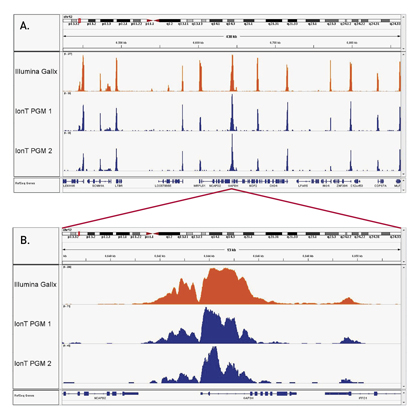
<p><strong>Figure 1A. The high consistency of the iDeal ChIP-seq kit on the Ion Torrent™ PGM™ (Life Technologies) and GAIIx (Illumina<sup>®</sup>)</strong><br /> ChIP was performed on sheared chromatin from 1 million HelaS3 cells using the iDeal ChIP-seq kit and 1 µg of H3K4me3 positive control antibody. Two different biological samples have been analyzed using two different sequencers - GAIIx (Illumina<sup>®</sup>) and PGM™ (Ion Torrent™). The expected ChIP-seq profile for H3K4me3 on the GAPDH promoter region has been obtained.<br /> Image A shows a several hundred bp along chr12 with high similarity of read distribution despite the radically different sequencers. Image B is a close capture focusing on the GAPDH that shows that even the peak structure is similar.</p>
<p class="text-center"><strong>Perfect match between ChIP-seq data obtained with the iDeal ChIP-seq workflow and reference dataset</strong></p>
<p><img src="https://www.diagenode.com/img/product/kits/perfect-match-between-chipseq-data.png" alt="Figure 1B" style="display: block; margin-left: auto; margin-right: auto;" /></p>
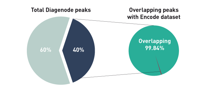
<p><strong>Figure 1B.</strong> The ChIP-seq dataset from this experiment has been compared with a reference dataset from the Broad Institute. We observed a perfect match between the top 40% of Diagenode peaks and the reference dataset. Based on the NIH Encode project criterion, ChIP-seq results are considered reproducible between an original and reproduced dataset if the top 40% of peaks have at least an 80% overlap ratio with the compared dataset.</p>
<p><img src="https://www.diagenode.com/img/product/kits/iDeal-kit-C01010053-figure-2.jpg" alt="Figure 2" style="display: block; margin-left: auto; margin-right: auto;" /></p>
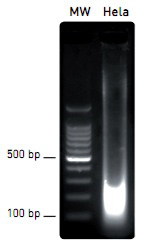
<p><strong>Figure 2. Efficient and easy chromatin shearing using the Bioruptor<sup>®</sup> and Shearing buffer iS1 from the iDeal ChIP-seq kit</strong><br /> Chromatin from 1 million of Hela cells was sheared using the Bioruptor<sup>®</sup> combined with the Bioruptor<sup>®</sup> Water cooler (Cat No. BioAcc-cool) during 3 rounds of 10 cycles of 30 seconds “ON” / 30 seconds “OFF” at HIGH power setting (position H). Diagenode 1.5 ml TPX tubes (Cat No. M-50001) were used for chromatin shearing. Samples were gently vortexed before and after performing each sonication round (rounds of 10 cycles), followed by a short centrifugation at 4°C to recover the sample volume at the bottom of the tube. The sheared chromatin was then decross-linked as described in the kit manual and analyzed by agarose gel electrophoresis.</p>
<p><img src="https://www.diagenode.com/img/product/kits/iDeal-kit-C01010053-figure-3.jpg" alt="Figure 3" style="display: block; margin-left: auto; margin-right: auto;" width="264" height="320" /></p>
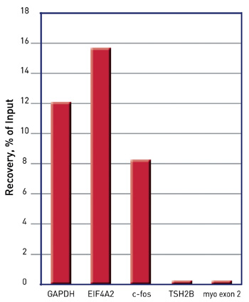
<p><strong>Figure 3. Validation of ChIP by qPCR: reliable results using Diagenode’s ChIP-seq grade H3K4me3 antibody, isotype control and sets of validated primers</strong><br /> Specific enrichment on positive loci (GAPDH, EIF4A2, c-fos promoter regions) comparing to no enrichment on negative loci (TSH2B promoter region and Myoglobin exon 2) was detected by qPCR. Samples were prepared using the Diagenode iDeal ChIP-seq kit. Diagenode ChIP-seq grade antibody against H3K4me3 and the corresponding isotype control IgG were used for immunoprecipitation. qPCR amplification was performed with sets of validated primers.</p>
<h3>ChIP-seq on tissue</h3>
<p><img src="https://www.diagenode.com/img/product/kits/ideal-figure-h3k4me3.jpg" alt="Figure 4A" style="display: block; margin-left: auto; margin-right: auto;" /></p>
<p><strong>Figure 4A.</strong> Chromatin Immunoprecipitation has been performed using chromatin from mouse liver tissue, the iDeal ChIP-seq kit for Histones and the Diagenode ChIP-seq-grade H3K4me3 (Cat. No. C15410003) antibody. The IP'd DNA was subsequently analysed on an Illumina® HiSeq. Library preparation, cluster generation and sequencing were performed according to the manufacturer's instructions. This figure shows the peak distribution in a region surrounding the GAPDH positive control gene.</p>
<p><img src="https://www.diagenode.com/img/product/kits/match-of-the-top40-peaks-2.png" alt="Figure 4B" caption="false" style="display: block; margin-left: auto; margin-right: auto;" width="700" height="280" /></p>
<p><strong>Figure 4B.</strong> The ChIP-seq dataset from this experiment has been compared with a reference dataset from the Broad Institute. We observed a perfect match between the top 40% of Diagenode peaks and the reference dataset. Based on the NIH Encode project criterion, ChIP-seq results are considered reproducible between an original and reproduced dataset if the top 40% of peaks have at least an 80% overlap ratio with the compared dataset.</p>',
'label2' => 'Species, cell lines, tissues tested',
'info2' => '<p>The iDeal ChIP-seq Kit for Histones is compatible with a broad variety of cell lines, tissues and species - some examples are shown below. Other species / cell lines / tissues can be used with this kit.</p>
<p><u>Cell lines:</u></p>
<p>Human: A549, A673, CD8+ T, Blood vascular endothelial cells, Lymphatic endothelial cells, fibroblasts, K562, MDA-MB231</p>
<p>Pig: Alveolar macrophages</p>
<p>Mouse: C2C12, primary HSPC, synovial fibroblasts, HeLa-S3, FACS sorted cells from embryonic kidneys, macrophages, mesodermal cells, myoblasts, NPC, salivary glands, spermatids, spermatocytes, skeletal muscle stem cells, stem cells, Th2</p>
<p>Hamster: CHO</p>
<p>Other cell lines / species: compatible, not tested</p>
<p><u>Tissues</u></p>
<p>Bee – brain</p>
<p>Daphnia – whole animal</p>
<p>Horse – brain, heart, lamina, liver, lung, skeletal muscles, ovary</p>
<p>Human – Erwing sarcoma tumor samples</p>
<p>Other tissues: compatible, not tested</p>
<p>Did you use the iDeal ChIP-seq for Histones Kit on other cell line / tissue / species? <a href="mailto:agnieszka.zelisko@diagenode.com?subject=Species, cell lines, tissues tested with the iDeal ChIP-seq Kit for TF&body=Dear Customer,%0D%0A%0D%0APlease, leave below your feedback about the iDeal ChIP-seq for Transcription Factors (cell / tissue type, species, other information...).%0D%0A%0D%0AThank you for sharing with us your experience !%0D%0A%0D%0ABest regards,%0D%0A%0D%0AAgnieszka Zelisko-Schmidt, PhD">Let us know!</a></p>',
'label3' => ' Additional solutions compatible with iDeal ChIP-seq Kit for Histones',
'info3' => '<p><a href="../p/chromatin-shearing-optimization-kit-low-sds-100-million-cells">Chromatin EasyShear Kit - Ultra Low SDS </a>optimizes chromatin shearing, a critical step for ChIP.</p>
<p> The <a href="https://www.diagenode.com/en/categories/library-preparation-for-ChIP-seq">MicroPlex Library Preparation Kit </a>provides easy and optimal library preparation of ChIPed samples.</p>
<p><a href="../categories/chip-seq-grade-antibodies">ChIP-seq grade anti-histone antibodies</a> provide high yields with excellent specificity and sensitivity.</p>
<p> Plus, for our IP-Star Automation users for automated ChIP, check out our <a href="../p/auto-ideal-chip-seq-kit-for-histones-x24-24-rxns">automated</a> version of this kit.</p>',
'format' => '4 chrom. prep./24 IPs',
'catalog_number' => 'C01010051',
'old_catalog_number' => 'AB-001-0024',
'sf_code' => 'C01010051-',
'type' => 'RFR',
'search_order' => '04-undefined',
'price_EUR' => '915',
'price_USD' => '1130',
'price_GBP' => '840',
'price_JPY' => '143335',
'price_CNY' => '',
'price_AUD' => '2825',
'country' => 'ALL',
'except_countries' => 'None',
'quote' => false,
'in_stock' => false,
'featured' => true,
'no_promo' => false,
'online' => true,
'master' => true,
'last_datasheet_update' => '0000-00-00',
'slug' => 'ideal-chip-seq-kit-x24-24-rxns',
'meta_title' => 'iDeal ChIP-seq kit x24',
'meta_keywords' => '',
'meta_description' => 'iDeal ChIP-seq kit x24',
'modified' => '2023-04-20 16:00:20',
'created' => '2015-06-29 14:08:20',
'locale' => 'zho'
),
'Antibody' => array(
'host' => '*****',
'id' => null,
'name' => null,
'description' => null,
'clonality' => null,
'isotype' => null,
'lot' => null,
'concentration' => null,
'reactivity' => null,
'type' => null,
'purity' => null,
'classification' => null,
'application_table' => null,
'storage_conditions' => null,
'storage_buffer' => null,
'precautions' => null,
'uniprot_acc' => null,
'slug' => null,
'meta_keywords' => null,
'meta_description' => null,
'modified' => null,
'created' => null,
'select_label' => null
),
'Slave' => array(
(int) 0 => array(
[maximum depth reached]
)
),
'Group' => array(
'Group' => array(
[maximum depth reached]
),
'Master' => array(
[maximum depth reached]
),
'Product' => array(
[maximum depth reached]
)
),
'Related' => array(
(int) 0 => array(
[maximum depth reached]
),
(int) 1 => array(
[maximum depth reached]
),
(int) 2 => array(
[maximum depth reached]
),
(int) 3 => array(
[maximum depth reached]
),
(int) 4 => array(
[maximum depth reached]
)
),
'Application' => array(
(int) 0 => array(
[maximum depth reached]
)
),
'Category' => array(
(int) 0 => array(
[maximum depth reached]
)
),
'Document' => array(
(int) 0 => array(
[maximum depth reached]
),
(int) 1 => array(
[maximum depth reached]
),
(int) 2 => array(
[maximum depth reached]
),
(int) 3 => array(
[maximum depth reached]
)
),
'Feature' => array(),
'Image' => array(
(int) 0 => array(
[maximum depth reached]
)
),
'Promotion' => array(),
'Protocol' => array(
(int) 0 => array(
[maximum depth reached]
)
),
'Publication' => array(
(int) 0 => array(
[maximum depth reached]
),
(int) 1 => array(
[maximum depth reached]
),
(int) 2 => array(
[maximum depth reached]
),
(int) 3 => array(
[maximum depth reached]
),
(int) 4 => array(
[maximum depth reached]
),
(int) 5 => array(
[maximum depth reached]
),
(int) 6 => array(
[maximum depth reached]
),
(int) 7 => array(
[maximum depth reached]
),
(int) 8 => array(
[maximum depth reached]
),
(int) 9 => array(
[maximum depth reached]
),
(int) 10 => array(
[maximum depth reached]
),
(int) 11 => array(
[maximum depth reached]
),
(int) 12 => array(
[maximum depth reached]
),
(int) 13 => array(
[maximum depth reached]
),
(int) 14 => array(
[maximum depth reached]
),
(int) 15 => array(
[maximum depth reached]
),
(int) 16 => array(
[maximum depth reached]
),
(int) 17 => array(
[maximum depth reached]
),
(int) 18 => array(
[maximum depth reached]
),
(int) 19 => array(
[maximum depth reached]
),
(int) 20 => array(
[maximum depth reached]
),
(int) 21 => array(
[maximum depth reached]
),
(int) 22 => array(
[maximum depth reached]
),
(int) 23 => array(
[maximum depth reached]
),
(int) 24 => array(
[maximum depth reached]
),
(int) 25 => array(
[maximum depth reached]
),
(int) 26 => array(
[maximum depth reached]
),
(int) 27 => array(
[maximum depth reached]
),
(int) 28 => array(
[maximum depth reached]
),
(int) 29 => array(
[maximum depth reached]
),
(int) 30 => array(
[maximum depth reached]
),
(int) 31 => array(
[maximum depth reached]
),
(int) 32 => array(
[maximum depth reached]
),
(int) 33 => array(
[maximum depth reached]
),
(int) 34 => array(
[maximum depth reached]
),
(int) 35 => array(
[maximum depth reached]
),
(int) 36 => array(
[maximum depth reached]
),
(int) 37 => array(
[maximum depth reached]
),
(int) 38 => array(
[maximum depth reached]
),
(int) 39 => array(
[maximum depth reached]
),
(int) 40 => array(
[maximum depth reached]
),
(int) 41 => array(
[maximum depth reached]
),
(int) 42 => array(
[maximum depth reached]
),
(int) 43 => array(
[maximum depth reached]
),
(int) 44 => array(
[maximum depth reached]
),
(int) 45 => array(
[maximum depth reached]
),
(int) 46 => array(
[maximum depth reached]
),
(int) 47 => array(
[maximum depth reached]
),
(int) 48 => array(
[maximum depth reached]
),
(int) 49 => array(
[maximum depth reached]
),
(int) 50 => array(
[maximum depth reached]
),
(int) 51 => array(
[maximum depth reached]
),
(int) 52 => array(
[maximum depth reached]
),
(int) 53 => array(
[maximum depth reached]
),
(int) 54 => array(
[maximum depth reached]
),
(int) 55 => array(
[maximum depth reached]
),
(int) 56 => array(
[maximum depth reached]
),
(int) 57 => array(
[maximum depth reached]
),
(int) 58 => array(
[maximum depth reached]
),
(int) 59 => array(
[maximum depth reached]
),
(int) 60 => array(
[maximum depth reached]
),
(int) 61 => array(
[maximum depth reached]
),
(int) 62 => array(
[maximum depth reached]
),
(int) 63 => array(
[maximum depth reached]
),
(int) 64 => array(
[maximum depth reached]
),
(int) 65 => array(
[maximum depth reached]
),
(int) 66 => array(
[maximum depth reached]
),
(int) 67 => array(
[maximum depth reached]
),
(int) 68 => array(
[maximum depth reached]
),
(int) 69 => array(
[maximum depth reached]
),
(int) 70 => array(
[maximum depth reached]
),
(int) 71 => array(
[maximum depth reached]
),
(int) 72 => array(
[maximum depth reached]
),
(int) 73 => array(
[maximum depth reached]
),
(int) 74 => array(
[maximum depth reached]
),
(int) 75 => array(
[maximum depth reached]
),
(int) 76 => array(
[maximum depth reached]
),
(int) 77 => array(
[maximum depth reached]
),
(int) 78 => array(
[maximum depth reached]
),
(int) 79 => array(
[maximum depth reached]
),
(int) 80 => array(
[maximum depth reached]
),
(int) 81 => array(
[maximum depth reached]
),
(int) 82 => array(
[maximum depth reached]
)
),
'Testimonial' => array(
(int) 0 => array(
[maximum depth reached]
),
(int) 1 => array(
[maximum depth reached]
)
),
'Area' => array(),
'SafetySheet' => array(
(int) 0 => array(
[maximum depth reached]
),
(int) 1 => array(
[maximum depth reached]
),
(int) 2 => array(
[maximum depth reached]
),
(int) 3 => array(
[maximum depth reached]
),
(int) 4 => array(
[maximum depth reached]
),
(int) 5 => array(
[maximum depth reached]
),
(int) 6 => array(
[maximum depth reached]
),
(int) 7 => array(
[maximum depth reached]
)
)
),
'meta_canonical' => 'https://www.diagenode.com/cn/p/ideal-chip-seq-kit-x24-24-rxns'
)
$language = 'cn'
$meta_keywords = ''
$meta_description = 'iDeal ChIP-seq kit x24'
$meta_title = 'iDeal ChIP-seq kit x24'
$product = array(
'Product' => array(
'id' => '1836',
'antibody_id' => null,
'name' => 'iDeal ChIP-seq kit for Histones',
'description' => '<p>Don’t risk wasting your precious sequencing samples. Diagenode’s validated <strong>iDeal ChIP-seq kit for Histones</strong> has everything you need for a successful start-to-finish <strong>ChIP of histones prior to Next-Generation Sequencing</strong>. The complete kit contains all buffers and reagents for cell lysis, chromatin shearing, immunoprecipitation and DNA purification. In addition, unlike competing solutions, the kit contains positive and negative control antibodies (H3K4me3 and IgG, respectively) as well as positive and negative control PCR primers pairs (GAPDH TSS and Myoglobin exon 2, respectively) for your convenience and a guarantee of optimal results. The kit has been validated on multiple histone marks.</p>
<p> The iDeal ChIP-seq kit for Histones<strong> </strong>is perfect for <strong>cells</strong> (<strong>100,000 cells</strong> to <strong>1,000,000 cells</strong> per IP) and has been validated for <strong>tissues</strong> (<strong>1.5 mg</strong> to <strong>5 mg</strong> of tissue per IP).</p>
<p> The iDeal ChIP-seq kit is the only kit on the market validated for the major sequencing systems. Our expertise in ChIP-seq tools allows reproducible and efficient results every time.</p>
<p></p>
<p> <strong></strong></p>
<p></p>',
'label1' => 'Characteristics',
'info1' => '<ul style="list-style-type: disc;">
<li>Highly <strong>optimized</strong> protocol for ChIP-seq from cells and tissues</li>
<li><strong>Validated</strong> for ChIP-seq with multiple histones marks</li>
<li>Most <strong>complete</strong> kit available (covers all steps, including the control antibodies and primers)</li>
<li>Optimized chromatin preparation in combination with the Bioruptor ensuring the best <strong>epitope integrity</strong></li>
<li>Magnetic beads make ChIP easy, fast and more <strong>reproducible</strong></li>
<li>Combination with Diagenode ChIP-seq antibodies provides high yields with excellent <strong>specificity</strong> and <strong>sensitivity</strong></li>
<li>Purified DNA suitable for any downstream application</li>
<li>Easy-to-follow protocol</li>
</ul>
<p>Note: to obtain optimal results, this kit should be used in combination with the DiaMag1.5 - magnetic rack.</p>
<h3>ChIP-seq on cells</h3>
<p><img src="https://www.diagenode.com/img/product/kits/iDeal-kit-C01010053-figure-1.jpg" alt="Figure 1A" style="display: block; margin-left: auto; margin-right: auto;" /></p>
<p><strong>Figure 1A. The high consistency of the iDeal ChIP-seq kit on the Ion Torrent™ PGM™ (Life Technologies) and GAIIx (Illumina<sup>®</sup>)</strong><br /> ChIP was performed on sheared chromatin from 1 million HelaS3 cells using the iDeal ChIP-seq kit and 1 µg of H3K4me3 positive control antibody. Two different biological samples have been analyzed using two different sequencers - GAIIx (Illumina<sup>®</sup>) and PGM™ (Ion Torrent™). The expected ChIP-seq profile for H3K4me3 on the GAPDH promoter region has been obtained.<br /> Image A shows a several hundred bp along chr12 with high similarity of read distribution despite the radically different sequencers. Image B is a close capture focusing on the GAPDH that shows that even the peak structure is similar.</p>
<p class="text-center"><strong>Perfect match between ChIP-seq data obtained with the iDeal ChIP-seq workflow and reference dataset</strong></p>
<p><img src="https://www.diagenode.com/img/product/kits/perfect-match-between-chipseq-data.png" alt="Figure 1B" style="display: block; margin-left: auto; margin-right: auto;" /></p>
<p><strong>Figure 1B.</strong> The ChIP-seq dataset from this experiment has been compared with a reference dataset from the Broad Institute. We observed a perfect match between the top 40% of Diagenode peaks and the reference dataset. Based on the NIH Encode project criterion, ChIP-seq results are considered reproducible between an original and reproduced dataset if the top 40% of peaks have at least an 80% overlap ratio with the compared dataset.</p>
<p><img src="https://www.diagenode.com/img/product/kits/iDeal-kit-C01010053-figure-2.jpg" alt="Figure 2" style="display: block; margin-left: auto; margin-right: auto;" /></p>
<p><strong>Figure 2. Efficient and easy chromatin shearing using the Bioruptor<sup>®</sup> and Shearing buffer iS1 from the iDeal ChIP-seq kit</strong><br /> Chromatin from 1 million of Hela cells was sheared using the Bioruptor<sup>®</sup> combined with the Bioruptor<sup>®</sup> Water cooler (Cat No. BioAcc-cool) during 3 rounds of 10 cycles of 30 seconds “ON” / 30 seconds “OFF” at HIGH power setting (position H). Diagenode 1.5 ml TPX tubes (Cat No. M-50001) were used for chromatin shearing. Samples were gently vortexed before and after performing each sonication round (rounds of 10 cycles), followed by a short centrifugation at 4°C to recover the sample volume at the bottom of the tube. The sheared chromatin was then decross-linked as described in the kit manual and analyzed by agarose gel electrophoresis.</p>
<p><img src="https://www.diagenode.com/img/product/kits/iDeal-kit-C01010053-figure-3.jpg" alt="Figure 3" style="display: block; margin-left: auto; margin-right: auto;" width="264" height="320" /></p>
<p><strong>Figure 3. Validation of ChIP by qPCR: reliable results using Diagenode’s ChIP-seq grade H3K4me3 antibody, isotype control and sets of validated primers</strong><br /> Specific enrichment on positive loci (GAPDH, EIF4A2, c-fos promoter regions) comparing to no enrichment on negative loci (TSH2B promoter region and Myoglobin exon 2) was detected by qPCR. Samples were prepared using the Diagenode iDeal ChIP-seq kit. Diagenode ChIP-seq grade antibody against H3K4me3 and the corresponding isotype control IgG were used for immunoprecipitation. qPCR amplification was performed with sets of validated primers.</p>
<h3>ChIP-seq on tissue</h3>
<p><img src="https://www.diagenode.com/img/product/kits/ideal-figure-h3k4me3.jpg" alt="Figure 4A" style="display: block; margin-left: auto; margin-right: auto;" /></p>
<p><strong>Figure 4A.</strong> Chromatin Immunoprecipitation has been performed using chromatin from mouse liver tissue, the iDeal ChIP-seq kit for Histones and the Diagenode ChIP-seq-grade H3K4me3 (Cat. No. C15410003) antibody. The IP'd DNA was subsequently analysed on an Illumina® HiSeq. Library preparation, cluster generation and sequencing were performed according to the manufacturer's instructions. This figure shows the peak distribution in a region surrounding the GAPDH positive control gene.</p>
<p><img src="https://www.diagenode.com/img/product/kits/match-of-the-top40-peaks-2.png" alt="Figure 4B" caption="false" style="display: block; margin-left: auto; margin-right: auto;" width="700" height="280" /></p>
<p><strong>Figure 4B.</strong> The ChIP-seq dataset from this experiment has been compared with a reference dataset from the Broad Institute. We observed a perfect match between the top 40% of Diagenode peaks and the reference dataset. Based on the NIH Encode project criterion, ChIP-seq results are considered reproducible between an original and reproduced dataset if the top 40% of peaks have at least an 80% overlap ratio with the compared dataset.</p>',
'label2' => 'Species, cell lines, tissues tested',
'info2' => '<p>The iDeal ChIP-seq Kit for Histones is compatible with a broad variety of cell lines, tissues and species - some examples are shown below. Other species / cell lines / tissues can be used with this kit.</p>
<p><u>Cell lines:</u></p>
<p>Human: A549, A673, CD8+ T, Blood vascular endothelial cells, Lymphatic endothelial cells, fibroblasts, K562, MDA-MB231</p>
<p>Pig: Alveolar macrophages</p>
<p>Mouse: C2C12, primary HSPC, synovial fibroblasts, HeLa-S3, FACS sorted cells from embryonic kidneys, macrophages, mesodermal cells, myoblasts, NPC, salivary glands, spermatids, spermatocytes, skeletal muscle stem cells, stem cells, Th2</p>
<p>Hamster: CHO</p>
<p>Other cell lines / species: compatible, not tested</p>
<p><u>Tissues</u></p>
<p>Bee – brain</p>
<p>Daphnia – whole animal</p>
<p>Horse – brain, heart, lamina, liver, lung, skeletal muscles, ovary</p>
<p>Human – Erwing sarcoma tumor samples</p>
<p>Other tissues: compatible, not tested</p>
<p>Did you use the iDeal ChIP-seq for Histones Kit on other cell line / tissue / species? <a href="mailto:agnieszka.zelisko@diagenode.com?subject=Species, cell lines, tissues tested with the iDeal ChIP-seq Kit for TF&body=Dear Customer,%0D%0A%0D%0APlease, leave below your feedback about the iDeal ChIP-seq for Transcription Factors (cell / tissue type, species, other information...).%0D%0A%0D%0AThank you for sharing with us your experience !%0D%0A%0D%0ABest regards,%0D%0A%0D%0AAgnieszka Zelisko-Schmidt, PhD">Let us know!</a></p>',
'label3' => ' Additional solutions compatible with iDeal ChIP-seq Kit for Histones',
'info3' => '<p><a href="../p/chromatin-shearing-optimization-kit-low-sds-100-million-cells">Chromatin EasyShear Kit - Ultra Low SDS </a>optimizes chromatin shearing, a critical step for ChIP.</p>
<p> The <a href="https://www.diagenode.com/en/categories/library-preparation-for-ChIP-seq">MicroPlex Library Preparation Kit </a>provides easy and optimal library preparation of ChIPed samples.</p>
<p><a href="../categories/chip-seq-grade-antibodies">ChIP-seq grade anti-histone antibodies</a> provide high yields with excellent specificity and sensitivity.</p>
<p> Plus, for our IP-Star Automation users for automated ChIP, check out our <a href="../p/auto-ideal-chip-seq-kit-for-histones-x24-24-rxns">automated</a> version of this kit.</p>',
'format' => '4 chrom. prep./24 IPs',
'catalog_number' => 'C01010051',
'old_catalog_number' => 'AB-001-0024',
'sf_code' => 'C01010051-',
'type' => 'RFR',
'search_order' => '04-undefined',
'price_EUR' => '915',
'price_USD' => '1130',
'price_GBP' => '840',
'price_JPY' => '143335',
'price_CNY' => '',
'price_AUD' => '2825',
'country' => 'ALL',
'except_countries' => 'None',
'quote' => false,
'in_stock' => false,
'featured' => true,
'no_promo' => false,
'online' => true,
'master' => true,
'last_datasheet_update' => '0000-00-00',
'slug' => 'ideal-chip-seq-kit-x24-24-rxns',
'meta_title' => 'iDeal ChIP-seq kit x24',
'meta_keywords' => '',
'meta_description' => 'iDeal ChIP-seq kit x24',
'modified' => '2023-04-20 16:00:20',
'created' => '2015-06-29 14:08:20',
'locale' => 'zho'
),
'Antibody' => array(
'host' => '*****',
'id' => null,
'name' => null,
'description' => null,
'clonality' => null,
'isotype' => null,
'lot' => null,
'concentration' => null,
'reactivity' => null,
'type' => null,
'purity' => null,
'classification' => null,
'application_table' => null,
'storage_conditions' => null,
'storage_buffer' => null,
'precautions' => null,
'uniprot_acc' => null,
'slug' => null,
'meta_keywords' => null,
'meta_description' => null,
'modified' => null,
'created' => null,
'select_label' => null
),
'Slave' => array(
(int) 0 => array(
'id' => '9',
'name' => 'C01010051',
'product_id' => '1836',
'modified' => '2016-02-18 17:14:29',
'created' => '2016-02-18 17:14:29'
)
),
'Group' => array(
'Group' => array(
'id' => '9',
'name' => 'C01010051',
'product_id' => '1836',
'modified' => '2016-02-18 17:14:29',
'created' => '2016-02-18 17:14:29'
),
'Master' => array(
'id' => '1836',
'antibody_id' => null,
'name' => 'iDeal ChIP-seq kit for Histones',
'description' => '<p><a href="https://www.diagenode.com/files/products/kits/ideal-chipseq-for-histones-complete-manual.pdf"><img src="https://www.diagenode.com/img/buttons/bt-manual.png" /></a></p>
<p>Don’t risk wasting your precious sequencing samples. Diagenode’s validated <strong>iDeal ChIP-seq kit for Histones</strong> has everything you need for a successful start-to-finish <strong>ChIP of histones prior to Next-Generation Sequencing</strong>. The complete kit contains all buffers and reagents for cell lysis, chromatin shearing, immunoprecipitation and DNA purification. In addition, unlike competing solutions, the kit contains positive and negative control antibodies (H3K4me3 and IgG, respectively) as well as positive and negative control PCR primers pairs (GAPDH TSS and Myoglobin exon 2, respectively) for your convenience and a guarantee of optimal results. The kit has been validated on multiple histone marks.</p>
<p> The iDeal ChIP-seq kit for Histones<strong> </strong>is perfect for <strong>cells</strong> (<strong>100,000 cells</strong> to <strong>1,000,000 cells</strong> per IP) and has been validated for <strong>tissues</strong> (<strong>1.5 mg</strong> to <strong>5 mg</strong> of tissue per IP).</p>
<p> The iDeal ChIP-seq kit is the only kit on the market validated for the major sequencing systems. Our expertise in ChIP-seq tools allows reproducible and efficient results every time.</p>
<p></p>
<p> <strong></strong></p>
<p></p>',
'label1' => 'Characteristics',
'info1' => '<ul style="list-style-type: disc;">
<li>Highly <strong>optimized</strong> protocol for ChIP-seq from cells and tissues</li>
<li><strong>Validated</strong> for ChIP-seq with multiple histones marks</li>
<li>Most <strong>complete</strong> kit available (covers all steps, including the control antibodies and primers)</li>
<li>Optimized chromatin preparation in combination with the Bioruptor ensuring the best <strong>epitope integrity</strong></li>
<li>Magnetic beads make ChIP easy, fast and more <strong>reproducible</strong></li>
<li>Combination with Diagenode ChIP-seq antibodies provides high yields with excellent <strong>specificity</strong> and <strong>sensitivity</strong></li>
<li>Purified DNA suitable for any downstream application</li>
<li>Easy-to-follow protocol</li>
</ul>
<p>Note: to obtain optimal results, this kit should be used in combination with the DiaMag1.5 - magnetic rack.</p>
<h3>ChIP-seq on cells</h3>
<p><img src="https://www.diagenode.com/img/product/kits/iDeal-kit-C01010053-figure-1.jpg" alt="Figure 1A" style="display: block; margin-left: auto; margin-right: auto;" /></p>
<p><strong>Figure 1A. The high consistency of the iDeal ChIP-seq kit on the Ion Torrent™ PGM™ (Life Technologies) and GAIIx (Illumina<sup>®</sup>)</strong><br /> ChIP was performed on sheared chromatin from 1 million HelaS3 cells using the iDeal ChIP-seq kit and 1 µg of H3K4me3 positive control antibody. Two different biological samples have been analyzed using two different sequencers - GAIIx (Illumina<sup>®</sup>) and PGM™ (Ion Torrent™). The expected ChIP-seq profile for H3K4me3 on the GAPDH promoter region has been obtained.<br /> Image A shows a several hundred bp along chr12 with high similarity of read distribution despite the radically different sequencers. Image B is a close capture focusing on the GAPDH that shows that even the peak structure is similar.</p>
<p class="text-center"><strong>Perfect match between ChIP-seq data obtained with the iDeal ChIP-seq workflow and reference dataset</strong></p>
<p><img src="https://www.diagenode.com/img/product/kits/perfect-match-between-chipseq-data.png" alt="Figure 1B" style="display: block; margin-left: auto; margin-right: auto;" /></p>
<p><strong>Figure 1B.</strong> The ChIP-seq dataset from this experiment has been compared with a reference dataset from the Broad Institute. We observed a perfect match between the top 40% of Diagenode peaks and the reference dataset. Based on the NIH Encode project criterion, ChIP-seq results are considered reproducible between an original and reproduced dataset if the top 40% of peaks have at least an 80% overlap ratio with the compared dataset.</p>
<p><img src="https://www.diagenode.com/img/product/kits/iDeal-kit-C01010053-figure-2.jpg" alt="Figure 2" style="display: block; margin-left: auto; margin-right: auto;" /></p>
<p><strong>Figure 2. Efficient and easy chromatin shearing using the Bioruptor<sup>®</sup> and Shearing buffer iS1 from the iDeal ChIP-seq kit</strong><br /> Chromatin from 1 million of Hela cells was sheared using the Bioruptor<sup>®</sup> combined with the Bioruptor<sup>®</sup> Water cooler (Cat No. BioAcc-cool) during 3 rounds of 10 cycles of 30 seconds “ON” / 30 seconds “OFF” at HIGH power setting (position H). Diagenode 1.5 ml TPX tubes (Cat No. M-50001) were used for chromatin shearing. Samples were gently vortexed before and after performing each sonication round (rounds of 10 cycles), followed by a short centrifugation at 4°C to recover the sample volume at the bottom of the tube. The sheared chromatin was then decross-linked as described in the kit manual and analyzed by agarose gel electrophoresis.</p>
<p><img src="https://www.diagenode.com/img/product/kits/iDeal-kit-C01010053-figure-3.jpg" alt="Figure 3" style="display: block; margin-left: auto; margin-right: auto;" width="264" height="320" /></p>
<p><strong>Figure 3. Validation of ChIP by qPCR: reliable results using Diagenode’s ChIP-seq grade H3K4me3 antibody, isotype control and sets of validated primers</strong><br /> Specific enrichment on positive loci (GAPDH, EIF4A2, c-fos promoter regions) comparing to no enrichment on negative loci (TSH2B promoter region and Myoglobin exon 2) was detected by qPCR. Samples were prepared using the Diagenode iDeal ChIP-seq kit. Diagenode ChIP-seq grade antibody against H3K4me3 and the corresponding isotype control IgG were used for immunoprecipitation. qPCR amplification was performed with sets of validated primers.</p>
<h3>ChIP-seq on tissue</h3>
<p><img src="https://www.diagenode.com/img/product/kits/ideal-figure-h3k4me3.jpg" alt="Figure 4A" style="display: block; margin-left: auto; margin-right: auto;" /></p>
<p><strong>Figure 4A.</strong> Chromatin Immunoprecipitation has been performed using chromatin from mouse liver tissue, the iDeal ChIP-seq kit for Histones and the Diagenode ChIP-seq-grade H3K4me3 (Cat. No. C15410003) antibody. The IP'd DNA was subsequently analysed on an Illumina® HiSeq. Library preparation, cluster generation and sequencing were performed according to the manufacturer's instructions. This figure shows the peak distribution in a region surrounding the GAPDH positive control gene.</p>
<p><img src="https://www.diagenode.com/img/product/kits/match-of-the-top40-peaks-2.png" alt="Figure 4B" caption="false" style="display: block; margin-left: auto; margin-right: auto;" width="700" height="280" /></p>
<p><strong>Figure 4B.</strong> The ChIP-seq dataset from this experiment has been compared with a reference dataset from the Broad Institute. We observed a perfect match between the top 40% of Diagenode peaks and the reference dataset. Based on the NIH Encode project criterion, ChIP-seq results are considered reproducible between an original and reproduced dataset if the top 40% of peaks have at least an 80% overlap ratio with the compared dataset.</p>',
'label2' => 'Species, cell lines, tissues tested',
'info2' => '<p>The iDeal ChIP-seq Kit for Histones is compatible with a broad variety of cell lines, tissues and species - some examples are shown below. Other species / cell lines / tissues can be used with this kit.</p>
<p><u>Cell lines:</u></p>
<p>Human: A549, A673, CD8+ T, Blood vascular endothelial cells, Lymphatic endothelial cells, fibroblasts, K562, MDA-MB231</p>
<p>Pig: Alveolar macrophages</p>
<p>Mouse: C2C12, primary HSPC, synovial fibroblasts, HeLa-S3, FACS sorted cells from embryonic kidneys, macrophages, mesodermal cells, myoblasts, NPC, salivary glands, spermatids, spermatocytes, skeletal muscle stem cells, stem cells, Th2</p>
<p>Hamster: CHO</p>
<p>Other cell lines / species: compatible, not tested</p>
<p><u>Tissues</u></p>
<p>Bee – brain</p>
<p>Daphnia – whole animal</p>
<p>Horse – brain, heart, lamina, liver, lung, skeletal muscles, ovary</p>
<p>Human – Erwing sarcoma tumor samples</p>
<p>Other tissues: compatible, not tested</p>
<p>Did you use the iDeal ChIP-seq for Histones Kit on other cell line / tissue / species? <a href="mailto:agnieszka.zelisko@diagenode.com?subject=Species, cell lines, tissues tested with the iDeal ChIP-seq Kit for TF&body=Dear Customer,%0D%0A%0D%0APlease, leave below your feedback about the iDeal ChIP-seq for Transcription Factors (cell / tissue type, species, other information...).%0D%0A%0D%0AThank you for sharing with us your experience !%0D%0A%0D%0ABest regards,%0D%0A%0D%0AAgnieszka Zelisko-Schmidt, PhD">Let us know!</a></p>',
'label3' => ' Additional solutions compatible with iDeal ChIP-seq Kit for Histones',
'info3' => '<p><a href="../p/chromatin-shearing-optimization-kit-low-sds-100-million-cells">Chromatin EasyShear Kit - Ultra Low SDS </a>optimizes chromatin shearing, a critical step for ChIP.</p>
<p> The <a href="https://www.diagenode.com/en/categories/library-preparation-for-ChIP-seq">MicroPlex Library Preparation Kit </a>provides easy and optimal library preparation of ChIPed samples.</p>
<p><a href="../categories/chip-seq-grade-antibodies">ChIP-seq grade anti-histone antibodies</a> provide high yields with excellent specificity and sensitivity.</p>
<p> Plus, for our IP-Star Automation users for automated ChIP, check out our <a href="../p/auto-ideal-chip-seq-kit-for-histones-x24-24-rxns">automated</a> version of this kit.</p>',
'format' => '4 chrom. prep./24 IPs',
'catalog_number' => 'C01010051',
'old_catalog_number' => 'AB-001-0024',
'sf_code' => 'C01010051-',
'type' => 'RFR',
'search_order' => '04-undefined',
'price_EUR' => '915',
'price_USD' => '1130',
'price_GBP' => '840',
'price_JPY' => '143335',
'price_CNY' => '',
'price_AUD' => '2825',
'country' => 'ALL',
'except_countries' => 'None',
'quote' => false,
'in_stock' => false,
'featured' => true,
'no_promo' => false,
'online' => true,
'master' => true,
'last_datasheet_update' => '0000-00-00',
'slug' => 'ideal-chip-seq-kit-x24-24-rxns',
'meta_title' => 'iDeal ChIP-seq kit x24',
'meta_keywords' => '',
'meta_description' => 'iDeal ChIP-seq kit x24',
'modified' => '2023-04-20 16:00:20',
'created' => '2015-06-29 14:08:20'
),
'Product' => array(
(int) 0 => array(
[maximum depth reached]
),
(int) 1 => array(
[maximum depth reached]
)
)
),
'Related' => array(
(int) 0 => array(
'id' => '1839',
'antibody_id' => null,
'name' => 'iDeal ChIP-seq kit for Transcription Factors',
'description' => '<p><a href="https://www.diagenode.com/files/products/kits/ideal-chipseq-transcription-factors-x10-manual.pdf"><img src="https://www.diagenode.com/img/buttons/bt-manual.png" /></a></p>
<div class="row">
<div class="small-12 medium-8 large-8 columns"><br />
<p><span style="font-weight: 400;">Diagenode’s <strong>iDeal ChIP-seq Kit for Transcription Factors</strong> is a highly validated solution for robust transcription factor and other non-histone proteins ChIP-seq results and contains everything you need for start-to-finish </span><b>ChIP </b><span style="font-weight: 400;">prior to </span><b>Next-Generation Sequencing</b><span style="font-weight: 400;">. This complete solution contains all buffers and reagents for cell lysis, chromatin shearing, immunoprecipitation, and DNA purification. In addition, unlike competing solutions, the kit contains positive and negative control antibodies (CTCF and IgG, respectively) as well as positive and negative control PCR primers pairs (H19 and Myoglobin exon 2, respectively) for your convenience and a guarantee of optimal results. <br /></span></p>
</div>
<div class="small-12 medium-4 large-4 columns"><center><br /><br />
<script>// <![CDATA[
var date = new Date(); var heure = date.getHours(); var jour = date.getDay(); var semaine = Math.floor(date.getDate() / 7) + 1; if (jour === 2 && ( (heure >= 9 && heure < 9.5) || (heure >= 18 && heure < 18.5) )) { document.write('<a href="https://us02web.zoom.us/j/85467619762"><img src="https://www.diagenode.com/img/epicafe-ON.gif"></a>'); } else { document.write('<a href="https://go.diagenode.com/l/928883/2023-04-26/3kq1v"><img src="https://www.diagenode.com/img/epicafe-OFF.png"></a>'); }
// ]]></script>
</center></div>
</div>
<p><span style="font-weight: 400;">The </span><b> iDeal ChIP-seq kit for Transcription Factors </b><span style="font-weight: 400;">is compatible for cells or tissues:</span></p>
<table style="width: 419px; margin-left: auto; margin-right: auto;">
<tbody>
<tr>
<td style="width: 144px;"></td>
<td style="width: 267px; text-align: center;"><span style="font-weight: 400;">Amount per IP</span></td>
</tr>
<tr>
<td style="width: 144px;">Cells</td>
<td style="width: 267px; text-align: center;"><strong>4,000,000</strong></td>
</tr>
<tr>
<td style="width: 144px;">Tissues</td>
<td style="width: 267px; text-align: center;"><strong>30 mg</strong></td>
</tr>
</tbody>
</table>
<p><span style="font-weight: 400;">The iDeal ChIP-seq kit is the only kit on the market validated for major sequencing systems. Our expertise in ChIP-seq tools allows reproducible and efficient results every time. </span></p>
<p></p>
<p></p>',
'label1' => 'Characteristics',
'info1' => '<ul>
<li><span style="font-weight: 400;"><strong>Highly optimized protocol</strong> for ChIP-seq from cells and tissues</span></li>
<li><span style="font-weight: 400;"><strong>Validated</strong> for <strong>ChIP-seq</strong> with multiple transcription factors and non-histone targets<br /></span></li>
<li><span style="font-weight: 400;"><strong>Most complete kit</strong> available (covers all steps, including the control antibodies and primers)<br /></span></li>
<li><span style="font-weight: 400;"><strong>Magnetic beads</strong> make ChIP <strong>easy</strong>, <strong>fast</strong> and more <strong>reproducible</strong></span></li>
<li><span style="font-weight: 400;">Combination with Diagenode ChIP-seq antibodies provides <strong>high yields</strong> with excellent <strong>specificity</strong> and <strong>sensitivity</strong><br /></span></li>
<li><span style="font-weight: 400;">Purified DNA suitable for any downstream application</span></li>
<li><span style="font-weight: 400;">Easy-to-follow protocol</span></li>
</ul>
<p><span style="font-weight: 400;"></span></p>
<p> </p>
<h3>ChIP-seq on cells</h3>
<p><img src="https://www.diagenode.com/img/product/kits/ideal-ctcf-diagenode.jpg" alt="CTCF Diagenode" style="display: block; margin-left: auto; margin-right: auto;" /></p>
<p><strong>Figure 1.</strong> (A) Chromatin Immunoprecipitation has been performed using chromatin from HeLa cells, the iDeal ChIP-seq kit for Transcription Factors and the Diagenode ChIP-seq-grade CTCF antibody. The IP'd DNA was subsequently analysed on an Illumina<sup>®</sup> HiSeq. Library preparation, cluster generation and sequencing were performed according to the manufacturer's instructions. This figure shows the peak distribution in a region surrounding the GAPDH positive control gene.</p>
<p><img src="https://www.diagenode.com/img/product/kits/ideal-figure-b-total-diagendoe-peaks.png" alt="CTCF Diagenode" style="display: block; margin-left: auto; margin-right: auto;" /></p>
<p><strong>Figure 1B.</strong> The ChIP-seq dataset from this experiment has been compared with a reference dataset from the Broad Institute. We observed a perfect match between the top 40% of Diagenode peaks and the reference dataset. Based on the NIH Encode project criterion, ChIP-seq results are considered reproducible between an original and reproduced dataset if the top 40% of peaks have at least an 80% overlap ratio with the compared dataset.</p>
<p> </p>
<p><img src="https://www.diagenode.com/img/product/kits/ideal-TF-chip-seq-A.png" alt="ChIP-seq figure A" style="display: block; margin-left: auto; margin-right: auto;" /></p>
<p><img src="https://www.diagenode.com/img/product/kits/ideal-TF-chip-seq-B.png" alt="ChIP-seq figure B" style="display: block; margin-left: auto; margin-right: auto;" /></p>
<p><img src="https://www.diagenode.com/img/product/kits/ideal-TF-chip-seq-C.png" alt="ChIP-seq figure C" style="display: block; margin-left: auto; margin-right: auto;" /></p>
<p><strong>Figure 2.</strong> Chromatin Immunoprecipitation has been performed using chromatin from HeLa cells, the iDeal ChIP-seq kit for Transcription Factors and the Diagenode ChIP-seq-grade HDAC1 (A), LSD1 (B) and p53 antibody (C). The IP'd DNA was subsequently analysed on an Illumina<sup>®</sup> Genome Analyzer. Library preparation, cluster generation and sequencing were performed according to the manufacturer's instructions. This figure shows the peak distribution in regions of chromosome 3 (A), chromosome 12 (B) and chromosome 6 (C) respectively.</p>
<p> </p>
<h3>ChIP-seq on tissue</h3>
<p><img src="https://www.diagenode.com/img/product/kits/ideal-figure-3a.jpg" alt="ChIP-seq figure A" style="display: block; margin-left: auto; margin-right: auto;" /></p>
<p><strong>Figure 3A.</strong> Chromatin Immunoprecipitation has been performed using chromatin from mouse liver tissue, the iDeal ChIP-seq kit for Transcription Factors and the Diagenode ChIP-seq-grade CTCF antibody. The IP'd DNA was subsequently analysed on an Illumina® HiSeq. Library preparation, cluster generation and sequencing were performed according to the manufacturer's instructions. This figure shows the peak distribution in a region surrounding the Vwf positive control gene.</p>
<p><img src="https://www.diagenode.com/img/product/kits/match-of-the-top40-peaks.png" alt="Match of the Top40 peaks" style="display: block; margin-left: auto; margin-right: auto;" /></p>
<p><strong>Figure 3B.</strong> The ChIP-seq dataset from this experiment has been compared with a reference dataset from the Broad Institute. We observed a perfect match between the top 40% of Diagenode peaks and the reference dataset. Based on the NIH Encode project criterion, ChIP-seq results are considered reproducible between an original and reproduced dataset if the top 40% of peaks have at least an 80% overlap ratio with the compared dataset.</p>',
'label2' => 'Species, cell lines, tissues tested',
'info2' => '<p>The iDeal ChIP-seq Kit for Transcription Factors is compatible with a broad variety of cell lines, tissues and species - some examples are shown below. Other species / cell lines / tissues can be used with this kit.</p>
<p><span style="text-decoration: underline;">Cell lines:</span></p>
<p>Human: A549, A673, BT-549, CD4 T, HCC1806, HeLa, HepG2, HFF, HK-GFP-MR, ILC, K562, KYSE-180, LapC4, M14, MCF7, MDA-MB-231, MDA-MB-436, RDES, SKNO1, VCaP, U2-OS, ZR-75-1 </p>
<p>Mouse: ESC, NPCs, BZ, GT1-7, acinar cells, HSPCs, Th2 cells, keratinocytes</p>
<p>Cattle: pbMEC, <span>MAC-T</span></p>
<p><span>Other cell lines / species: compatible, not tested</span></p>
<p><span style="text-decoration: underline;">Tissues:</span></p>
<p>Mouse: kidney, heart, brain, iris, liver, limbs from E10.5 embryos</p>
<p><span>Horse: l</span>iver, brain, heart, lung, skeletal muscle, lamina, ovary</p>
<p>Other tissues: compatible, not tested</p>
<p><span style="text-decoration: underline;">ChIP on yeast</span></p>
<p>The iDeal ChIP-seq kit for TF is compatible with yeast samples. Check out our <strong><a href="https://www.diagenode.com/files/products/kits/Application_Note-ChIP_on_Yeast.pdf">Application Note</a></strong> presenting an optimized detailed protocol for ChIP on yeast.</p>
<p></p>
<p>Did you use the iDeal ChIP-seq for Transcription Factors Kit on other cell line / tissue / species? <a href="mailto:agnieszka.zelisko@diagenode.com?subject=Species, cell lines, tissues tested with the iDeal ChIP-seq Kit for TF&body=Dear Customer,%0D%0A%0D%0APlease, leave below your feedback about the iDeal ChIP-seq for Transcription Factors (cell / tissue type, species, other information...).%0D%0A%0D%0AThank you for sharing with us your experience !%0D%0A%0D%0ABest regards,%0D%0A%0D%0AAgnieszka Zelisko-Schmidt, PhD">Let us know!</a></p>',
'label3' => 'Additional solutions compatible with iDeal ChIP-seq kit for Transcription Factors',
'info3' => '<p><span style="font-weight: 400;">The</span> <a href="https://www.diagenode.com/en/p/chromatin-shearing-optimization-kit-low-sds-for-tfs-25-rxns"><span style="font-weight: 400;">Chromatin EasyShear Kit – Low SDS </span></a><span style="font-weight: 400;">is the kit compatible with the iDeal ChIP-seq kit for TF, recommended for the optimization of chromatin shearing, a critical step for ChIP.</span></p>
<p><a href="https://www.diagenode.com/en/p/chip-cross-link-gold-600-ul"><span style="font-weight: 400;">ChIP Cross-link Gold</span></a> <span style="font-weight: 400;">should be used in combination with formaldehyde when working with higher order and/or dynamic interactions, for efficient protein-protein fixation.</span></p>
<p><span style="font-weight: 400;">For library preparation of immunoprecipitated samples we recommend to use the </span><b> </b><a href="https://www.diagenode.com/en/categories/library-preparation-for-ChIP-seq"><span style="font-weight: 400;">MicroPlex Library Preparation Kit</span></a><span style="font-weight: 400;"> - validated for library preparation from picogram inputs.</span></p>
<p><a href="https://www.diagenode.com/en/categories/chip-seq-grade-antibodies"><span style="font-weight: 400;">ChIP-seq grade antibodies</span></a><span style="font-weight: 400;"> provide high yields with excellent specificity and sensitivity.</span></p>
<p><span style="font-weight: 400;">Check the list of available </span><a href="https://www.diagenode.com/en/categories/primer-pairs"><span style="font-weight: 400;">Primer pairs</span></a><span style="font-weight: 400;"> designed for high specificity to specific genomic regions.</span></p>
<p><span style="font-weight: 400;">Plus, for our <a href="https://www.diagenode.com/en/categories/ip-star">IP-Star Automation</a> users for automated ChIP, check out our <a href="https://www.diagenode.com/en/p/auto-ideal-chip-seq-kit-for-transcription-factors-x24-24-rxns">automated version</a> of this kit.</span></p>',
'format' => '4 chrom. prep./24 IPs',
'catalog_number' => 'C01010055',
'old_catalog_number' => '',
'sf_code' => 'C01010055-',
'type' => 'RFR',
'search_order' => '04-undefined',
'price_EUR' => '915',
'price_USD' => '1130',
'price_GBP' => '840',
'price_JPY' => '143335',
'price_CNY' => '',
'price_AUD' => '2825',
'country' => 'ALL',
'except_countries' => 'None',
'quote' => false,
'in_stock' => false,
'featured' => true,
'no_promo' => false,
'online' => true,
'master' => true,
'last_datasheet_update' => '0000-00-00',
'slug' => 'ideal-chip-seq-kit-for-transcription-factors-x24-24-rxns',
'meta_title' => 'iDeal ChIP-seq kit for Transcription Factors x24',
'meta_keywords' => '',
'meta_description' => 'iDeal ChIP-seq kit for Transcription Factors x24',
'modified' => '2023-06-20 18:27:37',
'created' => '2015-06-29 14:08:20',
'ProductsRelated' => array(
[maximum depth reached]
),
'Image' => array(
[maximum depth reached]
)
),
(int) 1 => array(
'id' => '2173',
'antibody_id' => '115',
'name' => 'H3K4me3 Antibody',
'description' => '<p><span>Polyclonal antibody raised in rabbit against the region of histone <strong>H3 containing the trimethylated lysine 4</strong> (<strong>H3K4me3</strong>), using a KLH-conjugated synthetic peptide.</span></p>',
'label1' => 'Validation data',
'info1' => '<div class="row">
<div class="small-6 columns"><center><img src="https://www.diagenode.com/img/product/antibodies/C15410003-fig1-ChIP.jpg" /></center></div>
<div class="small-6 columns">
<p><small><strong>Figure 1. ChIP results obtained with the Diagenode antibody directed against H3K4me3</strong><br />ChIP assays were performed using human K562 cells, the Diagenode antibody against H3K4me3 (cat. No. C15410003) and optimized PCR primer pairs for qPCR. ChIP was performed with the iDeal ChIP-seq kit (cat. No. C01010051), using sheared chromatin from 500,000 cells. A titration consisting of 0.5, 1, 2 and 5 µg of antibody per ChIP experiment was analyzed. IgG (1 µg/IP) was used as a negative IP control. Quantitative PCR was performed with primers specific for the promoter of the active genes GAPDH and EIF4A2, used as positive controls, and for the inactive MYOD1 gene and the Sat2 satellite repeat, used as negative controls. Figure 1 shows the recovery, expressed as a % of input (the relative amount of immunoprecipitated DNA compared to input DNA after qPCR analysis). </small></p>
</div>
</div>
<p></p>
<div class="row">
<div class="small-12 columns"><center>A.<img src="https://www.diagenode.com/img/product/antibodies/C15410003-fig2a-ChIP-seq.jpg" width="800" /></center><center>B.<img src="https://www.diagenode.com/img/product/antibodies/C15410003-fig2b-ChIP-seq.jpg" width="800" /></center><center>C.<img src="https://www.diagenode.com/img/product/antibodies/C15410003-fig2c-ChIP-seq.jpg" width="800" /></center><center>D.<img src="https://www.diagenode.com/img/product/antibodies/C15410003-fig2d-ChIP-seq.jpg" width="800" /></center></div>
</div>
<div class="row">
<div class="small-12 columns">
<p><small><strong>Figure 2. ChIP-seq results obtained with the Diagenode antibody directed against H3K4me3</strong><br />ChIP was performed on sheared chromatin from 1 million HeLaS3 cells using 1 µg of the Diagenode antibody against H3K4me3 (cat. No. C15410003) as described above. The IP'd DNA was subsequently analysed on an Illumina Genome Analyzer. Library preparation, cluster generation and sequencing were performed according to the manufacturer's instructions. The 36 bp tags were aligned to the human genome using the ELAND algorithm. Figure 2 shows the peak distribution along the complete sequence and a 600 kb region of the X-chromosome (figure 2A and B) and in two regions surrounding the GAPDH and EIF4A2 positive control genes, respectively (figure 2C and D). These results clearly show an enrichment of the H3K4 trimethylation at the promoters of active genes.</small></p>
</div>
</div>
<div class="row">
<div class="small-12 columns"><center>A.<img src="https://www.diagenode.com/img/product/antibodies/C15410003-cuttag-a.png" width="800" /></center></div>
<div class="small-12 columns"><center>B.<img src="https://www.diagenode.com/img/product/antibodies/C15410003-cuttag-b.png" width="800" /></center></div>
</div>
<div class="row">
<div class="small-12 columns">
<p><small><strong>Figure 3. Cut&Tag results obtained with the Diagenode antibody directed against H3K4me3</strong><br />CUT&TAG (Kaya-Okur, H.S., Nat Commun 10, 1930, 2019) was performed on 50,000 K562 cells using 0.5 µg of the Diagenode antibody against H3K4me3 (cat. No. C15410003) and the Diagenode pA-Tn5 transposase (C01070001). The libraries were subsequently analysed on an Illumina NextSeq 500 sequencer (2x75 paired-end reads) according to the manufacturer's instructions. The tags were aligned to the human genome (hg19) using the BWA algorithm. Figure 3 shows the peak distribution in 2 genomic regions surrounding the FOS gene on chromosome 14 and the ACTB gene on chromosome 7 (figure 3A and B, respectively).</small></p>
</div>
</div>
<div class="row">
<div class="small-6 columns"><center><img src="https://www.diagenode.com/img/product/antibodies/C15410003-fig3-ELISA.jpg" width="350" /></center><center></center><center></center><center></center><center></center></div>
<div class="small-6 columns">
<p><small><strong>Figure 4. Determination of the antibody titer</strong><br />To determine the titer of the antibody, an ELISA was performed using a serial dilution of the Diagenode antibody against H3K4me3 (cat. No. C15410003). The antigen used was a peptide containing the histone modification of interest. By plotting the absorbance against the antibody dilution (Figure 4), the titer of the antibody was estimated to be 1:11,000.</small></p>
</div>
</div>
<div class="row">
<div class="small-6 columns"><img src="https://www.diagenode.com/img/product/antibodies/C15410003-fig4-DB.jpg" /></div>
<div class="small-6 columns">
<p><small><strong>Figure 5. Cross reactivity tests using the Diagenode antibody directed against H3K4me3</strong><br />To test the cross reactivity of the Diagenode antibody against H3K4me3 (cat. No. C15410003), a Dot Blot analysis was performed with peptides containing other histone modifications and the unmodified H3K4. One hundred to 0.2 pmol of the respective peptides were spotted on a membrane. The antibody was used at a dilution of 1:2,000. Figure 5A shows a high specificity of the antibody for the modification of interest.</small></p>
</div>
</div>
<div class="row">
<div class="small-4 columns"><img src="https://www.diagenode.com/img/product/antibodies/C15410003-fig5-WB.jpg" /></div>
<div class="small-8 columns">
<p><small><strong>Figure 6. Western blot analysis using the Diagenode antibody directed against H3K4me3</strong><br />Western blot was performed on whole cell extracts (40 µg, lane 1) from HeLa cells, and on 1 µg of recombinant histone H3 (lane 2) using the Diagenode antibody against H3K4me3 (cat. No. C15410003). The antibody was diluted 1:1,000 in TBS-Tween containing 5% skimmed milk. The position of the protein of interest is indicated on the right; the marker (in kDa) is shown on the left.</small></p>
</div>
</div>
<div class="row">
<div class="small-12 columns"><center><img src="https://www.diagenode.com/img/product/antibodies/C15410003-fig6-if.jpg" /></center></div>
</div>
<div class="row">
<div class="small-12 columns">
<p><small><strong>Figure 7. Immunofluorescence using the Diagenode antibody directed against H3K4me3</strong><br />HeLa cells were stained with the Diagenode antibody against H3K4me3 (cat. No. C15410003) and with DAPI. Cells were fixed with 4% formaldehyde for 20’ and blocked with PBS/TX-100 containing 5% normal goat serum. The cells were immunofluorescently labelled with the H3K4me3 antibody (left) diluted 1:200 in blocking solution followed by an anti-rabbit antibody conjugated to Alexa568 or with DAPI (middle), which specifically labels DNA. The right picture shows a merge of both stainings.</small></p>
</div>
</div>
<script src="chrome-extension://hhojmcideegachlhfgfdhailpfhgknjm/web_accessible_resources/index.js"></script>',
'label2' => 'Target Description',
'info2' => '<p>Histones are the main constituents of the protein part of chromosomes of eukaryotic cells. They are rich in the amino acids arginine and lysine and have been greatly conserved during evolution. Histones pack the DNA into tight masses of chromatin. Two core histones of each class H2A, H2B, H3 and H4 assemble and are wrapped by 146 base pairs of DNA to form one octameric nucleosome. Histone tails undergo numerous post-translational modifications, which either directly or indirectly alter chromatin structure to facilitate transcriptional activation or repression or other nuclear processes. In addition to the genetic code, combinations of the different histone modifications reveal the so-called "histone code". Histone methylation and demethylation is dynamically regulated by respectively histone methyl transferases and histone demethylases. Methylation of histone H3K4 is associated with activation of gene transcription.</p>
<script src="chrome-extension://hhojmcideegachlhfgfdhailpfhgknjm/web_accessible_resources/index.js"></script>',
'label3' => '',
'info3' => '<p></p>
<script src="chrome-extension://hhojmcideegachlhfgfdhailpfhgknjm/web_accessible_resources/index.js"></script>',
'format' => '50 µg',
'catalog_number' => 'C15410003-50',
'old_catalog_number' => 'pAb-003-050',
'sf_code' => 'C15410003-D001-000581',
'type' => 'FRE',
'search_order' => '03-Antibody',
'price_EUR' => '480',
'price_USD' => '470',
'price_GBP' => '430',
'price_JPY' => '75190',
'price_CNY' => '',
'price_AUD' => '1175',
'country' => 'ALL',
'except_countries' => 'None',
'quote' => false,
'in_stock' => false,
'featured' => false,
'no_promo' => false,
'online' => true,
'master' => true,
'last_datasheet_update' => 'January 8, 2021',
'slug' => 'h3k4me3-polyclonal-antibody-premium-50-ug-50-ul',
'meta_title' => 'H3K4me3 Antibody - ChIP-seq Grade (C15410003) | Diagenode',
'meta_keywords' => '',
'meta_description' => 'H3K4me3 (Histone H3 trimethylated at lysine 4) Polyclonal Antibody validated in ChIP-seq, ChIP-qPCR, CUT&Tag, ELISA, DB, WB and IF. Specificity confirmed by Peptide array. Batch-specific data available on the website. Sample size available.',
'modified' => '2024-01-28 11:25:44',
'created' => '2015-06-29 14:08:20',
'ProductsRelated' => array(
[maximum depth reached]
),
'Image' => array(
[maximum depth reached]
)
),
(int) 2 => array(
'id' => '2268',
'antibody_id' => '70',
'name' => 'H3K27me3 Antibody',
'description' => '<p>Polyclonal antibody raised in rabbit against the region of histone <strong>H3 containing the trimethylated lysine 27</strong> (<strong>H3K27me3</strong>), using a KLH-conjugated synthetic peptide.</p>',
'label1' => 'Validation Data',
'info1' => '<div class="row">
<div class="small-6 columns">
<p>A. <img src="https://www.diagenode.com/img/product/antibodies/C15410195-ChIP-Fig1.png" alt="H3K27me3 Antibody ChIP Grade" /></p>
<p>B. <img src="https://www.diagenode.com/img/product/antibodies/C15410195-ChIP-Fig2.png" alt="H3K27me3 Antibody for ChIP" /></p>
</div>
<div class="small-6 columns">
<p><small><strong>Figure 1. ChIP results obtained with the Diagenode antibody directed against H3K27me3</strong><br />ChIP assays were performed using human HeLa cells, the Diagenode antibody against H3K27me3 (Cat. No. C15410195) and optimized PCR primer pairs for qPCR. ChIP was performed with the “iDeal ChIP-seq” kit (Cat. No. C01010051), using sheared chromatin from 1 million cells. The chromatin was spiked with a panel of in vitro assembled nucleosomes, each containing a specific lysine methylation. A titration consisting of 0.5, 1, 2 and 5 µg of antibody per ChIP experiment was analyzed. IgG (1 µg/IP) was used as a negative IP control.</small></p>
<p><small><strong>Figure 1A.</strong> Quantitative PCR was performed with primers specific for the promoter of the active GAPDH and EIF4A2 genes, used as negative controls, and for the inactive TSH2B and MYT1 genes, used as positive controls. The graph shows the recovery, expressed as a % of input (the relative amount of immunoprecipitated DNA compared to input DNA after qPCR analysis).</small></p>
<p><small><strong>Figure 1B.</strong> Recovery of the nucleosomes carrying the H3K27me1, H3K27me2, H3K27me3, H3K4me3, H3K9me3 and H3K36me3 modifications and the unmodified H3K27 as determined by qPCR. The figure clearly shows the antibody is very specific in ChIP for the H3K27me3 modification.</small></p>
</div>
</div>
<div class="row">
<div class="small-12 columns">
<p>A. <img src="https://www.diagenode.com/img/product/antibodies/C15410195-ChIP-Fig2a.png" alt="H3K27me3 Antibody ChIP-seq Grade" /></p>
</div>
</div>
<div class="extra-spaced"></div>
<div class="row">
<div class="small-12 columns">
<p>B. <img src="https://www.diagenode.com/img/product/antibodies/C15410195-ChIP-Fig2b.png" alt="H3K27me3 Antibody for ChIP-seq" /></p>
<p>C. <img src="https://www.diagenode.com/img/product/antibodies/C15410195-ChIP-Fig2c.png" alt="H3K27me3 Antibody for ChIP-seq assay" /></p>
<p>D. <img src="https://www.diagenode.com/img/product/antibodies/C15410195-ChIP-Fig2d.png" alt="H3K27me3 Antibody validated in ChIP-seq" /></p>
</div>
</div>
<div class="row">
<div class="small-12 columns">
<p><small><strong>Figure 2. ChIP-seq results obtained with the Diagenode antibody directed against H3K27me3</strong><br />ChIP was performed on sheared chromatin from 1 million HeLa cells using 1 µg of the Diagenode antibody against H3K27me3 (Cat. No. C15410195) as described above. The IP'd DNA was subsequently analysed on an Illumina HiSeq. Library preparation, cluster generation and sequencing were performed according to the manufacturer's instructions. The 50 bp tags were aligned to the human genome using the BWA algorithm. Figure 2 shows the enrichment in genomic regions of chromosome 6 and 20, surrounding the TSH2B and MYT1 positive control genes (fig 2A and 2B, respectively), and in two genomic regions of chromosome 1 and X (figure 2C and D).</small></p>
</div>
</div>
<div class="extra-spaced"></div>
<div class="row">
<div class="small-12 columns">
<p>A. <img src="https://www.diagenode.com/img/product/antibodies/C15410195-CUTTAG-Fig3A.png" /></p>
<p>B. <img src="https://www.diagenode.com/img/product/antibodies/C15410195-CUTTAG-Fig3B.png" /></p>
</div>
</div>
<div class="row">
<div class="small-12 columns">
<p><small><strong>Figure 3. Cut&Tag results obtained with the Diagenode antibody directed against H3K27me3</strong><br />CUT&TAG (Kaya-Okur, H.S., Nat Commun 10, 1930, 2019) was performed on 50,000 K562 cells using 1 µg of the Diagenode antibody against H3K27me3 (cat. No. C15410195) and the Diagenode pA-Tn5 transposase (C01070001). The libraries were subsequently analysed on an Illumina NextSeq 500 sequencer (2x75 paired-end reads) according to the manufacturer's instructions. The tags were aligned to the human genome (hg19) using the BWA algorithm. Figure 3 shows the peak distribution in 2 genomic regions on chromosome and 13 and 20 (figure 3A and B, respectively).</small></p>
</div>
</div>
<div class="extra-spaced"></div>
<div class="row">
<div class="small-6 columns">
<p><img src="https://www.diagenode.com/img/product/antibodies/C15410195-ELISA-Fig4.png" alt="H3K27me3 Antibody ELISA Validation " /></p>
</div>
<div class="small-6 columns">
<p><small><strong>Figure 4. Determination of the antibody titer</strong><br />To determine the titer of the antibody, an ELISA was performed using a serial dilution of the Diagenode antibody directed against H3K27me3 (Cat. No. C15410195). The antigen used was a peptide containing the histone modification of interest. By plotting the absorbance against the antibody dilution (Figure 4), the titer of the antibody was estimated to be 1:3,000.</small></p>
</div>
</div>
<div class="extra-spaced"></div>
<div class="row">
<div class="small-6 columns">
<p><img src="https://www.diagenode.com/img/product/antibodies/C15410195-DB-Fig5a.png" alt="H3K27me3 Antibody Dot Blot Validation " /></p>
</div>
<div class="small-6 columns">
<p><small><strong>Figure 5. Cross reactivity tests using the Diagenode antibody directed against H3K27me3</strong><br />A Dot Blot analysis was performed to test the cross reactivity of the Diagenode antibody against H3K27me3 (Cat. No. C15410195) with peptides containing other modifications of histone H3 and H4 and the unmodified H3K27 sequence. One hundred to 0.2 pmol of the peptide containing the respective histone modification were spotted on a membrane. The antibody was used at a dilution of 1:5,000. Figure 5 shows a high specificity of the antibody for the modification of interest. Please note that the antibody also recognizes the modification if S28 is phosphorylated.</small></p>
</div>
</div>
<div class="row">
<div class="small-6 columns">
<p><img src="https://www.diagenode.com/img/product/antibodies/C15410195-WB-Fig6.png" alt="H3K27me3 Antibody validated in Western Blot" /></p>
</div>
<div class="small-6 columns">
<p><small><strong>Figure 6. Western blot analysis using the Diagenode antibody directed against H3K27me3</strong><br />Western blot was performed on whole cell (25 µg, lane 1) and histone extracts (15 µg, lane 2) from HeLa cells, and on 1 µg of recombinant histone H2A, H2B, H3 and H4 (lane 3, 4, 5 and 6, respectively) using the Diagenode antibody against H3K27me3 (cat. No. C15410195) diluted 1:500 in TBS-Tween containing 5% skimmed milk. The position of the protein of interest is indicated on the right; the marker (in kDa) is shown on the left.</small></p>
</div>
</div>
<div class="row">
<div class="small-12 columns">
<p><img src="https://www.diagenode.com/img/product/antibodies/C15410195-IF-Fig7.png" alt="H3K27me3 Antibody validated for Immunofluorescence" /></p>
</div>
</div>
<div class="row">
<div class="small-12 columns">
<p><small><strong>Figure 7. Immunofluorescence using the Diagenode antibody directed against H3K27me3</strong><br />Human HeLa cells were stained with the Diagenode antibody against H3K27me3 (Cat. No. C15410195) and with DAPI. Cells were fixed with 4% formaldehyde for 10’ and blocked with PBS/TX-100 containing 5% normal goat serum and 1% BSA. The cells were immunofluorescently labelled with the H3K27me3 antibody (left) diluted 1:200 in blocking solution followed by an anti-rabbit antibody conjugated to Alexa488. The middle panel shows staining of the nuclei with DAPI. A merge of the two stainings is shown on the right.</small></p>
</div>
</div>',
'label2' => 'Target Description',
'info2' => '<p><small>Histones are the main constituents of the protein part of chromosomes of eukaryotic cells. They are rich in the amino acids arginine and lysine and have been greatly conserved during evolution. Histones pack the DNA into tight masses of chromatin. Two core histones of each class H2A, H2B, H3 and H4 assemble and are wrapped by 146 base pairs of DNA to form one octameric nucleosome. Histone tails undergo numerous post-translational modifications, which alter chromatin structure to facilitate transcriptional activation, repression or other nuclear processes. In addition to the genetic code, combinations of the different histone modifications reveal the so-called “histone code”. Histone methylation and demethylation is regulated by histone methyl transferases and histone demethylases. Methylation of histone H3K27 is associated with inactive genomic regions.</small></p>',
'label3' => '',
'info3' => '',
'format' => '50 μg',
'catalog_number' => 'C15410195',
'old_catalog_number' => 'pAb-195-050',
'sf_code' => 'C15410195-D001-000581',
'type' => 'FRE',
'search_order' => '03-Antibody',
'price_EUR' => '480',
'price_USD' => '470',
'price_GBP' => '430',
'price_JPY' => '75190',
'price_CNY' => '',
'price_AUD' => '1175',
'country' => 'ALL',
'except_countries' => 'None',
'quote' => false,
'in_stock' => false,
'featured' => false,
'no_promo' => false,
'online' => true,
'master' => true,
'last_datasheet_update' => 'January 14, 2021',
'slug' => 'h3k27me3-polyclonal-antibody-premium-50-mg-27-ml',
'meta_title' => 'H3K27me3 Antibody - ChIP-seq Grade (C15410195) | Diagenode',
'meta_keywords' => '',
'meta_description' => 'H3K27me3 (Histone H3 trimethylated at lysine 27) Polyclonal Antibody validated in ChIP-seq, ChIP-qPCR, CUT&Tag, ELISA, DB, WB and IF. Specificity confirmed by Peptide array assay. Batch-specific data available on the website. Sample size available.',
'modified' => '2024-01-17 13:55:58',
'created' => '2015-06-29 14:08:20',
'ProductsRelated' => array(
[maximum depth reached]
),
'Image' => array(
[maximum depth reached]
)
),
(int) 3 => array(
'id' => '2264',
'antibody_id' => '121',
'name' => 'H3K9me3 Antibody',
'description' => '<p><span>Polyclonal antibody raised in rabbit against the region of histone<strong> H3 containing the trimethylated lysine 9</strong> (<strong>H3K9me3</strong>), using a KLH-conjugated synthetic peptide.</span></p>',
'label1' => 'Validation Data',
'info1' => '<div class="row">
<div class="small-6 columns"><center><img src="https://www.diagenode.com/img/product/antibodies/C15410193-ChIP-Fig1.png" /></center></div>
<div class="small-6 columns">
<p><small><strong>Figure 1. ChIP results obtained with the Diagenode antibody directed against H3K9me3</strong><br />ChIP assays were performed using human HeLa cells, the Diagenode antibody against H3K9me3 (cat. No. C15410193) and optimized PCR primer sets for qPCR. ChIP was performed on sheared chromatin from 1 million HeLaS3 cells using the “iDeal ChIP-seq” kit (cat. No. C01010051). A titration of the antibody consisting of 0.5, 1, 2, and 5 µg per ChIP experiment was analysed. IgG (1 µg/IP) was used as negative IP control. QPCR was performed with primers for the heterochromatin marker Sat2 and for the ZNF510 gene, used as positive controls, and for the promoters of the active EIF4A2 and GAPDH genes, used as negative controls. Figure 1 shows the recovery, expressed as a % of input (the relative amount of immunoprecipitated DNA compared to input DNA after qPCR analysis).</small></p>
</div>
</div>
<div class="row">
<div class="small-12 columns"><center>A.<img src="https://www.diagenode.com/img/product/antibodies/C15410193-ChIP-Fig2a.png" width="700" /></center><center>B.<img src="https://www.diagenode.com/img/product/antibodies/C15410193-ChIP-Fig2b.png" width="700" /></center><center>C.<img src="https://www.diagenode.com/img/product/antibodies/C15410193-ChIP-Fig2c.png" width="700" /></center><center>D.<img src="https://www.diagenode.com/img/product/antibodies/C15410193-ChIP-Fig2d.png" width="700" /></center></div>
</div>
<div class="row">
<div class="small-12 columns">
<p><small><strong>Figure 2. ChIP-seq results obtained with the Diagenode antibody directed against H3K9me3</strong><br />ChIP was performed with 0.5 µg of the Diagenode antibody against H3K9me3 (cat. No. C15410193) on sheared chromatin from 1,000,000 HeLa cells using the “iDeal ChIP-seq” kit as described above. The IP'd DNA was subsequently analysed on an Illumina HiSeq 2000. Library preparation, cluster generation and sequencing were performed according to the manufacturer's instructions. The 50 bp tags were aligned to the human genome using the BWA algorithm. Figure 2A shows the signal distribution along the long arm of chromosome 19 and a zoomin to an enriched region containing several ZNF repeat genes. The arrows indicate two satellite repeat regions which exhibit a stronger signal. Figures 2B, 2C and 2D show the enrichment along the ZNF510 positive control target and at the H19 and KCNQ1 imprinted genes.</small></p>
</div>
</div>
<div class="row">
<div class="small-12 columns"><center>A.<img src="https://www.diagenode.com/img/product/antibodies/C15410193-CT-Fig3a.png" width="700" /></center><center>B.<img src="https://www.diagenode.com/img/product/antibodies/C15410193-CT-Fig3b.png" width="700" /></center></div>
</div>
<div class="row">
<div class="small-12 columns">
<p><small><strong>Figure 3. Cut&Tag results obtained with the Diagenode antibody directed against H3K9me3</strong><br />CUT&TAG (Kaya-Okur, H.S., Nat Commun 10, 1930, 2019) was performed on 50,000 K562 cells using 1 µg of the Diagenode antibody against H3K9me3 (cat. No. C15410193) and the Diagenode pA-Tn5 transposase (C01070001). The libraries were subsequently analysed on an Illumina NextSeq 500 sequencer (2x75 paired-end reads) according to the manufacturer's instructions. The tags were aligned to the human genome (hg19) using the BWA algorithm. Figure 3 shows the peak distribution in a genomic regions on chromosome 1 containing several ZNF repeat genes and in a genomic region surrounding the KCNQ1 imprinting control gene on chromosome 11 (figure 3A and B, respectively).</small></p>
</div>
</div>
<div class="row">
<div class="small-6 columns"><center><img src="https://www.diagenode.com/img/product/antibodies/C15410193-Elisa-Fig4.png" /></center></div>
<div class="small-6 columns">
<p><small><strong>Figure 4. Determination of the antibody titer</strong><br />To determine the titer of the antibody, an ELISA was performed using a serial dilution of the antibody directed against human H3K9me3 (cat. No. C15410193) in antigen coated wells. The antigen used was a peptide containing the histone modification of interest. By plotting the absorbance against the antibody dilution (Figure 4), the titer of the antibody was estimated to be 1:87,000.</small></p>
</div>
</div>
<div class="row">
<div class="small-4 columns"><center><img src="https://www.diagenode.com/img/product/antibodies/C15410193-DB-Fig5.png" /></center></div>
<div class="small-8 columns">
<p><small><strong>Figure 5. Cross reactivity tests using the Diagenode antibody directed against H3K9me3</strong><br />A Dot Blot analysis was performed to test the cross reactivity of the Diagenode antibody against H3K9me3 (cat. No. C15410193) with peptides containing other modifications and unmodified sequences of histone H3 and H4. One hundred to 0.2 pmol of the peptide containing the respective histone modification were spotted on a membrane. The antibody was used at a dilution of 1:20,000. Figure 5 shows a high specificity of the antibody for the modification of interest.</small></p>
</div>
</div>
<div class="row">
<div class="small-4 columns"><center><img src="https://www.diagenode.com/img/product/antibodies/C15410193-WB-Fig6.png" /></center></div>
<div class="small-8 columns">
<p><small><strong>Figure 6. Western blot analysis using the Diagenode antibody directed against H3K9me3</strong><br />Western blot was performed on whole cell (25 µg, lane 1) and histone extracts (15 µg, lane 2) from HeLa cells, and on 1 µg of recombinant histone H2A, H2B, H3 and H4 (lane 3, 4, 5 and 6, respectively) using the Diagenode antibody against H3K9me3 (cat. No. C15410193). The antibody was diluted 1:1,000 in TBS-Tween containing 5% skimmed milk. The position of the protein of interest is indicated on the right; the marker (in kDa) is shown on the left.</small></p>
</div>
</div>
<div class="row">
<div class="small-12 columns"><center><img src="https://www.diagenode.com/img/product/antibodies/C15410193-IF-Fig7.png" /></center></div>
</div>
<div class="row">
<div class="small-12 columns">
<p><small><strong>Figure 7. Immunofluorescence using the Diagenode antibody directed against H3K9me3</strong><br />HeLa cells were stained with the Diagenode antibody against H3K9me3 (cat. No. C15410193) and with DAPI. Cells were fixed with 4% formaldehyde for 10’ and blocked with PBS/TX-100 containing 5% normal goat serum and 1% BSA. The cells were immunofluorescently labelled with the H3K9me3 antibody (middle) diluted 1:500 in blocking solution followed by an anti-rabbit antibody conjugated to Alexa488. The left panel shows staining of the nuclei with DAPI. A merge of both stainings is shown on the right.</small></p>
</div>
</div>',
'label2' => 'Target Description',
'info2' => '<p>Histones are the main constituents of the protein part of chromosomes of eukaryotic cells. They are rich in the amino acids arginine and lysine and have been greatly conserved during evolution. Histones pack the DNA into tight masses of chromatin. Two core histones of each class H2A, H2B, H3 and H4 assemble and are wrapped by 146 base pairs of DNA to form one octameric nucleosome. Histone tails undergo numerous post-translational modifications, which either directly or indirectly alter chromatin structure to facilitate transcriptional activation or repression or other nuclear processes. In addition to the genetic code, combinations of the different histone modifications reveal the so-called “histone code”. Histone methylation and demethylation is dynamically regulated by respectively histone methyl transferases and histone demethylases. Trimethylation of histone H3K9 is associated with inactive genomic regions, satellite repeats and ZNF gene repeats.</p>',
'label3' => '',
'info3' => '',
'format' => '50 μg',
'catalog_number' => 'C15410193',
'old_catalog_number' => 'pAb-193-050',
'sf_code' => 'C15410193-D001-000581',
'type' => 'FRE',
'search_order' => '03-Antibody',
'price_EUR' => '480',
'price_USD' => '470',
'price_GBP' => '430',
'price_JPY' => '75190',
'price_CNY' => '0',
'price_AUD' => '1175',
'country' => 'ALL',
'except_countries' => 'None',
'quote' => false,
'in_stock' => false,
'featured' => false,
'no_promo' => false,
'online' => true,
'master' => true,
'last_datasheet_update' => 'December 12, 2017',
'slug' => 'h3k9me3-polyclonal-antibody-premium-50-mg',
'meta_title' => 'H3K9me3 Antibody - ChIP-seq Grade (C15410193) | Diagenode',
'meta_keywords' => '',
'meta_description' => 'H3K9me3 (Histone H3 trimethylated at lysine 9) Polyclonal Antibody validated in ChIP-seq, ChIP-qPCR, CUT&Tag, ELISA, DB, WB and IF. Specificity confirmed by Peptide array assay. Batch-specific data available on the website. Sample size available.',
'modified' => '2021-10-20 09:55:53',
'created' => '2015-06-29 14:08:20',
'ProductsRelated' => array(
[maximum depth reached]
),
'Image' => array(
[maximum depth reached]
)
),
(int) 4 => array(
'id' => '2270',
'antibody_id' => '109',
'name' => 'H3K27ac Antibody',
'description' => '<p><span>Polyclonal antibody raised in rabbit against the region of histone <strong>H3 containing the acetylated lysine 27</strong> (<strong>H3K27ac</strong>), using a KLH-conjugated synthetic peptide.</span></p>',
'label1' => 'Validation Data',
'info1' => '<div class="row">
<div class="small-6 columns">A.<img src="https://www.diagenode.com/img/product/antibodies/C15410196-ChIP-Fig1a.png" width="356" /><br /> B.<img src="https://www.diagenode.com/img/product/antibodies/C15410196-ChIP-Fig1b.png" width="356" /></div>
<div class="small-6 columns">
<p><strong>Figure 1. ChIP results obtained with the Diagenode antibody directed against H3K27ac</strong></p>
<p>Figure 1A ChIP assays were performed using human HeLa cells, the Diagenode antibody against H3K27ac (Cat. No. C15410196) and optimized PCR primer pairs for qPCR. ChIP was performed with the “Auto Histone ChIP-seq” kit on the IP-Star automated system, using sheared chromatin from 1,000,000 cells. A titration consisting of 1, 2, 5 and 10 µg of antibody per ChIP experiment was analyzed. IgG (2 µg/IP) was used as a negative IP control. Quantitative PCR was performed with primers for the promoters of the active EIF4A2 and ACTB genes, used as positive controls, and for the inactive TSH2B and MYT1 genes, used as negative controls.</p>
<p>Figure 1B ChIP assays were performed using human K562 cells, the Diagenode antibody against H3K27ac (Cat. No. C15410196)and optimized PCR primer pairs for qPCR. ChIP was performed with the “iDeal ChIP-seq” kit (Cat. No. C01010051), using sheared chromatin from 100,000 cells. A titration consisting of 0.2, 0.5, 1 and 2 µg of antibody per ChIP experiment was analyzed. IgG (1 µg/IP) was used as a negative IP control. Quantitative PCR was performed with primers for the promoters of the active GAPDH and EIF4A2 genes, used as positive controls, and for the coding regions of the inactive MB and MYT1 genes, used as negative controls. Figure 1 shows the recovery, expressed as a % of input (the relative amount of immunoprecipitated DNA compared to input DNA after qPCR analysis)</p>
</div>
</div>
<div class="extra-spaced"></div>
<div class="row">
<div class="small-12 columns"><center>
<p>A.<img src="https://www.diagenode.com/img/product/antibodies/C15410196-ChIP-Fig2a.png" /></p>
</center><center>
<p>B.<img src="https://www.diagenode.com/img/product/antibodies/C15410196-ChIP-Fig2b.png" /></p>
</center><center>
<p>C.<img src="https://www.diagenode.com/img/product/antibodies/C15410196-ChIP-Fig2c.png" /></p>
</center></div>
</div>
<div class="row">
<div class="small-12 columns">
<p><strong>Figure 2. ChIP-seq results obtained with the Diagenode antibody directed against H3K27ac</strong></p>
<p>ChIP was performed on sheared chromatin from 100,000 K562 cells using 1 µg of the Diagenode antibody against H3K27ac (Cat. No. C15410196<span class="label-primary"></span>) as described above. The IP’d DNA was subsequently analysed on an Illumina Genome Analyzer. Library preparation, cluster generation and sequencing were performed according to the manufacturer’s instructions. The 36 bp tags were aligned to the human genome using the ELAND algorithm. Figure 2A shows the peak distribution along the complete human X-chromosome. Figure 2 B and C show the peak distribution in two regions surrounding the EIF4A2 and GAPDH positive control genes, respectively. The position of the PCR amplicon, used for validating the ChIP assay is indicated with an arrow.</p>
</div>
</div>
<div class="row">
<div class="small-12 columns"><center><img src="https://www.diagenode.com/img/product/antibodies/C15410196-fig3.jpg" /></center></div>
</div>
<div class="row">
<div class="small-12 columns">
<p><strong>Figure 3. Cut&Tag results obtained with the Diagenode antibody directed against H3K27ac</strong></p>
<p>CUT&TAG (Kaya-Okur, H.S., Nat Commun 10, 1930, 2019) was performed on 50,000 K562 cells using 1 µg of the Diagenode antibody against H3K27ac (cat. No. C15410196) and the Diagenode pA-Tn5 transposase (C01070001). The libraries were subsequently analysed on an Illumina NextSeq 500 sequencer (2x75 paired-end reads) according to the manufacturer's instructions. The tags were aligned to the human genome (hg19) using the BWA algorithm. Figure 3 shows the peak distribution in 2 genomic regions surrounding the EIF2S3 gene on the X-chromosome and the CCT5 gene on chromosome 5 (figure 3A and B, respectively).</p>
</div>
</div>
<div class="row">
<div class="small-6 columns"><img src="https://www.diagenode.com/img/product/antibodies/C15410196-ELISA-Fig3.png" /></div>
<div class="small-6 columns">
<p><strong>Figure 4. Determination of the antibody titer</strong></p>
<p>To determine the titer of the antibody, an ELISA was performed using a serial dilution of the Diagenode antibody against H3K27ac (Cat. No. C15410196<span class="label-primary"></span>). The antigen used was a peptide containing the histone modification of interest. By plotting the absorbance against the antibody dilution (Figure 4), the titer of the antibody was estimated to be 1:8,300.</p>
</div>
</div>
<div class="row">
<div class="small-4 columns"><center><img src="https://www.diagenode.com/img/product/antibodies/C15410196-DB-Fig4.png" /></center></div>
<div class="small-8 columns">
<p><strong>Figure 5. Cross reactivity tests using the Diagenode antibody directed against H3K27ac</strong><br />To test the cross reactivity of the Diagenode antibody against H3K27ac (Cat. No. C15410196<span class="label-primary"></span>), a Dot Blot analysis was performed with peptides containing other histone modifications and the unmodified H3K27. One hundred to 0.2 pmol of the respective peptides were spotted on a membrane. The antibody was used at a dilution of 1:20,000. Figure 5 shows a high specificity of the antibody for the modification of interest.</p>
</div>
</div>
<div class="row">
<div class="small-4 columns"><center><img src="https://www.diagenode.com/img/product/antibodies/C15410196-WB-Fig5.png" /></center></div>
<div class="small-8 columns">
<p><strong>Figure 6. Western blot analysis using the Diagenode antibody directed against H3K27ac</strong><br />Western blot was performed on whole cell (25 µg, lane 1) and histone extracts (15 µg, lane 2) from HeLa cells, and on 1 µg of recombinant histone H2A, H2B, H3 and H4 (lane 3, 4, 5 and 6, respectively) using the Diagenode antibody against H3K27ac (Cat. No. C1541196). The antibody was diluted 1:1,000 in TBS-Tween containing 5% skimmed milk. The marker (in kDa) is shown on the left.</p>
</div>
</div>
<div class="row">
<div class="small-4 columns"><img src="https://www.diagenode.com/img/product/antibodies/C15410196-IF-Fig6.png" /></div>
<div class="small-8 columns">
<p><strong>Figure 7. Immunofluorescence using the Diagenode antibody directed against H3K27ac</strong></p>
<p>HeLa cells were stained with the Diagenode antibody against H3K27ac (Cat. No. C15410196<span class="label-primary"></span>) and with DAPI. Cells were fixed with 4% formaldehyde for 10’ and blocked with PBS/ TX-100 containing 5% normal goat serum and 1% BSA. The cells were immunofluorescently labeled with the H3K27ac antibody (top) diluted 1:500 in blocking solution followed by an anti-rabbit antibody conjugated to Alexa488. The middle panel shows staining of the nuclei with DAPI. A merge of the two stainings is shown at the bottom.</p>
</div>
</div>',
'label2' => 'Target Description',
'info2' => '<p style="text-align: justify;">Histones are the main constituents of the protein part of chromosomes of eukaryotic cells. They are rich in the amino acids arginine and lysine and have been greatly conserved during evolution. Histones pack the DNA into tight masses of chromatin. Two core histones of each class H2A, H2B, H3 and H4 assemble and are wrapped by 146 base pairs of DNA to form one octameric nucleosome. Histone tails undergo numerous post-translational modifications, which either directly or indirectly alter chromatin structure to facilitate transcriptional activation or repression or other nuclear processes. In addition to the genetic code, combinations of the different histone modifications reveal the so-called “histone code”. Histone methylation and demethylation is dynamically regulated by respectively histone methyl transferases and histone demethylases. Acetylation of histone H3K27 is associated with active promoters and enhancers.</p>',
'label3' => '',
'info3' => '',
'format' => '50 μg',
'catalog_number' => 'C15410196',
'old_catalog_number' => 'pAb-196-050',
'sf_code' => 'C15410196-D001-000581',
'type' => 'FRE',
'search_order' => '03-Antibody',
'price_EUR' => '480',
'price_USD' => '470',
'price_GBP' => '430',
'price_JPY' => '75190',
'price_CNY' => '',
'price_AUD' => '1175',
'country' => 'ALL',
'except_countries' => 'None',
'quote' => false,
'in_stock' => false,
'featured' => false,
'no_promo' => false,
'online' => true,
'master' => true,
'last_datasheet_update' => 'January 11, 2021',
'slug' => 'h3k27ac-polyclonal-antibody-premium-50-mg-18-ml',
'meta_title' => 'H3K27ac Antibody - ChIP-seq Grade (C15410196) | Diagenode',
'meta_keywords' => '',
'meta_description' => 'H3K27ac (Histone H3 acetylated at lysine 27) Polyclonal Antibody validated in ChIP-seq, ChIP-qPCR, CUT&Tag, ELISA, DB, WB and IF. Batch-specific data available on the website. Sample size available. ',
'modified' => '2021-10-20 10:28:57',
'created' => '2015-06-29 14:08:20',
'ProductsRelated' => array(
[maximum depth reached]
),
'Image' => array(
[maximum depth reached]
)
)
),
'Application' => array(
(int) 0 => array(
'id' => '9',
'position' => '10',
'parent_id' => '2',
'name' => 'ChIP-seq',
'description' => '<div class="row">
<div class="large-12 columns">Chromatin Immunoprecipitation (ChIP) coupled with high-throughput massively parallel sequencing as a detection method (ChIP-seq) has become one of the primary methods for epigenomics researchers, namely to investigate protein-DNA interaction on a genome-wide scale. This technique is now used in a variety of life science disciplines including cellular differentiation, tumor suppressor gene silencing, and the effect of histone modifications on gene expression.</div>
<div class="large-12 columns"></div>
<h5 class="large-12 columns"><strong></strong></h5>
<h5 class="large-12 columns"><strong>The ChIP-seq workflow</strong></h5>
<div class="small-12 medium-12 large-12 columns text-center"><br /><img src="https://www.diagenode.com/img/chip-seq-diagram.png" /></div>
<div class="large-12 columns"><br />
<ol>
<li class="large-12 columns"><strong>Chromatin preparation: </strong>Crosslink chromatin-bound proteins (histones or transcription factors) to DNA followed by cell lysis.</li>
<li class="large-12 columns"><strong>Chromatin shearing:</strong> Fragment chromatin by sonication to desired fragment size (100-500 bp)</li>
<li class="large-12 columns"><strong>Chromatin IP</strong>: Capture protein-DNA complexes with <strong><a href="../categories/chip-seq-grade-antibodies">specific ChIP-seq grade antibodies</a></strong> against the histone or transcription factor of interest</li>
<li class="large-12 columns"><strong>DNA purification</strong>: Reverse cross-links, elute, and purify </li>
<li class="large-12 columns"><strong>NGS Library Preparation</strong>: Ligate adapters and amplify IP'd material</li>
<li class="large-12 columns"><strong>Bioinformatic analysis</strong>: Perform r<span style="font-weight: 400;">ead filtering and trimming</span>, r<span style="font-weight: 400;">ead specific alignment, enrichment specific peak calling, QC metrics, multi-sample cross-comparison etc. </span></li>
</ol>
</div>
</div>
<div class="row" style="margin-top: 32px;">
<div class="small-12 medium-10 large-9 small-centered columns">
<div class="radius panel" style="background-color: #fff;">
<h3 class="text-center" style="color: #b21329;">Need guidance?</h3>
<p class="text-justify">Choose our full ChIP kits or simply choose what you need from antibodies, buffers, beads, chromatin shearing and purification reagents. With the ChIP Kit Customizer, you have complete flexibility on which components you want from our validated ChIP kits.</p>
<div class="row">
<div class="small-6 medium-6 large-6 columns"><a href="../pages/which-kit-to-choose"><img alt="" src="https://www.diagenode.com/img/banners/banner-decide.png" /></a></div>
<div class="small-6 medium-6 large-6 columns"><a href="../pages/chip-kit-customizer-1"><img alt="" src="https://www.diagenode.com/img/banners/banner-customizer.png" /></a></div>
</div>
</div>
</div>
</div>',
'in_footer' => false,
'in_menu' => true,
'online' => true,
'tabular' => true,
'slug' => 'chromatin-immunoprecipitation-sequencing',
'meta_keywords' => 'Chromatin Immunoprecipitation Sequencing,ChIP-Seq,ChIP-seq grade antibodies,DNA purification,qPCR,Shearing of chromatin',
'meta_description' => 'Diagenode offers wide range of kits and antibodies for Chromatin Immunoprecipitation Sequencing (ChIP-Seq) and also provides Bioruptor for chromatin shearing',
'meta_title' => 'Chromatin Immunoprecipitation - ChIP-seq Kits - Dna methylation | Diagenode',
'modified' => '2017-11-14 09:57:16',
'created' => '2015-04-12 18:08:46',
'ProductsApplication' => array(
[maximum depth reached]
)
)
),
'Category' => array(
(int) 0 => array(
'id' => '115',
'position' => '2',
'parent_id' => '59',
'name' => 'ChIP-seq kits',
'description' => '',
'no_promo' => false,
'in_menu' => false,
'online' => true,
'tabular' => true,
'hide' => false,
'all_format' => false,
'is_antibody' => false,
'slug' => 'chromatin-ip-chip-seq-kits',
'cookies_tag_id' => null,
'meta_keywords' => '',
'meta_description' => '',
'meta_title' => '',
'modified' => '2017-10-19 12:31:03',
'created' => '2016-07-19 16:52:57',
'ProductsCategory' => array(
[maximum depth reached]
),
'CookiesTag' => array([maximum depth reached])
)
),
'Document' => array(
(int) 0 => array(
'id' => '1004',
'name' => 'iDeal ChIP Seq histones kit x24 x100マニュアル',
'description' => '<p>iDeal ChIP Seq histone kitx24 x100マニュアル</p>',
'image_id' => null,
'type' => 'マニュアル',
'url' => 'files/products/kits/iDeal_ChIP_Seq_histones_kitマニュアル.pdf',
'slug' => 'iDeal-ChIP-Seq-histones-kitマニュアル',
'meta_keywords' => '',
'meta_description' => '',
'modified' => '2018-04-17 16:36:18',
'created' => '2018-04-17 11:43:52',
'ProductsDocument' => array(
[maximum depth reached]
)
),
(int) 1 => array(
'id' => '1008',
'name' => 'iDeal ChIP-seq Kit for Histones manual',
'description' => '<p>The iDeal ChIP-seq Kit for Histones has been validated on IP-Star® Compact Automated System. Two versions of this protocol (manual and automated) are described in this document.</p>',
'image_id' => null,
'type' => 'Manual',
'url' => 'files/products/kits/ideal-chipseq-for-histones-complete-manual.pdf',
'slug' => 'ideal-chipseq-kit-for-histones-manual',
'meta_keywords' => '',
'meta_description' => '',
'modified' => '2020-03-31 10:26:16',
'created' => '2018-06-18 15:02:07',
'ProductsDocument' => array(
[maximum depth reached]
)
),
(int) 2 => array(
'id' => '2',
'name' => 'Bioinformatics pipeline for ChIPseq analyses',
'description' => '<p>Chromatin ImmunoPrecipitation (ChIP) coupled with high-throughput massively parallel sequencing as a detection method (ChIP-seq) has become one of the primary methods for epigenomic researches, namely to investigate protein-DNA interaction on a genome-wide<br />scale.</p>',
'image_id' => '200',
'type' => 'Poster',
'url' => 'files/posters/Bioinformatics_pipeline_for_ChIPseq_analyses.pdf',
'slug' => 'bioinformatics-pipeline-for-chipseq-analyses',
'meta_keywords' => '',
'meta_description' => '',
'modified' => '2015-09-04 19:00:32',
'created' => '2015-07-03 16:05:15',
'ProductsDocument' => array(
[maximum depth reached]
)
),
(int) 3 => array(
'id' => '37',
'name' => 'Chromatin Brochure',
'description' => '<p>Whether you are experienced or new to the field of chromatin immunoprecipitation, Diagenode has everything you need to make ChIP easy and convenient while ensuring consistent data between samples and experiments. As an expert in the field of epigenetics, Diagenode is committed to providing complete solutions from chromatin shearing reagents, shearing instruments such as the Bioruptor® (the gold standard for chromatin shearing), ChIP kits, the largest number of validated and trusted antibodies on the market, and the SX-8G IP-Star® Compact Automated System to achieve unparalleled productivity and reproducibility.</p>',
'image_id' => null,
'type' => 'Brochure',
'url' => 'files/brochures/Chromatin_Immunoprecipitation_Brochure.pdf',
'slug' => 'chromatin-immunoprecipitation-brochure',
'meta_keywords' => '',
'meta_description' => '',
'modified' => '2022-03-24 12:34:11',
'created' => '2015-07-03 16:05:27',
'ProductsDocument' => array(
[maximum depth reached]
)
)
),
'Feature' => array(),
'Image' => array(
(int) 0 => array(
'id' => '1775',
'name' => 'product/kits/chip-kit-icon.png',
'alt' => 'ChIP kit icon',
'modified' => '2018-04-17 11:52:29',
'created' => '2018-03-15 15:50:34',
'ProductsImage' => array(
[maximum depth reached]
)
)
),
'Promotion' => array(),
'Protocol' => array(
(int) 0 => array(
'id' => '1',
'name' => 'Chromatin shearing from tissue protocol using Diagenode’s Chromatin shearing optimization kit - Low SDS and Bioruptor<sup>®</sup> Standard, Plus or Pico',
'description' => '<p>This protocol describes the chromatin preparation from fresh or frozen tissues. The isolated chromatin can be used for chromatin immunoprecipitation assays using Diagenode’s iDeal ChIP-seq kit (C01010051). The following protocol is optimized for 30-40 mg of tissue allowing up to 18 ChIP samples (1.5-2 mg of tissue per sample). However, the exact amount of tissue needed for ChIP may vary depending on protein abundance, antibody affinity etc. and should be determined for each tissue type.</p>',
'image_id' => '222',
'type' => 'Protocol',
'url' => 'files/protocols/Chromatin_shearing_from_tissue_protocol.pdf',
'slug' => 'chromatin-shearing-from-tissue-protocol',
'meta_keywords' => '',
'meta_description' => '',
'modified' => '2015-09-29 22:10:32',
'created' => '2015-07-20 10:35:07',
'ProductsProtocol' => array(
[maximum depth reached]
)
)
),
'Publication' => array(
(int) 0 => array(
'id' => '4968',
'name' => 'Innate immune training restores pro-reparative myeloid functions to promote remyelination in the aged central nervous system',
'authors' => 'Tiwari V. et al.',
'description' => '<p><span>The reduced ability of the central nervous system to regenerate with increasing age limits functional recovery following demyelinating injury. Previous work has shown that myelin debris can overwhelm the metabolic capacity of microglia, thereby impeding tissue regeneration in aging, but the underlying mechanisms are unknown. In a model of demyelination, we found that a substantial number of genes that were not effectively activated in aged myeloid cells displayed epigenetic modifications associated with restricted chromatin accessibility. Ablation of two class I histone deacetylases in microglia was sufficient to restore the capacity of aged mice to remyelinate lesioned tissue. We used Bacillus Calmette-Guerin (BCG), a live-attenuated vaccine, to train the innate immune system and detected epigenetic reprogramming of brain-resident myeloid cells and functional restoration of myelin debris clearance and lesion recovery. Our results provide insight into aging-associated decline in myeloid function and how this decay can be prevented by innate immune reprogramming.</span></p>',
'date' => '2024-07-24',
'pmid' => 'https://www.cell.com/immunity/fulltext/S1074-7613(24)00348-0',
'doi' => '',
'modified' => '2024-09-02 17:05:54',
'created' => '2024-09-02 17:05:54',
'ProductsPublication' => array(
[maximum depth reached]
)
),
(int) 1 => array(
'id' => '4954',
'name' => 'A multiomic atlas of the aging hippocampus reveals molecular changes in response to environmental enrichment',
'authors' => 'Perez R. F. at al. ',
'description' => '<p><span>Aging involves the deterioration of organismal function, leading to the emergence of multiple pathologies. Environmental stimuli, including lifestyle, can influence the trajectory of this process and may be used as tools in the pursuit of healthy aging. To evaluate the role of epigenetic mechanisms in this context, we have generated bulk tissue and single cell multi-omic maps of the male mouse dorsal hippocampus in young and old animals exposed to environmental stimulation in the form of enriched environments. We present a molecular atlas of the aging process, highlighting two distinct axes, related to inflammation and to the dysregulation of mRNA metabolism, at the functional RNA and protein level. Additionally, we report the alteration of heterochromatin domains, including the loss of bivalent chromatin and the uncovering of a heterochromatin-switch phenomenon whereby constitutive heterochromatin loss is partially mitigated through gains in facultative heterochromatin. Notably, we observed the multi-omic reversal of a great number of aging-associated alterations in the context of environmental enrichment, which was particularly linked to glial and oligodendrocyte pathways. In conclusion, our work describes the epigenomic landscape of environmental stimulation in the context of aging and reveals how lifestyle intervention can lead to the multi-layered reversal of aging-associated decline.</span></p>',
'date' => '2024-07-16',
'pmid' => 'https://www.nature.com/articles/s41467-024-49608-z',
'doi' => 'https://doi.org/10.1038/s41467-024-49608-z',
'modified' => '2024-07-29 11:33:49',
'created' => '2024-07-29 11:33:49',
'ProductsPublication' => array(
[maximum depth reached]
)
),
(int) 2 => array(
'id' => '4946',
'name' => 'The landscape of RNA-chromatin interaction reveals small non-coding RNAs as essential mediators of leukemia maintenance',
'authors' => 'Haiyang Yun et al.',
'description' => '<p><span>RNA constitutes a large fraction of chromatin. Spatial distribution and functional relevance of most of RNA-chromatin interactions remain unknown. We established a landscape analysis of RNA-chromatin interactions in human acute myeloid leukemia (AML). In total more than 50 million interactions were captured in an AML cell line. Protein-coding mRNAs and long non-coding RNAs exhibited a substantial number of interactions with chromatin in </span><i>cis</i><span><span> </span>suggesting transcriptional activity. In contrast, small nucleolar RNAs (snoRNAs) and small nuclear RNAs (snRNAs) associated with chromatin predominantly in<span> </span></span><i>trans</i><span><span> </span>suggesting chromatin specific functions. Of note, snoRNA-chromatin interaction was associated with chromatin modifications and occurred independently of the classical snoRNA-RNP complex. Two C/D box snoRNAs, namely<span> </span></span><i>SNORD118</i><span><span> </span>and<span> </span></span><i>SNORD3A</i><span>, displayed high frequency of<span> </span></span><i>trans</i><span>-association with chromatin. The transcription of<span> </span></span><i>SNORD118</i><span><span> </span>and<span> </span></span><i>SNORD3A</i><span><span> </span>was increased upon leukemia transformation and enriched in leukemia stem cells, but decreased during myeloid differentiation. Suppression of<span> </span></span><i>SNORD118</i><span><span> </span>and<span> </span></span><i>SNORD3A</i><span><span> </span>impaired leukemia cell proliferation and colony forming capacity in AML cell lines and primary patient samples. Notably, this effect was leukemia specific with less impact on healthy CD34+ hematopoietic stem and progenitor cells. These findings highlight the functional importance of chromatin-associated RNAs overall and in particular of<span> </span></span><i>SNORD118</i><span><span> </span>and<span> </span></span><i>SNORD3A</i><span><span> </span>in maintaining leukemia propagation.</span></p>',
'date' => '2024-06-28',
'pmid' => 'https://www.nature.com/articles/s41375-024-02322-7',
'doi' => 'https://doi.org/10.1038/s41375-024-02322-7',
'modified' => '2024-07-04 14:32:41',
'created' => '2024-07-04 14:32:41',
'ProductsPublication' => array(
[maximum depth reached]
)
),
(int) 3 => array(
'id' => '4940',
'name' => 'Positive and negative feedback regulation of the TGF-β1 explains two equilibrium states in skin aging',
'authors' => 'Haga M. et al.',
'description' => '<div class="abstract-content selected" id="eng-abstract">
<p>During aging, skin homeostasis is essential for maintaining appearance, as well as biological defense of the human body. In this study, we identified thrombospondin-1 (THBS1) and fibromodulin (FMOD) as positive and negative regulators, respectively, of the TGF-β1-SMAD4 axis in human skin aging, based on<span> </span><i>in vitro</i><span> </span>and<span> </span><i>in vivo</i><span> </span>omics analyses and mathematical modeling. Using transcriptomic and epigenetic analyses of senescent dermal fibroblasts, TGF-β1 was identified as the key upstream regulator. Bifurcation analysis revealed a binary high-/low-TGF-β1 switch, with THBS1 as the main controller. Computational simulation of the TGF-β1 signaling pathway indicated that THBS1 expression was sensitively regulated, whereas FMOD was regulated robustly. Results of sensitivity analysis and validation showed that inhibition of SMAD4 complex formation was a promising method to control THBS1 production and senescence. Therefore, this study demonstrated the potential of combining data-driven target discovery with mathematical approaches to determine the mechanisms underlying skin aging.</p>
</div>
<p><strong class="sub-title">Keywords:<span> </span></strong>Cell biology; Dermatology; Mathematical biosciences; Omics; Systems biology.</p>',
'date' => '2024-04-10',
'pmid' => 'https://pubmed.ncbi.nlm.nih.gov/38706856/',
'doi' => '10.1016/j.isci.2024.109708',
'modified' => '2024-06-24 10:07:20',
'created' => '2024-06-24 10:07:20',
'ProductsPublication' => array(
[maximum depth reached]
)
),
(int) 4 => array(
'id' => '4925',
'name' => 'Chromatin profiling reveals TFAP4 as a critical transcriptional regulator of bovine satellite cell differentiation',
'authors' => 'Pengcheng Lyu et al.',
'description' => '<h3 class="c-article__sub-heading" data-test="abstract-sub-heading">Background</h3>
<p>Satellite cells are myogenic precursor cells in adult skeletal muscle and play a crucial role in skeletal muscle regeneration, maintenance, and growth. Like embryonic myoblasts, satellite cells have the ability to proliferate, differentiate, and fuse to form multinucleated myofibers. In this study, we aimed to identify additional transcription factors that control gene expression during bovine satellite cell proliferation and differentiation.</p>
<h3 class="c-article__sub-heading" data-test="abstract-sub-heading">Results</h3>
<p>Using chromatin immunoprecipitation followed by sequencing, we identified 56,973 and 54,470 genomic regions marked with both the histone modifications H3K4me1 and H3K27ac, which were considered active enhancers, and 50,956 and 59,174 genomic regions marked with H3K27me3, which were considered repressed enhancers, in proliferating and differentiating bovine satellite cells, respectively. In addition, we identified 1,216 and 1,171 super-enhancers in proliferating and differentiating bovine satellite cells, respectively. Analyzing these enhancers showed that in proliferating bovine satellite cells, active enhancers were associated with genes stimulating cell proliferation or inhibiting myoblast differentiation whereas repressed enhancers were associated with genes essential for myoblast differentiation, and that in differentiating satellite cells, active enhancers were associated with genes essential for myoblast differentiation or muscle contraction whereas repressed enhancers were associated with genes stimulating cell proliferation or inhibiting myoblast differentiation. Active enhancers in proliferating bovine satellite cells were enriched with binding sites for many transcription factors such as MYF5 and the AP-1 family transcription factors; active enhancers in differentiating bovine satellite cells were enriched with binding sites for many transcription factors such as MYOG and TFAP4; and repressed enhancers in both proliferating and differentiating bovine satellite cells were enriched with binding sites for NF-kB, ZEB-1, and several other transcription factors. The role of TFAP4 in satellite cell or myoblast differentiation was previously unknown, and through gene knockdown and overexpression, we experimentally validated a critical role for TFAP4 in the differentiation and fusion of bovine satellite cells into myofibers.</p>
<h3 class="c-article__sub-heading" data-test="abstract-sub-heading">Conclusions</h3>
<p>Satellite cell proliferation and differentiation are controlled by many transcription factors such as AP-1, TFAP4, NF-kB, and ZEB-1 whose roles in these processes were previously unknown in addition to those transcription factors such as MYF5 and MYOG whose roles in these processes are widely known.</p>',
'date' => '2024-03-12',
'pmid' => 'https://bmcgenomics.biomedcentral.com/articles/10.1186/s12864-024-10189-2',
'doi' => 'https://doi.org/10.1186/s12864-024-10189-2',
'modified' => '2024-03-15 15:06:06',
'created' => '2024-03-15 15:06:06',
'ProductsPublication' => array(
[maximum depth reached]
)
),
(int) 5 => array(
'id' => '4881',
'name' => 'LEO1 Is Required for Efficient Entry into Quiescence, Control of H3K9 Methylation and Gene Expression in Human Fibroblasts',
'authors' => 'Laurent M. et al.',
'description' => '<p><span>(1) Background: The LEO1 (Left open reading frame 1) protein is a conserved subunit of the PAF1C complex (RNA polymerase II-associated factor 1 complex). PAF1C has well-established mechanistic functions in elongation of transcription and RNA processing. We previously showed, in fission yeast, that LEO1 controls histone H3K9 methylation levels by affecting the turnover of histone H3 in chromatin, and that it is essential for the proper regulation of gene expression during cellular quiescence. Human fibroblasts enter a reversible quiescence state upon serum deprivation in the growth media. Here we investigate the function of LEO1 in human fibroblasts. (2) Methods: We knocked out the </span><span class="html-italic">LEO1</span><span><span> </span>gene using CRISPR/Cas9 methodology in human fibroblasts and verified that the LEO1 protein was undetectable by Western blot. We characterized the phenotype of the<span> </span></span><span class="html-italic">ΔLEO1</span><span><span> </span>knockout cells with FACS analysis and cell growth assays. We used RNA-sequencing using spike-in controls to measure gene expression and spike-in controlled ChIP-sequencing experiments to measure the histone modification H3K9me2 genome-wide. (3) Results: Gene expression levels are altered in quiescent cells, however factors controlling chromatin and gene expression changes in quiescent human cells are largely unknown. The<span> </span></span><span class="html-italic">ΔLEO1</span><span><span> </span>knockout fibroblasts are viable but have reduced metabolic activity compared to wild-type cells.<span> </span></span><span class="html-italic">ΔLEO1</span><span><span> </span>cells showed a slower entry into quiescence and a different morphology compared to wild-type cells. Gene expression was generally reduced in quiescent wild-type cells. The downregulated genes included genes involved in cell proliferation. A small number of genes were upregulated in quiescent wild-type cells including several genes involved in ERK1/ERK2 and Wnt signaling. In quiescent<span> </span></span><span class="html-italic">ΔLEO1</span><span><span> </span>cells, many genes were mis-regulated compared to wild-type cells. This included genes involved in Calcium ion transport and cell morphogenesis. Finally, spike-in controlled ChIP-sequencing experiments demonstrated that the histone modification H3K9me2 levels are globally increased in quiescent<span> </span></span><span class="html-italic">ΔLEO1</span><span><span> </span>cells. (4) Conclusions: Thus, LEO1 is important for proper entry into cellular quiescence, control of H3K9me2 levels, and gene expression in human fibroblasts.</span></p>',
'date' => '2023-11-17',
'pmid' => 'https://www.mdpi.com/2218-273X/13/11/1662',
'doi' => 'https://doi.org/10.3390/biom13111662',
'modified' => '2023-11-21 12:01:53',
'created' => '2023-11-21 12:01:53',
'ProductsPublication' => array(
[maximum depth reached]
)
),
(int) 6 => array(
'id' => '4852',
'name' => 'In skeletal muscle and neural crest cells, SMCHD1 regulates biologicalpathways relevant for Bosma syndrome and facioscapulohumeral dystrophyphenotype.',
'authors' => 'Laberthonnière C. et al.',
'description' => '<p>Many genetic syndromes are linked to mutations in genes encoding factors that guide chromatin organization. Among them, several distinct rare genetic diseases are linked to mutations in SMCHD1 that encodes the structural maintenance of chromosomes flexible hinge domain containing 1 chromatin-associated factor. In humans, its function as well as the impact of its mutations remains poorly defined. To fill this gap, we determined the episignature associated with heterozygous SMCHD1 variants in primary cells and cell lineages derived from induced pluripotent stem cells for Bosma arhinia and microphthalmia syndrome (BAMS) and type 2 facioscapulohumeral dystrophy (FSHD2). In human tissues, SMCHD1 regulates the distribution of methylated CpGs, H3K27 trimethylation and CTCF at repressed chromatin but also at euchromatin. Based on the exploration of tissues affected either in FSHD or in BAMS, i.e. skeletal muscle fibers and neural crest stem cells, respectively, our results emphasize multiple functions for SMCHD1, in chromatin compaction, chromatin insulation and gene regulation with variable targets or phenotypical outcomes. We concluded that in rare genetic diseases, SMCHD1 variants impact gene expression in two ways: (i) by changing the chromatin context at a number of euchromatin loci or (ii) by directly regulating some loci encoding master transcription factors required for cell fate determination and tissue differentiation.</p>',
'date' => '2023-06-01',
'pmid' => 'https://www.ncbi.nlm.nih.gov/pubmed/37334829',
'doi' => '10.1093/nar/gkad523',
'modified' => '2023-08-01 14:35:38',
'created' => '2023-08-01 15:59:38',
'ProductsPublication' => array(
[maximum depth reached]
)
),
(int) 7 => array(
'id' => '4804',
'name' => 'Neonatal immune challenge poses a sex-specific risk for epigeneticmicroglial reprogramming and behavioral impairment.',
'authors' => 'Schwabenland M. et al.',
'description' => '<p><span>While the precise processes underlying a sex bias in the development of central nervous system (CNS) disorders are unknown, there is growing evidence that an early life immune activation can contribute to the disease pathogenesis. When we mimicked an early systemic viral infection or applied murine cytomegalovirus (MCMV) systemically in neonatal female and male mice, only male adolescent mice presented behavioral deficits, including reduced social behavior and cognition. This was paralleled by an increased amount of infiltrating T cells in the brain parenchyma, enhanced interferon-γ (IFNγ) signaling, and epigenetic reprogramming of microglial cells. These microglial cells showed increased phagocytic activity, which resulted in abnormal loss of excitatory synapses within the hippocampal brain region. None of these alterations were seen in female adolescent mice. Our findings underscore the early postnatal period's susceptibility to cause sex-dependent long-term CNS deficiencies following infections.</span></p>',
'date' => '2023-05-01',
'pmid' => 'https://www.ncbi.nlm.nih.gov/pubmed/37169749',
'doi' => '10.1038/s41467-023-38373-0',
'modified' => '2023-06-15 08:46:33',
'created' => '2023-06-13 21:11:31',
'ProductsPublication' => array(
[maximum depth reached]
)
),
(int) 8 => array(
'id' => '4819',
'name' => 'Reversible transitions between noradrenergic and mesenchymal tumoridentities define cell plasticity in neuroblastoma.',
'authors' => 'Thirant C. et al.',
'description' => '<p><span>Noradrenergic and mesenchymal identities have been characterized in neuroblastoma cell lines according to their epigenetic landscapes and core regulatory circuitries. However, their relationship and relative contribution in patient tumors remain poorly defined. We now document spontaneous and reversible plasticity between the two identities, associated with epigenetic reprogramming, in several neuroblastoma models. Interestingly, xenografts with cells from each identity eventually harbor a noradrenergic phenotype suggesting that the microenvironment provides a powerful pressure towards this phenotype. Accordingly, such a noradrenergic cell identity is systematically observed in single-cell RNA-seq of 18 tumor biopsies and 15 PDX models. Yet, a subpopulation of these noradrenergic tumor cells presents with mesenchymal features that are shared with plasticity models, indicating that the plasticity described in these models has relevance in neuroblastoma patients. This work therefore emphasizes that intrinsic plasticity properties of neuroblastoma cells are dependent upon external cues of the environment to drive cell identity.</span></p>',
'date' => '2023-05-01',
'pmid' => 'https://www.ncbi.nlm.nih.gov/pubmed/37142597',
'doi' => '10.1038/s41467-023-38239-5',
'modified' => '2023-06-19 10:09:07',
'created' => '2023-06-13 21:11:31',
'ProductsPublication' => array(
[maximum depth reached]
)
),
(int) 9 => array(
'id' => '4765',
'name' => 'Epigenetic dosage identifies two major and functionally distinct beta cells ubtypes.',
'authors' => 'Dror E.et al.',
'description' => '<p>The mechanisms that specify and stabilize cell subtypes remain poorly understood. Here, we identify two major subtypes of pancreatic β cells based on histone mark heterogeneity (beta HI and beta LO). Beta HI cells exhibit 4-fold higher levels of H3K27me3, distinct chromatin organization and compaction, and a specific transcriptional pattern. B<span>eta HI and beta LO</span> cells also differ in size, morphology, cytosolic and nuclear ultrastructure, epigenomes, cell surface marker expression, and function, and can be FACS separated into CD24 and CD24 fractions. Functionally, β cells have increased mitochondrial mass, activity, and insulin secretion in vivo and ex vivo. Partial loss of function indicates that H3K27me3 dosage regulates <span>beta HI/beta LO </span>ratio in vivo, suggesting that control of <span>beta HI </span>cell subtype identity and ratio is at least partially uncoupled. Both subtypes are conserved in humans, with <span>beta HI</span> cells enriched in humans with type 2 diabetes. Thus, epigenetic dosage is a novel regulator of cell subtype specification and identifies two functionally distinct beta cell subtypes.</p>',
'date' => '2023-03-01',
'pmid' => 'https://www.ncbi.nlm.nih.gov/pubmed/36948185',
'doi' => '10.1016/j.cmet.2023.03.008',
'modified' => '2023-04-17 09:26:02',
'created' => '2023-04-14 13:41:22',
'ProductsPublication' => array(
[maximum depth reached]
)
),
(int) 10 => array(
'id' => '4725',
'name' => 'Developmental phenomics suggests that H3K4 monomethylation confersmulti-level phenotypic robustness.',
'authors' => 'Gandara Lautaro et al.',
'description' => '<p>How histone modifications affect animal development remains difficult to ascertain. Despite the prevalence of histone 3 lysine 4 monomethylation (H3K4me1) on enhancers, hypomethylation appears to have minor effects on phenotype and viability. Here, we genetically reduce H3K4me1 deposition in Drosophila melanogaster and find that hypomethylation reduces transcription factor enrichment in nuclear microenvironments, disrupts gene expression, and reduces phenotypic robustness. Using a developmental phenomics approach, we find changes in morphology, metabolism, behavior, and offspring production. However, many phenotypic changes are only detected when hypomethylated flies develop outside of standard laboratory environments or with specific genetic backgrounds. Therefore, quantitative phenomics measurements can unravel how pleiotropic modulators of gene expression affect developmental robustness under conditions resembling the natural environments of a species.</p>',
'date' => '2022-12-01',
'pmid' => 'https://www.ncbi.nlm.nih.gov/pubmed/36516782',
'doi' => '10.1016/j.celrep.2022.111832',
'modified' => '2023-03-13 10:50:28',
'created' => '2023-02-28 12:19:11',
'ProductsPublication' => array(
[maximum depth reached]
)
),
(int) 11 => array(
'id' => '4882',
'name' => 'ΔNp63α facilitates proliferation and migration, and modulates the chromatin landscape in intrahepatic cholangiocarcinoma cells',
'authors' => 'Anghui Peng et al.',
'description' => '<p><span>p63 plays a crucial role in epithelia-originating tumours; however, its role in intrahepatic cholangiocarcinoma (iCCA) has not been completely explored. Our study revealed the oncogenic properties of p63 in iCCA and identified the major expressed isoform as ΔNp63α. We collected iCCA clinical data from The Cancer Genome Atlas database and analyzed p63 expression in iCCA tissue samples. We further established genetically modified iCCA cell lines in which p63 was overexpressed or knocked down to study the protein function/function of p63 in iCCA. We found that cells overexpressing p63, but not p63 knockdown counterparts, displayed increased proliferation, migration, and invasion. Transcriptome analysis showed that p63 altered the iCCA transcriptome, particularly by affecting cell adhesion-related genes. Moreover, chromatin accessibility decreased at p63 target sites when p63 binding was lost and increased when p63 binding was gained. The majority of the p63 bound sites were located in the distal intergenic regions and showed strong enhancer marks; however, active histone modifications around the Transcription Start Site changed as p63 expression changed. We also detected an interaction between p63 and the chromatin structural protein YY1. Taken together, our results suggest an oncogenic role for p63 in iCCA.</span></p>',
'date' => '2022-11-27',
'pmid' => 'https://pubmed.ncbi.nlm.nih.gov/38012140/',
'doi' => '10.1038/s41419-023-06309-7',
'modified' => '2023-11-30 08:30:33',
'created' => '2023-11-30 08:30:33',
'ProductsPublication' => array(
[maximum depth reached]
)
),
(int) 12 => array(
'id' => '4490',
'name' => 'Repression and 3D-restructuring resolves