Diagenode

H3K27me3S28p Antibody

Catalog Number
Format
Price
C15410293
50 μg
$480.00
  Bulk order


Polyclonal antibody raised in rabbit against Histone H3 (trimethyl Lys27, p Ser28), using a KLH-conjugated synthetic peptide.

Lot001
Concentration0.66 μg/μl
Species reactivityHuman, mouse, C. elegans, rat, chicken, Xenopus, Drosophila, plant
TypePolyclonal
PurityAffinity purified
HostRabbit
PrecautionsThis product is for research use only. Not for use in diagnostic or therapeutic procedures.
Applications Suggested dilution * References
ChIP 2-5 μg/million cells Fig 1
Immunofluorescence 1:200 Fig 2, 3
Western Blotting 1:500 Fig 4
Immunochemistry 1:50
Dot Blotting 1:500 - 1:1,000 Fig 5
  • Validation Data

    H3K27me3S28p Antibody ChIP Grade

    Figure 1. ChIP
    Chromatin Immunoprecipitation using the H3K27me3S28p antibody. Chromatin from one million formaldehyde cross-linked HeLa cells was used with 2 μg of H3K27me3S28p and 20 μl of magnetic IgG beads per immunoprecipitation. A no antibody (No Ab) control was also used. Immunoprecipitated DNA was quantified using quantitative PCR and normalized to the input chromatin.

    H3K27me3S28p Antibody validated in Immunofluorescence

    Figure 2. Immunofluorescence
    Immunofluorescence of H3K27me3S28p antibody. Tissue: HeLa cells. Fixation: 0.5% PFA. Primary antibody used at a 1:200 dilution for 1 h at RT. Secondary antibody: Dylight 488 secondary antibody at 1:10,000 for 45 min at RT. Localization: Histone H3K27me3S28p is nuclear and chromosomal. Staining: Histone H3K27me3S28p is expressed in green, nuclei and alpha-tubulin are counterstained with DAPI (blue) and Dylight 550 (red).

    H3K27me3S28p Antibody validated in Immunofluorescence

    Figure 3. Immunofluorescence
    Immunofluorescence of H3K27me3S28p antibody. Tissue: HeLa cells. Fixation: 0.5% PFA. Primary antibody used at a 1:200 dilution for 1 h at RT. Secondary antibody: Dylight 488 secondary antibody at 1:10,000 for 45 min at RT. Localization: Histone H3K27me3S28p is nuclear and chromosomal. Staining: Histone H3K27me3S28p is expressed in green, nuclei and alpha-tubulin are counterstained with DAPI (blue) and Dylight 550 (red).

    H3K27me3S28p Antibody validated in Western Blot

    Figure 4. Western Blot
    Western Blot of H3K27me3S28p antibody. Lane 1: HeLa histone extracts. Lane 2: NIH-3T3 histone extracts. Lane 3: C. elegans embryo cell lysate. Load: 30 μg per lane. Primary antibody used at 1 μg/ml overnight at 4°C. Secondary antibody: IRDye800TM rabbit secondary antibody at 1:10,000 for 45 min at RT. Predicted/ Observed size: ~15 kDa. Other band(s): None.

    H3K27me3S28p Antibody validated in Dot Blot

    Figure 5. Dot Blot
    Dot Blot of H3K27me3S28p antibody. Lane 1: S28p/K27 unmodified. Lane 2: S28p N-Term. Lane 3: S28p C-term. Lane 4: K27Me3. Lane 5: S28p/K27Me3. Load: 1, 10, and 100 picomoles of peptide. Primary antibody at 1 μg/ml for 45 min at 4°C. Secondary antibody: DylightTM488 rabbit secondary antibody at 1:10,000 for 45 min at RT.

  • Target Description

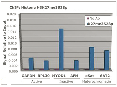
    Chromatin is the arrangement of DNA and proteins in which chromosomes are formed. Correspondingly, chromatin is formed from nucleosomes, which are comprised of a set of four histone proteins (H2A, H2B, H3, H4) wrapped with DNA. Chromatin is a very dynamic structure in which numerous post-translational modifications work together to activate or repress the availability of DNA to be copied, transcribed, or repaired. These marks decide which DNA will be open and commonly active (euchromatin) or tightly wound to prevent access and activation (heterochromatin). Common histone modifications include methylation of lysine and arginine, acetylation of lysine, phosphorylation of threonine and serine, and sumoylation, biotinylation, and ubiquitylation of lysine. Specifically, trimethylation of K27 is associated with gene silencing, whereas pS28 is associated with mitotis and immediate early genes.

  •  Applications
    IF
    Immunofluorescence: Diagenode offers huge selection of highly sensitive antibodies validated in IF. Immunofluorescence using the Diagenode monoclonal antibody directed against CRISPR/Cas9 HeLa cells transfected with a Cas9 expression vector (... Read more
    WB
    Western blot : The quality of antibodies used in this technique is crucial for correct and specific protein identification. Diagenode offers huge selection of highly sensitive and specific western blot-validated antibodies. Learn more about: Load... Read more
    DB
    Dot blotting Read more
    ChIP-qPCR (ab)
    Read more
  •  Documents
    Datasheet H3K27me3S28p C15410293 DATASHEET
    Datasheet description
    Download
  •  Publications

    How to properly cite this product in your work

    Diagenode strongly recommends using this: H3K27me3S28p Antibody (Diagenode Cat# C15410293 Lot# 001). Click here to copy to clipboard.

    Using our products in your publication? Let us know!

  •  Related products

Events

  • APHL 2024
    Milwaukee, Wisconsin, USA
    May 6-May 9, 2024
 See all events

News

 See all news


The European Regional Development Fund and Wallonia are investing in your future.

Extension of industrial buildings and new laboratories.


       Site map   |   Contact us   |   Conditions of sales   |   Conditions of purchase   |   Privacy policy   |   Diagenode Diagnostics