Histones are the main constituents of the protein part of chromosomes of eukaryotic cells. They are rich in the amino acids arginine and lysine and have been greatly conserved during evolution. Histones pack the DNA into tight masses of chromatin. Two core histones of each class H2A, H2B, H3 and H4 assemble and are wrapped by 146 base pairs of DNA to form one octameric nucleosome. Histone tails undergo numerous post-translational modifications, which either directly or indirectly alter chromatin structure to facilitate transcriptional activation or repression or other nuclear processes. In addition to the genetic code, combinations of the different histone modifications reveal the so-called “histone code”. Histone methylation and demethylation is dynamically regulated by respectively histone methyl transferases and histone demethylases. Acetylation of histone H4 is associated with active genes.
H4K5,8,12ac Antibody

Polyclonal antibody raised in rabbit against the region of histone H4 containing the acetylated lysines 5, 8 and 12 (H4K5,8,12ac), using a KLH-conjugated synthetic peptide.
| Lot | A2022P |
|---|---|
| Concentration | 0.76 µg/µl |
| Species reactivity | Human, mouse, wide range expected |
| Type | Polyclonal |
| Purity | Affinity purified |
| Host | Rabbit |
| Precautions | This product is for research use only. Not for use in diagnostic or therapeutic procedures. |
| Applications | Suggested dilution | References |
|---|---|---|
| ChIP/ChIP-seq* | 1 μg | Fig 1, 2 |
| CUT&TAG | 1:1,000 | Fig 3 |
| ELISA | 1:1,000 | Fig 4 |
| Dot blotting | 1:20,000 | Fig 5 |
| Western blotting | 1:1,000 | Fig 6 |
| IF | 1:500 | Fig 7 |
* Please note that the optimal antibody amount per ChIP should be determined by the end-user. We recommend testing 0.5-5 μg per IP.
- Validation Data
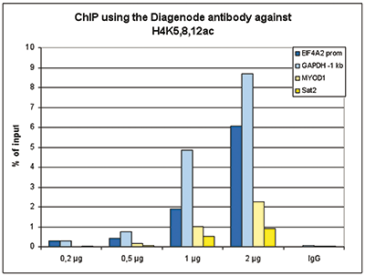
Figure 1. ChIP results obtained with the Diagenode antibody directed against H4K5,8,12ac.
ChIP assays were performed using human K562 cells, the Diagenode antibody against H4K5,8,12ac (cat. No. C15410021) and optimized PCR primer sets for qPCR. ChIP was performed with the “iDeal ChIP- seq” kit (cat. No. AB-001-0024) on sheared chromatin from 100,000 cells. A titration of the antibody consisting of 0.2, 0.5, 1 and 2 μg per ChIP experiment was analysed. IgG (1 μg/IP) was used as negative IP control. QPCR was performed with primers for promoter of the active gene EIF4A2 and for a region 1 kb upstream of the GAPDH gene, used as positive controls, and for the inactive MYOD1 gene and the Sat2 satellite repeat region used as negative controls. Figure 1 shows the recovery, expressed as a % of input (the relative amount of immunoprecipitated DNA compared to input DNA after qPCR analysis).A.
B.
C.
D.
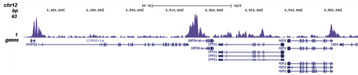
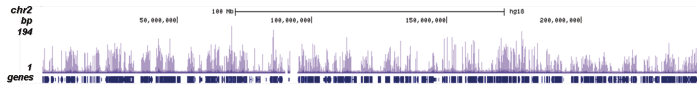
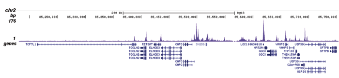
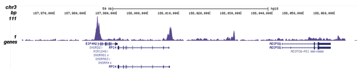
Figure 2. ChIP-seq results obtained with the Diagenode antibody directed against H4K5,8,12ac.
ChIP was performed with 0.5 μg of the Diagenode antibody against H4K5,8,12ac (cat. No. C15410021) on sheared chromatin from 100,000 K562 cells using the “iDeal ChIP-seq” kit as described above. The IP’d DNA was subsequently analysed on an Illumina Genome Analyzer. Library preparation, cluster generation and sequencing were performed according to the manufacturer’s instructions. The 36 bp tags were aligned to the human genome using the ELAND algorithm. Figure 2 shows the signal distribution along the complete length of chromosome 2 (figure 2A) and a zoomin to a 600 kb region (figure 2B). Figure 2C and D show the enrichment in two genomic regions on chromosome 3 and 12, respectively, containing EIF4A2 and GAPDH positive controls.A.

B.
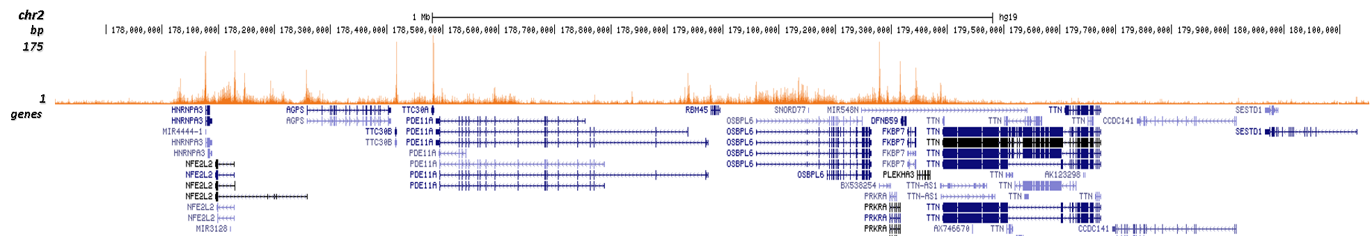
Figure 3. Cut&Tag results obtained with the Diagenode antibody directed against H4K5,8,12ac.
CUT&TAG (Kaya-Okur, H.S., Nat Commun 10, 1930, 2019) was performed on 50,000 K562 cells using 1 µg of the Diagenode antibody against H4K5,8,12ac (cat. No. C15410021) and the Diagenode pA-Tn5 transposase (C01070001). The libraries were subsequently analysed on an Illumina NextSeq 500 sequencer (2x75 paired-end reads) according to the manufacturer's instructions. The tags were aligned to the human genome (hg19) using the BWA algorithm. Figure 3 shows the peak distribution along the complete sequence and a 2 Mb zoomin of chromosome 2 (figure 3A and B, respectively).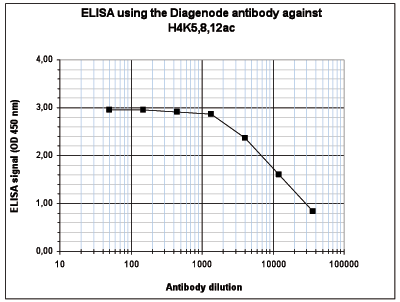
Figure 4. Determination of the antibody titer.
To determine the titer of the antibody, an ELISA was performed using a serial dilution of the Diagenode antibody directed against H4K5,8,12ac (cat. No. C15410021) in antigen coated wells. The antigen used was a peptide containing the histone modification of interest. By plotting the absorbance against the antibody dilution (Figure 4), the titer of the antibody was estimated to be 1:14,500.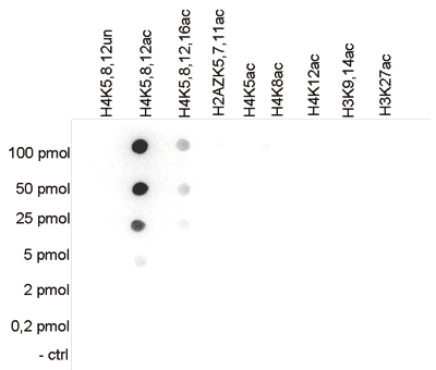
Figure 5. Cross reactivity tests using the Diagenode antibody directed against H4K5,8,12ac.
To test the cross reactivity of the Diagenode antibody against H4K5,8,12ac (cat. No. C15410021), a Dot Blot analysis was performed with peptides containing other histone modifications and the unmodified H4. One hundred to 0.2 pmol of the respective peptides were spotted on a membrane. The antibody was used at a dilution of 1:20,000. Figure 5 shows a high specificity of the antibody for the modification of interest.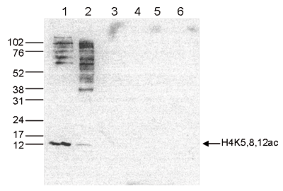
Figure 6. Western blot analysis using the Diagenode antibody directed against H4K5,8,12ac.
Western blot was performed on whole cell (25 μg, lane 1) and histone extracts (15 μg, lane 2) from HeLa cells, and on 1 μg of recombinant histone H2A, H2B, H3 and H4 (lane 3, 4, 5 and 6, respectively) using the Diagenode antibody against H4K5,8,12ac (cat. No. C15410021). The antibody was diluted 1:1,000 in TBS-Tween containing 5% skimmed milk. The position of the protein of interest is indicated on the right, the marker (in kDa) is shown on the left.A.
B.
C.
D.
E.
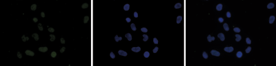
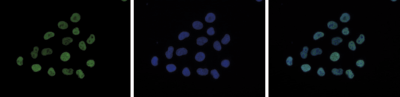
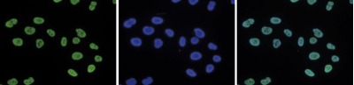
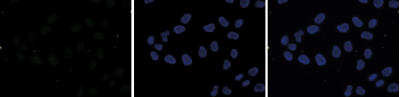
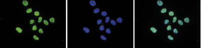
Figure 7. Immunofluorescence using the Diagenode antibody directed against H4K5,8,12ac.
HeLa cells were stained with the Diagenode antibody against H4K5,8,12ac (cat. No. C15410021) and with DAPI. Cells were fixed with 4% formaldehyde for 10’ and blocked with PBS/TX-100 containing 5% normal goat serum and 1% BSA. Figure 7A: cells were immunofluorescently labeled with the H4K5,8,12ac antibody (left) diluted 1:500 in blocking solution followed by an anti-rabbit antibody conjugated to Alexa488. The middle panel shows staining of the nuclei with DAPI. A merge of the two stainings is shown on the right. Figure 7B, C, D and E: staining of the cells with the H4K5,8,12ac antibody after incubation of the antibody with 10 ng/μl of the following blocking peptides: H4K5,8,12 unmodified (figure 7B), H4K5,8,12ac (figure 7C), H2A.ZK5,7,11ac (figure 7D) and H4K5,8,12,16ac (figure 7E). - Publications

How to properly cite our product/service in your work
We strongly recommend using this: H4K5,8,12ac Antibody (Hologic Diagenode Cat# C15410021 Lot# A2022P). Click here to copy to clipboard.
Using our products or services in your publication? Let us know!