Histones are the main constituents of the protein part of chromosomes of eukaryotic cells. They are rich in the amino acids arginine and lysine and have been greatly conserved during evolution. Histones pack the DNA into tight masses of chromatin. Two core histones of each class H2A, H2B, H3 and H4 assemble and are wrapped by 146 base pairs of DNA to form one octameric nucleosome. Histone tails undergo numerous post-translational modifications, which either directly or indirectly alter chromatin structure to facilitate transcriptional activation or repression or other nuclear processes. In addition to the genetic code, combinations of the different histone modifications reveal the so-called “histone code”. Histone methylation and demethylation is dynamically regulated by respectively histone methyl transferases and histone demethylases. Trimethylation of histone H4K20 is associated with inactive genomic regions, satellite repeats and ZNF gene repeats.
H4K20me3 Antibody

Polyclonal antibody raised in rabbit against the region of histone H4 containing the trimethylated lysine 20 (H4K20me3), using a KLH-conjugated synthetic peptide.
| Lot | A2730P |
|---|---|
| Concentration | 0.94 µg/µl |
| Species reactivity | Human, mouse, wide range expected |
| Type | Polyclonal |
| Purity | Affinity purified polyclonal antibody. |
| Host | Rabbit |
| Storage Conditions | Store at -20°C; for long storage, store at -80°C. Avoid multiple freeze-thaw cycles. |
| Storage Buffer | PBS containing 0.05% azide and 0.05% ProClin 300. |
| Precautions | This product is for research use only. Not for use in diagnostic or therapeutic procedures. |
| Applications | Suggested dilution | References |
|---|---|---|
| ChIP/ChIP-seq * | 0.5-1 µg per IP | Fig 1, 2 |
| ELISA | 1:500 | Fig 3 |
| Dot Blotting/Peptide array | 1:20,000/1:10,000 | Fig 4 |
| Western Blotting | 1:1,000 | Fig 5 |
| Immunofluorescence | 1:500 | Fig 6 |
* Please note that the optimal antibody amount per IP should be determined by the end-user. We recommend testing 0.5-5 µg per IP.
- Validation data
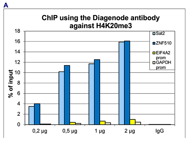
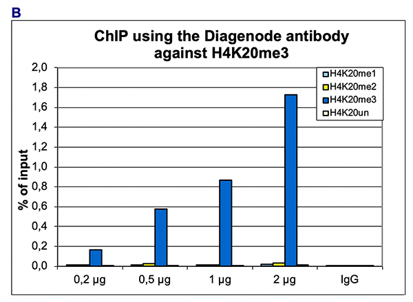
Figure 1. ChIP results obtained with the Diagenode antibody directed against H4K20me3
ChIP assays were performed using human HeLa cells, the Diagenode antibody against H4K20me3 (Cat. No. C15410207) and optimized PCR primer pairs for qPCR. ChIP was performed with the “iDeal ChIP-seq” kit (Cat. No. C01010051), using sheared chromatin from 1 million cells. The chromatin was spiked with a panel of in vitro assembled nucleosomes, each containing a specific lysine methylation (SNAP-ChIP K-MetStat Panel, Epicypher). A titration consisting of 0.2, 0.5, 1 and 2 µg of antibody per ChIP experiment was analyzed. IgG (1 µg/IP) was used as a negative IP control.
Figure 1A. Quantitative PCR was performed with primers specific for the promoter of the active GAPDH and EIF4A2 genes, used as negative controls, and for the ZNF10 gene and the Sat2 satellite repeat, used as positive controls. The graph shows the recovery, expressed as a % of input (the relative amount of immunoprecipitated DNA compared to input DNA after qPCR analysis).
Figure 1B. Recovery of the nucleosomes carrying the H4K20me1, H4K20me2, H4K2me3 and the unmodified H4K20 as determined by qPCR. The figure clearly shows the antibody is very specific in ChIP for the H4K20me3 modification.A.
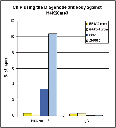
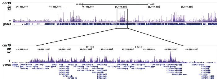
Figure 2. ChIP-seq results obtained with the Diagenode antibody directed against H4K20me3
ChIP was performed with 0.5 µg of the Diagenode antibody against H4K20me3 (Cat. No. C15410207) on sheared chromatin from 100,000 K562 cells using the “iDeal ChIP-seq” kit. The IP’d DNA was analysed by QPCR as described above (figure 2A). The IP’d DNA was subsequently analysed on an Illumina Genome Analyzer. Library preparation, cluster generation and sequencing were performed according to the manufacturer’s instructions. The 36 bp tags were aligned to the human genome using the ELAND algorithm. Figure 2B shows the signal distribution along the long arm of chromosome 19 and a zoomin to an enriched region containing several ZNF repeat genes. Figure 2C and D show the enrichment in the telomeric region of chromosome 12, also containing several ZNF repeat genes, and at ZNF510 on chromosome 9, respectively. The position of the amplicon used for ChIP-qPCR is indicated by an arrow.B.
C.

D.
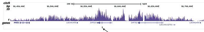
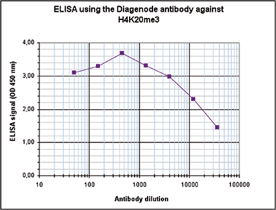
Figure 3. Determination of the antibody titer
To determine the titer of the antibody, an ELISA was performed using a serial dilution of the Diagenode antibody directed against H4K20me3 (Cat. No. C15410207) in antigen coated wells. The antigen used was a peptide containing the histone modification of interest. By plotting the absorbance against the antibody dilution (Figure 3), the titer of the antibody was estimated to be 1:21,700.A.
B.
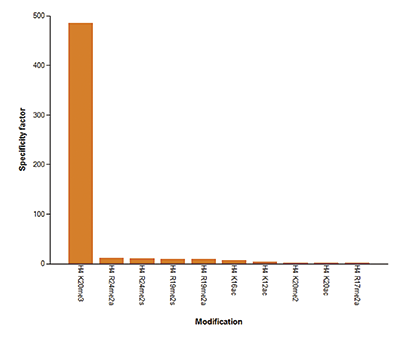
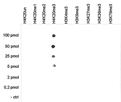
Figure 4. Cross reactivity tests using the Diagenode antibody directed against H4K20me3
Figure 4A To test the cross reactivity of the Diagenode antibody against H4K20me3 (Cat. No. C15410207), a Dot Blot analysis was performed with peptides containing other histone modifications and the unmodified H4K20. One hundred to 0.2 pmol of the respective peptides were spotted on a membrane. The antibody was used at a dilution of 1:20,000. Figure 4A shows a high specificity of the antibody for the modification of interest. Figure 4B The specificity of the antibody was further demonstrated by peptide array analyses on an array containing 384 peptides with different combinations of modifications from histone H3, H4, H2A and H2B. The antibody was used at a dilution of 1:10,000. Figure 4B shows the specificity factor, calculated as the ratio of the average intensity of all spots containing the mark, divided by the average intensity of all spots not containing the mark.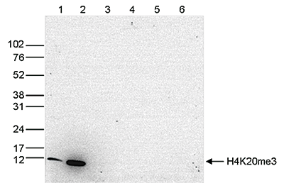
Figure 5. Western blot analysis using the Diagenode antibody directed against H4K20me3
Western blot was performed on whole cell (25 µg, lane 1) and histone extracts (15 µg, lane 2) from HeLa cells, and on 1 µg of recombinant histone H2A, H2B, H3 and H4 (lane 3, 4, 5 and 6, respectively) using the Diagenode antibody against H4K20me3 (Cat. No. C15410207). The antibody was diluted 1:1,000 in TBS-Tween containing 5% skimmed milk. The position of the protein of interest is indicated on the right, the marker (in kDa) is shown on the left.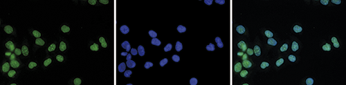
Figure 6. Immunofluorescence using the Diagenode antibody directed against H4K20me3
HeLa cells were stained with the Diagenode antibody against H4K20me3 (Cat. No. C15410207) and with DAPI. Cells were fixed with methanol and blocked with PBS/TX-100 containing 5% normal goat serum and 1% BSA. The cells were immunofluorescently labeled with the H4K20me3 antibody (left) diluted 1:500 in blocking solution followed by an anti-rabbit antibody conjugated to Alexa488. The middle panel shows staining of the nuclei with DAPI. A merge of the two stainings is shown on the right. - Publications

How to properly cite our product/service in your work
We strongly recommend using this: H4K20me3 Antibody (Hologic Diagenode Cat# C15410207 Lot# A2730P). Click here to copy to clipboard.
Using our products or services in your publication? Let us know!
The 20S proteasome activator PA28γ controls the compaction of chromatin
Didier Fesquet, David Llères, Cristina Viganò, Francisca Méchali, Séverine Boulon, Robert Feil, Olivier Coux, Catherine Bonne-Andrea, Véronique Baldin
The nuclear PA28γ is known to activate the 20S proteasome, but its precise cellular functions remains unclear. Here, we identify PA28γ as a key factor that structures heterochromatin. We find that in human cells, a fraction of PA28γ-20S proteasome complexes localizes within HP1-linked heterochromat...In vitro capture and characterization of embryonic rosette-stage pluripotency between naive and primed states.
Neagu A, van Genderen E, Escudero I, Verwegen L, Kurek D, Lehmann J, Stel J, Dirks RAM, van Mierlo G, Maas A, Eleveld C, Ge Y, den Dekker AT, Brouwer RWW, van IJcken WFJ, Modic M, Drukker M, Jansen JH, Rivron NC, Baart EB, Marks H, Ten Berge D
Following implantation, the naive pluripotent epiblast of the mouse blastocyst generates a rosette, undergoes lumenogenesis and forms the primed pluripotent egg cylinder, which is able to generate the embryonic tissues. How pluripotency progression and morphogenesis are linked and whether intermediate pluripotent st...Myc Regulates Chromatin Decompaction and Nuclear Architecture during B Cell Activation
Kieffer-Kwon K.R. et al.
50 years ago, Vincent Allfrey and colleagues discovered that lymphocyte activation triggers massive acetylation of chromatin. However, the molecular mechanisms driving epigenetic accessibility are still unknown. We here show that stimulated lymphocytes decondense chromatin by three differentially regulated steps. Fi... - Related products
-
 C01011000
Auto ChIPmentation Kit for Histones - Replaced ...
-