
| ELISA Enzyme-linked immunosorbent assay. Read more |
| DB Dot blotting Read more |
| WB Western blot : The quality of antibodies used in this technique is crucial for correct and specific protein identification. Diagenode offers huge selection of highly sensitive and specific western blot-validated antibodies. Learn more about: Load... Read more |
| IF Immunofluorescence: Diagenode offers huge selection of highly sensitive antibodies validated in IF. Immunofluorescence using the Diagenode monoclonal antibody directed against CRISPR/Cas9 HeLa cells transfected with a Cas9 expression vector (... Read more |
H3K79me3 Antibody
Catalog Number
Format
Price
C15310068
(CS-068-100)
(CS-068-100)
100 µl
DISCONTINUED 
As an alternative we offer the purified H3K79me3 polyclonal antibody (C15410068).
Polyclonal antibody raised in rabbit against histone H3 containing the trimethylated lysine 79 (H3K79me3), using a KLH-conjugated synthetic peptide.
| Lot | A86-001 |
|---|---|
| Concentration | not determined |
| Species reactivity | Human, mouse |
| Type | Polyclonal |
| Purity | Whole antiserum |
| Host | Rabbit |
| Precautions | This product is for research use only. Not for use in diagnostic or therapeutic procedures. |
| Applications | Suggested dilution | References |
|---|---|---|
| ELISA | 1:500 - 1:1,000 | Fig 1 |
| Dot Blotting | 1:50,000 | Fig 2 |
| Western Blotting | 1:500 | Fig 3 |
| Immunofluorescence | 1:200 | Fig 4 |
- Validation Data
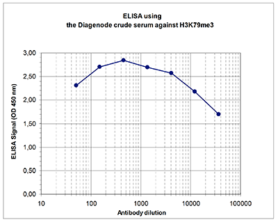
Figure 1. Determination of the antibody titer
To determine the titer of the antibody, an ELISA was performed using a serial dilution of Diagenode antibody directed against H3K79me3 (Cat. No. CS-068-100) in antigen coated wells. The antigen used was a peptide containing the histone modification of interest. By plotting the absorbance against the antibody dilution (Figure 1), the titer of the antibody was estimated to be 1:70,000.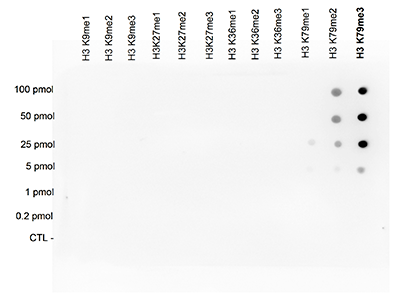
Figure 2. Cross reactivity test of the Diagenode antibody directed against H3K79me3
A Dot Blot analysis was performed to test the cross reactivity of the Diagenode antibody against H3K79me3 (Cat. No. CS-068-100) with peptides containing other modifications of histone H3. These include mono- and dimethylation of the same lysine and mono-, di- and trimethylation of lysine 9, 27 and 36. One hundred to 0.2 pmol of the peptides were spotted on a membrane. The antibody was used at a dilution of 1:50,000. Figure 2 shows a high specificity of the antibody for the modification of interest.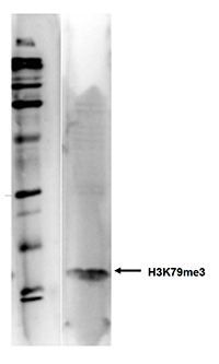
Figure 3. Western blot analysis using the Diagenode antibody directed against H3K79me3
Western blot was performed on histone extracts from HeLa cells (15 μg) with the Diagenode antibody against H3K79me3 (Cat. No. CS-068-100), diluted 1:500 in TBS-Tween containing 5% skimmed milk. The molecular weight marker (Bio-Rad, broad range biotinylated SDS-PAGE standard) is shown on the left, the location of the protein of interest is indicated on the right.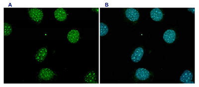
Figure 4. Immunofluorescence using the Diagenode antibody directed against H3K79me3
Mouse fibroblasts (NIH3T3 cells) were stained with the Diagenode antibody against H3K79me3 (Cat. No. CS- 068-100) and with DAPI. Cells were formaldehyde fixed, permeabilized with Triton X100 and blocked with PBS containing 2.5% BSA. Figure 4A: cells were immunofluorescently labeled with the H3K79me3 antibody (diluted 1:200 and incubated for 1 hour at room temperature) followed by goat anti-rabbit antibody conjugated to FITC. Figure 1B: staining of the nuclei with DAPI, which specifically labels DNA. Both antibody and DAPI staining are restricted to the nucleus. - Publications

How to properly cite our product/service in your work
We strongly recommend using this: H3K79me3 Antibody (Hologic Diagenode Cat# C15310068 Lot# A86-001). Click here to copy to clipboard.
Using our products or services in your publication? Let us know!
PWWP2A binds distinct chromatin moieties and interacts with an MTA1-specific core NuRD complex.
Link S, Spitzer RMM, Sana M, Torrado M, Völker-Albert MC, Keilhauer EC, Burgold T, Pünzeler S, Low JKK, Lindström I, Nist A, Regnard C, Stiewe T, Hendrich B, Imhof A, Mann M, Mackay JP, Bartkuhn M, Hake SB
Chromatin structure and function is regulated by reader proteins recognizing histone modifications and/or histone variants. We recently identified that PWWP2A tightly binds to H2A.Z-containing nucleosomes and is involved in mitotic progression and cranial-facial development. Here, using in vitro assays, we show that...