Chromatin is the arrangement of DNA and proteins in which chromosomes are formed. Correspondingly, chromatin is formed from nucleosomes, which are comprised of a set of four histone proteins (H2A, H2B, H3, H4) wrapped with DNA. Chromatin is a very dynamic structure in which numerous post-translational modifications work together to activate or repress the availability of DNA to be copied, transcribed, or repaired. These marks decide which DNA will be open and commonly active (euchromatin) or tightly wound to prevent access and activation (heterochromatin). Common histone modifications include methylation of lysine and arginine, acetylation of lysine, phosphorylation of threonine and serine, and sumoylation, biotinylation, and ubiquitylation of lysine. In particular, phosphorylation of H4 Ser1 (H4S1p) has been linked to mitosis and DNA repair. This modification is enriched in sites proximal to double stranded brakes, but not those associated with UV damage. Casein kinase II (CK2) phosphorylates H4 Ser1, and it also implicated in regulating the DNA damage response. Furthermore, recruitment of CK2 requires the SIN3/RPD3 histone deacetylase complex.
H4S1p Antibody

Polyclonal antibody raised in rabbit against Histone H4 (p Ser1), using a KLH-conjugated synthetic peptide.
| Lot | 001 |
|---|---|
| Concentration | 0.89 μg/μl |
| Species reactivity | Human, mouse, rat |
| Type | Polyclonal |
| Purity | Affinity purified |
| Host | Rabbit |
| Precautions | This product is for research use only. Not for use in diagnostic or therapeutic procedures. |
| Applications | Suggested dilution | References |
|---|---|---|
| ChIP | 2-5 μg/million cells | Figure 1 |
| Immunofluorescence | 1:50 | Figure 2, 3 |
| Western Blotting | 1:1,000 | Figure 4 |
| Immunohistochemistry | 1:50 | |
| Dot Blotting | 1:10,000 | Figure 5 |
- Validation Data
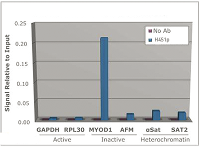
Figure 1. ChIP
Chromatin Immunoprecipitation with the H4S1p antibody. Chromatin from one million formaldehyde cross-linked HeLa cells was used with 2 ug of H4S1p alongside a no antibody (No Ab) control. DNA was measured by qPCR and normalized to total input.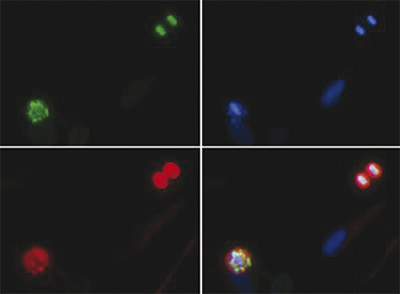
Figure 2. Immunofluorescence
Immunofluorescence with the H4S1p antibody. Tissue: Nonmitotic, prophase, and telophase HeLa cells. Fixation: 0.5% PFA. Primary antibody used at a 1:50 dilution for 1 h at RT. Secondary antibody: FITC secondary antibody at 1:10,000 for 45 min at RT. Localization: Histone H4S1p is nuclear. Staining: Histone H4S1p is expressed in green, nuclei and alpha-tubulin are counterstained with DAPI (blue) and Dylight 594 (red).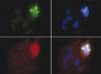
Figure 3. Immunofluorescence
Immunofluorescence with the H4S1p antibody. Tissue: Nonmitotic and telophase HeLa cells. Fixation: 0.5% PFA. Primary antibody used at a 1:50 dilution for 1 h at RT. Secondary antibody: FITC secondary antibody at 1:10,000 for 45 min at RT. Localization: Histone H4S1p is nuclear. Staining: Histone H4S1p is expressed in green, nuclei and alpha-tubulin are counterstained with DAPI (blue) and Dylight 594 (red).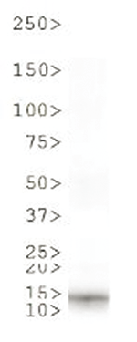
Figure 4. Western Blot
Western Blot with the H4S1p antibody. 30 μg HeLa histone extracts. Primary antibody used at 1:1000 dilution overnight at 4°C. Secondary antibody: IRDye800TM rabbit secondary antibody at a 1:10,000 for 45 min at RT. Predicted/Observed size: ~13 kDa. Other band(s): None.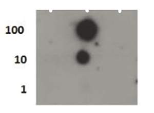
Figure 5. Dot Blot
Dot Blot with the H4S1p antibody. Lane 1: H4S1 unmodfied. Lane 2: H4S1p. Lane 3: H4S1 unmodified. Load: 1, 10, and 100 picomoles of peptide. Primary antibodyused at 0,1 μg/ml for 45 min at 4°C. Secondary antibody: DylightTM488 rabbit secondary antibody at 1:10,000 for 45 min at RT. - Publications

How to properly cite our product/service in your work
We strongly recommend using this: H4S1p Antibody (Hologic Diagenode Cat# C15410298 Lot# 001). Click here to copy to clipboard.
Using our products or services in your publication? Let us know!