Histones are the main constituents of the protein part of chromosomes of eukaryotic cells. They are rich in the amino acids arginine and lysine and have been greatly conserved during evolution. Histones pack the DNA into tight masses of chromatin. Two core histones of each class H2A, H2B, H3 and H4 assemble and are wrapped by 146 base pairs of DNA to form one octameric nucleosome. Histone tails undergo numerous post-translational modifications, which either directly or indirectly alter chromatin structure to facilitate transcriptional activation or repression or other nuclear processes. In addition to the genetic code, combinations of the different histone modifications reveal the so-called “histone code”. Histone methylation and demethylation is dynamically regulated by respectively histone methyl transferases and histone demethylases. Methylation of histone H3K9 is associated with gene repression and heterochromatin formation, although higher levels of H3K9me1 have been found in some more active promoters.
H3K9me1 Antibody

Polyclonal antibody raised in rabbit against histone H3 containing the monomethylated lysine 9 (H3K9me1), using a KLH-conjugated synthetic peptide.
| Lot | A89-001 |
|---|---|
| Concentration | not determined |
| Species reactivity | Human |
| Type | Polyclonal |
| Purity | Whole antiserum |
| Host | Rabbit |
| Precautions | This product is for research use only. Not for use in diagnostic or therapeutic procedures. |
| Applications | Suggested dilution | References |
|---|---|---|
| ChIP * | 10 μl/ChIP | Fig 1 |
| ELISA | 1:2,000 - 1:3,000 | Fig 2 |
| Dot Blotting | 1:200,000 | Fig 3 |
| Western Blotting | 1:1,000 | Fig 4 |
| Immunofluorescence | 1:200 | Fig 5 |
- Validation Data
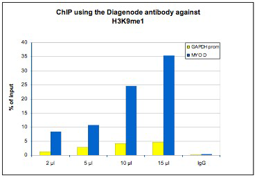
Figure 1. ChIP results obtained with the Diagenode antibody directed against H3K9me1
ChIP assays were performed using human osteosarcoma (U2OS) cells, the Diagenode antibody against H3K9me1 (cat. No. CS-065-100) and optimized PCR primer sets for qPCR. Chromatin was sheared with the Diagenode “Shearing ChIP” kit (cat. No. kch-redmod-100). ChIP was performed with the “OneDay ChIP” kit (cat. No. kch-oneDIP-060), using sheared chromatin from 1.6 million cells. A titration of the antibody consisting of 2, 5, 10 and 15 μl per ChIP experiment was analysed. IgG (5 μg/IP) was used as negative IP control. Quantitative PCR was performed using primers for the promoter of the housekeeping gene GAPDH and for the coding region of the myogenic differentiation gene (MYOD), a gene that is inactive at normal conditions. Figure 1 shows the recovery, expressed as a % of input (the relative amount of immunoprecipitated DNA compared to input DNA after qPCR analysis).
Figure 2. Determination of the titer
To determine the titer, an ELISA was performed using a serial dilution of the Diagenode antibody directed against H3K9me1 (cat. No. CS-065-100). The antigen used was a peptide containing the histone modification of interest. By plotting the absorbance against the antibody dilution (Figure 2), the titer of the antibody was estimated to be 1:50,000.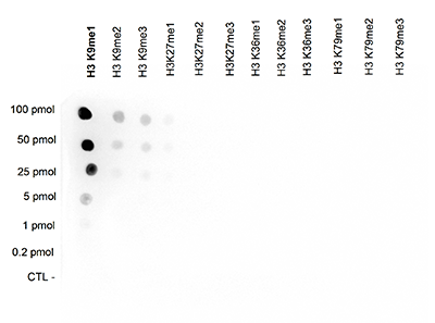
Figure 3. Cross reactivity test using the Diagenode antibody directed against H3K9me1
A Dot Blot analysis was performed to test the cross reactivity of the Diagenode antibody against H3K9me1 (cat. No. CS-065-100) with peptides containing other modifications of histone H3. Other histone modifications include di- and trimethylation of the same lysine and mono-, di- and trimethylation of lysine 27, 36 and 79. One hundred to 0.2 pmol of peptide containing the respective histone modification were spotted on a membrane. The antibody was used at a dilution of 1:200,000. Figure 3 shows a high specificity of the antibody for the modification of interest.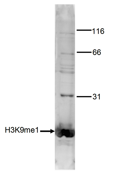
Figure 4. Western blot analysis using the Diagenode antibody directed against H3K9me1
Histone extracts of HeLa cells (15 μg) were analysed by Western blot using the Diagenode antibody against H3K9me1 (cat. No. CS-065-100) diluted 1:1000 in TBS-Tween containing 5% skimmed milk. The position of the protein of interest is indicated on the left; the marker (in kDa) is shown on the right.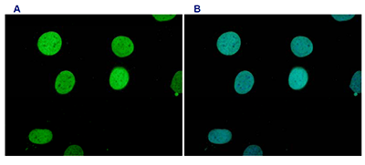
Figure 5. Immunofluorescence using the Diagenode antibody directed against H3K9me1
HeLa cells were stained with the Diagenode antibody against H3K9me1 (cat. No. CS-065-100) and with DAPI. Cells were formaldehyde fixated, permeabilized with Triton X-100 and blocked with PBS containing 2.5% BSA. Figure 5A: cells were immunofluorescently labelled with the H3K9me1 antibody (diluted 1:200 and incubated for 1 hour at room temperature) followed by goat anti-rabbit antibody conjugated to DyLight. Figure 5B: staining of the nuclei with DAPI, which specifically labels DNA. Both antibody and DAPI staining are restricted to the nucleus. - Publications

How to properly cite our product/service in your work
We strongly recommend using this: H3K9me1 Antibody (Hologic Diagenode Cat# C15310065 Lot# A89-001). Click here to copy to clipboard.
Using our products or services in your publication? Let us know!
Chromatin states of core pluripotency-associated genes in pluripotent, multipotent and differentiated cells.
Barrand S, Collas P
Oct4, Nanog and Sox2 constitute a core of transcription factors controlling pluripotency. Differentiation and reprogramming studies have unraveled a few epigenetic modifications associated in relation to the expression state of OCT4, NANOG and SOX2. There is, however, no comprehensive map of chromatin states on thes...