
| WB Western blot : The quality of antibodies used in this technique is crucial for correct and specific protein identification. Diagenode offers huge selection of highly sensitive and specific western blot-validated antibodies. Learn more about: Load... Read more |
| DB Dot blotting Read more |
H3K37me1 Antibody
Catalog Number
Format
Price

Polyclonal antibody raised in rabbit against Histone H3 (Monomethyl Lys37), using a KLH-conjugated synthetic peptide.
| Lot | 001 |
|---|---|
| Concentration | 0.42 μg/μl |
| Species reactivity | Human, mouse, C. elegans, rat, chicken, Xenopus, Drosophila, plant |
| Type | Polyclonal |
| Purity | Affinity purified |
| Host | Rabbit |
| Precautions | This product is for research use only. Not for use in diagnostic or therapeutic procedures. |
| Applications | Suggested dilution | References |
|---|---|---|
| Western Blotting | 1 μg/ml | Fig 1 |
| Dot Blotting | 1:1,000 | Fig 2, 3 |
- Validation Data
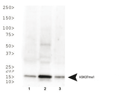
Figure 1. Western Blot
Western Blot with the H3K37me1 antibody. Lane 1: HeLa histone extracts. Lane 2: NIH-3T3 histone extracts. Lane 3: C. elegans embryo lysate. Load: 30 μg per lane. Primary antibody used at 1 μg/ml overnight at 4°C. Secondary antibody: IRDye800TM rabbit secondary antibody at 1:10,000 for 45 min at RT. Predicted/Observed size: ~15 kDa. Other band(s): None.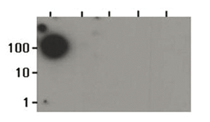
Figure 2. Dot Blot
Dot Blot with the H3K37me1 antibody. Lane 1: K37me1. Lane 2: K36me1. Lane 3: K36me2. Lane 4: K36me3. Lane 5: K36ac. Load: 1, 10, and 100 picomoles of peptide. Primary antibody used at 1:1000 for 45 min at 4°C. Secondary antibody: DylightTM488 rabbit secondary antibody at 1:10,000 for 45 min at RT.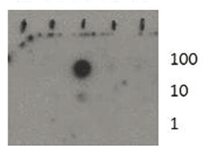
Figure 3. Dot Blot
Dot Blot with the H3K37me1 antibody. Lane 1: K37. Lane 2: K37Ac. Lane 3: K37Me1. Lane 4: K37Me2. Lane 5: K37Me3. Load: 1, 10, and 100 picomoles of peptide. Primary antibody used at 1:1000 for 45 min at 4°C. Secondary antibody: DylightTM488 rabbit secondary antibody at 1:10,000 for 45 min at RT. - Publications

How to properly cite our product/service in your work
We strongly recommend using this: H3K37me1 Antibody (Hologic Diagenode Cat# C15410295 Lot# 001). Click here to copy to clipboard.
Using our products or services in your publication? Let us know!
SMYD5 is a histone H3-specific methyltransferase mediatingmono-methylation of histone H3 lysine 36 and 37.
Aljazi Mohammad B. et al.
Although post-translational modifications (-PTMs) of some histone H3 lysine residues are well studied, the PTMs of histone H3 lysine 37 in mammalian cells remain largely unknown. In this study, we provide evidence to show that SMYD family member 5 (SMYD5) is a histone H3-specfic methyltransferase that catalyzes mono...