FOXM1 (UniProt/Swiss-Prot entry Q08050) is a transcription factor which is involved in cell proliferation. It regulates the expression of cell cycle genes essential for DNA replication and mitosis such as cyclin B1 and cyclin A2. It may also play a role in DNA breaks repair participating in the DNA damage checkpoint response.
FOXM1 Antibody
Catalog Number
Format
Price

Polyclonal antibody raised in rabbit against FOXM1 (forkhead box M1), using a KLH-conjugated synthetic peptide.
| Lot | 42613 |
|---|---|
| Concentration | 1.52 μg/μl |
| Species reactivity | Human |
| Type | Polyclonal |
| Purity | Affinity purified |
| Host | Rabbit |
| Precautions | This product is for research use only. Not for use in diagnostic or therapeutic procedures. |
| Applications | Suggested dilution * | References |
|---|---|---|
| ChIP/ChIP-seq * | 2 μg/ChIP | Fig 1, 2 |
| Western Blotting | 1:1,000 - 1:10,000 | Fig 3 |
| Immunohistochemistry | 1:100 - 1:1,000 | Fig 4 |
* Please note that the optimal antibody amount per IP should be determined by the end-user. We recommend testing 1-5 μg per IP.
- Validation Data
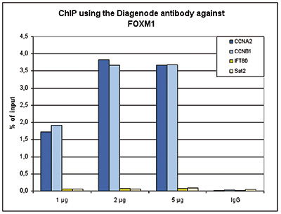
Figure 1. ChIP results obtained with the Diagenode antibody directed against FOXM1
ChIP assays were performed using HeLa cells, the Diagenode antibody against FOXM1 (Cat. No. C15410232) and optimized primer sets for qPCR. ChIP was performed with the “iDeal ChIP-seq” kit (Cat. No. C01010055), using sheared chromatin from 4 million cells. A titration of the antibody consisting of 1, 2 and 5 μg per ChIP experiment was analysed. IgG (1 μg/IP) was used as negative IP control. QPCR was performed with primers for the promoters of the CCNA2 and CCNB1 genes, used as positive controls, and for the IFT80 gene and the Sat2 satellite repeat, used as negative controls. Figure 1 shows the recovery, expressed as a % of input (the relative amount of immunoprecipitated DNA compared to input DNA after qPCR analysis).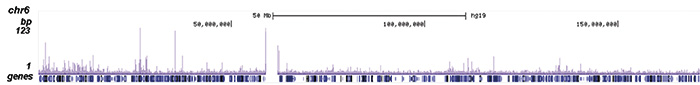
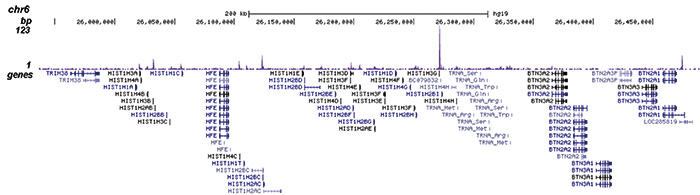
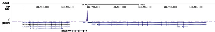
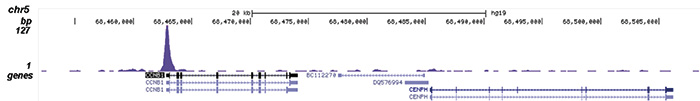
Figure 2. ChIP-seq results obtained with the Diagenode antibody directed against FOXM1
ChIP was performed on sheared chromatin from 4 million HeLa cells using 2 μg of the Diagenode antibody against FOXM1 (Cat. No. C15410232) as described above. The IP’d DNA was subsequently analysed on an Illumina HiSeq. Library preparation, cluster generation and sequencing were performed according to the manufacturer’s instructions. The 50 bp tags were aligned to the human genome using the BWA algorithm. Figure 2 shows the enrichment along the complete sequence and a 600 kb region of human chromosome 6 (fig 2A and B), and in two genomic regions surrounding the CCNA2 and CCNB1 positive control genes (fig 2C and D).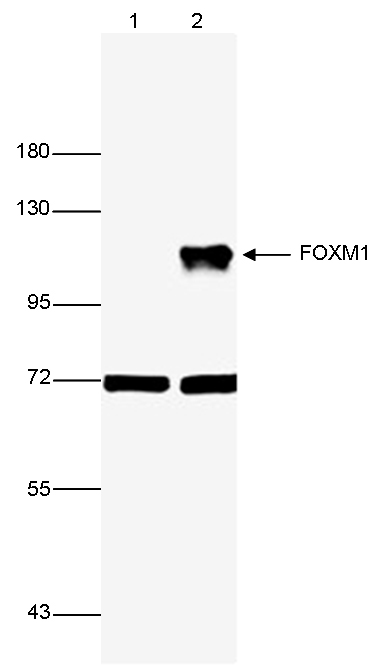
Figure 3. Western blot analysis using the Diagenode antibody directed against FOXM1
Whole cell extracts from 293T (30 μg, lane 1) or 293T cells transfected with a FOXM1 expression vector (lane 2) were analysed by Western blot using the Diagenode antibody against FOXM1 (Cat. No. C15410232) diluted 1:5,000. The position of the protein of interest is indicated on the right; the marker (in kDa) is shown on the left.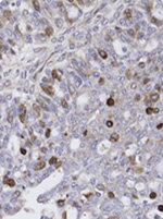
Figure 4. Immunohistochemistry using the Diagenode antibody directed against FOXM1
Formalin fixed paraffin embedded human ovarian cancer tissue was stained with the Diagenode antibody against FOXM1 (Cat. No. C15410232) diluted 1:100 followed by a peroxidase labelled goat anti-rabbit secondary antibody. - Publications

How to properly cite our product/service in your work
We strongly recommend using this: FOXM1 Antibody (Hologic Diagenode Cat# C15410232-100 Lot# 42613). Click here to copy to clipboard.
Using our products or services in your publication? Let us know!