Notice (8): Undefined variable: solution_of_interest [APP/View/Products/view.ctp, line 755]Code Context<!-- BEGIN: REQUEST_FORM MODAL -->
<div id="request_formModal" class="reveal-modal medium" data-reveal aria-labelledby="modalTitle" aria-hidden="true" role="dialog">
<?= $this->element('Forms/simple_form', array('solution_of_interest' => $solution_of_interest, 'header' => $header, 'message' => $message, 'campaign_id' => $campaign_id)) ?>
$viewFile = '/home/website-server/www/app/View/Products/view.ctp'
$dataForView = array(
'language' => 'cn',
'meta_keywords' => '5-hydroxymethylcytosine,5-hmC, 5-mC,polyclonal antibody ,Diagenode',
'meta_description' => '5-hydroxymethylcytosine (5-hmC) Polyclonal Antibody (rabbit) validated in hMeDIP, ELISA and DB. Batch-specific data available on the website. Sample size available',
'meta_title' => '5-hydroxymethylcytosine (5-hmC) Polyclonal Antibody(rabbit) | Diagenode',
'product' => array(
'Product' => array(
'id' => '2138',
'antibody_id' => '37',
'name' => '5-hydroxymethylcytosine (5-hmC) polyclonal antibody (rabbit) ',
'description' => '<p><span>Polyclonal antibody raised against 5-hydroxymethylcytosine (5-hmC). 5-hmC is a recently discovered DNA modification which results from the enzymatic conversion of 5-methylcytosine into 5-hydroxymethylcytosine by the TET family of oxygenases. Preliminary results indicate that 5-hmC may have important roles distinct from 5-methylcytosine (5-mC). Although its precise role has still to be shown, early evidence suggests a few putative mechanisms that could have big implications in epigenetics.</span></p>',
'label1' => 'Validation Data',
'info1' => '<div class="row">
<div class="small-6 columns">
<p class="text-center"><img src="https://www.diagenode.com/img/product/antibodies/C15310210-elisa.png" alt="ELISA" width="342" height="266" /></p>
</div>
<div class="small-6 columns">
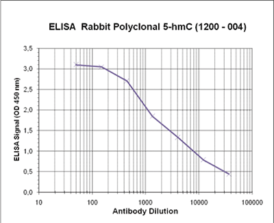
<p><small><strong>Figure 1. Determination of the 5-hmC rabbit polyclonal antibody titer</strong><br />To determine the titer, an ELISA was performed using a serial dilution of the Diagenode rabbit polyclonal antibody directed against 5-hmC in antigen coated wells. The antigen used was BSA coupled to the 5-hmC base. By plotting the absorbance against the antibody dilution, the titer of the antibody was estimated to be 1: 3,500. </small></p>
</div>
</div>
<div class="row">
<div class="small-6 columns">
<p class="text-center"><img src="https://www.diagenode.com/img/product/antibodies/C15310210-fig2.png" alt="" width="161" height="399" /></p>
</div>
<div class="small-6 columns">
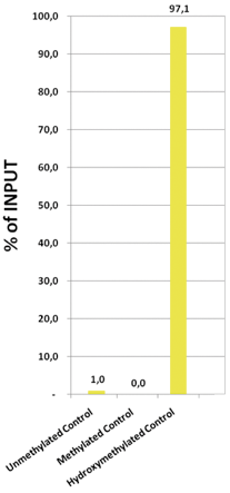
<p><small><strong>Figure 2. An hydroxymethylated DNA IP (hMeDIP) was performed using the Diagenode rabbit polyclonal antibody directed against 5-hydroxymethylcytosine (Cat. No. CS-HMC-100).</strong><br />The IgG isotype antibodies from rabbit (Cat. No. kch-504-250) was used as negative control. The DNA was prepared with the GenDNA module of the hMeDIP kit and sonicated with our Bioruptor® (UCD-200/300 series) to have DNA fragments of 300-500 bp. 1 μg of human Hela cells DNA were spiked with non-methylated, methylated, and hydroxymethylated fragments. The IP’d material has been analysed by qPCR using the primer pair specific for the 3 different control sequences. The obtained results show that the Diagenode rabbit polyclonal for 5-hmC is highly specific for this base modification (no IP with non-methylated or methylated C bases containing fragments). </small></p>
</div>
</div>
<div class="row">
<div class="small-6 columns">
<p class="text-center"><img src="https://www.diagenode.com/img/product/antibodies/C15310210-fig3.png" alt="Dot Blot" width="135" height="119" /></p>
</div>
<div class="small-6 columns">
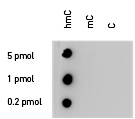
<p><small><strong>Figure 3. Dotblot analysis of the Diagenode 5-hmC rabbit polyclonal antibody with the C, mC and hmC PCR controls</strong><br />100 to 4 ng (equivalent of 5 to 0.2 pmol of C-bases) of the hmC, mC and C PCR controls from the Diagenode “5-hmC, 5-mC & cytosine DNA Standard Pack” (Cat No. AF-101-0002) were spotted on a membrane (Amersham Hybond-N+). The membrane was incubated with the rabbit 5-hydroxymethylcytosine polyclonal antibody (dilution 1:200). The membranes were exposed for 30 seconds. </small></p>
</div>
</div>',
'label2' => 'Target description',
'info2' => '<p>5-hydroxymethylcytosine (5-hmC) has been recently discovered in mammalian DNA. This results from the enzymatic conversion of 5-methylcytosine into 5-hydroxymethylcytosine by the TET family of oxygenases. So far, the 5-hmC bases have been identified in Purkinje neurons, in granule cells and embryonic stem cells where they are present at high levels (up to 0,6% of total nucleotides in Purkinje cells).</p>
<p>Preliminary results indicate that 5-hmC may have important roles distinct from 5-mC. Although its precise role has still to be shown, early evidence suggests a few putative mechanisms that could have big implications in epigenetics : 5-hydroxymethylcytosine may well represent a new pathway to demethylate DNA involving a repair mechanism converting 5-hmC to cytosine and, as such open up entirely new perspectives in epigenetic studies.</p>
<p>Due to the structural similarity between 5-mC and 5-hmC, these bases are experimentally almost indistinguishable. Recent articles demonstrated that the most common approaches (e.g. enzymatic approaches, bisulfite sequencing) do not account for 5-hmC. The development of the affinity-based technologies appears to be the most powerful way to differentially and specifically enrich 5-mC and 5-hmC sequences. The results shown here illustrate the use of this unique monoclonal antibody against 5-hydroxymethylcytosine that has been fully validated in various technologies.</p>',
'label3' => '',
'info3' => '',
'format' => '100 µl',
'catalog_number' => 'C15310210-100',
'old_catalog_number' => 'CS-HMC-100',
'sf_code' => 'C15310210-D001-001161',
'type' => 'FRE',
'search_order' => '03-Antibody',
'price_EUR' => '380',
'price_USD' => '380',
'price_GBP' => '340',
'price_JPY' => '59525',
'price_CNY' => '',
'price_AUD' => '950',
'country' => 'ALL',
'except_countries' => 'Japan',
'quote' => false,
'in_stock' => false,
'featured' => false,
'no_promo' => false,
'online' => true,
'master' => true,
'last_datasheet_update' => '0000-00-00',
'slug' => '5-hmc-polyclonal-antibody-rabbit-classic-100-ul',
'meta_title' => '5-hydroxymethylcytosine (5-hmC) Polyclonal Antibody(rabbit) | Diagenode',
'meta_keywords' => '5-hydroxymethylcytosine,5-hmC, 5-mC,polyclonal antibody ,Diagenode',
'meta_description' => '5-hydroxymethylcytosine (5-hmC) Polyclonal Antibody (rabbit) validated in hMeDIP, ELISA and DB. Batch-specific data available on the website. Sample size available',
'modified' => '2022-01-05 15:27:19',
'created' => '2015-06-29 14:08:20',
'locale' => 'zho'
),
'Antibody' => array(
'host' => '*****',
'id' => '37',
'name' => '5-hmC polyclonal antibody (rabbit)',
'description' => '5-hydroxymethylcytosine (5-hmC) has been recently discovered in mammalian DNA. This results from the enzymatic conversion of 5-methylcytosine into 5-hydroxymethylcytosine by the TET family of oxygenases. Initially, the 5-hmC bases have been identified in Purkinje neurons, in granule cells and embryonic stem cells where they are present at high levels (up to 0,6% of total nucleotides in Purkinje cells). A recent report indicates that 5-hmC is abundant in brain tissue, especially in areas that are associated with higher cognitive functions.
Preliminary results indicate that 5-hmC may have important roles distinct from 5-mC. Although its precise role has still to be shown, early evidence suggests a few putative mechanisms that could have big implications in epigenetics: 5-hydroxymethylcytosine may well represent a new pathway to demethylate DNA involving a repair mechanism converting 5-hmC to cytosine and, as such open up entirely new perspectives in epigenetic studies
Due to the structural similarity between 5-mC and 5-hmC, these bases are experimentally almost indistinguishable. Recent articles demonstrated that the most common approaches (e.g. enzymatic approaches, bisulfite sequencing) do not account for 5-hmC. The development of the affinity-based technologies appears to be the most powerful way to differentially and specifically enrich 5-mC and 5-hmC sequences. The results shown here illustrate the use of this unique rabbit polyclonal antibody against 5-hydroxymethylcytosine that has been fully validated in various technologies.',
'clonality' => '',
'isotype' => '',
'lot' => 'A1204-004',
'concentration' => 'not determined',
'reactivity' => 'Human, mouse, other (wide range)',
'type' => 'Polyclonal',
'purity' => 'Whole antiserum',
'classification' => 'Classic',
'application_table' => '<table>
<thead>
<tr>
<th>Applications</th>
<th>Suggested dilution</th>
<th>References</th>
</tr>
</thead>
<tbody>
<tr>
<td>ELISA</td>
<td>1/500</td>
<td>Fig 1</td>
</tr>
<tr>
<td>hMeDIP</td>
<td>2.5 μl/IP</td>
<td>Fig 2</td>
</tr>
<tr>
<td>Dot Blotting</td>
<td>1:200</td>
<td>Fig 3</td>
</tr>
</tbody>
</table>',
'storage_conditions' => '',
'storage_buffer' => '',
'precautions' => 'This product is for research use only. Not for use in diagnostic or therapeutic procedures.',
'uniprot_acc' => '',
'slug' => '',
'meta_keywords' => '',
'meta_description' => '',
'modified' => '2016-08-03 10:32:57',
'created' => '0000-00-00 00:00:00',
'select_label' => '37 - 5-hmC polyclonal antibody (rabbit) (A1204-004 - not determined - Human, mouse, other (wide range) - Whole antiserum - Rabbit)'
),
'Slave' => array(
(int) 0 => array(
[maximum depth reached]
)
),
'Group' => array(
'Group' => array(
[maximum depth reached]
),
'Master' => array(
[maximum depth reached]
),
'Product' => array(
[maximum depth reached]
)
),
'Related' => array(
(int) 0 => array(
[maximum depth reached]
),
(int) 1 => array(
[maximum depth reached]
),
(int) 2 => array(
[maximum depth reached]
),
(int) 3 => array(
[maximum depth reached]
),
(int) 4 => array(
[maximum depth reached]
),
(int) 5 => array(
[maximum depth reached]
),
(int) 6 => array(
[maximum depth reached]
),
(int) 7 => array(
[maximum depth reached]
),
(int) 8 => array(
[maximum depth reached]
),
(int) 9 => array(
[maximum depth reached]
),
(int) 10 => array(
[maximum depth reached]
)
),
'Application' => array(
(int) 0 => array(
[maximum depth reached]
),
(int) 1 => array(
[maximum depth reached]
)
),
'Category' => array(
(int) 0 => array(
[maximum depth reached]
),
(int) 1 => array(
[maximum depth reached]
)
),
'Document' => array(
(int) 0 => array(
[maximum depth reached]
),
(int) 1 => array(
[maximum depth reached]
),
(int) 2 => array(
[maximum depth reached]
)
),
'Feature' => array(),
'Image' => array(
(int) 0 => array(
[maximum depth reached]
)
),
'Promotion' => array(),
'Protocol' => array(),
'Publication' => array(
(int) 0 => array(
[maximum depth reached]
),
(int) 1 => array(
[maximum depth reached]
),
(int) 2 => array(
[maximum depth reached]
),
(int) 3 => array(
[maximum depth reached]
),
(int) 4 => array(
[maximum depth reached]
),
(int) 5 => array(
[maximum depth reached]
),
(int) 6 => array(
[maximum depth reached]
)
),
'Testimonial' => array(),
'Area' => array(),
'SafetySheet' => array(
(int) 0 => array(
[maximum depth reached]
),
(int) 1 => array(
[maximum depth reached]
),
(int) 2 => array(
[maximum depth reached]
),
(int) 3 => array(
[maximum depth reached]
),
(int) 4 => array(
[maximum depth reached]
),
(int) 5 => array(
[maximum depth reached]
),
(int) 6 => array(
[maximum depth reached]
),
(int) 7 => array(
[maximum depth reached]
)
)
),
'meta_canonical' => 'https://www.diagenode.com/cn/p/5-hmc-polyclonal-antibody-rabbit-classic-100-ul'
)
$language = 'cn'
$meta_keywords = '5-hydroxymethylcytosine,5-hmC, 5-mC,polyclonal antibody ,Diagenode'
$meta_description = '5-hydroxymethylcytosine (5-hmC) Polyclonal Antibody (rabbit) validated in hMeDIP, ELISA and DB. Batch-specific data available on the website. Sample size available'
$meta_title = '5-hydroxymethylcytosine (5-hmC) Polyclonal Antibody(rabbit) | Diagenode'
$product = array(
'Product' => array(
'id' => '2138',
'antibody_id' => '37',
'name' => '5-hydroxymethylcytosine (5-hmC) polyclonal antibody (rabbit) ',
'description' => '<p><span>Polyclonal antibody raised against 5-hydroxymethylcytosine (5-hmC). 5-hmC is a recently discovered DNA modification which results from the enzymatic conversion of 5-methylcytosine into 5-hydroxymethylcytosine by the TET family of oxygenases. Preliminary results indicate that 5-hmC may have important roles distinct from 5-methylcytosine (5-mC). Although its precise role has still to be shown, early evidence suggests a few putative mechanisms that could have big implications in epigenetics.</span></p>',
'label1' => 'Validation Data',
'info1' => '<div class="row">
<div class="small-6 columns">
<p class="text-center"><img src="https://www.diagenode.com/img/product/antibodies/C15310210-elisa.png" alt="ELISA" width="342" height="266" /></p>
</div>
<div class="small-6 columns">
<p><small><strong>Figure 1. Determination of the 5-hmC rabbit polyclonal antibody titer</strong><br />To determine the titer, an ELISA was performed using a serial dilution of the Diagenode rabbit polyclonal antibody directed against 5-hmC in antigen coated wells. The antigen used was BSA coupled to the 5-hmC base. By plotting the absorbance against the antibody dilution, the titer of the antibody was estimated to be 1: 3,500. </small></p>
</div>
</div>
<div class="row">
<div class="small-6 columns">
<p class="text-center"><img src="https://www.diagenode.com/img/product/antibodies/C15310210-fig2.png" alt="" width="161" height="399" /></p>
</div>
<div class="small-6 columns">
<p><small><strong>Figure 2. An hydroxymethylated DNA IP (hMeDIP) was performed using the Diagenode rabbit polyclonal antibody directed against 5-hydroxymethylcytosine (Cat. No. CS-HMC-100).</strong><br />The IgG isotype antibodies from rabbit (Cat. No. kch-504-250) was used as negative control. The DNA was prepared with the GenDNA module of the hMeDIP kit and sonicated with our Bioruptor® (UCD-200/300 series) to have DNA fragments of 300-500 bp. 1 μg of human Hela cells DNA were spiked with non-methylated, methylated, and hydroxymethylated fragments. The IP’d material has been analysed by qPCR using the primer pair specific for the 3 different control sequences. The obtained results show that the Diagenode rabbit polyclonal for 5-hmC is highly specific for this base modification (no IP with non-methylated or methylated C bases containing fragments). </small></p>
</div>
</div>
<div class="row">
<div class="small-6 columns">
<p class="text-center"><img src="https://www.diagenode.com/img/product/antibodies/C15310210-fig3.png" alt="Dot Blot" width="135" height="119" /></p>
</div>
<div class="small-6 columns">
<p><small><strong>Figure 3. Dotblot analysis of the Diagenode 5-hmC rabbit polyclonal antibody with the C, mC and hmC PCR controls</strong><br />100 to 4 ng (equivalent of 5 to 0.2 pmol of C-bases) of the hmC, mC and C PCR controls from the Diagenode “5-hmC, 5-mC & cytosine DNA Standard Pack” (Cat No. AF-101-0002) were spotted on a membrane (Amersham Hybond-N+). The membrane was incubated with the rabbit 5-hydroxymethylcytosine polyclonal antibody (dilution 1:200). The membranes were exposed for 30 seconds. </small></p>
</div>
</div>',
'label2' => 'Target description',
'info2' => '<p>5-hydroxymethylcytosine (5-hmC) has been recently discovered in mammalian DNA. This results from the enzymatic conversion of 5-methylcytosine into 5-hydroxymethylcytosine by the TET family of oxygenases. So far, the 5-hmC bases have been identified in Purkinje neurons, in granule cells and embryonic stem cells where they are present at high levels (up to 0,6% of total nucleotides in Purkinje cells).</p>
<p>Preliminary results indicate that 5-hmC may have important roles distinct from 5-mC. Although its precise role has still to be shown, early evidence suggests a few putative mechanisms that could have big implications in epigenetics : 5-hydroxymethylcytosine may well represent a new pathway to demethylate DNA involving a repair mechanism converting 5-hmC to cytosine and, as such open up entirely new perspectives in epigenetic studies.</p>
<p>Due to the structural similarity between 5-mC and 5-hmC, these bases are experimentally almost indistinguishable. Recent articles demonstrated that the most common approaches (e.g. enzymatic approaches, bisulfite sequencing) do not account for 5-hmC. The development of the affinity-based technologies appears to be the most powerful way to differentially and specifically enrich 5-mC and 5-hmC sequences. The results shown here illustrate the use of this unique monoclonal antibody against 5-hydroxymethylcytosine that has been fully validated in various technologies.</p>',
'label3' => '',
'info3' => '',
'format' => '100 µl',
'catalog_number' => 'C15310210-100',
'old_catalog_number' => 'CS-HMC-100',
'sf_code' => 'C15310210-D001-001161',
'type' => 'FRE',
'search_order' => '03-Antibody',
'price_EUR' => '380',
'price_USD' => '380',
'price_GBP' => '340',
'price_JPY' => '59525',
'price_CNY' => '',
'price_AUD' => '950',
'country' => 'ALL',
'except_countries' => 'Japan',
'quote' => false,
'in_stock' => false,
'featured' => false,
'no_promo' => false,
'online' => true,
'master' => true,
'last_datasheet_update' => '0000-00-00',
'slug' => '5-hmc-polyclonal-antibody-rabbit-classic-100-ul',
'meta_title' => '5-hydroxymethylcytosine (5-hmC) Polyclonal Antibody(rabbit) | Diagenode',
'meta_keywords' => '5-hydroxymethylcytosine,5-hmC, 5-mC,polyclonal antibody ,Diagenode',
'meta_description' => '5-hydroxymethylcytosine (5-hmC) Polyclonal Antibody (rabbit) validated in hMeDIP, ELISA and DB. Batch-specific data available on the website. Sample size available',
'modified' => '2022-01-05 15:27:19',
'created' => '2015-06-29 14:08:20',
'locale' => 'zho'
),
'Antibody' => array(
'host' => '*****',
'id' => '37',
'name' => '5-hmC polyclonal antibody (rabbit)',
'description' => '5-hydroxymethylcytosine (5-hmC) has been recently discovered in mammalian DNA. This results from the enzymatic conversion of 5-methylcytosine into 5-hydroxymethylcytosine by the TET family of oxygenases. Initially, the 5-hmC bases have been identified in Purkinje neurons, in granule cells and embryonic stem cells where they are present at high levels (up to 0,6% of total nucleotides in Purkinje cells). A recent report indicates that 5-hmC is abundant in brain tissue, especially in areas that are associated with higher cognitive functions.
Preliminary results indicate that 5-hmC may have important roles distinct from 5-mC. Although its precise role has still to be shown, early evidence suggests a few putative mechanisms that could have big implications in epigenetics: 5-hydroxymethylcytosine may well represent a new pathway to demethylate DNA involving a repair mechanism converting 5-hmC to cytosine and, as such open up entirely new perspectives in epigenetic studies
Due to the structural similarity between 5-mC and 5-hmC, these bases are experimentally almost indistinguishable. Recent articles demonstrated that the most common approaches (e.g. enzymatic approaches, bisulfite sequencing) do not account for 5-hmC. The development of the affinity-based technologies appears to be the most powerful way to differentially and specifically enrich 5-mC and 5-hmC sequences. The results shown here illustrate the use of this unique rabbit polyclonal antibody against 5-hydroxymethylcytosine that has been fully validated in various technologies.',
'clonality' => '',
'isotype' => '',
'lot' => 'A1204-004',
'concentration' => 'not determined',
'reactivity' => 'Human, mouse, other (wide range)',
'type' => 'Polyclonal',
'purity' => 'Whole antiserum',
'classification' => 'Classic',
'application_table' => '<table>
<thead>
<tr>
<th>Applications</th>
<th>Suggested dilution</th>
<th>References</th>
</tr>
</thead>
<tbody>
<tr>
<td>ELISA</td>
<td>1/500</td>
<td>Fig 1</td>
</tr>
<tr>
<td>hMeDIP</td>
<td>2.5 μl/IP</td>
<td>Fig 2</td>
</tr>
<tr>
<td>Dot Blotting</td>
<td>1:200</td>
<td>Fig 3</td>
</tr>
</tbody>
</table>',
'storage_conditions' => '',
'storage_buffer' => '',
'precautions' => 'This product is for research use only. Not for use in diagnostic or therapeutic procedures.',
'uniprot_acc' => '',
'slug' => '',
'meta_keywords' => '',
'meta_description' => '',
'modified' => '2016-08-03 10:32:57',
'created' => '0000-00-00 00:00:00',
'select_label' => '37 - 5-hmC polyclonal antibody (rabbit) (A1204-004 - not determined - Human, mouse, other (wide range) - Whole antiserum - Rabbit)'
),
'Slave' => array(
(int) 0 => array(
'id' => '83',
'name' => 'C15310210',
'product_id' => '2138',
'modified' => '2016-02-18 21:53:03',
'created' => '2016-02-18 21:53:03'
)
),
'Group' => array(
'Group' => array(
'id' => '83',
'name' => 'C15310210',
'product_id' => '2138',
'modified' => '2016-02-18 21:53:03',
'created' => '2016-02-18 21:53:03'
),
'Master' => array(
'id' => '2138',
'antibody_id' => '37',
'name' => '5-hydroxymethylcytosine (5-hmC) Antibody (rabbit) ',
'description' => '<p><span>Polyclonal antibody raised against 5-hydroxymethylcytosine (5-hmC). 5-hmC is a recently discovered DNA modification which results from the enzymatic conversion of 5-methylcytosine into 5-hydroxymethylcytosine by the TET family of oxygenases. Preliminary results indicate that 5-hmC may have important roles distinct from 5-methylcytosine (5-mC). Although its precise role has still to be shown, early evidence suggests a few putative mechanisms that could have big implications in epigenetics.</span></p>',
'label1' => 'Validation Data',
'info1' => '<div class="row">
<div class="small-6 columns">
<p class="text-center"><img src="https://www.diagenode.com/img/product/antibodies/C15310210-elisa.png" alt="ELISA" width="342" height="266" /></p>
</div>
<div class="small-6 columns">
<p><small><strong>Figure 1. Determination of the 5-hmC rabbit polyclonal antibody titer</strong><br />To determine the titer, an ELISA was performed using a serial dilution of the Diagenode rabbit polyclonal antibody directed against 5-hmC in antigen coated wells. The antigen used was BSA coupled to the 5-hmC base. By plotting the absorbance against the antibody dilution, the titer of the antibody was estimated to be 1: 3,500. </small></p>
</div>
</div>
<div class="row">
<div class="small-6 columns">
<p class="text-center"><img src="https://www.diagenode.com/img/product/antibodies/C15310210-fig2.png" alt="" width="161" height="399" /></p>
</div>
<div class="small-6 columns">
<p><small><strong>Figure 2. An hydroxymethylated DNA IP (hMeDIP) was performed using the Diagenode rabbit polyclonal antibody directed against 5-hydroxymethylcytosine (Cat. No. CS-HMC-100).</strong><br />The IgG isotype antibodies from rabbit (Cat. No. kch-504-250) was used as negative control. The DNA was prepared with the GenDNA module of the hMeDIP kit and sonicated with our Bioruptor® (UCD-200/300 series) to have DNA fragments of 300-500 bp. 1 μg of human Hela cells DNA were spiked with non-methylated, methylated, and hydroxymethylated fragments. The IP’d material has been analysed by qPCR using the primer pair specific for the 3 different control sequences. The obtained results show that the Diagenode rabbit polyclonal for 5-hmC is highly specific for this base modification (no IP with non-methylated or methylated C bases containing fragments). </small></p>
</div>
</div>
<div class="row">
<div class="small-6 columns">
<p class="text-center"><img src="https://www.diagenode.com/img/product/antibodies/C15310210-fig3.png" alt="Dot Blot" width="135" height="119" /></p>
</div>
<div class="small-6 columns">
<p><small><strong>Figure 3. Dotblot analysis of the Diagenode 5-hmC rabbit polyclonal antibody with the C, mC and hmC PCR controls</strong><br />100 to 4 ng (equivalent of 5 to 0.2 pmol of C-bases) of the hmC, mC and C PCR controls from the Diagenode “5-hmC, 5-mC & cytosine DNA Standard Pack” (Cat No. AF-101-0002) were spotted on a membrane (Amersham Hybond-N+). The membrane was incubated with the rabbit 5-hydroxymethylcytosine polyclonal antibody (dilution 1:200). The membranes were exposed for 30 seconds. </small></p>
</div>
</div>',
'label2' => 'Target description',
'info2' => '<p>5-hydroxymethylcytosine (5-hmC) has been recently discovered in mammalian DNA. This results from the enzymatic conversion of 5-methylcytosine into 5-hydroxymethylcytosine by the TET family of oxygenases. So far, the 5-hmC bases have been identified in Purkinje neurons, in granule cells and embryonic stem cells where they are present at high levels (up to 0,6% of total nucleotides in Purkinje cells).</p>
<p>Preliminary results indicate that 5-hmC may have important roles distinct from 5-mC. Although its precise role has still to be shown, early evidence suggests a few putative mechanisms that could have big implications in epigenetics : 5-hydroxymethylcytosine may well represent a new pathway to demethylate DNA involving a repair mechanism converting 5-hmC to cytosine and, as such open up entirely new perspectives in epigenetic studies.</p>
<p>Due to the structural similarity between 5-mC and 5-hmC, these bases are experimentally almost indistinguishable. Recent articles demonstrated that the most common approaches (e.g. enzymatic approaches, bisulfite sequencing) do not account for 5-hmC. The development of the affinity-based technologies appears to be the most powerful way to differentially and specifically enrich 5-mC and 5-hmC sequences. The results shown here illustrate the use of this unique monoclonal antibody against 5-hydroxymethylcytosine that has been fully validated in various technologies.</p>',
'label3' => '',
'info3' => '',
'format' => '100 µl',
'catalog_number' => 'C15310210-100',
'old_catalog_number' => 'CS-HMC-100',
'sf_code' => 'C15310210-D001-001161',
'type' => 'FRE',
'search_order' => '03-Antibody',
'price_EUR' => '380',
'price_USD' => '380',
'price_GBP' => '340',
'price_JPY' => '59525',
'price_CNY' => '',
'price_AUD' => '950',
'country' => 'ALL',
'except_countries' => 'Japan',
'quote' => false,
'in_stock' => false,
'featured' => false,
'no_promo' => false,
'online' => true,
'master' => true,
'last_datasheet_update' => '0000-00-00',
'slug' => '5-hmc-polyclonal-antibody-rabbit-classic-100-ul',
'meta_title' => '5-hydroxymethylcytosine (5-hmC) Polyclonal Antibody(rabbit) | Diagenode',
'meta_keywords' => '5-hydroxymethylcytosine,5-hmC, 5-mC,polyclonal antibody ,Diagenode',
'meta_description' => '5-hydroxymethylcytosine (5-hmC) Polyclonal Antibody (rabbit) validated in hMeDIP, ELISA and DB. Batch-specific data available on the website. Sample size available',
'modified' => '2022-01-05 15:27:19',
'created' => '2015-06-29 14:08:20'
),
'Product' => array(
(int) 0 => array(
[maximum depth reached]
)
)
),
'Related' => array(
(int) 0 => array(
'id' => '1885',
'antibody_id' => null,
'name' => 'Auto hMeDIP kit x16 (monoclonal mouse antibody)',
'description' => '<p><span>The Auro hMeDIP kit is designed for enrichment of hydroxymethylated DNA from fragmented genomic DNA samples for use in genome-wide methylation analysis. It features</span><span> a highly specific monoclonal antibody against </span><span>5-hydroxymethylcytosine (5-hmC) for the immunoprecipitation of hydroxymethylated DNA</span><span>. It includes control DNA and primers to assess the effiency of the assay. </span><span>Performing hydroxymethylation profiling with the hMeDIP kit is fast, reliable and highly specific.</span></p>',
'label1' => ' Characteristics',
'info1' => '<ul style="list-style-type: disc;">
<li><span>Robust enrichment & immunoprecipitation of hydroxymethylated DNA</span></li>
<li>Highly specific monoclonal antibody against 5-hmC<span> for reliable, reproducible results</span></li>
<li>Including control DNA and primers to <span>monitor the efficiency of the assay</span>
<ul style="list-style-type: circle;">
<li>5-hmC, 5-mC and unmethylated DNA sequences and primer pairs</li>
<li>Mouse primer pairs against Sfi1 targeting hydroxymethylated gene in mouse</li>
</ul>
</li>
</ul>
<ul style="list-style-type: disc;">
<li>Improved single-tube, magnetic bead-based protocol</li>
</ul>',
'label2' => '',
'info2' => '',
'label3' => '',
'info3' => '',
'format' => '16 rxns',
'catalog_number' => 'C02010034',
'old_catalog_number' => '',
'sf_code' => 'C02010034-',
'type' => 'RFR',
'search_order' => '04-undefined',
'price_EUR' => '630',
'price_USD' => '690',
'price_GBP' => '580',
'price_JPY' => '98690',
'price_CNY' => '',
'price_AUD' => '1725',
'country' => 'ALL',
'except_countries' => 'Japan',
'quote' => false,
'in_stock' => false,
'featured' => false,
'no_promo' => false,
'online' => true,
'master' => true,
'last_datasheet_update' => '0000-00-00',
'slug' => 'auto-hmedip-kit-x16-monoclonal-mouse-antibody-16-rxns',
'meta_title' => 'Auto hMeDIP kit x16 (monoclonal mouse antibody)',
'meta_keywords' => '',
'meta_description' => 'Auto hMeDIP kit x16 (monoclonal mouse antibody)',
'modified' => '2021-01-18 10:37:19',
'created' => '2015-06-29 14:08:20',
'ProductsRelated' => array(
[maximum depth reached]
),
'Image' => array(
[maximum depth reached]
)
),
(int) 1 => array(
'id' => '1882',
'antibody_id' => null,
'name' => 'hMeDIP kit x16 (monoclonal mouse antibody)',
'description' => '<p><a href="https://www.diagenode.com/files/products/kits/hMeDIP_kit_manual.pdf"><img src="https://www.diagenode.com/img/buttons/bt-manual.png" /></a></p>
<p><span>The hMeDIP kit is designed for enrichment of hydroxymethylated DNA from fragmented genomic DNA<span><span> </span>samples for use in genome-wide methylation analysis. It features</span></span><span> a highly specific monoclonal antibody against </span>5-hydroxymethylcytosine (5-hmC) for the immunoprecipitation of hydroxymethylated DNA<span>. It includes control DNA and primers to assess the effiency of the assay. </span>Performing hydroxymethylation profiling with the hMeDIP kit is fast, reliable and highly specific.</p>
<p><em>Looking for hMeDIP-seq protocol? <a href="https://go.diagenode.com/l/928883/2022-01-07/2m1ht" target="_blank" title="Contact us">Contact us</a></em></p>
<p><span></span></p>
<p><span></span></p>',
'label1' => 'Characteristics',
'info1' => '<ul style="list-style-type: disc;">
<li><span>Robust enrichment & immunoprecipitation of hydroxymethylated DNA</span></li>
<li>Highly specific monoclonal antibody against 5-hmC<span> for reliable, reproducible results</span></li>
<li>Including control DNA and primers to <span>monitor the efficiency of the assay</span>
<ul style="list-style-type: circle;">
<li>hmeDNA and unmethylated DNA sequences and primer pairs</li>
<li>Mouse primer pairs against Sfi1 targeting hydroxymethylated gene in mouse</li>
</ul>
</li>
<li>Improved single-tube, magnetic bead-based protocol</li>
</ul>',
'label2' => '',
'info2' => '',
'label3' => '',
'info3' => '',
'format' => '16 rxns',
'catalog_number' => 'C02010031',
'old_catalog_number' => 'AF-110-0016',
'sf_code' => 'C02010031-',
'type' => 'RFR',
'search_order' => '04-undefined',
'price_EUR' => '630',
'price_USD' => '690',
'price_GBP' => '580',
'price_JPY' => '98690',
'price_CNY' => '',
'price_AUD' => '1725',
'country' => 'ALL',
'except_countries' => 'None',
'quote' => false,
'in_stock' => false,
'featured' => true,
'no_promo' => false,
'online' => true,
'master' => true,
'last_datasheet_update' => '0000-00-00',
'slug' => 'hmedip-kit-x16-monoclonal-mouse-antibody-16-rxns',
'meta_title' => 'hMeDIP kit x16 (monoclonal mouse antibody)',
'meta_keywords' => '',
'meta_description' => 'hMeDIP kit x16 (monoclonal mouse antibody)',
'modified' => '2023-04-20 16:12:48',
'created' => '2015-06-29 14:08:20',
'ProductsRelated' => array(
[maximum depth reached]
),
'Image' => array(
[maximum depth reached]
)
),
(int) 2 => array(
'id' => '1880',
'antibody_id' => null,
'name' => 'MagMeDIP Kit',
'description' => '<p><a href="https://www.diagenode.com/files/products/kits/magmedip-kit-manual-C02010020-21.pdf"><img src="https://www.diagenode.com/img/buttons/bt-manual.png" /></a></p>
<p> </p>
<div class="small-12 medium-4 large-4 columns"><center></center><center></center><center></center><center><a href="https://www.ncbi.nlm.nih.gov/pubmed/30429608" target="_blank"><img src="https://www.diagenode.com/img/banners/banner-nature-publication-580.png" alt="Click here to read more about MeDIP " caption="false" width="80%" /></a></center></div>
<div class="small-12 medium-8 large-8 columns">
<h3 style="text-align: justify;">Sensitive tumour detection and classification using plasma cell-free DNA methylomes<br /><a href="https://www.ncbi.nlm.nih.gov/pubmed/30429608" target="_blank">Read the publication</a></h3>
<h3 class="c-article-title u-h1" data-test="article-title" itemprop="name headline" style="text-align: justify;">Preparation of cfMeDIP-seq libraries for methylome profiling of plasma cell-free DNA<br /><a href="https://www.nature.com/articles/s41596-019-0202-2" target="_blank" title="cfMeDIP-seq Nature Method">Read the method</a></h3>
</div>
<p></p>
<p></p>
<p></p>
<div class="row">
<div class="small-12 medium-4 large-4 columns"><center>
<script>// <![CDATA[
var date = new Date(); var heure = date.getHours(); var jour = date.getDay(); var semaine = Math.floor(date.getDate() / 7) + 1; if (jour === 2 && ( (heure >= 9 && heure < 9.5) || (heure >= 18 && heure < 18.5) )) { document.write('<a href="https://us02web.zoom.us/j/85467619762"><img src="https://www.diagenode.com/img/epicafe-ON.gif"></a>'); } else { document.write('<a href="https://go.diagenode.com/l/928883/2023-04-26/3kq1v"><img src="https://www.diagenode.com/img/epicafe-OFF.png"></a>'); }
// ]]></script>
</center></div>
<div class="small-12 medium-8 large-8 columns"><br />
<p>Perform <strong>MeDIP</strong> (<strong>Me</strong>thylated <strong>D</strong>NA <strong>I</strong>mmuno<strong>p</strong>recipitation) followed by qPCR or NGS to estimate DNA methylation status of your sample using a highly sensitive 5-methylcytosine antibody. Our MagMeDIP kit contains high quality reagents to get the highest enrichment of methylated DNA with an optimized user-friendly protocol.</p>
</div>
</div>
<h3><span>Features</span></h3>
<ul>
<li>Starting DNA amount: <strong>10 ng – 1 µg</strong></li>
<li>Content: <strong>all reagents included</strong> for DNA extraction, immunoprecipitation (including the 5-mC antibody, spike-in controls and their corresponding qPCR primer pairs) as well as DNA isolation after IP.</li>
<li>Application: <strong>qPCR</strong> and <strong>NGS</strong></li>
<li>Robust method, <strong>superior enrichment</strong>, and easy-to-use protocol</li>
<li><strong>High reproducibility</strong> between replicates and repetitive experiments</li>
<li>Compatible with <strong>all species </strong></li>
</ul>',
'label1' => 'MagMeDIP workflow',
'info1' => '<p>DNA methylation occurs primarily as 5-methylcytosine (5-mC), and the Diagenode MagMeDIP Kit takes advantage of a specific antibody targeting this 5-mC to immunoprecipitate methylated DNA, which can be thereafter directly analyzed by qPCR or Next-Generation Sequencing (NGS).</p>
<h3><span>How it works</span></h3>
<p>In brief, after the cell collection and lysis, the genomic DNA is extracted, sheared, and then denatured. In the next step the antibody directed against 5 methylcytosine and antibody binding beads are used for immunoselection and immunoprecipitation of methylated DNA fragments. Then, the IP’d methylated DNA is isolated and can be used for any subsequent analysis as qPCR, amplification, hybridization on microarrays or next generation sequencing.</p>
<center><img src="https://www.diagenode.com/img/product/kits/MagMeDIP-workflow.png" width="70%" alt="5-methylcytosine" caption="false" /></center>
<script async="" src="https://edge.fullstory.com/s/fs.js" crossorigin="anonymous"></script>
<script src="chrome-extension://hhojmcideegachlhfgfdhailpfhgknjm/web_accessible_resources/index.js"></script>',
'label2' => 'MeDIP-qPCR',
'info2' => '<p>The kit MagMeDIP contains all reagents necessary for a complete MeDIP-qPCR workflow. Two MagMeDIP protocols have been validated: for manual processing as well as for automated processing, using the Diagenode’s IP-Star Compact Automated System (please refer to the kit manual).</p>
<ul>
<li><strong>Complete kit</strong> including DNA extraction module, IP antibody and reagents, DNA isolation buffer</li>
<li><strong>Quality control of the IP:</strong> due to methylated and unmethylated DNA spike-in controls and their associated qPCR primers</li>
<li><strong>Easy to use</strong> with user-friendly magnetic beads and rack</li>
<li><strong>Highly validated protocol</strong></li>
<li>Automated protocol supplied</li>
</ul>
<center><img src="https://www.diagenode.com/img/product/kits/fig1-magmedipkit.png" width="85%" alt="Methylated DNA Immunoprecipitation" caption="false" /></center>
<p style="font-size: 0.9em;"><em><strong>Figure 1.</strong> Immunoprecipitation results obtained with Diagenode MagMeDIP Kit</em></p>
<p style="font-size: 0.9em;">MeDIP assays were performed manually using 1 µg or 50 ng gDNA from blood cells with the MagMeDIP kit (Diagenode). The IP was performed with the Methylated and Unmethylated spike-in controls included in the kit, together with the human DNA samples. The DNA was isolated/purified using DIB. Afterwards, qPCR was performed using the primer pairs included in this kit.</p>
<script async="" src="https://edge.fullstory.com/s/fs.js" crossorigin="anonymous"></script>
<script src="chrome-extension://hhojmcideegachlhfgfdhailpfhgknjm/web_accessible_resources/index.js"></script>',
'label3' => 'MeDIP-seq',
'info3' => '<p>For DNA methylation analysis on the whole genome, MagMeDIP kit can be coupled with Next-Generation Sequencing. To perform MeDIP-sequencing we recommend the following strategy:</p>
<ul style="list-style-type: circle;">
<li>Choose a library preparation solution which is compatible with the starting amount of DNA you are planning to use (from 10 ng to 1 μg). It can be a home-made solution or a commercial one.</li>
<li>Choose the indexing system that fits your needs considering the following features:</li>
<ul>
<ul>
<ul>
<li>Single-indexing, combinatorial dual-indexing or unique dual-indexing</li>
<li>Number of barcodes</li>
<li>Full-length adaptors containing the barcodes or barcoding at the final amplification step</li>
<li>Presence / absence of Unique Molecular Identifiers (for PCR duplicates removal)</li>
</ul>
</ul>
</ul>
<li>Standard library preparation protocols are compatible with double-stranded DNA only, therefore the first steps of the library preparation (end repair, A-tailing, adaptor ligation and clean-up) will have to be performed on sheared DNA, before the IP.</li>
</ul>
<p style="padding-left: 30px;"><strong>CAUTION:</strong> As the immunoprecipitation step occurs at the middle of the library preparation workflow, single-tube solutions for library preparation are usually not compatible with MeDIP-sequencing.</p>
<ul style="list-style-type: circle;">
<li>For DNA isolation after the IP, we recommend using the <a href="https://www.diagenode.com/en/p/ipure-kit-v2-x24" title="IPure kit v2">IPure kit v2</a> (available separately, Cat. No. C03010014) instead of DNA isolation Buffer.</li>
</ul>
<ul style="list-style-type: circle;">
<li>Perform library amplification after the DNA isolation following the standard protocol of the chosen library preparation solution.</li>
</ul>
<h3><span>MeDIP-seq workflow</span></h3>
<center><img src="https://www.diagenode.com/img/product/kits/MeDIP-seq-workflow.png" width="110%" alt="MagMeDIP qPCR Kit x10 workflow" caption="false" /></center>
<h3><span>Example of results</span></h3>
<center><img src="https://www.diagenode.com/img/product/kits/medip-specificity.png" alt="MagMeDIP qPCR Kit Result" caption="false" width="951" height="488" /></center>
<p></p>
<p style="font-size: 0.9em;"><strong>Figure 1. qPCR analysis of external spike-in DNA controls (methylated and unmethylated) after IP.</strong> Samples were prepared using 1μg – 100ng -10ng sheared human gDNA with the MagMeDIP kit (Diagenode) and a commercially available library prep kit. DNA isolation after IP has been performed with IPure kit V2 (Diagenode).</p>
<p></p>
<p></p>
<center><img src="https://www.diagenode.com/img/product/kits/medip-saturation-analysis.png" alt=" MagMeDIP kit " caption="false" width="951" height="461" /></center>
<p></p>
<p style="font-size: 0.9em;"><strong>Figure 2. Saturation analysis.</strong> Clean reads were aligned to the human genome (hg19) using Burrows-Wheeler aligner (BWA) algorithm after which duplicated and unmapped reads were removed resulting in a mapping efficiency >98% for all samples. Quality and validity check of the mapped MeDIP-seq data was performed using MEDIPS R package. Saturation plots show that all sets of reads have sufficient complexity and depth to saturate the coverage profile of the reference genome and that this is reproducible between replicates and repetitive experiments (data shown for 50 ng gDNA input: left panel = replicate a, right panel = replicate b).</p>
<p></p>
<p></p>
<center><img src="https://www.diagenode.com/img/product/kits/medip-libraries-prep.png" alt="MagMeDIP x10 " caption="false" width="951" height="708" /></center>
<p></p>
<p style="font-size: 0.9em;"><strong>Figure 3. Sequencing profiles of MeDIP-seq libraries prepared from different starting amounts of sheared gDNA on the positive and negative methylated control regions.</strong> MeDIP-seq libraries were prepared from decreasing starting amounts of gDNA (1 μg (green), 50 ng (red), and 10ng (blue)) originating from human blood with the MagMeDIP kit (Diagenode) and a commercially available library prep kit. DNA isolation after IP has been performed with IPure kit V2 (Diagenode). IP and corresponding INPUT samples were sequenced on Illumina NovaSeq SP with 2x50 PE reads. The reads were mapped to the human genome (hg19) with bwa and the alignments were loaded into IGV (the tracks use an identical scale). The top IGV figure shows the TSH2B (also known as H2BC1) gene (marked by blue boxes in the bottom track) and its surroundings. The TSH2B gene is coding for a histone variant that does not occur in blood cells, and it is known to be silenced by methylation. Accordingly, we see a high coverage in the vicinity of this gene. The bottom IGV figure shows the GADPH locus (marked by blue boxes in the bottom track) and its surroundings. The GADPH gene is a highly active transcription region and should not be methylated, resulting in no reads accumulation following MeDIP-seq experiment.</p>
<p></p>
<ul>
<ul>
<script async="" src="https://edge.fullstory.com/s/fs.js" crossorigin="anonymous"></script>
<script src="chrome-extension://hhojmcideegachlhfgfdhailpfhgknjm/web_accessible_resources/index.js"></script>
</ul>
</ul>',
'format' => '48 rxns (IP)',
'catalog_number' => 'C02010021',
'old_catalog_number' => 'mc-magme-048',
'sf_code' => 'C02010021-',
'type' => 'RFR',
'search_order' => '04-undefined',
'price_EUR' => '745',
'price_USD' => '750',
'price_GBP' => '680',
'price_JPY' => '116705',
'price_CNY' => '',
'price_AUD' => '1875',
'country' => 'ALL',
'except_countries' => 'None',
'quote' => false,
'in_stock' => false,
'featured' => true,
'no_promo' => false,
'online' => true,
'master' => true,
'last_datasheet_update' => '0000-00-00',
'slug' => 'magmedip-kit-x48-48-rxns',
'meta_title' => 'MagMeDIP Kit for efficient immunoprecipitation of methylated DNA | Diagenode',
'meta_keywords' => '',
'meta_description' => 'Perform Methylated DNA Immunoprecipitation (MeDIP) to estimate DNA methylation status of your sample using highly specific 5-mC antibody. This kit allows the preparation of cfMeDIP-seq libraries.',
'modified' => '2023-07-06 14:15:10',
'created' => '2015-06-29 14:08:20',
'ProductsRelated' => array(
[maximum depth reached]
),
'Image' => array(
[maximum depth reached]
)
),
(int) 3 => array(
'id' => '1888',
'antibody_id' => null,
'name' => 'Auto MethylCap kit',
'description' => '<p>The Auto MethylCap kit allows to specifically capture DNA fragments containing methylated CpGs. The assay is based on the affinity purification of methylated DNA using methyl-CpG-binding domain (MBD) of human MeCP2 protein. This procedure has been optimized to perform automated immunoprecipitation of chromatin using the <a href="https://www.diagenode.com/en/p/sx-8g-ip-star-compact-automated-system-1-unit">IP-Star® Compact Automated System</a> enabling highly reproducible results and allowing for high throughput.</p>',
'label1' => ' Characteristics',
'info1' => '<ul>
<li><strong>Fast & sensitive capture</strong> of methylated DNA</li>
<li><strong>High capture efficiency</strong></li>
<li><strong>Differential fractionation</strong> of methylated DNA by CpG density (3 eluted fractions)</li>
<li><strong>Automation compatibility</strong><strong></strong>
<h3>MBD-seq allows for detection of genomic regions with different CpG density</h3>
<p><img src="https://www.diagenode.com/img/product/kits/mbd_results1.png" alt="MBD-sequencing results have been validated by bisulfite sequencing" style="display: block; margin-left: auto; margin-right: auto;" /></p>
<p><strong>F</strong><strong>igure 1.</strong><span> </span>Using the MBD approach, two methylated regions were detected in different elution fractions according to their methylated CpG density (A). Low, Medium and High refer to the sequenced DNA from different elution fractions with increasing salt concentration. Methylated patterns of these two different methylated regions were validated by bisulfite conversion assay (B).<br /><strong></strong></p>
</li>
</ul>',
'label2' => '',
'info2' => '',
'label3' => '',
'info3' => '',
'format' => '48 rxns',
'catalog_number' => 'C02020011',
'old_catalog_number' => 'AF-Auto01-0048',
'sf_code' => 'C02020011-',
'type' => 'RFR',
'search_order' => '04-undefined',
'price_EUR' => '740',
'price_USD' => '695',
'price_GBP' => '675',
'price_JPY' => '115920',
'price_CNY' => '',
'price_AUD' => '1738',
'country' => 'ALL',
'except_countries' => 'Japan',
'quote' => false,
'in_stock' => true,
'featured' => false,
'no_promo' => false,
'online' => true,
'master' => true,
'last_datasheet_update' => '0000-00-00',
'slug' => 'auto-methylcap-kit-x48-48-rxns',
'meta_title' => 'Auto MethylCap kit x48',
'meta_keywords' => '',
'meta_description' => 'Auto MethylCap kit x48',
'modified' => '2024-12-03 14:12:54',
'created' => '2015-06-29 14:08:20',
'ProductsRelated' => array(
[maximum depth reached]
),
'Image' => array(
[maximum depth reached]
)
),
(int) 4 => array(
'id' => '1887',
'antibody_id' => null,
'name' => 'MethylCap kit',
'description' => '<p>The MethylCap kit allows to specifically capture DNA fragments containing methylated CpGs. The assay is based on the affinity purification of methylated DNA using methyl-CpG-binding domain (MBD) of human MeCP2 protein. The procedure has been adapted to both manual process or <a href="https://www.diagenode.com/en/p/sx-8g-ip-star-compact-automated-system-1-unit">IP-Star® Compact Automated System</a>. Libraries of captured methylated DNA can be prepared for next-generation sequencing (NGS) by combining MBD technology with the <a href="https://www.diagenode.com/en/p/microplex-lib-prep-kit-v3-48-rxns">MicroPlex Library Preparation Kit v3</a>.</p>',
'label1' => 'Characteristics',
'info1' => '<ul style="list-style-type: circle;">
<li><strong>Fast & sensitive capture</strong> of methylated DNA</li>
<li><strong>High capture efficiency</strong></li>
<li><strong>Differential fractionation</strong> of methylated DNA by CpG density (3 eluted fractions)</li>
<li><strong>On-day protocol</strong></li>
<li><strong>NGS compatibility</strong></li>
</ul>
<h3>MBD-seq allows for detection of genomic regions with different CpG density</h3>
<p><img src="https://www.diagenode.com/img/product/kits/mbd_results1.png" alt="MBD-sequencing results have been validated by bisulfite sequencing" style="display: block; margin-left: auto; margin-right: auto;" /></p>
<p><strong></strong></p>
<p><strong></strong><strong>F</strong><strong>igure 1.</strong> Using the MBD approach, two methylated regions were detected in different elution fractions according to their methylated CpG density (A). Low, Medium and High refer to the sequenced DNA from different elution fractions with increasing salt concentration. Methylated patterns of these two different methylated regions were validated by bisulfite conversion assay (B).</p>',
'label2' => '',
'info2' => '',
'label3' => '',
'info3' => '',
'format' => '48 rxns',
'catalog_number' => 'C02020010',
'old_catalog_number' => 'AF-100-0048',
'sf_code' => 'C02020010-',
'type' => 'RFR',
'search_order' => '04-undefined',
'price_EUR' => '740',
'price_USD' => '695',
'price_GBP' => '675',
'price_JPY' => '115920',
'price_CNY' => '',
'price_AUD' => '1738',
'country' => 'ALL',
'except_countries' => 'None',
'quote' => false,
'in_stock' => true,
'featured' => true,
'no_promo' => false,
'online' => true,
'master' => true,
'last_datasheet_update' => '0000-00-00',
'slug' => 'methylcap-kit-x48-48-rxns',
'meta_title' => 'MethylCap kit x48',
'meta_keywords' => '',
'meta_description' => 'MethylCap kit x48',
'modified' => '2024-11-21 06:38:46',
'created' => '2015-06-29 14:08:20',
'ProductsRelated' => array(
[maximum depth reached]
),
'Image' => array(
[maximum depth reached]
)
),
(int) 5 => array(
'id' => '1892',
'antibody_id' => null,
'name' => 'Premium Bisulfite kit',
'description' => '<p style="text-align: center;"><a href="https://www.diagenode.com/files/products/kits/Premium_Bisulfite_kit_manual.pdf"><img src="https://www.diagenode.com/img/buttons/bt-manual.png" /></a></p>
<p style="text-align: center;"><strong>Make your Bisulfite conversion now in only 60 minutes !</strong></p>
<p>Diagenode's Premium Bisulfite Kit rapidly converts DNA through bisulfite treatment. Our conversion reagent is added directly to DNA, requires no intermediate steps, and results in high yields of DNA ready for downstream analysis methods including PCR and Next-Generation Sequencing.</p>',
'label1' => '',
'info1' => '',
'label2' => '',
'info2' => '',
'label3' => '',
'info3' => '',
'format' => '50 rxns',
'catalog_number' => 'C02030030',
'old_catalog_number' => '',
'sf_code' => 'C02030030-',
'type' => 'REF',
'search_order' => '04-undefined',
'price_EUR' => '255',
'price_USD' => '240',
'price_GBP' => '230',
'price_JPY' => '39945',
'price_CNY' => '',
'price_AUD' => '600',
'country' => 'ALL',
'except_countries' => 'None',
'quote' => false,
'in_stock' => false,
'featured' => true,
'no_promo' => false,
'online' => true,
'master' => true,
'last_datasheet_update' => '0000-00-00',
'slug' => 'premium-bisulfite-kit-50-rxns',
'meta_title' => 'Premium Bisulfite kit',
'meta_keywords' => '',
'meta_description' => 'Premium Bisulfite kit',
'modified' => '2023-04-20 16:13:50',
'created' => '2015-06-29 14:08:20',
'ProductsRelated' => array(
[maximum depth reached]
),
'Image' => array([maximum depth reached])
),
(int) 6 => array(
'id' => '2362',
'antibody_id' => '428',
'name' => 'TET2 Antibody',
'description' => '<p><span>Polyclonal antibody raised in rabbit against <strong>TET2 (tet oncogene family member 2)</strong>, using a recombinant protein.</span></p>',
'label1' => 'Validation Data',
'info1' => '<div class="row">
<div class="small-4 columns">
<p><img src="https://www.diagenode.com/img/product/antibodies/C15410255-TET2-Fig4.jpg" alt="TET2 Antibody ChIP Grade" width="284" height="208" /></p>
</div>
<div class="small-8 columns">
<p><small><strong> Figure 1. TET2 ChIP results</strong><br /> ChIP was performed with U2OS chromatin extract and 5 μg of either control rabbit IgG or TET2 antibody. The precipitated DNA was detected by PCR with primer set targeting to CCND2. </small></p>
</div>
</div>
<div class="row">
<div class="small-4 columns">
<p><img src="https://www.diagenode.com/img/product/antibodies/C15410255-TET2-Fig1.jpg" alt="TET2 Antibody validated in Immunoprecipitates" width="284" height="345" /></p>
</div>
<div class="small-8 columns">
<p><small><strong> Figure 2. TET2 IP results</strong> TET2 antibody immunoprecipitates TET2 protein in IP experiments. IP samples: 30 μg whole cell extract of TET2-transfected 293T cells. A. Control with 3 μg of preimmune Rabbit IgG B. Immunoprecipitation of TET2 protein by 3 μg TET2 antibody (Cat. No. C15410255) 5 % SDS-PAGE The immunoprecipitated TET2 protein was detected by TET2 antibody diluted 1:3,000. </small></p>
</div>
</div>
<div class="row">
<div class="small-4 columns">
<p><img src="https://www.diagenode.com/img/product/antibodies/C15410255-TET2-Fig2.jpg" alt="TET2 Antibody validated in Immunofluorescent" width="284" height="112" /></p>
</div>
<div class="small-8 columns">
<p><small><strong> Figure 3. TET2 IF results</strong> TET2 antibody detects TET2 protein in the nucleus by immunofluorescent analysis. Sample: HeLa cells were fixed in 4% paraformaldehyde at RT for 15 min. Green: TET2 protein stained by TET2 antibody (Cat. No. C15410255) diluted 1:500. Blue: Hoechst 33342 staining. </small></p>
</div>
</div>
<div class="row">
<div class="small-4 columns">
<p><img src="https://www.diagenode.com/img/product/antibodies/C15410255-TET2-Fig3.jpg" alt="TET2 Antibody validated in Western Blot" width="150" height="258" /></p>
</div>
<div class="small-8 columns">
<p><small><strong> Figure 4. TET2 Western blot results</strong> TET2 antibody detects TET2 protein by Western blot analysis. A. 30 μg 293T whole cell extract B. 30 μg whole cell extract of human TET2-transfected 293T cells 5 % SDS-PAGE TET2 antibody (Cat. No. C15410255) dilution: 1:5000. </small></p>
</div>
</div>',
'label2' => 'Target description',
'info2' => '<p>TET2 (UniProt/Swiss-Prot entry Q6N021) is a methylcytosine dioxygenase that catalyzes the conversion of 5-methylcytosine to 5-hydroxymethylcytosine (5-hmC). 5-hmC has been recently discovered in mammalian DNA and is abundant in Purkinje neurons, granule cells, embryonic stem cells, and brain tissue, especially in areas that are associated with higher cognitive function. Although its precise role has still to be shown, recent studies indicate that 5-hmC plays important roles distinct from 5-mC. Early evidence suggests that 5-hmC may represent a new pathway to demethylate DNA involving a repair mechanism converting 5-hmC to cytosine. Mutations in TET2 have been associated with myeloproliferative diseases such as essential thrombocythemia, polycythemia vera and primary myelofibrosis.</p>',
'label3' => '',
'info3' => '',
'format' => '100 μl',
'catalog_number' => 'C15410255-100',
'old_catalog_number' => '',
'sf_code' => 'C15410255-D001-001161',
'type' => 'FRE',
'search_order' => '03-Antibody',
'price_EUR' => '395',
'price_USD' => '410',
'price_GBP' => '345',
'price_JPY' => '61875',
'price_CNY' => '',
'price_AUD' => '1025',
'country' => 'ALL',
'except_countries' => 'None',
'quote' => false,
'in_stock' => false,
'featured' => false,
'no_promo' => false,
'online' => true,
'master' => true,
'last_datasheet_update' => '0000-00-00',
'slug' => 'tet2-polyclonal-antibody-classic-100-mg',
'meta_title' => 'TET2 Antibody - ChIP Grade (C15410255) | Diagenode',
'meta_keywords' => '',
'meta_description' => 'TET2 (Tet oncogene family member 2) Polyclonal Antibody validated in ChIP-qPCR, IP, WB and IF.',
'modified' => '2022-01-05 15:05:23',
'created' => '2015-06-29 14:08:20',
'ProductsRelated' => array(
[maximum depth reached]
),
'Image' => array(
[maximum depth reached]
)
),
(int) 7 => array(
'id' => '2429',
'antibody_id' => '429',
'name' => 'TET3 Antibody ',
'description' => '<p><span>Polyclonal antibody raised in rabbit against TET3 (Tet Methylcytosine Dioxygenase 3), using 4 KLH-conjugated synthetic peptides containing sequences from different parts of the protein.</span></p>',
'label1' => 'Validation Data',
'info1' => '<div class="row">
<div class="small-5 columns">
<p><img src="https://www.diagenode.com/img/product/antibodies/C15410311-ELISA.jpg" alt="ELISA" height="301" width="400" /></p>
</div>
<div class="small-7 columns">
<p><small><strong>Figure 1. Determination of the antibody titer</strong><br />To determine the titer of the antibody, an ELISA was performed using a serial dilution of Diagenode antibody directed against mouse TET3 (cat. No. C15410311). The plates were coated with the peptides used for immunization of the rabbit. By plotting the absorbance against the antibody dilution (Figure 2), the titer of the antibody was estimated to be 1:20,300.</small></p>
</div>
</div>
<div class="row">
<div class="small-5 columns">
<p class="text-center"><img src="https://www.diagenode.com/img/product/antibodies/C15410311-WB.jpg" alt="Western blot" height="167" width="123" /></p>
</div>
<div class="small-7 columns">
<p><small> <strong>Figure 2. Western blot analysis using the Diagenode antibody directed against TET3</strong><br />Whole cell extracts (25 μg) from Jurkat cells were analysed by Western blot using the Diagenode antibody against TET3 (cat. No. C15410311) diluted 1:1,000 in TBS-Tween containing 5% skimmed milk. The position of the protein of interest is indicated on the right; the marker (in kDa) is shown on the left.</small></p>
</div>
</div>
<div class="row">
<div class="small-5 columns">
<p class="text-center"><img src="https://www.diagenode.com/img/product/antibodies/C15410311-WB2.jpg" alt="Western blot" height="185" width="142" /></p>
</div>
<div class="small-7 columns">
<p><small> <strong>Figure 3. Western blot analysis using the Diagenode antibody directed against TET3</strong><br /> Whole cell extracts (25 μg) from Jurkat cells were analysed by Western blot using the Diagenode antibody against TET3 (cat. No. C15410311) diluted 1:200 in TBS- Tween containing 5% skimmed milk. Lane 2 shows the results after incubation of the antibody with the immunizing peptides. The position of the protein of interest is indicated on the right; the marker (in kDa) is shown on the left.</small></p>
</div>
</div>',
'label2' => 'Target description',
'info2' => '<p>TET3 (UniProtKB/Swiss-Prot entry O43151) is a member of the ten-eleven translocation (TET) gene family which play a role in the DNA methylation process. It catalyzes the conversion of the modified genomic base 5-methylcytosine (5mC) into 5-hydroxymethylcytosine (5hmC) which is the first step in demethylation of the DNA. TET3 may therefore play an important role in gene activation and plays a key role in epigenetic chromatin reprogramming in the zygote following fertilization. Diseases associated with TET3 include acute myeloid leukemia.</p>',
'label3' => '',
'info3' => '',
'format' => '50 μg',
'catalog_number' => 'C15410311',
'old_catalog_number' => '',
'sf_code' => 'C15410311-D001-000581',
'type' => 'FRE',
'search_order' => '03-Antibody',
'price_EUR' => '260',
'price_USD' => '260',
'price_GBP' => '245',
'price_JPY' => '40730',
'price_CNY' => '',
'price_AUD' => '650',
'country' => 'ALL',
'except_countries' => 'None',
'quote' => false,
'in_stock' => false,
'featured' => false,
'no_promo' => false,
'online' => true,
'master' => true,
'last_datasheet_update' => '0000-00-00',
'slug' => 'tet3-polyclonal-antibody-pioneer-50-mg',
'meta_title' => 'TET3 Polyclonal Antibody | Diagenode',
'meta_keywords' => '',
'meta_description' => 'TET3 (Tet Methylcytosine Dioxygenase 3) Polyclonal Antibody validated in WB and ELISA. Batch-specific data available on the website. ',
'modified' => '2022-01-05 16:06:44',
'created' => '2015-06-29 14:08:20',
'ProductsRelated' => array(
[maximum depth reached]
),
'Image' => array(
[maximum depth reached]
)
),
(int) 8 => array(
'id' => '1980',
'antibody_id' => '630',
'name' => '5-methylcytosine (5-mC) Antibody - clone 33D3',
'description' => '<p><span>Monoclonal antibody raised in mouse against </span><b>5-mC</b><span><span> </span>(</span><b>5-methylcytosine</b><span>) conjugated to ovalbumine (</span><b>33D3 clone</b><span>).</span></p>',
'label1' => 'Validation Data',
'info1' => '<div class="row">
<div class="small-12 columns">
<p class="text-center"><img src="https://www.diagenode.com/img/product/antibodies/C15200081_ChIPSeq-A.png" alt="5-mC (5-methylcytosine) Antibody validated in MeDIP-seq" caption="false" width="886" height="173" /></p>
<p class="text-center"><img src="https://www.diagenode.com/img/product/antibodies/C15200081_ChIPSeq-B.png" alt="5-mC (5-methylcytosine) Antibody validated in MeDIP-seq" caption="false" width="886" height="184" /></p>
</div>
</div>
<div class="row">
<div class="small-12 columns">
<p><small><strong>Figure 1. MeDIP-seq with the Diagenode monoclonal antibody directed against 5-mC</strong><br /> Genomic DNA from E14 ES cells was sheared with the Bioruptor® to generate random fragments (size range 300 to 700 bp). One µg of the fragmented DNA was ligated to Illumina adapters and the resulting DNA was used for a standard MeDIP assay, using 2 µg of the Diagenode monoclonal against 5-mC (Cat. No. C15200081). After recovery of the methylated DNA, Illumina sequencing libraries were generated and sequenced on an Illumina Genome Analyzer according to the manufacturer’s instructions. Figure 1A and 1B show Genome browser views of CA simple repeat elements with read distributions specific for 5-mC at 2 gene locations (SigleC15 and Mfsd4). Visual inspection of the peak profiles in a genome browser reveals high enrichment of CA simple repeats in affinity-enriched methylated fragments after MeDIP with the Diagenode 5-mC monoclonal antibody.</small></p>
</div>
</div>
<div class="row">
<div class="small-5 columns">
<p><img src="https://www.diagenode.com/img/product/antibodies/C15200081_medip.png" alt="5-mC (5-methylcytosine) Antibody validated in MeDIP" caption="false" width="355" height="372" /></p>
</div>
<div class="small-7 columns">
<p><small><strong>Figure 2. MeDIP results obtained with the Diagenode monoclonal antibody directed against 5-mC</strong><br /> MeDIP (Methylated DNA immunoprecipitation) was performed on 1 µg fragmented human genomic DNA using 0.2 µg of the Diagenode monoclonal antibody against 5-mC (cat. No. C15200081) and the MagMeDIP Kit (cat. No. C02010021). The fragmented DNA was spiked with the internal controls present in the kit (methylated DNA (meDNA) as a positive and unmethylated DNA (unDNA) as a negative control) prior to performing the IP. QPCR was performed with optimized primer sets, included in the kit, specific for the methylated and unmethylated DNA controls, and for a known methylated (TSH2B) and unmethylated (GAPDH) genomic region. Figure 2 shows the recovery expressed as a % of input (the relative amount of immunoprecipitated DNA compared to input DNA after qPCR analysis).</small></p>
</div>
</div>
<div class="row">
<div class="small-3 columns">
<p><img src="https://www.diagenode.com/img/product/antibodies/C15200081_Dotblot.png" alt=" 5-mC (5-methylcytosine) Antibody validated in dot blot" caption="false" width="201" height="196" /></p>
</div>
<div class="small-9 columns">
<p><small><strong>Figure 3. Dot blot analysis using the Diagenode monoclonal antibody directed against 5-mC</strong><br />To demonstrate the specificity of the Diagenode antibody against 5-mC (cat. No. C15200081), a Dot blot analysis was performed using the hmC, mC and C controls from the Diagenode “5-hmC, 5-mC & cytosine DNA Standard Pack” (cat. No. C02040010). One hundred to 4 ng (equivalent of 5 to 0.2 pmol of C-bases) of the controls were spotted on a membrane. Figure 3 shows a high specificity of the antibody for the methylated control.</small></p>
</div>
</div>
<div class="row">
<div class="small-12 columns"><center><img src="https://www.diagenode.com/img/product/antibodies/C15200081_IF1.png" alt="5-mC (5-methylcytosine) Antibody for immunofluorescence" height="121" width="500" caption="false" /></center></div>
</div>
<div class="row">
<div class="small-12 columns">
<p><small><strong>Figure 4. Immunofluorescence using the Diagenode monoclonal antibody directed against 5-mC</strong><br />HeLa cells were stained with the Diagenode antibody against 5-mC (Cat. No. C15200081) and with DAPI. Cells were fixed with 4% formaldehyde for 10’ and blocked with PBS/TX-100 containing 1% BSA. The cells were immunofluorescently labelled with the 5-mC antibody (middle) diluted 1:500 in blocking solution followed by an anti-mouse antibody conjugated to Alexa594. The left panel shows staining of the nuclei with DAPI. A merge of the two stainings is shown on the right.</small></p>
</div>
</div>
<!--
<div class="row">
<div class="small-12 columns"><center><img src="https://www.diagenode.com/img/product/antibodies/C15200081_SPR.png" alt="5-methylcytosine (5-mC) Antibody" surface="" plasmon="" resonance="" caption="false" width="700" height="372" /></center></div>
</div>
<div class="row">
<div class="small-12 columns">
<p><small><strong>Figure 5. Surface plasmon resonance (SPR) analysis of the the Diagenode monoclonal antibody directed against 5-mC</strong><br />A synthesized biotin-labeled 5-mC conjugate was immobilized on a CM4 BIAcore sensorchip (GE Healthcare, France). Briefly, two flowcells were prepared by sequential injections of EDC/NHS, streptavidin, and ethanolamine. One of these flowcells served as negative control (biotinylated spacer without 5-mC), while biotinylated 5-mC conjugate was injected in the other one, to get an immobilization level of 55 response units (RU). All SPR experiments were performed, using HBS-N buffer (10 mM HEPES,150 mM NaCl, pH 7.4), at a flow rate of 5 µl/min. Interaction assays involved injections of 2 different dilutions of the Diagenode 5-mC monoclonal antibody (Cat. No. C15200081) over the biotinylated 5-mC conjugate and negative control surfaces, followed by a 3 min washing step with HBS-N buffer to allow dissociation of the complexes formed. At the end of each cycle, the streptavidin surface was regenerated by injection of 0.1M citric acid (pH=3).</small></p>
<p><small>The sensorgrams correspond to the biotinylated 5-mC conjugate surface signal subtracted with the negative control. Data from the sensorgrams that reached binding equilibrium were used for Scatchard analysis. The value of the dissociation constant (kd) obtained by global fitting and 1:1 Langmuir model is 65 nM.</small></p>
</div>
</div>-->',
'label2' => '',
'info2' => '',
'label3' => '',
'info3' => '',
'format' => '100 µg',
'catalog_number' => 'C15200081-100',
'old_catalog_number' => 'MAb-081-100',
'sf_code' => 'C15200081-D001-000526',
'type' => 'FRE',
'search_order' => '03-Antibody',
'price_EUR' => '505',
'price_USD' => '575',
'price_GBP' => '450',
'price_JPY' => '79110',
'price_CNY' => '0',
'price_AUD' => '1438',
'country' => 'ALL',
'except_countries' => 'None',
'quote' => false,
'in_stock' => false,
'featured' => false,
'no_promo' => false,
'online' => true,
'master' => true,
'last_datasheet_update' => 'October 27, 2020',
'slug' => '5-mc-monoclonal-antibody-33d3-premium-100-ug-50-ul',
'meta_title' => '5-methylcytosine (5-mC) Antibody - clone 33D3 (C15200081) | Diagenode',
'meta_keywords' => '5-methylcytosine (5-mC),monoclonal antibody,Methylated DNA Immunoprecipitation',
'meta_description' => '5-methylcytosine (5-mC) Monoclonal Antibody, clone 33D3 validated in MeDIP-seq, MeDIP, DB and IF. Batch-specific data available on the website. Sample size available.',
'modified' => '2023-05-17 10:08:33',
'created' => '2015-06-29 14:08:20',
'ProductsRelated' => array(
[maximum depth reached]
),
'Image' => array(
[maximum depth reached]
)
),
(int) 9 => array(
'id' => '2280',
'antibody_id' => '234',
'name' => '5-Carboxylcytosine (5-caC) Antibody ',
'description' => '<div data-canvas-width="124.25999999999996" style="left: 329.401px; top: 425.793px; font-size: 15px; font-family: sans-serif; transform: scaleX(1.0021);">Polyclonal antibody raised in rabbit against 5-Carboxylcytosine (5ca-CMP monophosphate) conjugated to BSA.</div>
<p><span> </span></p>
<p><strong></strong></p>',
'label1' => 'Validation Data',
'info1' => '<div class="row">
<div class="small-3 columns">
<p><img src="https://www.diagenode.com/img/product/antibodies/C15410204-Dotblot.jpg" alt="Dot blot" style="display: block; margin-left: auto; margin-right: auto;" /></p>
</div>
<div class="small-9 columns">
<p><small><strong> Fig. 1. Dot blot analysis using the Diagenode antibody directed against 5-caC</strong><br /> To demonstrate the specificity of the Diagenode antibody against 5-caC (cat. No. pAb-CaC-020/050), a Dot Blot analysis was performed using synthetic oligonucleotides containing different modified C-bases (indicated in red). 125 and 25 ng of the respective oligo’s were bound to a Streptavindin-coated multi-well plate. The antibody was used at a dilution of 1:1,000. The binding of antibody to the DNA was measured by ECL chemiluminescence. Figure 1 shows a high specificity of the antibody for the carboxylated cytosine. </small></p>
</div>
</div>
<div class="row">
<div class="small-4 columns">
<p><img src="https://www.diagenode.com/img/product/antibodies/C15410204-Immunostaining.jpg" alt="Immunofluorescence" style="display: block; margin-left: auto; margin-right: auto;" /></p>
</div>
<div class="small-8 columns">
<p><small><strong> Fig. 2. Immunofluorescence assay using the Diagenode antibody directed against 5-caC</strong><br /> 293T cells were transfected with either the mouse FLAG-tagged wild-type Tet1 (Tet1 CD) or the catalytically inactive FLAG-tagged C-terminal domain of Tet1 (Tet1 mCD) and stained with the Diagenode antibody against 5-caC (cat. No. pAb-CaC-020/050), diluted 1:500, and with an anti-FLAG antibody, followed by DAPI counterstaining. </small></p>
</div>
</div>
<div class="row">
<div class="small-4 columns">
<p><img src="https://www.diagenode.com/img/product/antibodies/C15410204-chip.jpg" alt="Immunoprecipitation" style="display: block; margin-left: auto; margin-right: auto;" /></p>
</div>
<div class="small-8 columns">
<p><small><strong> Fig. 3. Immunoprecipitation using the Diagenode antibody directed against 5-caC</strong><br /> Immunoprecipitation was performed with the Diagenode antibody against 5-caC (cat. No. pAb-CaC-020/050) on 2 μg of J1 ES genomic DNA, spiked with 1 pg of a control DNA fragment (approximately 700 bp from the RFP (Ring finger protein) gene) containing different cytosine modifications. The mC and hmC control DNA was generated by PCR with the corresponding nucleotide. The caC control fragment was obtained by in vitro methylation using M.SssI methyltransferase followed by oxidation with purified Tet2. The IP’d DNA was subsequently anaysed by qPCR using primers specific for the control DNA fragments and for GAPDH, used as a negative control. Figure 3 shows the enrichment calculated as the ratio of the recovery of the control DNA versus the recovery of the GAPDH negative control. </small></p>
</div>
</div>',
'label2' => 'Target description',
'info2' => '<p>Until recently, 5-methylcytosine (5-mC) was the only known modification of DNA for epigenetic regulation. In 2009, however, a second methylated cytosine, 5-hydroxymethylcytosine (5-hmC) was discovered. This new modified base (also called the Sixth base) is generated by enzymatic conversion of 5-mC into 5-hmC by the TET family of oxygenases.</p>
<p>Recent results indicate that 5-hmC plays important roles distinct from 5-mC. Although its precise role has still to be shown, early evidence suggests that 5-hmC may well represent a new pathway to demethylate DNA involving a repair mechanism converting 5-hmC to cytosine. This pathway could involve further oxidation of the hydroxymethyl group to a formyl or carboxyl group followed by either deformylation or decarboxylation. The carboxyl and formyl groups of 5-Formylcytosine (5-fC) and 5-Carboxylcytosine (5-caC) could be enzymatically removed without excision of the base.</p>
<p>Due to their structural similarity, the different modified cytosine analogues are difficult to discriminate. The development of highly specific affinity-based reagents, such as antibodies, appears to be the most powerful way to differentially and specifically enrich 5-mC and 5-hmC sequences. We previously released highly specific antibodies directed against 5-mC and 5-hmC. Now, we also present a unique rabbit polyclonal antibody against 5-Carboxycytosine.</p>',
'label3' => '',
'info3' => '',
'format' => '100 µg',
'catalog_number' => 'C15410204-100',
'old_catalog_number' => 'pAb-caC-100',
'sf_code' => 'C15410204-D001-000526',
'type' => 'FRE',
'search_order' => '03-Antibody',
'price_EUR' => '380',
'price_USD' => '380',
'price_GBP' => '340',
'price_JPY' => '59525',
'price_CNY' => '',
'price_AUD' => '950',
'country' => 'ALL',
'except_countries' => 'None',
'quote' => false,
'in_stock' => false,
'featured' => false,
'no_promo' => false,
'online' => true,
'master' => true,
'last_datasheet_update' => '0000-00-00',
'slug' => '5-cac-polyclonal-antibody-classic-100-ug',
'meta_title' => '5-Carboxylcytosine (5-caC) Polyclonal Antibody | Diagenode',
'meta_keywords' => 'Immunoprecipitation,5-Carboxylcytosine (5-caC),polyclonal antibody',
'meta_description' => '5-Carboxylcytosine (5-caC) Polyclonal Antibody validated in DB, IF and IP. Batch-specific data available on the website. Sample size available',
'modified' => '2024-01-17 20:11:37',
'created' => '2015-06-29 14:08:20',
'ProductsRelated' => array(
[maximum depth reached]
),
'Image' => array(
[maximum depth reached]
)
),
(int) 10 => array(
'id' => '2136',
'antibody_id' => '440',
'name' => '5-formylcytosine (5-fC) Antibody ',
'description' => '<p><span>Polyclonal antibody raised in rabbit against 5-formylcytosine (5-fC) conjugated to KLH.</span></p>',
'label1' => 'Validation Data',
'info1' => '<div class="row">
<div class="small-4 columns">
<p><img src="https://www.diagenode.com/img/product/antibodies/C15310200-DIP.png" alt="DIP" height="433" width="400" /></p>
</div>
<div class="small-8 columns">
<p><small><strong>Figure 1. DIP results obtained with the Diagenode antibody directed against 5-fC</strong><br />HEK293 cells were transfected with a reporter gene and hydroxymethylated in vitro with either a pCAG expression vector containing the TET2 catalytic domain (TET2cd) or a negative control pCAG vector. DIP assays were performed on 4 μg of sheared and denatured DNA using 3 μl of the Diagenode antibody against 5-fC (Cat. No. C15310200) in a total of 500 μl IP buffer. QPCR was performed with primers specific for the reporter gene. Figure 1 shows the recovery, expressed as a % of input (mean +standard deviation of 3 different experiments).</small></p>
</div>
</div>
<div class="row">
<div class="small-4 columns">
<p><img src="https://www.diagenode.com/img/product/antibodies/C15310200-fig1.jpg" alt="ELISA" height="277" width="379" /></p>
</div>
<div class="small-8 columns">
<p><small><strong>Figure 2. Determination of the titer</strong><br />To determine the titer of the antibody, an ELISA was performed using a serial dilution of the Diagenode antibody directed against 5-fC (Cat. No. C15310200). The plates were coated with the immunogen. By plotting the absorbance against the antibody dilution (Figure 2), the titer of the antibody was estimated to be >1:100,000.</small></p>
</div>
</div>',
'label2' => 'Target description',
'info2' => '<p>Until a few years ago, 5-methylcytosine (5-mC) was the only known modification of DNA for epigenetic regulation. In 2009, however, a second methylated cytosine, 5-hydroxymethylcytosine (5-hmC) was discovered. This new modified base is generated by enzymatic conversion of 5-mC into 5-hmC by the TET family of oxygenases.</p>
<p>Recent results indicate that 5-hmC plays important roles distinct from 5-mC. Although its precise role has still to be shown, early evidence suggests that 5-hmC may well represent a new pathway to demethylate DNA involving a repair mechanism converting 5-hmC to cytosine. As such it may play a role in the regulation of gene activity. This pathway includes further oxidation of the hydroxymethyl group to a formyl or carboxyl group, both catalyzed by TET oxygenases. The formyl and carboxyl groups of 5-Formylcytosine (5-fC) and 5-Carboxylcytosine (5-caC) can be enzymatically removed without excision of the base.</p>
<p>Due to their structural similarity, the different modified cytosine analogues are difficult to discriminate. The development of highly specific affinity-based reagents, such as antibodies, appears to be the most powerful way to differentially and specifically enrich 5-mC and 5-hmC sequences. We previously released highly specific antibodies directed against 5-mC, 5-hmC and 5-caC. Now, we also present a unique rabbit polyclonal antibody against 5-fC.</p>',
'label3' => '',
'info3' => '',
'format' => '100 µl',
'catalog_number' => 'C15310200',
'old_catalog_number' => '',
'sf_code' => 'C15310200-D001-001161',
'type' => 'FRE',
'search_order' => '03-Antibody',
'price_EUR' => '380',
'price_USD' => '380',
'price_GBP' => '340',
'price_JPY' => '59525',
'price_CNY' => '',
'price_AUD' => '950',
'country' => 'ALL',
'except_countries' => 'Japan',
'quote' => false,
'in_stock' => false,
'featured' => false,
'no_promo' => false,
'online' => true,
'master' => true,
'last_datasheet_update' => '0000-00-00',
'slug' => '5-formylcytosine-polyclonal-antibody-classic-100-ul',
'meta_title' => '5-formylcytosine (5-fC) Polyclonal Antibody | Diagenode',
'meta_keywords' => '5-formylcytosine (5-fC), polyclonal antibody,Diagenode',
'meta_description' => '5-formylcytosine (5-fC) Polyclonal Antibody validated in DIP and ELISA. Batch-specific data available on the website. Sample size available.',
'modified' => '2023-01-30 14:16:16',
'created' => '2015-06-29 14:08:20',
'ProductsRelated' => array(
[maximum depth reached]
),
'Image' => array(
[maximum depth reached]
)
)
),
'Application' => array(
(int) 0 => array(
'id' => '28',
'position' => '10',
'parent_id' => '40',
'name' => 'DB',
'description' => '<p>Dot blotting</p>',
'in_footer' => false,
'in_menu' => false,
'online' => true,
'tabular' => true,
'slug' => 'dot-blotting',
'meta_keywords' => 'Dot blotting,Monoclonal & Polyclonal antibody,',
'meta_description' => 'Diagenode offers Monoclonal & Polyclonal antibodies for Dot blotting applications',
'meta_title' => 'Dot blotting Antibodies - Monoclonal & Polyclonal antibody | Diagenode',
'modified' => '2016-01-13 14:40:49',
'created' => '2015-07-08 13:45:05',
'ProductsApplication' => array(
[maximum depth reached]
)
),
(int) 1 => array(
'id' => '20',
'position' => '10',
'parent_id' => '40',
'name' => 'ELISA',
'description' => '<div class="row">
<div class="small-12 medium-12 large-12 columns">Enzyme-linked immunosorbent assay.</div>
</div>',
'in_footer' => false,
'in_menu' => false,
'online' => true,
'tabular' => true,
'slug' => 'elisa-antibodies',
'meta_keywords' => ' ELISA Antibodies,Monoclonal antibody, Polyclonal antibody',
'meta_description' => 'Diagenode offers Monoclonal & Polyclonal antibodies for ELISA applications',
'meta_title' => 'ELISA Antibodies - Monoclonal & Polyclonal antibody | Diagenode',
'modified' => '2016-01-13 12:21:41',
'created' => '2014-07-08 08:13:28',
'ProductsApplication' => array(
[maximum depth reached]
)
)
),
'Category' => array(
(int) 0 => array(
'id' => '23',
'position' => '40',
'parent_id' => '4',
'name' => 'DNA modifications',
'description' => '<p><span style="font-weight: 400;">T</span><span style="font-weight: 400;">he pattern of <strong>DNA modifications</strong> is critical for genome stability and the control of gene expression in the cell. Methylation of 5-cytosine (5-mC), one of the best-studied epigenetic marks, is carried out by the <strong>DNA methyltransferases</strong> DNMT3A and B and DNMT1. DNMT3A and DNMT3B are responsible for </span><i><span style="font-weight: 400;">de novo</span></i><span style="font-weight: 400;"> DNA methylation, whereas DNMT1 maintains existing methylation. 5-mC undergoes active demethylation which is performed by the <strong>Ten-Eleven Translocation</strong> (TET) familly of DNA hydroxylases. The latter consists of 3 members TET1, 2 and 3. All 3 members catalyze the conversion of <strong>5-methylcytosine</strong> (5-mC) into <strong>5-hydroxymethylcytosine</strong> (5-hmC), and further into <strong>5-formylcytosine</strong> (5-fC) and <strong>5-carboxycytosine</strong> (5-caC). 5-fC and 5-caC can be converted to unmodified cytosine by <strong>Thymine DNA Glycosylase</strong> (TDG). It is not yet clear if 5-hmC, 5-fC and 5-caC have specific functions or are simply intermediates in the demethylation of 5-mC.</span></p>
<p><span style="font-weight: 400;">DNA methylation is generally considered as a repressive mark and is usually associated with gene silencing. It is essential that the balance between DNA methylation and demethylation is precisely maintained. Dysregulation of DNA methylation may lead to many different human diseases and is often observed in cancer cells.</span></p>
<p><span style="font-weight: 400;">Diagenode offers highly validated antibodies against different proteins involved in DNA modifications as well as against the modified bases allowing the study of all steps and intermediates in the DNA methylation/demethylation pathway:</span></p>
<p><img src="https://www.diagenode.com/img/categories/antibodies/dna-methylation.jpg" height="599" width="816" /></p>
<p><strong>Diagenode exclusively sources the original 5-methylcytosine monoclonal antibody (clone 33D3).</strong></p>
<p>Check out the list below to see all proposed antibodies for DNA modifications.</p>
<p>Diagenode’s highly validated antibodies:</p>
<ul>
<li>Highly sensitive and specific</li>
<li>Cost-effective (requires less antibody per reaction)</li>
<li>Batch-specific data is available on the website</li>
<li>Expert technical support</li>
<li>Sample sizes available</li>
<li>100% satisfaction guarantee</li>
</ul>',
'no_promo' => false,
'in_menu' => true,
'online' => true,
'tabular' => false,
'hide' => true,
'all_format' => false,
'is_antibody' => true,
'slug' => 'dna-methylation-antibody',
'cookies_tag_id' => null,
'meta_keywords' => 'Monoclonal Antibodies,Polyclonal antibodies,DNA methylation,Diagenode',
'meta_description' => 'Diagenode offers Monoclonal and Polyclonal antibodies for DNA Methylation. The pattern of DNA modifications is critical for genome stability and the control of gene expression in the cell. ',
'meta_title' => 'DNA modifications - Monoclonal and Polyclonal Antibodies for DNA Methylation | Diagenode',
'modified' => '2020-08-03 15:25:37',
'created' => '2014-07-31 03:51:53',
'ProductsCategory' => array(
[maximum depth reached]
),
'CookiesTag' => array([maximum depth reached])
),
(int) 1 => array(
'id' => '103',
'position' => '0',
'parent_id' => '4',
'name' => 'All antibodies',
'description' => '<p><span style="font-weight: 400;">All Diagenode’s antibodies are listed below. Please, use our Quick search field to find the antibody of interest by target name, application, purity.</span></p>
<p><span style="font-weight: 400;">Diagenode’s highly validated antibodies:</span></p>
<ul>
<li>Highly sensitive and specific</li>
<li>Cost-effective (requires less antibody per reaction)</li>
<li>Batch-specific data is available on the website</li>
<li>Expert technical support</li>
<li>Sample sizes available</li>
<li>100% satisfaction guarantee</li>
</ul>',
'no_promo' => false,
'in_menu' => true,
'online' => true,
'tabular' => false,
'hide' => true,
'all_format' => false,
'is_antibody' => true,
'slug' => 'all-antibodies',
'cookies_tag_id' => null,
'meta_keywords' => 'Antibodies,Premium Antibodies,Classic,Pioneer',
'meta_description' => 'Diagenode Offers Strict quality standards with Rigorous QC and validated Antibodies. Classified based on level of validation for flexibility of Application. Comprehensive selection of histone and non-histone Antibodies',
'meta_title' => 'Diagenode's selection of Antibodies is exclusively dedicated for Epigenetic Research | Diagenode',
'modified' => '2019-07-03 10:55:44',
'created' => '2015-11-02 14:49:22',
'ProductsCategory' => array(
[maximum depth reached]
),
'CookiesTag' => array([maximum depth reached])
)
),
'Document' => array(
(int) 0 => array(
'id' => '563',
'name' => 'Datasheet 5hmC CS-HMC-050',
'description' => '<p>Polyclonal antibody raised in rabbit against 5-hydroxymethylcytosine conjugated to KLH.</p>',
'image_id' => null,
'type' => 'Datasheet',
'url' => 'files/products/antibodies/Datasheet_5hmC_CS-HMC-050.pdf',
'slug' => 'datasheet-5hmc-cs-hmc-050',
'meta_keywords' => '',
'meta_description' => '',
'modified' => '2016-07-08 16:06:32',
'created' => '2015-07-07 11:47:44',
'ProductsDocument' => array(
[maximum depth reached]
)
),
(int) 1 => array(
'id' => '11',
'name' => 'Antibodies you can trust',
'description' => '<p style="text-align: justify;"><span>Epigenetic research tools have evolved over time from endpoint PCR to qPCR to the analyses of large sets of genome-wide sequencing data. ChIP sequencing (ChIP-seq) has now become the gold standard method for chromatin studies, given the accuracy and coverage scale of the approach over other methods. Successful ChIP-seq, however, requires a higher level of experimental accuracy and consistency in all steps of ChIP than ever before. Particularly crucial is the quality of ChIP antibodies. </span></p>',
'image_id' => null,
'type' => 'Poster',
'url' => 'files/posters/Antibodies_you_can_trust_Poster.pdf',
'slug' => 'antibodies-you-can-trust-poster',
'meta_keywords' => '',
'meta_description' => '',
'modified' => '2015-10-01 20:18:31',
'created' => '2015-07-03 16:05:15',
'ProductsDocument' => array(
[maximum depth reached]
)
),
(int) 2 => array(
'id' => '38',
'name' => 'Epigenetic Antibodies Brochure',
'description' => '<p>More than in any other immuoprecipitation assays, quality antibodies are critical tools in many epigenetics experiments. Since 10 years, Diagenode has developed the most stringent quality production available on the market for antibodies exclusively focused on epigenetic uses. All our antibodies have been qualified to work in epigenetic applications.</p>',
'image_id' => null,
'type' => 'Brochure',
'url' => 'files/brochures/Epigenetic_Antibodies_Brochure.pdf',
'slug' => 'epigenetic-antibodies-brochure',
'meta_keywords' => '',
'meta_description' => '',
'modified' => '2016-06-15 11:24:06',
'created' => '2015-07-03 16:05:27',
'ProductsDocument' => array(
[maximum depth reached]
)
)
),
'Feature' => array(),
'Image' => array(
(int) 0 => array(
'id' => '250',
'name' => 'product/antibodies/antibody.png',
'alt' => 'Mouse IgG',
'modified' => '2020-11-27 07:00:09',
'created' => '2015-07-17 10:12:18',
'ProductsImage' => array(
[maximum depth reached]
)
)
),
'Promotion' => array(),
'Protocol' => array(),
'Publication' => array(
(int) 0 => array(
'id' => '4079',
'name' => 'Epigenetic Blockade of Hippocampal SOD2 Via DNMT3b-Mediated DNAMethylation: Implications in Mild Traumatic Brain Injury-Induced PersistentOxidative Damage.',
'authors' => 'Balasubramanian, Nagalakshmi and Sagarkar, Sneha and Choudhary, Amit G andKokare, Dadasaheb M and Sakharkar, Amul J',
'description' => '<p>The recurrent events of mild trauma exacerbate the vulnerability for post-traumatic stress disorder; however, the underlying molecular mechanisms are scarcely known. The repeated mild traumatic brain injury (rMTBI) perturbs redox homeostasis which is primarily managed by superoxide dismutase 2 (SOD2). The current study investigates the role of DNA methylation in SOD2 gene regulation and its involvement in rMTBI-induced persistent neuropathology inflicted by weight drop injury paradigm. The oxidative damage, neurodegenerative indicators, and SOD2 function and its regulation in the hippocampus were analyzed after 48 h and 30 days of rMTBI. The temporal and episodic increase in ROS levels (oxidative stress) heightened 8-hydroxyguanosine levels indicating oxidative damage after rMTBI that was concomitant with decline in SOD2 function. In parallel, occupancy of DNMT3b at SOD2 promoter was higher post 30 days of the first episode of rMTBI causing hypermethylation at SOD2 promoter. This epigenetic silencing of SOD2 promoter was sustained after the second episode of rMTBI causing permanent blockade in SOD2 response. The resultant oxidative stress further culminated into the increasing number of degenerating neurons. The treatment with 5-azacytidine, a pan DNMT inhibitor, normalized DNA methylation levels and revived SOD2 function after the second episode of rMTBI. The release of blockade in SOD2 expression by DNMT inhibition also normalized the post-traumatic oxidative consequences and relieved the neurodegeneration and deficits in learning and memory as measured by novel object recognition test. In conclusion, DNMT3b-mediated DNA methylation plays a critical role in SOD2 gene regulation in the hippocampus, and the perturbations therein post rMTBI are detrimental to redox homeostasis manifesting into neurological consequences.</p>',
'date' => '2020-10-01',
'pmid' => 'https://www.ncbi.nlm.nih.gov/pubmed/33099744',
'doi' => '10.1007/s12035-020-02166-z',
'modified' => '2021-03-15 16:53:12',
'created' => '2021-02-18 10:21:53',
'ProductsPublication' => array(
[maximum depth reached]
)
),
(int) 1 => array(
'id' => '3580',
'name' => 'Genomic integrity of ground-state pluripotency.',
'authors' => 'Jafari N, Giehr P, Hesaraki M, Baas R, de Graaf P, Timmers HTM, Walter J, Baharvand H, Totonchi M',
'description' => '<p>Pluripotent cells appear to be in a transient state during early development. These cells have the capability to transition into embryonic stem cells (ESCs). It has been reported that mouse pluripotent cells cultivated in chemically defined media sustain the ground state of pluripotency. Because the epigenetic pattern of pluripotent cells reflects their environment, culture under different conditions causes epigenetic changes, which could lead to genomic instability. This study focused on the DNA methylation pattern of repetitive elements (REs) and their activation levels under two ground-state conditions and assessed the genomic integrity of ESCs. We measured the methylation and expression level of REs in different media. The results indicated that although the ground-state conditions show higher REs activity, they did not lead to DNA damage; therefore, the level of genomic instability is lower under the ground-state compared with the conventional condition. Our results indicated that when choosing an optimum condition, different features of the condition must be considered to have epigenetically and genomically stable stem cells.</p>',
'date' => '2018-12-01',
'pmid' => 'http://www.pubmed.gov/30171711',
'doi' => '10.1002/jcb.27296',
'modified' => '2019-04-17 15:53:51',
'created' => '2019-04-16 12:25:30',
'ProductsPublication' => array(
[maximum depth reached]
)
),
(int) 2 => array(
'id' => '3312',
'name' => 'Alterations in the placental methylome with maternal obesity and evidence for metabolic regulation',
'authors' => 'Mitsuya K. et al.',
'description' => '<p>The inflammatory and metabolic derangements of obesity in pregnant women generate an adverse intrauterine environment, increase pregnancy complications and adverse fetal outcomes and program the fetus for obesity and metabolic syndrome in later life. We hypothesized that epigenetic modifications in placenta including altered DNA methylation/hydroxymethylation may mediate these effects. Term placental villous tissue was collected following cesarean section from lean (prepregnancy BMI<25) or obese (BMI>30) women. Genomic DNA was isolated, methylated and hydroxymethylated DNA immunoprecipitated and hybridized to the NimbleGen 2.1M human DNA methylation array. Intermediate metabolites in placental tissues were measured by HPLC-ESI-MS, ascorbate levels by reverse phase HPLC and gene expression by RT-PCR. Differentially methylated and hydroxymethylated regions occurred across the genome, with a 21% increase in methylated but a 31% decrease in hydroxymethylated regions in obese vs lean groups. Whereas increased methylation and decreased methylation was evident around transcription start sites of multiple genes in the GH/CSH and PSG gene clusters on chromosomes 17 and 19 in other areas there was no relationship. Increased methylation was associated with decreased expression only for some genes in these clusters. Biological pathway analysis revealed the 262 genes which showed reciprocal differential methylation/ hydroxymethylation were enriched for pregnancy, immune response and cell adhesion-linked processes. We found a negative relationship for maternal BMI but a positive relationship for ascorbate with α-ketoglutarate a metabolite that regulates ten eleven translocase (TET) which mediates DNA methylation. We provide evidence for the obese maternal metabolic milieu being linked to an altered DNA methylome that may affect placental gene expression in relation to adverse outcomes.</p>',
'date' => '2017-10-18',
'pmid' => 'https://www.ncbi.nlm.nih.gov/pubmed/29045485',
'doi' => '',
'modified' => '2018-01-10 16:11:14',
'created' => '2018-01-10 16:11:14',
'ProductsPublication' => array(
[maximum depth reached]
)
),
(int) 3 => array(
'id' => '3220',
'name' => 'Maternal obesity programs increased leptin gene expression in rat male offspring via epigenetic modifications in a depot-specific manner',
'authors' => 'Lecoutre S. et al.',
'description' => '<div class="">
<h4>OBJECTIVE:</h4>
<p><abstracttext label="OBJECTIVE" nlmcategory="OBJECTIVE">According to the Developmental Origin of Health and Disease (DOHaD) concept, maternal obesity and accelerated growth in neonates predispose offspring to white adipose tissue (WAT) accumulation. In rodents, adipogenesis mainly develops during lactation. The mechanisms underlying the phenomenon known as developmental programming remain elusive. We previously reported that adult rat offspring from high-fat diet-fed dams (called HF) exhibited hypertrophic adipocyte, hyperleptinemia and increased leptin mRNA levels in a depot-specific manner. We hypothesized that leptin upregulation occurs via epigenetic malprogramming, which takes place early during development of WAT.</abstracttext></p>
<h4>METHODS:</h4>
<p><abstracttext label="METHODS" nlmcategory="METHODS">As a first step, we identified <i>in silico</i> two potential enhancers located upstream and downstream of the leptin transcription start site that exhibit strong dynamic epigenomic remodeling during adipocyte differentiation. We then focused on epigenetic modifications (methylation, hydroxymethylation, and histone modifications) of the promoter and the two potential enhancers regulating leptin gene expression in perirenal (pWAT) and inguinal (iWAT) fat pads of HF offspring during lactation (postnatal days 12 (PND12) and 21 (PND21)) and in adulthood.</abstracttext></p>
<h4>RESULTS:</h4>
<p><abstracttext label="RESULTS" nlmcategory="RESULTS">PND12 is an active period for epigenomic remodeling in both deposits especially in the upstream enhancer, consistent with leptin gene induction during adipogenesis. Unlike iWAT, some of these epigenetic marks were still observable in pWAT of weaned HF offspring. Retained marks were only visible in pWAT of 9-month-old HF rats that showed a persistent "expandable" phenotype.</abstracttext></p>
<h4>CONCLUSIONS:</h4>
<p><abstracttext label="CONCLUSIONS" nlmcategory="CONCLUSIONS">Consistent with the DOHaD hypothesis, persistent epigenetic remodeling occurs at regulatory regions especially within intergenic sequences, linked to higher leptin gene expression in adult HF offspring in a depot-specific manner.</abstracttext></p>
</div>',
'date' => '2017-08-01',
'pmid' => 'https://www.ncbi.nlm.nih.gov/pmc/articles/PMC5518658/',
'doi' => '',
'modified' => '2017-08-18 13:56:40',
'created' => '2017-08-18 13:56:40',
'ProductsPublication' => array(
[maximum depth reached]
)
),
(int) 4 => array(
'id' => '2935',
'name' => 'RESEARCH RESOURCE: Changes in gene expression and Estrogen Receptor cistrome in mouse liver upon acute E2 treatment.',
'authors' => 'Palierne G et al.',
'description' => '<p>Transcriptional regulation by the Estrogen Receptor α (ER) has been investigated mainly in breast cancer cell lines but estrogens such as 17β-Estradiol (E2) exert numerous extra-reproductive effects, particularly in the liver where E2 exhibits both protective metabolic and deleterious thrombotic actions. To analyze the direct and early transcriptional effects of estrogens in the liver, we determined the E2-sensitive transcriptome and ER cistrome in mice following acute administration of E2 or placebo. These analyses revealed the early induction of genes involved in lipid metabolism, which fits with the crucial role of ER in the prevention of liver steatosis. Characterization of the chromatin state of ER binding sites (BSs) in mice expressing or not ER demonstrated that ER is not required per se for the establishment and/or maintenance of chromatin modifications at the majority of its BSs. This is presumably a consequence of a strong overlap between ER and Hepatocyte nuclear factor 4 α (Hnf4α) BSs. In contrast, 40% of the BSs of the pioneer factor Foxa2 were dependent upon ER expression, and ER expression also affected the distribution of nucleosomes harboring dimethylated H3K4 around Foxa2 BSs. We finally show that, in addition to a network of liver-specific transcription factors including Cebpα/β and Hnf4α, ER might be required for proper Foxa2 function in this tissue.</p>',
'date' => '2016-05-10',
'pmid' => 'http://www.ncbi.nlm.nih.gov/pubmed/27164166',
'doi' => 'http://dx.doi.org/10.1210/me.2015-1311#sthash.HbVbN8aR.dpuf',
'modified' => '2016-05-26 10:04:48',
'created' => '2016-05-26 10:04:48',
'ProductsPublication' => array(
[maximum depth reached]
)
),
(int) 5 => array(
'id' => '1809',
'name' => 'Peroxisome proliferator-activated receptor γ and C/EBPα synergistically activate key metabolic adipocyte genes by assisted loading.',
'authors' => 'Madsen MS, Siersbæk R, Boergesen M, Nielsen R, Mandrup S',
'description' => '<p>Peroxisome proliferator-activated receptor γ (PPARγ) and CCAAT/enhancer binding protein α (C/EBPα) are key activators of adipogenesis. They mutually induce the expression of each other and have been reported to cooperate in activation of a few adipocyte genes. Recently, genome-wide profiling revealed a high degree of overlap between PPARγ and C/EBPα binding in adipocytes, suggesting that cooperativeness could be mediated through common binding sites. To directly investigate the interplay between PPARγ and C/EBPα at shared binding sites, we established a fibroblastic model system in which PPARγ and C/EBPα can be independently expressed. Using RNA sequencing, we demonstrate that coexpression of PPARγ and C/EBPα leads to synergistic activation of many key metabolic adipocyte genes. This is associated with extensive C/EBPα-mediated reprogramming of PPARγ binding and vice versa in the vicinity of these genes, as determined by chromatin immunoprecipitation combined with deep sequencing. Our results indicate that this is at least partly mediated by assisted loading involving chromatin remodeling directed by the leading factor. In conclusion, we report a novel mechanism by which the key adipogenic transcription factors, PPARγ and C/EBPα, cooperate in activation of the adipocyte gene program.</p>',
'date' => '2014-03-01',
'pmid' => 'https://www.ncbi.nlm.nih.gov/pubmed/24379442',
'doi' => '',
'modified' => '2016-04-04 10:13:44',
'created' => '2015-07-24 15:39:01',
'ProductsPublication' => array(
[maximum depth reached]
)
),
(int) 6 => array(
'id' => '906',
'name' => 'Dynamic hydroxymethylation of deoxyribonucleic acid marks differentiation-associated enhancers.',
'authors' => 'Sérandour AA, Avner S, Oger F, Bizot M, Percevault F, Lucchetti-Miganeh C, Palierne G, Gheeraert C, Barloy-Hubler F, Péron CL, Madigou T, Durand E, Froguel P, Staels B, Lefebvre P, Métivier R, Eeckhoute J, Salbert G',
'description' => '<p>Enhancers are developmentally controlled transcriptional regulatory regions whose activities are modulated through histone modifications or histone variant deposition. In this study, we show by genome-wide mapping that the newly discovered deoxyribonucleic acid (DNA) modification 5-hydroxymethylcytosine (5hmC) is dynamically associated with transcription factor binding to distal regulatory sites during neural differentiation of mouse P19 cells and during adipocyte differentiation of mouse 3T3-L1 cells. Functional annotation reveals that regions gaining 5hmC are associated with genes expressed either in neural tissues when P19 cells undergo neural differentiation or in adipose tissue when 3T3-L1 cells undergo adipocyte differentiation. Furthermore, distal regions gaining 5hmC together with H3K4me2 and H3K27ac in P19 cells behave as differentiation-dependent transcriptional enhancers. Identified regions are enriched in motifs for transcription factors regulating specific cell fates such as Meis1 in P19 cells and PPARγ in 3T3-L1 cells. Accordingly, a fraction of hydroxymethylated Meis1 sites were associated with a dynamic engagement of the 5-methylcytosine hydroxylase Tet1. In addition, kinetic studies of cytosine hydroxymethylation of selected enhancers indicated that DNA hydroxymethylation is an early event of enhancer activation. Hence, acquisition of 5hmC in cell-specific distal regulatory regions may represent a major event of enhancer progression toward an active state and participate in selective activation of tissue-specific genes.</p>',
'date' => '2012-06-22',
'pmid' => 'https://www.ncbi.nlm.nih.gov/pubmed/22730288',
'doi' => '',
'modified' => '2016-04-04 10:15:19',
'created' => '2015-07-24 15:38:58',
'ProductsPublication' => array(
[maximum depth reached]
)
)
),
'Testimonial' => array(),
'Area' => array(),
'SafetySheet' => array(
(int) 0 => array(
'id' => '2934',
'name' => '5-hmC Antibody (rabbit) SDS GB en',
'language' => 'en',
'url' => 'files/SDS/5-hmC/SDS-C15310210-5-hydroxymethylcytosine_5-hmC_Antibody_rabbit_-GB-en-GHS_1_0.pdf',
'countries' => 'GB',
'modified' => '2023-01-10 11:31:04',
'created' => '2023-01-10 11:31:04',
'ProductsSafetySheet' => array(
[maximum depth reached]
)
),
(int) 1 => array(
'id' => '2936',
'name' => '5-hmC Antibody (rabbit) SDS US en',
'language' => 'en',
'url' => 'files/SDS/5-hmC/SDS-C15310210-5-hydroxymethylcytosine_5-hmC_Antibody_rabbit_-US-en-GHS_1_0.pdf',
'countries' => 'US',
'modified' => '2023-01-10 11:31:04',
'created' => '2023-01-10 11:31:04',
'ProductsSafetySheet' => array(
[maximum depth reached]
)
),
(int) 2 => array(
'id' => '2931',
'name' => '5-hmC Antibody (rabbit) SDS DE de',
'language' => 'de',
'url' => 'files/SDS/5-hmC/SDS-C15310210-5-hydroxymethylcytosine_5-hmC_Antibody_rabbit_-DE-de-GHS_1_0.pdf',
'countries' => 'DE',
'modified' => '2023-01-10 11:31:04',
'created' => '2023-01-10 11:31:04',
'ProductsSafetySheet' => array(
[maximum depth reached]
)
),
(int) 3 => array(
'id' => '2935',
'name' => '5-hmC Antibody (rabbit) SDS JP ja',
'language' => 'ja',
'url' => 'files/SDS/5-hmC/SDS-C15310210-5-hydroxymethylcytosine_5-hmC_Antibody_rabbit_-JP-ja-GHS_1_0.pdf',
'countries' => 'JP',
'modified' => '2023-01-10 11:31:04',
'created' => '2023-01-10 11:31:04',
'ProductsSafetySheet' => array(
[maximum depth reached]
)
),
(int) 4 => array(
'id' => '2930',
'name' => '5-hmC Antibody (rabbit) SDS BE nl',
'language' => 'nl',
'url' => 'files/SDS/5-hmC/SDS-C15310210-5-hydroxymethylcytosine_5-hmC_Antibody_rabbit_-BE-nl-GHS_1_0.pdf',
'countries' => 'BE',
'modified' => '2023-01-10 11:31:04',
'created' => '2023-01-10 11:31:04',
'ProductsSafetySheet' => array(
[maximum depth reached]
)
),
(int) 5 => array(
'id' => '2929',
'name' => '5-hmC Antibody (rabbit) SDS BE fr',
'language' => 'fr',
'url' => 'files/SDS/5-hmC/SDS-C15310210-5-hydroxymethylcytosine_5-hmC_Antibody_rabbit_-BE-fr-GHS_1_0.pdf',
'countries' => 'BE',
'modified' => '2023-01-10 11:31:04',
'created' => '2023-01-10 11:31:04',
'ProductsSafetySheet' => array(
[maximum depth reached]
)
),
(int) 6 => array(
'id' => '2933',
'name' => '5-hmC Antibody (rabbit) SDS FR fr',
'language' => 'fr',
'url' => 'files/SDS/5-hmC/SDS-C15310210-5-hydroxymethylcytosine_5-hmC_Antibody_rabbit_-FR-fr-GHS_1_0.pdf',
'countries' => 'FR',
'modified' => '2023-01-10 11:31:04',
'created' => '2023-01-10 11:31:04',
'ProductsSafetySheet' => array(
[maximum depth reached]
)
),
(int) 7 => array(
'id' => '2932',
'name' => '5-hmC Antibody (rabbit) SDS ES es',
'language' => 'es',
'url' => 'files/SDS/5-hmC/SDS-C15310210-5-hydroxymethylcytosine_5-hmC_Antibody_rabbit_-ES-es-GHS_1_0.pdf',
'countries' => 'ES',
'modified' => '2023-01-10 11:31:04',
'created' => '2023-01-10 11:31:04',
'ProductsSafetySheet' => array(
[maximum depth reached]
)
)
)
)
$meta_canonical = 'https://www.diagenode.com/cn/p/5-hmc-polyclonal-antibody-rabbit-classic-100-ul'
$country = 'US'
$countries_allowed = array(
(int) 0 => 'CA',
(int) 1 => 'US',
(int) 2 => 'IE',
(int) 3 => 'GB',
(int) 4 => 'DK',
(int) 5 => 'NO',
(int) 6 => 'SE',
(int) 7 => 'FI',
(int) 8 => 'NL',
(int) 9 => 'BE',
(int) 10 => 'LU',
(int) 11 => 'FR',
(int) 12 => 'DE',
(int) 13 => 'CH',
(int) 14 => 'AT',
(int) 15 => 'ES',
(int) 16 => 'IT',
(int) 17 => 'PT'
)
$outsource = false
$other_formats = array(
(int) 0 => array(
'id' => '2678',
'antibody_id' => '37',
'name' => '5-hydroxymethylcytosine (5-hmC) Antibody (rabbit) ',
'description' => '<p><span>Polyclonal antibody raised against 5-hydroxymethylcytosine (5-hmC). 5-hmC is a recently discovered DNA modification which results from the enzymatic conversion of 5-methylcytosine into 5-hydroxymethylcytosine by the TET family of oxygenases. Preliminary results indicate that 5-hmC may have important roles distinct from 5-methylcytosine (5-mC). Although its precise role has still to be shown, early evidence suggests a few putative mechanisms that could have big implications in epigenetics.</span></p>',
'label1' => '',
'info1' => '',
'label2' => '',
'info2' => '',
'label3' => '',
'info3' => '',
'format' => '20 µl',
'catalog_number' => 'C15310210-20',
'old_catalog_number' => '',
'sf_code' => 'C15310210-361',
'type' => 'FRE',
'search_order' => '03-Antibody',
'price_EUR' => '125',
'price_USD' => '125',
'price_GBP' => '115',
'price_JPY' => '19580',
'price_CNY' => '',
'price_AUD' => '312',
'country' => 'ALL',
'except_countries' => 'Japan',
'quote' => false,
'in_stock' => false,
'featured' => false,
'no_promo' => false,
'online' => true,
'master' => false,
'last_datasheet_update' => '0000-00-00',
'slug' => '5-hmc-polyclonal-antibody-rabbit-classic-20-ul',
'meta_title' => '5-hmC Polyclonal Antibody(rabbit) | Diagenode',
'meta_keywords' => 'Polyclonal antibody, 5-hydroxymethylcytosine,5-hmC',
'meta_description' => '5-hydroxymethylcytosine (5-hmC) Polyclonal Antibody (rabbit) validated in hMeDIP, DB and ELISA. Batch-specific data available on the website. Sample size available.',
'modified' => '2022-01-05 15:27:31',
'created' => '2015-07-31 15:04:14',
'ProductsGroup' => array(
'id' => '90',
'product_id' => '2678',
'group_id' => '83'
)
)
)
$pro = array(
'id' => '2678',
'antibody_id' => '37',
'name' => '5-hydroxymethylcytosine (5-hmC) Antibody (rabbit) ',
'description' => '<p><span>Polyclonal antibody raised against 5-hydroxymethylcytosine (5-hmC). 5-hmC is a recently discovered DNA modification which results from the enzymatic conversion of 5-methylcytosine into 5-hydroxymethylcytosine by the TET family of oxygenases. Preliminary results indicate that 5-hmC may have important roles distinct from 5-methylcytosine (5-mC). Although its precise role has still to be shown, early evidence suggests a few putative mechanisms that could have big implications in epigenetics.</span></p>',
'label1' => '',
'info1' => '',
'label2' => '',
'info2' => '',
'label3' => '',
'info3' => '',
'format' => '20 µl',
'catalog_number' => 'C15310210-20',
'old_catalog_number' => '',
'sf_code' => 'C15310210-361',
'type' => 'FRE',
'search_order' => '03-Antibody',
'price_EUR' => '125',
'price_USD' => '125',
'price_GBP' => '115',
'price_JPY' => '19580',
'price_CNY' => '',
'price_AUD' => '312',
'country' => 'ALL',
'except_countries' => 'Japan',
'quote' => false,
'in_stock' => false,
'featured' => false,
'no_promo' => false,
'online' => true,
'master' => false,
'last_datasheet_update' => '0000-00-00',
'slug' => '5-hmc-polyclonal-antibody-rabbit-classic-20-ul',
'meta_title' => '5-hmC Polyclonal Antibody(rabbit) | Diagenode',
'meta_keywords' => 'Polyclonal antibody, 5-hydroxymethylcytosine,5-hmC',
'meta_description' => '5-hydroxymethylcytosine (5-hmC) Polyclonal Antibody (rabbit) validated in hMeDIP, DB and ELISA. Batch-specific data available on the website. Sample size available.',
'modified' => '2022-01-05 15:27:31',
'created' => '2015-07-31 15:04:14',
'ProductsGroup' => array(
'id' => '90',
'product_id' => '2678',
'group_id' => '83'
)
)
$edit = ''
$testimonials = ''
$featured_testimonials = ''
$related_products = '<li>
<div class="row">
<div class="small-12 columns">
<a href="/cn/p/auto-hmedip-kit-x16-monoclonal-mouse-antibody-16-rxns"><img src="/img/product/kits/methyl-kit-icon.png" alt="Methylation kit icon" class="th"/></a> </div>
<div class="small-12 columns">
<div class="small-6 columns" style="padding-left:0px;padding-right:0px;margin-top:-6px;margin-left:-1px">
<span class="success label" style="">C02010034</span>
</div>
<div class="small-6 columns text-right" style="padding-left:0px;padding-right:0px;margin-top:-6px">
<!--a href="#" style="color:#B21329"><i class="fa fa-info-circle"></i></a-->
<!-- BEGIN: ADD TO CART MODAL --><div id="cartModal-1885" class="reveal-modal small" data-reveal aria-labelledby="modalTitle" aria-hidden="true" role="dialog">
<form action="/cn/carts/add/1885" id="CartAdd/1885Form" method="post" accept-charset="utf-8"><div style="display:none;"><input type="hidden" name="_method" value="POST"/></div><input type="hidden" name="data[Cart][product_id]" value="1885" id="CartProductId"/>
<div class="row">
<div class="small-12 medium-12 large-12 columns">
<p>将 <input name="data[Cart][quantity]" placeholder="1" value="1" min="1" style="width:60px;display:inline" type="number" id="CartQuantity" required="required"/> <strong> Auto hMeDIP kit x16 (monoclonal mouse antibody)</strong> 添加至我的购物车。</p>
<div class="row">
<div class="small-6 medium-6 large-6 columns">
<button class="alert small button expand" onclick="$(this).addToCart('Auto hMeDIP kit x16 (monoclonal mouse antibody)',
'C02010034',
'690',
$('#CartQuantity').val());" name="checkout" id="checkout" value="checkout" type="submit">结账</button> </div>
<div class="small-6 medium-6 large-6 columns">
<button class="alert small button expand" onclick="$(this).addToCart('Auto hMeDIP kit x16 (monoclonal mouse antibody)',
'C02010034',
'690',
$('#CartQuantity').val());" name="keepshop" id="keepshop" type="submit">继续购物</button> </div>
</div>
</div>
</div>
</form><a class="close-reveal-modal" aria-label="Close">×</a></div><!-- END: ADD TO CART MODAL --><a href="#" id="auto-hmedip-kit-x16-monoclonal-mouse-antibody-16-rxns" data-reveal-id="cartModal-1885" class="" style="color:#B21329"><i class="fa fa-cart-plus"></i></a>
</div>
</div>
<div class="small-12 columns" >
<h6 style="height:60px">Auto hMeDIP kit x16 (monoclonal mouse antibody)</h6>
</div>
</div>
</li>
<li>
<div class="row">
<div class="small-12 columns">
<a href="/cn/p/hmedip-kit-x16-monoclonal-mouse-antibody-16-rxns"><img src="/img/product/kits/methyl-kit-icon.png" alt="Methylation kit icon" class="th"/></a> </div>
<div class="small-12 columns">
<div class="small-6 columns" style="padding-left:0px;padding-right:0px;margin-top:-6px;margin-left:-1px">
<span class="success label" style="">C02010031</span>
</div>
<div class="small-6 columns text-right" style="padding-left:0px;padding-right:0px;margin-top:-6px">
<!--a href="#" style="color:#B21329"><i class="fa fa-info-circle"></i></a-->
<!-- BEGIN: ADD TO CART MODAL --><div id="cartModal-1882" class="reveal-modal small" data-reveal aria-labelledby="modalTitle" aria-hidden="true" role="dialog">
<form action="/cn/carts/add/1882" id="CartAdd/1882Form" method="post" accept-charset="utf-8"><div style="display:none;"><input type="hidden" name="_method" value="POST"/></div><input type="hidden" name="data[Cart][product_id]" value="1882" id="CartProductId"/>
<div class="row">
<div class="small-12 medium-12 large-12 columns">
<p>将 <input name="data[Cart][quantity]" placeholder="1" value="1" min="1" style="width:60px;display:inline" type="number" id="CartQuantity" required="required"/> <strong> hMeDIP kit x16 (monoclonal mouse antibody)</strong> 添加至我的购物车。</p>
<div class="row">
<div class="small-6 medium-6 large-6 columns">
<button class="alert small button expand" onclick="$(this).addToCart('hMeDIP kit x16 (monoclonal mouse antibody)',
'C02010031',
'690',
$('#CartQuantity').val());" name="checkout" id="checkout" value="checkout" type="submit">结账</button> </div>
<div class="small-6 medium-6 large-6 columns">
<button class="alert small button expand" onclick="$(this).addToCart('hMeDIP kit x16 (monoclonal mouse antibody)',
'C02010031',
'690',
$('#CartQuantity').val());" name="keepshop" id="keepshop" type="submit">继续购物</button> </div>
</div>
</div>
</div>
</form><a class="close-reveal-modal" aria-label="Close">×</a></div><!-- END: ADD TO CART MODAL --><a href="#" id="hmedip-kit-x16-monoclonal-mouse-antibody-16-rxns" data-reveal-id="cartModal-1882" class="" style="color:#B21329"><i class="fa fa-cart-plus"></i></a>
</div>
</div>
<div class="small-12 columns" >
<h6 style="height:60px">hMeDIP kit x16 (monoclonal mouse antibody)</h6>
</div>
</div>
</li>
<li>
<div class="row">
<div class="small-12 columns">
<a href="/cn/p/magmedip-kit-x48-48-rxns"><img src="/img/product/kits/C02010021-magmedip-qpcr.jpg" alt="MagMeDIP qPCR Kit box" class="th"/></a> </div>
<div class="small-12 columns">
<div class="small-6 columns" style="padding-left:0px;padding-right:0px;margin-top:-6px;margin-left:-1px">
<span class="success label" style="">C02010021</span>
</div>
<div class="small-6 columns text-right" style="padding-left:0px;padding-right:0px;margin-top:-6px">
<!--a href="#" style="color:#B21329"><i class="fa fa-info-circle"></i></a-->
<!-- BEGIN: ADD TO CART MODAL --><div id="cartModal-1880" class="reveal-modal small" data-reveal aria-labelledby="modalTitle" aria-hidden="true" role="dialog">
<form action="/cn/carts/add/1880" id="CartAdd/1880Form" method="post" accept-charset="utf-8"><div style="display:none;"><input type="hidden" name="_method" value="POST"/></div><input type="hidden" name="data[Cart][product_id]" value="1880" id="CartProductId"/>
<div class="row">
<div class="small-12 medium-12 large-12 columns">
<p>将 <input name="data[Cart][quantity]" placeholder="1" value="1" min="1" style="width:60px;display:inline" type="number" id="CartQuantity" required="required"/> <strong> MagMeDIP Kit</strong> 添加至我的购物车。</p>
<div class="row">
<div class="small-6 medium-6 large-6 columns">
<button class="alert small button expand" onclick="$(this).addToCart('MagMeDIP Kit',
'C02010021',
'750',
$('#CartQuantity').val());" name="checkout" id="checkout" value="checkout" type="submit">结账</button> </div>
<div class="small-6 medium-6 large-6 columns">
<button class="alert small button expand" onclick="$(this).addToCart('MagMeDIP Kit',
'C02010021',
'750',
$('#CartQuantity').val());" name="keepshop" id="keepshop" type="submit">继续购物</button> </div>
</div>
</div>
</div>
</form><a class="close-reveal-modal" aria-label="Close">×</a></div><!-- END: ADD TO CART MODAL --><a href="#" id="magmedip-kit-x48-48-rxns" data-reveal-id="cartModal-1880" class="" style="color:#B21329"><i class="fa fa-cart-plus"></i></a>
</div>
</div>
<div class="small-12 columns" >
<h6 style="height:60px">MagMeDIP qPCR Kit</h6>
</div>
</div>
</li>
<li>
<div class="row">
<div class="small-12 columns">
<a href="/cn/p/auto-methylcap-kit-x48-48-rxns"><img src="/img/product/kits/methyl-kit-icon.png" alt="Methylation kit icon" class="th"/></a> </div>
<div class="small-12 columns">
<div class="small-6 columns" style="padding-left:0px;padding-right:0px;margin-top:-6px;margin-left:-1px">
<span class="success label" style="">C02020011</span>
</div>
<div class="small-6 columns text-right" style="padding-left:0px;padding-right:0px;margin-top:-6px">
<!--a href="#" style="color:#B21329"><i class="fa fa-info-circle"></i></a-->
</div>
</div>
<div class="small-12 columns" >
<h6 style="height:60px">Auto MethylCap kit</h6>
</div>
</div>
</li>
<li>
<div class="row">
<div class="small-12 columns">
<a href="/cn/p/methylcap-kit-x48-48-rxns"><img src="/img/product/kits/methyl-kit-icon.png" alt="Methylation kit icon" class="th"/></a> </div>
<div class="small-12 columns">
<div class="small-6 columns" style="padding-left:0px;padding-right:0px;margin-top:-6px;margin-left:-1px">
<span class="success label" style="">C02020010</span>
</div>
<div class="small-6 columns text-right" style="padding-left:0px;padding-right:0px;margin-top:-6px">
<!--a href="#" style="color:#B21329"><i class="fa fa-info-circle"></i></a-->
</div>
</div>
<div class="small-12 columns" >
<h6 style="height:60px">MethylCap kit</h6>
</div>
</div>
</li>
<li>
<div class="row">
<div class="small-12 columns">
<a href="/cn/p/premium-bisulfite-kit-50-rxns"><img src="/img/grey-logo.jpg" alt="default alt" class="th"/></a> </div>
<div class="small-12 columns">
<div class="small-6 columns" style="padding-left:0px;padding-right:0px;margin-top:-6px;margin-left:-1px">
<span class="success label" style="">C02030030</span>
</div>
<div class="small-6 columns text-right" style="padding-left:0px;padding-right:0px;margin-top:-6px">
<!--a href="#" style="color:#B21329"><i class="fa fa-info-circle"></i></a-->
<!-- BEGIN: ADD TO CART MODAL --><div id="cartModal-1892" class="reveal-modal small" data-reveal aria-labelledby="modalTitle" aria-hidden="true" role="dialog">
<form action="/cn/carts/add/1892" id="CartAdd/1892Form" method="post" accept-charset="utf-8"><div style="display:none;"><input type="hidden" name="_method" value="POST"/></div><input type="hidden" name="data[Cart][product_id]" value="1892" id="CartProductId"/>
<div class="row">
<div class="small-12 medium-12 large-12 columns">
<p>将 <input name="data[Cart][quantity]" placeholder="1" value="1" min="1" style="width:60px;display:inline" type="number" id="CartQuantity" required="required"/> <strong> Premium Bisulfite kit</strong> 添加至我的购物车。</p>
<div class="row">
<div class="small-6 medium-6 large-6 columns">
<button class="alert small button expand" onclick="$(this).addToCart('Premium Bisulfite kit',
'C02030030',
'240',
$('#CartQuantity').val());" name="checkout" id="checkout" value="checkout" type="submit">结账</button> </div>
<div class="small-6 medium-6 large-6 columns">
<button class="alert small button expand" onclick="$(this).addToCart('Premium Bisulfite kit',
'C02030030',
'240',
$('#CartQuantity').val());" name="keepshop" id="keepshop" type="submit">继续购物</button> </div>
</div>
</div>
</div>
</form><a class="close-reveal-modal" aria-label="Close">×</a></div><!-- END: ADD TO CART MODAL --><a href="#" id="premium-bisulfite-kit-50-rxns" data-reveal-id="cartModal-1892" class="" style="color:#B21329"><i class="fa fa-cart-plus"></i></a>
</div>
</div>
<div class="small-12 columns" >
<h6 style="height:60px">Premium Bisulfite kit</h6>
</div>
</div>
</li>
<li>
<div class="row">
<div class="small-12 columns">
<a href="/cn/p/tet2-polyclonal-antibody-classic-100-mg"><img src="/img/product/antibodies/ab-chip-icon.png" alt="Antibody ChIP icon" class="th"/></a> </div>
<div class="small-12 columns">
<div class="small-6 columns" style="padding-left:0px;padding-right:0px;margin-top:-6px;margin-left:-1px">
<span class="success label" style="">C15410255-100</span>
</div>
<div class="small-6 columns text-right" style="padding-left:0px;padding-right:0px;margin-top:-6px">
<!--a href="#" style="color:#B21329"><i class="fa fa-info-circle"></i></a-->
<!-- BEGIN: ADD TO CART MODAL --><div id="cartModal-2362" class="reveal-modal small" data-reveal aria-labelledby="modalTitle" aria-hidden="true" role="dialog">
<form action="/cn/carts/add/2362" id="CartAdd/2362Form" method="post" accept-charset="utf-8"><div style="display:none;"><input type="hidden" name="_method" value="POST"/></div><input type="hidden" name="data[Cart][product_id]" value="2362" id="CartProductId"/>
<div class="row">
<div class="small-12 medium-12 large-12 columns">
<p>将 <input name="data[Cart][quantity]" placeholder="1" value="1" min="1" style="width:60px;display:inline" type="number" id="CartQuantity" required="required"/> <strong> TET2 Antibody</strong> 添加至我的购物车。</p>
<div class="row">
<div class="small-6 medium-6 large-6 columns">
<button class="alert small button expand" onclick="$(this).addToCart('TET2 Antibody',
'C15410255-100',
'410',
$('#CartQuantity').val());" name="checkout" id="checkout" value="checkout" type="submit">结账</button> </div>
<div class="small-6 medium-6 large-6 columns">
<button class="alert small button expand" onclick="$(this).addToCart('TET2 Antibody',
'C15410255-100',
'410',
$('#CartQuantity').val());" name="keepshop" id="keepshop" type="submit">继续购物</button> </div>
</div>
</div>
</div>
</form><a class="close-reveal-modal" aria-label="Close">×</a></div><!-- END: ADD TO CART MODAL --><a href="#" id="tet2-polyclonal-antibody-classic-100-mg" data-reveal-id="cartModal-2362" class="" style="color:#B21329"><i class="fa fa-cart-plus"></i></a>
</div>
</div>
<div class="small-12 columns" >
<h6 style="height:60px">TET2 polyclonal antibody </h6>
</div>
</div>
</li>
<li>
<div class="row">
<div class="small-12 columns">
<a href="/cn/p/tet3-polyclonal-antibody-pioneer-50-mg"><img src="/img/product/antibodies/antibody.png" alt="Mouse IgG" class="th"/></a> </div>
<div class="small-12 columns">
<div class="small-6 columns" style="padding-left:0px;padding-right:0px;margin-top:-6px;margin-left:-1px">
<span class="success label" style="">C15410311</span>
</div>
<div class="small-6 columns text-right" style="padding-left:0px;padding-right:0px;margin-top:-6px">
<!--a href="#" style="color:#B21329"><i class="fa fa-info-circle"></i></a-->
<!-- BEGIN: ADD TO CART MODAL --><div id="cartModal-2429" class="reveal-modal small" data-reveal aria-labelledby="modalTitle" aria-hidden="true" role="dialog">
<form action="/cn/carts/add/2429" id="CartAdd/2429Form" method="post" accept-charset="utf-8"><div style="display:none;"><input type="hidden" name="_method" value="POST"/></div><input type="hidden" name="data[Cart][product_id]" value="2429" id="CartProductId"/>
<div class="row">
<div class="small-12 medium-12 large-12 columns">
<p>将 <input name="data[Cart][quantity]" placeholder="1" value="1" min="1" style="width:60px;display:inline" type="number" id="CartQuantity" required="required"/> <strong> TET3 Antibody </strong> 添加至我的购物车。</p>
<div class="row">
<div class="small-6 medium-6 large-6 columns">
<button class="alert small button expand" onclick="$(this).addToCart('TET3 Antibody ',
'C15410311',
'260',
$('#CartQuantity').val());" name="checkout" id="checkout" value="checkout" type="submit">结账</button> </div>
<div class="small-6 medium-6 large-6 columns">
<button class="alert small button expand" onclick="$(this).addToCart('TET3 Antibody ',
'C15410311',
'260',
$('#CartQuantity').val());" name="keepshop" id="keepshop" type="submit">继续购物</button> </div>
</div>
</div>
</div>
</form><a class="close-reveal-modal" aria-label="Close">×</a></div><!-- END: ADD TO CART MODAL --><a href="#" id="tet3-polyclonal-antibody-pioneer-50-mg" data-reveal-id="cartModal-2429" class="" style="color:#B21329"><i class="fa fa-cart-plus"></i></a>
</div>
</div>
<div class="small-12 columns" >
<h6 style="height:60px">TET3 polyclonal antibody </h6>
</div>
</div>
</li>
<li>
<div class="row">
<div class="small-12 columns">
<a href="/cn/p/5-mc-monoclonal-antibody-33d3-premium-100-ug-50-ul"><img src="/img/product/antibodies/antibody.png" alt="Mouse IgG" class="th"/></a> </div>
<div class="small-12 columns">
<div class="small-6 columns" style="padding-left:0px;padding-right:0px;margin-top:-6px;margin-left:-1px">
<span class="success label" style="">C15200081-100</span>
</div>
<div class="small-6 columns text-right" style="padding-left:0px;padding-right:0px;margin-top:-6px">
<!--a href="#" style="color:#B21329"><i class="fa fa-info-circle"></i></a-->
<!-- BEGIN: ADD TO CART MODAL --><div id="cartModal-1980" class="reveal-modal small" data-reveal aria-labelledby="modalTitle" aria-hidden="true" role="dialog">
<form action="/cn/carts/add/1980" id="CartAdd/1980Form" method="post" accept-charset="utf-8"><div style="display:none;"><input type="hidden" name="_method" value="POST"/></div><input type="hidden" name="data[Cart][product_id]" value="1980" id="CartProductId"/>
<div class="row">
<div class="small-12 medium-12 large-12 columns">
<p>将 <input name="data[Cart][quantity]" placeholder="1" value="1" min="1" style="width:60px;display:inline" type="number" id="CartQuantity" required="required"/> <strong> 5-methylcytosine (5-mC) Antibody - clone 33D3</strong> 添加至我的购物车。</p>
<div class="row">
<div class="small-6 medium-6 large-6 columns">
<button class="alert small button expand" onclick="$(this).addToCart('5-methylcytosine (5-mC) Antibody - clone 33D3',
'C15200081-100',
'575',
$('#CartQuantity').val());" name="checkout" id="checkout" value="checkout" type="submit">结账</button> </div>
<div class="small-6 medium-6 large-6 columns">
<button class="alert small button expand" onclick="$(this).addToCart('5-methylcytosine (5-mC) Antibody - clone 33D3',
'C15200081-100',
'575',
$('#CartQuantity').val());" name="keepshop" id="keepshop" type="submit">继续购物</button> </div>
</div>
</div>
</div>
</form><a class="close-reveal-modal" aria-label="Close">×</a></div><!-- END: ADD TO CART MODAL --><a href="#" id="5-mc-monoclonal-antibody-33d3-premium-100-ug-50-ul" data-reveal-id="cartModal-1980" class="" style="color:#B21329"><i class="fa fa-cart-plus"></i></a>
</div>
</div>
<div class="small-12 columns" >
<h6 style="height:60px">5-methylcytosine (5-mC) Antibody - clone 33D3</h6>
</div>
</div>
</li>
<li>
<div class="row">
<div class="small-12 columns">
<a href="/cn/p/5-cac-polyclonal-antibody-classic-100-ug"><img src="/img/product/antibodies/antibody.png" alt="Mouse IgG" class="th"/></a> </div>
<div class="small-12 columns">
<div class="small-6 columns" style="padding-left:0px;padding-right:0px;margin-top:-6px;margin-left:-1px">
<span class="success label" style="">C15410204-100</span>
</div>
<div class="small-6 columns text-right" style="padding-left:0px;padding-right:0px;margin-top:-6px">
<!--a href="#" style="color:#B21329"><i class="fa fa-info-circle"></i></a-->
<!-- BEGIN: ADD TO CART MODAL --><div id="cartModal-2280" class="reveal-modal small" data-reveal aria-labelledby="modalTitle" aria-hidden="true" role="dialog">
<form action="/cn/carts/add/2280" id="CartAdd/2280Form" method="post" accept-charset="utf-8"><div style="display:none;"><input type="hidden" name="_method" value="POST"/></div><input type="hidden" name="data[Cart][product_id]" value="2280" id="CartProductId"/>
<div class="row">
<div class="small-12 medium-12 large-12 columns">
<p>将 <input name="data[Cart][quantity]" placeholder="1" value="1" min="1" style="width:60px;display:inline" type="number" id="CartQuantity" required="required"/> <strong> 5-Carboxylcytosine (5-caC) Antibody </strong> 添加至我的购物车。</p>
<div class="row">
<div class="small-6 medium-6 large-6 columns">
<button class="alert small button expand" onclick="$(this).addToCart('5-Carboxylcytosine (5-caC) Antibody ',
'C15410204-100',
'380',
$('#CartQuantity').val());" name="checkout" id="checkout" value="checkout" type="submit">结账</button> </div>
<div class="small-6 medium-6 large-6 columns">
<button class="alert small button expand" onclick="$(this).addToCart('5-Carboxylcytosine (5-caC) Antibody ',
'C15410204-100',
'380',
$('#CartQuantity').val());" name="keepshop" id="keepshop" type="submit">继续购物</button> </div>
</div>
</div>
</div>
</form><a class="close-reveal-modal" aria-label="Close">×</a></div><!-- END: ADD TO CART MODAL --><a href="#" id="5-cac-polyclonal-antibody-classic-100-ug" data-reveal-id="cartModal-2280" class="" style="color:#B21329"><i class="fa fa-cart-plus"></i></a>
</div>
</div>
<div class="small-12 columns" >
<h6 style="height:60px">5-Carboxylcytosine (5-caC) polyclonal antibody </h6>
</div>
</div>
</li>
<li>
<div class="row">
<div class="small-12 columns">
<a href="/cn/p/5-formylcytosine-polyclonal-antibody-classic-100-ul"><img src="/img/product/antibodies/antibody.png" alt="Mouse IgG" class="th"/></a> </div>
<div class="small-12 columns">
<div class="small-6 columns" style="padding-left:0px;padding-right:0px;margin-top:-6px;margin-left:-1px">
<span class="success label" style="">C15310200</span>
</div>
<div class="small-6 columns text-right" style="padding-left:0px;padding-right:0px;margin-top:-6px">
<!--a href="#" style="color:#B21329"><i class="fa fa-info-circle"></i></a-->
<!-- BEGIN: ADD TO CART MODAL --><div id="cartModal-2136" class="reveal-modal small" data-reveal aria-labelledby="modalTitle" aria-hidden="true" role="dialog">
<form action="/cn/carts/add/2136" id="CartAdd/2136Form" method="post" accept-charset="utf-8"><div style="display:none;"><input type="hidden" name="_method" value="POST"/></div><input type="hidden" name="data[Cart][product_id]" value="2136" id="CartProductId"/>
<div class="row">
<div class="small-12 medium-12 large-12 columns">
<p>将 <input name="data[Cart][quantity]" placeholder="1" value="1" min="1" style="width:60px;display:inline" type="number" id="CartQuantity" required="required"/> <strong> 5-formylcytosine (5-fC) Antibody </strong> 添加至我的购物车。</p>
<div class="row">
<div class="small-6 medium-6 large-6 columns">
<button class="alert small button expand" onclick="$(this).addToCart('5-formylcytosine (5-fC) Antibody ',
'C15310200',
'380',
$('#CartQuantity').val());" name="checkout" id="checkout" value="checkout" type="submit">结账</button> </div>
<div class="small-6 medium-6 large-6 columns">
<button class="alert small button expand" onclick="$(this).addToCart('5-formylcytosine (5-fC) Antibody ',
'C15310200',
'380',
$('#CartQuantity').val());" name="keepshop" id="keepshop" type="submit">继续购物</button> </div>
</div>
</div>
</div>
</form><a class="close-reveal-modal" aria-label="Close">×</a></div><!-- END: ADD TO CART MODAL --><a href="#" id="5-formylcytosine-polyclonal-antibody-classic-100-ul" data-reveal-id="cartModal-2136" class="" style="color:#B21329"><i class="fa fa-cart-plus"></i></a>
</div>
</div>
<div class="small-12 columns" >
<h6 style="height:60px">5-formylcytosine (5-fC) polyclonal antibody </h6>
</div>
</div>
</li>
'
$related = array(
'id' => '2136',
'antibody_id' => '440',
'name' => '5-formylcytosine (5-fC) Antibody ',
'description' => '<p><span>Polyclonal antibody raised in rabbit against 5-formylcytosine (5-fC) conjugated to KLH.</span></p>',
'label1' => 'Validation Data',
'info1' => '<div class="row">
<div class="small-4 columns">
<p><img src="https://www.diagenode.com/img/product/antibodies/C15310200-DIP.png" alt="DIP" height="433" width="400" /></p>
</div>
<div class="small-8 columns">
<p><small><strong>Figure 1. DIP results obtained with the Diagenode antibody directed against 5-fC</strong><br />HEK293 cells were transfected with a reporter gene and hydroxymethylated in vitro with either a pCAG expression vector containing the TET2 catalytic domain (TET2cd) or a negative control pCAG vector. DIP assays were performed on 4 μg of sheared and denatured DNA using 3 μl of the Diagenode antibody against 5-fC (Cat. No. C15310200) in a total of 500 μl IP buffer. QPCR was performed with primers specific for the reporter gene. Figure 1 shows the recovery, expressed as a % of input (mean +standard deviation of 3 different experiments).</small></p>
</div>
</div>
<div class="row">
<div class="small-4 columns">
<p><img src="https://www.diagenode.com/img/product/antibodies/C15310200-fig1.jpg" alt="ELISA" height="277" width="379" /></p>
</div>
<div class="small-8 columns">
<p><small><strong>Figure 2. Determination of the titer</strong><br />To determine the titer of the antibody, an ELISA was performed using a serial dilution of the Diagenode antibody directed against 5-fC (Cat. No. C15310200). The plates were coated with the immunogen. By plotting the absorbance against the antibody dilution (Figure 2), the titer of the antibody was estimated to be >1:100,000.</small></p>
</div>
</div>',
'label2' => 'Target description',
'info2' => '<p>Until a few years ago, 5-methylcytosine (5-mC) was the only known modification of DNA for epigenetic regulation. In 2009, however, a second methylated cytosine, 5-hydroxymethylcytosine (5-hmC) was discovered. This new modified base is generated by enzymatic conversion of 5-mC into 5-hmC by the TET family of oxygenases.</p>
<p>Recent results indicate that 5-hmC plays important roles distinct from 5-mC. Although its precise role has still to be shown, early evidence suggests that 5-hmC may well represent a new pathway to demethylate DNA involving a repair mechanism converting 5-hmC to cytosine. As such it may play a role in the regulation of gene activity. This pathway includes further oxidation of the hydroxymethyl group to a formyl or carboxyl group, both catalyzed by TET oxygenases. The formyl and carboxyl groups of 5-Formylcytosine (5-fC) and 5-Carboxylcytosine (5-caC) can be enzymatically removed without excision of the base.</p>
<p>Due to their structural similarity, the different modified cytosine analogues are difficult to discriminate. The development of highly specific affinity-based reagents, such as antibodies, appears to be the most powerful way to differentially and specifically enrich 5-mC and 5-hmC sequences. We previously released highly specific antibodies directed against 5-mC, 5-hmC and 5-caC. Now, we also present a unique rabbit polyclonal antibody against 5-fC.</p>',
'label3' => '',
'info3' => '',
'format' => '100 µl',
'catalog_number' => 'C15310200',
'old_catalog_number' => '',
'sf_code' => 'C15310200-D001-001161',
'type' => 'FRE',
'search_order' => '03-Antibody',
'price_EUR' => '380',
'price_USD' => '380',
'price_GBP' => '340',
'price_JPY' => '59525',
'price_CNY' => '',
'price_AUD' => '950',
'country' => 'ALL',
'except_countries' => 'Japan',
'quote' => false,
'in_stock' => false,
'featured' => false,
'no_promo' => false,
'online' => true,
'master' => true,
'last_datasheet_update' => '0000-00-00',
'slug' => '5-formylcytosine-polyclonal-antibody-classic-100-ul',
'meta_title' => '5-formylcytosine (5-fC) Polyclonal Antibody | Diagenode',
'meta_keywords' => '5-formylcytosine (5-fC), polyclonal antibody,Diagenode',
'meta_description' => '5-formylcytosine (5-fC) Polyclonal Antibody validated in DIP and ELISA. Batch-specific data available on the website. Sample size available.',
'modified' => '2023-01-30 14:16:16',
'created' => '2015-06-29 14:08:20',
'ProductsRelated' => array(
'id' => '3817',
'product_id' => '2138',
'related_id' => '2136'
),
'Image' => array(
(int) 0 => array(
'id' => '250',
'name' => 'product/antibodies/antibody.png',
'alt' => 'Mouse IgG',
'modified' => '2020-11-27 07:00:09',
'created' => '2015-07-17 10:12:18',
'ProductsImage' => array(
[maximum depth reached]
)
)
)
)
$rrbs_service = array(
(int) 0 => (int) 1894,
(int) 1 => (int) 1895
)
$chipseq_service = array(
(int) 0 => (int) 2683,
(int) 1 => (int) 1835,
(int) 2 => (int) 1836,
(int) 3 => (int) 2684,
(int) 4 => (int) 1838,
(int) 5 => (int) 1839,
(int) 6 => (int) 1856
)
$labelize = object(Closure) {
}
$old_catalog_number = ''
$country_code = 'US'
$other_format = array(
'id' => '2678',
'antibody_id' => '37',
'name' => '5-hydroxymethylcytosine (5-hmC) Antibody (rabbit) ',
'description' => '<p><span>Polyclonal antibody raised against 5-hydroxymethylcytosine (5-hmC). 5-hmC is a recently discovered DNA modification which results from the enzymatic conversion of 5-methylcytosine into 5-hydroxymethylcytosine by the TET family of oxygenases. Preliminary results indicate that 5-hmC may have important roles distinct from 5-methylcytosine (5-mC). Although its precise role has still to be shown, early evidence suggests a few putative mechanisms that could have big implications in epigenetics.</span></p>',
'label1' => '',
'info1' => '',
'label2' => '',
'info2' => '',
'label3' => '',
'info3' => '',
'format' => '20 µl',
'catalog_number' => 'C15310210-20',
'old_catalog_number' => '',
'sf_code' => 'C15310210-361',
'type' => 'FRE',
'search_order' => '03-Antibody',
'price_EUR' => '125',
'price_USD' => '125',
'price_GBP' => '115',
'price_JPY' => '19580',
'price_CNY' => '',
'price_AUD' => '312',
'country' => 'ALL',
'except_countries' => 'Japan',
'quote' => false,
'in_stock' => false,
'featured' => false,
'no_promo' => false,
'online' => true,
'master' => false,
'last_datasheet_update' => '0000-00-00',
'slug' => '5-hmc-polyclonal-antibody-rabbit-classic-20-ul',
'meta_title' => '5-hmC Polyclonal Antibody(rabbit) | Diagenode',
'meta_keywords' => 'Polyclonal antibody, 5-hydroxymethylcytosine,5-hmC',
'meta_description' => '5-hydroxymethylcytosine (5-hmC) Polyclonal Antibody (rabbit) validated in hMeDIP, DB and ELISA. Batch-specific data available on the website. Sample size available.',
'modified' => '2022-01-05 15:27:31',
'created' => '2015-07-31 15:04:14',
'ProductsGroup' => array(
'id' => '90',
'product_id' => '2678',
'group_id' => '83'
)
)
$img = 'banners/banner-cut_tag-chipmentation-500.jpg'
$label = '<img src="/img/banners/banner-customizer-back.png" alt=""/>'
$application = array(
'id' => '20',
'position' => '10',
'parent_id' => '40',
'name' => 'ELISA',
'description' => '<div class="row">
<div class="small-12 medium-12 large-12 columns">Enzyme-linked immunosorbent assay.</div>
</div>',
'in_footer' => false,
'in_menu' => false,
'online' => true,
'tabular' => true,
'slug' => 'elisa-antibodies',
'meta_keywords' => ' ELISA Antibodies,Monoclonal antibody, Polyclonal antibody',
'meta_description' => 'Diagenode offers Monoclonal & Polyclonal antibodies for ELISA applications',
'meta_title' => 'ELISA Antibodies - Monoclonal & Polyclonal antibody | Diagenode',
'modified' => '2016-01-13 12:21:41',
'created' => '2014-07-08 08:13:28',
'ProductsApplication' => array(
'id' => '1905',
'product_id' => '2138',
'application_id' => '20'
)
)
$slugs = array(
(int) 0 => 'elisa-antibodies'
)
$applications = array(
'id' => '20',
'position' => '10',
'parent_id' => '40',
'name' => 'ELISA',
'description' => '<div class="row">
<div class="small-12 medium-12 large-12 columns">Enzyme-linked immunosorbent assay.</div>
</div>',
'in_footer' => false,
'in_menu' => false,
'online' => true,
'tabular' => true,
'slug' => 'elisa-antibodies',
'meta_keywords' => ' ELISA Antibodies,Monoclonal antibody, Polyclonal antibody',
'meta_description' => 'Diagenode offers Monoclonal & Polyclonal antibodies for ELISA applications',
'meta_title' => 'ELISA Antibodies - Monoclonal & Polyclonal antibody | Diagenode',
'modified' => '2016-01-13 12:21:41',
'created' => '2014-07-08 08:13:28',
'locale' => 'zho'
)
$description = '<div class="row">
<div class="small-12 medium-12 large-12 columns">Enzyme-linked immunosorbent assay.</div>
</div>'
$name = 'ELISA'
$document = array(
'id' => '38',
'name' => 'Epigenetic Antibodies Brochure',
'description' => '<p>More than in any other immuoprecipitation assays, quality antibodies are critical tools in many epigenetics experiments. Since 10 years, Diagenode has developed the most stringent quality production available on the market for antibodies exclusively focused on epigenetic uses. All our antibodies have been qualified to work in epigenetic applications.</p>',
'image_id' => null,
'type' => 'Brochure',
'url' => 'files/brochures/Epigenetic_Antibodies_Brochure.pdf',
'slug' => 'epigenetic-antibodies-brochure',
'meta_keywords' => '',
'meta_description' => '',
'modified' => '2016-06-15 11:24:06',
'created' => '2015-07-03 16:05:27',
'ProductsDocument' => array(
'id' => '2159',
'product_id' => '2138',
'document_id' => '38'
)
)
$sds = array(
'id' => '2932',
'name' => '5-hmC Antibody (rabbit) SDS ES es',
'language' => 'es',
'url' => 'files/SDS/5-hmC/SDS-C15310210-5-hydroxymethylcytosine_5-hmC_Antibody_rabbit_-ES-es-GHS_1_0.pdf',
'countries' => 'ES',
'modified' => '2023-01-10 11:31:04',
'created' => '2023-01-10 11:31:04',
'ProductsSafetySheet' => array(
'id' => '4816',
'product_id' => '2138',
'safety_sheet_id' => '2932'
)
)
$publication = array(
'id' => '906',
'name' => 'Dynamic hydroxymethylation of deoxyribonucleic acid marks differentiation-associated enhancers.',
'authors' => 'Sérandour AA, Avner S, Oger F, Bizot M, Percevault F, Lucchetti-Miganeh C, Palierne G, Gheeraert C, Barloy-Hubler F, Péron CL, Madigou T, Durand E, Froguel P, Staels B, Lefebvre P, Métivier R, Eeckhoute J, Salbert G',
'description' => '<p>Enhancers are developmentally controlled transcriptional regulatory regions whose activities are modulated through histone modifications or histone variant deposition. In this study, we show by genome-wide mapping that the newly discovered deoxyribonucleic acid (DNA) modification 5-hydroxymethylcytosine (5hmC) is dynamically associated with transcription factor binding to distal regulatory sites during neural differentiation of mouse P19 cells and during adipocyte differentiation of mouse 3T3-L1 cells. Functional annotation reveals that regions gaining 5hmC are associated with genes expressed either in neural tissues when P19 cells undergo neural differentiation or in adipose tissue when 3T3-L1 cells undergo adipocyte differentiation. Furthermore, distal regions gaining 5hmC together with H3K4me2 and H3K27ac in P19 cells behave as differentiation-dependent transcriptional enhancers. Identified regions are enriched in motifs for transcription factors regulating specific cell fates such as Meis1 in P19 cells and PPARγ in 3T3-L1 cells. Accordingly, a fraction of hydroxymethylated Meis1 sites were associated with a dynamic engagement of the 5-methylcytosine hydroxylase Tet1. In addition, kinetic studies of cytosine hydroxymethylation of selected enhancers indicated that DNA hydroxymethylation is an early event of enhancer activation. Hence, acquisition of 5hmC in cell-specific distal regulatory regions may represent a major event of enhancer progression toward an active state and participate in selective activation of tissue-specific genes.</p>',
'date' => '2012-06-22',
'pmid' => 'https://www.ncbi.nlm.nih.gov/pubmed/22730288',
'doi' => '',
'modified' => '2016-04-04 10:15:19',
'created' => '2015-07-24 15:38:58',
'ProductsPublication' => array(
'id' => '604',
'product_id' => '2138',
'publication_id' => '906'
)
)
$externalLink = ' <a href="https://www.ncbi.nlm.nih.gov/pubmed/22730288" target="_blank"><i class="fa fa-external-link"></i></a>'
include - APP/View/Products/view.ctp, line 755
View::_evaluate() - CORE/Cake/View/View.php, line 971
View::_render() - CORE/Cake/View/View.php, line 933
View::render() - CORE/Cake/View/View.php, line 473
Controller::render() - CORE/Cake/Controller/Controller.php, line 963
ProductsController::slug() - APP/Controller/ProductsController.php, line 1052
ReflectionMethod::invokeArgs() - [internal], line ??
Controller::invokeAction() - CORE/Cake/Controller/Controller.php, line 491
Dispatcher::_invoke() - CORE/Cake/Routing/Dispatcher.php, line 193
Dispatcher::dispatch() - CORE/Cake/Routing/Dispatcher.php, line 167
[main] - APP/webroot/index.php, line 118
Notice (8): Undefined variable: header [APP/View/Products/view.ctp, line 755]Code Context<!-- BEGIN: REQUEST_FORM MODAL -->
<div id="request_formModal" class="reveal-modal medium" data-reveal aria-labelledby="modalTitle" aria-hidden="true" role="dialog">
<?= $this->element('Forms/simple_form', array('solution_of_interest' => $solution_of_interest, 'header' => $header, 'message' => $message, 'campaign_id' => $campaign_id)) ?>
$viewFile = '/home/website-server/www/app/View/Products/view.ctp'
$dataForView = array(
'language' => 'cn',
'meta_keywords' => '5-hydroxymethylcytosine,5-hmC, 5-mC,polyclonal antibody ,Diagenode',
'meta_description' => '5-hydroxymethylcytosine (5-hmC) Polyclonal Antibody (rabbit) validated in hMeDIP, ELISA and DB. Batch-specific data available on the website. Sample size available',
'meta_title' => '5-hydroxymethylcytosine (5-hmC) Polyclonal Antibody(rabbit) | Diagenode',
'product' => array(
'Product' => array(
'id' => '2138',
'antibody_id' => '37',
'name' => '5-hydroxymethylcytosine (5-hmC) polyclonal antibody (rabbit) ',
'description' => '<p><span>Polyclonal antibody raised against 5-hydroxymethylcytosine (5-hmC). 5-hmC is a recently discovered DNA modification which results from the enzymatic conversion of 5-methylcytosine into 5-hydroxymethylcytosine by the TET family of oxygenases. Preliminary results indicate that 5-hmC may have important roles distinct from 5-methylcytosine (5-mC). Although its precise role has still to be shown, early evidence suggests a few putative mechanisms that could have big implications in epigenetics.</span></p>',
'label1' => 'Validation Data',
'info1' => '<div class="row">
<div class="small-6 columns">
<p class="text-center"><img src="https://www.diagenode.com/img/product/antibodies/C15310210-elisa.png" alt="ELISA" width="342" height="266" /></p>
</div>
<div class="small-6 columns">
<p><small><strong>Figure 1. Determination of the 5-hmC rabbit polyclonal antibody titer</strong><br />To determine the titer, an ELISA was performed using a serial dilution of the Diagenode rabbit polyclonal antibody directed against 5-hmC in antigen coated wells. The antigen used was BSA coupled to the 5-hmC base. By plotting the absorbance against the antibody dilution, the titer of the antibody was estimated to be 1: 3,500. </small></p>
</div>
</div>
<div class="row">
<div class="small-6 columns">
<p class="text-center"><img src="https://www.diagenode.com/img/product/antibodies/C15310210-fig2.png" alt="" width="161" height="399" /></p>
</div>
<div class="small-6 columns">
<p><small><strong>Figure 2. An hydroxymethylated DNA IP (hMeDIP) was performed using the Diagenode rabbit polyclonal antibody directed against 5-hydroxymethylcytosine (Cat. No. CS-HMC-100).</strong><br />The IgG isotype antibodies from rabbit (Cat. No. kch-504-250) was used as negative control. The DNA was prepared with the GenDNA module of the hMeDIP kit and sonicated with our Bioruptor® (UCD-200/300 series) to have DNA fragments of 300-500 bp. 1 μg of human Hela cells DNA were spiked with non-methylated, methylated, and hydroxymethylated fragments. The IP’d material has been analysed by qPCR using the primer pair specific for the 3 different control sequences. The obtained results show that the Diagenode rabbit polyclonal for 5-hmC is highly specific for this base modification (no IP with non-methylated or methylated C bases containing fragments). </small></p>
</div>
</div>
<div class="row">
<div class="small-6 columns">
<p class="text-center"><img src="https://www.diagenode.com/img/product/antibodies/C15310210-fig3.png" alt="Dot Blot" width="135" height="119" /></p>
</div>
<div class="small-6 columns">
<p><small><strong>Figure 3. Dotblot analysis of the Diagenode 5-hmC rabbit polyclonal antibody with the C, mC and hmC PCR controls</strong><br />100 to 4 ng (equivalent of 5 to 0.2 pmol of C-bases) of the hmC, mC and C PCR controls from the Diagenode “5-hmC, 5-mC & cytosine DNA Standard Pack” (Cat No. AF-101-0002) were spotted on a membrane (Amersham Hybond-N+). The membrane was incubated with the rabbit 5-hydroxymethylcytosine polyclonal antibody (dilution 1:200). The membranes were exposed for 30 seconds. </small></p>
</div>
</div>',
'label2' => 'Target description',
'info2' => '<p>5-hydroxymethylcytosine (5-hmC) has been recently discovered in mammalian DNA. This results from the enzymatic conversion of 5-methylcytosine into 5-hydroxymethylcytosine by the TET family of oxygenases. So far, the 5-hmC bases have been identified in Purkinje neurons, in granule cells and embryonic stem cells where they are present at high levels (up to 0,6% of total nucleotides in Purkinje cells).</p>
<p>Preliminary results indicate that 5-hmC may have important roles distinct from 5-mC. Although its precise role has still to be shown, early evidence suggests a few putative mechanisms that could have big implications in epigenetics : 5-hydroxymethylcytosine may well represent a new pathway to demethylate DNA involving a repair mechanism converting 5-hmC to cytosine and, as such open up entirely new perspectives in epigenetic studies.</p>
<p>Due to the structural similarity between 5-mC and 5-hmC, these bases are experimentally almost indistinguishable. Recent articles demonstrated that the most common approaches (e.g. enzymatic approaches, bisulfite sequencing) do not account for 5-hmC. The development of the affinity-based technologies appears to be the most powerful way to differentially and specifically enrich 5-mC and 5-hmC sequences. The results shown here illustrate the use of this unique monoclonal antibody against 5-hydroxymethylcytosine that has been fully validated in various technologies.</p>',
'label3' => '',
'info3' => '',
'format' => '100 µl',
'catalog_number' => 'C15310210-100',
'old_catalog_number' => 'CS-HMC-100',
'sf_code' => 'C15310210-D001-001161',
'type' => 'FRE',
'search_order' => '03-Antibody',
'price_EUR' => '380',
'price_USD' => '380',
'price_GBP' => '340',
'price_JPY' => '59525',
'price_CNY' => '',
'price_AUD' => '950',
'country' => 'ALL',
'except_countries' => 'Japan',
'quote' => false,
'in_stock' => false,
'featured' => false,
'no_promo' => false,
'online' => true,
'master' => true,
'last_datasheet_update' => '0000-00-00',
'slug' => '5-hmc-polyclonal-antibody-rabbit-classic-100-ul',
'meta_title' => '5-hydroxymethylcytosine (5-hmC) Polyclonal Antibody(rabbit) | Diagenode',
'meta_keywords' => '5-hydroxymethylcytosine,5-hmC, 5-mC,polyclonal antibody ,Diagenode',
'meta_description' => '5-hydroxymethylcytosine (5-hmC) Polyclonal Antibody (rabbit) validated in hMeDIP, ELISA and DB. Batch-specific data available on the website. Sample size available',
'modified' => '2022-01-05 15:27:19',
'created' => '2015-06-29 14:08:20',
'locale' => 'zho'
),
'Antibody' => array(
'host' => '*****',
'id' => '37',
'name' => '5-hmC polyclonal antibody (rabbit)',
'description' => '5-hydroxymethylcytosine (5-hmC) has been recently discovered in mammalian DNA. This results from the enzymatic conversion of 5-methylcytosine into 5-hydroxymethylcytosine by the TET family of oxygenases. Initially, the 5-hmC bases have been identified in Purkinje neurons, in granule cells and embryonic stem cells where they are present at high levels (up to 0,6% of total nucleotides in Purkinje cells). A recent report indicates that 5-hmC is abundant in brain tissue, especially in areas that are associated with higher cognitive functions.
Preliminary results indicate that 5-hmC may have important roles distinct from 5-mC. Although its precise role has still to be shown, early evidence suggests a few putative mechanisms that could have big implications in epigenetics: 5-hydroxymethylcytosine may well represent a new pathway to demethylate DNA involving a repair mechanism converting 5-hmC to cytosine and, as such open up entirely new perspectives in epigenetic studies
Due to the structural similarity between 5-mC and 5-hmC, these bases are experimentally almost indistinguishable. Recent articles demonstrated that the most common approaches (e.g. enzymatic approaches, bisulfite sequencing) do not account for 5-hmC. The development of the affinity-based technologies appears to be the most powerful way to differentially and specifically enrich 5-mC and 5-hmC sequences. The results shown here illustrate the use of this unique rabbit polyclonal antibody against 5-hydroxymethylcytosine that has been fully validated in various technologies.',
'clonality' => '',
'isotype' => '',
'lot' => 'A1204-004',
'concentration' => 'not determined',
'reactivity' => 'Human, mouse, other (wide range)',
'type' => 'Polyclonal',
'purity' => 'Whole antiserum',
'classification' => 'Classic',
'application_table' => '<table>
<thead>
<tr>
<th>Applications</th>
<th>Suggested dilution</th>
<th>References</th>
</tr>
</thead>
<tbody>
<tr>
<td>ELISA</td>
<td>1/500</td>
<td>Fig 1</td>
</tr>
<tr>
<td>hMeDIP</td>
<td>2.5 μl/IP</td>
<td>Fig 2</td>
</tr>
<tr>
<td>Dot Blotting</td>
<td>1:200</td>
<td>Fig 3</td>
</tr>
</tbody>
</table>',
'storage_conditions' => '',
'storage_buffer' => '',
'precautions' => 'This product is for research use only. Not for use in diagnostic or therapeutic procedures.',
'uniprot_acc' => '',
'slug' => '',
'meta_keywords' => '',
'meta_description' => '',
'modified' => '2016-08-03 10:32:57',
'created' => '0000-00-00 00:00:00',
'select_label' => '37 - 5-hmC polyclonal antibody (rabbit) (A1204-004 - not determined - Human, mouse, other (wide range) - Whole antiserum - Rabbit)'
),
'Slave' => array(
(int) 0 => array(
[maximum depth reached]
)
),
'Group' => array(
'Group' => array(
[maximum depth reached]
),
'Master' => array(
[maximum depth reached]
),
'Product' => array(
[maximum depth reached]
)
),
'Related' => array(
(int) 0 => array(
[maximum depth reached]
),
(int) 1 => array(
[maximum depth reached]
),
(int) 2 => array(
[maximum depth reached]
),
(int) 3 => array(
[maximum depth reached]
),
(int) 4 => array(
[maximum depth reached]
),
(int) 5 => array(
[maximum depth reached]
),
(int) 6 => array(
[maximum depth reached]
),
(int) 7 => array(
[maximum depth reached]
),
(int) 8 => array(
[maximum depth reached]
),
(int) 9 => array(
[maximum depth reached]
),
(int) 10 => array(
[maximum depth reached]
)
),
'Application' => array(
(int) 0 => array(
[maximum depth reached]
),
(int) 1 => array(
[maximum depth reached]
)
),
'Category' => array(
(int) 0 => array(
[maximum depth reached]
),
(int) 1 => array(
[maximum depth reached]
)
),
'Document' => array(
(int) 0 => array(
[maximum depth reached]
),
(int) 1 => array(
[maximum depth reached]
),
(int) 2 => array(
[maximum depth reached]
)
),
'Feature' => array(),
'Image' => array(
(int) 0 => array(
[maximum depth reached]
)
),
'Promotion' => array(),
'Protocol' => array(),
'Publication' => array(
(int) 0 => array(
[maximum depth reached]
),
(int) 1 => array(
[maximum depth reached]
),
(int) 2 => array(
[maximum depth reached]
),
(int) 3 => array(
[maximum depth reached]
),
(int) 4 => array(
[maximum depth reached]
),
(int) 5 => array(
[maximum depth reached]
),
(int) 6 => array(
[maximum depth reached]
)
),
'Testimonial' => array(),
'Area' => array(),
'SafetySheet' => array(
(int) 0 => array(
[maximum depth reached]
),
(int) 1 => array(
[maximum depth reached]
),
(int) 2 => array(
[maximum depth reached]
),
(int) 3 => array(
[maximum depth reached]
),
(int) 4 => array(
[maximum depth reached]
),
(int) 5 => array(
[maximum depth reached]
),
(int) 6 => array(
[maximum depth reached]
),
(int) 7 => array(
[maximum depth reached]
)
)
),
'meta_canonical' => 'https://www.diagenode.com/cn/p/5-hmc-polyclonal-antibody-rabbit-classic-100-ul'
)
$language = 'cn'
$meta_keywords = '5-hydroxymethylcytosine,5-hmC, 5-mC,polyclonal antibody ,Diagenode'
$meta_description = '5-hydroxymethylcytosine (5-hmC) Polyclonal Antibody (rabbit) validated in hMeDIP, ELISA and DB. Batch-specific data available on the website. Sample size available'
$meta_title = '5-hydroxymethylcytosine (5-hmC) Polyclonal Antibody(rabbit) | Diagenode'
$product = array(
'Product' => array(
'id' => '2138',
'antibody_id' => '37',
'name' => '5-hydroxymethylcytosine (5-hmC) polyclonal antibody (rabbit) ',
'description' => '<p><span>Polyclonal antibody raised against 5-hydroxymethylcytosine (5-hmC). 5-hmC is a recently discovered DNA modification which results from the enzymatic conversion of 5-methylcytosine into 5-hydroxymethylcytosine by the TET family of oxygenases. Preliminary results indicate that 5-hmC may have important roles distinct from 5-methylcytosine (5-mC). Although its precise role has still to be shown, early evidence suggests a few putative mechanisms that could have big implications in epigenetics.</span></p>',
'label1' => 'Validation Data',
'info1' => '<div class="row">
<div class="small-6 columns">
<p class="text-center"><img src="https://www.diagenode.com/img/product/antibodies/C15310210-elisa.png" alt="ELISA" width="342" height="266" /></p>
</div>
<div class="small-6 columns">
<p><small><strong>Figure 1. Determination of the 5-hmC rabbit polyclonal antibody titer</strong><br />To determine the titer, an ELISA was performed using a serial dilution of the Diagenode rabbit polyclonal antibody directed against 5-hmC in antigen coated wells. The antigen used was BSA coupled to the 5-hmC base. By plotting the absorbance against the antibody dilution, the titer of the antibody was estimated to be 1: 3,500. </small></p>
</div>
</div>
<div class="row">
<div class="small-6 columns">
<p class="text-center"><img src="https://www.diagenode.com/img/product/antibodies/C15310210-fig2.png" alt="" width="161" height="399" /></p>
</div>
<div class="small-6 columns">
<p><small><strong>Figure 2. An hydroxymethylated DNA IP (hMeDIP) was performed using the Diagenode rabbit polyclonal antibody directed against 5-hydroxymethylcytosine (Cat. No. CS-HMC-100).</strong><br />The IgG isotype antibodies from rabbit (Cat. No. kch-504-250) was used as negative control. The DNA was prepared with the GenDNA module of the hMeDIP kit and sonicated with our Bioruptor® (UCD-200/300 series) to have DNA fragments of 300-500 bp. 1 μg of human Hela cells DNA were spiked with non-methylated, methylated, and hydroxymethylated fragments. The IP’d material has been analysed by qPCR using the primer pair specific for the 3 different control sequences. The obtained results show that the Diagenode rabbit polyclonal for 5-hmC is highly specific for this base modification (no IP with non-methylated or methylated C bases containing fragments). </small></p>
</div>
</div>
<div class="row">
<div class="small-6 columns">
<p class="text-center"><img src="https://www.diagenode.com/img/product/antibodies/C15310210-fig3.png" alt="Dot Blot" width="135" height="119" /></p>
</div>
<div class="small-6 columns">
<p><small><strong>Figure 3. Dotblot analysis of the Diagenode 5-hmC rabbit polyclonal antibody with the C, mC and hmC PCR controls</strong><br />100 to 4 ng (equivalent of 5 to 0.2 pmol of C-bases) of the hmC, mC and C PCR controls from the Diagenode “5-hmC, 5-mC & cytosine DNA Standard Pack” (Cat No. AF-101-0002) were spotted on a membrane (Amersham Hybond-N+). The membrane was incubated with the rabbit 5-hydroxymethylcytosine polyclonal antibody (dilution 1:200). The membranes were exposed for 30 seconds. </small></p>
</div>
</div>',
'label2' => 'Target description',
'info2' => '<p>5-hydroxymethylcytosine (5-hmC) has been recently discovered in mammalian DNA. This results from the enzymatic conversion of 5-methylcytosine into 5-hydroxymethylcytosine by the TET family of oxygenases. So far, the 5-hmC bases have been identified in Purkinje neurons, in granule cells and embryonic stem cells where they are present at high levels (up to 0,6% of total nucleotides in Purkinje cells).</p>
<p>Preliminary results indicate that 5-hmC may have important roles distinct from 5-mC. Although its precise role has still to be shown, early evidence suggests a few putative mechanisms that could have big implications in epigenetics : 5-hydroxymethylcytosine may well represent a new pathway to demethylate DNA involving a repair mechanism converting 5-hmC to cytosine and, as such open up entirely new perspectives in epigenetic studies.</p>
<p>Due to the structural similarity between 5-mC and 5-hmC, these bases are experimentally almost indistinguishable. Recent articles demonstrated that the most common approaches (e.g. enzymatic approaches, bisulfite sequencing) do not account for 5-hmC. The development of the affinity-based technologies appears to be the most powerful way to differentially and specifically enrich 5-mC and 5-hmC sequences. The results shown here illustrate the use of this unique monoclonal antibody against 5-hydroxymethylcytosine that has been fully validated in various technologies.</p>',
'label3' => '',
'info3' => '',
'format' => '100 µl',
'catalog_number' => 'C15310210-100',
'old_catalog_number' => 'CS-HMC-100',
'sf_code' => 'C15310210-D001-001161',
'type' => 'FRE',
'search_order' => '03-Antibody',
'price_EUR' => '380',
'price_USD' => '380',
'price_GBP' => '340',
'price_JPY' => '59525',
'price_CNY' => '',
'price_AUD' => '950',
'country' => 'ALL',
'except_countries' => 'Japan',
'quote' => false,
'in_stock' => false,
'featured' => false,
'no_promo' => false,
'online' => true,
'master' => true,
'last_datasheet_update' => '0000-00-00',
'slug' => '5-hmc-polyclonal-antibody-rabbit-classic-100-ul',
'meta_title' => '5-hydroxymethylcytosine (5-hmC) Polyclonal Antibody(rabbit) | Diagenode',
'meta_keywords' => '5-hydroxymethylcytosine,5-hmC, 5-mC,polyclonal antibody ,Diagenode',
'meta_description' => '5-hydroxymethylcytosine (5-hmC) Polyclonal Antibody (rabbit) validated in hMeDIP, ELISA and DB. Batch-specific data available on the website. Sample size available',
'modified' => '2022-01-05 15:27:19',
'created' => '2015-06-29 14:08:20',
'locale' => 'zho'
),
'Antibody' => array(
'host' => '*****',
'id' => '37',
'name' => '5-hmC polyclonal antibody (rabbit)',
'description' => '5-hydroxymethylcytosine (5-hmC) has been recently discovered in mammalian DNA. This results from the enzymatic conversion of 5-methylcytosine into 5-hydroxymethylcytosine by the TET family of oxygenases. Initially, the 5-hmC bases have been identified in Purkinje neurons, in granule cells and embryonic stem cells where they are present at high levels (up to 0,6% of total nucleotides in Purkinje cells). A recent report indicates that 5-hmC is abundant in brain tissue, especially in areas that are associated with higher cognitive functions.
Preliminary results indicate that 5-hmC may have important roles distinct from 5-mC. Although its precise role has still to be shown, early evidence suggests a few putative mechanisms that could have big implications in epigenetics: 5-hydroxymethylcytosine may well represent a new pathway to demethylate DNA involving a repair mechanism converting 5-hmC to cytosine and, as such open up entirely new perspectives in epigenetic studies
Due to the structural similarity between 5-mC and 5-hmC, these bases are experimentally almost indistinguishable. Recent articles demonstrated that the most common approaches (e.g. enzymatic approaches, bisulfite sequencing) do not account for 5-hmC. The development of the affinity-based technologies appears to be the most powerful way to differentially and specifically enrich 5-mC and 5-hmC sequences. The results shown here illustrate the use of this unique rabbit polyclonal antibody against 5-hydroxymethylcytosine that has been fully validated in various technologies.',
'clonality' => '',
'isotype' => '',
'lot' => 'A1204-004',
'concentration' => 'not determined',
'reactivity' => 'Human, mouse, other (wide range)',
'type' => 'Polyclonal',
'purity' => 'Whole antiserum',
'classification' => 'Classic',
'application_table' => '<table>
<thead>
<tr>
<th>Applications</th>
<th>Suggested dilution</th>
<th>References</th>
</tr>
</thead>
<tbody>
<tr>
<td>ELISA</td>
<td>1/500</td>
<td>Fig 1</td>
</tr>
<tr>
<td>hMeDIP</td>
<td>2.5 μl/IP</td>
<td>Fig 2</td>
</tr>
<tr>
<td>Dot Blotting</td>
<td>1:200</td>
<td>Fig 3</td>
</tr>
</tbody>
</table>',
'storage_conditions' => '',
'storage_buffer' => '',
'precautions' => 'This product is for research use only. Not for use in diagnostic or therapeutic procedures.',
'uniprot_acc' => '',
'slug' => '',
'meta_keywords' => '',
'meta_description' => '',
'modified' => '2016-08-03 10:32:57',
'created' => '0000-00-00 00:00:00',
'select_label' => '37 - 5-hmC polyclonal antibody (rabbit) (A1204-004 - not determined - Human, mouse, other (wide range) - Whole antiserum - Rabbit)'
),
'Slave' => array(
(int) 0 => array(
'id' => '83',
'name' => 'C15310210',
'product_id' => '2138',
'modified' => '2016-02-18 21:53:03',
'created' => '2016-02-18 21:53:03'
)
),
'Group' => array(
'Group' => array(
'id' => '83',
'name' => 'C15310210',
'product_id' => '2138',
'modified' => '2016-02-18 21:53:03',
'created' => '2016-02-18 21:53:03'
),
'Master' => array(
'id' => '2138',
'antibody_id' => '37',
'name' => '5-hydroxymethylcytosine (5-hmC) Antibody (rabbit) ',
'description' => '<p><span>Polyclonal antibody raised against 5-hydroxymethylcytosine (5-hmC). 5-hmC is a recently discovered DNA modification which results from the enzymatic conversion of 5-methylcytosine into 5-hydroxymethylcytosine by the TET family of oxygenases. Preliminary results indicate that 5-hmC may have important roles distinct from 5-methylcytosine (5-mC). Although its precise role has still to be shown, early evidence suggests a few putative mechanisms that could have big implications in epigenetics.</span></p>',
'label1' => 'Validation Data',
'info1' => '<div class="row">
<div class="small-6 columns">
<p class="text-center"><img src="https://www.diagenode.com/img/product/antibodies/C15310210-elisa.png" alt="ELISA" width="342" height="266" /></p>
</div>
<div class="small-6 columns">
<p><small><strong>Figure 1. Determination of the 5-hmC rabbit polyclonal antibody titer</strong><br />To determine the titer, an ELISA was performed using a serial dilution of the Diagenode rabbit polyclonal antibody directed against 5-hmC in antigen coated wells. The antigen used was BSA coupled to the 5-hmC base. By plotting the absorbance against the antibody dilution, the titer of the antibody was estimated to be 1: 3,500. </small></p>
</div>
</div>
<div class="row">
<div class="small-6 columns">
<p class="text-center"><img src="https://www.diagenode.com/img/product/antibodies/C15310210-fig2.png" alt="" width="161" height="399" /></p>
</div>
<div class="small-6 columns">
<p><small><strong>Figure 2. An hydroxymethylated DNA IP (hMeDIP) was performed using the Diagenode rabbit polyclonal antibody directed against 5-hydroxymethylcytosine (Cat. No. CS-HMC-100).</strong><br />The IgG isotype antibodies from rabbit (Cat. No. kch-504-250) was used as negative control. The DNA was prepared with the GenDNA module of the hMeDIP kit and sonicated with our Bioruptor® (UCD-200/300 series) to have DNA fragments of 300-500 bp. 1 μg of human Hela cells DNA were spiked with non-methylated, methylated, and hydroxymethylated fragments. The IP’d material has been analysed by qPCR using the primer pair specific for the 3 different control sequences. The obtained results show that the Diagenode rabbit polyclonal for 5-hmC is highly specific for this base modification (no IP with non-methylated or methylated C bases containing fragments). </small></p>
</div>
</div>
<div class="row">
<div class="small-6 columns">
<p class="text-center"><img src="https://www.diagenode.com/img/product/antibodies/C15310210-fig3.png" alt="Dot Blot" width="135" height="119" /></p>
</div>
<div class="small-6 columns">
<p><small><strong>Figure 3. Dotblot analysis of the Diagenode 5-hmC rabbit polyclonal antibody with the C, mC and hmC PCR controls</strong><br />100 to 4 ng (equivalent of 5 to 0.2 pmol of C-bases) of the hmC, mC and C PCR controls from the Diagenode “5-hmC, 5-mC & cytosine DNA Standard Pack” (Cat No. AF-101-0002) were spotted on a membrane (Amersham Hybond-N+). The membrane was incubated with the rabbit 5-hydroxymethylcytosine polyclonal antibody (dilution 1:200). The membranes were exposed for 30 seconds. </small></p>
</div>
</div>',
'label2' => 'Target description',
'info2' => '<p>5-hydroxymethylcytosine (5-hmC) has been recently discovered in mammalian DNA. This results from the enzymatic conversion of 5-methylcytosine into 5-hydroxymethylcytosine by the TET family of oxygenases. So far, the 5-hmC bases have been identified in Purkinje neurons, in granule cells and embryonic stem cells where they are present at high levels (up to 0,6% of total nucleotides in Purkinje cells).</p>
<p>Preliminary results indicate that 5-hmC may have important roles distinct from 5-mC. Although its precise role has still to be shown, early evidence suggests a few putative mechanisms that could have big implications in epigenetics : 5-hydroxymethylcytosine may well represent a new pathway to demethylate DNA involving a repair mechanism converting 5-hmC to cytosine and, as such open up entirely new perspectives in epigenetic studies.</p>
<p>Due to the structural similarity between 5-mC and 5-hmC, these bases are experimentally almost indistinguishable. Recent articles demonstrated that the most common approaches (e.g. enzymatic approaches, bisulfite sequencing) do not account for 5-hmC. The development of the affinity-based technologies appears to be the most powerful way to differentially and specifically enrich 5-mC and 5-hmC sequences. The results shown here illustrate the use of this unique monoclonal antibody against 5-hydroxymethylcytosine that has been fully validated in various technologies.</p>',
'label3' => '',
'info3' => '',
'format' => '100 µl',
'catalog_number' => 'C15310210-100',
'old_catalog_number' => 'CS-HMC-100',
'sf_code' => 'C15310210-D001-001161',
'type' => 'FRE',
'search_order' => '03-Antibody',
'price_EUR' => '380',
'price_USD' => '380',
'price_GBP' => '340',
'price_JPY' => '59525',
'price_CNY' => '',
'price_AUD' => '950',
'country' => 'ALL',
'except_countries' => 'Japan',
'quote' => false,
'in_stock' => false,
'featured' => false,
'no_promo' => false,
'online' => true,
'master' => true,
'last_datasheet_update' => '0000-00-00',
'slug' => '5-hmc-polyclonal-antibody-rabbit-classic-100-ul',
'meta_title' => '5-hydroxymethylcytosine (5-hmC) Polyclonal Antibody(rabbit) | Diagenode',
'meta_keywords' => '5-hydroxymethylcytosine,5-hmC, 5-mC,polyclonal antibody ,Diagenode',
'meta_description' => '5-hydroxymethylcytosine (5-hmC) Polyclonal Antibody (rabbit) validated in hMeDIP, ELISA and DB. Batch-specific data available on the website. Sample size available',
'modified' => '2022-01-05 15:27:19',
'created' => '2015-06-29 14:08:20'
),
'Product' => array(
(int) 0 => array(
[maximum depth reached]
)
)
),
'Related' => array(
(int) 0 => array(
'id' => '1885',
'antibody_id' => null,
'name' => 'Auto hMeDIP kit x16 (monoclonal mouse antibody)',
'description' => '<p><span>The Auro hMeDIP kit is designed for enrichment of hydroxymethylated DNA from fragmented genomic DNA samples for use in genome-wide methylation analysis. It features</span><span> a highly specific monoclonal antibody against </span><span>5-hydroxymethylcytosine (5-hmC) for the immunoprecipitation of hydroxymethylated DNA</span><span>. It includes control DNA and primers to assess the effiency of the assay. </span><span>Performing hydroxymethylation profiling with the hMeDIP kit is fast, reliable and highly specific.</span></p>',
'label1' => ' Characteristics',
'info1' => '<ul style="list-style-type: disc;">
<li><span>Robust enrichment & immunoprecipitation of hydroxymethylated DNA</span></li>
<li>Highly specific monoclonal antibody against 5-hmC<span> for reliable, reproducible results</span></li>
<li>Including control DNA and primers to <span>monitor the efficiency of the assay</span>
<ul style="list-style-type: circle;">
<li>5-hmC, 5-mC and unmethylated DNA sequences and primer pairs</li>
<li>Mouse primer pairs against Sfi1 targeting hydroxymethylated gene in mouse</li>
</ul>
</li>
</ul>
<ul style="list-style-type: disc;">
<li>Improved single-tube, magnetic bead-based protocol</li>
</ul>',
'label2' => '',
'info2' => '',
'label3' => '',
'info3' => '',
'format' => '16 rxns',
'catalog_number' => 'C02010034',
'old_catalog_number' => '',
'sf_code' => 'C02010034-',
'type' => 'RFR',
'search_order' => '04-undefined',
'price_EUR' => '630',
'price_USD' => '690',
'price_GBP' => '580',
'price_JPY' => '98690',
'price_CNY' => '',
'price_AUD' => '1725',
'country' => 'ALL',
'except_countries' => 'Japan',
'quote' => false,
'in_stock' => false,
'featured' => false,
'no_promo' => false,
'online' => true,
'master' => true,
'last_datasheet_update' => '0000-00-00',
'slug' => 'auto-hmedip-kit-x16-monoclonal-mouse-antibody-16-rxns',
'meta_title' => 'Auto hMeDIP kit x16 (monoclonal mouse antibody)',
'meta_keywords' => '',
'meta_description' => 'Auto hMeDIP kit x16 (monoclonal mouse antibody)',
'modified' => '2021-01-18 10:37:19',
'created' => '2015-06-29 14:08:20',
'ProductsRelated' => array(
[maximum depth reached]
),
'Image' => array(
[maximum depth reached]
)
),
(int) 1 => array(
'id' => '1882',
'antibody_id' => null,
'name' => 'hMeDIP kit x16 (monoclonal mouse antibody)',
'description' => '<p><a href="https://www.diagenode.com/files/products/kits/hMeDIP_kit_manual.pdf"><img src="https://www.diagenode.com/img/buttons/bt-manual.png" /></a></p>
<p><span>The hMeDIP kit is designed for enrichment of hydroxymethylated DNA from fragmented genomic DNA<span><span> </span>samples for use in genome-wide methylation analysis. It features</span></span><span> a highly specific monoclonal antibody against </span>5-hydroxymethylcytosine (5-hmC) for the immunoprecipitation of hydroxymethylated DNA<span>. It includes control DNA and primers to assess the effiency of the assay. </span>Performing hydroxymethylation profiling with the hMeDIP kit is fast, reliable and highly specific.</p>
<p><em>Looking for hMeDIP-seq protocol? <a href="https://go.diagenode.com/l/928883/2022-01-07/2m1ht" target="_blank" title="Contact us">Contact us</a></em></p>
<p><span></span></p>
<p><span></span></p>',
'label1' => 'Characteristics',
'info1' => '<ul style="list-style-type: disc;">
<li><span>Robust enrichment & immunoprecipitation of hydroxymethylated DNA</span></li>
<li>Highly specific monoclonal antibody against 5-hmC<span> for reliable, reproducible results</span></li>
<li>Including control DNA and primers to <span>monitor the efficiency of the assay</span>
<ul style="list-style-type: circle;">
<li>hmeDNA and unmethylated DNA sequences and primer pairs</li>
<li>Mouse primer pairs against Sfi1 targeting hydroxymethylated gene in mouse</li>
</ul>
</li>
<li>Improved single-tube, magnetic bead-based protocol</li>
</ul>',
'label2' => '',
'info2' => '',
'label3' => '',
'info3' => '',
'format' => '16 rxns',
'catalog_number' => 'C02010031',
'old_catalog_number' => 'AF-110-0016',
'sf_code' => 'C02010031-',
'type' => 'RFR',
'search_order' => '04-undefined',
'price_EUR' => '630',
'price_USD' => '690',
'price_GBP' => '580',
'price_JPY' => '98690',
'price_CNY' => '',
'price_AUD' => '1725',
'country' => 'ALL',
'except_countries' => 'None',
'quote' => false,
'in_stock' => false,
'featured' => true,
'no_promo' => false,
'online' => true,
'master' => true,
'last_datasheet_update' => '0000-00-00',
'slug' => 'hmedip-kit-x16-monoclonal-mouse-antibody-16-rxns',
'meta_title' => 'hMeDIP kit x16 (monoclonal mouse antibody)',
'meta_keywords' => '',
'meta_description' => 'hMeDIP kit x16 (monoclonal mouse antibody)',
'modified' => '2023-04-20 16:12:48',
'created' => '2015-06-29 14:08:20',
'ProductsRelated' => array(
[maximum depth reached]
),
'Image' => array(
[maximum depth reached]
)
),
(int) 2 => array(
'id' => '1880',
'antibody_id' => null,
'name' => 'MagMeDIP Kit',
'description' => '<p><a href="https://www.diagenode.com/files/products/kits/magmedip-kit-manual-C02010020-21.pdf"><img src="https://www.diagenode.com/img/buttons/bt-manual.png" /></a></p>
<p> </p>
<div class="small-12 medium-4 large-4 columns"><center></center><center></center><center></center><center><a href="https://www.ncbi.nlm.nih.gov/pubmed/30429608" target="_blank"><img src="https://www.diagenode.com/img/banners/banner-nature-publication-580.png" alt="Click here to read more about MeDIP " caption="false" width="80%" /></a></center></div>
<div class="small-12 medium-8 large-8 columns">
<h3 style="text-align: justify;">Sensitive tumour detection and classification using plasma cell-free DNA methylomes<br /><a href="https://www.ncbi.nlm.nih.gov/pubmed/30429608" target="_blank">Read the publication</a></h3>
<h3 class="c-article-title u-h1" data-test="article-title" itemprop="name headline" style="text-align: justify;">Preparation of cfMeDIP-seq libraries for methylome profiling of plasma cell-free DNA<br /><a href="https://www.nature.com/articles/s41596-019-0202-2" target="_blank" title="cfMeDIP-seq Nature Method">Read the method</a></h3>
</div>
<p></p>
<p></p>
<p></p>
<div class="row">
<div class="small-12 medium-4 large-4 columns"><center>
<script>// <![CDATA[
var date = new Date(); var heure = date.getHours(); var jour = date.getDay(); var semaine = Math.floor(date.getDate() / 7) + 1; if (jour === 2 && ( (heure >= 9 && heure < 9.5) || (heure >= 18 && heure < 18.5) )) { document.write('<a href="https://us02web.zoom.us/j/85467619762"><img src="https://www.diagenode.com/img/epicafe-ON.gif"></a>'); } else { document.write('<a href="https://go.diagenode.com/l/928883/2023-04-26/3kq1v"><img src="https://www.diagenode.com/img/epicafe-OFF.png"></a>'); }
// ]]></script>
</center></div>
<div class="small-12 medium-8 large-8 columns"><br />
<p>Perform <strong>MeDIP</strong> (<strong>Me</strong>thylated <strong>D</strong>NA <strong>I</strong>mmuno<strong>p</strong>recipitation) followed by qPCR or NGS to estimate DNA methylation status of your sample using a highly sensitive 5-methylcytosine antibody. Our MagMeDIP kit contains high quality reagents to get the highest enrichment of methylated DNA with an optimized user-friendly protocol.</p>
</div>
</div>
<h3><span>Features</span></h3>
<ul>
<li>Starting DNA amount: <strong>10 ng – 1 µg</strong></li>
<li>Content: <strong>all reagents included</strong> for DNA extraction, immunoprecipitation (including the 5-mC antibody, spike-in controls and their corresponding qPCR primer pairs) as well as DNA isolation after IP.</li>
<li>Application: <strong>qPCR</strong> and <strong>NGS</strong></li>
<li>Robust method, <strong>superior enrichment</strong>, and easy-to-use protocol</li>
<li><strong>High reproducibility</strong> between replicates and repetitive experiments</li>
<li>Compatible with <strong>all species </strong></li>
</ul>',
'label1' => 'MagMeDIP workflow',
'info1' => '<p>DNA methylation occurs primarily as 5-methylcytosine (5-mC), and the Diagenode MagMeDIP Kit takes advantage of a specific antibody targeting this 5-mC to immunoprecipitate methylated DNA, which can be thereafter directly analyzed by qPCR or Next-Generation Sequencing (NGS).</p>
<h3><span>How it works</span></h3>
<p>In brief, after the cell collection and lysis, the genomic DNA is extracted, sheared, and then denatured. In the next step the antibody directed against 5 methylcytosine and antibody binding beads are used for immunoselection and immunoprecipitation of methylated DNA fragments. Then, the IP’d methylated DNA is isolated and can be used for any subsequent analysis as qPCR, amplification, hybridization on microarrays or next generation sequencing.</p>
<center><img src="https://www.diagenode.com/img/product/kits/MagMeDIP-workflow.png" width="70%" alt="5-methylcytosine" caption="false" /></center>
<script async="" src="https://edge.fullstory.com/s/fs.js" crossorigin="anonymous"></script>
<script src="chrome-extension://hhojmcideegachlhfgfdhailpfhgknjm/web_accessible_resources/index.js"></script>',
'label2' => 'MeDIP-qPCR',
'info2' => '<p>The kit MagMeDIP contains all reagents necessary for a complete MeDIP-qPCR workflow. Two MagMeDIP protocols have been validated: for manual processing as well as for automated processing, using the Diagenode’s IP-Star Compact Automated System (please refer to the kit manual).</p>
<ul>
<li><strong>Complete kit</strong> including DNA extraction module, IP antibody and reagents, DNA isolation buffer</li>
<li><strong>Quality control of the IP:</strong> due to methylated and unmethylated DNA spike-in controls and their associated qPCR primers</li>
<li><strong>Easy to use</strong> with user-friendly magnetic beads and rack</li>
<li><strong>Highly validated protocol</strong></li>
<li>Automated protocol supplied</li>
</ul>
<center><img src="https://www.diagenode.com/img/product/kits/fig1-magmedipkit.png" width="85%" alt="Methylated DNA Immunoprecipitation" caption="false" /></center>
<p style="font-size: 0.9em;"><em><strong>Figure 1.</strong> Immunoprecipitation results obtained with Diagenode MagMeDIP Kit</em></p>
<p style="font-size: 0.9em;">MeDIP assays were performed manually using 1 µg or 50 ng gDNA from blood cells with the MagMeDIP kit (Diagenode). The IP was performed with the Methylated and Unmethylated spike-in controls included in the kit, together with the human DNA samples. The DNA was isolated/purified using DIB. Afterwards, qPCR was performed using the primer pairs included in this kit.</p>
<script async="" src="https://edge.fullstory.com/s/fs.js" crossorigin="anonymous"></script>
<script src="chrome-extension://hhojmcideegachlhfgfdhailpfhgknjm/web_accessible_resources/index.js"></script>',
'label3' => 'MeDIP-seq',
'info3' => '<p>For DNA methylation analysis on the whole genome, MagMeDIP kit can be coupled with Next-Generation Sequencing. To perform MeDIP-sequencing we recommend the following strategy:</p>
<ul style="list-style-type: circle;">
<li>Choose a library preparation solution which is compatible with the starting amount of DNA you are planning to use (from 10 ng to 1 μg). It can be a home-made solution or a commercial one.</li>
<li>Choose the indexing system that fits your needs considering the following features:</li>
<ul>
<ul>
<ul>
<li>Single-indexing, combinatorial dual-indexing or unique dual-indexing</li>
<li>Number of barcodes</li>
<li>Full-length adaptors containing the barcodes or barcoding at the final amplification step</li>
<li>Presence / absence of Unique Molecular Identifiers (for PCR duplicates removal)</li>
</ul>
</ul>
</ul>
<li>Standard library preparation protocols are compatible with double-stranded DNA only, therefore the first steps of the library preparation (end repair, A-tailing, adaptor ligation and clean-up) will have to be performed on sheared DNA, before the IP.</li>
</ul>
<p style="padding-left: 30px;"><strong>CAUTION:</strong> As the immunoprecipitation step occurs at the middle of the library preparation workflow, single-tube solutions for library preparation are usually not compatible with MeDIP-sequencing.</p>
<ul style="list-style-type: circle;">
<li>For DNA isolation after the IP, we recommend using the <a href="https://www.diagenode.com/en/p/ipure-kit-v2-x24" title="IPure kit v2">IPure kit v2</a> (available separately, Cat. No. C03010014) instead of DNA isolation Buffer.</li>
</ul>
<ul style="list-style-type: circle;">
<li>Perform library amplification after the DNA isolation following the standard protocol of the chosen library preparation solution.</li>
</ul>
<h3><span>MeDIP-seq workflow</span></h3>
<center><img src="https://www.diagenode.com/img/product/kits/MeDIP-seq-workflow.png" width="110%" alt="MagMeDIP qPCR Kit x10 workflow" caption="false" /></center>
<h3><span>Example of results</span></h3>
<center><img src="https://www.diagenode.com/img/product/kits/medip-specificity.png" alt="MagMeDIP qPCR Kit Result" caption="false" width="951" height="488" /></center>
<p></p>
<p style="font-size: 0.9em;"><strong>Figure 1. qPCR analysis of external spike-in DNA controls (methylated and unmethylated) after IP.</strong> Samples were prepared using 1μg – 100ng -10ng sheared human gDNA with the MagMeDIP kit (Diagenode) and a commercially available library prep kit. DNA isolation after IP has been performed with IPure kit V2 (Diagenode).</p>
<p></p>
<p></p>
<center><img src="https://www.diagenode.com/img/product/kits/medip-saturation-analysis.png" alt=" MagMeDIP kit " caption="false" width="951" height="461" /></center>
<p></p>
<p style="font-size: 0.9em;"><strong>Figure 2. Saturation analysis.</strong> Clean reads were aligned to the human genome (hg19) using Burrows-Wheeler aligner (BWA) algorithm after which duplicated and unmapped reads were removed resulting in a mapping efficiency >98% for all samples. Quality and validity check of the mapped MeDIP-seq data was performed using MEDIPS R package. Saturation plots show that all sets of reads have sufficient complexity and depth to saturate the coverage profile of the reference genome and that this is reproducible between replicates and repetitive experiments (data shown for 50 ng gDNA input: left panel = replicate a, right panel = replicate b).</p>
<p></p>
<p></p>
<center><img src="https://www.diagenode.com/img/product/kits/medip-libraries-prep.png" alt="MagMeDIP x10 " caption="false" width="951" height="708" /></center>
<p></p>
<p style="font-size: 0.9em;"><strong>Figure 3. Sequencing profiles of MeDIP-seq libraries prepared from different starting amounts of sheared gDNA on the positive and negative methylated control regions.</strong> MeDIP-seq libraries were prepared from decreasing starting amounts of gDNA (1 μg (green), 50 ng (red), and 10ng (blue)) originating from human blood with the MagMeDIP kit (Diagenode) and a commercially available library prep kit. DNA isolation after IP has been performed with IPure kit V2 (Diagenode). IP and corresponding INPUT samples were sequenced on Illumina NovaSeq SP with 2x50 PE reads. The reads were mapped to the human genome (hg19) with bwa and the alignments were loaded into IGV (the tracks use an identical scale). The top IGV figure shows the TSH2B (also known as H2BC1) gene (marked by blue boxes in the bottom track) and its surroundings. The TSH2B gene is coding for a histone variant that does not occur in blood cells, and it is known to be silenced by methylation. Accordingly, we see a high coverage in the vicinity of this gene. The bottom IGV figure shows the GADPH locus (marked by blue boxes in the bottom track) and its surroundings. The GADPH gene is a highly active transcription region and should not be methylated, resulting in no reads accumulation following MeDIP-seq experiment.</p>
<p></p>
<ul>
<ul>
<script async="" src="https://edge.fullstory.com/s/fs.js" crossorigin="anonymous"></script>
<script src="chrome-extension://hhojmcideegachlhfgfdhailpfhgknjm/web_accessible_resources/index.js"></script>
</ul>
</ul>',
'format' => '48 rxns (IP)',
'catalog_number' => 'C02010021',
'old_catalog_number' => 'mc-magme-048',
'sf_code' => 'C02010021-',
'type' => 'RFR',
'search_order' => '04-undefined',
'price_EUR' => '745',
'price_USD' => '750',
'price_GBP' => '680',
'price_JPY' => '116705',
'price_CNY' => '',
'price_AUD' => '1875',
'country' => 'ALL',
'except_countries' => 'None',
'quote' => false,
'in_stock' => false,
'featured' => true,
'no_promo' => false,
'online' => true,
'master' => true,
'last_datasheet_update' => '0000-00-00',
'slug' => 'magmedip-kit-x48-48-rxns',
'meta_title' => 'MagMeDIP Kit for efficient immunoprecipitation of methylated DNA | Diagenode',
'meta_keywords' => '',
'meta_description' => 'Perform Methylated DNA Immunoprecipitation (MeDIP) to estimate DNA methylation status of your sample using highly specific 5-mC antibody. This kit allows the preparation of cfMeDIP-seq libraries.',
'modified' => '2023-07-06 14:15:10',
'created' => '2015-06-29 14:08:20',
'ProductsRelated' => array(
[maximum depth reached]
),
'Image' => array(
[maximum depth reached]
)
),
(int) 3 => array(
'id' => '1888',
'antibody_id' => null,
'name' => 'Auto MethylCap kit',
'description' => '<p>The Auto MethylCap kit allows to specifically capture DNA fragments containing methylated CpGs. The assay is based on the affinity purification of methylated DNA using methyl-CpG-binding domain (MBD) of human MeCP2 protein. This procedure has been optimized to perform automated immunoprecipitation of chromatin using the <a href="https://www.diagenode.com/en/p/sx-8g-ip-star-compact-automated-system-1-unit">IP-Star® Compact Automated System</a> enabling highly reproducible results and allowing for high throughput.</p>',
'label1' => ' Characteristics',
'info1' => '<ul>
<li><strong>Fast & sensitive capture</strong> of methylated DNA</li>
<li><strong>High capture efficiency</strong></li>
<li><strong>Differential fractionation</strong> of methylated DNA by CpG density (3 eluted fractions)</li>
<li><strong>Automation compatibility</strong><strong></strong>
<h3>MBD-seq allows for detection of genomic regions with different CpG density</h3>
<p><img src="https://www.diagenode.com/img/product/kits/mbd_results1.png" alt="MBD-sequencing results have been validated by bisulfite sequencing" style="display: block; margin-left: auto; margin-right: auto;" /></p>
<p><strong>F</strong><strong>igure 1.</strong><span> </span>Using the MBD approach, two methylated regions were detected in different elution fractions according to their methylated CpG density (A). Low, Medium and High refer to the sequenced DNA from different elution fractions with increasing salt concentration. Methylated patterns of these two different methylated regions were validated by bisulfite conversion assay (B).<br /><strong></strong></p>
</li>
</ul>',
'label2' => '',
'info2' => '',
'label3' => '',
'info3' => '',
'format' => '48 rxns',
'catalog_number' => 'C02020011',
'old_catalog_number' => 'AF-Auto01-0048',
'sf_code' => 'C02020011-',
'type' => 'RFR',
'search_order' => '04-undefined',
'price_EUR' => '740',
'price_USD' => '695',
'price_GBP' => '675',
'price_JPY' => '115920',
'price_CNY' => '',
'price_AUD' => '1738',
'country' => 'ALL',
'except_countries' => 'Japan',
'quote' => false,
'in_stock' => true,
'featured' => false,
'no_promo' => false,
'online' => true,
'master' => true,
'last_datasheet_update' => '0000-00-00',
'slug' => 'auto-methylcap-kit-x48-48-rxns',
'meta_title' => 'Auto MethylCap kit x48',
'meta_keywords' => '',
'meta_description' => 'Auto MethylCap kit x48',
'modified' => '2024-12-03 14:12:54',
'created' => '2015-06-29 14:08:20',
'ProductsRelated' => array(
[maximum depth reached]
),
'Image' => array(
[maximum depth reached]
)
),
(int) 4 => array(
'id' => '1887',
'antibody_id' => null,
'name' => 'MethylCap kit',
'description' => '<p>The MethylCap kit allows to specifically capture DNA fragments containing methylated CpGs. The assay is based on the affinity purification of methylated DNA using methyl-CpG-binding domain (MBD) of human MeCP2 protein. The procedure has been adapted to both manual process or <a href="https://www.diagenode.com/en/p/sx-8g-ip-star-compact-automated-system-1-unit">IP-Star® Compact Automated System</a>. Libraries of captured methylated DNA can be prepared for next-generation sequencing (NGS) by combining MBD technology with the <a href="https://www.diagenode.com/en/p/microplex-lib-prep-kit-v3-48-rxns">MicroPlex Library Preparation Kit v3</a>.</p>',
'label1' => 'Characteristics',
'info1' => '<ul style="list-style-type: circle;">
<li><strong>Fast & sensitive capture</strong> of methylated DNA</li>
<li><strong>High capture efficiency</strong></li>
<li><strong>Differential fractionation</strong> of methylated DNA by CpG density (3 eluted fractions)</li>
<li><strong>On-day protocol</strong></li>
<li><strong>NGS compatibility</strong></li>
</ul>
<h3>MBD-seq allows for detection of genomic regions with different CpG density</h3>
<p><img src="https://www.diagenode.com/img/product/kits/mbd_results1.png" alt="MBD-sequencing results have been validated by bisulfite sequencing" style="display: block; margin-left: auto; margin-right: auto;" /></p>
<p><strong></strong></p>
<p><strong></strong><strong>F</strong><strong>igure 1.</strong> Using the MBD approach, two methylated regions were detected in different elution fractions according to their methylated CpG density (A). Low, Medium and High refer to the sequenced DNA from different elution fractions with increasing salt concentration. Methylated patterns of these two different methylated regions were validated by bisulfite conversion assay (B).</p>',
'label2' => '',
'info2' => '',
'label3' => '',
'info3' => '',
'format' => '48 rxns',
'catalog_number' => 'C02020010',
'old_catalog_number' => 'AF-100-0048',
'sf_code' => 'C02020010-',
'type' => 'RFR',
'search_order' => '04-undefined',
'price_EUR' => '740',
'price_USD' => '695',
'price_GBP' => '675',
'price_JPY' => '115920',
'price_CNY' => '',
'price_AUD' => '1738',
'country' => 'ALL',
'except_countries' => 'None',
'quote' => false,
'in_stock' => true,
'featured' => true,
'no_promo' => false,
'online' => true,
'master' => true,
'last_datasheet_update' => '0000-00-00',
'slug' => 'methylcap-kit-x48-48-rxns',
'meta_title' => 'MethylCap kit x48',
'meta_keywords' => '',
'meta_description' => 'MethylCap kit x48',
'modified' => '2024-11-21 06:38:46',
'created' => '2015-06-29 14:08:20',
'ProductsRelated' => array(
[maximum depth reached]
),
'Image' => array(
[maximum depth reached]
)
),
(int) 5 => array(
'id' => '1892',
'antibody_id' => null,
'name' => 'Premium Bisulfite kit',
'description' => '<p style="text-align: center;"><a href="https://www.diagenode.com/files/products/kits/Premium_Bisulfite_kit_manual.pdf"><img src="https://www.diagenode.com/img/buttons/bt-manual.png" /></a></p>
<p style="text-align: center;"><strong>Make your Bisulfite conversion now in only 60 minutes !</strong></p>
<p>Diagenode's Premium Bisulfite Kit rapidly converts DNA through bisulfite treatment. Our conversion reagent is added directly to DNA, requires no intermediate steps, and results in high yields of DNA ready for downstream analysis methods including PCR and Next-Generation Sequencing.</p>',
'label1' => '',
'info1' => '',
'label2' => '',
'info2' => '',
'label3' => '',
'info3' => '',
'format' => '50 rxns',
'catalog_number' => 'C02030030',
'old_catalog_number' => '',
'sf_code' => 'C02030030-',
'type' => 'REF',
'search_order' => '04-undefined',
'price_EUR' => '255',
'price_USD' => '240',
'price_GBP' => '230',
'price_JPY' => '39945',
'price_CNY' => '',
'price_AUD' => '600',
'country' => 'ALL',
'except_countries' => 'None',
'quote' => false,
'in_stock' => false,
'featured' => true,
'no_promo' => false,
'online' => true,
'master' => true,
'last_datasheet_update' => '0000-00-00',
'slug' => 'premium-bisulfite-kit-50-rxns',
'meta_title' => 'Premium Bisulfite kit',
'meta_keywords' => '',
'meta_description' => 'Premium Bisulfite kit',
'modified' => '2023-04-20 16:13:50',
'created' => '2015-06-29 14:08:20',
'ProductsRelated' => array(
[maximum depth reached]
),
'Image' => array([maximum depth reached])
),
(int) 6 => array(
'id' => '2362',
'antibody_id' => '428',
'name' => 'TET2 Antibody',
'description' => '<p><span>Polyclonal antibody raised in rabbit against <strong>TET2 (tet oncogene family member 2)</strong>, using a recombinant protein.</span></p>',
'label1' => 'Validation Data',
'info1' => '<div class="row">
<div class="small-4 columns">
<p><img src="https://www.diagenode.com/img/product/antibodies/C15410255-TET2-Fig4.jpg" alt="TET2 Antibody ChIP Grade" width="284" height="208" /></p>
</div>
<div class="small-8 columns">
<p><small><strong> Figure 1. TET2 ChIP results</strong><br /> ChIP was performed with U2OS chromatin extract and 5 μg of either control rabbit IgG or TET2 antibody. The precipitated DNA was detected by PCR with primer set targeting to CCND2. </small></p>
</div>
</div>
<div class="row">
<div class="small-4 columns">
<p><img src="https://www.diagenode.com/img/product/antibodies/C15410255-TET2-Fig1.jpg" alt="TET2 Antibody validated in Immunoprecipitates" width="284" height="345" /></p>
</div>
<div class="small-8 columns">
<p><small><strong> Figure 2. TET2 IP results</strong> TET2 antibody immunoprecipitates TET2 protein in IP experiments. IP samples: 30 μg whole cell extract of TET2-transfected 293T cells. A. Control with 3 μg of preimmune Rabbit IgG B. Immunoprecipitation of TET2 protein by 3 μg TET2 antibody (Cat. No. C15410255) 5 % SDS-PAGE The immunoprecipitated TET2 protein was detected by TET2 antibody diluted 1:3,000. </small></p>
</div>
</div>
<div class="row">
<div class="small-4 columns">
<p><img src="https://www.diagenode.com/img/product/antibodies/C15410255-TET2-Fig2.jpg" alt="TET2 Antibody validated in Immunofluorescent" width="284" height="112" /></p>
</div>
<div class="small-8 columns">
<p><small><strong> Figure 3. TET2 IF results</strong> TET2 antibody detects TET2 protein in the nucleus by immunofluorescent analysis. Sample: HeLa cells were fixed in 4% paraformaldehyde at RT for 15 min. Green: TET2 protein stained by TET2 antibody (Cat. No. C15410255) diluted 1:500. Blue: Hoechst 33342 staining. </small></p>
</div>
</div>
<div class="row">
<div class="small-4 columns">
<p><img src="https://www.diagenode.com/img/product/antibodies/C15410255-TET2-Fig3.jpg" alt="TET2 Antibody validated in Western Blot" width="150" height="258" /></p>
</div>
<div class="small-8 columns">
<p><small><strong> Figure 4. TET2 Western blot results</strong> TET2 antibody detects TET2 protein by Western blot analysis. A. 30 μg 293T whole cell extract B. 30 μg whole cell extract of human TET2-transfected 293T cells 5 % SDS-PAGE TET2 antibody (Cat. No. C15410255) dilution: 1:5000. </small></p>
</div>
</div>',
'label2' => 'Target description',
'info2' => '<p>TET2 (UniProt/Swiss-Prot entry Q6N021) is a methylcytosine dioxygenase that catalyzes the conversion of 5-methylcytosine to 5-hydroxymethylcytosine (5-hmC). 5-hmC has been recently discovered in mammalian DNA and is abundant in Purkinje neurons, granule cells, embryonic stem cells, and brain tissue, especially in areas that are associated with higher cognitive function. Although its precise role has still to be shown, recent studies indicate that 5-hmC plays important roles distinct from 5-mC. Early evidence suggests that 5-hmC may represent a new pathway to demethylate DNA involving a repair mechanism converting 5-hmC to cytosine. Mutations in TET2 have been associated with myeloproliferative diseases such as essential thrombocythemia, polycythemia vera and primary myelofibrosis.</p>',
'label3' => '',
'info3' => '',
'format' => '100 μl',
'catalog_number' => 'C15410255-100',
'old_catalog_number' => '',
'sf_code' => 'C15410255-D001-001161',
'type' => 'FRE',
'search_order' => '03-Antibody',
'price_EUR' => '395',
'price_USD' => '410',
'price_GBP' => '345',
'price_JPY' => '61875',
'price_CNY' => '',
'price_AUD' => '1025',
'country' => 'ALL',
'except_countries' => 'None',
'quote' => false,
'in_stock' => false,
'featured' => false,
'no_promo' => false,
'online' => true,
'master' => true,
'last_datasheet_update' => '0000-00-00',
'slug' => 'tet2-polyclonal-antibody-classic-100-mg',
'meta_title' => 'TET2 Antibody - ChIP Grade (C15410255) | Diagenode',
'meta_keywords' => '',
'meta_description' => 'TET2 (Tet oncogene family member 2) Polyclonal Antibody validated in ChIP-qPCR, IP, WB and IF.',
'modified' => '2022-01-05 15:05:23',
'created' => '2015-06-29 14:08:20',
'ProductsRelated' => array(
[maximum depth reached]
),
'Image' => array(
[maximum depth reached]
)
),
(int) 7 => array(
'id' => '2429',
'antibody_id' => '429',
'name' => 'TET3 Antibody ',
'description' => '<p><span>Polyclonal antibody raised in rabbit against TET3 (Tet Methylcytosine Dioxygenase 3), using 4 KLH-conjugated synthetic peptides containing sequences from different parts of the protein.</span></p>',
'label1' => 'Validation Data',
'info1' => '<div class="row">
<div class="small-5 columns">
<p><img src="https://www.diagenode.com/img/product/antibodies/C15410311-ELISA.jpg" alt="ELISA" height="301" width="400" /></p>
</div>
<div class="small-7 columns">
<p><small><strong>Figure 1. Determination of the antibody titer</strong><br />To determine the titer of the antibody, an ELISA was performed using a serial dilution of Diagenode antibody directed against mouse TET3 (cat. No. C15410311). The plates were coated with the peptides used for immunization of the rabbit. By plotting the absorbance against the antibody dilution (Figure 2), the titer of the antibody was estimated to be 1:20,300.</small></p>
</div>
</div>
<div class="row">
<div class="small-5 columns">
<p class="text-center"><img src="https://www.diagenode.com/img/product/antibodies/C15410311-WB.jpg" alt="Western blot" height="167" width="123" /></p>
</div>
<div class="small-7 columns">
<p><small> <strong>Figure 2. Western blot analysis using the Diagenode antibody directed against TET3</strong><br />Whole cell extracts (25 μg) from Jurkat cells were analysed by Western blot using the Diagenode antibody against TET3 (cat. No. C15410311) diluted 1:1,000 in TBS-Tween containing 5% skimmed milk. The position of the protein of interest is indicated on the right; the marker (in kDa) is shown on the left.</small></p>
</div>
</div>
<div class="row">
<div class="small-5 columns">
<p class="text-center"><img src="https://www.diagenode.com/img/product/antibodies/C15410311-WB2.jpg" alt="Western blot" height="185" width="142" /></p>
</div>
<div class="small-7 columns">
<p><small> <strong>Figure 3. Western blot analysis using the Diagenode antibody directed against TET3</strong><br /> Whole cell extracts (25 μg) from Jurkat cells were analysed by Western blot using the Diagenode antibody against TET3 (cat. No. C15410311) diluted 1:200 in TBS- Tween containing 5% skimmed milk. Lane 2 shows the results after incubation of the antibody with the immunizing peptides. The position of the protein of interest is indicated on the right; the marker (in kDa) is shown on the left.</small></p>
</div>
</div>',
'label2' => 'Target description',
'info2' => '<p>TET3 (UniProtKB/Swiss-Prot entry O43151) is a member of the ten-eleven translocation (TET) gene family which play a role in the DNA methylation process. It catalyzes the conversion of the modified genomic base 5-methylcytosine (5mC) into 5-hydroxymethylcytosine (5hmC) which is the first step in demethylation of the DNA. TET3 may therefore play an important role in gene activation and plays a key role in epigenetic chromatin reprogramming in the zygote following fertilization. Diseases associated with TET3 include acute myeloid leukemia.</p>',
'label3' => '',
'info3' => '',
'format' => '50 μg',
'catalog_number' => 'C15410311',
'old_catalog_number' => '',
'sf_code' => 'C15410311-D001-000581',
'type' => 'FRE',
'search_order' => '03-Antibody',
'price_EUR' => '260',
'price_USD' => '260',
'price_GBP' => '245',
'price_JPY' => '40730',
'price_CNY' => '',
'price_AUD' => '650',
'country' => 'ALL',
'except_countries' => 'None',
'quote' => false,
'in_stock' => false,
'featured' => false,
'no_promo' => false,
'online' => true,
'master' => true,
'last_datasheet_update' => '0000-00-00',
'slug' => 'tet3-polyclonal-antibody-pioneer-50-mg',
'meta_title' => 'TET3 Polyclonal Antibody | Diagenode',
'meta_keywords' => '',
'meta_description' => 'TET3 (Tet Methylcytosine Dioxygenase 3) Polyclonal Antibody validated in WB and ELISA. Batch-specific data available on the website. ',
'modified' => '2022-01-05 16:06:44',
'created' => '2015-06-29 14:08:20',
'ProductsRelated' => array(
[maximum depth reached]
),
'Image' => array(
[maximum depth reached]
)
),
(int) 8 => array(
'id' => '1980',
'antibody_id' => '630',
'name' => '5-methylcytosine (5-mC) Antibody - clone 33D3',
'description' => '<p><span>Monoclonal antibody raised in mouse against </span><b>5-mC</b><span><span> </span>(</span><b>5-methylcytosine</b><span>) conjugated to ovalbumine (</span><b>33D3 clone</b><span>).</span></p>',
'label1' => 'Validation Data',
'info1' => '<div class="row">
<div class="small-12 columns">
<p class="text-center"><img src="https://www.diagenode.com/img/product/antibodies/C15200081_ChIPSeq-A.png" alt="5-mC (5-methylcytosine) Antibody validated in MeDIP-seq" caption="false" width="886" height="173" /></p>
<p class="text-center"><img src="https://www.diagenode.com/img/product/antibodies/C15200081_ChIPSeq-B.png" alt="5-mC (5-methylcytosine) Antibody validated in MeDIP-seq" caption="false" width="886" height="184" /></p>
</div>
</div>
<div class="row">
<div class="small-12 columns">
<p><small><strong>Figure 1. MeDIP-seq with the Diagenode monoclonal antibody directed against 5-mC</strong><br /> Genomic DNA from E14 ES cells was sheared with the Bioruptor® to generate random fragments (size range 300 to 700 bp). One µg of the fragmented DNA was ligated to Illumina adapters and the resulting DNA was used for a standard MeDIP assay, using 2 µg of the Diagenode monoclonal against 5-mC (Cat. No. C15200081). After recovery of the methylated DNA, Illumina sequencing libraries were generated and sequenced on an Illumina Genome Analyzer according to the manufacturer’s instructions. Figure 1A and 1B show Genome browser views of CA simple repeat elements with read distributions specific for 5-mC at 2 gene locations (SigleC15 and Mfsd4). Visual inspection of the peak profiles in a genome browser reveals high enrichment of CA simple repeats in affinity-enriched methylated fragments after MeDIP with the Diagenode 5-mC monoclonal antibody.</small></p>
</div>
</div>
<div class="row">
<div class="small-5 columns">
<p><img src="https://www.diagenode.com/img/product/antibodies/C15200081_medip.png" alt="5-mC (5-methylcytosine) Antibody validated in MeDIP" caption="false" width="355" height="372" /></p>
</div>
<div class="small-7 columns">
<p><small><strong>Figure 2. MeDIP results obtained with the Diagenode monoclonal antibody directed against 5-mC</strong><br /> MeDIP (Methylated DNA immunoprecipitation) was performed on 1 µg fragmented human genomic DNA using 0.2 µg of the Diagenode monoclonal antibody against 5-mC (cat. No. C15200081) and the MagMeDIP Kit (cat. No. C02010021). The fragmented DNA was spiked with the internal controls present in the kit (methylated DNA (meDNA) as a positive and unmethylated DNA (unDNA) as a negative control) prior to performing the IP. QPCR was performed with optimized primer sets, included in the kit, specific for the methylated and unmethylated DNA controls, and for a known methylated (TSH2B) and unmethylated (GAPDH) genomic region. Figure 2 shows the recovery expressed as a % of input (the relative amount of immunoprecipitated DNA compared to input DNA after qPCR analysis).</small></p>
</div>
</div>
<div class="row">
<div class="small-3 columns">
<p><img src="https://www.diagenode.com/img/product/antibodies/C15200081_Dotblot.png" alt=" 5-mC (5-methylcytosine) Antibody validated in dot blot" caption="false" width="201" height="196" /></p>
</div>
<div class="small-9 columns">
<p><small><strong>Figure 3. Dot blot analysis using the Diagenode monoclonal antibody directed against 5-mC</strong><br />To demonstrate the specificity of the Diagenode antibody against 5-mC (cat. No. C15200081), a Dot blot analysis was performed using the hmC, mC and C controls from the Diagenode “5-hmC, 5-mC & cytosine DNA Standard Pack” (cat. No. C02040010). One hundred to 4 ng (equivalent of 5 to 0.2 pmol of C-bases) of the controls were spotted on a membrane. Figure 3 shows a high specificity of the antibody for the methylated control.</small></p>
</div>
</div>
<div class="row">
<div class="small-12 columns"><center><img src="https://www.diagenode.com/img/product/antibodies/C15200081_IF1.png" alt="5-mC (5-methylcytosine) Antibody for immunofluorescence" height="121" width="500" caption="false" /></center></div>
</div>
<div class="row">
<div class="small-12 columns">
<p><small><strong>Figure 4. Immunofluorescence using the Diagenode monoclonal antibody directed against 5-mC</strong><br />HeLa cells were stained with the Diagenode antibody against 5-mC (Cat. No. C15200081) and with DAPI. Cells were fixed with 4% formaldehyde for 10’ and blocked with PBS/TX-100 containing 1% BSA. The cells were immunofluorescently labelled with the 5-mC antibody (middle) diluted 1:500 in blocking solution followed by an anti-mouse antibody conjugated to Alexa594. The left panel shows staining of the nuclei with DAPI. A merge of the two stainings is shown on the right.</small></p>
</div>
</div>
<!--
<div class="row">
<div class="small-12 columns"><center><img src="https://www.diagenode.com/img/product/antibodies/C15200081_SPR.png" alt="5-methylcytosine (5-mC) Antibody" surface="" plasmon="" resonance="" caption="false" width="700" height="372" /></center></div>
</div>
<div class="row">
<div class="small-12 columns">
<p><small><strong>Figure 5. Surface plasmon resonance (SPR) analysis of the the Diagenode monoclonal antibody directed against 5-mC</strong><br />A synthesized biotin-labeled 5-mC conjugate was immobilized on a CM4 BIAcore sensorchip (GE Healthcare, France). Briefly, two flowcells were prepared by sequential injections of EDC/NHS, streptavidin, and ethanolamine. One of these flowcells served as negative control (biotinylated spacer without 5-mC), while biotinylated 5-mC conjugate was injected in the other one, to get an immobilization level of 55 response units (RU). All SPR experiments were performed, using HBS-N buffer (10 mM HEPES,150 mM NaCl, pH 7.4), at a flow rate of 5 µl/min. Interaction assays involved injections of 2 different dilutions of the Diagenode 5-mC monoclonal antibody (Cat. No. C15200081) over the biotinylated 5-mC conjugate and negative control surfaces, followed by a 3 min washing step with HBS-N buffer to allow dissociation of the complexes formed. At the end of each cycle, the streptavidin surface was regenerated by injection of 0.1M citric acid (pH=3).</small></p>
<p><small>The sensorgrams correspond to the biotinylated 5-mC conjugate surface signal subtracted with the negative control. Data from the sensorgrams that reached binding equilibrium were used for Scatchard analysis. The value of the dissociation constant (kd) obtained by global fitting and 1:1 Langmuir model is 65 nM.</small></p>
</div>
</div>-->',
'label2' => '',
'info2' => '',
'label3' => '',
'info3' => '',
'format' => '100 µg',
'catalog_number' => 'C15200081-100',
'old_catalog_number' => 'MAb-081-100',
'sf_code' => 'C15200081-D001-000526',
'type' => 'FRE',
'search_order' => '03-Antibody',
'price_EUR' => '505',
'price_USD' => '575',
'price_GBP' => '450',
'price_JPY' => '79110',
'price_CNY' => '0',
'price_AUD' => '1438',
'country' => 'ALL',
'except_countries' => 'None',
'quote' => false,
'in_stock' => false,
'featured' => false,
'no_promo' => false,
'online' => true,
'master' => true,
'last_datasheet_update' => 'October 27, 2020',
'slug' => '5-mc-monoclonal-antibody-33d3-premium-100-ug-50-ul',
'meta_title' => '5-methylcytosine (5-mC) Antibody - clone 33D3 (C15200081) | Diagenode',
'meta_keywords' => '5-methylcytosine (5-mC),monoclonal antibody,Methylated DNA Immunoprecipitation',
'meta_description' => '5-methylcytosine (5-mC) Monoclonal Antibody, clone 33D3 validated in MeDIP-seq, MeDIP, DB and IF. Batch-specific data available on the website. Sample size available.',
'modified' => '2023-05-17 10:08:33',
'created' => '2015-06-29 14:08:20',
'ProductsRelated' => array(
[maximum depth reached]
),
'Image' => array(
[maximum depth reached]
)
),
(int) 9 => array(
'id' => '2280',
'antibody_id' => '234',
'name' => '5-Carboxylcytosine (5-caC) Antibody ',
'description' => '<div data-canvas-width="124.25999999999996" style="left: 329.401px; top: 425.793px; font-size: 15px; font-family: sans-serif; transform: scaleX(1.0021);">Polyclonal antibody raised in rabbit against 5-Carboxylcytosine (5ca-CMP monophosphate) conjugated to BSA.</div>
<p><span> </span></p>
<p><strong></strong></p>',
'label1' => 'Validation Data',
'info1' => '<div class="row">
<div class="small-3 columns">
<p><img src="https://www.diagenode.com/img/product/antibodies/C15410204-Dotblot.jpg" alt="Dot blot" style="display: block; margin-left: auto; margin-right: auto;" /></p>
</div>
<div class="small-9 columns">
<p><small><strong> Fig. 1. Dot blot analysis using the Diagenode antibody directed against 5-caC</strong><br /> To demonstrate the specificity of the Diagenode antibody against 5-caC (cat. No. pAb-CaC-020/050), a Dot Blot analysis was performed using synthetic oligonucleotides containing different modified C-bases (indicated in red). 125 and 25 ng of the respective oligo’s were bound to a Streptavindin-coated multi-well plate. The antibody was used at a dilution of 1:1,000. The binding of antibody to the DNA was measured by ECL chemiluminescence. Figure 1 shows a high specificity of the antibody for the carboxylated cytosine. </small></p>
</div>
</div>
<div class="row">
<div class="small-4 columns">
<p><img src="https://www.diagenode.com/img/product/antibodies/C15410204-Immunostaining.jpg" alt="Immunofluorescence" style="display: block; margin-left: auto; margin-right: auto;" /></p>
</div>
<div class="small-8 columns">
<p><small><strong> Fig. 2. Immunofluorescence assay using the Diagenode antibody directed against 5-caC</strong><br /> 293T cells were transfected with either the mouse FLAG-tagged wild-type Tet1 (Tet1 CD) or the catalytically inactive FLAG-tagged C-terminal domain of Tet1 (Tet1 mCD) and stained with the Diagenode antibody against 5-caC (cat. No. pAb-CaC-020/050), diluted 1:500, and with an anti-FLAG antibody, followed by DAPI counterstaining. </small></p>
</div>
</div>
<div class="row">
<div class="small-4 columns">
<p><img src="https://www.diagenode.com/img/product/antibodies/C15410204-chip.jpg" alt="Immunoprecipitation" style="display: block; margin-left: auto; margin-right: auto;" /></p>
</div>
<div class="small-8 columns">
<p><small><strong> Fig. 3. Immunoprecipitation using the Diagenode antibody directed against 5-caC</strong><br /> Immunoprecipitation was performed with the Diagenode antibody against 5-caC (cat. No. pAb-CaC-020/050) on 2 μg of J1 ES genomic DNA, spiked with 1 pg of a control DNA fragment (approximately 700 bp from the RFP (Ring finger protein) gene) containing different cytosine modifications. The mC and hmC control DNA was generated by PCR with the corresponding nucleotide. The caC control fragment was obtained by in vitro methylation using M.SssI methyltransferase followed by oxidation with purified Tet2. The IP’d DNA was subsequently anaysed by qPCR using primers specific for the control DNA fragments and for GAPDH, used as a negative control. Figure 3 shows the enrichment calculated as the ratio of the recovery of the control DNA versus the recovery of the GAPDH negative control. </small></p>
</div>
</div>',
'label2' => 'Target description',
'info2' => '<p>Until recently, 5-methylcytosine (5-mC) was the only known modification of DNA for epigenetic regulation. In 2009, however, a second methylated cytosine, 5-hydroxymethylcytosine (5-hmC) was discovered. This new modified base (also called the Sixth base) is generated by enzymatic conversion of 5-mC into 5-hmC by the TET family of oxygenases.</p>
<p>Recent results indicate that 5-hmC plays important roles distinct from 5-mC. Although its precise role has still to be shown, early evidence suggests that 5-hmC may well represent a new pathway to demethylate DNA involving a repair mechanism converting 5-hmC to cytosine. This pathway could involve further oxidation of the hydroxymethyl group to a formyl or carboxyl group followed by either deformylation or decarboxylation. The carboxyl and formyl groups of 5-Formylcytosine (5-fC) and 5-Carboxylcytosine (5-caC) could be enzymatically removed without excision of the base.</p>
<p>Due to their structural similarity, the different modified cytosine analogues are difficult to discriminate. The development of highly specific affinity-based reagents, such as antibodies, appears to be the most powerful way to differentially and specifically enrich 5-mC and 5-hmC sequences. We previously released highly specific antibodies directed against 5-mC and 5-hmC. Now, we also present a unique rabbit polyclonal antibody against 5-Carboxycytosine.</p>',
'label3' => '',
'info3' => '',
'format' => '100 µg',
'catalog_number' => 'C15410204-100',
'old_catalog_number' => 'pAb-caC-100',
'sf_code' => 'C15410204-D001-000526',
'type' => 'FRE',
'search_order' => '03-Antibody',
'price_EUR' => '380',
'price_USD' => '380',
'price_GBP' => '340',
'price_JPY' => '59525',
'price_CNY' => '',
'price_AUD' => '950',
'country' => 'ALL',
'except_countries' => 'None',
'quote' => false,
'in_stock' => false,
'featured' => false,
'no_promo' => false,
'online' => true,
'master' => true,
'last_datasheet_update' => '0000-00-00',
'slug' => '5-cac-polyclonal-antibody-classic-100-ug',
'meta_title' => '5-Carboxylcytosine (5-caC) Polyclonal Antibody | Diagenode',
'meta_keywords' => 'Immunoprecipitation,5-Carboxylcytosine (5-caC),polyclonal antibody',
'meta_description' => '5-Carboxylcytosine (5-caC) Polyclonal Antibody validated in DB, IF and IP. Batch-specific data available on the website. Sample size available',
'modified' => '2024-01-17 20:11:37',
'created' => '2015-06-29 14:08:20',
'ProductsRelated' => array(
[maximum depth reached]
),
'Image' => array(
[maximum depth reached]
)
),
(int) 10 => array(
'id' => '2136',
'antibody_id' => '440',
'name' => '5-formylcytosine (5-fC) Antibody ',
'description' => '<p><span>Polyclonal antibody raised in rabbit against 5-formylcytosine (5-fC) conjugated to KLH.</span></p>',
'label1' => 'Validation Data',
'info1' => '<div class="row">
<div class="small-4 columns">
<p><img src="https://www.diagenode.com/img/product/antibodies/C15310200-DIP.png" alt="DIP" height="433" width="400" /></p>
</div>
<div class="small-8 columns">
<p><small><strong>Figure 1. DIP results obtained with the Diagenode antibody directed against 5-fC</strong><br />HEK293 cells were transfected with a reporter gene and hydroxymethylated in vitro with either a pCAG expression vector containing the TET2 catalytic domain (TET2cd) or a negative control pCAG vector. DIP assays were performed on 4 μg of sheared and denatured DNA using 3 μl of the Diagenode antibody against 5-fC (Cat. No. C15310200) in a total of 500 μl IP buffer. QPCR was performed with primers specific for the reporter gene. Figure 1 shows the recovery, expressed as a % of input (mean +standard deviation of 3 different experiments).</small></p>
</div>
</div>
<div class="row">
<div class="small-4 columns">
<p><img src="https://www.diagenode.com/img/product/antibodies/C15310200-fig1.jpg" alt="ELISA" height="277" width="379" /></p>
</div>
<div class="small-8 columns">
<p><small><strong>Figure 2. Determination of the titer</strong><br />To determine the titer of the antibody, an ELISA was performed using a serial dilution of the Diagenode antibody directed against 5-fC (Cat. No. C15310200). The plates were coated with the immunogen. By plotting the absorbance against the antibody dilution (Figure 2), the titer of the antibody was estimated to be >1:100,000.</small></p>
</div>
</div>',
'label2' => 'Target description',
'info2' => '<p>Until a few years ago, 5-methylcytosine (5-mC) was the only known modification of DNA for epigenetic regulation. In 2009, however, a second methylated cytosine, 5-hydroxymethylcytosine (5-hmC) was discovered. This new modified base is generated by enzymatic conversion of 5-mC into 5-hmC by the TET family of oxygenases.</p>
<p>Recent results indicate that 5-hmC plays important roles distinct from 5-mC. Although its precise role has still to be shown, early evidence suggests that 5-hmC may well represent a new pathway to demethylate DNA involving a repair mechanism converting 5-hmC to cytosine. As such it may play a role in the regulation of gene activity. This pathway includes further oxidation of the hydroxymethyl group to a formyl or carboxyl group, both catalyzed by TET oxygenases. The formyl and carboxyl groups of 5-Formylcytosine (5-fC) and 5-Carboxylcytosine (5-caC) can be enzymatically removed without excision of the base.</p>
<p>Due to their structural similarity, the different modified cytosine analogues are difficult to discriminate. The development of highly specific affinity-based reagents, such as antibodies, appears to be the most powerful way to differentially and specifically enrich 5-mC and 5-hmC sequences. We previously released highly specific antibodies directed against 5-mC, 5-hmC and 5-caC. Now, we also present a unique rabbit polyclonal antibody against 5-fC.</p>',
'label3' => '',
'info3' => '',
'format' => '100 µl',
'catalog_number' => 'C15310200',
'old_catalog_number' => '',
'sf_code' => 'C15310200-D001-001161',
'type' => 'FRE',
'search_order' => '03-Antibody',
'price_EUR' => '380',
'price_USD' => '380',
'price_GBP' => '340',
'price_JPY' => '59525',
'price_CNY' => '',
'price_AUD' => '950',
'country' => 'ALL',
'except_countries' => 'Japan',
'quote' => false,
'in_stock' => false,
'featured' => false,
'no_promo' => false,
'online' => true,
'master' => true,
'last_datasheet_update' => '0000-00-00',
'slug' => '5-formylcytosine-polyclonal-antibody-classic-100-ul',
'meta_title' => '5-formylcytosine (5-fC) Polyclonal Antibody | Diagenode',
'meta_keywords' => '5-formylcytosine (5-fC), polyclonal antibody,Diagenode',
'meta_description' => '5-formylcytosine (5-fC) Polyclonal Antibody validated in DIP and ELISA. Batch-specific data available on the website. Sample size available.',
'modified' => '2023-01-30 14:16:16',
'created' => '2015-06-29 14:08:20',
'ProductsRelated' => array(
[maximum depth reached]
),
'Image' => array(
[maximum depth reached]
)
)
),
'Application' => array(
(int) 0 => array(
'id' => '28',
'position' => '10',
'parent_id' => '40',
'name' => 'DB',
'description' => '<p>Dot blotting</p>',
'in_footer' => false,
'in_menu' => false,
'online' => true,
'tabular' => true,
'slug' => 'dot-blotting',
'meta_keywords' => 'Dot blotting,Monoclonal & Polyclonal antibody,',
'meta_description' => 'Diagenode offers Monoclonal & Polyclonal antibodies for Dot blotting applications',
'meta_title' => 'Dot blotting Antibodies - Monoclonal & Polyclonal antibody | Diagenode',
'modified' => '2016-01-13 14:40:49',
'created' => '2015-07-08 13:45:05',
'ProductsApplication' => array(
[maximum depth reached]
)
),
(int) 1 => array(
'id' => '20',
'position' => '10',
'parent_id' => '40',
'name' => 'ELISA',
'description' => '<div class="row">
<div class="small-12 medium-12 large-12 columns">Enzyme-linked immunosorbent assay.</div>
</div>',
'in_footer' => false,
'in_menu' => false,
'online' => true,
'tabular' => true,
'slug' => 'elisa-antibodies',
'meta_keywords' => ' ELISA Antibodies,Monoclonal antibody, Polyclonal antibody',
'meta_description' => 'Diagenode offers Monoclonal & Polyclonal antibodies for ELISA applications',
'meta_title' => 'ELISA Antibodies - Monoclonal & Polyclonal antibody | Diagenode',
'modified' => '2016-01-13 12:21:41',
'created' => '2014-07-08 08:13:28',
'ProductsApplication' => array(
[maximum depth reached]
)
)
),
'Category' => array(
(int) 0 => array(
'id' => '23',
'position' => '40',
'parent_id' => '4',
'name' => 'DNA modifications',
'description' => '<p><span style="font-weight: 400;">T</span><span style="font-weight: 400;">he pattern of <strong>DNA modifications</strong> is critical for genome stability and the control of gene expression in the cell. Methylation of 5-cytosine (5-mC), one of the best-studied epigenetic marks, is carried out by the <strong>DNA methyltransferases</strong> DNMT3A and B and DNMT1. DNMT3A and DNMT3B are responsible for </span><i><span style="font-weight: 400;">de novo</span></i><span style="font-weight: 400;"> DNA methylation, whereas DNMT1 maintains existing methylation. 5-mC undergoes active demethylation which is performed by the <strong>Ten-Eleven Translocation</strong> (TET) familly of DNA hydroxylases. The latter consists of 3 members TET1, 2 and 3. All 3 members catalyze the conversion of <strong>5-methylcytosine</strong> (5-mC) into <strong>5-hydroxymethylcytosine</strong> (5-hmC), and further into <strong>5-formylcytosine</strong> (5-fC) and <strong>5-carboxycytosine</strong> (5-caC). 5-fC and 5-caC can be converted to unmodified cytosine by <strong>Thymine DNA Glycosylase</strong> (TDG). It is not yet clear if 5-hmC, 5-fC and 5-caC have specific functions or are simply intermediates in the demethylation of 5-mC.</span></p>
<p><span style="font-weight: 400;">DNA methylation is generally considered as a repressive mark and is usually associated with gene silencing. It is essential that the balance between DNA methylation and demethylation is precisely maintained. Dysregulation of DNA methylation may lead to many different human diseases and is often observed in cancer cells.</span></p>
<p><span style="font-weight: 400;">Diagenode offers highly validated antibodies against different proteins involved in DNA modifications as well as against the modified bases allowing the study of all steps and intermediates in the DNA methylation/demethylation pathway:</span></p>
<p><img src="https://www.diagenode.com/img/categories/antibodies/dna-methylation.jpg" height="599" width="816" /></p>
<p><strong>Diagenode exclusively sources the original 5-methylcytosine monoclonal antibody (clone 33D3).</strong></p>
<p>Check out the list below to see all proposed antibodies for DNA modifications.</p>
<p>Diagenode’s highly validated antibodies:</p>
<ul>
<li>Highly sensitive and specific</li>
<li>Cost-effective (requires less antibody per reaction)</li>
<li>Batch-specific data is available on the website</li>
<li>Expert technical support</li>
<li>Sample sizes available</li>
<li>100% satisfaction guarantee</li>
</ul>',
'no_promo' => false,
'in_menu' => true,
'online' => true,
'tabular' => false,
'hide' => true,
'all_format' => false,
'is_antibody' => true,
'slug' => 'dna-methylation-antibody',
'cookies_tag_id' => null,
'meta_keywords' => 'Monoclonal Antibodies,Polyclonal antibodies,DNA methylation,Diagenode',
'meta_description' => 'Diagenode offers Monoclonal and Polyclonal antibodies for DNA Methylation. The pattern of DNA modifications is critical for genome stability and the control of gene expression in the cell. ',
'meta_title' => 'DNA modifications - Monoclonal and Polyclonal Antibodies for DNA Methylation | Diagenode',
'modified' => '2020-08-03 15:25:37',
'created' => '2014-07-31 03:51:53',
'ProductsCategory' => array(
[maximum depth reached]
),
'CookiesTag' => array([maximum depth reached])
),
(int) 1 => array(
'id' => '103',
'position' => '0',
'parent_id' => '4',
'name' => 'All antibodies',
'description' => '<p><span style="font-weight: 400;">All Diagenode’s antibodies are listed below. Please, use our Quick search field to find the antibody of interest by target name, application, purity.</span></p>
<p><span style="font-weight: 400;">Diagenode’s highly validated antibodies:</span></p>
<ul>
<li>Highly sensitive and specific</li>
<li>Cost-effective (requires less antibody per reaction)</li>
<li>Batch-specific data is available on the website</li>
<li>Expert technical support</li>
<li>Sample sizes available</li>
<li>100% satisfaction guarantee</li>
</ul>',
'no_promo' => false,
'in_menu' => true,
'online' => true,
'tabular' => false,
'hide' => true,
'all_format' => false,
'is_antibody' => true,
'slug' => 'all-antibodies',
'cookies_tag_id' => null,
'meta_keywords' => 'Antibodies,Premium Antibodies,Classic,Pioneer',
'meta_description' => 'Diagenode Offers Strict quality standards with Rigorous QC and validated Antibodies. Classified based on level of validation for flexibility of Application. Comprehensive selection of histone and non-histone Antibodies',
'meta_title' => 'Diagenode's selection of Antibodies is exclusively dedicated for Epigenetic Research | Diagenode',
'modified' => '2019-07-03 10:55:44',
'created' => '2015-11-02 14:49:22',
'ProductsCategory' => array(
[maximum depth reached]
),
'CookiesTag' => array([maximum depth reached])
)
),
'Document' => array(
(int) 0 => array(
'id' => '563',
'name' => 'Datasheet 5hmC CS-HMC-050',
'description' => '<p>Polyclonal antibody raised in rabbit against 5-hydroxymethylcytosine conjugated to KLH.</p>',
'image_id' => null,
'type' => 'Datasheet',
'url' => 'files/products/antibodies/Datasheet_5hmC_CS-HMC-050.pdf',
'slug' => 'datasheet-5hmc-cs-hmc-050',
'meta_keywords' => '',
'meta_description' => '',
'modified' => '2016-07-08 16:06:32',
'created' => '2015-07-07 11:47:44',
'ProductsDocument' => array(
[maximum depth reached]
)
),
(int) 1 => array(
'id' => '11',
'name' => 'Antibodies you can trust',
'description' => '<p style="text-align: justify;"><span>Epigenetic research tools have evolved over time from endpoint PCR to qPCR to the analyses of large sets of genome-wide sequencing data. ChIP sequencing (ChIP-seq) has now become the gold standard method for chromatin studies, given the accuracy and coverage scale of the approach over other methods. Successful ChIP-seq, however, requires a higher level of experimental accuracy and consistency in all steps of ChIP than ever before. Particularly crucial is the quality of ChIP antibodies. </span></p>',
'image_id' => null,
'type' => 'Poster',
'url' => 'files/posters/Antibodies_you_can_trust_Poster.pdf',
'slug' => 'antibodies-you-can-trust-poster',
'meta_keywords' => '',
'meta_description' => '',
'modified' => '2015-10-01 20:18:31',
'created' => '2015-07-03 16:05:15',
'ProductsDocument' => array(
[maximum depth reached]
)
),
(int) 2 => array(
'id' => '38',
'name' => 'Epigenetic Antibodies Brochure',
'description' => '<p>More than in any other immuoprecipitation assays, quality antibodies are critical tools in many epigenetics experiments. Since 10 years, Diagenode has developed the most stringent quality production available on the market for antibodies exclusively focused on epigenetic uses. All our antibodies have been qualified to work in epigenetic applications.</p>',
'image_id' => null,
'type' => 'Brochure',
'url' => 'files/brochures/Epigenetic_Antibodies_Brochure.pdf',
'slug' => 'epigenetic-antibodies-brochure',
'meta_keywords' => '',
'meta_description' => '',
'modified' => '2016-06-15 11:24:06',
'created' => '2015-07-03 16:05:27',
'ProductsDocument' => array(
[maximum depth reached]
)
)
),
'Feature' => array(),
'Image' => array(
(int) 0 => array(
'id' => '250',
'name' => 'product/antibodies/antibody.png',
'alt' => 'Mouse IgG',
'modified' => '2020-11-27 07:00:09',
'created' => '2015-07-17 10:12:18',
'ProductsImage' => array(
[maximum depth reached]
)
)
),
'Promotion' => array(),
'Protocol' => array(),
'Publication' => array(
(int) 0 => array(
'id' => '4079',
'name' => 'Epigenetic Blockade of Hippocampal SOD2 Via DNMT3b-Mediated DNAMethylation: Implications in Mild Traumatic Brain Injury-Induced PersistentOxidative Damage.',
'authors' => 'Balasubramanian, Nagalakshmi and Sagarkar, Sneha and Choudhary, Amit G andKokare, Dadasaheb M and Sakharkar, Amul J',
'description' => '<p>The recurrent events of mild trauma exacerbate the vulnerability for post-traumatic stress disorder; however, the underlying molecular mechanisms are scarcely known. The repeated mild traumatic brain injury (rMTBI) perturbs redox homeostasis which is primarily managed by superoxide dismutase 2 (SOD2). The current study investigates the role of DNA methylation in SOD2 gene regulation and its involvement in rMTBI-induced persistent neuropathology inflicted by weight drop injury paradigm. The oxidative damage, neurodegenerative indicators, and SOD2 function and its regulation in the hippocampus were analyzed after 48 h and 30 days of rMTBI. The temporal and episodic increase in ROS levels (oxidative stress) heightened 8-hydroxyguanosine levels indicating oxidative damage after rMTBI that was concomitant with decline in SOD2 function. In parallel, occupancy of DNMT3b at SOD2 promoter was higher post 30 days of the first episode of rMTBI causing hypermethylation at SOD2 promoter. This epigenetic silencing of SOD2 promoter was sustained after the second episode of rMTBI causing permanent blockade in SOD2 response. The resultant oxidative stress further culminated into the increasing number of degenerating neurons. The treatment with 5-azacytidine, a pan DNMT inhibitor, normalized DNA methylation levels and revived SOD2 function after the second episode of rMTBI. The release of blockade in SOD2 expression by DNMT inhibition also normalized the post-traumatic oxidative consequences and relieved the neurodegeneration and deficits in learning and memory as measured by novel object recognition test. In conclusion, DNMT3b-mediated DNA methylation plays a critical role in SOD2 gene regulation in the hippocampus, and the perturbations therein post rMTBI are detrimental to redox homeostasis manifesting into neurological consequences.</p>',
'date' => '2020-10-01',
'pmid' => 'https://www.ncbi.nlm.nih.gov/pubmed/33099744',
'doi' => '10.1007/s12035-020-02166-z',
'modified' => '2021-03-15 16:53:12',
'created' => '2021-02-18 10:21:53',
'ProductsPublication' => array(
[maximum depth reached]
)
),
(int) 1 => array(
'id' => '3580',
'name' => 'Genomic integrity of ground-state pluripotency.',
'authors' => 'Jafari N, Giehr P, Hesaraki M, Baas R, de Graaf P, Timmers HTM, Walter J, Baharvand H, Totonchi M',
'description' => '<p>Pluripotent cells appear to be in a transient state during early development. These cells have the capability to transition into embryonic stem cells (ESCs). It has been reported that mouse pluripotent cells cultivated in chemically defined media sustain the ground state of pluripotency. Because the epigenetic pattern of pluripotent cells reflects their environment, culture under different conditions causes epigenetic changes, which could lead to genomic instability. This study focused on the DNA methylation pattern of repetitive elements (REs) and their activation levels under two ground-state conditions and assessed the genomic integrity of ESCs. We measured the methylation and expression level of REs in different media. The results indicated that although the ground-state conditions show higher REs activity, they did not lead to DNA damage; therefore, the level of genomic instability is lower under the ground-state compared with the conventional condition. Our results indicated that when choosing an optimum condition, different features of the condition must be considered to have epigenetically and genomically stable stem cells.</p>',
'date' => '2018-12-01',
'pmid' => 'http://www.pubmed.gov/30171711',
'doi' => '10.1002/jcb.27296',
'modified' => '2019-04-17 15:53:51',
'created' => '2019-04-16 12:25:30',
'ProductsPublication' => array(
[maximum depth reached]
)
),
(int) 2 => array(
'id' => '3312',
'name' => 'Alterations in the placental methylome with maternal obesity and evidence for metabolic regulation',
'authors' => 'Mitsuya K. et al.',
'description' => '<p>The inflammatory and metabolic derangements of obesity in pregnant women generate an adverse intrauterine environment, increase pregnancy complications and adverse fetal outcomes and program the fetus for obesity and metabolic syndrome in later life. We hypothesized that epigenetic modifications in placenta including altered DNA methylation/hydroxymethylation may mediate these effects. Term placental villous tissue was collected following cesarean section from lean (prepregnancy BMI<25) or obese (BMI>30) women. Genomic DNA was isolated, methylated and hydroxymethylated DNA immunoprecipitated and hybridized to the NimbleGen 2.1M human DNA methylation array. Intermediate metabolites in placental tissues were measured by HPLC-ESI-MS, ascorbate levels by reverse phase HPLC and gene expression by RT-PCR. Differentially methylated and hydroxymethylated regions occurred across the genome, with a 21% increase in methylated but a 31% decrease in hydroxymethylated regions in obese vs lean groups. Whereas increased methylation and decreased methylation was evident around transcription start sites of multiple genes in the GH/CSH and PSG gene clusters on chromosomes 17 and 19 in other areas there was no relationship. Increased methylation was associated with decreased expression only for some genes in these clusters. Biological pathway analysis revealed the 262 genes which showed reciprocal differential methylation/ hydroxymethylation were enriched for pregnancy, immune response and cell adhesion-linked processes. We found a negative relationship for maternal BMI but a positive relationship for ascorbate with α-ketoglutarate a metabolite that regulates ten eleven translocase (TET) which mediates DNA methylation. We provide evidence for the obese maternal metabolic milieu being linked to an altered DNA methylome that may affect placental gene expression in relation to adverse outcomes.</p>',
'date' => '2017-10-18',
'pmid' => 'https://www.ncbi.nlm.nih.gov/pubmed/29045485',
'doi' => '',
'modified' => '2018-01-10 16:11:14',
'created' => '2018-01-10 16:11:14',
'ProductsPublication' => array(
[maximum depth reached]
)
),
(int) 3 => array(
'id' => '3220',
'name' => 'Maternal obesity programs increased leptin gene expression in rat male offspring via epigenetic modifications in a depot-specific manner',
'authors' => 'Lecoutre S. et al.',
'description' => '<div class="">
<h4>OBJECTIVE:</h4>
<p><abstracttext label="OBJECTIVE" nlmcategory="OBJECTIVE">According to the Developmental Origin of Health and Disease (DOHaD) concept, maternal obesity and accelerated growth in neonates predispose offspring to white adipose tissue (WAT) accumulation. In rodents, adipogenesis mainly develops during lactation. The mechanisms underlying the phenomenon known as developmental programming remain elusive. We previously reported that adult rat offspring from high-fat diet-fed dams (called HF) exhibited hypertrophic adipocyte, hyperleptinemia and increased leptin mRNA levels in a depot-specific manner. We hypothesized that leptin upregulation occurs via epigenetic malprogramming, which takes place early during development of WAT.</abstracttext></p>
<h4>METHODS:</h4>
<p><abstracttext label="METHODS" nlmcategory="METHODS">As a first step, we identified <i>in silico</i> two potential enhancers located upstream and downstream of the leptin transcription start site that exhibit strong dynamic epigenomic remodeling during adipocyte differentiation. We then focused on epigenetic modifications (methylation, hydroxymethylation, and histone modifications) of the promoter and the two potential enhancers regulating leptin gene expression in perirenal (pWAT) and inguinal (iWAT) fat pads of HF offspring during lactation (postnatal days 12 (PND12) and 21 (PND21)) and in adulthood.</abstracttext></p>
<h4>RESULTS:</h4>
<p><abstracttext label="RESULTS" nlmcategory="RESULTS">PND12 is an active period for epigenomic remodeling in both deposits especially in the upstream enhancer, consistent with leptin gene induction during adipogenesis. Unlike iWAT, some of these epigenetic marks were still observable in pWAT of weaned HF offspring. Retained marks were only visible in pWAT of 9-month-old HF rats that showed a persistent "expandable" phenotype.</abstracttext></p>
<h4>CONCLUSIONS:</h4>
<p><abstracttext label="CONCLUSIONS" nlmcategory="CONCLUSIONS">Consistent with the DOHaD hypothesis, persistent epigenetic remodeling occurs at regulatory regions especially within intergenic sequences, linked to higher leptin gene expression in adult HF offspring in a depot-specific manner.</abstracttext></p>
</div>',
'date' => '2017-08-01',
'pmid' => 'https://www.ncbi.nlm.nih.gov/pmc/articles/PMC5518658/',
'doi' => '',
'modified' => '2017-08-18 13:56:40',
'created' => '2017-08-18 13:56:40',
'ProductsPublication' => array(
[maximum depth reached]
)
),
(int) 4 => array(
'id' => '2935',
'name' => 'RESEARCH RESOURCE: Changes in gene expression and Estrogen Receptor cistrome in mouse liver upon acute E2 treatment.',
'authors' => 'Palierne G et al.',
'description' => '<p>Transcriptional regulation by the Estrogen Receptor α (ER) has been investigated mainly in breast cancer cell lines but estrogens such as 17β-Estradiol (E2) exert numerous extra-reproductive effects, particularly in the liver where E2 exhibits both protective metabolic and deleterious thrombotic actions. To analyze the direct and early transcriptional effects of estrogens in the liver, we determined the E2-sensitive transcriptome and ER cistrome in mice following acute administration of E2 or placebo. These analyses revealed the early induction of genes involved in lipid metabolism, which fits with the crucial role of ER in the prevention of liver steatosis. Characterization of the chromatin state of ER binding sites (BSs) in mice expressing or not ER demonstrated that ER is not required per se for the establishment and/or maintenance of chromatin modifications at the majority of its BSs. This is presumably a consequence of a strong overlap between ER and Hepatocyte nuclear factor 4 α (Hnf4α) BSs. In contrast, 40% of the BSs of the pioneer factor Foxa2 were dependent upon ER expression, and ER expression also affected the distribution of nucleosomes harboring dimethylated H3K4 around Foxa2 BSs. We finally show that, in addition to a network of liver-specific transcription factors including Cebpα/β and Hnf4α, ER might be required for proper Foxa2 function in this tissue.</p>',
'date' => '2016-05-10',
'pmid' => 'http://www.ncbi.nlm.nih.gov/pubmed/27164166',
'doi' => 'http://dx.doi.org/10.1210/me.2015-1311#sthash.HbVbN8aR.dpuf',
'modified' => '2016-05-26 10:04:48',
'created' => '2016-05-26 10:04:48',
'ProductsPublication' => array(
[maximum depth reached]
)
),
(int) 5 => array(
'id' => '1809',
'name' => 'Peroxisome proliferator-activated receptor γ and C/EBPα synergistically activate key metabolic adipocyte genes by assisted loading.',
'authors' => 'Madsen MS, Siersbæk R, Boergesen M, Nielsen R, Mandrup S',
'description' => '<p>Peroxisome proliferator-activated receptor γ (PPARγ) and CCAAT/enhancer binding protein α (C/EBPα) are key activators of adipogenesis. They mutually induce the expression of each other and have been reported to cooperate in activation of a few adipocyte genes. Recently, genome-wide profiling revealed a high degree of overlap between PPARγ and C/EBPα binding in adipocytes, suggesting that cooperativeness could be mediated through common binding sites. To directly investigate the interplay between PPARγ and C/EBPα at shared binding sites, we established a fibroblastic model system in which PPARγ and C/EBPα can be independently expressed. Using RNA sequencing, we demonstrate that coexpression of PPARγ and C/EBPα leads to synergistic activation of many key metabolic adipocyte genes. This is associated with extensive C/EBPα-mediated reprogramming of PPARγ binding and vice versa in the vicinity of these genes, as determined by chromatin immunoprecipitation combined with deep sequencing. Our results indicate that this is at least partly mediated by assisted loading involving chromatin remodeling directed by the leading factor. In conclusion, we report a novel mechanism by which the key adipogenic transcription factors, PPARγ and C/EBPα, cooperate in activation of the adipocyte gene program.</p>',
'date' => '2014-03-01',
'pmid' => 'https://www.ncbi.nlm.nih.gov/pubmed/24379442',
'doi' => '',
'modified' => '2016-04-04 10:13:44',
'created' => '2015-07-24 15:39:01',
'ProductsPublication' => array(
[maximum depth reached]
)
),
(int) 6 => array(
'id' => '906',
'name' => 'Dynamic hydroxymethylation of deoxyribonucleic acid marks differentiation-associated enhancers.',
'authors' => 'Sérandour AA, Avner S, Oger F, Bizot M, Percevault F, Lucchetti-Miganeh C, Palierne G, Gheeraert C, Barloy-Hubler F, Péron CL, Madigou T, Durand E, Froguel P, Staels B, Lefebvre P, Métivier R, Eeckhoute J, Salbert G',
'description' => '<p>Enhancers are developmentally controlled transcriptional regulatory regions whose activities are modulated through histone modifications or histone variant deposition. In this study, we show by genome-wide mapping that the newly discovered deoxyribonucleic acid (DNA) modification 5-hydroxymethylcytosine (5hmC) is dynamically associated with transcription factor binding to distal regulatory sites during neural differentiation of mouse P19 cells and during adipocyte differentiation of mouse 3T3-L1 cells. Functional annotation reveals that regions gaining 5hmC are associated with genes expressed either in neural tissues when P19 cells undergo neural differentiation or in adipose tissue when 3T3-L1 cells undergo adipocyte differentiation. Furthermore, distal regions gaining 5hmC together with H3K4me2 and H3K27ac in P19 cells behave as differentiation-dependent transcriptional enhancers. Identified regions are enriched in motifs for transcription factors regulating specific cell fates such as Meis1 in P19 cells and PPARγ in 3T3-L1 cells. Accordingly, a fraction of hydroxymethylated Meis1 sites were associated with a dynamic engagement of the 5-methylcytosine hydroxylase Tet1. In addition, kinetic studies of cytosine hydroxymethylation of selected enhancers indicated that DNA hydroxymethylation is an early event of enhancer activation. Hence, acquisition of 5hmC in cell-specific distal regulatory regions may represent a major event of enhancer progression toward an active state and participate in selective activation of tissue-specific genes.</p>',
'date' => '2012-06-22',
'pmid' => 'https://www.ncbi.nlm.nih.gov/pubmed/22730288',
'doi' => '',
'modified' => '2016-04-04 10:15:19',
'created' => '2015-07-24 15:38:58',
'ProductsPublication' => array(
[maximum depth reached]
)
)
),
'Testimonial' => array(),
'Area' => array(),
'SafetySheet' => array(
(int) 0 => array(
'id' => '2934',
'name' => '5-hmC Antibody (rabbit) SDS GB en',
'language' => 'en',
'url' => 'files/SDS/5-hmC/SDS-C15310210-5-hydroxymethylcytosine_5-hmC_Antibody_rabbit_-GB-en-GHS_1_0.pdf',
'countries' => 'GB',
'modified' => '2023-01-10 11:31:04',
'created' => '2023-01-10 11:31:04',
'ProductsSafetySheet' => array(
[maximum depth reached]
)
),
(int) 1 => array(
'id' => '2936',
'name' => '5-hmC Antibody (rabbit) SDS US en',
'language' => 'en',
'url' => 'files/SDS/5-hmC/SDS-C15310210-5-hydroxymethylcytosine_5-hmC_Antibody_rabbit_-US-en-GHS_1_0.pdf',
'countries' => 'US',
'modified' => '2023-01-10 11:31:04',
'created' => '2023-01-10 11:31:04',
'ProductsSafetySheet' => array(
[maximum depth reached]
)
),
(int) 2 => array(
'id' => '2931',
'name' => '5-hmC Antibody (rabbit) SDS DE de',
'language' => 'de',
'url' => 'files/SDS/5-hmC/SDS-C15310210-5-hydroxymethylcytosine_5-hmC_Antibody_rabbit_-DE-de-GHS_1_0.pdf',
'countries' => 'DE',
'modified' => '2023-01-10 11:31:04',
'created' => '2023-01-10 11:31:04',
'ProductsSafetySheet' => array(
[maximum depth reached]
)
),
(int) 3 => array(
'id' => '2935',
'name' => '5-hmC Antibody (rabbit) SDS JP ja',
'language' => 'ja',
'url' => 'files/SDS/5-hmC/SDS-C15310210-5-hydroxymethylcytosine_5-hmC_Antibody_rabbit_-JP-ja-GHS_1_0.pdf',
'countries' => 'JP',
'modified' => '2023-01-10 11:31:04',
'created' => '2023-01-10 11:31:04',
'ProductsSafetySheet' => array(
[maximum depth reached]
)
),
(int) 4 => array(
'id' => '2930',
'name' => '5-hmC Antibody (rabbit) SDS BE nl',
'language' => 'nl',
'url' => 'files/SDS/5-hmC/SDS-C15310210-5-hydroxymethylcytosine_5-hmC_Antibody_rabbit_-BE-nl-GHS_1_0.pdf',
'countries' => 'BE',
'modified' => '2023-01-10 11:31:04',
'created' => '2023-01-10 11:31:04',
'ProductsSafetySheet' => array(
[maximum depth reached]
)
),
(int) 5 => array(
'id' => '2929',
'name' => '5-hmC Antibody (rabbit) SDS BE fr',
'language' => 'fr',
'url' => 'files/SDS/5-hmC/SDS-C15310210-5-hydroxymethylcytosine_5-hmC_Antibody_rabbit_-BE-fr-GHS_1_0.pdf',
'countries' => 'BE',
'modified' => '2023-01-10 11:31:04',
'created' => '2023-01-10 11:31:04',
'ProductsSafetySheet' => array(
[maximum depth reached]
)
),
(int) 6 => array(
'id' => '2933',
'name' => '5-hmC Antibody (rabbit) SDS FR fr',
'language' => 'fr',
'url' => 'files/SDS/5-hmC/SDS-C15310210-5-hydroxymethylcytosine_5-hmC_Antibody_rabbit_-FR-fr-GHS_1_0.pdf',
'countries' => 'FR',
'modified' => '2023-01-10 11:31:04',
'created' => '2023-01-10 11:31:04',
'ProductsSafetySheet' => array(
[maximum depth reached]
)
),
(int) 7 => array(
'id' => '2932',
'name' => '5-hmC Antibody (rabbit) SDS ES es',
'language' => 'es',
'url' => 'files/SDS/5-hmC/SDS-C15310210-5-hydroxymethylcytosine_5-hmC_Antibody_rabbit_-ES-es-GHS_1_0.pdf',
'countries' => 'ES',
'modified' => '2023-01-10 11:31:04',
'created' => '2023-01-10 11:31:04',
'ProductsSafetySheet' => array(
[maximum depth reached]
)
)
)
)
$meta_canonical = 'https://www.diagenode.com/cn/p/5-hmc-polyclonal-antibody-rabbit-classic-100-ul'
$country = 'US'
$countries_allowed = array(
(int) 0 => 'CA',
(int) 1 => 'US',
(int) 2 => 'IE',
(int) 3 => 'GB',
(int) 4 => 'DK',
(int) 5 => 'NO',
(int) 6 => 'SE',
(int) 7 => 'FI',
(int) 8 => 'NL',
(int) 9 => 'BE',
(int) 10 => 'LU',
(int) 11 => 'FR',
(int) 12 => 'DE',
(int) 13 => 'CH',
(int) 14 => 'AT',
(int) 15 => 'ES',
(int) 16 => 'IT',
(int) 17 => 'PT'
)
$outsource = false
$other_formats = array(
(int) 0 => array(
'id' => '2678',
'antibody_id' => '37',
'name' => '5-hydroxymethylcytosine (5-hmC) Antibody (rabbit) ',
'description' => '<p><span>Polyclonal antibody raised against 5-hydroxymethylcytosine (5-hmC). 5-hmC is a recently discovered DNA modification which results from the enzymatic conversion of 5-methylcytosine into 5-hydroxymethylcytosine by the TET family of oxygenases. Preliminary results indicate that 5-hmC may have important roles distinct from 5-methylcytosine (5-mC). Although its precise role has still to be shown, early evidence suggests a few putative mechanisms that could have big implications in epigenetics.</span></p>',
'label1' => '',
'info1' => '',
'label2' => '',
'info2' => '',
'label3' => '',
'info3' => '',
'format' => '20 µl',
'catalog_number' => 'C15310210-20',
'old_catalog_number' => '',
'sf_code' => 'C15310210-361',
'type' => 'FRE',
'search_order' => '03-Antibody',
'price_EUR' => '125',
'price_USD' => '125',
'price_GBP' => '115',
'price_JPY' => '19580',
'price_CNY' => '',
'price_AUD' => '312',
'country' => 'ALL',
'except_countries' => 'Japan',
'quote' => false,
'in_stock' => false,
'featured' => false,
'no_promo' => false,
'online' => true,
'master' => false,
'last_datasheet_update' => '0000-00-00',
'slug' => '5-hmc-polyclonal-antibody-rabbit-classic-20-ul',
'meta_title' => '5-hmC Polyclonal Antibody(rabbit) | Diagenode',
'meta_keywords' => 'Polyclonal antibody, 5-hydroxymethylcytosine,5-hmC',
'meta_description' => '5-hydroxymethylcytosine (5-hmC) Polyclonal Antibody (rabbit) validated in hMeDIP, DB and ELISA. Batch-specific data available on the website. Sample size available.',
'modified' => '2022-01-05 15:27:31',
'created' => '2015-07-31 15:04:14',
'ProductsGroup' => array(
'id' => '90',
'product_id' => '2678',
'group_id' => '83'
)
)
)
$pro = array(
'id' => '2678',
'antibody_id' => '37',
'name' => '5-hydroxymethylcytosine (5-hmC) Antibody (rabbit) ',
'description' => '<p><span>Polyclonal antibody raised against 5-hydroxymethylcytosine (5-hmC). 5-hmC is a recently discovered DNA modification which results from the enzymatic conversion of 5-methylcytosine into 5-hydroxymethylcytosine by the TET family of oxygenases. Preliminary results indicate that 5-hmC may have important roles distinct from 5-methylcytosine (5-mC). Although its precise role has still to be shown, early evidence suggests a few putative mechanisms that could have big implications in epigenetics.</span></p>',
'label1' => '',
'info1' => '',
'label2' => '',
'info2' => '',
'label3' => '',
'info3' => '',
'format' => '20 µl',
'catalog_number' => 'C15310210-20',
'old_catalog_number' => '',
'sf_code' => 'C15310210-361',
'type' => 'FRE',
'search_order' => '03-Antibody',
'price_EUR' => '125',
'price_USD' => '125',
'price_GBP' => '115',
'price_JPY' => '19580',
'price_CNY' => '',
'price_AUD' => '312',
'country' => 'ALL',
'except_countries' => 'Japan',
'quote' => false,
'in_stock' => false,
'featured' => false,
'no_promo' => false,
'online' => true,
'master' => false,
'last_datasheet_update' => '0000-00-00',
'slug' => '5-hmc-polyclonal-antibody-rabbit-classic-20-ul',
'meta_title' => '5-hmC Polyclonal Antibody(rabbit) | Diagenode',
'meta_keywords' => 'Polyclonal antibody, 5-hydroxymethylcytosine,5-hmC',
'meta_description' => '5-hydroxymethylcytosine (5-hmC) Polyclonal Antibody (rabbit) validated in hMeDIP, DB and ELISA. Batch-specific data available on the website. Sample size available.',
'modified' => '2022-01-05 15:27:31',
'created' => '2015-07-31 15:04:14',
'ProductsGroup' => array(
'id' => '90',
'product_id' => '2678',
'group_id' => '83'
)
)
$edit = ''
$testimonials = ''
$featured_testimonials = ''
$related_products = '<li>
<div class="row">
<div class="small-12 columns">
<a href="/cn/p/auto-hmedip-kit-x16-monoclonal-mouse-antibody-16-rxns"><img src="/img/product/kits/methyl-kit-icon.png" alt="Methylation kit icon" class="th"/></a> </div>
<div class="small-12 columns">
<div class="small-6 columns" style="padding-left:0px;padding-right:0px;margin-top:-6px;margin-left:-1px">
<span class="success label" style="">C02010034</span>
</div>
<div class="small-6 columns text-right" style="padding-left:0px;padding-right:0px;margin-top:-6px">
<!--a href="#" style="color:#B21329"><i class="fa fa-info-circle"></i></a-->
<!-- BEGIN: ADD TO CART MODAL --><div id="cartModal-1885" class="reveal-modal small" data-reveal aria-labelledby="modalTitle" aria-hidden="true" role="dialog">
<form action="/cn/carts/add/1885" id="CartAdd/1885Form" method="post" accept-charset="utf-8"><div style="display:none;"><input type="hidden" name="_method" value="POST"/></div><input type="hidden" name="data[Cart][product_id]" value="1885" id="CartProductId"/>
<div class="row">
<div class="small-12 medium-12 large-12 columns">
<p>将 <input name="data[Cart][quantity]" placeholder="1" value="1" min="1" style="width:60px;display:inline" type="number" id="CartQuantity" required="required"/> <strong> Auto hMeDIP kit x16 (monoclonal mouse antibody)</strong> 添加至我的购物车。</p>
<div class="row">
<div class="small-6 medium-6 large-6 columns">
<button class="alert small button expand" onclick="$(this).addToCart('Auto hMeDIP kit x16 (monoclonal mouse antibody)',
'C02010034',
'690',
$('#CartQuantity').val());" name="checkout" id="checkout" value="checkout" type="submit">结账</button> </div>
<div class="small-6 medium-6 large-6 columns">
<button class="alert small button expand" onclick="$(this).addToCart('Auto hMeDIP kit x16 (monoclonal mouse antibody)',
'C02010034',
'690',
$('#CartQuantity').val());" name="keepshop" id="keepshop" type="submit">继续购物</button> </div>
</div>
</div>
</div>
</form><a class="close-reveal-modal" aria-label="Close">×</a></div><!-- END: ADD TO CART MODAL --><a href="#" id="auto-hmedip-kit-x16-monoclonal-mouse-antibody-16-rxns" data-reveal-id="cartModal-1885" class="" style="color:#B21329"><i class="fa fa-cart-plus"></i></a>
</div>
</div>
<div class="small-12 columns" >
<h6 style="height:60px">Auto hMeDIP kit x16 (monoclonal mouse antibody)</h6>
</div>
</div>
</li>
<li>
<div class="row">
<div class="small-12 columns">
<a href="/cn/p/hmedip-kit-x16-monoclonal-mouse-antibody-16-rxns"><img src="/img/product/kits/methyl-kit-icon.png" alt="Methylation kit icon" class="th"/></a> </div>
<div class="small-12 columns">
<div class="small-6 columns" style="padding-left:0px;padding-right:0px;margin-top:-6px;margin-left:-1px">
<span class="success label" style="">C02010031</span>
</div>
<div class="small-6 columns text-right" style="padding-left:0px;padding-right:0px;margin-top:-6px">
<!--a href="#" style="color:#B21329"><i class="fa fa-info-circle"></i></a-->
<!-- BEGIN: ADD TO CART MODAL --><div id="cartModal-1882" class="reveal-modal small" data-reveal aria-labelledby="modalTitle" aria-hidden="true" role="dialog">
<form action="/cn/carts/add/1882" id="CartAdd/1882Form" method="post" accept-charset="utf-8"><div style="display:none;"><input type="hidden" name="_method" value="POST"/></div><input type="hidden" name="data[Cart][product_id]" value="1882" id="CartProductId"/>
<div class="row">
<div class="small-12 medium-12 large-12 columns">
<p>将 <input name="data[Cart][quantity]" placeholder="1" value="1" min="1" style="width:60px;display:inline" type="number" id="CartQuantity" required="required"/> <strong> hMeDIP kit x16 (monoclonal mouse antibody)</strong> 添加至我的购物车。</p>
<div class="row">
<div class="small-6 medium-6 large-6 columns">
<button class="alert small button expand" onclick="$(this).addToCart('hMeDIP kit x16 (monoclonal mouse antibody)',
'C02010031',
'690',
$('#CartQuantity').val());" name="checkout" id="checkout" value="checkout" type="submit">结账</button> </div>
<div class="small-6 medium-6 large-6 columns">
<button class="alert small button expand" onclick="$(this).addToCart('hMeDIP kit x16 (monoclonal mouse antibody)',
'C02010031',
'690',
$('#CartQuantity').val());" name="keepshop" id="keepshop" type="submit">继续购物</button> </div>
</div>
</div>
</div>
</form><a class="close-reveal-modal" aria-label="Close">×</a></div><!-- END: ADD TO CART MODAL --><a href="#" id="hmedip-kit-x16-monoclonal-mouse-antibody-16-rxns" data-reveal-id="cartModal-1882" class="" style="color:#B21329"><i class="fa fa-cart-plus"></i></a>
</div>
</div>
<div class="small-12 columns" >
<h6 style="height:60px">hMeDIP kit x16 (monoclonal mouse antibody)</h6>
</div>
</div>
</li>
<li>
<div class="row">
<div class="small-12 columns">
<a href="/cn/p/magmedip-kit-x48-48-rxns"><img src="/img/product/kits/C02010021-magmedip-qpcr.jpg" alt="MagMeDIP qPCR Kit box" class="th"/></a> </div>
<div class="small-12 columns">
<div class="small-6 columns" style="padding-left:0px;padding-right:0px;margin-top:-6px;margin-left:-1px">
<span class="success label" style="">C02010021</span>
</div>
<div class="small-6 columns text-right" style="padding-left:0px;padding-right:0px;margin-top:-6px">
<!--a href="#" style="color:#B21329"><i class="fa fa-info-circle"></i></a-->
<!-- BEGIN: ADD TO CART MODAL --><div id="cartModal-1880" class="reveal-modal small" data-reveal aria-labelledby="modalTitle" aria-hidden="true" role="dialog">
<form action="/cn/carts/add/1880" id="CartAdd/1880Form" method="post" accept-charset="utf-8"><div style="display:none;"><input type="hidden" name="_method" value="POST"/></div><input type="hidden" name="data[Cart][product_id]" value="1880" id="CartProductId"/>
<div class="row">
<div class="small-12 medium-12 large-12 columns">
<p>将 <input name="data[Cart][quantity]" placeholder="1" value="1" min="1" style="width:60px;display:inline" type="number" id="CartQuantity" required="required"/> <strong> MagMeDIP Kit</strong> 添加至我的购物车。</p>
<div class="row">
<div class="small-6 medium-6 large-6 columns">
<button class="alert small button expand" onclick="$(this).addToCart('MagMeDIP Kit',
'C02010021',
'750',
$('#CartQuantity').val());" name="checkout" id="checkout" value="checkout" type="submit">结账</button> </div>
<div class="small-6 medium-6 large-6 columns">
<button class="alert small button expand" onclick="$(this).addToCart('MagMeDIP Kit',
'C02010021',
'750',
$('#CartQuantity').val());" name="keepshop" id="keepshop" type="submit">继续购物</button> </div>
</div>
</div>
</div>
</form><a class="close-reveal-modal" aria-label="Close">×</a></div><!-- END: ADD TO CART MODAL --><a href="#" id="magmedip-kit-x48-48-rxns" data-reveal-id="cartModal-1880" class="" style="color:#B21329"><i class="fa fa-cart-plus"></i></a>
</div>
</div>
<div class="small-12 columns" >
<h6 style="height:60px">MagMeDIP qPCR Kit</h6>
</div>
</div>
</li>
<li>
<div class="row">
<div class="small-12 columns">
<a href="/cn/p/auto-methylcap-kit-x48-48-rxns"><img src="/img/product/kits/methyl-kit-icon.png" alt="Methylation kit icon" class="th"/></a> </div>
<div class="small-12 columns">
<div class="small-6 columns" style="padding-left:0px;padding-right:0px;margin-top:-6px;margin-left:-1px">
<span class="success label" style="">C02020011</span>
</div>
<div class="small-6 columns text-right" style="padding-left:0px;padding-right:0px;margin-top:-6px">
<!--a href="#" style="color:#B21329"><i class="fa fa-info-circle"></i></a-->
</div>
</div>
<div class="small-12 columns" >
<h6 style="height:60px">Auto MethylCap kit</h6>
</div>
</div>
</li>
<li>
<div class="row">
<div class="small-12 columns">
<a href="/cn/p/methylcap-kit-x48-48-rxns"><img src="/img/product/kits/methyl-kit-icon.png" alt="Methylation kit icon" class="th"/></a> </div>
<div class="small-12 columns">
<div class="small-6 columns" style="padding-left:0px;padding-right:0px;margin-top:-6px;margin-left:-1px">
<span class="success label" style="">C02020010</span>
</div>
<div class="small-6 columns text-right" style="padding-left:0px;padding-right:0px;margin-top:-6px">
<!--a href="#" style="color:#B21329"><i class="fa fa-info-circle"></i></a-->
</div>
</div>
<div class="small-12 columns" >
<h6 style="height:60px">MethylCap kit</h6>
</div>
</div>
</li>
<li>
<div class="row">
<div class="small-12 columns">
<a href="/cn/p/premium-bisulfite-kit-50-rxns"><img src="/img/grey-logo.jpg" alt="default alt" class="th"/></a> </div>
<div class="small-12 columns">
<div class="small-6 columns" style="padding-left:0px;padding-right:0px;margin-top:-6px;margin-left:-1px">
<span class="success label" style="">C02030030</span>
</div>
<div class="small-6 columns text-right" style="padding-left:0px;padding-right:0px;margin-top:-6px">
<!--a href="#" style="color:#B21329"><i class="fa fa-info-circle"></i></a-->
<!-- BEGIN: ADD TO CART MODAL --><div id="cartModal-1892" class="reveal-modal small" data-reveal aria-labelledby="modalTitle" aria-hidden="true" role="dialog">
<form action="/cn/carts/add/1892" id="CartAdd/1892Form" method="post" accept-charset="utf-8"><div style="display:none;"><input type="hidden" name="_method" value="POST"/></div><input type="hidden" name="data[Cart][product_id]" value="1892" id="CartProductId"/>
<div class="row">
<div class="small-12 medium-12 large-12 columns">
<p>将 <input name="data[Cart][quantity]" placeholder="1" value="1" min="1" style="width:60px;display:inline" type="number" id="CartQuantity" required="required"/> <strong> Premium Bisulfite kit</strong> 添加至我的购物车。</p>
<div class="row">
<div class="small-6 medium-6 large-6 columns">
<button class="alert small button expand" onclick="$(this).addToCart('Premium Bisulfite kit',
'C02030030',
'240',
$('#CartQuantity').val());" name="checkout" id="checkout" value="checkout" type="submit">结账</button> </div>
<div class="small-6 medium-6 large-6 columns">
<button class="alert small button expand" onclick="$(this).addToCart('Premium Bisulfite kit',
'C02030030',
'240',
$('#CartQuantity').val());" name="keepshop" id="keepshop" type="submit">继续购物</button> </div>
</div>
</div>
</div>
</form><a class="close-reveal-modal" aria-label="Close">×</a></div><!-- END: ADD TO CART MODAL --><a href="#" id="premium-bisulfite-kit-50-rxns" data-reveal-id="cartModal-1892" class="" style="color:#B21329"><i class="fa fa-cart-plus"></i></a>
</div>
</div>
<div class="small-12 columns" >
<h6 style="height:60px">Premium Bisulfite kit</h6>
</div>
</div>
</li>
<li>
<div class="row">
<div class="small-12 columns">
<a href="/cn/p/tet2-polyclonal-antibody-classic-100-mg"><img src="/img/product/antibodies/ab-chip-icon.png" alt="Antibody ChIP icon" class="th"/></a> </div>
<div class="small-12 columns">
<div class="small-6 columns" style="padding-left:0px;padding-right:0px;margin-top:-6px;margin-left:-1px">
<span class="success label" style="">C15410255-100</span>
</div>
<div class="small-6 columns text-right" style="padding-left:0px;padding-right:0px;margin-top:-6px">
<!--a href="#" style="color:#B21329"><i class="fa fa-info-circle"></i></a-->
<!-- BEGIN: ADD TO CART MODAL --><div id="cartModal-2362" class="reveal-modal small" data-reveal aria-labelledby="modalTitle" aria-hidden="true" role="dialog">
<form action="/cn/carts/add/2362" id="CartAdd/2362Form" method="post" accept-charset="utf-8"><div style="display:none;"><input type="hidden" name="_method" value="POST"/></div><input type="hidden" name="data[Cart][product_id]" value="2362" id="CartProductId"/>
<div class="row">
<div class="small-12 medium-12 large-12 columns">
<p>将 <input name="data[Cart][quantity]" placeholder="1" value="1" min="1" style="width:60px;display:inline" type="number" id="CartQuantity" required="required"/> <strong> TET2 Antibody</strong> 添加至我的购物车。</p>
<div class="row">
<div class="small-6 medium-6 large-6 columns">
<button class="alert small button expand" onclick="$(this).addToCart('TET2 Antibody',
'C15410255-100',
'410',
$('#CartQuantity').val());" name="checkout" id="checkout" value="checkout" type="submit">结账</button> </div>
<div class="small-6 medium-6 large-6 columns">
<button class="alert small button expand" onclick="$(this).addToCart('TET2 Antibody',
'C15410255-100',
'410',
$('#CartQuantity').val());" name="keepshop" id="keepshop" type="submit">继续购物</button> </div>
</div>
</div>
</div>
</form><a class="close-reveal-modal" aria-label="Close">×</a></div><!-- END: ADD TO CART MODAL --><a href="#" id="tet2-polyclonal-antibody-classic-100-mg" data-reveal-id="cartModal-2362" class="" style="color:#B21329"><i class="fa fa-cart-plus"></i></a>
</div>
</div>
<div class="small-12 columns" >
<h6 style="height:60px">TET2 polyclonal antibody </h6>
</div>
</div>
</li>
<li>
<div class="row">
<div class="small-12 columns">
<a href="/cn/p/tet3-polyclonal-antibody-pioneer-50-mg"><img src="/img/product/antibodies/antibody.png" alt="Mouse IgG" class="th"/></a> </div>
<div class="small-12 columns">
<div class="small-6 columns" style="padding-left:0px;padding-right:0px;margin-top:-6px;margin-left:-1px">
<span class="success label" style="">C15410311</span>
</div>
<div class="small-6 columns text-right" style="padding-left:0px;padding-right:0px;margin-top:-6px">
<!--a href="#" style="color:#B21329"><i class="fa fa-info-circle"></i></a-->
<!-- BEGIN: ADD TO CART MODAL --><div id="cartModal-2429" class="reveal-modal small" data-reveal aria-labelledby="modalTitle" aria-hidden="true" role="dialog">
<form action="/cn/carts/add/2429" id="CartAdd/2429Form" method="post" accept-charset="utf-8"><div style="display:none;"><input type="hidden" name="_method" value="POST"/></div><input type="hidden" name="data[Cart][product_id]" value="2429" id="CartProductId"/>
<div class="row">
<div class="small-12 medium-12 large-12 columns">
<p>将 <input name="data[Cart][quantity]" placeholder="1" value="1" min="1" style="width:60px;display:inline" type="number" id="CartQuantity" required="required"/> <strong> TET3 Antibody </strong> 添加至我的购物车。</p>
<div class="row">
<div class="small-6 medium-6 large-6 columns">
<button class="alert small button expand" onclick="$(this).addToCart('TET3 Antibody ',
'C15410311',
'260',
$('#CartQuantity').val());" name="checkout" id="checkout" value="checkout" type="submit">结账</button> </div>
<div class="small-6 medium-6 large-6 columns">
<button class="alert small button expand" onclick="$(this).addToCart('TET3 Antibody ',
'C15410311',
'260',
$('#CartQuantity').val());" name="keepshop" id="keepshop" type="submit">继续购物</button> </div>
</div>
</div>
</div>
</form><a class="close-reveal-modal" aria-label="Close">×</a></div><!-- END: ADD TO CART MODAL --><a href="#" id="tet3-polyclonal-antibody-pioneer-50-mg" data-reveal-id="cartModal-2429" class="" style="color:#B21329"><i class="fa fa-cart-plus"></i></a>
</div>
</div>
<div class="small-12 columns" >
<h6 style="height:60px">TET3 polyclonal antibody </h6>
</div>
</div>
</li>
<li>
<div class="row">
<div class="small-12 columns">
<a href="/cn/p/5-mc-monoclonal-antibody-33d3-premium-100-ug-50-ul"><img src="/img/product/antibodies/antibody.png" alt="Mouse IgG" class="th"/></a> </div>
<div class="small-12 columns">
<div class="small-6 columns" style="padding-left:0px;padding-right:0px;margin-top:-6px;margin-left:-1px">
<span class="success label" style="">C15200081-100</span>
</div>
<div class="small-6 columns text-right" style="padding-left:0px;padding-right:0px;margin-top:-6px">
<!--a href="#" style="color:#B21329"><i class="fa fa-info-circle"></i></a-->
<!-- BEGIN: ADD TO CART MODAL --><div id="cartModal-1980" class="reveal-modal small" data-reveal aria-labelledby="modalTitle" aria-hidden="true" role="dialog">
<form action="/cn/carts/add/1980" id="CartAdd/1980Form" method="post" accept-charset="utf-8"><div style="display:none;"><input type="hidden" name="_method" value="POST"/></div><input type="hidden" name="data[Cart][product_id]" value="1980" id="CartProductId"/>
<div class="row">
<div class="small-12 medium-12 large-12 columns">
<p>将 <input name="data[Cart][quantity]" placeholder="1" value="1" min="1" style="width:60px;display:inline" type="number" id="CartQuantity" required="required"/> <strong> 5-methylcytosine (5-mC) Antibody - clone 33D3</strong> 添加至我的购物车。</p>
<div class="row">
<div class="small-6 medium-6 large-6 columns">
<button class="alert small button expand" onclick="$(this).addToCart('5-methylcytosine (5-mC) Antibody - clone 33D3',
'C15200081-100',
'575',
$('#CartQuantity').val());" name="checkout" id="checkout" value="checkout" type="submit">结账</button> </div>
<div class="small-6 medium-6 large-6 columns">
<button class="alert small button expand" onclick="$(this).addToCart('5-methylcytosine (5-mC) Antibody - clone 33D3',
'C15200081-100',
'575',
$('#CartQuantity').val());" name="keepshop" id="keepshop" type="submit">继续购物</button> </div>
</div>
</div>
</div>
</form><a class="close-reveal-modal" aria-label="Close">×</a></div><!-- END: ADD TO CART MODAL --><a href="#" id="5-mc-monoclonal-antibody-33d3-premium-100-ug-50-ul" data-reveal-id="cartModal-1980" class="" style="color:#B21329"><i class="fa fa-cart-plus"></i></a>
</div>
</div>
<div class="small-12 columns" >
<h6 style="height:60px">5-methylcytosine (5-mC) Antibody - clone 33D3</h6>
</div>
</div>
</li>
<li>
<div class="row">
<div class="small-12 columns">
<a href="/cn/p/5-cac-polyclonal-antibody-classic-100-ug"><img src="/img/product/antibodies/antibody.png" alt="Mouse IgG" class="th"/></a> </div>
<div class="small-12 columns">
<div class="small-6 columns" style="padding-left:0px;padding-right:0px;margin-top:-6px;margin-left:-1px">
<span class="success label" style="">C15410204-100</span>
</div>
<div class="small-6 columns text-right" style="padding-left:0px;padding-right:0px;margin-top:-6px">
<!--a href="#" style="color:#B21329"><i class="fa fa-info-circle"></i></a-->
<!-- BEGIN: ADD TO CART MODAL --><div id="cartModal-2280" class="reveal-modal small" data-reveal aria-labelledby="modalTitle" aria-hidden="true" role="dialog">
<form action="/cn/carts/add/2280" id="CartAdd/2280Form" method="post" accept-charset="utf-8"><div style="display:none;"><input type="hidden" name="_method" value="POST"/></div><input type="hidden" name="data[Cart][product_id]" value="2280" id="CartProductId"/>
<div class="row">
<div class="small-12 medium-12 large-12 columns">
<p>将 <input name="data[Cart][quantity]" placeholder="1" value="1" min="1" style="width:60px;display:inline" type="number" id="CartQuantity" required="required"/> <strong> 5-Carboxylcytosine (5-caC) Antibody </strong> 添加至我的购物车。</p>
<div class="row">
<div class="small-6 medium-6 large-6 columns">
<button class="alert small button expand" onclick="$(this).addToCart('5-Carboxylcytosine (5-caC) Antibody ',
'C15410204-100',
'380',
$('#CartQuantity').val());" name="checkout" id="checkout" value="checkout" type="submit">结账</button> </div>
<div class="small-6 medium-6 large-6 columns">
<button class="alert small button expand" onclick="$(this).addToCart('5-Carboxylcytosine (5-caC) Antibody ',
'C15410204-100',
'380',
$('#CartQuantity').val());" name="keepshop" id="keepshop" type="submit">继续购物</button> </div>
</div>
</div>
</div>
</form><a class="close-reveal-modal" aria-label="Close">×</a></div><!-- END: ADD TO CART MODAL --><a href="#" id="5-cac-polyclonal-antibody-classic-100-ug" data-reveal-id="cartModal-2280" class="" style="color:#B21329"><i class="fa fa-cart-plus"></i></a>
</div>
</div>
<div class="small-12 columns" >
<h6 style="height:60px">5-Carboxylcytosine (5-caC) polyclonal antibody </h6>
</div>
</div>
</li>
<li>
<div class="row">
<div class="small-12 columns">
<a href="/cn/p/5-formylcytosine-polyclonal-antibody-classic-100-ul"><img src="/img/product/antibodies/antibody.png" alt="Mouse IgG" class="th"/></a> </div>
<div class="small-12 columns">
<div class="small-6 columns" style="padding-left:0px;padding-right:0px;margin-top:-6px;margin-left:-1px">
<span class="success label" style="">C15310200</span>
</div>
<div class="small-6 columns text-right" style="padding-left:0px;padding-right:0px;margin-top:-6px">
<!--a href="#" style="color:#B21329"><i class="fa fa-info-circle"></i></a-->
<!-- BEGIN: ADD TO CART MODAL --><div id="cartModal-2136" class="reveal-modal small" data-reveal aria-labelledby="modalTitle" aria-hidden="true" role="dialog">
<form action="/cn/carts/add/2136" id="CartAdd/2136Form" method="post" accept-charset="utf-8"><div style="display:none;"><input type="hidden" name="_method" value="POST"/></div><input type="hidden" name="data[Cart][product_id]" value="2136" id="CartProductId"/>
<div class="row">
<div class="small-12 medium-12 large-12 columns">
<p>将 <input name="data[Cart][quantity]" placeholder="1" value="1" min="1" style="width:60px;display:inline" type="number" id="CartQuantity" required="required"/> <strong> 5-formylcytosine (5-fC) Antibody </strong> 添加至我的购物车。</p>
<div class="row">
<div class="small-6 medium-6 large-6 columns">
<button class="alert small button expand" onclick="$(this).addToCart('5-formylcytosine (5-fC) Antibody ',
'C15310200',
'380',
$('#CartQuantity').val());" name="checkout" id="checkout" value="checkout" type="submit">结账</button> </div>
<div class="small-6 medium-6 large-6 columns">
<button class="alert small button expand" onclick="$(this).addToCart('5-formylcytosine (5-fC) Antibody ',
'C15310200',
'380',
$('#CartQuantity').val());" name="keepshop" id="keepshop" type="submit">继续购物</button> </div>
</div>
</div>
</div>
</form><a class="close-reveal-modal" aria-label="Close">×</a></div><!-- END: ADD TO CART MODAL --><a href="#" id="5-formylcytosine-polyclonal-antibody-classic-100-ul" data-reveal-id="cartModal-2136" class="" style="color:#B21329"><i class="fa fa-cart-plus"></i></a>
</div>
</div>
<div class="small-12 columns" >
<h6 style="height:60px">5-formylcytosine (5-fC) polyclonal antibody </h6>
</div>
</div>
</li>
'
$related = array(
'id' => '2136',
'antibody_id' => '440',
'name' => '5-formylcytosine (5-fC) Antibody ',
'description' => '<p><span>Polyclonal antibody raised in rabbit against 5-formylcytosine (5-fC) conjugated to KLH.</span></p>',
'label1' => 'Validation Data',
'info1' => '<div class="row">
<div class="small-4 columns">
<p><img src="https://www.diagenode.com/img/product/antibodies/C15310200-DIP.png" alt="DIP" height="433" width="400" /></p>
</div>
<div class="small-8 columns">
<p><small><strong>Figure 1. DIP results obtained with the Diagenode antibody directed against 5-fC</strong><br />HEK293 cells were transfected with a reporter gene and hydroxymethylated in vitro with either a pCAG expression vector containing the TET2 catalytic domain (TET2cd) or a negative control pCAG vector. DIP assays were performed on 4 μg of sheared and denatured DNA using 3 μl of the Diagenode antibody against 5-fC (Cat. No. C15310200) in a total of 500 μl IP buffer. QPCR was performed with primers specific for the reporter gene. Figure 1 shows the recovery, expressed as a % of input (mean +standard deviation of 3 different experiments).</small></p>
</div>
</div>
<div class="row">
<div class="small-4 columns">
<p><img src="https://www.diagenode.com/img/product/antibodies/C15310200-fig1.jpg" alt="ELISA" height="277" width="379" /></p>
</div>
<div class="small-8 columns">
<p><small><strong>Figure 2. Determination of the titer</strong><br />To determine the titer of the antibody, an ELISA was performed using a serial dilution of the Diagenode antibody directed against 5-fC (Cat. No. C15310200). The plates were coated with the immunogen. By plotting the absorbance against the antibody dilution (Figure 2), the titer of the antibody was estimated to be >1:100,000.</small></p>
</div>
</div>',
'label2' => 'Target description',
'info2' => '<p>Until a few years ago, 5-methylcytosine (5-mC) was the only known modification of DNA for epigenetic regulation. In 2009, however, a second methylated cytosine, 5-hydroxymethylcytosine (5-hmC) was discovered. This new modified base is generated by enzymatic conversion of 5-mC into 5-hmC by the TET family of oxygenases.</p>
<p>Recent results indicate that 5-hmC plays important roles distinct from 5-mC. Although its precise role has still to be shown, early evidence suggests that 5-hmC may well represent a new pathway to demethylate DNA involving a repair mechanism converting 5-hmC to cytosine. As such it may play a role in the regulation of gene activity. This pathway includes further oxidation of the hydroxymethyl group to a formyl or carboxyl group, both catalyzed by TET oxygenases. The formyl and carboxyl groups of 5-Formylcytosine (5-fC) and 5-Carboxylcytosine (5-caC) can be enzymatically removed without excision of the base.</p>
<p>Due to their structural similarity, the different modified cytosine analogues are difficult to discriminate. The development of highly specific affinity-based reagents, such as antibodies, appears to be the most powerful way to differentially and specifically enrich 5-mC and 5-hmC sequences. We previously released highly specific antibodies directed against 5-mC, 5-hmC and 5-caC. Now, we also present a unique rabbit polyclonal antibody against 5-fC.</p>',
'label3' => '',
'info3' => '',
'format' => '100 µl',
'catalog_number' => 'C15310200',
'old_catalog_number' => '',
'sf_code' => 'C15310200-D001-001161',
'type' => 'FRE',
'search_order' => '03-Antibody',
'price_EUR' => '380',
'price_USD' => '380',
'price_GBP' => '340',
'price_JPY' => '59525',
'price_CNY' => '',
'price_AUD' => '950',
'country' => 'ALL',
'except_countries' => 'Japan',
'quote' => false,
'in_stock' => false,
'featured' => false,
'no_promo' => false,
'online' => true,
'master' => true,
'last_datasheet_update' => '0000-00-00',
'slug' => '5-formylcytosine-polyclonal-antibody-classic-100-ul',
'meta_title' => '5-formylcytosine (5-fC) Polyclonal Antibody | Diagenode',
'meta_keywords' => '5-formylcytosine (5-fC), polyclonal antibody,Diagenode',
'meta_description' => '5-formylcytosine (5-fC) Polyclonal Antibody validated in DIP and ELISA. Batch-specific data available on the website. Sample size available.',
'modified' => '2023-01-30 14:16:16',
'created' => '2015-06-29 14:08:20',
'ProductsRelated' => array(
'id' => '3817',
'product_id' => '2138',
'related_id' => '2136'
),
'Image' => array(
(int) 0 => array(
'id' => '250',
'name' => 'product/antibodies/antibody.png',
'alt' => 'Mouse IgG',
'modified' => '2020-11-27 07:00:09',
'created' => '2015-07-17 10:12:18',
'ProductsImage' => array(
[maximum depth reached]
)
)
)
)
$rrbs_service = array(
(int) 0 => (int) 1894,
(int) 1 => (int) 1895
)
$chipseq_service = array(
(int) 0 => (int) 2683,
(int) 1 => (int) 1835,
(int) 2 => (int) 1836,
(int) 3 => (int) 2684,
(int) 4 => (int) 1838,
(int) 5 => (int) 1839,
(int) 6 => (int) 1856
)
$labelize = object(Closure) {
}
$old_catalog_number = ''
$country_code = 'US'
$other_format = array(
'id' => '2678',
'antibody_id' => '37',
'name' => '5-hydroxymethylcytosine (5-hmC) Antibody (rabbit) ',
'description' => '<p><span>Polyclonal antibody raised against 5-hydroxymethylcytosine (5-hmC). 5-hmC is a recently discovered DNA modification which results from the enzymatic conversion of 5-methylcytosine into 5-hydroxymethylcytosine by the TET family of oxygenases. Preliminary results indicate that 5-hmC may have important roles distinct from 5-methylcytosine (5-mC). Although its precise role has still to be shown, early evidence suggests a few putative mechanisms that could have big implications in epigenetics.</span></p>',
'label1' => '',
'info1' => '',
'label2' => '',
'info2' => '',
'label3' => '',
'info3' => '',
'format' => '20 µl',
'catalog_number' => 'C15310210-20',
'old_catalog_number' => '',
'sf_code' => 'C15310210-361',
'type' => 'FRE',
'search_order' => '03-Antibody',
'price_EUR' => '125',
'price_USD' => '125',
'price_GBP' => '115',
'price_JPY' => '19580',
'price_CNY' => '',
'price_AUD' => '312',
'country' => 'ALL',
'except_countries' => 'Japan',
'quote' => false,
'in_stock' => false,
'featured' => false,
'no_promo' => false,
'online' => true,
'master' => false,
'last_datasheet_update' => '0000-00-00',
'slug' => '5-hmc-polyclonal-antibody-rabbit-classic-20-ul',
'meta_title' => '5-hmC Polyclonal Antibody(rabbit) | Diagenode',
'meta_keywords' => 'Polyclonal antibody, 5-hydroxymethylcytosine,5-hmC',
'meta_description' => '5-hydroxymethylcytosine (5-hmC) Polyclonal Antibody (rabbit) validated in hMeDIP, DB and ELISA. Batch-specific data available on the website. Sample size available.',
'modified' => '2022-01-05 15:27:31',
'created' => '2015-07-31 15:04:14',
'ProductsGroup' => array(
'id' => '90',
'product_id' => '2678',
'group_id' => '83'
)
)
$img = 'banners/banner-cut_tag-chipmentation-500.jpg'
$label = '<img src="/img/banners/banner-customizer-back.png" alt=""/>'
$application = array(
'id' => '20',
'position' => '10',
'parent_id' => '40',
'name' => 'ELISA',
'description' => '<div class="row">
<div class="small-12 medium-12 large-12 columns">Enzyme-linked immunosorbent assay.</div>
</div>',
'in_footer' => false,
'in_menu' => false,
'online' => true,
'tabular' => true,
'slug' => 'elisa-antibodies',
'meta_keywords' => ' ELISA Antibodies,Monoclonal antibody, Polyclonal antibody',
'meta_description' => 'Diagenode offers Monoclonal & Polyclonal antibodies for ELISA applications',
'meta_title' => 'ELISA Antibodies - Monoclonal & Polyclonal antibody | Diagenode',
'modified' => '2016-01-13 12:21:41',
'created' => '2014-07-08 08:13:28',
'ProductsApplication' => array(
'id' => '1905',
'product_id' => '2138',
'application_id' => '20'
)
)
$slugs = array(
(int) 0 => 'elisa-antibodies'
)
$applications = array(
'id' => '20',
'position' => '10',
'parent_id' => '40',
'name' => 'ELISA',
'description' => '<div class="row">
<div class="small-12 medium-12 large-12 columns">Enzyme-linked immunosorbent assay.</div>
</div>',
'in_footer' => false,
'in_menu' => false,
'online' => true,
'tabular' => true,
'slug' => 'elisa-antibodies',
'meta_keywords' => ' ELISA Antibodies,Monoclonal antibody, Polyclonal antibody',
'meta_description' => 'Diagenode offers Monoclonal & Polyclonal antibodies for ELISA applications',
'meta_title' => 'ELISA Antibodies - Monoclonal & Polyclonal antibody | Diagenode',
'modified' => '2016-01-13 12:21:41',
'created' => '2014-07-08 08:13:28',
'locale' => 'zho'
)
$description = '<div class="row">
<div class="small-12 medium-12 large-12 columns">Enzyme-linked immunosorbent assay.</div>
</div>'
$name = 'ELISA'
$document = array(
'id' => '38',
'name' => 'Epigenetic Antibodies Brochure',
'description' => '<p>More than in any other immuoprecipitation assays, quality antibodies are critical tools in many epigenetics experiments. Since 10 years, Diagenode has developed the most stringent quality production available on the market for antibodies exclusively focused on epigenetic uses. All our antibodies have been qualified to work in epigenetic applications.</p>',
'image_id' => null,
'type' => 'Brochure',
'url' => 'files/brochures/Epigenetic_Antibodies_Brochure.pdf',
'slug' => 'epigenetic-antibodies-brochure',
'meta_keywords' => '',
'meta_description' => '',
'modified' => '2016-06-15 11:24:06',
'created' => '2015-07-03 16:05:27',
'ProductsDocument' => array(
'id' => '2159',
'product_id' => '2138',
'document_id' => '38'
)
)
$sds = array(
'id' => '2932',
'name' => '5-hmC Antibody (rabbit) SDS ES es',
'language' => 'es',
'url' => 'files/SDS/5-hmC/SDS-C15310210-5-hydroxymethylcytosine_5-hmC_Antibody_rabbit_-ES-es-GHS_1_0.pdf',
'countries' => 'ES',
'modified' => '2023-01-10 11:31:04',
'created' => '2023-01-10 11:31:04',
'ProductsSafetySheet' => array(
'id' => '4816',
'product_id' => '2138',
'safety_sheet_id' => '2932'
)
)
$publication = array(
'id' => '906',
'name' => 'Dynamic hydroxymethylation of deoxyribonucleic acid marks differentiation-associated enhancers.',
'authors' => 'Sérandour AA, Avner S, Oger F, Bizot M, Percevault F, Lucchetti-Miganeh C, Palierne G, Gheeraert C, Barloy-Hubler F, Péron CL, Madigou T, Durand E, Froguel P, Staels B, Lefebvre P, Métivier R, Eeckhoute J, Salbert G',
'description' => '<p>Enhancers are developmentally controlled transcriptional regulatory regions whose activities are modulated through histone modifications or histone variant deposition. In this study, we show by genome-wide mapping that the newly discovered deoxyribonucleic acid (DNA) modification 5-hydroxymethylcytosine (5hmC) is dynamically associated with transcription factor binding to distal regulatory sites during neural differentiation of mouse P19 cells and during adipocyte differentiation of mouse 3T3-L1 cells. Functional annotation reveals that regions gaining 5hmC are associated with genes expressed either in neural tissues when P19 cells undergo neural differentiation or in adipose tissue when 3T3-L1 cells undergo adipocyte differentiation. Furthermore, distal regions gaining 5hmC together with H3K4me2 and H3K27ac in P19 cells behave as differentiation-dependent transcriptional enhancers. Identified regions are enriched in motifs for transcription factors regulating specific cell fates such as Meis1 in P19 cells and PPARγ in 3T3-L1 cells. Accordingly, a fraction of hydroxymethylated Meis1 sites were associated with a dynamic engagement of the 5-methylcytosine hydroxylase Tet1. In addition, kinetic studies of cytosine hydroxymethylation of selected enhancers indicated that DNA hydroxymethylation is an early event of enhancer activation. Hence, acquisition of 5hmC in cell-specific distal regulatory regions may represent a major event of enhancer progression toward an active state and participate in selective activation of tissue-specific genes.</p>',
'date' => '2012-06-22',
'pmid' => 'https://www.ncbi.nlm.nih.gov/pubmed/22730288',
'doi' => '',
'modified' => '2016-04-04 10:15:19',
'created' => '2015-07-24 15:38:58',
'ProductsPublication' => array(
'id' => '604',
'product_id' => '2138',
'publication_id' => '906'
)
)
$externalLink = ' <a href="https://www.ncbi.nlm.nih.gov/pubmed/22730288" target="_blank"><i class="fa fa-external-link"></i></a>'
include - APP/View/Products/view.ctp, line 755
View::_evaluate() - CORE/Cake/View/View.php, line 971
View::_render() - CORE/Cake/View/View.php, line 933
View::render() - CORE/Cake/View/View.php, line 473
Controller::render() - CORE/Cake/Controller/Controller.php, line 963
ProductsController::slug() - APP/Controller/ProductsController.php, line 1052
ReflectionMethod::invokeArgs() - [internal], line ??
Controller::invokeAction() - CORE/Cake/Controller/Controller.php, line 491
Dispatcher::_invoke() - CORE/Cake/Routing/Dispatcher.php, line 193
Dispatcher::dispatch() - CORE/Cake/Routing/Dispatcher.php, line 167
[main] - APP/webroot/index.php, line 118
Notice (8): Undefined variable: message [APP/View/Products/view.ctp, line 755]Code Context<!-- BEGIN: REQUEST_FORM MODAL -->
<div id="request_formModal" class="reveal-modal medium" data-reveal aria-labelledby="modalTitle" aria-hidden="true" role="dialog">
<?= $this->element('Forms/simple_form', array('solution_of_interest' => $solution_of_interest, 'header' => $header, 'message' => $message, 'campaign_id' => $campaign_id)) ?>
$viewFile = '/home/website-server/www/app/View/Products/view.ctp'
$dataForView = array(
'language' => 'cn',
'meta_keywords' => '5-hydroxymethylcytosine,5-hmC, 5-mC,polyclonal antibody ,Diagenode',
'meta_description' => '5-hydroxymethylcytosine (5-hmC) Polyclonal Antibody (rabbit) validated in hMeDIP, ELISA and DB. Batch-specific data available on the website. Sample size available',
'meta_title' => '5-hydroxymethylcytosine (5-hmC) Polyclonal Antibody(rabbit) | Diagenode',
'product' => array(
'Product' => array(
'id' => '2138',
'antibody_id' => '37',
'name' => '5-hydroxymethylcytosine (5-hmC) polyclonal antibody (rabbit) ',
'description' => '<p><span>Polyclonal antibody raised against 5-hydroxymethylcytosine (5-hmC). 5-hmC is a recently discovered DNA modification which results from the enzymatic conversion of 5-methylcytosine into 5-hydroxymethylcytosine by the TET family of oxygenases. Preliminary results indicate that 5-hmC may have important roles distinct from 5-methylcytosine (5-mC). Although its precise role has still to be shown, early evidence suggests a few putative mechanisms that could have big implications in epigenetics.</span></p>',
'label1' => 'Validation Data',
'info1' => '<div class="row">
<div class="small-6 columns">
<p class="text-center"><img src="https://www.diagenode.com/img/product/antibodies/C15310210-elisa.png" alt="ELISA" width="342" height="266" /></p>
</div>
<div class="small-6 columns">
<p><small><strong>Figure 1. Determination of the 5-hmC rabbit polyclonal antibody titer</strong><br />To determine the titer, an ELISA was performed using a serial dilution of the Diagenode rabbit polyclonal antibody directed against 5-hmC in antigen coated wells. The antigen used was BSA coupled to the 5-hmC base. By plotting the absorbance against the antibody dilution, the titer of the antibody was estimated to be 1: 3,500. </small></p>
</div>
</div>
<div class="row">
<div class="small-6 columns">
<p class="text-center"><img src="https://www.diagenode.com/img/product/antibodies/C15310210-fig2.png" alt="" width="161" height="399" /></p>
</div>
<div class="small-6 columns">
<p><small><strong>Figure 2. An hydroxymethylated DNA IP (hMeDIP) was performed using the Diagenode rabbit polyclonal antibody directed against 5-hydroxymethylcytosine (Cat. No. CS-HMC-100).</strong><br />The IgG isotype antibodies from rabbit (Cat. No. kch-504-250) was used as negative control. The DNA was prepared with the GenDNA module of the hMeDIP kit and sonicated with our Bioruptor® (UCD-200/300 series) to have DNA fragments of 300-500 bp. 1 μg of human Hela cells DNA were spiked with non-methylated, methylated, and hydroxymethylated fragments. The IP’d material has been analysed by qPCR using the primer pair specific for the 3 different control sequences. The obtained results show that the Diagenode rabbit polyclonal for 5-hmC is highly specific for this base modification (no IP with non-methylated or methylated C bases containing fragments). </small></p>
</div>
</div>
<div class="row">
<div class="small-6 columns">
<p class="text-center"><img src="https://www.diagenode.com/img/product/antibodies/C15310210-fig3.png" alt="Dot Blot" width="135" height="119" /></p>
</div>
<div class="small-6 columns">
<p><small><strong>Figure 3. Dotblot analysis of the Diagenode 5-hmC rabbit polyclonal antibody with the C, mC and hmC PCR controls</strong><br />100 to 4 ng (equivalent of 5 to 0.2 pmol of C-bases) of the hmC, mC and C PCR controls from the Diagenode “5-hmC, 5-mC & cytosine DNA Standard Pack” (Cat No. AF-101-0002) were spotted on a membrane (Amersham Hybond-N+). The membrane was incubated with the rabbit 5-hydroxymethylcytosine polyclonal antibody (dilution 1:200). The membranes were exposed for 30 seconds. </small></p>
</div>
</div>',
'label2' => 'Target description',
'info2' => '<p>5-hydroxymethylcytosine (5-hmC) has been recently discovered in mammalian DNA. This results from the enzymatic conversion of 5-methylcytosine into 5-hydroxymethylcytosine by the TET family of oxygenases. So far, the 5-hmC bases have been identified in Purkinje neurons, in granule cells and embryonic stem cells where they are present at high levels (up to 0,6% of total nucleotides in Purkinje cells).</p>
<p>Preliminary results indicate that 5-hmC may have important roles distinct from 5-mC. Although its precise role has still to be shown, early evidence suggests a few putative mechanisms that could have big implications in epigenetics : 5-hydroxymethylcytosine may well represent a new pathway to demethylate DNA involving a repair mechanism converting 5-hmC to cytosine and, as such open up entirely new perspectives in epigenetic studies.</p>
<p>Due to the structural similarity between 5-mC and 5-hmC, these bases are experimentally almost indistinguishable. Recent articles demonstrated that the most common approaches (e.g. enzymatic approaches, bisulfite sequencing) do not account for 5-hmC. The development of the affinity-based technologies appears to be the most powerful way to differentially and specifically enrich 5-mC and 5-hmC sequences. The results shown here illustrate the use of this unique monoclonal antibody against 5-hydroxymethylcytosine that has been fully validated in various technologies.</p>',
'label3' => '',
'info3' => '',
'format' => '100 µl',
'catalog_number' => 'C15310210-100',
'old_catalog_number' => 'CS-HMC-100',
'sf_code' => 'C15310210-D001-001161',
'type' => 'FRE',
'search_order' => '03-Antibody',
'price_EUR' => '380',
'price_USD' => '380',
'price_GBP' => '340',
'price_JPY' => '59525',
'price_CNY' => '',
'price_AUD' => '950',
'country' => 'ALL',
'except_countries' => 'Japan',
'quote' => false,
'in_stock' => false,
'featured' => false,
'no_promo' => false,
'online' => true,
'master' => true,
'last_datasheet_update' => '0000-00-00',
'slug' => '5-hmc-polyclonal-antibody-rabbit-classic-100-ul',
'meta_title' => '5-hydroxymethylcytosine (5-hmC) Polyclonal Antibody(rabbit) | Diagenode',
'meta_keywords' => '5-hydroxymethylcytosine,5-hmC, 5-mC,polyclonal antibody ,Diagenode',
'meta_description' => '5-hydroxymethylcytosine (5-hmC) Polyclonal Antibody (rabbit) validated in hMeDIP, ELISA and DB. Batch-specific data available on the website. Sample size available',
'modified' => '2022-01-05 15:27:19',
'created' => '2015-06-29 14:08:20',
'locale' => 'zho'
),
'Antibody' => array(
'host' => '*****',
'id' => '37',
'name' => '5-hmC polyclonal antibody (rabbit)',
'description' => '5-hydroxymethylcytosine (5-hmC) has been recently discovered in mammalian DNA. This results from the enzymatic conversion of 5-methylcytosine into 5-hydroxymethylcytosine by the TET family of oxygenases. Initially, the 5-hmC bases have been identified in Purkinje neurons, in granule cells and embryonic stem cells where they are present at high levels (up to 0,6% of total nucleotides in Purkinje cells). A recent report indicates that 5-hmC is abundant in brain tissue, especially in areas that are associated with higher cognitive functions.
Preliminary results indicate that 5-hmC may have important roles distinct from 5-mC. Although its precise role has still to be shown, early evidence suggests a few putative mechanisms that could have big implications in epigenetics: 5-hydroxymethylcytosine may well represent a new pathway to demethylate DNA involving a repair mechanism converting 5-hmC to cytosine and, as such open up entirely new perspectives in epigenetic studies
Due to the structural similarity between 5-mC and 5-hmC, these bases are experimentally almost indistinguishable. Recent articles demonstrated that the most common approaches (e.g. enzymatic approaches, bisulfite sequencing) do not account for 5-hmC. The development of the affinity-based technologies appears to be the most powerful way to differentially and specifically enrich 5-mC and 5-hmC sequences. The results shown here illustrate the use of this unique rabbit polyclonal antibody against 5-hydroxymethylcytosine that has been fully validated in various technologies.',
'clonality' => '',
'isotype' => '',
'lot' => 'A1204-004',
'concentration' => 'not determined',
'reactivity' => 'Human, mouse, other (wide range)',
'type' => 'Polyclonal',
'purity' => 'Whole antiserum',
'classification' => 'Classic',
'application_table' => '<table>
<thead>
<tr>
<th>Applications</th>
<th>Suggested dilution</th>
<th>References</th>
</tr>
</thead>
<tbody>
<tr>
<td>ELISA</td>
<td>1/500</td>
<td>Fig 1</td>
</tr>
<tr>
<td>hMeDIP</td>
<td>2.5 μl/IP</td>
<td>Fig 2</td>
</tr>
<tr>
<td>Dot Blotting</td>
<td>1:200</td>
<td>Fig 3</td>
</tr>
</tbody>
</table>',
'storage_conditions' => '',
'storage_buffer' => '',
'precautions' => 'This product is for research use only. Not for use in diagnostic or therapeutic procedures.',
'uniprot_acc' => '',
'slug' => '',
'meta_keywords' => '',
'meta_description' => '',
'modified' => '2016-08-03 10:32:57',
'created' => '0000-00-00 00:00:00',
'select_label' => '37 - 5-hmC polyclonal antibody (rabbit) (A1204-004 - not determined - Human, mouse, other (wide range) - Whole antiserum - Rabbit)'
),
'Slave' => array(
(int) 0 => array(
[maximum depth reached]
)
),
'Group' => array(
'Group' => array(
[maximum depth reached]
),
'Master' => array(
[maximum depth reached]
),
'Product' => array(
[maximum depth reached]
)
),
'Related' => array(
(int) 0 => array(
[maximum depth reached]
),
(int) 1 => array(
[maximum depth reached]
),
(int) 2 => array(
[maximum depth reached]
),
(int) 3 => array(
[maximum depth reached]
),
(int) 4 => array(
[maximum depth reached]
),
(int) 5 => array(
[maximum depth reached]
),
(int) 6 => array(
[maximum depth reached]
),
(int) 7 => array(
[maximum depth reached]
),
(int) 8 => array(
[maximum depth reached]
),
(int) 9 => array(
[maximum depth reached]
),
(int) 10 => array(
[maximum depth reached]
)
),
'Application' => array(
(int) 0 => array(
[maximum depth reached]
),
(int) 1 => array(
[maximum depth reached]
)
),
'Category' => array(
(int) 0 => array(
[maximum depth reached]
),
(int) 1 => array(
[maximum depth reached]
)
),
'Document' => array(
(int) 0 => array(
[maximum depth reached]
),
(int) 1 => array(
[maximum depth reached]
),
(int) 2 => array(
[maximum depth reached]
)
),
'Feature' => array(),
'Image' => array(
(int) 0 => array(
[maximum depth reached]
)
),
'Promotion' => array(),
'Protocol' => array(),
'Publication' => array(
(int) 0 => array(
[maximum depth reached]
),
(int) 1 => array(
[maximum depth reached]
),
(int) 2 => array(
[maximum depth reached]
),
(int) 3 => array(
[maximum depth reached]
),
(int) 4 => array(
[maximum depth reached]
),
(int) 5 => array(
[maximum depth reached]
),
(int) 6 => array(
[maximum depth reached]
)
),
'Testimonial' => array(),
'Area' => array(),
'SafetySheet' => array(
(int) 0 => array(
[maximum depth reached]
),
(int) 1 => array(
[maximum depth reached]
),
(int) 2 => array(
[maximum depth reached]
),
(int) 3 => array(
[maximum depth reached]
),
(int) 4 => array(
[maximum depth reached]
),
(int) 5 => array(
[maximum depth reached]
),
(int) 6 => array(
[maximum depth reached]
),
(int) 7 => array(
[maximum depth reached]
)
)
),
'meta_canonical' => 'https://www.diagenode.com/cn/p/5-hmc-polyclonal-antibody-rabbit-classic-100-ul'
)
$language = 'cn'
$meta_keywords = '5-hydroxymethylcytosine,5-hmC, 5-mC,polyclonal antibody ,Diagenode'
$meta_description = '5-hydroxymethylcytosine (5-hmC) Polyclonal Antibody (rabbit) validated in hMeDIP, ELISA and DB. Batch-specific data available on the website. Sample size available'
$meta_title = '5-hydroxymethylcytosine (5-hmC) Polyclonal Antibody(rabbit) | Diagenode'
$product = array(
'Product' => array(
'id' => '2138',
'antibody_id' => '37',
'name' => '5-hydroxymethylcytosine (5-hmC) polyclonal antibody (rabbit) ',
'description' => '<p><span>Polyclonal antibody raised against 5-hydroxymethylcytosine (5-hmC). 5-hmC is a recently discovered DNA modification which results from the enzymatic conversion of 5-methylcytosine into 5-hydroxymethylcytosine by the TET family of oxygenases. Preliminary results indicate that 5-hmC may have important roles distinct from 5-methylcytosine (5-mC). Although its precise role has still to be shown, early evidence suggests a few putative mechanisms that could have big implications in epigenetics.</span></p>',
'label1' => 'Validation Data',
'info1' => '<div class="row">
<div class="small-6 columns">
<p class="text-center"><img src="https://www.diagenode.com/img/product/antibodies/C15310210-elisa.png" alt="ELISA" width="342" height="266" /></p>
</div>
<div class="small-6 columns">
<p><small><strong>Figure 1. Determination of the 5-hmC rabbit polyclonal antibody titer</strong><br />To determine the titer, an ELISA was performed using a serial dilution of the Diagenode rabbit polyclonal antibody directed against 5-hmC in antigen coated wells. The antigen used was BSA coupled to the 5-hmC base. By plotting the absorbance against the antibody dilution, the titer of the antibody was estimated to be 1: 3,500. </small></p>
</div>
</div>
<div class="row">
<div class="small-6 columns">
<p class="text-center"><img src="https://www.diagenode.com/img/product/antibodies/C15310210-fig2.png" alt="" width="161" height="399" /></p>
</div>
<div class="small-6 columns">
<p><small><strong>Figure 2. An hydroxymethylated DNA IP (hMeDIP) was performed using the Diagenode rabbit polyclonal antibody directed against 5-hydroxymethylcytosine (Cat. No. CS-HMC-100).</strong><br />The IgG isotype antibodies from rabbit (Cat. No. kch-504-250) was used as negative control. The DNA was prepared with the GenDNA module of the hMeDIP kit and sonicated with our Bioruptor® (UCD-200/300 series) to have DNA fragments of 300-500 bp. 1 μg of human Hela cells DNA were spiked with non-methylated, methylated, and hydroxymethylated fragments. The IP’d material has been analysed by qPCR using the primer pair specific for the 3 different control sequences. The obtained results show that the Diagenode rabbit polyclonal for 5-hmC is highly specific for this base modification (no IP with non-methylated or methylated C bases containing fragments). </small></p>
</div>
</div>
<div class="row">
<div class="small-6 columns">
<p class="text-center"><img src="https://www.diagenode.com/img/product/antibodies/C15310210-fig3.png" alt="Dot Blot" width="135" height="119" /></p>
</div>
<div class="small-6 columns">
<p><small><strong>Figure 3. Dotblot analysis of the Diagenode 5-hmC rabbit polyclonal antibody with the C, mC and hmC PCR controls</strong><br />100 to 4 ng (equivalent of 5 to 0.2 pmol of C-bases) of the hmC, mC and C PCR controls from the Diagenode “5-hmC, 5-mC & cytosine DNA Standard Pack” (Cat No. AF-101-0002) were spotted on a membrane (Amersham Hybond-N+). The membrane was incubated with the rabbit 5-hydroxymethylcytosine polyclonal antibody (dilution 1:200). The membranes were exposed for 30 seconds. </small></p>
</div>
</div>',
'label2' => 'Target description',
'info2' => '<p>5-hydroxymethylcytosine (5-hmC) has been recently discovered in mammalian DNA. This results from the enzymatic conversion of 5-methylcytosine into 5-hydroxymethylcytosine by the TET family of oxygenases. So far, the 5-hmC bases have been identified in Purkinje neurons, in granule cells and embryonic stem cells where they are present at high levels (up to 0,6% of total nucleotides in Purkinje cells).</p>
<p>Preliminary results indicate that 5-hmC may have important roles distinct from 5-mC. Although its precise role has still to be shown, early evidence suggests a few putative mechanisms that could have big implications in epigenetics : 5-hydroxymethylcytosine may well represent a new pathway to demethylate DNA involving a repair mechanism converting 5-hmC to cytosine and, as such open up entirely new perspectives in epigenetic studies.</p>
<p>Due to the structural similarity between 5-mC and 5-hmC, these bases are experimentally almost indistinguishable. Recent articles demonstrated that the most common approaches (e.g. enzymatic approaches, bisulfite sequencing) do not account for 5-hmC. The development of the affinity-based technologies appears to be the most powerful way to differentially and specifically enrich 5-mC and 5-hmC sequences. The results shown here illustrate the use of this unique monoclonal antibody against 5-hydroxymethylcytosine that has been fully validated in various technologies.</p>',
'label3' => '',
'info3' => '',
'format' => '100 µl',
'catalog_number' => 'C15310210-100',
'old_catalog_number' => 'CS-HMC-100',
'sf_code' => 'C15310210-D001-001161',
'type' => 'FRE',
'search_order' => '03-Antibody',
'price_EUR' => '380',
'price_USD' => '380',
'price_GBP' => '340',
'price_JPY' => '59525',
'price_CNY' => '',
'price_AUD' => '950',
'country' => 'ALL',
'except_countries' => 'Japan',
'quote' => false,
'in_stock' => false,
'featured' => false,
'no_promo' => false,
'online' => true,
'master' => true,
'last_datasheet_update' => '0000-00-00',
'slug' => '5-hmc-polyclonal-antibody-rabbit-classic-100-ul',
'meta_title' => '5-hydroxymethylcytosine (5-hmC) Polyclonal Antibody(rabbit) | Diagenode',
'meta_keywords' => '5-hydroxymethylcytosine,5-hmC, 5-mC,polyclonal antibody ,Diagenode',
'meta_description' => '5-hydroxymethylcytosine (5-hmC) Polyclonal Antibody (rabbit) validated in hMeDIP, ELISA and DB. Batch-specific data available on the website. Sample size available',
'modified' => '2022-01-05 15:27:19',
'created' => '2015-06-29 14:08:20',
'locale' => 'zho'
),
'Antibody' => array(
'host' => '*****',
'id' => '37',
'name' => '5-hmC polyclonal antibody (rabbit)',
'description' => '5-hydroxymethylcytosine (5-hmC) has been recently discovered in mammalian DNA. This results from the enzymatic conversion of 5-methylcytosine into 5-hydroxymethylcytosine by the TET family of oxygenases. Initially, the 5-hmC bases have been identified in Purkinje neurons, in granule cells and embryonic stem cells where they are present at high levels (up to 0,6% of total nucleotides in Purkinje cells). A recent report indicates that 5-hmC is abundant in brain tissue, especially in areas that are associated with higher cognitive functions.
Preliminary results indicate that 5-hmC may have important roles distinct from 5-mC. Although its precise role has still to be shown, early evidence suggests a few putative mechanisms that could have big implications in epigenetics: 5-hydroxymethylcytosine may well represent a new pathway to demethylate DNA involving a repair mechanism converting 5-hmC to cytosine and, as such open up entirely new perspectives in epigenetic studies
Due to the structural similarity between 5-mC and 5-hmC, these bases are experimentally almost indistinguishable. Recent articles demonstrated that the most common approaches (e.g. enzymatic approaches, bisulfite sequencing) do not account for 5-hmC. The development of the affinity-based technologies appears to be the most powerful way to differentially and specifically enrich 5-mC and 5-hmC sequences. The results shown here illustrate the use of this unique rabbit polyclonal antibody against 5-hydroxymethylcytosine that has been fully validated in various technologies.',
'clonality' => '',
'isotype' => '',
'lot' => 'A1204-004',
'concentration' => 'not determined',
'reactivity' => 'Human, mouse, other (wide range)',
'type' => 'Polyclonal',
'purity' => 'Whole antiserum',
'classification' => 'Classic',
'application_table' => '<table>
<thead>
<tr>
<th>Applications</th>
<th>Suggested dilution</th>
<th>References</th>
</tr>
</thead>
<tbody>
<tr>
<td>ELISA</td>
<td>1/500</td>
<td>Fig 1</td>
</tr>
<tr>
<td>hMeDIP</td>
<td>2.5 μl/IP</td>
<td>Fig 2</td>
</tr>
<tr>
<td>Dot Blotting</td>
<td>1:200</td>
<td>Fig 3</td>
</tr>
</tbody>
</table>',
'storage_conditions' => '',
'storage_buffer' => '',
'precautions' => 'This product is for research use only. Not for use in diagnostic or therapeutic procedures.',
'uniprot_acc' => '',
'slug' => '',
'meta_keywords' => '',
'meta_description' => '',
'modified' => '2016-08-03 10:32:57',
'created' => '0000-00-00 00:00:00',
'select_label' => '37 - 5-hmC polyclonal antibody (rabbit) (A1204-004 - not determined - Human, mouse, other (wide range) - Whole antiserum - Rabbit)'
),
'Slave' => array(
(int) 0 => array(
'id' => '83',
'name' => 'C15310210',
'product_id' => '2138',
'modified' => '2016-02-18 21:53:03',
'created' => '2016-02-18 21:53:03'
)
),
'Group' => array(
'Group' => array(
'id' => '83',
'name' => 'C15310210',
'product_id' => '2138',
'modified' => '2016-02-18 21:53:03',
'created' => '2016-02-18 21:53:03'
),
'Master' => array(
'id' => '2138',
'antibody_id' => '37',
'name' => '5-hydroxymethylcytosine (5-hmC) Antibody (rabbit) ',
'description' => '<p><span>Polyclonal antibody raised against 5-hydroxymethylcytosine (5-hmC). 5-hmC is a recently discovered DNA modification which results from the enzymatic conversion of 5-methylcytosine into 5-hydroxymethylcytosine by the TET family of oxygenases. Preliminary results indicate that 5-hmC may have important roles distinct from 5-methylcytosine (5-mC). Although its precise role has still to be shown, early evidence suggests a few putative mechanisms that could have big implications in epigenetics.</span></p>',
'label1' => 'Validation Data',
'info1' => '<div class="row">
<div class="small-6 columns">
<p class="text-center"><img src="https://www.diagenode.com/img/product/antibodies/C15310210-elisa.png" alt="ELISA" width="342" height="266" /></p>
</div>
<div class="small-6 columns">
<p><small><strong>Figure 1. Determination of the 5-hmC rabbit polyclonal antibody titer</strong><br />To determine the titer, an ELISA was performed using a serial dilution of the Diagenode rabbit polyclonal antibody directed against 5-hmC in antigen coated wells. The antigen used was BSA coupled to the 5-hmC base. By plotting the absorbance against the antibody dilution, the titer of the antibody was estimated to be 1: 3,500. </small></p>
</div>
</div>
<div class="row">
<div class="small-6 columns">
<p class="text-center"><img src="https://www.diagenode.com/img/product/antibodies/C15310210-fig2.png" alt="" width="161" height="399" /></p>
</div>
<div class="small-6 columns">
<p><small><strong>Figure 2. An hydroxymethylated DNA IP (hMeDIP) was performed using the Diagenode rabbit polyclonal antibody directed against 5-hydroxymethylcytosine (Cat. No. CS-HMC-100).</strong><br />The IgG isotype antibodies from rabbit (Cat. No. kch-504-250) was used as negative control. The DNA was prepared with the GenDNA module of the hMeDIP kit and sonicated with our Bioruptor® (UCD-200/300 series) to have DNA fragments of 300-500 bp. 1 μg of human Hela cells DNA were spiked with non-methylated, methylated, and hydroxymethylated fragments. The IP’d material has been analysed by qPCR using the primer pair specific for the 3 different control sequences. The obtained results show that the Diagenode rabbit polyclonal for 5-hmC is highly specific for this base modification (no IP with non-methylated or methylated C bases containing fragments). </small></p>
</div>
</div>
<div class="row">
<div class="small-6 columns">
<p class="text-center"><img src="https://www.diagenode.com/img/product/antibodies/C15310210-fig3.png" alt="Dot Blot" width="135" height="119" /></p>
</div>
<div class="small-6 columns">
<p><small><strong>Figure 3. Dotblot analysis of the Diagenode 5-hmC rabbit polyclonal antibody with the C, mC and hmC PCR controls</strong><br />100 to 4 ng (equivalent of 5 to 0.2 pmol of C-bases) of the hmC, mC and C PCR controls from the Diagenode “5-hmC, 5-mC & cytosine DNA Standard Pack” (Cat No. AF-101-0002) were spotted on a membrane (Amersham Hybond-N+). The membrane was incubated with the rabbit 5-hydroxymethylcytosine polyclonal antibody (dilution 1:200). The membranes were exposed for 30 seconds. </small></p>
</div>
</div>',
'label2' => 'Target description',
'info2' => '<p>5-hydroxymethylcytosine (5-hmC) has been recently discovered in mammalian DNA. This results from the enzymatic conversion of 5-methylcytosine into 5-hydroxymethylcytosine by the TET family of oxygenases. So far, the 5-hmC bases have been identified in Purkinje neurons, in granule cells and embryonic stem cells where they are present at high levels (up to 0,6% of total nucleotides in Purkinje cells).</p>
<p>Preliminary results indicate that 5-hmC may have important roles distinct from 5-mC. Although its precise role has still to be shown, early evidence suggests a few putative mechanisms that could have big implications in epigenetics : 5-hydroxymethylcytosine may well represent a new pathway to demethylate DNA involving a repair mechanism converting 5-hmC to cytosine and, as such open up entirely new perspectives in epigenetic studies.</p>
<p>Due to the structural similarity between 5-mC and 5-hmC, these bases are experimentally almost indistinguishable. Recent articles demonstrated that the most common approaches (e.g. enzymatic approaches, bisulfite sequencing) do not account for 5-hmC. The development of the affinity-based technologies appears to be the most powerful way to differentially and specifically enrich 5-mC and 5-hmC sequences. The results shown here illustrate the use of this unique monoclonal antibody against 5-hydroxymethylcytosine that has been fully validated in various technologies.</p>',
'label3' => '',
'info3' => '',
'format' => '100 µl',
'catalog_number' => 'C15310210-100',
'old_catalog_number' => 'CS-HMC-100',
'sf_code' => 'C15310210-D001-001161',
'type' => 'FRE',
'search_order' => '03-Antibody',
'price_EUR' => '380',
'price_USD' => '380',
'price_GBP' => '340',
'price_JPY' => '59525',
'price_CNY' => '',
'price_AUD' => '950',
'country' => 'ALL',
'except_countries' => 'Japan',
'quote' => false,
'in_stock' => false,
'featured' => false,
'no_promo' => false,
'online' => true,
'master' => true,
'last_datasheet_update' => '0000-00-00',
'slug' => '5-hmc-polyclonal-antibody-rabbit-classic-100-ul',
'meta_title' => '5-hydroxymethylcytosine (5-hmC) Polyclonal Antibody(rabbit) | Diagenode',
'meta_keywords' => '5-hydroxymethylcytosine,5-hmC, 5-mC,polyclonal antibody ,Diagenode',
'meta_description' => '5-hydroxymethylcytosine (5-hmC) Polyclonal Antibody (rabbit) validated in hMeDIP, ELISA and DB. Batch-specific data available on the website. Sample size available',
'modified' => '2022-01-05 15:27:19',
'created' => '2015-06-29 14:08:20'
),
'Product' => array(
(int) 0 => array(
[maximum depth reached]
)
)
),
'Related' => array(
(int) 0 => array(
'id' => '1885',
'antibody_id' => null,
'name' => 'Auto hMeDIP kit x16 (monoclonal mouse antibody)',
'description' => '<p><span>The Auro hMeDIP kit is designed for enrichment of hydroxymethylated DNA from fragmented genomic DNA samples for use in genome-wide methylation analysis. It features</span><span> a highly specific monoclonal antibody against </span><span>5-hydroxymethylcytosine (5-hmC) for the immunoprecipitation of hydroxymethylated DNA</span><span>. It includes control DNA and primers to assess the effiency of the assay. </span><span>Performing hydroxymethylation profiling with the hMeDIP kit is fast, reliable and highly specific.</span></p>',
'label1' => ' Characteristics',
'info1' => '<ul style="list-style-type: disc;">
<li><span>Robust enrichment & immunoprecipitation of hydroxymethylated DNA</span></li>
<li>Highly specific monoclonal antibody against 5-hmC<span> for reliable, reproducible results</span></li>
<li>Including control DNA and primers to <span>monitor the efficiency of the assay</span>
<ul style="list-style-type: circle;">
<li>5-hmC, 5-mC and unmethylated DNA sequences and primer pairs</li>
<li>Mouse primer pairs against Sfi1 targeting hydroxymethylated gene in mouse</li>
</ul>
</li>
</ul>
<ul style="list-style-type: disc;">
<li>Improved single-tube, magnetic bead-based protocol</li>
</ul>',
'label2' => '',
'info2' => '',
'label3' => '',
'info3' => '',
'format' => '16 rxns',
'catalog_number' => 'C02010034',
'old_catalog_number' => '',
'sf_code' => 'C02010034-',
'type' => 'RFR',
'search_order' => '04-undefined',
'price_EUR' => '630',
'price_USD' => '690',
'price_GBP' => '580',
'price_JPY' => '98690',
'price_CNY' => '',
'price_AUD' => '1725',
'country' => 'ALL',
'except_countries' => 'Japan',
'quote' => false,
'in_stock' => false,
'featured' => false,
'no_promo' => false,
'online' => true,
'master' => true,
'last_datasheet_update' => '0000-00-00',
'slug' => 'auto-hmedip-kit-x16-monoclonal-mouse-antibody-16-rxns',
'meta_title' => 'Auto hMeDIP kit x16 (monoclonal mouse antibody)',
'meta_keywords' => '',
'meta_description' => 'Auto hMeDIP kit x16 (monoclonal mouse antibody)',
'modified' => '2021-01-18 10:37:19',
'created' => '2015-06-29 14:08:20',
'ProductsRelated' => array(
[maximum depth reached]
),
'Image' => array(
[maximum depth reached]
)
),
(int) 1 => array(
'id' => '1882',
'antibody_id' => null,
'name' => 'hMeDIP kit x16 (monoclonal mouse antibody)',
'description' => '<p><a href="https://www.diagenode.com/files/products/kits/hMeDIP_kit_manual.pdf"><img src="https://www.diagenode.com/img/buttons/bt-manual.png" /></a></p>
<p><span>The hMeDIP kit is designed for enrichment of hydroxymethylated DNA from fragmented genomic DNA<span><span> </span>samples for use in genome-wide methylation analysis. It features</span></span><span> a highly specific monoclonal antibody against </span>5-hydroxymethylcytosine (5-hmC) for the immunoprecipitation of hydroxymethylated DNA<span>. It includes control DNA and primers to assess the effiency of the assay. </span>Performing hydroxymethylation profiling with the hMeDIP kit is fast, reliable and highly specific.</p>
<p><em>Looking for hMeDIP-seq protocol? <a href="https://go.diagenode.com/l/928883/2022-01-07/2m1ht" target="_blank" title="Contact us">Contact us</a></em></p>
<p><span></span></p>
<p><span></span></p>',
'label1' => 'Characteristics',
'info1' => '<ul style="list-style-type: disc;">
<li><span>Robust enrichment & immunoprecipitation of hydroxymethylated DNA</span></li>
<li>Highly specific monoclonal antibody against 5-hmC<span> for reliable, reproducible results</span></li>
<li>Including control DNA and primers to <span>monitor the efficiency of the assay</span>
<ul style="list-style-type: circle;">
<li>hmeDNA and unmethylated DNA sequences and primer pairs</li>
<li>Mouse primer pairs against Sfi1 targeting hydroxymethylated gene in mouse</li>
</ul>
</li>
<li>Improved single-tube, magnetic bead-based protocol</li>
</ul>',
'label2' => '',
'info2' => '',
'label3' => '',
'info3' => '',
'format' => '16 rxns',
'catalog_number' => 'C02010031',
'old_catalog_number' => 'AF-110-0016',
'sf_code' => 'C02010031-',
'type' => 'RFR',
'search_order' => '04-undefined',
'price_EUR' => '630',
'price_USD' => '690',
'price_GBP' => '580',
'price_JPY' => '98690',
'price_CNY' => '',
'price_AUD' => '1725',
'country' => 'ALL',
'except_countries' => 'None',
'quote' => false,
'in_stock' => false,
'featured' => true,
'no_promo' => false,
'online' => true,
'master' => true,
'last_datasheet_update' => '0000-00-00',
'slug' => 'hmedip-kit-x16-monoclonal-mouse-antibody-16-rxns',
'meta_title' => 'hMeDIP kit x16 (monoclonal mouse antibody)',
'meta_keywords' => '',
'meta_description' => 'hMeDIP kit x16 (monoclonal mouse antibody)',
'modified' => '2023-04-20 16:12:48',
'created' => '2015-06-29 14:08:20',
'ProductsRelated' => array(
[maximum depth reached]
),
'Image' => array(
[maximum depth reached]
)
),
(int) 2 => array(
'id' => '1880',
'antibody_id' => null,
'name' => 'MagMeDIP Kit',
'description' => '<p><a href="https://www.diagenode.com/files/products/kits/magmedip-kit-manual-C02010020-21.pdf"><img src="https://www.diagenode.com/img/buttons/bt-manual.png" /></a></p>
<p> </p>
<div class="small-12 medium-4 large-4 columns"><center></center><center></center><center></center><center><a href="https://www.ncbi.nlm.nih.gov/pubmed/30429608" target="_blank"><img src="https://www.diagenode.com/img/banners/banner-nature-publication-580.png" alt="Click here to read more about MeDIP " caption="false" width="80%" /></a></center></div>
<div class="small-12 medium-8 large-8 columns">
<h3 style="text-align: justify;">Sensitive tumour detection and classification using plasma cell-free DNA methylomes<br /><a href="https://www.ncbi.nlm.nih.gov/pubmed/30429608" target="_blank">Read the publication</a></h3>
<h3 class="c-article-title u-h1" data-test="article-title" itemprop="name headline" style="text-align: justify;">Preparation of cfMeDIP-seq libraries for methylome profiling of plasma cell-free DNA<br /><a href="https://www.nature.com/articles/s41596-019-0202-2" target="_blank" title="cfMeDIP-seq Nature Method">Read the method</a></h3>
</div>
<p></p>
<p></p>
<p></p>
<div class="row">
<div class="small-12 medium-4 large-4 columns"><center>
<script>// <![CDATA[
var date = new Date(); var heure = date.getHours(); var jour = date.getDay(); var semaine = Math.floor(date.getDate() / 7) + 1; if (jour === 2 && ( (heure >= 9 && heure < 9.5) || (heure >= 18 && heure < 18.5) )) { document.write('<a href="https://us02web.zoom.us/j/85467619762"><img src="https://www.diagenode.com/img/epicafe-ON.gif"></a>'); } else { document.write('<a href="https://go.diagenode.com/l/928883/2023-04-26/3kq1v"><img src="https://www.diagenode.com/img/epicafe-OFF.png"></a>'); }
// ]]></script>
</center></div>
<div class="small-12 medium-8 large-8 columns"><br />
<p>Perform <strong>MeDIP</strong> (<strong>Me</strong>thylated <strong>D</strong>NA <strong>I</strong>mmuno<strong>p</strong>recipitation) followed by qPCR or NGS to estimate DNA methylation status of your sample using a highly sensitive 5-methylcytosine antibody. Our MagMeDIP kit contains high quality reagents to get the highest enrichment of methylated DNA with an optimized user-friendly protocol.</p>
</div>
</div>
<h3><span>Features</span></h3>
<ul>
<li>Starting DNA amount: <strong>10 ng – 1 µg</strong></li>
<li>Content: <strong>all reagents included</strong> for DNA extraction, immunoprecipitation (including the 5-mC antibody, spike-in controls and their corresponding qPCR primer pairs) as well as DNA isolation after IP.</li>
<li>Application: <strong>qPCR</strong> and <strong>NGS</strong></li>
<li>Robust method, <strong>superior enrichment</strong>, and easy-to-use protocol</li>
<li><strong>High reproducibility</strong> between replicates and repetitive experiments</li>
<li>Compatible with <strong>all species </strong></li>
</ul>',
'label1' => 'MagMeDIP workflow',
'info1' => '<p>DNA methylation occurs primarily as 5-methylcytosine (5-mC), and the Diagenode MagMeDIP Kit takes advantage of a specific antibody targeting this 5-mC to immunoprecipitate methylated DNA, which can be thereafter directly analyzed by qPCR or Next-Generation Sequencing (NGS).</p>
<h3><span>How it works</span></h3>
<p>In brief, after the cell collection and lysis, the genomic DNA is extracted, sheared, and then denatured. In the next step the antibody directed against 5 methylcytosine and antibody binding beads are used for immunoselection and immunoprecipitation of methylated DNA fragments. Then, the IP’d methylated DNA is isolated and can be used for any subsequent analysis as qPCR, amplification, hybridization on microarrays or next generation sequencing.</p>
<center><img src="https://www.diagenode.com/img/product/kits/MagMeDIP-workflow.png" width="70%" alt="5-methylcytosine" caption="false" /></center>
<script async="" src="https://edge.fullstory.com/s/fs.js" crossorigin="anonymous"></script>
<script src="chrome-extension://hhojmcideegachlhfgfdhailpfhgknjm/web_accessible_resources/index.js"></script>',
'label2' => 'MeDIP-qPCR',
'info2' => '<p>The kit MagMeDIP contains all reagents necessary for a complete MeDIP-qPCR workflow. Two MagMeDIP protocols have been validated: for manual processing as well as for automated processing, using the Diagenode’s IP-Star Compact Automated System (please refer to the kit manual).</p>
<ul>
<li><strong>Complete kit</strong> including DNA extraction module, IP antibody and reagents, DNA isolation buffer</li>
<li><strong>Quality control of the IP:</strong> due to methylated and unmethylated DNA spike-in controls and their associated qPCR primers</li>
<li><strong>Easy to use</strong> with user-friendly magnetic beads and rack</li>
<li><strong>Highly validated protocol</strong></li>
<li>Automated protocol supplied</li>
</ul>
<center><img src="https://www.diagenode.com/img/product/kits/fig1-magmedipkit.png" width="85%" alt="Methylated DNA Immunoprecipitation" caption="false" /></center>
<p style="font-size: 0.9em;"><em><strong>Figure 1.</strong> Immunoprecipitation results obtained with Diagenode MagMeDIP Kit</em></p>
<p style="font-size: 0.9em;">MeDIP assays were performed manually using 1 µg or 50 ng gDNA from blood cells with the MagMeDIP kit (Diagenode). The IP was performed with the Methylated and Unmethylated spike-in controls included in the kit, together with the human DNA samples. The DNA was isolated/purified using DIB. Afterwards, qPCR was performed using the primer pairs included in this kit.</p>
<script async="" src="https://edge.fullstory.com/s/fs.js" crossorigin="anonymous"></script>
<script src="chrome-extension://hhojmcideegachlhfgfdhailpfhgknjm/web_accessible_resources/index.js"></script>',
'label3' => 'MeDIP-seq',
'info3' => '<p>For DNA methylation analysis on the whole genome, MagMeDIP kit can be coupled with Next-Generation Sequencing. To perform MeDIP-sequencing we recommend the following strategy:</p>
<ul style="list-style-type: circle;">
<li>Choose a library preparation solution which is compatible with the starting amount of DNA you are planning to use (from 10 ng to 1 μg). It can be a home-made solution or a commercial one.</li>
<li>Choose the indexing system that fits your needs considering the following features:</li>
<ul>
<ul>
<ul>
<li>Single-indexing, combinatorial dual-indexing or unique dual-indexing</li>
<li>Number of barcodes</li>
<li>Full-length adaptors containing the barcodes or barcoding at the final amplification step</li>
<li>Presence / absence of Unique Molecular Identifiers (for PCR duplicates removal)</li>
</ul>
</ul>
</ul>
<li>Standard library preparation protocols are compatible with double-stranded DNA only, therefore the first steps of the library preparation (end repair, A-tailing, adaptor ligation and clean-up) will have to be performed on sheared DNA, before the IP.</li>
</ul>
<p style="padding-left: 30px;"><strong>CAUTION:</strong> As the immunoprecipitation step occurs at the middle of the library preparation workflow, single-tube solutions for library preparation are usually not compatible with MeDIP-sequencing.</p>
<ul style="list-style-type: circle;">
<li>For DNA isolation after the IP, we recommend using the <a href="https://www.diagenode.com/en/p/ipure-kit-v2-x24" title="IPure kit v2">IPure kit v2</a> (available separately, Cat. No. C03010014) instead of DNA isolation Buffer.</li>
</ul>
<ul style="list-style-type: circle;">
<li>Perform library amplification after the DNA isolation following the standard protocol of the chosen library preparation solution.</li>
</ul>
<h3><span>MeDIP-seq workflow</span></h3>
<center><img src="https://www.diagenode.com/img/product/kits/MeDIP-seq-workflow.png" width="110%" alt="MagMeDIP qPCR Kit x10 workflow" caption="false" /></center>
<h3><span>Example of results</span></h3>
<center><img src="https://www.diagenode.com/img/product/kits/medip-specificity.png" alt="MagMeDIP qPCR Kit Result" caption="false" width="951" height="488" /></center>
<p></p>
<p style="font-size: 0.9em;"><strong>Figure 1. qPCR analysis of external spike-in DNA controls (methylated and unmethylated) after IP.</strong> Samples were prepared using 1μg – 100ng -10ng sheared human gDNA with the MagMeDIP kit (Diagenode) and a commercially available library prep kit. DNA isolation after IP has been performed with IPure kit V2 (Diagenode).</p>
<p></p>
<p></p>
<center><img src="https://www.diagenode.com/img/product/kits/medip-saturation-analysis.png" alt=" MagMeDIP kit " caption="false" width="951" height="461" /></center>
<p></p>
<p style="font-size: 0.9em;"><strong>Figure 2. Saturation analysis.</strong> Clean reads were aligned to the human genome (hg19) using Burrows-Wheeler aligner (BWA) algorithm after which duplicated and unmapped reads were removed resulting in a mapping efficiency >98% for all samples. Quality and validity check of the mapped MeDIP-seq data was performed using MEDIPS R package. Saturation plots show that all sets of reads have sufficient complexity and depth to saturate the coverage profile of the reference genome and that this is reproducible between replicates and repetitive experiments (data shown for 50 ng gDNA input: left panel = replicate a, right panel = replicate b).</p>
<p></p>
<p></p>
<center><img src="https://www.diagenode.com/img/product/kits/medip-libraries-prep.png" alt="MagMeDIP x10 " caption="false" width="951" height="708" /></center>
<p></p>
<p style="font-size: 0.9em;"><strong>Figure 3. Sequencing profiles of MeDIP-seq libraries prepared from different starting amounts of sheared gDNA on the positive and negative methylated control regions.</strong> MeDIP-seq libraries were prepared from decreasing starting amounts of gDNA (1 μg (green), 50 ng (red), and 10ng (blue)) originating from human blood with the MagMeDIP kit (Diagenode) and a commercially available library prep kit. DNA isolation after IP has been performed with IPure kit V2 (Diagenode). IP and corresponding INPUT samples were sequenced on Illumina NovaSeq SP with 2x50 PE reads. The reads were mapped to the human genome (hg19) with bwa and the alignments were loaded into IGV (the tracks use an identical scale). The top IGV figure shows the TSH2B (also known as H2BC1) gene (marked by blue boxes in the bottom track) and its surroundings. The TSH2B gene is coding for a histone variant that does not occur in blood cells, and it is known to be silenced by methylation. Accordingly, we see a high coverage in the vicinity of this gene. The bottom IGV figure shows the GADPH locus (marked by blue boxes in the bottom track) and its surroundings. The GADPH gene is a highly active transcription region and should not be methylated, resulting in no reads accumulation following MeDIP-seq experiment.</p>
<p></p>
<ul>
<ul>
<script async="" src="https://edge.fullstory.com/s/fs.js" crossorigin="anonymous"></script>
<script src="chrome-extension://hhojmcideegachlhfgfdhailpfhgknjm/web_accessible_resources/index.js"></script>
</ul>
</ul>',
'format' => '48 rxns (IP)',
'catalog_number' => 'C02010021',
'old_catalog_number' => 'mc-magme-048',
'sf_code' => 'C02010021-',
'type' => 'RFR',
'search_order' => '04-undefined',
'price_EUR' => '745',
'price_USD' => '750',
'price_GBP' => '680',
'price_JPY' => '116705',
'price_CNY' => '',
'price_AUD' => '1875',
'country' => 'ALL',
'except_countries' => 'None',
'quote' => false,
'in_stock' => false,
'featured' => true,
'no_promo' => false,
'online' => true,
'master' => true,
'last_datasheet_update' => '0000-00-00',
'slug' => 'magmedip-kit-x48-48-rxns',
'meta_title' => 'MagMeDIP Kit for efficient immunoprecipitation of methylated DNA | Diagenode',
'meta_keywords' => '',
'meta_description' => 'Perform Methylated DNA Immunoprecipitation (MeDIP) to estimate DNA methylation status of your sample using highly specific 5-mC antibody. This kit allows the preparation of cfMeDIP-seq libraries.',
'modified' => '2023-07-06 14:15:10',
'created' => '2015-06-29 14:08:20',
'ProductsRelated' => array(
[maximum depth reached]
),
'Image' => array(
[maximum depth reached]
)
),
(int) 3 => array(
'id' => '1888',
'antibody_id' => null,
'name' => 'Auto MethylCap kit',
'description' => '<p>The Auto MethylCap kit allows to specifically capture DNA fragments containing methylated CpGs. The assay is based on the affinity purification of methylated DNA using methyl-CpG-binding domain (MBD) of human MeCP2 protein. This procedure has been optimized to perform automated immunoprecipitation of chromatin using the <a href="https://www.diagenode.com/en/p/sx-8g-ip-star-compact-automated-system-1-unit">IP-Star® Compact Automated System</a> enabling highly reproducible results and allowing for high throughput.</p>',
'label1' => ' Characteristics',
'info1' => '<ul>
<li><strong>Fast & sensitive capture</strong> of methylated DNA</li>
<li><strong>High capture efficiency</strong></li>
<li><strong>Differential fractionation</strong> of methylated DNA by CpG density (3 eluted fractions)</li>
<li><strong>Automation compatibility</strong><strong></strong>
<h3>MBD-seq allows for detection of genomic regions with different CpG density</h3>
<p><img src="https://www.diagenode.com/img/product/kits/mbd_results1.png" alt="MBD-sequencing results have been validated by bisulfite sequencing" style="display: block; margin-left: auto; margin-right: auto;" /></p>
<p><strong>F</strong><strong>igure 1.</strong><span> </span>Using the MBD approach, two methylated regions were detected in different elution fractions according to their methylated CpG density (A). Low, Medium and High refer to the sequenced DNA from different elution fractions with increasing salt concentration. Methylated patterns of these two different methylated regions were validated by bisulfite conversion assay (B).<br /><strong></strong></p>
</li>
</ul>',
'label2' => '',
'info2' => '',
'label3' => '',
'info3' => '',
'format' => '48 rxns',
'catalog_number' => 'C02020011',
'old_catalog_number' => 'AF-Auto01-0048',
'sf_code' => 'C02020011-',
'type' => 'RFR',
'search_order' => '04-undefined',
'price_EUR' => '740',
'price_USD' => '695',
'price_GBP' => '675',
'price_JPY' => '115920',
'price_CNY' => '',
'price_AUD' => '1738',
'country' => 'ALL',
'except_countries' => 'Japan',
'quote' => false,
'in_stock' => true,
'featured' => false,
'no_promo' => false,
'online' => true,
'master' => true,
'last_datasheet_update' => '0000-00-00',
'slug' => 'auto-methylcap-kit-x48-48-rxns',
'meta_title' => 'Auto MethylCap kit x48',
'meta_keywords' => '',
'meta_description' => 'Auto MethylCap kit x48',
'modified' => '2024-12-03 14:12:54',
'created' => '2015-06-29 14:08:20',
'ProductsRelated' => array(
[maximum depth reached]
),
'Image' => array(
[maximum depth reached]
)
),
(int) 4 => array(
'id' => '1887',
'antibody_id' => null,
'name' => 'MethylCap kit',
'description' => '<p>The MethylCap kit allows to specifically capture DNA fragments containing methylated CpGs. The assay is based on the affinity purification of methylated DNA using methyl-CpG-binding domain (MBD) of human MeCP2 protein. The procedure has been adapted to both manual process or <a href="https://www.diagenode.com/en/p/sx-8g-ip-star-compact-automated-system-1-unit">IP-Star® Compact Automated System</a>. Libraries of captured methylated DNA can be prepared for next-generation sequencing (NGS) by combining MBD technology with the <a href="https://www.diagenode.com/en/p/microplex-lib-prep-kit-v3-48-rxns">MicroPlex Library Preparation Kit v3</a>.</p>',
'label1' => 'Characteristics',
'info1' => '<ul style="list-style-type: circle;">
<li><strong>Fast & sensitive capture</strong> of methylated DNA</li>
<li><strong>High capture efficiency</strong></li>
<li><strong>Differential fractionation</strong> of methylated DNA by CpG density (3 eluted fractions)</li>
<li><strong>On-day protocol</strong></li>
<li><strong>NGS compatibility</strong></li>
</ul>
<h3>MBD-seq allows for detection of genomic regions with different CpG density</h3>
<p><img src="https://www.diagenode.com/img/product/kits/mbd_results1.png" alt="MBD-sequencing results have been validated by bisulfite sequencing" style="display: block; margin-left: auto; margin-right: auto;" /></p>
<p><strong></strong></p>
<p><strong></strong><strong>F</strong><strong>igure 1.</strong> Using the MBD approach, two methylated regions were detected in different elution fractions according to their methylated CpG density (A). Low, Medium and High refer to the sequenced DNA from different elution fractions with increasing salt concentration. Methylated patterns of these two different methylated regions were validated by bisulfite conversion assay (B).</p>',
'label2' => '',
'info2' => '',
'label3' => '',
'info3' => '',
'format' => '48 rxns',
'catalog_number' => 'C02020010',
'old_catalog_number' => 'AF-100-0048',
'sf_code' => 'C02020010-',
'type' => 'RFR',
'search_order' => '04-undefined',
'price_EUR' => '740',
'price_USD' => '695',
'price_GBP' => '675',
'price_JPY' => '115920',
'price_CNY' => '',
'price_AUD' => '1738',
'country' => 'ALL',
'except_countries' => 'None',
'quote' => false,
'in_stock' => true,
'featured' => true,
'no_promo' => false,
'online' => true,
'master' => true,
'last_datasheet_update' => '0000-00-00',
'slug' => 'methylcap-kit-x48-48-rxns',
'meta_title' => 'MethylCap kit x48',
'meta_keywords' => '',
'meta_description' => 'MethylCap kit x48',
'modified' => '2024-11-21 06:38:46',
'created' => '2015-06-29 14:08:20',
'ProductsRelated' => array(
[maximum depth reached]
),
'Image' => array(
[maximum depth reached]
)
),
(int) 5 => array(
'id' => '1892',
'antibody_id' => null,
'name' => 'Premium Bisulfite kit',
'description' => '<p style="text-align: center;"><a href="https://www.diagenode.com/files/products/kits/Premium_Bisulfite_kit_manual.pdf"><img src="https://www.diagenode.com/img/buttons/bt-manual.png" /></a></p>
<p style="text-align: center;"><strong>Make your Bisulfite conversion now in only 60 minutes !</strong></p>
<p>Diagenode's Premium Bisulfite Kit rapidly converts DNA through bisulfite treatment. Our conversion reagent is added directly to DNA, requires no intermediate steps, and results in high yields of DNA ready for downstream analysis methods including PCR and Next-Generation Sequencing.</p>',
'label1' => '',
'info1' => '',
'label2' => '',
'info2' => '',
'label3' => '',
'info3' => '',
'format' => '50 rxns',
'catalog_number' => 'C02030030',
'old_catalog_number' => '',
'sf_code' => 'C02030030-',
'type' => 'REF',
'search_order' => '04-undefined',
'price_EUR' => '255',
'price_USD' => '240',
'price_GBP' => '230',
'price_JPY' => '39945',
'price_CNY' => '',
'price_AUD' => '600',
'country' => 'ALL',
'except_countries' => 'None',
'quote' => false,
'in_stock' => false,
'featured' => true,
'no_promo' => false,
'online' => true,
'master' => true,
'last_datasheet_update' => '0000-00-00',
'slug' => 'premium-bisulfite-kit-50-rxns',
'meta_title' => 'Premium Bisulfite kit',
'meta_keywords' => '',
'meta_description' => 'Premium Bisulfite kit',
'modified' => '2023-04-20 16:13:50',
'created' => '2015-06-29 14:08:20',
'ProductsRelated' => array(
[maximum depth reached]
),
'Image' => array([maximum depth reached])
),
(int) 6 => array(
'id' => '2362',
'antibody_id' => '428',
'name' => 'TET2 Antibody',
'description' => '<p><span>Polyclonal antibody raised in rabbit against <strong>TET2 (tet oncogene family member 2)</strong>, using a recombinant protein.</span></p>',
'label1' => 'Validation Data',
'info1' => '<div class="row">
<div class="small-4 columns">
<p><img src="https://www.diagenode.com/img/product/antibodies/C15410255-TET2-Fig4.jpg" alt="TET2 Antibody ChIP Grade" width="284" height="208" /></p>
</div>
<div class="small-8 columns">
<p><small><strong> Figure 1. TET2 ChIP results</strong><br /> ChIP was performed with U2OS chromatin extract and 5 μg of either control rabbit IgG or TET2 antibody. The precipitated DNA was detected by PCR with primer set targeting to CCND2. </small></p>
</div>
</div>
<div class="row">
<div class="small-4 columns">
<p><img src="https://www.diagenode.com/img/product/antibodies/C15410255-TET2-Fig1.jpg" alt="TET2 Antibody validated in Immunoprecipitates" width="284" height="345" /></p>
</div>
<div class="small-8 columns">
<p><small><strong> Figure 2. TET2 IP results</strong> TET2 antibody immunoprecipitates TET2 protein in IP experiments. IP samples: 30 μg whole cell extract of TET2-transfected 293T cells. A. Control with 3 μg of preimmune Rabbit IgG B. Immunoprecipitation of TET2 protein by 3 μg TET2 antibody (Cat. No. C15410255) 5 % SDS-PAGE The immunoprecipitated TET2 protein was detected by TET2 antibody diluted 1:3,000. </small></p>
</div>
</div>
<div class="row">
<div class="small-4 columns">
<p><img src="https://www.diagenode.com/img/product/antibodies/C15410255-TET2-Fig2.jpg" alt="TET2 Antibody validated in Immunofluorescent" width="284" height="112" /></p>
</div>
<div class="small-8 columns">
<p><small><strong> Figure 3. TET2 IF results</strong> TET2 antibody detects TET2 protein in the nucleus by immunofluorescent analysis. Sample: HeLa cells were fixed in 4% paraformaldehyde at RT for 15 min. Green: TET2 protein stained by TET2 antibody (Cat. No. C15410255) diluted 1:500. Blue: Hoechst 33342 staining. </small></p>
</div>
</div>
<div class="row">
<div class="small-4 columns">
<p><img src="https://www.diagenode.com/img/product/antibodies/C15410255-TET2-Fig3.jpg" alt="TET2 Antibody validated in Western Blot" width="150" height="258" /></p>
</div>
<div class="small-8 columns">
<p><small><strong> Figure 4. TET2 Western blot results</strong> TET2 antibody detects TET2 protein by Western blot analysis. A. 30 μg 293T whole cell extract B. 30 μg whole cell extract of human TET2-transfected 293T cells 5 % SDS-PAGE TET2 antibody (Cat. No. C15410255) dilution: 1:5000. </small></p>
</div>
</div>',
'label2' => 'Target description',
'info2' => '<p>TET2 (UniProt/Swiss-Prot entry Q6N021) is a methylcytosine dioxygenase that catalyzes the conversion of 5-methylcytosine to 5-hydroxymethylcytosine (5-hmC). 5-hmC has been recently discovered in mammalian DNA and is abundant in Purkinje neurons, granule cells, embryonic stem cells, and brain tissue, especially in areas that are associated with higher cognitive function. Although its precise role has still to be shown, recent studies indicate that 5-hmC plays important roles distinct from 5-mC. Early evidence suggests that 5-hmC may represent a new pathway to demethylate DNA involving a repair mechanism converting 5-hmC to cytosine. Mutations in TET2 have been associated with myeloproliferative diseases such as essential thrombocythemia, polycythemia vera and primary myelofibrosis.</p>',
'label3' => '',
'info3' => '',
'format' => '100 μl',
'catalog_number' => 'C15410255-100',
'old_catalog_number' => '',
'sf_code' => 'C15410255-D001-001161',
'type' => 'FRE',
'search_order' => '03-Antibody',
'price_EUR' => '395',
'price_USD' => '410',
'price_GBP' => '345',
'price_JPY' => '61875',
'price_CNY' => '',
'price_AUD' => '1025',
'country' => 'ALL',
'except_countries' => 'None',
'quote' => false,
'in_stock' => false,
'featured' => false,
'no_promo' => false,
'online' => true,
'master' => true,
'last_datasheet_update' => '0000-00-00',
'slug' => 'tet2-polyclonal-antibody-classic-100-mg',
'meta_title' => 'TET2 Antibody - ChIP Grade (C15410255) | Diagenode',
'meta_keywords' => '',
'meta_description' => 'TET2 (Tet oncogene family member 2) Polyclonal Antibody validated in ChIP-qPCR, IP, WB and IF.',
'modified' => '2022-01-05 15:05:23',
'created' => '2015-06-29 14:08:20',
'ProductsRelated' => array(
[maximum depth reached]
),
'Image' => array(
[maximum depth reached]
)
),
(int) 7 => array(
'id' => '2429',
'antibody_id' => '429',
'name' => 'TET3 Antibody ',
'description' => '<p><span>Polyclonal antibody raised in rabbit against TET3 (Tet Methylcytosine Dioxygenase 3), using 4 KLH-conjugated synthetic peptides containing sequences from different parts of the protein.</span></p>',
'label1' => 'Validation Data',
'info1' => '<div class="row">
<div class="small-5 columns">
<p><img src="https://www.diagenode.com/img/product/antibodies/C15410311-ELISA.jpg" alt="ELISA" height="301" width="400" /></p>
</div>
<div class="small-7 columns">
<p><small><strong>Figure 1. Determination of the antibody titer</strong><br />To determine the titer of the antibody, an ELISA was performed using a serial dilution of Diagenode antibody directed against mouse TET3 (cat. No. C15410311). The plates were coated with the peptides used for immunization of the rabbit. By plotting the absorbance against the antibody dilution (Figure 2), the titer of the antibody was estimated to be 1:20,300.</small></p>
</div>
</div>
<div class="row">
<div class="small-5 columns">
<p class="text-center"><img src="https://www.diagenode.com/img/product/antibodies/C15410311-WB.jpg" alt="Western blot" height="167" width="123" /></p>
</div>
<div class="small-7 columns">
<p><small> <strong>Figure 2. Western blot analysis using the Diagenode antibody directed against TET3</strong><br />Whole cell extracts (25 μg) from Jurkat cells were analysed by Western blot using the Diagenode antibody against TET3 (cat. No. C15410311) diluted 1:1,000 in TBS-Tween containing 5% skimmed milk. The position of the protein of interest is indicated on the right; the marker (in kDa) is shown on the left.</small></p>
</div>
</div>
<div class="row">
<div class="small-5 columns">
<p class="text-center"><img src="https://www.diagenode.com/img/product/antibodies/C15410311-WB2.jpg" alt="Western blot" height="185" width="142" /></p>
</div>
<div class="small-7 columns">
<p><small> <strong>Figure 3. Western blot analysis using the Diagenode antibody directed against TET3</strong><br /> Whole cell extracts (25 μg) from Jurkat cells were analysed by Western blot using the Diagenode antibody against TET3 (cat. No. C15410311) diluted 1:200 in TBS- Tween containing 5% skimmed milk. Lane 2 shows the results after incubation of the antibody with the immunizing peptides. The position of the protein of interest is indicated on the right; the marker (in kDa) is shown on the left.</small></p>
</div>
</div>',
'label2' => 'Target description',
'info2' => '<p>TET3 (UniProtKB/Swiss-Prot entry O43151) is a member of the ten-eleven translocation (TET) gene family which play a role in the DNA methylation process. It catalyzes the conversion of the modified genomic base 5-methylcytosine (5mC) into 5-hydroxymethylcytosine (5hmC) which is the first step in demethylation of the DNA. TET3 may therefore play an important role in gene activation and plays a key role in epigenetic chromatin reprogramming in the zygote following fertilization. Diseases associated with TET3 include acute myeloid leukemia.</p>',
'label3' => '',
'info3' => '',
'format' => '50 μg',
'catalog_number' => 'C15410311',
'old_catalog_number' => '',
'sf_code' => 'C15410311-D001-000581',
'type' => 'FRE',
'search_order' => '03-Antibody',
'price_EUR' => '260',
'price_USD' => '260',
'price_GBP' => '245',
'price_JPY' => '40730',
'price_CNY' => '',
'price_AUD' => '650',
'country' => 'ALL',
'except_countries' => 'None',
'quote' => false,
'in_stock' => false,
'featured' => false,
'no_promo' => false,
'online' => true,
'master' => true,
'last_datasheet_update' => '0000-00-00',
'slug' => 'tet3-polyclonal-antibody-pioneer-50-mg',
'meta_title' => 'TET3 Polyclonal Antibody | Diagenode',
'meta_keywords' => '',
'meta_description' => 'TET3 (Tet Methylcytosine Dioxygenase 3) Polyclonal Antibody validated in WB and ELISA. Batch-specific data available on the website. ',
'modified' => '2022-01-05 16:06:44',
'created' => '2015-06-29 14:08:20',
'ProductsRelated' => array(
[maximum depth reached]
),
'Image' => array(
[maximum depth reached]
)
),
(int) 8 => array(
'id' => '1980',
'antibody_id' => '630',
'name' => '5-methylcytosine (5-mC) Antibody - clone 33D3',
'description' => '<p><span>Monoclonal antibody raised in mouse against </span><b>5-mC</b><span><span> </span>(</span><b>5-methylcytosine</b><span>) conjugated to ovalbumine (</span><b>33D3 clone</b><span>).</span></p>',
'label1' => 'Validation Data',
'info1' => '<div class="row">
<div class="small-12 columns">
<p class="text-center"><img src="https://www.diagenode.com/img/product/antibodies/C15200081_ChIPSeq-A.png" alt="5-mC (5-methylcytosine) Antibody validated in MeDIP-seq" caption="false" width="886" height="173" /></p>
<p class="text-center"><img src="https://www.diagenode.com/img/product/antibodies/C15200081_ChIPSeq-B.png" alt="5-mC (5-methylcytosine) Antibody validated in MeDIP-seq" caption="false" width="886" height="184" /></p>
</div>
</div>
<div class="row">
<div class="small-12 columns">
<p><small><strong>Figure 1. MeDIP-seq with the Diagenode monoclonal antibody directed against 5-mC</strong><br /> Genomic DNA from E14 ES cells was sheared with the Bioruptor® to generate random fragments (size range 300 to 700 bp). One µg of the fragmented DNA was ligated to Illumina adapters and the resulting DNA was used for a standard MeDIP assay, using 2 µg of the Diagenode monoclonal against 5-mC (Cat. No. C15200081). After recovery of the methylated DNA, Illumina sequencing libraries were generated and sequenced on an Illumina Genome Analyzer according to the manufacturer’s instructions. Figure 1A and 1B show Genome browser views of CA simple repeat elements with read distributions specific for 5-mC at 2 gene locations (SigleC15 and Mfsd4). Visual inspection of the peak profiles in a genome browser reveals high enrichment of CA simple repeats in affinity-enriched methylated fragments after MeDIP with the Diagenode 5-mC monoclonal antibody.</small></p>
</div>
</div>
<div class="row">
<div class="small-5 columns">
<p><img src="https://www.diagenode.com/img/product/antibodies/C15200081_medip.png" alt="5-mC (5-methylcytosine) Antibody validated in MeDIP" caption="false" width="355" height="372" /></p>
</div>
<div class="small-7 columns">
<p><small><strong>Figure 2. MeDIP results obtained with the Diagenode monoclonal antibody directed against 5-mC</strong><br /> MeDIP (Methylated DNA immunoprecipitation) was performed on 1 µg fragmented human genomic DNA using 0.2 µg of the Diagenode monoclonal antibody against 5-mC (cat. No. C15200081) and the MagMeDIP Kit (cat. No. C02010021). The fragmented DNA was spiked with the internal controls present in the kit (methylated DNA (meDNA) as a positive and unmethylated DNA (unDNA) as a negative control) prior to performing the IP. QPCR was performed with optimized primer sets, included in the kit, specific for the methylated and unmethylated DNA controls, and for a known methylated (TSH2B) and unmethylated (GAPDH) genomic region. Figure 2 shows the recovery expressed as a % of input (the relative amount of immunoprecipitated DNA compared to input DNA after qPCR analysis).</small></p>
</div>
</div>
<div class="row">
<div class="small-3 columns">
<p><img src="https://www.diagenode.com/img/product/antibodies/C15200081_Dotblot.png" alt=" 5-mC (5-methylcytosine) Antibody validated in dot blot" caption="false" width="201" height="196" /></p>
</div>
<div class="small-9 columns">
<p><small><strong>Figure 3. Dot blot analysis using the Diagenode monoclonal antibody directed against 5-mC</strong><br />To demonstrate the specificity of the Diagenode antibody against 5-mC (cat. No. C15200081), a Dot blot analysis was performed using the hmC, mC and C controls from the Diagenode “5-hmC, 5-mC & cytosine DNA Standard Pack” (cat. No. C02040010). One hundred to 4 ng (equivalent of 5 to 0.2 pmol of C-bases) of the controls were spotted on a membrane. Figure 3 shows a high specificity of the antibody for the methylated control.</small></p>
</div>
</div>
<div class="row">
<div class="small-12 columns"><center><img src="https://www.diagenode.com/img/product/antibodies/C15200081_IF1.png" alt="5-mC (5-methylcytosine) Antibody for immunofluorescence" height="121" width="500" caption="false" /></center></div>
</div>
<div class="row">
<div class="small-12 columns">
<p><small><strong>Figure 4. Immunofluorescence using the Diagenode monoclonal antibody directed against 5-mC</strong><br />HeLa cells were stained with the Diagenode antibody against 5-mC (Cat. No. C15200081) and with DAPI. Cells were fixed with 4% formaldehyde for 10’ and blocked with PBS/TX-100 containing 1% BSA. The cells were immunofluorescently labelled with the 5-mC antibody (middle) diluted 1:500 in blocking solution followed by an anti-mouse antibody conjugated to Alexa594. The left panel shows staining of the nuclei with DAPI. A merge of the two stainings is shown on the right.</small></p>
</div>
</div>
<!--
<div class="row">
<div class="small-12 columns"><center><img src="https://www.diagenode.com/img/product/antibodies/C15200081_SPR.png" alt="5-methylcytosine (5-mC) Antibody" surface="" plasmon="" resonance="" caption="false" width="700" height="372" /></center></div>
</div>
<div class="row">
<div class="small-12 columns">
<p><small><strong>Figure 5. Surface plasmon resonance (SPR) analysis of the the Diagenode monoclonal antibody directed against 5-mC</strong><br />A synthesized biotin-labeled 5-mC conjugate was immobilized on a CM4 BIAcore sensorchip (GE Healthcare, France). Briefly, two flowcells were prepared by sequential injections of EDC/NHS, streptavidin, and ethanolamine. One of these flowcells served as negative control (biotinylated spacer without 5-mC), while biotinylated 5-mC conjugate was injected in the other one, to get an immobilization level of 55 response units (RU). All SPR experiments were performed, using HBS-N buffer (10 mM HEPES,150 mM NaCl, pH 7.4), at a flow rate of 5 µl/min. Interaction assays involved injections of 2 different dilutions of the Diagenode 5-mC monoclonal antibody (Cat. No. C15200081) over the biotinylated 5-mC conjugate and negative control surfaces, followed by a 3 min washing step with HBS-N buffer to allow dissociation of the complexes formed. At the end of each cycle, the streptavidin surface was regenerated by injection of 0.1M citric acid (pH=3).</small></p>
<p><small>The sensorgrams correspond to the biotinylated 5-mC conjugate surface signal subtracted with the negative control. Data from the sensorgrams that reached binding equilibrium were used for Scatchard analysis. The value of the dissociation constant (kd) obtained by global fitting and 1:1 Langmuir model is 65 nM.</small></p>
</div>
</div>-->',
'label2' => '',
'info2' => '',
'label3' => '',
'info3' => '',
'format' => '100 µg',
'catalog_number' => 'C15200081-100',
'old_catalog_number' => 'MAb-081-100',
'sf_code' => 'C15200081-D001-000526',
'type' => 'FRE',
'search_order' => '03-Antibody',
'price_EUR' => '505',
'price_USD' => '575',
'price_GBP' => '450',
'price_JPY' => '79110',
'price_CNY' => '0',
'price_AUD' => '1438',
'country' => 'ALL',
'except_countries' => 'None',
'quote' => false,
'in_stock' => false,
'featured' => false,
'no_promo' => false,
'online' => true,
'master' => true,
'last_datasheet_update' => 'October 27, 2020',
'slug' => '5-mc-monoclonal-antibody-33d3-premium-100-ug-50-ul',
'meta_title' => '5-methylcytosine (5-mC) Antibody - clone 33D3 (C15200081) | Diagenode',
'meta_keywords' => '5-methylcytosine (5-mC),monoclonal antibody,Methylated DNA Immunoprecipitation',
'meta_description' => '5-methylcytosine (5-mC) Monoclonal Antibody, clone 33D3 validated in MeDIP-seq, MeDIP, DB and IF. Batch-specific data available on the website. Sample size available.',
'modified' => '2023-05-17 10:08:33',
'created' => '2015-06-29 14:08:20',
'ProductsRelated' => array(
[maximum depth reached]
),
'Image' => array(
[maximum depth reached]
)
),
(int) 9 => array(
'id' => '2280',
'antibody_id' => '234',
'name' => '5-Carboxylcytosine (5-caC) Antibody ',
'description' => '<div data-canvas-width="124.25999999999996" style="left: 329.401px; top: 425.793px; font-size: 15px; font-family: sans-serif; transform: scaleX(1.0021);">Polyclonal antibody raised in rabbit against 5-Carboxylcytosine (5ca-CMP monophosphate) conjugated to BSA.</div>
<p><span> </span></p>
<p><strong></strong></p>',
'label1' => 'Validation Data',
'info1' => '<div class="row">
<div class="small-3 columns">
<p><img src="https://www.diagenode.com/img/product/antibodies/C15410204-Dotblot.jpg" alt="Dot blot" style="display: block; margin-left: auto; margin-right: auto;" /></p>
</div>
<div class="small-9 columns">
<p><small><strong> Fig. 1. Dot blot analysis using the Diagenode antibody directed against 5-caC</strong><br /> To demonstrate the specificity of the Diagenode antibody against 5-caC (cat. No. pAb-CaC-020/050), a Dot Blot analysis was performed using synthetic oligonucleotides containing different modified C-bases (indicated in red). 125 and 25 ng of the respective oligo’s were bound to a Streptavindin-coated multi-well plate. The antibody was used at a dilution of 1:1,000. The binding of antibody to the DNA was measured by ECL chemiluminescence. Figure 1 shows a high specificity of the antibody for the carboxylated cytosine. </small></p>
</div>
</div>
<div class="row">
<div class="small-4 columns">
<p><img src="https://www.diagenode.com/img/product/antibodies/C15410204-Immunostaining.jpg" alt="Immunofluorescence" style="display: block; margin-left: auto; margin-right: auto;" /></p>
</div>
<div class="small-8 columns">
<p><small><strong> Fig. 2. Immunofluorescence assay using the Diagenode antibody directed against 5-caC</strong><br /> 293T cells were transfected with either the mouse FLAG-tagged wild-type Tet1 (Tet1 CD) or the catalytically inactive FLAG-tagged C-terminal domain of Tet1 (Tet1 mCD) and stained with the Diagenode antibody against 5-caC (cat. No. pAb-CaC-020/050), diluted 1:500, and with an anti-FLAG antibody, followed by DAPI counterstaining. </small></p>
</div>
</div>
<div class="row">
<div class="small-4 columns">
<p><img src="https://www.diagenode.com/img/product/antibodies/C15410204-chip.jpg" alt="Immunoprecipitation" style="display: block; margin-left: auto; margin-right: auto;" /></p>
</div>
<div class="small-8 columns">
<p><small><strong> Fig. 3. Immunoprecipitation using the Diagenode antibody directed against 5-caC</strong><br /> Immunoprecipitation was performed with the Diagenode antibody against 5-caC (cat. No. pAb-CaC-020/050) on 2 μg of J1 ES genomic DNA, spiked with 1 pg of a control DNA fragment (approximately 700 bp from the RFP (Ring finger protein) gene) containing different cytosine modifications. The mC and hmC control DNA was generated by PCR with the corresponding nucleotide. The caC control fragment was obtained by in vitro methylation using M.SssI methyltransferase followed by oxidation with purified Tet2. The IP’d DNA was subsequently anaysed by qPCR using primers specific for the control DNA fragments and for GAPDH, used as a negative control. Figure 3 shows the enrichment calculated as the ratio of the recovery of the control DNA versus the recovery of the GAPDH negative control. </small></p>
</div>
</div>',
'label2' => 'Target description',
'info2' => '<p>Until recently, 5-methylcytosine (5-mC) was the only known modification of DNA for epigenetic regulation. In 2009, however, a second methylated cytosine, 5-hydroxymethylcytosine (5-hmC) was discovered. This new modified base (also called the Sixth base) is generated by enzymatic conversion of 5-mC into 5-hmC by the TET family of oxygenases.</p>
<p>Recent results indicate that 5-hmC plays important roles distinct from 5-mC. Although its precise role has still to be shown, early evidence suggests that 5-hmC may well represent a new pathway to demethylate DNA involving a repair mechanism converting 5-hmC to cytosine. This pathway could involve further oxidation of the hydroxymethyl group to a formyl or carboxyl group followed by either deformylation or decarboxylation. The carboxyl and formyl groups of 5-Formylcytosine (5-fC) and 5-Carboxylcytosine (5-caC) could be enzymatically removed without excision of the base.</p>
<p>Due to their structural similarity, the different modified cytosine analogues are difficult to discriminate. The development of highly specific affinity-based reagents, such as antibodies, appears to be the most powerful way to differentially and specifically enrich 5-mC and 5-hmC sequences. We previously released highly specific antibodies directed against 5-mC and 5-hmC. Now, we also present a unique rabbit polyclonal antibody against 5-Carboxycytosine.</p>',
'label3' => '',
'info3' => '',
'format' => '100 µg',
'catalog_number' => 'C15410204-100',
'old_catalog_number' => 'pAb-caC-100',
'sf_code' => 'C15410204-D001-000526',
'type' => 'FRE',
'search_order' => '03-Antibody',
'price_EUR' => '380',
'price_USD' => '380',
'price_GBP' => '340',
'price_JPY' => '59525',
'price_CNY' => '',
'price_AUD' => '950',
'country' => 'ALL',
'except_countries' => 'None',
'quote' => false,
'in_stock' => false,
'featured' => false,
'no_promo' => false,
'online' => true,
'master' => true,
'last_datasheet_update' => '0000-00-00',
'slug' => '5-cac-polyclonal-antibody-classic-100-ug',
'meta_title' => '5-Carboxylcytosine (5-caC) Polyclonal Antibody | Diagenode',
'meta_keywords' => 'Immunoprecipitation,5-Carboxylcytosine (5-caC),polyclonal antibody',
'meta_description' => '5-Carboxylcytosine (5-caC) Polyclonal Antibody validated in DB, IF and IP. Batch-specific data available on the website. Sample size available',
'modified' => '2024-01-17 20:11:37',
'created' => '2015-06-29 14:08:20',
'ProductsRelated' => array(
[maximum depth reached]
),
'Image' => array(
[maximum depth reached]
)
),
(int) 10 => array(
'id' => '2136',
'antibody_id' => '440',
'name' => '5-formylcytosine (5-fC) Antibody ',
'description' => '<p><span>Polyclonal antibody raised in rabbit against 5-formylcytosine (5-fC) conjugated to KLH.</span></p>',
'label1' => 'Validation Data',
'info1' => '<div class="row">
<div class="small-4 columns">
<p><img src="https://www.diagenode.com/img/product/antibodies/C15310200-DIP.png" alt="DIP" height="433" width="400" /></p>
</div>
<div class="small-8 columns">
<p><small><strong>Figure 1. DIP results obtained with the Diagenode antibody directed against 5-fC</strong><br />HEK293 cells were transfected with a reporter gene and hydroxymethylated in vitro with either a pCAG expression vector containing the TET2 catalytic domain (TET2cd) or a negative control pCAG vector. DIP assays were performed on 4 μg of sheared and denatured DNA using 3 μl of the Diagenode antibody against 5-fC (Cat. No. C15310200) in a total of 500 μl IP buffer. QPCR was performed with primers specific for the reporter gene. Figure 1 shows the recovery, expressed as a % of input (mean +standard deviation of 3 different experiments).</small></p>
</div>
</div>
<div class="row">
<div class="small-4 columns">
<p><img src="https://www.diagenode.com/img/product/antibodies/C15310200-fig1.jpg" alt="ELISA" height="277" width="379" /></p>
</div>
<div class="small-8 columns">
<p><small><strong>Figure 2. Determination of the titer</strong><br />To determine the titer of the antibody, an ELISA was performed using a serial dilution of the Diagenode antibody directed against 5-fC (Cat. No. C15310200). The plates were coated with the immunogen. By plotting the absorbance against the antibody dilution (Figure 2), the titer of the antibody was estimated to be >1:100,000.</small></p>
</div>
</div>',
'label2' => 'Target description',
'info2' => '<p>Until a few years ago, 5-methylcytosine (5-mC) was the only known modification of DNA for epigenetic regulation. In 2009, however, a second methylated cytosine, 5-hydroxymethylcytosine (5-hmC) was discovered. This new modified base is generated by enzymatic conversion of 5-mC into 5-hmC by the TET family of oxygenases.</p>
<p>Recent results indicate that 5-hmC plays important roles distinct from 5-mC. Although its precise role has still to be shown, early evidence suggests that 5-hmC may well represent a new pathway to demethylate DNA involving a repair mechanism converting 5-hmC to cytosine. As such it may play a role in the regulation of gene activity. This pathway includes further oxidation of the hydroxymethyl group to a formyl or carboxyl group, both catalyzed by TET oxygenases. The formyl and carboxyl groups of 5-Formylcytosine (5-fC) and 5-Carboxylcytosine (5-caC) can be enzymatically removed without excision of the base.</p>
<p>Due to their structural similarity, the different modified cytosine analogues are difficult to discriminate. The development of highly specific affinity-based reagents, such as antibodies, appears to be the most powerful way to differentially and specifically enrich 5-mC and 5-hmC sequences. We previously released highly specific antibodies directed against 5-mC, 5-hmC and 5-caC. Now, we also present a unique rabbit polyclonal antibody against 5-fC.</p>',
'label3' => '',
'info3' => '',
'format' => '100 µl',
'catalog_number' => 'C15310200',
'old_catalog_number' => '',
'sf_code' => 'C15310200-D001-001161',
'type' => 'FRE',
'search_order' => '03-Antibody',
'price_EUR' => '380',
'price_USD' => '380',
'price_GBP' => '340',
'price_JPY' => '59525',
'price_CNY' => '',
'price_AUD' => '950',
'country' => 'ALL',
'except_countries' => 'Japan',
'quote' => false,
'in_stock' => false,
'featured' => false,
'no_promo' => false,
'online' => true,
'master' => true,
'last_datasheet_update' => '0000-00-00',
'slug' => '5-formylcytosine-polyclonal-antibody-classic-100-ul',
'meta_title' => '5-formylcytosine (5-fC) Polyclonal Antibody | Diagenode',
'meta_keywords' => '5-formylcytosine (5-fC), polyclonal antibody,Diagenode',
'meta_description' => '5-formylcytosine (5-fC) Polyclonal Antibody validated in DIP and ELISA. Batch-specific data available on the website. Sample size available.',
'modified' => '2023-01-30 14:16:16',
'created' => '2015-06-29 14:08:20',
'ProductsRelated' => array(
[maximum depth reached]
),
'Image' => array(
[maximum depth reached]
)
)
),
'Application' => array(
(int) 0 => array(
'id' => '28',
'position' => '10',
'parent_id' => '40',
'name' => 'DB',
'description' => '<p>Dot blotting</p>',
'in_footer' => false,
'in_menu' => false,
'online' => true,
'tabular' => true,
'slug' => 'dot-blotting',
'meta_keywords' => 'Dot blotting,Monoclonal & Polyclonal antibody,',
'meta_description' => 'Diagenode offers Monoclonal & Polyclonal antibodies for Dot blotting applications',
'meta_title' => 'Dot blotting Antibodies - Monoclonal & Polyclonal antibody | Diagenode',
'modified' => '2016-01-13 14:40:49',
'created' => '2015-07-08 13:45:05',
'ProductsApplication' => array(
[maximum depth reached]
)
),
(int) 1 => array(
'id' => '20',
'position' => '10',
'parent_id' => '40',
'name' => 'ELISA',
'description' => '<div class="row">
<div class="small-12 medium-12 large-12 columns">Enzyme-linked immunosorbent assay.</div>
</div>',
'in_footer' => false,
'in_menu' => false,
'online' => true,
'tabular' => true,
'slug' => 'elisa-antibodies',
'meta_keywords' => ' ELISA Antibodies,Monoclonal antibody, Polyclonal antibody',
'meta_description' => 'Diagenode offers Monoclonal & Polyclonal antibodies for ELISA applications',
'meta_title' => 'ELISA Antibodies - Monoclonal & Polyclonal antibody | Diagenode',
'modified' => '2016-01-13 12:21:41',
'created' => '2014-07-08 08:13:28',
'ProductsApplication' => array(
[maximum depth reached]
)
)
),
'Category' => array(
(int) 0 => array(
'id' => '23',
'position' => '40',
'parent_id' => '4',
'name' => 'DNA modifications',
'description' => '<p><span style="font-weight: 400;">T</span><span style="font-weight: 400;">he pattern of <strong>DNA modifications</strong> is critical for genome stability and the control of gene expression in the cell. Methylation of 5-cytosine (5-mC), one of the best-studied epigenetic marks, is carried out by the <strong>DNA methyltransferases</strong> DNMT3A and B and DNMT1. DNMT3A and DNMT3B are responsible for </span><i><span style="font-weight: 400;">de novo</span></i><span style="font-weight: 400;"> DNA methylation, whereas DNMT1 maintains existing methylation. 5-mC undergoes active demethylation which is performed by the <strong>Ten-Eleven Translocation</strong> (TET) familly of DNA hydroxylases. The latter consists of 3 members TET1, 2 and 3. All 3 members catalyze the conversion of <strong>5-methylcytosine</strong> (5-mC) into <strong>5-hydroxymethylcytosine</strong> (5-hmC), and further into <strong>5-formylcytosine</strong> (5-fC) and <strong>5-carboxycytosine</strong> (5-caC). 5-fC and 5-caC can be converted to unmodified cytosine by <strong>Thymine DNA Glycosylase</strong> (TDG). It is not yet clear if 5-hmC, 5-fC and 5-caC have specific functions or are simply intermediates in the demethylation of 5-mC.</span></p>
<p><span style="font-weight: 400;">DNA methylation is generally considered as a repressive mark and is usually associated with gene silencing. It is essential that the balance between DNA methylation and demethylation is precisely maintained. Dysregulation of DNA methylation may lead to many different human diseases and is often observed in cancer cells.</span></p>
<p><span style="font-weight: 400;">Diagenode offers highly validated antibodies against different proteins involved in DNA modifications as well as against the modified bases allowing the study of all steps and intermediates in the DNA methylation/demethylation pathway:</span></p>
<p><img src="https://www.diagenode.com/img/categories/antibodies/dna-methylation.jpg" height="599" width="816" /></p>
<p><strong>Diagenode exclusively sources the original 5-methylcytosine monoclonal antibody (clone 33D3).</strong></p>
<p>Check out the list below to see all proposed antibodies for DNA modifications.</p>
<p>Diagenode’s highly validated antibodies:</p>
<ul>
<li>Highly sensitive and specific</li>
<li>Cost-effective (requires less antibody per reaction)</li>
<li>Batch-specific data is available on the website</li>
<li>Expert technical support</li>
<li>Sample sizes available</li>
<li>100% satisfaction guarantee</li>
</ul>',
'no_promo' => false,
'in_menu' => true,
'online' => true,
'tabular' => false,
'hide' => true,
'all_format' => false,
'is_antibody' => true,
'slug' => 'dna-methylation-antibody',
'cookies_tag_id' => null,
'meta_keywords' => 'Monoclonal Antibodies,Polyclonal antibodies,DNA methylation,Diagenode',
'meta_description' => 'Diagenode offers Monoclonal and Polyclonal antibodies for DNA Methylation. The pattern of DNA modifications is critical for genome stability and the control of gene expression in the cell. ',
'meta_title' => 'DNA modifications - Monoclonal and Polyclonal Antibodies for DNA Methylation | Diagenode',
'modified' => '2020-08-03 15:25:37',
'created' => '2014-07-31 03:51:53',
'ProductsCategory' => array(
[maximum depth reached]
),
'CookiesTag' => array([maximum depth reached])
),
(int) 1 => array(
'id' => '103',
'position' => '0',
'parent_id' => '4',
'name' => 'All antibodies',
'description' => '<p><span style="font-weight: 400;">All Diagenode’s antibodies are listed below. Please, use our Quick search field to find the antibody of interest by target name, application, purity.</span></p>
<p><span style="font-weight: 400;">Diagenode’s highly validated antibodies:</span></p>
<ul>
<li>Highly sensitive and specific</li>
<li>Cost-effective (requires less antibody per reaction)</li>
<li>Batch-specific data is available on the website</li>
<li>Expert technical support</li>
<li>Sample sizes available</li>
<li>100% satisfaction guarantee</li>
</ul>',
'no_promo' => false,
'in_menu' => true,
'online' => true,
'tabular' => false,
'hide' => true,
'all_format' => false,
'is_antibody' => true,
'slug' => 'all-antibodies',
'cookies_tag_id' => null,
'meta_keywords' => 'Antibodies,Premium Antibodies,Classic,Pioneer',
'meta_description' => 'Diagenode Offers Strict quality standards with Rigorous QC and validated Antibodies. Classified based on level of validation for flexibility of Application. Comprehensive selection of histone and non-histone Antibodies',
'meta_title' => 'Diagenode's selection of Antibodies is exclusively dedicated for Epigenetic Research | Diagenode',
'modified' => '2019-07-03 10:55:44',
'created' => '2015-11-02 14:49:22',
'ProductsCategory' => array(
[maximum depth reached]
),
'CookiesTag' => array([maximum depth reached])
)
),
'Document' => array(
(int) 0 => array(
'id' => '563',
'name' => 'Datasheet 5hmC CS-HMC-050',
'description' => '<p>Polyclonal antibody raised in rabbit against 5-hydroxymethylcytosine conjugated to KLH.</p>',
'image_id' => null,
'type' => 'Datasheet',
'url' => 'files/products/antibodies/Datasheet_5hmC_CS-HMC-050.pdf',
'slug' => 'datasheet-5hmc-cs-hmc-050',
'meta_keywords' => '',
'meta_description' => '',
'modified' => '2016-07-08 16:06:32',
'created' => '2015-07-07 11:47:44',
'ProductsDocument' => array(
[maximum depth reached]
)
),
(int) 1 => array(
'id' => '11',
'name' => 'Antibodies you can trust',
'description' => '<p style="text-align: justify;"><span>Epigenetic research tools have evolved over time from endpoint PCR to qPCR to the analyses of large sets of genome-wide sequencing data. ChIP sequencing (ChIP-seq) has now become the gold standard method for chromatin studies, given the accuracy and coverage scale of the approach over other methods. Successful ChIP-seq, however, requires a higher level of experimental accuracy and consistency in all steps of ChIP than ever before. Particularly crucial is the quality of ChIP antibodies. </span></p>',
'image_id' => null,
'type' => 'Poster',
'url' => 'files/posters/Antibodies_you_can_trust_Poster.pdf',
'slug' => 'antibodies-you-can-trust-poster',
'meta_keywords' => '',
'meta_description' => '',
'modified' => '2015-10-01 20:18:31',
'created' => '2015-07-03 16:05:15',
'ProductsDocument' => array(
[maximum depth reached]
)
),
(int) 2 => array(
'id' => '38',
'name' => 'Epigenetic Antibodies Brochure',
'description' => '<p>More than in any other immuoprecipitation assays, quality antibodies are critical tools in many epigenetics experiments. Since 10 years, Diagenode has developed the most stringent quality production available on the market for antibodies exclusively focused on epigenetic uses. All our antibodies have been qualified to work in epigenetic applications.</p>',
'image_id' => null,
'type' => 'Brochure',
'url' => 'files/brochures/Epigenetic_Antibodies_Brochure.pdf',
'slug' => 'epigenetic-antibodies-brochure',
'meta_keywords' => '',
'meta_description' => '',
'modified' => '2016-06-15 11:24:06',
'created' => '2015-07-03 16:05:27',
'ProductsDocument' => array(
[maximum depth reached]
)
)
),
'Feature' => array(),
'Image' => array(
(int) 0 => array(
'id' => '250',
'name' => 'product/antibodies/antibody.png',
'alt' => 'Mouse IgG',
'modified' => '2020-11-27 07:00:09',
'created' => '2015-07-17 10:12:18',
'ProductsImage' => array(
[maximum depth reached]
)
)
),
'Promotion' => array(),
'Protocol' => array(),
'Publication' => array(
(int) 0 => array(
'id' => '4079',
'name' => 'Epigenetic Blockade of Hippocampal SOD2 Via DNMT3b-Mediated DNAMethylation: Implications in Mild Traumatic Brain Injury-Induced PersistentOxidative Damage.',
'authors' => 'Balasubramanian, Nagalakshmi and Sagarkar, Sneha and Choudhary, Amit G andKokare, Dadasaheb M and Sakharkar, Amul J',
'description' => '<p>The recurrent events of mild trauma exacerbate the vulnerability for post-traumatic stress disorder; however, the underlying molecular mechanisms are scarcely known. The repeated mild traumatic brain injury (rMTBI) perturbs redox homeostasis which is primarily managed by superoxide dismutase 2 (SOD2). The current study investigates the role of DNA methylation in SOD2 gene regulation and its involvement in rMTBI-induced persistent neuropathology inflicted by weight drop injury paradigm. The oxidative damage, neurodegenerative indicators, and SOD2 function and its regulation in the hippocampus were analyzed after 48 h and 30 days of rMTBI. The temporal and episodic increase in ROS levels (oxidative stress) heightened 8-hydroxyguanosine levels indicating oxidative damage after rMTBI that was concomitant with decline in SOD2 function. In parallel, occupancy of DNMT3b at SOD2 promoter was higher post 30 days of the first episode of rMTBI causing hypermethylation at SOD2 promoter. This epigenetic silencing of SOD2 promoter was sustained after the second episode of rMTBI causing permanent blockade in SOD2 response. The resultant oxidative stress further culminated into the increasing number of degenerating neurons. The treatment with 5-azacytidine, a pan DNMT inhibitor, normalized DNA methylation levels and revived SOD2 function after the second episode of rMTBI. The release of blockade in SOD2 expression by DNMT inhibition also normalized the post-traumatic oxidative consequences and relieved the neurodegeneration and deficits in learning and memory as measured by novel object recognition test. In conclusion, DNMT3b-mediated DNA methylation plays a critical role in SOD2 gene regulation in the hippocampus, and the perturbations therein post rMTBI are detrimental to redox homeostasis manifesting into neurological consequences.</p>',
'date' => '2020-10-01',
'pmid' => 'https://www.ncbi.nlm.nih.gov/pubmed/33099744',
'doi' => '10.1007/s12035-020-02166-z',
'modified' => '2021-03-15 16:53:12',
'created' => '2021-02-18 10:21:53',
'ProductsPublication' => array(
[maximum depth reached]
)
),
(int) 1 => array(
'id' => '3580',
'name' => 'Genomic integrity of ground-state pluripotency.',
'authors' => 'Jafari N, Giehr P, Hesaraki M, Baas R, de Graaf P, Timmers HTM, Walter J, Baharvand H, Totonchi M',
'description' => '<p>Pluripotent cells appear to be in a transient state during early development. These cells have the capability to transition into embryonic stem cells (ESCs). It has been reported that mouse pluripotent cells cultivated in chemically defined media sustain the ground state of pluripotency. Because the epigenetic pattern of pluripotent cells reflects their environment, culture under different conditions causes epigenetic changes, which could lead to genomic instability. This study focused on the DNA methylation pattern of repetitive elements (REs) and their activation levels under two ground-state conditions and assessed the genomic integrity of ESCs. We measured the methylation and expression level of REs in different media. The results indicated that although the ground-state conditions show higher REs activity, they did not lead to DNA damage; therefore, the level of genomic instability is lower under the ground-state compared with the conventional condition. Our results indicated that when choosing an optimum condition, different features of the condition must be considered to have epigenetically and genomically stable stem cells.</p>',
'date' => '2018-12-01',
'pmid' => 'http://www.pubmed.gov/30171711',
'doi' => '10.1002/jcb.27296',
'modified' => '2019-04-17 15:53:51',
'created' => '2019-04-16 12:25:30',
'ProductsPublication' => array(
[maximum depth reached]
)
),
(int) 2 => array(
'id' => '3312',
'name' => 'Alterations in the placental methylome with maternal obesity and evidence for metabolic regulation',
'authors' => 'Mitsuya K. et al.',
'description' => '<p>The inflammatory and metabolic derangements of obesity in pregnant women generate an adverse intrauterine environment, increase pregnancy complications and adverse fetal outcomes and program the fetus for obesity and metabolic syndrome in later life. We hypothesized that epigenetic modifications in placenta including altered DNA methylation/hydroxymethylation may mediate these effects. Term placental villous tissue was collected following cesarean section from lean (prepregnancy BMI<25) or obese (BMI>30) women. Genomic DNA was isolated, methylated and hydroxymethylated DNA immunoprecipitated and hybridized to the NimbleGen 2.1M human DNA methylation array. Intermediate metabolites in placental tissues were measured by HPLC-ESI-MS, ascorbate levels by reverse phase HPLC and gene expression by RT-PCR. Differentially methylated and hydroxymethylated regions occurred across the genome, with a 21% increase in methylated but a 31% decrease in hydroxymethylated regions in obese vs lean groups. Whereas increased methylation and decreased methylation was evident around transcription start sites of multiple genes in the GH/CSH and PSG gene clusters on chromosomes 17 and 19 in other areas there was no relationship. Increased methylation was associated with decreased expression only for some genes in these clusters. Biological pathway analysis revealed the 262 genes which showed reciprocal differential methylation/ hydroxymethylation were enriched for pregnancy, immune response and cell adhesion-linked processes. We found a negative relationship for maternal BMI but a positive relationship for ascorbate with α-ketoglutarate a metabolite that regulates ten eleven translocase (TET) which mediates DNA methylation. We provide evidence for the obese maternal metabolic milieu being linked to an altered DNA methylome that may affect placental gene expression in relation to adverse outcomes.</p>',
'date' => '2017-10-18',
'pmid' => 'https://www.ncbi.nlm.nih.gov/pubmed/29045485',
'doi' => '',
'modified' => '2018-01-10 16:11:14',
'created' => '2018-01-10 16:11:14',
'ProductsPublication' => array(
[maximum depth reached]
)
),
(int) 3 => array(
'id' => '3220',
'name' => 'Maternal obesity programs increased leptin gene expression in rat male offspring via epigenetic modifications in a depot-specific manner',
'authors' => 'Lecoutre S. et al.',
'description' => '<div class="">
<h4>OBJECTIVE:</h4>
<p><abstracttext label="OBJECTIVE" nlmcategory="OBJECTIVE">According to the Developmental Origin of Health and Disease (DOHaD) concept, maternal obesity and accelerated growth in neonates predispose offspring to white adipose tissue (WAT) accumulation. In rodents, adipogenesis mainly develops during lactation. The mechanisms underlying the phenomenon known as developmental programming remain elusive. We previously reported that adult rat offspring from high-fat diet-fed dams (called HF) exhibited hypertrophic adipocyte, hyperleptinemia and increased leptin mRNA levels in a depot-specific manner. We hypothesized that leptin upregulation occurs via epigenetic malprogramming, which takes place early during development of WAT.</abstracttext></p>
<h4>METHODS:</h4>
<p><abstracttext label="METHODS" nlmcategory="METHODS">As a first step, we identified <i>in silico</i> two potential enhancers located upstream and downstream of the leptin transcription start site that exhibit strong dynamic epigenomic remodeling during adipocyte differentiation. We then focused on epigenetic modifications (methylation, hydroxymethylation, and histone modifications) of the promoter and the two potential enhancers regulating leptin gene expression in perirenal (pWAT) and inguinal (iWAT) fat pads of HF offspring during lactation (postnatal days 12 (PND12) and 21 (PND21)) and in adulthood.</abstracttext></p>
<h4>RESULTS:</h4>
<p><abstracttext label="RESULTS" nlmcategory="RESULTS">PND12 is an active period for epigenomic remodeling in both deposits especially in the upstream enhancer, consistent with leptin gene induction during adipogenesis. Unlike iWAT, some of these epigenetic marks were still observable in pWAT of weaned HF offspring. Retained marks were only visible in pWAT of 9-month-old HF rats that showed a persistent "expandable" phenotype.</abstracttext></p>
<h4>CONCLUSIONS:</h4>
<p><abstracttext label="CONCLUSIONS" nlmcategory="CONCLUSIONS">Consistent with the DOHaD hypothesis, persistent epigenetic remodeling occurs at regulatory regions especially within intergenic sequences, linked to higher leptin gene expression in adult HF offspring in a depot-specific manner.</abstracttext></p>
</div>',
'date' => '2017-08-01',
'pmid' => 'https://www.ncbi.nlm.nih.gov/pmc/articles/PMC5518658/',
'doi' => '',
'modified' => '2017-08-18 13:56:40',
'created' => '2017-08-18 13:56:40',
'ProductsPublication' => array(
[maximum depth reached]
)
),
(int) 4 => array(
'id' => '2935',
'name' => 'RESEARCH RESOURCE: Changes in gene expression and Estrogen Receptor cistrome in mouse liver upon acute E2 treatment.',
'authors' => 'Palierne G et al.',
'description' => '<p>Transcriptional regulation by the Estrogen Receptor α (ER) has been investigated mainly in breast cancer cell lines but estrogens such as 17β-Estradiol (E2) exert numerous extra-reproductive effects, particularly in the liver where E2 exhibits both protective metabolic and deleterious thrombotic actions. To analyze the direct and early transcriptional effects of estrogens in the liver, we determined the E2-sensitive transcriptome and ER cistrome in mice following acute administration of E2 or placebo. These analyses revealed the early induction of genes involved in lipid metabolism, which fits with the crucial role of ER in the prevention of liver steatosis. Characterization of the chromatin state of ER binding sites (BSs) in mice expressing or not ER demonstrated that ER is not required per se for the establishment and/or maintenance of chromatin modifications at the majority of its BSs. This is presumably a consequence of a strong overlap between ER and Hepatocyte nuclear factor 4 α (Hnf4α) BSs. In contrast, 40% of the BSs of the pioneer factor Foxa2 were dependent upon ER expression, and ER expression also affected the distribution of nucleosomes harboring dimethylated H3K4 around Foxa2 BSs. We finally show that, in addition to a network of liver-specific transcription factors including Cebpα/β and Hnf4α, ER might be required for proper Foxa2 function in this tissue.</p>',
'date' => '2016-05-10',
'pmid' => 'http://www.ncbi.nlm.nih.gov/pubmed/27164166',
'doi' => 'http://dx.doi.org/10.1210/me.2015-1311#sthash.HbVbN8aR.dpuf',
'modified' => '2016-05-26 10:04:48',
'created' => '2016-05-26 10:04:48',
'ProductsPublication' => array(
[maximum depth reached]
)
),
(int) 5 => array(
'id' => '1809',
'name' => 'Peroxisome proliferator-activated receptor γ and C/EBPα synergistically activate key metabolic adipocyte genes by assisted loading.',
'authors' => 'Madsen MS, Siersbæk R, Boergesen M, Nielsen R, Mandrup S',
'description' => '<p>Peroxisome proliferator-activated receptor γ (PPARγ) and CCAAT/enhancer binding protein α (C/EBPα) are key activators of adipogenesis. They mutually induce the expression of each other and have been reported to cooperate in activation of a few adipocyte genes. Recently, genome-wide profiling revealed a high degree of overlap between PPARγ and C/EBPα binding in adipocytes, suggesting that cooperativeness could be mediated through common binding sites. To directly investigate the interplay between PPARγ and C/EBPα at shared binding sites, we established a fibroblastic model system in which PPARγ and C/EBPα can be independently expressed. Using RNA sequencing, we demonstrate that coexpression of PPARγ and C/EBPα leads to synergistic activation of many key metabolic adipocyte genes. This is associated with extensive C/EBPα-mediated reprogramming of PPARγ binding and vice versa in the vicinity of these genes, as determined by chromatin immunoprecipitation combined with deep sequencing. Our results indicate that this is at least partly mediated by assisted loading involving chromatin remodeling directed by the leading factor. In conclusion, we report a novel mechanism by which the key adipogenic transcription factors, PPARγ and C/EBPα, cooperate in activation of the adipocyte gene program.</p>',
'date' => '2014-03-01',
'pmid' => 'https://www.ncbi.nlm.nih.gov/pubmed/24379442',
'doi' => '',
'modified' => '2016-04-04 10:13:44',
'created' => '2015-07-24 15:39:01',
'ProductsPublication' => array(
[maximum depth reached]
)
),
(int) 6 => array(
'id' => '906',
'name' => 'Dynamic hydroxymethylation of deoxyribonucleic acid marks differentiation-associated enhancers.',
'authors' => 'Sérandour AA, Avner S, Oger F, Bizot M, Percevault F, Lucchetti-Miganeh C, Palierne G, Gheeraert C, Barloy-Hubler F, Péron CL, Madigou T, Durand E, Froguel P, Staels B, Lefebvre P, Métivier R, Eeckhoute J, Salbert G',
'description' => '<p>Enhancers are developmentally controlled transcriptional regulatory regions whose activities are modulated through histone modifications or histone variant deposition. In this study, we show by genome-wide mapping that the newly discovered deoxyribonucleic acid (DNA) modification 5-hydroxymethylcytosine (5hmC) is dynamically associated with transcription factor binding to distal regulatory sites during neural differentiation of mouse P19 cells and during adipocyte differentiation of mouse 3T3-L1 cells. Functional annotation reveals that regions gaining 5hmC are associated with genes expressed either in neural tissues when P19 cells undergo neural differentiation or in adipose tissue when 3T3-L1 cells undergo adipocyte differentiation. Furthermore, distal regions gaining 5hmC together with H3K4me2 and H3K27ac in P19 cells behave as differentiation-dependent transcriptional enhancers. Identified regions are enriched in motifs for transcription factors regulating specific cell fates such as Meis1 in P19 cells and PPARγ in 3T3-L1 cells. Accordingly, a fraction of hydroxymethylated Meis1 sites were associated with a dynamic engagement of the 5-methylcytosine hydroxylase Tet1. In addition, kinetic studies of cytosine hydroxymethylation of selected enhancers indicated that DNA hydroxymethylation is an early event of enhancer activation. Hence, acquisition of 5hmC in cell-specific distal regulatory regions may represent a major event of enhancer progression toward an active state and participate in selective activation of tissue-specific genes.</p>',
'date' => '2012-06-22',
'pmid' => 'https://www.ncbi.nlm.nih.gov/pubmed/22730288',
'doi' => '',
'modified' => '2016-04-04 10:15:19',
'created' => '2015-07-24 15:38:58',
'ProductsPublication' => array(
[maximum depth reached]
)
)
),
'Testimonial' => array(),
'Area' => array(),
'SafetySheet' => array(
(int) 0 => array(
'id' => '2934',
'name' => '5-hmC Antibody (rabbit) SDS GB en',
'language' => 'en',
'url' => 'files/SDS/5-hmC/SDS-C15310210-5-hydroxymethylcytosine_5-hmC_Antibody_rabbit_-GB-en-GHS_1_0.pdf',
'countries' => 'GB',
'modified' => '2023-01-10 11:31:04',
'created' => '2023-01-10 11:31:04',
'ProductsSafetySheet' => array(
[maximum depth reached]
)
),
(int) 1 => array(
'id' => '2936',
'name' => '5-hmC Antibody (rabbit) SDS US en',
'language' => 'en',
'url' => 'files/SDS/5-hmC/SDS-C15310210-5-hydroxymethylcytosine_5-hmC_Antibody_rabbit_-US-en-GHS_1_0.pdf',
'countries' => 'US',
'modified' => '2023-01-10 11:31:04',
'created' => '2023-01-10 11:31:04',
'ProductsSafetySheet' => array(
[maximum depth reached]
)
),
(int) 2 => array(
'id' => '2931',
'name' => '5-hmC Antibody (rabbit) SDS DE de',
'language' => 'de',
'url' => 'files/SDS/5-hmC/SDS-C15310210-5-hydroxymethylcytosine_5-hmC_Antibody_rabbit_-DE-de-GHS_1_0.pdf',
'countries' => 'DE',
'modified' => '2023-01-10 11:31:04',
'created' => '2023-01-10 11:31:04',
'ProductsSafetySheet' => array(
[maximum depth reached]
)
),
(int) 3 => array(
'id' => '2935',
'name' => '5-hmC Antibody (rabbit) SDS JP ja',
'language' => 'ja',
'url' => 'files/SDS/5-hmC/SDS-C15310210-5-hydroxymethylcytosine_5-hmC_Antibody_rabbit_-JP-ja-GHS_1_0.pdf',
'countries' => 'JP',
'modified' => '2023-01-10 11:31:04',
'created' => '2023-01-10 11:31:04',
'ProductsSafetySheet' => array(
[maximum depth reached]
)
),
(int) 4 => array(
'id' => '2930',
'name' => '5-hmC Antibody (rabbit) SDS BE nl',
'language' => 'nl',
'url' => 'files/SDS/5-hmC/SDS-C15310210-5-hydroxymethylcytosine_5-hmC_Antibody_rabbit_-BE-nl-GHS_1_0.pdf',
'countries' => 'BE',
'modified' => '2023-01-10 11:31:04',
'created' => '2023-01-10 11:31:04',
'ProductsSafetySheet' => array(
[maximum depth reached]
)
),
(int) 5 => array(
'id' => '2929',
'name' => '5-hmC Antibody (rabbit) SDS BE fr',
'language' => 'fr',
'url' => 'files/SDS/5-hmC/SDS-C15310210-5-hydroxymethylcytosine_5-hmC_Antibody_rabbit_-BE-fr-GHS_1_0.pdf',
'countries' => 'BE',
'modified' => '2023-01-10 11:31:04',
'created' => '2023-01-10 11:31:04',
'ProductsSafetySheet' => array(
[maximum depth reached]
)
),
(int) 6 => array(
'id' => '2933',
'name' => '5-hmC Antibody (rabbit) SDS FR fr',
'language' => 'fr',
'url' => 'files/SDS/5-hmC/SDS-C15310210-5-hydroxymethylcytosine_5-hmC_Antibody_rabbit_-FR-fr-GHS_1_0.pdf',
'countries' => 'FR',
'modified' => '2023-01-10 11:31:04',
'created' => '2023-01-10 11:31:04',
'ProductsSafetySheet' => array(
[maximum depth reached]
)
),
(int) 7 => array(
'id' => '2932',
'name' => '5-hmC Antibody (rabbit) SDS ES es',
'language' => 'es',
'url' => 'files/SDS/5-hmC/SDS-C15310210-5-hydroxymethylcytosine_5-hmC_Antibody_rabbit_-ES-es-GHS_1_0.pdf',
'countries' => 'ES',
'modified' => '2023-01-10 11:31:04',
'created' => '2023-01-10 11:31:04',
'ProductsSafetySheet' => array(
[maximum depth reached]
)
)
)
)
$meta_canonical = 'https://www.diagenode.com/cn/p/5-hmc-polyclonal-antibody-rabbit-classic-100-ul'
$country = 'US'
$countries_allowed = array(
(int) 0 => 'CA',
(int) 1 => 'US',
(int) 2 => 'IE',
(int) 3 => 'GB',
(int) 4 => 'DK',
(int) 5 => 'NO',
(int) 6 => 'SE',
(int) 7 => 'FI',
(int) 8 => 'NL',
(int) 9 => 'BE',
(int) 10 => 'LU',
(int) 11 => 'FR',
(int) 12 => 'DE',
(int) 13 => 'CH',
(int) 14 => 'AT',
(int) 15 => 'ES',
(int) 16 => 'IT',
(int) 17 => 'PT'
)
$outsource = false
$other_formats = array(
(int) 0 => array(
'id' => '2678',
'antibody_id' => '37',
'name' => '5-hydroxymethylcytosine (5-hmC) Antibody (rabbit) ',
'description' => '<p><span>Polyclonal antibody raised against 5-hydroxymethylcytosine (5-hmC). 5-hmC is a recently discovered DNA modification which results from the enzymatic conversion of 5-methylcytosine into 5-hydroxymethylcytosine by the TET family of oxygenases. Preliminary results indicate that 5-hmC may have important roles distinct from 5-methylcytosine (5-mC). Although its precise role has still to be shown, early evidence suggests a few putative mechanisms that could have big implications in epigenetics.</span></p>',
'label1' => '',
'info1' => '',
'label2' => '',
'info2' => '',
'label3' => '',
'info3' => '',
'format' => '20 µl',
'catalog_number' => 'C15310210-20',
'old_catalog_number' => '',
'sf_code' => 'C15310210-361',
'type' => 'FRE',
'search_order' => '03-Antibody',
'price_EUR' => '125',
'price_USD' => '125',
'price_GBP' => '115',
'price_JPY' => '19580',
'price_CNY' => '',
'price_AUD' => '312',
'country' => 'ALL',
'except_countries' => 'Japan',
'quote' => false,
'in_stock' => false,
'featured' => false,
'no_promo' => false,
'online' => true,
'master' => false,
'last_datasheet_update' => '0000-00-00',
'slug' => '5-hmc-polyclonal-antibody-rabbit-classic-20-ul',
'meta_title' => '5-hmC Polyclonal Antibody(rabbit) | Diagenode',
'meta_keywords' => 'Polyclonal antibody, 5-hydroxymethylcytosine,5-hmC',
'meta_description' => '5-hydroxymethylcytosine (5-hmC) Polyclonal Antibody (rabbit) validated in hMeDIP, DB and ELISA. Batch-specific data available on the website. Sample size available.',
'modified' => '2022-01-05 15:27:31',
'created' => '2015-07-31 15:04:14',
'ProductsGroup' => array(
'id' => '90',
'product_id' => '2678',
'group_id' => '83'
)
)
)
$pro = array(
'id' => '2678',
'antibody_id' => '37',
'name' => '5-hydroxymethylcytosine (5-hmC) Antibody (rabb