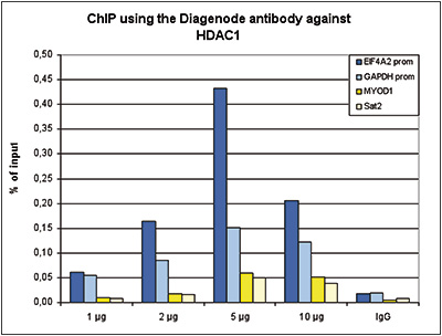
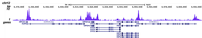
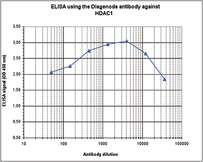
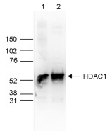
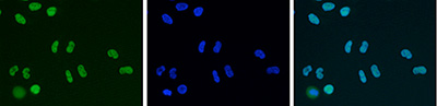
Diagenode

HDAC1 Antibody - replaced by the reference C15410325

Catalog Number
Format
Price
C15410053
(pAb-053-050)
50 µg


As the result of extensive validation, the antibody HDAC1 has been upgraded to Premium category.  Please, find it as HDAC1 polyclonal antibody - Premium (C15410325).

Alternative names: HD1, RPD3, RPD3L1, GON-10

Polyclonal antibody raised in rabbit against the C-terminal region of human HDAC1 (Histone deacetylase 1), using a KLH-conjugated synthetic peptide.

LotA21-001P
Concentration1.73 µg/µl
Species reactivityHuman, mouse
TypePolyclonal
PurityAffinity purified
HostRabbit
PrecautionsThis product is for research use only. Not for use in diagnostic or therapeutic procedures.
Applications Suggested dilution References
ChIP * 2 μg/ChIP Fig 1, 2
ELISA 1:4,000 Fig 3
Western Blotting 1:1,000 Fig 4
Immunofluorescence 1:500 Fig 5

* Please note that the optimal antibody amount per IP should be determined by the end-user. We recommend testing 1-5 μg per IP.

  • Validation Data

    HDAC1 Antibody ChIP Grade

    Figure 1. ChIP results obtained with the Diagenode antibody directed against HDAC1
    ChIP was performed with the Diagenode antibody against HDAC1 (Cat. No. C15410053) on sheared chromatin from 4,000,000 HeLa cells. An antibody titration consisting of 1, 2, 5 and 10 μg per ChIP experiment was analysed. IgG (2 μg/IP) was used as negative IP control. QPCR was performed with primers specific for the EIF4A2 and GAPDH promoters, used as positive controls, and for the MYOD1 gene and Sat2 satellite repeat, used as negative controls. Figure 1 shows the recovery, expressed as a % of input (the relative amount of immunoprecipitated DNA compared to input DNA after qPCR analysis).

    HDAC1 Antibody ChIP-seq Grade

    HDAC1 Antibody for ChIP-seq

    HDAC1 Antibody for ChIP-seq assay

    HDAC1 Antibody validated in ChIP-seq

    Figure 2. ChIP-seq results obtained with the Diagenode antibody directed against HDAC1
    ChIP was performed on sheared chromatin from 4,000,000 HeLa cells using 2 μg of the Diagenode antibody against HDAC1 (Cat. No. C15410053) as described above. The IP’d DNA was subsequently analysed on an Illumina HiSeq 2000. Library preparation, cluster generation and sequencing were performed according to the manufacturer’s instructions. The 50 bp tags were aligned to the human genome using the BWA algorithm. Figure 2 shows the peak distribution along the complete sequence and a 1 Mb region of the X-chromosome (figure 2A and B) and in two regions surrounding the GAPDH and EIF4A2 positive control genes, respectively (figure 2C and D).

    HDAC1 Antibody validated in ELISA

    Figure 3. Determination of the antibody titer
    To determine the titer of the antibody, an ELISA was performed using a serial dilution of Diagenode antibody directed against HDAC1 (Cat. No. pAb-053-050), crude serum and flow through. The plates were coated with the peptide used for immunization of the rabbit. By plotting the absorbance against the antibody dilution (Figure 2), the titer of the antibody was estimated to be 1:75,000.

    HDAC1 Antibody validated in Western Blot

    Figure 4. Western blot analysis using the Diagenode antibody directed against HDAC1
    Whole cell extracts (25 μg, lane 1) and nuclear extracts (25 μg, lane 2) from HeLa cells were analysed by Western blot using the Diagenode antibody against HDAC1 (Cat. No. pAb-053-050) diluted 1:1,000 in TBS-Tween containing 5% skimmed milk. The position of the protein of interest is indicated on the right (expected size: 55 kDa); the marker (in kDa) is shown on the left.

    HDAC1 Antibody validated in Immunofluorescence

    Figure 5. Immunofluorescence using the Diagenode antibody directed against HDAC1
    HeLa cells were stained with the Diagenode antibody against HDAC1 (Cat. No. C15410053) and with DAPI. Cells were fixed with 4% formaldehyde for 10’ and blocked with PBS/TX-100 containing 5% normal goat serum and 1% BSA. The cells were immunofluorescently labelled with the HDAC1 antibody (left) diluted 1:500 in blocking solution followed by an anti-rabbit antibody conjugated to Alexa488. The middle panel shows staining of the nuclei with DAPI. A merge of the two stainings is shown on the right.

  • Target Description

    HDAC1 (UniProt/Swiss-Prot entry Q13547) catalyses the deacetylation of lysine residues on the N-terminal part of the core histones (H2A, H2B, H3 and H4). Acetylation and deacetylation of these highly conserved lysine residues is important for the control of gene expression and HDAC activity is often associated with gene repression. Histone deacetylation is established by the formation of large multiprotein complexes. HDAC1 also interacts with the retinoblastoma tumor suppressor protein and is able to deacetylate p53. Therefore, it also plays an essential role in cell proliferation and differentiation and in apoptosis.

  •  Applications
    ELISA
    Enzyme-linked immunosorbent assay. Read more
    WB
    Western blot : The quality of antibodies used in this technique is crucial for correct and specific protein identification. Diagenode offers huge selection of highly sensitive and specific western blot-validated antibodies. Learn more about: Load... Read more
    IF
    Immunofluorescence: Diagenode offers huge selection of highly sensitive antibodies validated in IF. Immunofluorescence using the Diagenode monoclonal antibody directed against CRISPR/Cas9 HeLa cells transfected with a Cas9 expression vector (... Read more
    ChIP-seq (ab)
    Read more
    ChIP-qPCR (ab)
    Read more
  •  Documents
    Datasheet HDAC1 C15410053 DATASHEET
    Datasheet description
    Download
    Antibodies you can trust POSTER
    Epigenetic research tools have evolved over time from endpoint PCR to qPCR to the analyses of lar...
    Download
    Epigenetic Antibodies Brochure BROCHURE
    More than in any other immuoprecipitation assays, quality antibodies are critical tools in many e...
    Download
  •  Safety sheets
    HDAC1 Antibody SDS GB en Download
    HDAC1 Antibody SDS US en Download
    HDAC1 Antibody SDS BE nl Download
    HDAC1 Antibody SDS FR fr Download
    HDAC1 Antibody SDS BE fr Download
    HDAC1 Antibody SDS ES es Download
    HDAC1 Antibody SDS JP ja Download
    HDAC1 Antibody SDS DE de Download
  •  Publications

    How to properly cite this product in your work

    Diagenode strongly recommends using this: HDAC1 Antibody - replaced by the reference C15410325 (Diagenode Cat# C15410053 Lot# A21-001P). Click here to copy to clipboard.

    Using our products in your publication? Let us know!

    Comprehensive molecular phenotyping of -deficient gastric cancer revealspervasive epigenomic reprogramming and therapeutic opportunities.
    Xu C. et al.
    OBJECTIVE: Gastric cancer (GC) is a leading cause of cancer mortality, with being the second most frequently mutated driver gene in GC. We sought to decipher -specific GC regulatory networks and examine therapeutic vulnerabilities arising from loss. DESIGN: Genomic profiling of GC patients including a Singapore coho...

    Functional incompatibility between the generic NF-κB motif and a subtype-specific Sp1III element drives the formation of HIV-1 subtype C viral promoter
    Verma A et al.
    Of the various genetic subtypes of HIV-1, HIV-2 and SIV, only in subtype C of HIV-1, a genetically variant NF-κB binding site is found at the core of the viral promoter in association with a subtype-specific Sp1III motif. How the subtype-associated variations in the core transcription factor binding sites (TFB...

    Genome-wide hydroxymethylcytosine pattern changes in response to oxidative stress
    Delatte B, Jeschke J, Defrance M, Bachman M, Creppe C, Calonne E, Bizet M, Deplus R, Marroquí L, Libin M, Ravichandran M, Mascart F, Eizirik DL, Murrell A, Jurkowski TP, Fuks F
    The TET enzymes convert methylcytosine to the newly discovered base hydroxymethylcytosine. While recent reports suggest that TETs may play a role in response to oxidative stress, this role remains uncertain, and results lack in vivo models. Here we show a global decrease of hydroxymethylcytosine in cells treated w...

    SNAIL1 combines competitive displacement of ASCL2 and epigenetic mechanisms to rapidly silence the EPHB3 tumor suppressor in colorectal cancer.
    Rönsch K, Jägle S, Rose K, Seidl M, Baumgartner F, Freihen V, Yousaf A, Metzger E, Lassmann S, Schüle R, Zeiser R, Michoel T, Hecht A
    EPHB3 is a critical cellular guidance factor in the intestinal epithelium and an important tumor suppressor in colorectal cancer (CRC) whose expression is frequently lost at the adenoma-carcinoma transition when tumor cells become invasive. The molecular mechanisms underlying EPHB3 silencing are incompletely underst...

    Citrullination of DNMT3A by PADI4 regulates its stability and controls DNA methylation.
    Deplus R, Denis H, Putmans P, Calonne E, Fourrez M, Yamamoto K, Suzuki A, Fuks F
    DNA methylation is a central epigenetic modification in mammals, with essential roles in development and disease. De novo DNA methyltransferases establish DNA methylation patterns in specific regions within the genome by mechanisms that remain poorly understood. Here we show that protein citrullination by peptidylar...

    Dimethyl fumarate regulates histone deacetylase expression in astrocytes.
    Kalinin S, Polak PE, Lin SX, Braun D, Guizzetti M, Zhang X, Rubinstein I, Feinstein DL
    We previously showed that dimethyl fumarate (DMF) reduces inflammatory activation in astrocytes, involving activation of transcription factor Nrf2. However, the pathways causing Nrf2 activation were not examined. We now show that DMF modifies expression of histone deacetylases (HDACs) in primary rat astrocytes. Afte...

    Phosphorylation of p65(RelA) on Ser547 by ATM Represses NF-κB-Dependent Transcription of Specific Genes after Genotoxic Stress
    Sabatel H, Di Valentin E, Gloire G, Dequiedt F, Piette J, Habraken Y

    The histone demethylase Kdm3a is essential to progression through differentiation.
    Herzog M, Josseaux E, Dedeurwaerder S, Calonne E, Volkmar M, Fuks F
    Histone demethylation has important roles in regulating gene expression and forms part of the epigenetic memory system that regulates cell fate and identity by still poorly understood mechanisms. Here, we examined the role of histone demethylase Kdm3a during cell differentiation, showing that Kdm3a is essential for ...

    HDAC1 Regulates Fear Extinction in Mice.
    Bahari-Javan S, Maddalena A, Kerimoglu C, Wittnam J, Held T, Bähr M, Burkhardt S, Delalle I, Kügler S, Fischer A, Sananbenesi F
    Histone acetylation has been implicated with the pathogenesis of neuropsychiatric disorders and targeting histone deacetylases (HDACs) using HDAC inhibitors was shown to be neuroprotective and to initiate neuroregenerative processes. However, little is known about the role of individual HDAC proteins during the path...

    Enhancer of Zeste 2 (EZH2) is up-regulated in malignant gliomas and in glioma stem-like cells.
    Orzan F, Pellegatta S, Poliani PL, Pisati F, Caldera V, Menghi F, Kapetis D, Marras C, Schiffer D, Finocchiaro G
    AIMS: Proteins of the Polycomb repressive complex 2 (PRC2) are epigenetic gene silencers and are involved in tumour development. Their oncogenic function might be associated with their role in stem cell maintenance. The histone methyltransferase Enhancer of Zeste 2 (EZH2) is a key member of PRC2 function: we have in...

    The core binding factor CBF negatively regulates skeletal muscle terminal differentiation.
    Philipot O, Joliot V, Ait-Mohamed O, Pellentz C, Robin P, Fritsch L, Ait-Si-Ali S
    BACKGROUND: Core Binding Factor or CBF is a transcription factor composed of two subunits, Runx1/AML-1 and CBF beta or CBFbeta. CBF was originally described as a regulator of hematopoiesis. METHODOLOGY/PRINCIPAL FINDINGS: Here we show that CBF is involved in the control of skeletal muscle terminal differentiation. I...

    Functional connection between deimination and deacetylation of histones.
    Denis H, Deplus R, Putmans P, Yamada M, Métivier R, Fuks F
    Histone methylation plays key roles in regulating chromatin structure and function. The recent identification of enzymes that antagonize or remove histone methylation offers new opportunities to appreciate histone methylation plasticity in the regulation of epigenetic pathways. Peptidylarginine deiminase 4 (PADI4; a...

  •  Related products

Events

 See all events

News

 See all news


The European Regional Development Fund and Wallonia are investing in your future.

Extension of industrial buildings and new laboratories.


       Site map   |   Contact us   |   Conditions of sales   |   Conditions of purchase   |   Privacy policy   |   Diagenode Diagnostics