Diagenode

H3T6pK9me1 Antibody

Catalog Number
Format
Price
C15410283
50 μg
$480.00
  Bulk order


Polyclonal antibody raised in rabbit against Histone H3 (p Thr6, monomethyl Lys9), using a KLH-conjugated synthetic peptide.

Lot001
Concentration0.85 μg/μl
Species reactivityHuman, mouse, C. elegans, rat, chicken, Xenopus, Drosophila, plant
TypePolyclonal
PurityAffinity purified
HostRabbit
PrecautionsThis product is for research use only. Not for use in diagnostic or therapeutic procedures.
Applications Suggested dilution References
ChIP 2-5 μg/million cells Fig 1
Immunohistochemistry 1:200
Immunofluorescence 1:100 Fig 2
Western Blotting 1:500 - 1:1,000 Fig 3, 4, 5
Dot Blotting 1:1,000 Fig 6
  • Validation Data

    H3T6pK9me1 Antibody ChIP Grade

    Figure 1. H3T6pK9me1 antibody ChIP results
    Chromatin Immunoprecipitation of H3T6pK9me1 antibody. Chromatin from one million formaldehyde cross-linked Hela cells was used with 2ug of H3T6pK9me1 and 20ul of magnetic IgG beads per immunoprecipitation. A no antibody (No Ab) control was also used. Immunoprecipitated DNA was quantified using quantitative real-time PCR and normalized to the input chromatin.

    H3T6pK9me1 Antibody validated in Immunofluorescence

    Figure 2. H3T6pK9me1 antibody Immunofluorescence results
    Immunofluorescence of H3T6pK9me1 antibody. Tissue: HeLa cells. Fixation: 0.5% PFA. Primary antibody: incubated at a 1:100 dilution for 1 h at RT. Secondary antibody: FITC secondary antibody at 1:10,000 for 45 min at RT. Localization: H3T6pK9me1 is nuclear and chromosomal. Staining: H3T6pK9me1 is expressed in green and the nuclei are counterstained with DAPI (blue).

    H3T6pK9me1 Antibody validated in Western Blot

    Figure 3. H3T6pK9me1 antibody Western blot results
    Western Blot of H3T6pK9me1 antibody. 30 μg NIH-3T3 Histone extracts. Primary antibody diluted 1:1,000 overnight at 4°C. Secondary antibody: IRDye800TM rabbit secondary antibody at 1:10,000 for 45 min at RT. Predicted/Observed size: ~15 kDa. Other band(s): None.

    H3T6pK9me1 Antibody validated in Western Blot

    Figure 4. H3T6pK9me1 antibody Western blot results
    Western Blot of H3T6pK9me1 antibody. 30 μg C. elegans embryo lysate. Primary antibody diluted 1:1,000 overnight at 4°C. Secondary antibody: IRDye800TM rabbit secondary antibody at 1:10,000 for 45 min at RT. Predicted/Observed size: ~15 kDa. Other band(s): None.

    H3T6pK9me1 Antibody validated in Western Blot

    Figure 5. H3T6pK9me1 antibody Western blot results
    Western Blot of H3T6pK9me1 antibody. 30 μg HeLa Histone extracts. Primary antibody diluted 1:1,000 overnight at 4°C. Secondary antibody: IRDye800TM rabbit secondary antibody at 1:10,000 for 45 min at RT. Predicted/Observed size: ~15 kDa. Other band(s): None.

    H3T6pK9me1 Antibody validated in Dot Blot

    Figure 6. H3T6pK9me1 antibody Dot blot results
    Dot Blot of H3T6pK9me1 antibody. Lane 1: Histone H3 1-13. Lane 2: T6p. Lane 3: T6pK9Me1. Lane 4: T6pK9Me2. Lane 5: T6pK9Me3. Lane 6: Histone H3 2-17. Lane 7: K9Me1. Lane 8: K9Me2. Lane 9: K9Me3. Lane 10: K9Ac. Load: 1, 10, and 100 picomoles of peptide. Primary antibody diluted 1:1,000 for 45 min at 4°C. Secondary antibody: DylightTM488 rabbit secondary antibody at 1:10,000 for 45 min at RT.

  • Target Description

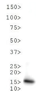
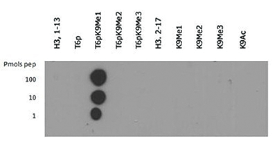
    Methylation of Histone H3 at Lys9 (K9) is an epigenetic silencer of transcription. Gene silencing from histone post translational modifications, as well as DNA methylation, play a key role in the development of normal tissues. If this silencing is disturbed through the artificial silencing of RIZ1, and thereby H3 K9Me1, it has been shown that normal apoptotic processes in precancerous cells can be reduced. Interestingly, data indicates that the conversion of the monomethyl to the trimethyl form requires mediation by SUVR4 in transposons and pseudogenes. Research also indicates that the presence of the G9a/GLP heterodimeric complex is required for this modification to exist. The additional phosphorylation at Thr6 (T6p) affects the ability of other proteins to bind to the H3 tail, along with amplifing the effects of other histone PTMs that are present. Because T6 phosphorylation is constitutive, its dephosphorylation may play a key role in DNA transcription, repair and replication.

  •  Applications
    IHC
    Immunohistochemistry Read more
    IF
    Immunofluorescence: Diagenode offers huge selection of highly sensitive antibodies validated in IF. Immunofluorescence using the Diagenode monoclonal antibody directed against CRISPR/Cas9 HeLa cells transfected with a Cas9 expression vector (... Read more
    WB
    Western blot : The quality of antibodies used in this technique is crucial for correct and specific protein identification. Diagenode offers huge selection of highly sensitive and specific western blot-validated antibodies. Learn more about: Load... Read more
    DB
    Dot blotting Read more
    ChIP-qPCR (ab)
    Read more
  •  Documents
    Datasheet H3T6pK9me1 C15410283 DATASHEET
    Datasheet description
    Download
  •  Publications

    How to properly cite this product in your work

    Diagenode strongly recommends using this: H3T6pK9me1 Antibody (Diagenode Cat# C15410283 Lot# 001). Click here to copy to clipboard.

    Using our products in your publication? Let us know!

  •  Related products

Events

  • APHL 2024
    Milwaukee, Wisconsin, USA
    May 6-May 9, 2024
 See all events

News

 See all news


The European Regional Development Fund and Wallonia are investing in your future.

Extension of industrial buildings and new laboratories.


       Site map   |   Contact us   |   Conditions of sales   |   Conditions of purchase   |   Privacy policy   |   Diagenode Diagnostics