Notice (8): Undefined variable: solution_of_interest [APP/View/Products/view.ctp, line 755]Code Context<!-- BEGIN: REQUEST_FORM MODAL -->
<div id="request_formModal" class="reveal-modal medium" data-reveal aria-labelledby="modalTitle" aria-hidden="true" role="dialog">
<?= $this->element('Forms/simple_form', array('solution_of_interest' => $solution_of_interest, 'header' => $header, 'message' => $message, 'campaign_id' => $campaign_id)) ?>
$viewFile = '/home/website-server/www/app/View/Products/view.ctp'
$dataForView = array(
'language' => 'cn',
'meta_keywords' => '',
'meta_description' => 'H3pan (Histone H3) Polyclonal Antibody validated in ChIP-qPCR, ELISA and WB. Batch-specific data available on the website. Sample size available.',
'meta_title' => 'H3pan Polyclonal Antibody | Diagenode',
'product' => array(
'Product' => array(
'id' => '3006',
'antibody_id' => '237',
'name' => 'H3pan polyclonal antibody (sample size)',
'description' => '',
'label1' => 'Validation data',
'info1' => '',
'label2' => '',
'info2' => '',
'label3' => '',
'info3' => '',
'format' => '20 µl',
'catalog_number' => 'C15310135-20',
'old_catalog_number' => '',
'sf_code' => 'C15310135-361',
'type' => 'FRE',
'search_order' => '03-Antibody',
'price_EUR' => '105',
'price_USD' => '115',
'price_GBP' => '100',
'price_JPY' => '16450',
'price_CNY' => '0',
'price_AUD' => '288',
'country' => 'ALL',
'except_countries' => 'None',
'quote' => false,
'in_stock' => false,
'featured' => false,
'no_promo' => false,
'online' => true,
'master' => false,
'last_datasheet_update' => '',
'slug' => 'h3pan-polyclonal-antibody-classic-20-ul',
'meta_title' => 'H3pan Polyclonal Antibody | Diagenode',
'meta_keywords' => '',
'meta_description' => 'H3pan (Histone H3) Polyclonal Antibody validated in ChIP-qPCR, ELISA and WB. Batch-specific data available on the website. Sample size available.',
'modified' => '2021-12-23 11:39:45',
'created' => '2018-11-27 10:50:23',
'locale' => 'zho'
),
'Antibody' => array(
'host' => '*****',
'id' => '237',
'name' => 'H3pan polyclonal antibody',
'description' => 'Histones are the main constituents of the protein part of chromosomes of eukaryotic cells. They are rich in the amino acids arginine and lysine and have been greatly conserved during evolution. Histones pack the DNA into tight masses of chromatin. Two core histones of each class H2A, H2B, H3 and H4 assemble and are wrapped by 146 base pairs of DNA to form one octameric nucleosome. Histones play a central role in the regulation of transcription, DNA repair, DNA replication and chromosomal stability. These different functions are established via a complex set of post-translational modifications which either directly or indirectly alter chromatin structure and DNA accessibility to facilitate transcriptional activation or repression or other nuclear processes.',
'clonality' => '',
'isotype' => '',
'lot' => 'A2566-001',
'concentration' => 'Not determined',
'reactivity' => 'Human, zebrafish, Daphnia: positive. Other species: not tested.',
'type' => 'Polyclonal',
'purity' => 'Whole antiserum',
'classification' => 'Classic',
'application_table' => '<table>
<thead>
<tr>
<th>Applications</th>
<th>Suggested dilution</th>
<th>References</th>
</tr>
</thead>
<tbody>
<tr>
<td>ChIP</td>
<td>1 μl/ChIP</td>
<td>Fig 1</td>
</tr>
<tr>
<td>ELISA</td>
<td>1:10,000</td>
<td>Fig 2</td>
</tr>
<tr>
<td>Western Blotting</td>
<td>1:500</td>
<td>Fig 3</td>
</tr>
</tbody>
</table>
<p><small><sup>*</sup> Please note that of the optimal antibody amount per IP should be determined by the end-user. We recommend testing 1-5 μg per IP.</small></p>',
'storage_conditions' => '',
'storage_buffer' => '',
'precautions' => 'This product is for research use only. Not for use in diagnostic or therapeutic procedures.',
'uniprot_acc' => '',
'slug' => '',
'meta_keywords' => '',
'meta_description' => '',
'modified' => '2020-10-02 12:09:11',
'created' => '0000-00-00 00:00:00',
'select_label' => '237 - H3pan polyclonal antibody (A2566-001 - Not determined - Human, zebrafish, Daphnia: positive. Other species: not tested. - Whole antiserum - Rabbit)'
),
'Slave' => array(),
'Group' => array(
'Group' => array(
[maximum depth reached]
),
'Master' => array(
[maximum depth reached]
),
'Product' => array(
[maximum depth reached]
)
),
'Related' => array(),
'Application' => array(
(int) 0 => array(
[maximum depth reached]
),
(int) 1 => array(
[maximum depth reached]
),
(int) 2 => array(
[maximum depth reached]
)
),
'Category' => array(
(int) 0 => array(
[maximum depth reached]
),
(int) 1 => array(
[maximum depth reached]
),
(int) 2 => array(
[maximum depth reached]
)
),
'Document' => array(
(int) 0 => array(
[maximum depth reached]
),
(int) 1 => array(
[maximum depth reached]
),
(int) 2 => array(
[maximum depth reached]
)
),
'Feature' => array(),
'Image' => array(
(int) 0 => array(
[maximum depth reached]
)
),
'Promotion' => array(),
'Protocol' => array(),
'Publication' => array(
(int) 0 => array(
[maximum depth reached]
),
(int) 1 => array(
[maximum depth reached]
),
(int) 2 => array(
[maximum depth reached]
),
(int) 3 => array(
[maximum depth reached]
),
(int) 4 => array(
[maximum depth reached]
),
(int) 5 => array(
[maximum depth reached]
),
(int) 6 => array(
[maximum depth reached]
),
(int) 7 => array(
[maximum depth reached]
),
(int) 8 => array(
[maximum depth reached]
),
(int) 9 => array(
[maximum depth reached]
),
(int) 10 => array(
[maximum depth reached]
),
(int) 11 => array(
[maximum depth reached]
),
(int) 12 => array(
[maximum depth reached]
),
(int) 13 => array(
[maximum depth reached]
),
(int) 14 => array(
[maximum depth reached]
),
(int) 15 => array(
[maximum depth reached]
),
(int) 16 => array(
[maximum depth reached]
),
(int) 17 => array(
[maximum depth reached]
)
),
'Testimonial' => array(),
'Area' => array(),
'SafetySheet' => array(
(int) 0 => array(
[maximum depth reached]
),
(int) 1 => array(
[maximum depth reached]
),
(int) 2 => array(
[maximum depth reached]
),
(int) 3 => array(
[maximum depth reached]
),
(int) 4 => array(
[maximum depth reached]
),
(int) 5 => array(
[maximum depth reached]
),
(int) 6 => array(
[maximum depth reached]
),
(int) 7 => array(
[maximum depth reached]
)
)
),
'meta_canonical' => 'https://www.diagenode.com/cn/p/h3pan-polyclonal-antibody-classic-100-ul'
)
$language = 'cn'
$meta_keywords = ''
$meta_description = 'H3pan (Histone H3) Polyclonal Antibody validated in ChIP-qPCR, ELISA and WB. Batch-specific data available on the website. Sample size available.'
$meta_title = 'H3pan Polyclonal Antibody | Diagenode'
$product = array(
'Product' => array(
'id' => '3006',
'antibody_id' => '237',
'name' => 'H3pan polyclonal antibody (sample size)',
'description' => '<p><span>This antibody has been raised in rabbit against two KLH-conjugated synthetic peptides containing an unmodified sequence from the central part and from the C-terminus of <strong>histone H3</strong>, respectively.</span></p>',
'label1' => 'Validation data',
'info1' => '<div class="row">
<div class="small-4 columns">
<p><img src="https://www.diagenode.com/img/product/antibodies/C15310135-chip.jpg" alt="H3pan Antibody ChIP Grade" style="display: block; margin-left: auto; margin-right: auto;" /></p>
</div>
<div class="small-8 columns">
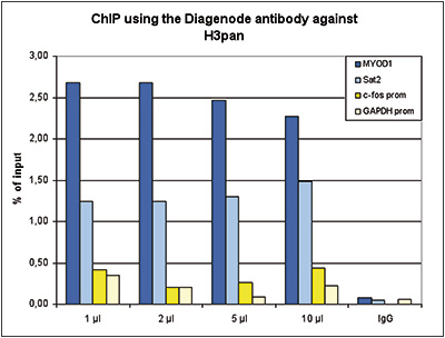
<p><small><strong>Figure 1. ChIP results obtained with the Diagenode antibody directed against H3pan</strong><br />ChIP assays were performed using human HeLa cells, the Diagenode antibody against H3pan (Cat. No. C15310135) and optimized PCR primer sets for qPCR. ChIP was performed with the Auto Histone ChIP-seq kit (Cat. No. C01010022), using sheared chromatin from 1 million cells. A titration of the antibody consisting of 1, 2, 5, and 10 μl per ChIP experiment was analysed. IgG (2 μg/IP) was used as negative IP control. QPCR was performed with primers for the promoters of the active GAPDH and EIF4A2 genes, used as negative controls, and for the inactive MYOD1 and the Sat2 satellite repeat, used as positive controls. Figure 1 shows the recovery, expressed as a % of input (the relative amount of immunoprecipitated DNA compared to input DNA after qPCR analysis).</small></p>
</div>
</div>
<div class="row">
<div class="small-4 columns">
<p><img src="https://www.diagenode.com/img/product/antibodies/C15310135-elisa.jpg" alt="H3pan Antibody ELISA validation" caption="false" width="288" height="217" /></p>
</div>
<div class="small-8 columns">
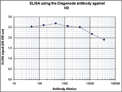
<p><small><strong>Figure 2. Determination of the antibody titer</strong><br />To determine the titer of the antibody, an ELISA was performed using a serial dilution of the Diagenode antibody directed against H3pan (Cat. No. C15310135). The plates were coated with the peptides used for immunization. By plotting the absorbance against the antibody dilution (Figure 2), the titer of the antibody was estimated to be >1:1,000,000.</small></p>
</div>
</div>
<div class="row">
<div class="small-4 columns">
<p><img src="https://www.diagenode.com/img/product/antibodies/C15310135-wb.jpg" alt="H3pan Antibody validated in Western Blot" style="display: block; margin-left: auto; margin-right: auto;" /></p>
</div>
<div class="small-8 columns">
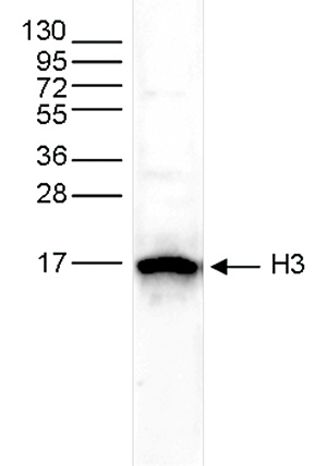
<p><small><strong>Figure 3. Western blot analysis using the Diagenode antibody directed against H3pan</strong><br />Whole cell extracts from HeLa cells (25 μg) were analysed by Western blot using the Diagenode antibody against H3pan (Cat. No. C15310135) diluted 1:500 in TBS-Tween containing 5% skimmed milk. The position of the protein of interest is indicated on the right; the marker (in kDa) is shown on the left.</small></p>
</div>
</div>',
'label2' => 'Target Description',
'info2' => '<p>Histones are the main constituents of the protein part of chromosomes of eukaryotic cells. They are rich in the amino acids arginine and lysine and have been greatly conserved during evolution. Histones pack the DNA into tight masses of chromatin. Two core histones of each class H2A, H2B, H3 and H4 assemble and are wrapped by 146 base pairs of DNA to form one octameric nucleosome. Histones play a central role in the regulation of transcription, DNA repair, DNA replication and chromosomal stability. These different functions are established via a complex set of post-translational modifications which either directly or indirectly alter chromatin structure and DNA accessibility to facilitate transcriptional activation or repression or other nuclear processes.</p>',
'label3' => '',
'info3' => '',
'format' => '20 µl',
'catalog_number' => 'C15310135-20',
'old_catalog_number' => '',
'sf_code' => 'C15310135-361',
'type' => 'FRE',
'search_order' => '03-Antibody',
'price_EUR' => '105',
'price_USD' => '115',
'price_GBP' => '100',
'price_JPY' => '16450',
'price_CNY' => '0',
'price_AUD' => '288',
'country' => 'ALL',
'except_countries' => 'None',
'quote' => false,
'in_stock' => false,
'featured' => false,
'no_promo' => false,
'online' => true,
'master' => false,
'last_datasheet_update' => '',
'slug' => 'h3pan-polyclonal-antibody-classic-20-ul',
'meta_title' => 'H3pan Polyclonal Antibody | Diagenode',
'meta_keywords' => '',
'meta_description' => 'H3pan (Histone H3) Polyclonal Antibody validated in ChIP-qPCR, ELISA and WB. Batch-specific data available on the website. Sample size available.',
'modified' => '2021-12-23 11:39:45',
'created' => '2018-11-27 10:50:23',
'locale' => 'zho'
),
'Antibody' => array(
'host' => '*****',
'id' => '237',
'name' => 'H3pan polyclonal antibody',
'description' => 'Histones are the main constituents of the protein part of chromosomes of eukaryotic cells. They are rich in the amino acids arginine and lysine and have been greatly conserved during evolution. Histones pack the DNA into tight masses of chromatin. Two core histones of each class H2A, H2B, H3 and H4 assemble and are wrapped by 146 base pairs of DNA to form one octameric nucleosome. Histones play a central role in the regulation of transcription, DNA repair, DNA replication and chromosomal stability. These different functions are established via a complex set of post-translational modifications which either directly or indirectly alter chromatin structure and DNA accessibility to facilitate transcriptional activation or repression or other nuclear processes.',
'clonality' => '',
'isotype' => '',
'lot' => 'A2566-001',
'concentration' => 'Not determined',
'reactivity' => 'Human, zebrafish, Daphnia: positive. Other species: not tested.',
'type' => 'Polyclonal',
'purity' => 'Whole antiserum',
'classification' => 'Classic',
'application_table' => '<table>
<thead>
<tr>
<th>Applications</th>
<th>Suggested dilution</th>
<th>References</th>
</tr>
</thead>
<tbody>
<tr>
<td>ChIP</td>
<td>1 μl/ChIP</td>
<td>Fig 1</td>
</tr>
<tr>
<td>ELISA</td>
<td>1:10,000</td>
<td>Fig 2</td>
</tr>
<tr>
<td>Western Blotting</td>
<td>1:500</td>
<td>Fig 3</td>
</tr>
</tbody>
</table>
<p><small><sup>*</sup> Please note that of the optimal antibody amount per IP should be determined by the end-user. We recommend testing 1-5 μg per IP.</small></p>',
'storage_conditions' => '',
'storage_buffer' => '',
'precautions' => 'This product is for research use only. Not for use in diagnostic or therapeutic procedures.',
'uniprot_acc' => '',
'slug' => '',
'meta_keywords' => '',
'meta_description' => '',
'modified' => '2020-10-02 12:09:11',
'created' => '0000-00-00 00:00:00',
'select_label' => '237 - H3pan polyclonal antibody (A2566-001 - Not determined - Human, zebrafish, Daphnia: positive. Other species: not tested. - Whole antiserum - Rabbit)'
),
'Slave' => array(),
'Group' => array(
'Group' => array(
'id' => '252',
'name' => 'C15310135',
'product_id' => '2122',
'modified' => '2018-11-27 10:50:45',
'created' => '2018-11-27 10:50:45'
),
'Master' => array(
'id' => '2122',
'antibody_id' => '237',
'name' => 'H3pan Antibody',
'description' => '<p><span>This antibody has been raised in rabbit against two KLH-conjugated synthetic peptides containing an unmodified sequence from the central part and from the C-terminus of <strong>histone H3</strong>, respectively.</span></p>',
'label1' => 'Validation Data',
'info1' => '<div class="row">
<div class="small-4 columns">
<p><img src="https://www.diagenode.com/img/product/antibodies/C15310135-chip.jpg" alt="H3pan Antibody ChIP Grade" style="display: block; margin-left: auto; margin-right: auto;" /></p>
</div>
<div class="small-8 columns">
<p><small><strong>Figure 1. ChIP results obtained with the Diagenode antibody directed against H3pan</strong><br />ChIP assays were performed using human HeLa cells, the Diagenode antibody against H3pan (Cat. No. C15310135) and optimized PCR primer sets for qPCR. ChIP was performed with the Auto Histone ChIP-seq kit (Cat. No. C01010022), using sheared chromatin from 1 million cells. A titration of the antibody consisting of 1, 2, 5, and 10 μl per ChIP experiment was analysed. IgG (2 μg/IP) was used as negative IP control. QPCR was performed with primers for the promoters of the active GAPDH and EIF4A2 genes, used as negative controls, and for the inactive MYOD1 and the Sat2 satellite repeat, used as positive controls. Figure 1 shows the recovery, expressed as a % of input (the relative amount of immunoprecipitated DNA compared to input DNA after qPCR analysis).</small></p>
</div>
</div>
<div class="row">
<div class="small-4 columns">
<p><img src="https://www.diagenode.com/img/product/antibodies/C15310135-elisa.jpg" alt="H3pan Antibody ELISA validation" caption="false" width="288" height="217" /></p>
</div>
<div class="small-8 columns">
<p><small><strong>Figure 2. Determination of the antibody titer</strong><br />To determine the titer of the antibody, an ELISA was performed using a serial dilution of the Diagenode antibody directed against H3pan (Cat. No. C15310135). The plates were coated with the peptides used for immunization. By plotting the absorbance against the antibody dilution (Figure 2), the titer of the antibody was estimated to be >1:1,000,000.</small></p>
</div>
</div>
<div class="row">
<div class="small-4 columns">
<p><img src="https://www.diagenode.com/img/product/antibodies/C15310135-wb.jpg" alt="H3pan Antibody validated in Western Blot" style="display: block; margin-left: auto; margin-right: auto;" /></p>
</div>
<div class="small-8 columns">
<p><small><strong>Figure 3. Western blot analysis using the Diagenode antibody directed against H3pan</strong><br />Whole cell extracts from HeLa cells (25 μg) were analysed by Western blot using the Diagenode antibody against H3pan (Cat. No. C15310135) diluted 1:500 in TBS-Tween containing 5% skimmed milk. The position of the protein of interest is indicated on the right; the marker (in kDa) is shown on the left.</small></p>
</div>
</div>',
'label2' => 'Target Description',
'info2' => '<p>Histones are the main constituents of the protein part of chromosomes of eukaryotic cells. They are rich in the amino acids arginine and lysine and have been greatly conserved during evolution. Histones pack the DNA into tight masses of chromatin. Two core histones of each class H2A, H2B, H3 and H4 assemble and are wrapped by 146 base pairs of DNA to form one octameric nucleosome. Histones play a central role in the regulation of transcription, DNA repair, DNA replication and chromosomal stability. These different functions are established via a complex set of post-translational modifications which either directly or indirectly alter chromatin structure and DNA accessibility to facilitate transcriptional activation or repression or other nuclear processes.</p>',
'label3' => '',
'info3' => '',
'format' => '100 µl',
'catalog_number' => 'C15310135',
'old_catalog_number' => 'CS-135-100',
'sf_code' => 'C15310135-D001-001161',
'type' => 'FRE',
'search_order' => '03-Antibody',
'price_EUR' => '380',
'price_USD' => '380',
'price_GBP' => '340',
'price_JPY' => '59525',
'price_CNY' => '',
'price_AUD' => '950',
'country' => 'ALL',
'except_countries' => 'None',
'quote' => false,
'in_stock' => false,
'featured' => false,
'no_promo' => false,
'online' => true,
'master' => true,
'last_datasheet_update' => '0000-00-00',
'slug' => 'h3pan-polyclonal-antibody-classic-100-ul',
'meta_title' => 'H3pan Antibody - ChIP Grade (C15310135) | Diagenode',
'meta_keywords' => '',
'meta_description' => 'H3pan (Histone H3) Polyclonal Antibody validated in ChIP-qPCR, WB and ELISA. Batch-specific data available on the website. Sample size available.',
'modified' => '2021-12-23 11:39:34',
'created' => '2015-06-29 14:08:20'
),
'Product' => array(
(int) 0 => array(
[maximum depth reached]
)
)
),
'Related' => array(),
'Application' => array(
(int) 0 => array(
'id' => '19',
'position' => '10',
'parent_id' => '40',
'name' => 'WB',
'description' => '<p><strong>Western blot</strong> : The quality of antibodies used in this technique is crucial for correct and specific protein identification. Diagenode offers huge selection of highly sensitive and specific western blot-validated antibodies.</p>
<p>Learn more about: <a href="https://www.diagenode.com/applications/western-blot">Loading control, MW marker visualization</a><em>. <br /></em></p>
<p><em></em>Check our selection of antibodies validated in Western blot.</p>',
'in_footer' => false,
'in_menu' => false,
'online' => true,
'tabular' => true,
'slug' => 'western-blot-antibodies',
'meta_keywords' => ' Western Blot Antibodies ,western blot protocol,Western Blotting Products,Polyclonal antibodies ,monoclonal antibodies ',
'meta_description' => 'Diagenode offers a wide range of antibodies and technical support for western blot applications',
'meta_title' => ' Western Blot - Monoclonal antibody - Polyclonal antibody | Diagenode',
'modified' => '2016-04-26 12:44:51',
'created' => '2015-01-07 09:20:00',
'ProductsApplication' => array(
[maximum depth reached]
)
),
(int) 1 => array(
'id' => '20',
'position' => '10',
'parent_id' => '40',
'name' => 'ELISA',
'description' => '<div class="row">
<div class="small-12 medium-12 large-12 columns">Enzyme-linked immunosorbent assay.</div>
</div>',
'in_footer' => false,
'in_menu' => false,
'online' => true,
'tabular' => true,
'slug' => 'elisa-antibodies',
'meta_keywords' => ' ELISA Antibodies,Monoclonal antibody, Polyclonal antibody',
'meta_description' => 'Diagenode offers Monoclonal & Polyclonal antibodies for ELISA applications',
'meta_title' => 'ELISA Antibodies - Monoclonal & Polyclonal antibody | Diagenode',
'modified' => '2016-01-13 12:21:41',
'created' => '2014-07-08 08:13:28',
'ProductsApplication' => array(
[maximum depth reached]
)
),
(int) 2 => array(
'id' => '10',
'position' => '10',
'parent_id' => '2',
'name' => 'ChIP-qPCR',
'description' => '<div class="row">
<div class="small-12 medium-12 large-12 columns text-justify">
<p class="text-justify">Chromatin Immunoprecipitation (ChIP) coupled with quantitative PCR can be used to investigate protein-DNA interaction at known genomic binding sites. if sites are not known, qPCR primers can also be designed against potential regulatory regions such as promoters. ChIP-qPCR is advantageous in studies that focus on specific genes and potential regulatory regions across differing experimental conditions as the cost of performing real-time PCR is minimal. This technique is now used in a variety of life science disciplines including cellular differentiation, tumor suppressor gene silencing, and the effect of histone modifications on gene expression.</p>
<p class="text-justify"><strong>The ChIP-qPCR workflow</strong></p>
</div>
<div class="small-12 medium-12 large-12 columns text-center"><br /> <img src="https://www.diagenode.com/img/chip-qpcr-diagram.png" /></div>
<div class="small-12 medium-12 large-12 columns"><br />
<ol>
<li class="large-12 columns"><strong>Chromatin preparation: </strong>cell fixation (cross-linking) of chromatin-bound proteins such as histones or transcription factors to DNA followed by cell lysis.</li>
<li class="large-12 columns"><strong>Chromatin shearing: </strong>fragmentation of chromatin<strong> </strong>by sonication down to desired fragment size (100-500 bp)</li>
<li class="large-12 columns"><strong>Chromatin IP</strong>: protein-DNA complexe capture using<strong> <a href="https://www.diagenode.com/en/categories/chip-grade-antibodies">specific ChIP-grade antibodies</a></strong> against the histone or transcription factor of interest</li>
<li class="large-12 columns"><strong>DNA purification</strong>: chromatin reverse cross-linking and elution followed by purification<strong> </strong></li>
<li class="large-12 columns"><strong>qPCR and analysis</strong>: using previously designed primers to amplify IP'd material at specific loci</li>
</ol>
</div>
</div>
<div class="row" style="margin-top: 32px;">
<div class="small-12 medium-10 large-9 small-centered columns">
<div class="radius panel" style="background-color: #fff;">
<h3 class="text-center" style="color: #b21329;">Need guidance?</h3>
<p class="text-justify">Choose our full ChIP kits or simply choose what you need from antibodies, buffers, beads, chromatin shearing and purification reagents. With the ChIP Kit Customizer, you have complete flexibility on which components you want from our validated ChIP kits.</p>
<div class="row">
<div class="small-6 medium-6 large-6 columns"><a href="https://www.diagenode.com/pages/which-kit-to-choose"><img src="https://www.diagenode.com/img/banners/banner-decide.png" alt="" /></a></div>
<div class="small-6 medium-6 large-6 columns"><a href="https://www.diagenode.com/pages/chip-kit-customizer-1"><img src="https://www.diagenode.com/img/banners/banner-customizer.png" alt="" /></a></div>
</div>
</div>
</div>
</div>',
'in_footer' => false,
'in_menu' => true,
'online' => true,
'tabular' => true,
'slug' => 'chip-qpcr',
'meta_keywords' => 'Chromatin immunoprecipitation,ChIP Quantitative PCR,polymerase chain reaction (PCR)',
'meta_description' => 'Diagenode's ChIP qPCR kits can be used to quantify enriched DNA after chromatin immunoprecipitation. ChIP-qPCR is advantageous in studies that focus on specific genes and potential regulatory regions across differing experimental conditions as the cost of',
'meta_title' => 'ChIP Quantitative PCR (ChIP-qPCR) | Diagenode',
'modified' => '2018-01-09 16:46:56',
'created' => '2014-12-11 00:22:08',
'ProductsApplication' => array(
[maximum depth reached]
)
)
),
'Category' => array(
(int) 0 => array(
'id' => '103',
'position' => '0',
'parent_id' => '4',
'name' => 'All antibodies',
'description' => '<p><span style="font-weight: 400;">All Diagenode’s antibodies are listed below. Please, use our Quick search field to find the antibody of interest by target name, application, purity.</span></p>
<p><span style="font-weight: 400;">Diagenode’s highly validated antibodies:</span></p>
<ul>
<li>Highly sensitive and specific</li>
<li>Cost-effective (requires less antibody per reaction)</li>
<li>Batch-specific data is available on the website</li>
<li>Expert technical support</li>
<li>Sample sizes available</li>
<li>100% satisfaction guarantee</li>
</ul>',
'no_promo' => false,
'in_menu' => true,
'online' => true,
'tabular' => false,
'hide' => true,
'all_format' => false,
'is_antibody' => true,
'slug' => 'all-antibodies',
'cookies_tag_id' => null,
'meta_keywords' => 'Antibodies,Premium Antibodies,Classic,Pioneer',
'meta_description' => 'Diagenode Offers Strict quality standards with Rigorous QC and validated Antibodies. Classified based on level of validation for flexibility of Application. Comprehensive selection of histone and non-histone Antibodies',
'meta_title' => 'Diagenode's selection of Antibodies is exclusively dedicated for Epigenetic Research | Diagenode',
'modified' => '2019-07-03 10:55:44',
'created' => '2015-11-02 14:49:22',
'ProductsCategory' => array(
[maximum depth reached]
),
'CookiesTag' => array([maximum depth reached])
),
(int) 1 => array(
'id' => '111',
'position' => '40',
'parent_id' => '4',
'name' => 'Histone antibodies',
'description' => '<p>Histones are the main protein components of chromatin involved in the compaction of DNA into nucleosomes, the basic units of chromatin. A <strong>nucleosome</strong> consists of one pair of each of the core histones (<strong>H2A</strong>, <strong>H2B</strong>, <strong>H3</strong> and <strong>H4</strong>) forming an octameric structure wrapped by 146 base pairs of DNA. The different nucleosomes are linked by the linker histone<strong> H1, </strong>allowing for further condensation of chromatin.</p>
<p>The core histones have a globular structure with large unstructured N-terminal tails protruding from the nucleosome. They can undergo to multiple post-translational modifications (PTM), mainly at the N-terminal tails. These <strong>post-translational modifications </strong>include methylation, acetylation, phosphorylation, ubiquitinylation, citrullination, sumoylation, deamination and crotonylation. The most well characterized PTMs are <strong>methylation,</strong> <strong>acetylation and phosphorylation</strong>. Histone methylation occurs mainly on lysine (K) residues, which can be mono-, di- or tri-methylated, and on arginines (R), which can be mono-methylated and symmetrically or asymmetrically di-methylated. Histone acetylation occurs on lysines and histone phosphorylation mainly on serines (S), threonines (T) and tyrosines (Y).</p>
<p>The PTMs of the different residues are involved in numerous processes such as DNA repair, DNA replication and chromosome condensation. They influence the chromatin organization and can be positively or negatively associated with gene expression. Trimethylation of H3K4, H3K36 and H3K79, and lysine acetylation generally result in an open chromatin configuration (figure below) and are therefore associated with <strong>euchromatin</strong> and gene activation. Trimethylation of H3K9, K3K27 and H4K20, on the other hand, is enriched in <strong>heterochromatin </strong>and associated with gene silencing. The combination of different histone modifications is called the "<strong>histone code</strong>”, analogous to the genetic code.</p>
<p><img src="https://www.diagenode.com/img/categories/antibodies/histone-marks-illustration.png" /></p>
<p>Diagenode is proud to offer a large range of antibodies against histones and histone modifications. Our antibodies are highly specific and have been validated in many applications, including <strong>ChIP</strong> and <strong>ChIP-seq</strong>.</p>
<p>Diagenode’s collection includes antibodies recognizing:</p>
<ul>
<li><strong>Histone H1 variants</strong></li>
<li><strong>Histone H2A, H2A variants and histone H2A</strong> <strong>modifications</strong> (serine phosphorylation, lysine acetylation, lysine ubiquitinylation)</li>
<li><strong>Histone H2B and H2B</strong> <strong>modifications </strong>(serine phosphorylation, lysine acetylation)</li>
<li><strong>Histone H3 and H3 modifications </strong>(lysine methylation (mono-, di- and tri-methylated), lysine acetylation, serine phosphorylation, threonine phosphorylation, arginine methylation (mono-methylated, symmetrically and asymmetrically di-methylated))</li>
<li><strong>Histone H4 and H4 modifications (</strong>lysine methylation (mono-, di- and tri-methylated), lysine acetylation, arginine methylation (mono-methylated and symmetrically di-methylated), serine phosphorylation )</li>
</ul>
<p><span style="font-weight: 400;"><strong>HDAC's HAT's, HMT's and other</strong> <strong>enzymes</strong> which modify histones can be found in the category <a href="../categories/chromatin-modifying-proteins-histone-transferase">Histone modifying enzymes</a><br /></span></p>
<p><span style="font-weight: 400;"> Diagenode’s highly validated antibodies:</span></p>
<ul>
<li><span style="font-weight: 400;"> Highly sensitive and specific</span></li>
<li><span style="font-weight: 400;"> Cost-effective (requires less antibody per reaction)</span></li>
<li><span style="font-weight: 400;"> Batch-specific data is available on the website</span></li>
<li><span style="font-weight: 400;"> Expert technical support</span></li>
<li><span style="font-weight: 400;"> Sample sizes available</span></li>
<li><span style="font-weight: 400;"> 100% satisfaction guarantee</span></li>
</ul>',
'no_promo' => false,
'in_menu' => false,
'online' => true,
'tabular' => false,
'hide' => true,
'all_format' => false,
'is_antibody' => true,
'slug' => 'histone-antibodies',
'cookies_tag_id' => null,
'meta_keywords' => 'Histone, antibody, histone h1, histone h2, histone h3, histone h4',
'meta_description' => 'Polyclonal and Monoclonal Antibodies against Histones and their modifications validated for many applications, including Chromatin Immunoprecipitation (ChIP) and ChIP-Sequencing (ChIP-seq)',
'meta_title' => 'Histone and Modified Histone Antibodies | Diagenode',
'modified' => '2020-09-17 13:34:56',
'created' => '2016-04-01 16:01:32',
'ProductsCategory' => array(
[maximum depth reached]
),
'CookiesTag' => array([maximum depth reached])
),
(int) 2 => array(
'id' => '127',
'position' => '10',
'parent_id' => '4',
'name' => 'ChIP-grade antibodies',
'description' => '<div class="row">
<div class="small-12 columns"><center></center>
<p><br />Chromatin immunoprecipitation (<b>ChIP</b>) is a technique to study the associations of proteins with the specific genomic regions in intact cells. One of the most important steps of this protocol is the immunoprecipitation of targeted protein using the antibody specifically recognizing it. The quality of antibodies used in ChIP is essential for the success of the experiment. Diagenode offers extensively validated ChIP-grade antibodies, confirmed for their specificity, and high level of performance in ChIP. Each batch is validated, and batch-specific data are available on the website.</p>
<p></p>
</div>
</div>
<p><strong>ChIP results</strong> obtained with the antibody directed against H3K4me3 (Cat. No. <a href="../p/h3k4me3-polyclonal-antibody-premium-50-ug-50-ul">C15410003</a>). </p>
<div class="row">
<div class="small-12 medium-6 large-6 columns"><img src="https://www.diagenode.com/img/product/antibodies/C15410003-fig1-ChIP.jpg" alt="" width="400" height="315" /> </div>
<div class="small-12 medium-6 large-6 columns">
<p></p>
<p></p>
<p></p>
</div>
</div>
<p></p>
<p>Our aim at Diagenode is to offer the largest collection of highly specific <strong>ChIP-grade antibodies</strong>. We add new antibodies monthly. Find your ChIP-grade antibody in the list below and check more information about tested applications, extensive validation data, and product information.</p>',
'no_promo' => false,
'in_menu' => true,
'online' => true,
'tabular' => false,
'hide' => true,
'all_format' => false,
'is_antibody' => true,
'slug' => 'chip-grade-antibodies',
'cookies_tag_id' => null,
'meta_keywords' => 'ChIP-grade antibodies, polyclonal antibody, monoclonal antibody, Diagenode',
'meta_description' => 'Diagenode Offers Extensively Validated ChIP-Grade Antibodies, Confirmed for their Specificity, and high level of Performance in Chromatin Immunoprecipitation ChIP',
'meta_title' => 'Chromatin immunoprecipitation ChIP-grade antibodies | Diagenode',
'modified' => '2024-11-19 17:27:07',
'created' => '2017-02-14 11:16:04',
'ProductsCategory' => array(
[maximum depth reached]
),
'CookiesTag' => array([maximum depth reached])
)
),
'Document' => array(
(int) 0 => array(
'id' => '11',
'name' => 'Antibodies you can trust',
'description' => '<p style="text-align: justify;"><span>Epigenetic research tools have evolved over time from endpoint PCR to qPCR to the analyses of large sets of genome-wide sequencing data. ChIP sequencing (ChIP-seq) has now become the gold standard method for chromatin studies, given the accuracy and coverage scale of the approach over other methods. Successful ChIP-seq, however, requires a higher level of experimental accuracy and consistency in all steps of ChIP than ever before. Particularly crucial is the quality of ChIP antibodies. </span></p>',
'image_id' => null,
'type' => 'Poster',
'url' => 'files/posters/Antibodies_you_can_trust_Poster.pdf',
'slug' => 'antibodies-you-can-trust-poster',
'meta_keywords' => '',
'meta_description' => '',
'modified' => '2015-10-01 20:18:31',
'created' => '2015-07-03 16:05:15',
'ProductsDocument' => array(
[maximum depth reached]
)
),
(int) 1 => array(
'id' => '38',
'name' => 'Epigenetic Antibodies Brochure',
'description' => '<p>More than in any other immuoprecipitation assays, quality antibodies are critical tools in many epigenetics experiments. Since 10 years, Diagenode has developed the most stringent quality production available on the market for antibodies exclusively focused on epigenetic uses. All our antibodies have been qualified to work in epigenetic applications.</p>',
'image_id' => null,
'type' => 'Brochure',
'url' => 'files/brochures/Epigenetic_Antibodies_Brochure.pdf',
'slug' => 'epigenetic-antibodies-brochure',
'meta_keywords' => '',
'meta_description' => '',
'modified' => '2016-06-15 11:24:06',
'created' => '2015-07-03 16:05:27',
'ProductsDocument' => array(
[maximum depth reached]
)
),
(int) 2 => array(
'id' => '691',
'name' => 'Datasheet H3pan C15310135',
'description' => '<p>Datasheet description</p>',
'image_id' => null,
'type' => 'Datasheet',
'url' => 'files/products/antibodies/Datasheet_H3pan_C15310135.pdf',
'slug' => 'datasheet-h3pan-C15310135',
'meta_keywords' => '',
'meta_description' => '',
'modified' => '2015-11-20 17:42:41',
'created' => '2015-07-07 11:47:44',
'ProductsDocument' => array(
[maximum depth reached]
)
)
),
'Feature' => array(),
'Image' => array(
(int) 0 => array(
'id' => '1779',
'name' => 'product/antibodies/ab-chip-icon.png',
'alt' => 'Antibody ChIP icon',
'modified' => '2020-08-12 11:52:55',
'created' => '2018-03-15 15:52:35',
'ProductsImage' => array(
[maximum depth reached]
)
)
),
'Promotion' => array(),
'Protocol' => array(),
'Publication' => array(
(int) 0 => array(
'id' => '3995',
'name' => 'Epigenetic, transcriptional and phenotypic responses in Daphnia magna exposed to low-level ionizing radiation',
'authors' => 'Thaulow Jens, Song You, Lindeman Leif C., Kamstra Jorke H., Lee YeonKyeong, Xie Li, Aleström Peter, Salbu Brit, Tollefsen Knut Erik',
'description' => '<p>Ionizing radiation is known to induce oxidative stress and DNA damage as well as epigenetic effects in aquatic organisms. Epigenetic changes can be part of the adaptive responses to protect organisms from radiation-induced damage, or act as drivers of toxicity pathways leading to adverse effects. To investigate the potential roles of epigenetic mechanisms in low-dose ionizing radiation-induced stress responses, an ecologically relevant crustacean, adult Daphnia magna were chronically exposed to low and medium level external 60Co gamma radiation ranging from 0.4, 1, 4, 10, and 40 mGy/h for seven days. Biological effects at the molecular (global DNA methylation, histone modification, gene expression), cellular (reactive oxygen species formation), tissue/organ (ovary, gut and epidermal histology) and organismal (fecundity) levels were investigated using a suite of effect assessment tools. The results showed an increase in global DNA methylation associated with loci-specific alterations of histone H3K9 methylation and acetylation, and downregulation of genes involved in DNA methylation, one-carbon metabolism, antioxidant defense, DNA repair, apoptosis, calcium signaling and endocrine regulation of development and reproduction. Temporal changes of reactive oxygen species (ROS) formation were also observed with an apparent transition from ROS suppression to induction from 2-7 days after gamma exposure. The cumulative fecundity, however, was not significantly changed by the gamma exposure. On the basis of the new experimental evidence and existing knowledge, a hypothetical model was proposed to provide in-depth mechanistic understanding of the roles of epigenetic mechanisms in low dose ionizing radiation induced stress responses in D. magna.</p>',
'date' => '2020-07-18',
'pmid' => 'https://www.sciencedirect.com/science/article/pii/S0013935120308252',
'doi' => '10.1016/j.envres.2020.109930',
'modified' => '2020-09-01 14:51:16',
'created' => '2020-08-21 16:41:39',
'ProductsPublication' => array(
[maximum depth reached]
)
),
(int) 1 => array(
'id' => '3964',
'name' => 'The 20S proteasome activator PA28γ controls the compaction of chromatin',
'authors' => 'Didier Fesquet, David Llères, Cristina Viganò, Francisca Méchali, Séverine Boulon, Robert Feil, Olivier Coux, Catherine Bonne-Andrea, Véronique Baldin',
'description' => '<p>The nuclear PA28γ is known to activate the 20S proteasome, but its precise cellular functions remains unclear. Here, we identify PA28γ as a key factor that structures heterochromatin. We find that in human cells, a fraction of PA28γ-20S proteasome complexes localizes within HP1-linked heterochromatin foci. Our biochemical studies show that PA28γ interacts with HP1 proteins, particularly HP1β, which recruits the PA28γ-20S proteasome complexes to heterochromatin. Loss of PA28γ does not modify the localization of HP1β, its mobility within nuclear foci, or the level of H3K9 tri-methylation, but reduces H4K20 mono- and tri-methylation, modifications involved in heterochromatin establishment. Concordantly, using a quantitative FRET-based microscopy assay to monitor nanometer-scale proximity between nucleosomes in living cells, we find that PA28γ regulates nucleosome proximity within heterochromatin, and thereby controls its compaction. This function of PA28γ is independent of the 20S proteasome. Importantly, HP1β on its own is unable to drive heterochromatin compaction without PA28γ. Combined, our data reveal an unexpected chromatin structural role of PA28γ, and provide new insights into the mechanism that controls HP1β-mediated heterochromatin compaction.</p>',
'date' => '2020-05-28',
'pmid' => 'https://www.biorxiv.org/content/10.1101/716332v1.article-info',
'doi' => 'https://doi.org/10.1101/716332',
'modified' => '2020-08-12 09:44:09',
'created' => '2020-08-10 12:12:25',
'ProductsPublication' => array(
[maximum depth reached]
)
),
(int) 2 => array(
'id' => '3929',
'name' => 'The TGF-β profibrotic cascade targets ecto-5'-nucleotidase gene in proximal tubule epithelial cells and is a traceable marker of progressive diabetic kidney disease.',
'authors' => 'Cappelli C, Tellez A, Jara C, Alarcón S, Torres A, Mendoza P, Podestá L, Flores C, Quezada C, Oyarzún C, Martín RS',
'description' => '<p>Progressive diabetic nephropathy (DN) and loss of renal function correlate with kidney fibrosis. Crosstalk between TGF-β and adenosinergic signaling contributes to the phenotypic transition of cells and to renal fibrosis in DN models. We evaluated the role of TGF-β on NT5E gene expression coding for the ecto-5`-nucleotidase CD73, the limiting enzyme in extracellular adenosine production. We showed that high d-glucose may predispose HK-2 cells towards active transcription of the proximal promoter region of the NT5E gene while additional TGF-β results in full activation. The epigenetic landscape of the NT5E gene promoter was modified by concurrent TGF-β with occupancy by the p300 co-activator and the phosphorylated forms of the Smad2/3 complex and RNA Pol II. Transcriptional induction at NT5E in response to TGF-β was earlier compared to the classic responsiveness genes PAI-1 and Fn1. CD73 levels and AMPase activity were concomitantly increased by TGF-β in HK-2 cells. Interestingly, we found increased CD73 content in urinary extracellular vesicles only in diabetic patients with renal repercussions. Further, CD73-mediated AMPase activity was increased in the urinary sediment of DN patients. We conclude that the NT5E gene is a target of the profibrotic TGF-β cascade and is a traceable marker of progressive DN.</p>',
'date' => '2020-04-11',
'pmid' => 'http://www.pubmed.gov/32289379',
'doi' => '10.1016/j.bbadis.2020.165796',
'modified' => '2020-08-17 10:46:30',
'created' => '2020-08-10 12:12:25',
'ProductsPublication' => array(
[maximum depth reached]
)
),
(int) 3 => array(
'id' => '3873',
'name' => 'Inhibition of methyltransferase activity of enhancer of zeste 2 leads to enhanced lipid accumulation and altered chromatin status in zebrafish.',
'authors' => 'den Broeder MJ, Ballangby J, Kamminga LM, Aleström P, Legler J, Lindeman LC, Kamstra JH',
'description' => '<p>BACKGROUND: Recent studies indicate that exposure to environmental chemicals may increase susceptibility to developing metabolic diseases. This susceptibility may in part be caused by changes to the epigenetic landscape which consequently affect gene expression and lead to changes in lipid metabolism. The epigenetic modifier enhancer of zeste 2 (Ezh2) is a histone H3K27 methyltransferase implicated to play a role in lipid metabolism and adipogenesis. In this study, we used the zebrafish (Danio rerio) to investigate the role of Ezh2 on lipid metabolism and chromatin status following developmental exposure to the Ezh1/2 inhibitor PF-06726304 acetate. We used the environmental chemical tributyltin (TBT) as a positive control, as this chemical is known to act on lipid metabolism via EZH-mediated pathways in mammals. RESULTS: Zebrafish embryos (0-5 days post-fertilization, dpf) exposed to non-toxic concentrations of PF-06726304 acetate (5 μM) and TBT (1 nM) exhibited increased lipid accumulation. Changes in chromatin were analyzed by the assay for transposase-accessible chromatin sequencing (ATAC-seq) at 50% epiboly (5.5 hpf). We observed 349 altered chromatin regions, predominantly located at H3K27me3 loci and mostly more open chromatin in the exposed samples. Genes associated to these loci were linked to metabolic pathways. In addition, a selection of genes involved in lipid homeostasis, adipogenesis and genes specifically targeted by PF-06726304 acetate via altered chromatin accessibility were differentially expressed after TBT and PF-06726304 acetate exposure at 5 dpf, but not at 50% epiboly stage. One gene, cebpa, did not show a change in chromatin, but did show a change in gene expression at 5 dpf. Interestingly, underlying H3K27me3 marks were significantly decreased at this locus at 50% epiboly. CONCLUSIONS: Here, we show for the first time the applicability of ATAC-seq as a tool to investigate toxicological responses in zebrafish. Our analysis indicates that Ezh2 inhibition leads to a partial primed state of chromatin linked to metabolic pathways which results in gene expression changes later in development, leading to enhanced lipid accumulation. Although ATAC-seq seems promising, our in-depth assessment of the cebpa locus indicates that we need to consider underlying epigenetic marks as well.</p>',
'date' => '2020-02-12',
'pmid' => 'http://www.pubmed.gov/32051014',
'doi' => '10.1186/s13072-020-0329-y',
'modified' => '2020-03-20 17:42:02',
'created' => '2020-03-13 13:45:54',
'ProductsPublication' => array(
[maximum depth reached]
)
),
(int) 4 => array(
'id' => '3691',
'name' => 'lncRNA KHPS1 Activates a Poised Enhancer by Triplex-Dependent Recruitment of Epigenomic Regulators.',
'authors' => 'Blank-Giwojna A, Postepska-Igielska A, Grummt I',
'description' => '<p>Transcription of the proto-oncogene SPHK1 is regulated by KHPS1, an antisense RNA that activates SPHK1 expression by forming a triple-helical RNA-DNA-DNA structure at the SPHK1 enhancer. Triplex-mediated tethering of KHPS1 to its target gene is required for recruitment of E2F1 and p300 and transcription of the RNA derived from the SPHK1 enhancer (eRNA-Sphk1). eRNA-Sphk1 evicts CTCF, which insulates the enhancer from the SPHK1 promoter, thus facilitating SPHK1 expression. Genomic deletion of the triplex-forming sequence attenuates SPHK1 expression, leading to decreased cell migration and invasion. Replacement of the triplex-forming region (TFR) of KHPS1 by the TFR of the lncRNA MEG3 tethers KHPS1 to the MEG3 target gene TGFBR1, underscoring the interchangeability and anchoring function of sequences involved in triplex formation. Altogether, the results reveal a triplex-driven feedforward mechanism involving lncRNA-dependent induction of eRNA, which enhances expression of specific target genes.</p>',
'date' => '2019-03-12',
'pmid' => 'http://www.pubmed.gov/30865882',
'doi' => '10.1016/j.celrep.2019.02.059',
'modified' => '2019-06-28 13:51:34',
'created' => '2019-06-21 14:55:31',
'ProductsPublication' => array(
[maximum depth reached]
)
),
(int) 5 => array(
'id' => '3686',
'name' => 'Gamma radiation induces locus specific changes to histone modification enrichment in zebrafish and Atlantic salmon.',
'authors' => 'Lindeman LC, Kamstra JH, Ballangby J, Hurem S, Martín LM, Brede DA, Teien HC, Oughton DH, Salbu B, Lyche JL, Aleström P',
'description' => '<p>Ionizing radiation is a recognized genotoxic agent, however, little is known about the role of the functional form of DNA in these processes. Post translational modifications on histone proteins control the organization of chromatin and hence control transcriptional responses that ultimately affect the phenotype. The purpose of this study was to investigate effects on chromatin caused by ionizing radiation in fish. Direct exposure of zebrafish (Danio rerio) embryos to gamma radiation (10.9 mGy/h for 3h) induced hyper-enrichment of H3K4me3 at the genes hnf4a, gmnn and vegfab. A similar relative hyper-enrichment was seen at the hnf4a loci of irradiated Atlantic salmon (Salmo salar) embryos (30 mGy/h for 10 days). At the selected genes in ovaries of adult zebrafish irradiated during gametogenesis (8.7 and 53 mGy/h for 27 days), a reduced enrichment of H3K4me3 was observed, which was correlated with reduced levels of histone H3 was observed. F1 embryos of the exposed parents showed hyper-methylation of H3K4me3, H3K9me3 and H3K27me3 on the same three loci, while these differences were almost negligible in F2 embryos. Our results from three selected loci suggest that ionizing radiation can affect chromatin structure and organization, and that these changes can be detected in F1 offspring, but not in subsequent generations.</p>',
'date' => '2019-01-01',
'pmid' => 'http://www.pubmed.gov/30759148',
'doi' => '10.1371/journal.pone.0212123',
'modified' => '2019-06-28 13:57:39',
'created' => '2019-06-21 14:55:31',
'ProductsPublication' => array(
[maximum depth reached]
)
),
(int) 6 => array(
'id' => '3605',
'name' => 'SIRT7-Dependent Deacetylation of Fibrillarin Controls Histone H2A Methylation and rRNA Synthesis during the Cell Cycle.',
'authors' => 'Iyer-Bierhoff A, Krogh N, Tessarz P, Ruppert T, Nielsen H, Grummt I',
'description' => '<p>Fibrillarin (FBL) is a dual-function nucleolar protein that catalyzes 2'-O methylation of pre-rRNA and methylation of histone H2A at glutamine 104 (H2AQ104me). The mechanisms that regulate FBL activity are unexplored. Here, we show that FBL is acetylated at several lysine residues by the acetyltransferase CBP and deacetylated by SIRT7. While reversible acetylation does not impact FBL-mediated pre-rRNA methylation, hyperacetylation impairs the interaction of FBL with histone H2A and chromatin, thereby compromising H2AQ104 methylation (H2AQ104me) and rDNA transcription. SIRT7-dependent deacetylation of FBL ensures H2AQ104me and high levels of rRNA synthesis during interphase. At the onset of mitosis, nucleolar disassembly is accompanied by hyperacetylation of FBL, loss of H2AQ104me, and repression of polymerase I (Pol I) transcription. Overexpression of an acetylation-deficient, but not an acetylation-mimicking, FBL mutant restores H2AQ104me and transcriptional activity. The results reveal that SIRT7-dependent deacetylation impacts nucleolar activity by an FBL-driven circuitry that mediates cell-cycle-dependent fluctuation of rDNA transcription.</p>',
'date' => '2018-12-11',
'pmid' => 'http://www.pubmed.gov/30540930',
'doi' => '10.1016/j.celrep.2018.11.051',
'modified' => '2019-04-17 14:54:34',
'created' => '2019-04-16 12:25:30',
'ProductsPublication' => array(
[maximum depth reached]
)
),
(int) 7 => array(
'id' => '3551',
'name' => 'HIV-2/SIV viral protein X counteracts HUSH repressor complex.',
'authors' => 'Ghina Chougui, Soundasse Munir-Matloob, Roy Matkovic, Michaël M Martin, Marina Morel, Hichem Lahouassa, Marjorie Leduc, Bertha Cecilia Ramirez, Lucie Etienne and Florence Margottin-Goguet',
'description' => '<p>To evade host immune defences, human immunodeficiency viruses 1 and 2 (HIV-1 and HIV-2) have evolved auxiliary proteins that target cell restriction factors. Viral protein X (Vpx) from the HIV-2/SIVsmm lineage enhances viral infection by antagonizing SAMHD1 (refs ), but this antagonism is not sufficient to explain all Vpx phenotypes. Here, through a proteomic screen, we identified another Vpx target-HUSH (TASOR, MPP8 and periphilin)-a complex involved in position-effect variegation. HUSH downregulation by Vpx is observed in primary cells and HIV-2-infected cells. Vpx binds HUSH and induces its proteasomal degradation through the recruitment of the DCAF1 ubiquitin ligase adaptor, independently from SAMHD1 antagonism. As a consequence, Vpx is able to reactivate HIV latent proviruses, unlike Vpx mutants, which are unable to induce HUSH degradation. Although antagonism of human HUSH is not conserved among all lentiviral lineages including HIV-1, it is a feature of viral protein R (Vpr) from simian immunodeficiency viruses (SIVs) of African green monkeys and from the divergent SIV of l'Hoest's monkey, arguing in favour of an ancient lentiviral species-specific vpx/vpr gene function. Altogether, our results suggest the HUSH complex as a restriction factor, active in primary CD4 T cells and counteracted by Vpx, therefore providing a molecular link between intrinsic immunity and epigenetic control.</p>',
'date' => '2018-08-01',
'pmid' => 'http://www.pubmed.gov/29891865',
'doi' => '10.1038/s41564-018-0179-6',
'modified' => '2019-02-28 10:20:23',
'created' => '2019-02-27 12:54:44',
'ProductsPublication' => array(
[maximum depth reached]
)
),
(int) 8 => array(
'id' => '3345',
'name' => 'The transcription factors Runx3 and ThPOK cross-regulate acquisition of cytotoxic function by human Th1 lymphocytes',
'authors' => 'Yasmina Serroukh et al',
'description' => '<p><span>Cytotoxic CD4 (CD4</span><sub>CTX</sub><span>) T cells are emerging as an important component of antiviral and antitumor immunity, but the molecular basis of their development remains poorly understood. In the context of human cytomegalovirus infection, a significant proportion of CD4 T cells displays cytotoxic functions. We observed that the transcriptional program of these cells was enriched in CD8 T cell lineage genes despite the absence of ThPOK downregulation. We further show that establishment of CD4</span><sub>CTX</sub><span>-specific transcriptional and epigenetic programs occurred in a stepwise fashion along the Th1-differentiation pathway. In vitro, prolonged activation of naive CD4 T cells in presence of Th1 polarizing cytokines led to the acquisition of perforin-dependent cytotoxic activity. This process was dependent on the Th1 transcription factor Runx3 and was limited by the sustained expression of ThPOK. This work elucidates the molecular program of human CD4</span><sub>CTX</sub><span><span> </span>T cells and identifies potential targets for immunotherapy against viral infections and cancer.</span></p>',
'date' => '2018-02-28',
'pmid' => 'https://www.ncbi.nlm.nih.gov/pubmed/29488879',
'doi' => '10.7554/eLife.30496',
'modified' => '2018-03-11 17:44:23',
'created' => '2018-03-11 17:44:23',
'ProductsPublication' => array(
[maximum depth reached]
)
),
(int) 9 => array(
'id' => '3462',
'name' => 'The transcription factors Runx3 and ThPOK cross-regulate acquisition of cytotoxic function by human Th1 lymphocytes.',
'authors' => 'Serroukh Y, Gu-Trantien C, Hooshiar Kashani B, Defrance M, Vu Manh TP, Azouz A, Detavernier A, Hoyois A, Das J, Bizet M, Pollet E, Tabbuso T, Calonne E, van Gisbergen K, Dalod M, Fuks F, Goriely S, Marchant A',
'description' => '<p>Cytotoxic CD4 (CD4) T cells are emerging as an important component of antiviral and antitumor immunity, but the molecular basis of their development remains poorly understood. In the context of human cytomegalovirus infection, a significant proportion of CD4 T cells displays cytotoxic functions. We observed that the transcriptional program of these cells was enriched in CD8 T cell lineage genes despite the absence of ThPOK downregulation. We further show that establishment of CD4-specific transcriptional and epigenetic programs occurred in a stepwise fashion along the Th1-differentiation pathway. In vitro, prolonged activation of naive CD4 T cells in presence of Th1 polarizing cytokines led to the acquisition of perforin-dependent cytotoxic activity. This process was dependent on the Th1 transcription factor Runx3 and was limited by the sustained expression of ThPOK. This work elucidates the molecular program of human CD4 T cells and identifies potential targets for immunotherapy against viral infections and cancer.</p>',
'date' => '2018-02-28',
'pmid' => 'http://www.pubmed.gov/29488879',
'doi' => '10.7554/eLife.30496',
'modified' => '2019-02-15 21:28:32',
'created' => '2019-02-14 15:01:22',
'ProductsPublication' => array(
[maximum depth reached]
)
),
(int) 10 => array(
'id' => '3002',
'name' => 'Phenotypic Plasticity through Transcriptional Regulation of the Evolutionary Hotspot Gene tan in Drosophila melanogaster',
'authors' => 'Gibert JM et al.',
'description' => '<p>Phenotypic plasticity is the ability of a given genotype to produce different phenotypes in response to distinct environmental conditions. Phenotypic plasticity can be adaptive. Furthermore, it is thought to facilitate evolution. Although phenotypic plasticity is a widespread phenomenon, its molecular mechanisms are only beginning to be unravelled. Environmental conditions can affect gene expression through modification of chromatin structure, mainly via histone modifications, nucleosome remodelling or DNA methylation, suggesting that phenotypic plasticity might partly be due to chromatin plasticity. As a model of phenotypic plasticity, we study abdominal pigmentation of Drosophila melanogaster females, which is temperature sensitive. Abdominal pigmentation is indeed darker in females grown at 18°C than at 29°C. This phenomenon is thought to be adaptive as the dark pigmentation produced at lower temperature increases body temperature. We show here that temperature modulates the expression of tan (t), a pigmentation gene involved in melanin production. t is expressed 7 times more at 18°C than at 29°C in female abdominal epidermis. Genetic experiments show that modulation of t expression by temperature is essential for female abdominal pigmentation plasticity. Temperature modulates the activity of an enhancer of t without modifying compaction of its chromatin or level of the active histone mark H3K27ac. By contrast, the active mark H3K4me3 on the t promoter is strongly modulated by temperature. The H3K4 methyl-transferase involved in this process is likely Trithorax, as we show that it regulates t expression and the H3K4me3 level on the t promoter and also participates in female pigmentation and its plasticity. Interestingly, t was previously shown to be involved in inter-individual variation of female abdominal pigmentation in Drosophila melanogaster, and in abdominal pigmentation divergence between Drosophila species. Sensitivity of t expression to environmental conditions might therefore give more substrate for selection, explaining why this gene has frequently been involved in evolution of pigmentation.</p>',
'date' => '2016-08-10',
'pmid' => 'http://www.ncbi.nlm.nih.gov/pubmed/27508387',
'doi' => '',
'modified' => '2016-08-25 17:23:22',
'created' => '2016-08-25 17:23:22',
'ProductsPublication' => array(
[maximum depth reached]
)
),
(int) 11 => array(
'id' => '2943',
'name' => 'Heat shock represses rRNA synthesis by inactivation of TIF-IA and lncRNA-dependent changes in nucleosome positioning',
'authors' => 'Zhao Z et al.',
'description' => '<p>Attenuation of ribosome biogenesis in suboptimal growth environments is crucial for cellular homeostasis and genetic integrity. Here, we show that shutdown of rRNA synthesis in response to elevated temperature is brought about by mechanisms that target both the RNA polymerase I (Pol I) transcription machinery and the epigenetic signature of the rDNA promoter. Upon heat shock, the basal transcription factor TIF-IA is inactivated by inhibition of CK2-dependent phosphorylations at Ser170/172. Attenuation of pre-rRNA synthesis in response to heat stress is accompanied by upregulation of <em>PAPAS</em>, a long non-coding RNA (lncRNA) that is transcribed in antisense orientation to pre-rRNA. <em>PAPAS</em> interacts with CHD4, the adenosine triphosphatase subunit of NuRD, leading to deacetylation of histones and movement of the promoter-bound nucleosome into a position that is refractory to transcription initiation. The results exemplify how stress-induced inactivation of TIF-IA and lncRNA-dependent changes of chromatin structure ensure repression of rRNA synthesis in response to thermo-stress.</p>',
'date' => '2016-06-01',
'pmid' => 'http://nar.oxfordjournals.org/content/early/2016/06/01/nar.gkw496.abstract',
'doi' => ' 10.1093/nar/gkw496',
'modified' => '2016-06-08 09:55:03',
'created' => '2016-06-08 09:55:03',
'ProductsPublication' => array(
[maximum depth reached]
)
),
(int) 12 => array(
'id' => '2886',
'name' => 'Role of Annexin gene and its regulation during zebrafish caudal fin regeneration',
'authors' => 'Saxena S, Purushothaman S, Meghah V, Bhatti B, Poruri A, Meena Lakshmi MG, Sarath Babu N, Murthy CL, Mandal KK, Kumar A, Idris MM',
'description' => '<p>The molecular mechanism of epimorphic regeneration is elusive due to its complexity and limitation in mammals. Epigenetic regulatory mechanisms play a crucial role in development and regeneration. This investigation attempted to reveal the role of epigenetic regulatory mechanisms, such as histone H3 and H4 lysine acetylation and methylation during zebrafish caudal fin regeneration. It was intriguing to observe that H3K9,14 acetylation, H4K20 trimethylation, H3K4 trimethylation and H3K9 dimethylation along with their respective regulatory genes, such as <em>GCN5, SETd8b, SETD7/9</em> and <em>SUV39h1</em>, were differentially regulated in the regenerating fin at various time points of post-amputation. Annexin genes have been associated with regeneration; this study reveals the significant upregulation of <em>ANXA2a</em> and <em>ANXA2b</em> transcripts and their protein products during the regeneration process. Chromatin Immunoprecipitation (ChIP) and PCR analysis of the regulatory regions of the <em>ANXA2a</em> and <em>ANXA2b</em> genes demonstrated the ability to repress two histone methylations, H3K27me3 and H4K20me3, in transcriptional regulation during regeneration. It is hypothesized that this novel insight into the diverse epigenetic mechanisms that play a critical role during the regeneration process may help to strategize the translational efforts, in addition to identifying the molecules involved in vertebrate regeneration.</p>',
'date' => '2016-03-12',
'pmid' => 'http://onlinelibrary.wiley.com/doi/10.1111/wrr.12429/abstract',
'doi' => '10.1111/wrr.12429',
'modified' => '2016-04-08 17:24:06',
'created' => '2016-04-08 17:24:06',
'ProductsPublication' => array(
[maximum depth reached]
)
),
(int) 13 => array(
'id' => '2810',
'name' => 'Standardizing chromatin research: a simple and universal method for ChIP-seq',
'authors' => 'Laura Arrigoni, Andreas S. Richter, Emily Betancourt, Kerstin Bruder, Sarah Diehl, Thomas Manke and Ulrike Bönisch',
'description' => '<p><span>Here we demonstrate that harmonization of ChIP-seq workflows across cell types and conditions is possible when obtaining chromatin from properly isolated nuclei. We established an ultrasound-based nuclei extraction method (Nuclei Extraction by Sonication) that is highly effective across various organisms, cell types and cell numbers. The described method has the potential to replace complex cell-type-specific, but largely ineffective, nuclei isolation protocols. This article demonstrates protocol standardization using the Bioruptor shearing systems and the IP-Star Automation System for ChIP automation.</span></p>',
'date' => '2015-12-23',
'pmid' => 'http://pubmed.gov/26704968',
'doi' => '10.1093/nar/gkv1495',
'modified' => '2016-06-09 09:47:00',
'created' => '2016-01-10 08:32:58',
'ProductsPublication' => array(
[maximum depth reached]
)
),
(int) 14 => array(
'id' => '2928',
'name' => 'Nurr1 and Retinoid X Receptor Ligands Stimulate Ret Signaling in Dopamine Neurons and Can Alleviate α-Synuclein Disrupted Gene Expression',
'authors' => 'Volakakis N et al.',
'description' => '<p>α-synuclein, a protein enriched in Lewy bodies and highly implicated in neurotoxicity in Parkinson's disease, is distributed both at nerve terminals and in the cell nucleus. Here we show that a nuclear derivative of α-synuclein induces more pronounced changes at the gene expression level in mouse primary dopamine (DA) neurons compared to a derivative that is excluded from the nucleus. Moreover, by RNA sequencing we analyzed the extent of genome-wide effects on gene expression resulting from expression of human α-synuclein in primary mouse DA neurons. The results implicated the transcription factor Nurr1 as a key dysregulated target of α-synuclein toxicity. Forced Nurr1 expression restored the expression of hundreds of dysregulated genes in primary DA neurons expressing α-synuclein, and therefore prompted us to test the possibility that Nurr1 can be pharmacologically targeted by bexarotene, a ligand for the retinoid X receptor that forms heterodimers with Nurr1. Although our data demonstrated that bexarotene was ineffective in neuroprotection in rats in vivo, the results revealed that bexarotene has the capacity to coregulate subsets of Nurr1 target genes including the receptor tyrosine kinase subunit Ret. Moreover, bexarotene was able to restore dysfunctional Ret-dependent neurotrophic signaling in α-synuclein-overexpressing mouse DA neurons. These data highlight the role of the Nurr1-Ret signaling pathway as a target of α-synuclein toxicity and suggest that retinoid X receptor ligands with appropriate pharmacological properties could have therapeutic potential in Parkinson's disease.</p>',
'date' => '2015-10-21',
'pmid' => 'http://www.ncbi.nlm.nih.gov/pubmed/26490873',
'doi' => '10.1523/JNEUROSCI.1155-15.2015',
'modified' => '2016-05-19 09:48:25',
'created' => '2016-05-19 09:48:25',
'ProductsPublication' => array(
[maximum depth reached]
)
),
(int) 15 => array(
'id' => '2522',
'name' => 'The Integrase Cofactor LEDGF/p75 Associates with Iws1 and Spt6 for Postintegration Silencing of HIV-1 Gene Expression in Latently Infected Cells.',
'authors' => 'Gérard A, Ségéral E, Naughtin M, Abdouni A, Charmeteau B, Cheynier R, Rain JC, Emiliani S',
'description' => '<p>The persistence of a latent reservoir containing transcriptionally silent, but replication-competent, integrated provirus is a serious challenge to HIV eradication. HIV integration is under the control of LEDGF/p75, the cellular cofactor of viral integrase. Investigating possible postintegration roles for LEDGF/p75, we find that LEDGF/p75 represses HIV expression in latently infected cells. LEDGF/p75 associated with two proteins involved in the control of gene expression and chromatin structure, Spt6 and Iws1, to form a stable complex. Iws1 plays a role in the establishment of latent infection, whereas Spt6 functions to recruit Iws1 and LEDGF/p75 to the silenced provirus and maintains histone occupancy at the HIV promoter. In latently infected cells, depletion of the complex results in reactivation of HIV expression Altogether, our results indicate that a complex containing LEDGF/p75, Iws1, and Spt6 participates in regulating postintegration steps of HIV latency.</p>',
'date' => '2015-01-14',
'pmid' => 'https://www.ncbi.nlm.nih.gov/pubmed/25590759',
'doi' => '',
'modified' => '2019-02-22 11:08:52',
'created' => '2015-07-24 15:39:04',
'ProductsPublication' => array(
[maximum depth reached]
)
),
(int) 16 => array(
'id' => '2423',
'name' => 'Endonuclease G preferentially cleaves 5-hydroxymethylcytosine-modified DNA creating a substrate for recombination.',
'authors' => 'Robertson AB, Robertson J, Fusser M, Klungland A',
'description' => '5-hydroxymethylcytosine (5hmC) has been suggested to be involved in various nucleic acid transactions and cellular processes, including transcriptional regulation, demethylation of 5-methylcytosine and stem cell pluripotency. We have identified an activity that preferentially catalyzes the cleavage of double-stranded 5hmC-modified DNA. Using biochemical methods we purified this activity from mouse liver extracts and demonstrate that the enzyme responsible for the cleavage of 5hmC-modified DNA is Endonuclease G (EndoG). We show that recombinant EndoG preferentially recognizes and cleaves a core sequence when one specific cytosine within that core sequence is hydroxymethylated. Additionally, we provide in vivo evidence that EndoG catalyzes the formation of double-stranded DNA breaks and that this cleavage is dependent upon the core sequence, EndoG and 5hmC. Finally, we demonstrate that the 5hmC modification can promote conservative recombination in an EndoG-dependent manner.',
'date' => '2014-12-01',
'pmid' => 'https://www.ncbi.nlm.nih.gov/pubmed/25355512',
'doi' => '',
'modified' => '2015-07-24 15:39:04',
'created' => '2015-07-24 15:39:04',
'ProductsPublication' => array(
[maximum depth reached]
)
),
(int) 17 => array(
'id' => '2027',
'name' => 'Nitric oxide-induced neuronal to glial lineage fate-change depends on NRSF/REST function in neural progenitor cells.',
'authors' => 'Bergsland M, Covacu R, Perez Estrada C, Svensson M, Brundin L',
'description' => 'Degeneration of CNS tissue commonly occurs during neuroinflammatory conditions, such as multiple sclerosis (MS) and neurotrauma. During such conditions, neural stem/progenitor cell (NPC) populations have been suggested to provide new cells to degenerated areas. In the normal brain, NPCs from the SVZ generate neurons that settle in the olfactory bulb or striatum. However, during neuroinflammatory conditions NPCs migrate toward the site of injury to form oligodendrocytes and astrocytes, whereas newly formed neurons are less abundant. Thus, the specific NPC lineage fate decisions appear to respond to signals from the local environment. The instructive signals from inflammation have been suggested to rely on excessive levels of the free radical nitric oxide (NO), which is an essential component of the innate immune response, as NO promotes neuronal to glial cell fate conversion of differentiating rat NPCs in vitro. Here we demonstrate that the NO-induced neuronal to glial fate conversion is dependent on the transcription factor NRSF/REST. Chromatin modification status of a number of neuronal and glial lineage restricted genes was altered upon NO-exposure. These changes coincided with gene expression alterations, demonstrating a global shift towards glial potential. Interestingly, by blocking the function of NRSF/REST, alterations in chromatin modifications were lost and the NO-induced neuronal to glial switch was suppressed. This implicates NRSF/REST as a key factor in the NPC-specific response to innate immunity and suggests a novel mechanism by which signaling from inflamed tissue promotes the formation of glial cells. Stem Cells 2014.',
'date' => '2014-05-08',
'pmid' => 'https://www.ncbi.nlm.nih.gov/pubmed/24807147',
'doi' => '',
'modified' => '2015-07-24 15:39:02',
'created' => '2015-07-24 15:39:02',
'ProductsPublication' => array(
[maximum depth reached]
)
)
),
'Testimonial' => array(),
'Area' => array(),
'SafetySheet' => array(
(int) 0 => array(
'id' => '2960',
'name' => 'H3pan antibody SDS US en',
'language' => 'en',
'url' => 'files/SDS/H3pan/SDS-C15310135-H3pan_antibody-US-en-GHS_1_0.pdf',
'countries' => 'US',
'modified' => '2023-01-10 11:46:31',
'created' => '2023-01-10 11:46:31',
'ProductsSafetySheet' => array(
[maximum depth reached]
)
),
(int) 1 => array(
'id' => '2958',
'name' => 'H3pan antibody SDS GB en',
'language' => 'en',
'url' => 'files/SDS/H3pan/SDS-C15310135-H3pan_antibody-GB-en-GHS_1_0.pdf',
'countries' => 'GB',
'modified' => '2023-01-10 11:46:31',
'created' => '2023-01-10 11:46:31',
'ProductsSafetySheet' => array(
[maximum depth reached]
)
),
(int) 2 => array(
'id' => '2956',
'name' => 'H3pan antibody SDS ES es',
'language' => 'es',
'url' => 'files/SDS/H3pan/SDS-C15310135-H3pan_antibody-ES-es-GHS_1_0.pdf',
'countries' => 'ES',
'modified' => '2023-01-10 11:46:31',
'created' => '2023-01-10 11:46:31',
'ProductsSafetySheet' => array(
[maximum depth reached]
)
),
(int) 3 => array(
'id' => '2955',
'name' => 'H3pan antibody SDS DE de',
'language' => 'de',
'url' => 'files/SDS/H3pan/SDS-C15310135-H3pan_antibody-DE-de-GHS_1_0.pdf',
'countries' => 'DE',
'modified' => '2023-01-10 11:46:31',
'created' => '2023-01-10 11:46:31',
'ProductsSafetySheet' => array(
[maximum depth reached]
)
),
(int) 4 => array(
'id' => '2959',
'name' => 'H3pan antibody SDS JP ja',
'language' => 'ja',
'url' => 'files/SDS/H3pan/SDS-C15310135-H3pan_antibody-JP-ja-GHS_1_0.pdf',
'countries' => 'JP',
'modified' => '2023-01-10 11:46:31',
'created' => '2023-01-10 11:46:31',
'ProductsSafetySheet' => array(
[maximum depth reached]
)
),
(int) 5 => array(
'id' => '2954',
'name' => 'H3pan antibody SDS BE nl',
'language' => 'nl',
'url' => 'files/SDS/H3pan/SDS-C15310135-H3pan_antibody-BE-nl-GHS_1_0.pdf',
'countries' => 'BE',
'modified' => '2023-01-10 11:46:31',
'created' => '2023-01-10 11:46:31',
'ProductsSafetySheet' => array(
[maximum depth reached]
)
),
(int) 6 => array(
'id' => '2953',
'name' => 'H3pan antibody SDS BE fr',
'language' => 'fr',
'url' => 'files/SDS/H3pan/SDS-C15310135-H3pan_antibody-BE-fr-GHS_1_0.pdf',
'countries' => 'BE',
'modified' => '2023-01-10 11:46:31',
'created' => '2023-01-10 11:46:31',
'ProductsSafetySheet' => array(
[maximum depth reached]
)
),
(int) 7 => array(
'id' => '2957',
'name' => 'H3pan antibody SDS FR fr',
'language' => 'fr',
'url' => 'files/SDS/H3pan/SDS-C15310135-H3pan_antibody-FR-fr-GHS_1_0.pdf',
'countries' => 'FR',
'modified' => '2023-01-10 11:46:31',
'created' => '2023-01-10 11:46:31',
'ProductsSafetySheet' => array(
[maximum depth reached]
)
)
)
)
$meta_canonical = 'https://www.diagenode.com/cn/p/h3pan-polyclonal-antibody-classic-100-ul'
$country = 'US'
$countries_allowed = array(
(int) 0 => 'CA',
(int) 1 => 'US',
(int) 2 => 'IE',
(int) 3 => 'GB',
(int) 4 => 'DK',
(int) 5 => 'NO',
(int) 6 => 'SE',
(int) 7 => 'FI',
(int) 8 => 'NL',
(int) 9 => 'BE',
(int) 10 => 'LU',
(int) 11 => 'FR',
(int) 12 => 'DE',
(int) 13 => 'CH',
(int) 14 => 'AT',
(int) 15 => 'ES',
(int) 16 => 'IT',
(int) 17 => 'PT'
)
$outsource = true
$other_formats = array(
(int) 0 => array(
'id' => '2122',
'antibody_id' => '237',
'name' => 'H3pan Antibody',
'description' => '<p><span>This antibody has been raised in rabbit against two KLH-conjugated synthetic peptides containing an unmodified sequence from the central part and from the C-terminus of <strong>histone H3</strong>, respectively.</span></p>',
'label1' => 'Validation Data',
'info1' => '<div class="row">
<div class="small-4 columns">
<p><img src="https://www.diagenode.com/img/product/antibodies/C15310135-chip.jpg" alt="H3pan Antibody ChIP Grade" style="display: block; margin-left: auto; margin-right: auto;" /></p>
</div>
<div class="small-8 columns">
<p><small><strong>Figure 1. ChIP results obtained with the Diagenode antibody directed against H3pan</strong><br />ChIP assays were performed using human HeLa cells, the Diagenode antibody against H3pan (Cat. No. C15310135) and optimized PCR primer sets for qPCR. ChIP was performed with the Auto Histone ChIP-seq kit (Cat. No. C01010022), using sheared chromatin from 1 million cells. A titration of the antibody consisting of 1, 2, 5, and 10 μl per ChIP experiment was analysed. IgG (2 μg/IP) was used as negative IP control. QPCR was performed with primers for the promoters of the active GAPDH and EIF4A2 genes, used as negative controls, and for the inactive MYOD1 and the Sat2 satellite repeat, used as positive controls. Figure 1 shows the recovery, expressed as a % of input (the relative amount of immunoprecipitated DNA compared to input DNA after qPCR analysis).</small></p>
</div>
</div>
<div class="row">
<div class="small-4 columns">
<p><img src="https://www.diagenode.com/img/product/antibodies/C15310135-elisa.jpg" alt="H3pan Antibody ELISA validation" caption="false" width="288" height="217" /></p>
</div>
<div class="small-8 columns">
<p><small><strong>Figure 2. Determination of the antibody titer</strong><br />To determine the titer of the antibody, an ELISA was performed using a serial dilution of the Diagenode antibody directed against H3pan (Cat. No. C15310135). The plates were coated with the peptides used for immunization. By plotting the absorbance against the antibody dilution (Figure 2), the titer of the antibody was estimated to be >1:1,000,000.</small></p>
</div>
</div>
<div class="row">
<div class="small-4 columns">
<p><img src="https://www.diagenode.com/img/product/antibodies/C15310135-wb.jpg" alt="H3pan Antibody validated in Western Blot" style="display: block; margin-left: auto; margin-right: auto;" /></p>
</div>
<div class="small-8 columns">
<p><small><strong>Figure 3. Western blot analysis using the Diagenode antibody directed against H3pan</strong><br />Whole cell extracts from HeLa cells (25 μg) were analysed by Western blot using the Diagenode antibody against H3pan (Cat. No. C15310135) diluted 1:500 in TBS-Tween containing 5% skimmed milk. The position of the protein of interest is indicated on the right; the marker (in kDa) is shown on the left.</small></p>
</div>
</div>',
'label2' => 'Target Description',
'info2' => '<p>Histones are the main constituents of the protein part of chromosomes of eukaryotic cells. They are rich in the amino acids arginine and lysine and have been greatly conserved during evolution. Histones pack the DNA into tight masses of chromatin. Two core histones of each class H2A, H2B, H3 and H4 assemble and are wrapped by 146 base pairs of DNA to form one octameric nucleosome. Histones play a central role in the regulation of transcription, DNA repair, DNA replication and chromosomal stability. These different functions are established via a complex set of post-translational modifications which either directly or indirectly alter chromatin structure and DNA accessibility to facilitate transcriptional activation or repression or other nuclear processes.</p>',
'label3' => '',
'info3' => '',
'format' => '100 µl',
'catalog_number' => 'C15310135',
'old_catalog_number' => 'CS-135-100',
'sf_code' => 'C15310135-D001-001161',
'type' => 'FRE',
'search_order' => '03-Antibody',
'price_EUR' => '380',
'price_USD' => '380',
'price_GBP' => '340',
'price_JPY' => '59525',
'price_CNY' => '',
'price_AUD' => '950',
'country' => 'ALL',
'except_countries' => 'None',
'quote' => false,
'in_stock' => false,
'featured' => false,
'no_promo' => false,
'online' => true,
'master' => true,
'last_datasheet_update' => '0000-00-00',
'slug' => 'h3pan-polyclonal-antibody-classic-100-ul',
'meta_title' => 'H3pan Antibody - ChIP Grade (C15310135) | Diagenode',
'meta_keywords' => '',
'meta_description' => 'H3pan (Histone H3) Polyclonal Antibody validated in ChIP-qPCR, WB and ELISA. Batch-specific data available on the website. Sample size available.',
'modified' => '2021-12-23 11:39:34',
'created' => '2015-06-29 14:08:20'
)
)
$pro = array(
'id' => '3006',
'antibody_id' => '237',
'name' => 'H3pan Antibody (sample size)',
'description' => '',
'label1' => 'Validation data',
'info1' => '',
'label2' => '',
'info2' => '',
'label3' => '',
'info3' => '',
'format' => '20 µl',
'catalog_number' => 'C15310135-20',
'old_catalog_number' => '',
'sf_code' => 'C15310135-361',
'type' => 'FRE',
'search_order' => '03-Antibody',
'price_EUR' => '105',
'price_USD' => '115',
'price_GBP' => '100',
'price_JPY' => '16450',
'price_CNY' => '0',
'price_AUD' => '288',
'country' => 'ALL',
'except_countries' => 'None',
'quote' => false,
'in_stock' => false,
'featured' => false,
'no_promo' => false,
'online' => true,
'master' => false,
'last_datasheet_update' => '',
'slug' => 'h3pan-polyclonal-antibody-classic-20-ul',
'meta_title' => 'H3pan Antibody - ChIP Grade (C15310135) | Diagenode',
'meta_keywords' => '',
'meta_description' => 'H3pan (Histone H3) Polyclonal Antibody validated in ChIP-qPCR, ELISA and WB. Batch-specific data available on the website. Sample size available.',
'modified' => '2021-12-23 11:39:45',
'created' => '2018-11-27 10:50:23',
'ProductsGroup' => array(
'id' => '288',
'product_id' => '3006',
'group_id' => '252'
)
)
$edit = ''
$testimonials = ''
$featured_testimonials = ''
$related_products = ''
$rrbs_service = array(
(int) 0 => (int) 1894,
(int) 1 => (int) 1895
)
$chipseq_service = array(
(int) 0 => (int) 2683,
(int) 1 => (int) 1835,
(int) 2 => (int) 1836,
(int) 3 => (int) 2684,
(int) 4 => (int) 1838,
(int) 5 => (int) 1839,
(int) 6 => (int) 1856
)
$labelize = object(Closure) {
}
$old_catalog_number = ' <span style="color:#CCC">(CS-135-100)</span>'
$country_code = 'US'
$other_format = array(
'id' => '2122',
'antibody_id' => '237',
'name' => 'H3pan Antibody',
'description' => '<p><span>This antibody has been raised in rabbit against two KLH-conjugated synthetic peptides containing an unmodified sequence from the central part and from the C-terminus of <strong>histone H3</strong>, respectively.</span></p>',
'label1' => 'Validation Data',
'info1' => '<div class="row">
<div class="small-4 columns">
<p><img src="https://www.diagenode.com/img/product/antibodies/C15310135-chip.jpg" alt="H3pan Antibody ChIP Grade" style="display: block; margin-left: auto; margin-right: auto;" /></p>
</div>
<div class="small-8 columns">
<p><small><strong>Figure 1. ChIP results obtained with the Diagenode antibody directed against H3pan</strong><br />ChIP assays were performed using human HeLa cells, the Diagenode antibody against H3pan (Cat. No. C15310135) and optimized PCR primer sets for qPCR. ChIP was performed with the Auto Histone ChIP-seq kit (Cat. No. C01010022), using sheared chromatin from 1 million cells. A titration of the antibody consisting of 1, 2, 5, and 10 μl per ChIP experiment was analysed. IgG (2 μg/IP) was used as negative IP control. QPCR was performed with primers for the promoters of the active GAPDH and EIF4A2 genes, used as negative controls, and for the inactive MYOD1 and the Sat2 satellite repeat, used as positive controls. Figure 1 shows the recovery, expressed as a % of input (the relative amount of immunoprecipitated DNA compared to input DNA after qPCR analysis).</small></p>
</div>
</div>
<div class="row">
<div class="small-4 columns">
<p><img src="https://www.diagenode.com/img/product/antibodies/C15310135-elisa.jpg" alt="H3pan Antibody ELISA validation" caption="false" width="288" height="217" /></p>
</div>
<div class="small-8 columns">
<p><small><strong>Figure 2. Determination of the antibody titer</strong><br />To determine the titer of the antibody, an ELISA was performed using a serial dilution of the Diagenode antibody directed against H3pan (Cat. No. C15310135). The plates were coated with the peptides used for immunization. By plotting the absorbance against the antibody dilution (Figure 2), the titer of the antibody was estimated to be >1:1,000,000.</small></p>
</div>
</div>
<div class="row">
<div class="small-4 columns">
<p><img src="https://www.diagenode.com/img/product/antibodies/C15310135-wb.jpg" alt="H3pan Antibody validated in Western Blot" style="display: block; margin-left: auto; margin-right: auto;" /></p>
</div>
<div class="small-8 columns">
<p><small><strong>Figure 3. Western blot analysis using the Diagenode antibody directed against H3pan</strong><br />Whole cell extracts from HeLa cells (25 μg) were analysed by Western blot using the Diagenode antibody against H3pan (Cat. No. C15310135) diluted 1:500 in TBS-Tween containing 5% skimmed milk. The position of the protein of interest is indicated on the right; the marker (in kDa) is shown on the left.</small></p>
</div>
</div>',
'label2' => 'Target Description',
'info2' => '<p>Histones are the main constituents of the protein part of chromosomes of eukaryotic cells. They are rich in the amino acids arginine and lysine and have been greatly conserved during evolution. Histones pack the DNA into tight masses of chromatin. Two core histones of each class H2A, H2B, H3 and H4 assemble and are wrapped by 146 base pairs of DNA to form one octameric nucleosome. Histones play a central role in the regulation of transcription, DNA repair, DNA replication and chromosomal stability. These different functions are established via a complex set of post-translational modifications which either directly or indirectly alter chromatin structure and DNA accessibility to facilitate transcriptional activation or repression or other nuclear processes.</p>',
'label3' => '',
'info3' => '',
'format' => '100 µl',
'catalog_number' => 'C15310135',
'old_catalog_number' => 'CS-135-100',
'sf_code' => 'C15310135-D001-001161',
'type' => 'FRE',
'search_order' => '03-Antibody',
'price_EUR' => '380',
'price_USD' => '380',
'price_GBP' => '340',
'price_JPY' => '59525',
'price_CNY' => '',
'price_AUD' => '950',
'country' => 'ALL',
'except_countries' => 'None',
'quote' => false,
'in_stock' => false,
'featured' => false,
'no_promo' => false,
'online' => true,
'master' => true,
'last_datasheet_update' => '0000-00-00',
'slug' => 'h3pan-polyclonal-antibody-classic-100-ul',
'meta_title' => 'H3pan Antibody - ChIP Grade (C15310135) | Diagenode',
'meta_keywords' => '',
'meta_description' => 'H3pan (Histone H3) Polyclonal Antibody validated in ChIP-qPCR, WB and ELISA. Batch-specific data available on the website. Sample size available.',
'modified' => '2021-12-23 11:39:34',
'created' => '2015-06-29 14:08:20'
)
$img = 'banners/banner-cut_tag-chipmentation-500.jpg'
$label = '<img src="/img/banners/banner-customizer-back.png" alt=""/>'
$application = array(
'id' => '10',
'position' => '10',
'parent_id' => '2',
'name' => 'ChIP-qPCR',
'description' => '<div class="row">
<div class="small-12 medium-12 large-12 columns text-justify">
<p class="text-justify">Chromatin Immunoprecipitation (ChIP) coupled with quantitative PCR can be used to investigate protein-DNA interaction at known genomic binding sites. if sites are not known, qPCR primers can also be designed against potential regulatory regions such as promoters. ChIP-qPCR is advantageous in studies that focus on specific genes and potential regulatory regions across differing experimental conditions as the cost of performing real-time PCR is minimal. This technique is now used in a variety of life science disciplines including cellular differentiation, tumor suppressor gene silencing, and the effect of histone modifications on gene expression.</p>
<p class="text-justify"><strong>The ChIP-qPCR workflow</strong></p>
</div>
<div class="small-12 medium-12 large-12 columns text-center"><br /> <img src="https://www.diagenode.com/img/chip-qpcr-diagram.png" /></div>
<div class="small-12 medium-12 large-12 columns"><br />
<ol>
<li class="large-12 columns"><strong>Chromatin preparation: </strong>cell fixation (cross-linking) of chromatin-bound proteins such as histones or transcription factors to DNA followed by cell lysis.</li>
<li class="large-12 columns"><strong>Chromatin shearing: </strong>fragmentation of chromatin<strong> </strong>by sonication down to desired fragment size (100-500 bp)</li>
<li class="large-12 columns"><strong>Chromatin IP</strong>: protein-DNA complexe capture using<strong> <a href="https://www.diagenode.com/en/categories/chip-grade-antibodies">specific ChIP-grade antibodies</a></strong> against the histone or transcription factor of interest</li>
<li class="large-12 columns"><strong>DNA purification</strong>: chromatin reverse cross-linking and elution followed by purification<strong> </strong></li>
<li class="large-12 columns"><strong>qPCR and analysis</strong>: using previously designed primers to amplify IP'd material at specific loci</li>
</ol>
</div>
</div>
<div class="row" style="margin-top: 32px;">
<div class="small-12 medium-10 large-9 small-centered columns">
<div class="radius panel" style="background-color: #fff;">
<h3 class="text-center" style="color: #b21329;">Need guidance?</h3>
<p class="text-justify">Choose our full ChIP kits or simply choose what you need from antibodies, buffers, beads, chromatin shearing and purification reagents. With the ChIP Kit Customizer, you have complete flexibility on which components you want from our validated ChIP kits.</p>
<div class="row">
<div class="small-6 medium-6 large-6 columns"><a href="https://www.diagenode.com/pages/which-kit-to-choose"><img src="https://www.diagenode.com/img/banners/banner-decide.png" alt="" /></a></div>
<div class="small-6 medium-6 large-6 columns"><a href="https://www.diagenode.com/pages/chip-kit-customizer-1"><img src="https://www.diagenode.com/img/banners/banner-customizer.png" alt="" /></a></div>
</div>
</div>
</div>
</div>',
'in_footer' => false,
'in_menu' => true,
'online' => true,
'tabular' => true,
'slug' => 'chip-qpcr',
'meta_keywords' => 'Chromatin immunoprecipitation,ChIP Quantitative PCR,polymerase chain reaction (PCR)',
'meta_description' => 'Diagenode's ChIP qPCR kits can be used to quantify enriched DNA after chromatin immunoprecipitation. ChIP-qPCR is advantageous in studies that focus on specific genes and potential regulatory regions across differing experimental conditions as the cost of',
'meta_title' => 'ChIP Quantitative PCR (ChIP-qPCR) | Diagenode',
'modified' => '2018-01-09 16:46:56',
'created' => '2014-12-11 00:22:08',
'ProductsApplication' => array(
'id' => '4989',
'product_id' => '3006',
'application_id' => '10'
)
)
$slugs = array(
(int) 0 => 'chip-qpcr'
)
$applications = array(
'id' => '10',
'position' => '10',
'parent_id' => '2',
'name' => 'ChIP-qPCR',
'description' => '<div class="row">
<div class="small-12 medium-12 large-12 columns text-justify">
<p class="text-justify">Chromatin Immunoprecipitation (ChIP) coupled with quantitative PCR can be used to investigate protein-DNA interaction at known genomic binding sites. if sites are not known, qPCR primers can also be designed against potential regulatory regions such as promoters. ChIP-qPCR is advantageous in studies that focus on specific genes and potential regulatory regions across differing experimental conditions as the cost of performing real-time PCR is minimal. This technique is now used in a variety of life science disciplines including cellular differentiation, tumor suppressor gene silencing, and the effect of histone modifications on gene expression.</p>
<p class="text-justify"><strong>The ChIP-qPCR workflow</strong></p>
</div>
<div class="small-12 medium-12 large-12 columns text-center"><br /> <img src="https://www.diagenode.com/img/chip-qpcr-diagram.png" /></div>
<div class="small-12 medium-12 large-12 columns"><br />
<ol>
<li class="large-12 columns"><strong>Chromatin preparation: </strong>cell fixation (cross-linking) of chromatin-bound proteins such as histones or transcription factors to DNA followed by cell lysis.</li>
<li class="large-12 columns"><strong>Chromatin shearing: </strong>fragmentation of chromatin<strong> </strong>by sonication down to desired fragment size (100-500 bp)</li>
<li class="large-12 columns"><strong>Chromatin IP</strong>: protein-DNA complexe capture using<strong> <a href="../categories/chip-grade-antibodies">specific ChIP-grade antibodies</a></strong> against the histone or transcription factor of interest</li>
<li class="large-12 columns"><strong>DNA purification</strong>: chromatin reverse cross-linking and elution followed by purification<strong> </strong></li>
<li class="large-12 columns"><strong>qPCR and analysis</strong>: using previously designed primers to amplify IP'd material at specific loci</li>
</ol>
</div>
</div>
<div class="row" style="margin-top: 32px;">
<div class="small-12 medium-10 large-9 small-centered columns">
<div class="radius panel" style="background-color: #fff;">
<h3 class="text-center" style="color: #b21329;">Need guidance?</h3>
<p class="text-justify">Choose our full ChIP kits or simply choose what you need from antibodies, buffers, beads, chromatin shearing and purification reagents. With the ChIP Kit Customizer, you have complete flexibility on which components you want from our validated ChIP kits.</p>
<div class="row">
<div class="small-6 medium-6 large-6 columns"><a href="../pages/which-kit-to-choose"><img src="https://www.diagenode.com/img/banners/banner-decide.png" alt="" /></a></div>
<div class="small-6 medium-6 large-6 columns"><a href="../pages/chip-kit-customizer-1"><img src="https://www.diagenode.com/img/banners/banner-customizer.png" alt="" /></a></div>
</div>
</div>
</div>
</div>',
'in_footer' => false,
'in_menu' => true,
'online' => true,
'tabular' => true,
'slug' => 'chip-qpcr',
'meta_keywords' => 'Chromatin immunoprecipitation,ChIP Quantitative PCR,polymerase chain reaction (PCR)',
'meta_description' => 'Diagenode's ChIP qPCR kits can be used to quantify enriched DNA after chromatin immunoprecipitation. ChIP-qPCR is advantageous in studies that focus on specific genes and potential regulatory regions across differing experimental conditions as the cost of',
'meta_title' => 'ChIP Quantitative PCR (ChIP-qPCR) | Diagenode',
'modified' => '2018-01-09 16:46:56',
'created' => '2014-12-11 00:22:08',
'locale' => 'zho'
)
$description = '<div class="row">
<div class="small-12 medium-12 large-12 columns text-justify">
<p class="text-justify">Chromatin Immunoprecipitation (ChIP) coupled with quantitative PCR can be used to investigate protein-DNA interaction at known genomic binding sites. if sites are not known, qPCR primers can also be designed against potential regulatory regions such as promoters. ChIP-qPCR is advantageous in studies that focus on specific genes and potential regulatory regions across differing experimental conditions as the cost of performing real-time PCR is minimal. This technique is now used in a variety of life science disciplines including cellular differentiation, tumor suppressor gene silencing, and the effect of histone modifications on gene expression.</p>
<p class="text-justify"><strong>The ChIP-qPCR workflow</strong></p>
</div>
<div class="small-12 medium-12 large-12 columns text-center"><br /> <img src="https://www.diagenode.com/img/chip-qpcr-diagram.png" /></div>
<div class="small-12 medium-12 large-12 columns"><br />
<ol>
<li class="large-12 columns"><strong>Chromatin preparation: </strong>cell fixation (cross-linking) of chromatin-bound proteins such as histones or transcription factors to DNA followed by cell lysis.</li>
<li class="large-12 columns"><strong>Chromatin shearing: </strong>fragmentation of chromatin<strong> </strong>by sonication down to desired fragment size (100-500 bp)</li>
<li class="large-12 columns"><strong>Chromatin IP</strong>: protein-DNA complexe capture using<strong> <a href="../categories/chip-grade-antibodies">specific ChIP-grade antibodies</a></strong> against the histone or transcription factor of interest</li>
<li class="large-12 columns"><strong>DNA purification</strong>: chromatin reverse cross-linking and elution followed by purification<strong> </strong></li>
<li class="large-12 columns"><strong>qPCR and analysis</strong>: using previously designed primers to amplify IP'd material at specific loci</li>
</ol>
</div>
</div>
<div class="row" style="margin-top: 32px;">
<div class="small-12 medium-10 large-9 small-centered columns">
<div class="radius panel" style="background-color: #fff;">
<h3 class="text-center" style="color: #b21329;">Need guidance?</h3>
<p class="text-justify">Choose our full ChIP kits or simply choose what you need from antibodies, buffers, beads, chromatin shearing and purification reagents. With the ChIP Kit Customizer, you have complete flexibility on which components you want from our validated ChIP kits.</p>
<div class="row">
<div class="small-6 medium-6 large-6 columns"><a href="../pages/which-kit-to-choose"><img src="https://www.diagenode.com/img/banners/banner-decide.png" alt="" /></a></div>
<div class="small-6 medium-6 large-6 columns"><a href="../pages/chip-kit-customizer-1"><img src="https://www.diagenode.com/img/banners/banner-customizer.png" alt="" /></a></div>
</div>
</div>
</div>
</div>'
$name = 'ChIP-qPCR'
$document = array(
'id' => '691',
'name' => 'Datasheet H3pan C15310135',
'description' => '<p>Datasheet description</p>',
'image_id' => null,
'type' => 'Datasheet',
'url' => 'files/products/antibodies/Datasheet_H3pan_C15310135.pdf',
'slug' => 'datasheet-h3pan-C15310135',
'meta_keywords' => '',
'meta_description' => '',
'modified' => '2015-11-20 17:42:41',
'created' => '2015-07-07 11:47:44',
'ProductsDocument' => array(
'id' => '2711',
'product_id' => '3006',
'document_id' => '691'
)
)
$sds = array(
'id' => '2957',
'name' => 'H3pan antibody SDS FR fr',
'language' => 'fr',
'url' => 'files/SDS/H3pan/SDS-C15310135-H3pan_antibody-FR-fr-GHS_1_0.pdf',
'countries' => 'FR',
'modified' => '2023-01-10 11:46:31',
'created' => '2023-01-10 11:46:31',
'ProductsSafetySheet' => array(
'id' => '4850',
'product_id' => '3006',
'safety_sheet_id' => '2957'
)
)
$publication = array(
'id' => '2027',
'name' => 'Nitric oxide-induced neuronal to glial lineage fate-change depends on NRSF/REST function in neural progenitor cells.',
'authors' => 'Bergsland M, Covacu R, Perez Estrada C, Svensson M, Brundin L',
'description' => 'Degeneration of CNS tissue commonly occurs during neuroinflammatory conditions, such as multiple sclerosis (MS) and neurotrauma. During such conditions, neural stem/progenitor cell (NPC) populations have been suggested to provide new cells to degenerated areas. In the normal brain, NPCs from the SVZ generate neurons that settle in the olfactory bulb or striatum. However, during neuroinflammatory conditions NPCs migrate toward the site of injury to form oligodendrocytes and astrocytes, whereas newly formed neurons are less abundant. Thus, the specific NPC lineage fate decisions appear to respond to signals from the local environment. The instructive signals from inflammation have been suggested to rely on excessive levels of the free radical nitric oxide (NO), which is an essential component of the innate immune response, as NO promotes neuronal to glial cell fate conversion of differentiating rat NPCs in vitro. Here we demonstrate that the NO-induced neuronal to glial fate conversion is dependent on the transcription factor NRSF/REST. Chromatin modification status of a number of neuronal and glial lineage restricted genes was altered upon NO-exposure. These changes coincided with gene expression alterations, demonstrating a global shift towards glial potential. Interestingly, by blocking the function of NRSF/REST, alterations in chromatin modifications were lost and the NO-induced neuronal to glial switch was suppressed. This implicates NRSF/REST as a key factor in the NPC-specific response to innate immunity and suggests a novel mechanism by which signaling from inflamed tissue promotes the formation of glial cells. Stem Cells 2014.',
'date' => '2014-05-08',
'pmid' => 'https://www.ncbi.nlm.nih.gov/pubmed/24807147',
'doi' => '',
'modified' => '2015-07-24 15:39:02',
'created' => '2015-07-24 15:39:02',
'ProductsPublication' => array(
'id' => '2913',
'product_id' => '3006',
'publication_id' => '2027'
)
)
$externalLink = ' <a href="https://www.ncbi.nlm.nih.gov/pubmed/24807147" target="_blank"><i class="fa fa-external-link"></i></a>'
include - APP/View/Products/view.ctp, line 755
View::_evaluate() - CORE/Cake/View/View.php, line 971
View::_render() - CORE/Cake/View/View.php, line 933
View::render() - CORE/Cake/View/View.php, line 473
Controller::render() - CORE/Cake/Controller/Controller.php, line 963
ProductsController::slug() - APP/Controller/ProductsController.php, line 1052
ReflectionMethod::invokeArgs() - [internal], line ??
Controller::invokeAction() - CORE/Cake/Controller/Controller.php, line 491
Dispatcher::_invoke() - CORE/Cake/Routing/Dispatcher.php, line 193
Dispatcher::dispatch() - CORE/Cake/Routing/Dispatcher.php, line 167
[main] - APP/webroot/index.php, line 118
Notice (8): Undefined variable: header [APP/View/Products/view.ctp, line 755]Code Context<!-- BEGIN: REQUEST_FORM MODAL -->
<div id="request_formModal" class="reveal-modal medium" data-reveal aria-labelledby="modalTitle" aria-hidden="true" role="dialog">
<?= $this->element('Forms/simple_form', array('solution_of_interest' => $solution_of_interest, 'header' => $header, 'message' => $message, 'campaign_id' => $campaign_id)) ?>
$viewFile = '/home/website-server/www/app/View/Products/view.ctp'
$dataForView = array(
'language' => 'cn',
'meta_keywords' => '',
'meta_description' => 'H3pan (Histone H3) Polyclonal Antibody validated in ChIP-qPCR, ELISA and WB. Batch-specific data available on the website. Sample size available.',
'meta_title' => 'H3pan Polyclonal Antibody | Diagenode',
'product' => array(
'Product' => array(
'id' => '3006',
'antibody_id' => '237',
'name' => 'H3pan polyclonal antibody (sample size)',
'description' => '',
'label1' => 'Validation data',
'info1' => '',
'label2' => '',
'info2' => '',
'label3' => '',
'info3' => '',
'format' => '20 µl',
'catalog_number' => 'C15310135-20',
'old_catalog_number' => '',
'sf_code' => 'C15310135-361',
'type' => 'FRE',
'search_order' => '03-Antibody',
'price_EUR' => '105',
'price_USD' => '115',
'price_GBP' => '100',
'price_JPY' => '16450',
'price_CNY' => '0',
'price_AUD' => '288',
'country' => 'ALL',
'except_countries' => 'None',
'quote' => false,
'in_stock' => false,
'featured' => false,
'no_promo' => false,
'online' => true,
'master' => false,
'last_datasheet_update' => '',
'slug' => 'h3pan-polyclonal-antibody-classic-20-ul',
'meta_title' => 'H3pan Polyclonal Antibody | Diagenode',
'meta_keywords' => '',
'meta_description' => 'H3pan (Histone H3) Polyclonal Antibody validated in ChIP-qPCR, ELISA and WB. Batch-specific data available on the website. Sample size available.',
'modified' => '2021-12-23 11:39:45',
'created' => '2018-11-27 10:50:23',
'locale' => 'zho'
),
'Antibody' => array(
'host' => '*****',
'id' => '237',
'name' => 'H3pan polyclonal antibody',
'description' => 'Histones are the main constituents of the protein part of chromosomes of eukaryotic cells. They are rich in the amino acids arginine and lysine and have been greatly conserved during evolution. Histones pack the DNA into tight masses of chromatin. Two core histones of each class H2A, H2B, H3 and H4 assemble and are wrapped by 146 base pairs of DNA to form one octameric nucleosome. Histones play a central role in the regulation of transcription, DNA repair, DNA replication and chromosomal stability. These different functions are established via a complex set of post-translational modifications which either directly or indirectly alter chromatin structure and DNA accessibility to facilitate transcriptional activation or repression or other nuclear processes.',
'clonality' => '',
'isotype' => '',
'lot' => 'A2566-001',
'concentration' => 'Not determined',
'reactivity' => 'Human, zebrafish, Daphnia: positive. Other species: not tested.',
'type' => 'Polyclonal',
'purity' => 'Whole antiserum',
'classification' => 'Classic',
'application_table' => '<table>
<thead>
<tr>
<th>Applications</th>
<th>Suggested dilution</th>
<th>References</th>
</tr>
</thead>
<tbody>
<tr>
<td>ChIP</td>
<td>1 μl/ChIP</td>
<td>Fig 1</td>
</tr>
<tr>
<td>ELISA</td>
<td>1:10,000</td>
<td>Fig 2</td>
</tr>
<tr>
<td>Western Blotting</td>
<td>1:500</td>
<td>Fig 3</td>
</tr>
</tbody>
</table>
<p><small><sup>*</sup> Please note that of the optimal antibody amount per IP should be determined by the end-user. We recommend testing 1-5 μg per IP.</small></p>',
'storage_conditions' => '',
'storage_buffer' => '',
'precautions' => 'This product is for research use only. Not for use in diagnostic or therapeutic procedures.',
'uniprot_acc' => '',
'slug' => '',
'meta_keywords' => '',
'meta_description' => '',
'modified' => '2020-10-02 12:09:11',
'created' => '0000-00-00 00:00:00',
'select_label' => '237 - H3pan polyclonal antibody (A2566-001 - Not determined - Human, zebrafish, Daphnia: positive. Other species: not tested. - Whole antiserum - Rabbit)'
),
'Slave' => array(),
'Group' => array(
'Group' => array(
[maximum depth reached]
),
'Master' => array(
[maximum depth reached]
),
'Product' => array(
[maximum depth reached]
)
),
'Related' => array(),
'Application' => array(
(int) 0 => array(
[maximum depth reached]
),
(int) 1 => array(
[maximum depth reached]
),
(int) 2 => array(
[maximum depth reached]
)
),
'Category' => array(
(int) 0 => array(
[maximum depth reached]
),
(int) 1 => array(
[maximum depth reached]
),
(int) 2 => array(
[maximum depth reached]
)
),
'Document' => array(
(int) 0 => array(
[maximum depth reached]
),
(int) 1 => array(
[maximum depth reached]
),
(int) 2 => array(
[maximum depth reached]
)
),
'Feature' => array(),
'Image' => array(
(int) 0 => array(
[maximum depth reached]
)
),
'Promotion' => array(),
'Protocol' => array(),
'Publication' => array(
(int) 0 => array(
[maximum depth reached]
),
(int) 1 => array(
[maximum depth reached]
),
(int) 2 => array(
[maximum depth reached]
),
(int) 3 => array(
[maximum depth reached]
),
(int) 4 => array(
[maximum depth reached]
),
(int) 5 => array(
[maximum depth reached]
),
(int) 6 => array(
[maximum depth reached]
),
(int) 7 => array(
[maximum depth reached]
),
(int) 8 => array(
[maximum depth reached]
),
(int) 9 => array(
[maximum depth reached]
),
(int) 10 => array(
[maximum depth reached]
),
(int) 11 => array(
[maximum depth reached]
),
(int) 12 => array(
[maximum depth reached]
),
(int) 13 => array(
[maximum depth reached]
),
(int) 14 => array(
[maximum depth reached]
),
(int) 15 => array(
[maximum depth reached]
),
(int) 16 => array(
[maximum depth reached]
),
(int) 17 => array(
[maximum depth reached]
)
),
'Testimonial' => array(),
'Area' => array(),
'SafetySheet' => array(
(int) 0 => array(
[maximum depth reached]
),
(int) 1 => array(
[maximum depth reached]
),
(int) 2 => array(
[maximum depth reached]
),
(int) 3 => array(
[maximum depth reached]
),
(int) 4 => array(
[maximum depth reached]
),
(int) 5 => array(
[maximum depth reached]
),
(int) 6 => array(
[maximum depth reached]
),
(int) 7 => array(
[maximum depth reached]
)
)
),
'meta_canonical' => 'https://www.diagenode.com/cn/p/h3pan-polyclonal-antibody-classic-100-ul'
)
$language = 'cn'
$meta_keywords = ''
$meta_description = 'H3pan (Histone H3) Polyclonal Antibody validated in ChIP-qPCR, ELISA and WB. Batch-specific data available on the website. Sample size available.'
$meta_title = 'H3pan Polyclonal Antibody | Diagenode'
$product = array(
'Product' => array(
'id' => '3006',
'antibody_id' => '237',
'name' => 'H3pan polyclonal antibody (sample size)',
'description' => '<p><span>This antibody has been raised in rabbit against two KLH-conjugated synthetic peptides containing an unmodified sequence from the central part and from the C-terminus of <strong>histone H3</strong>, respectively.</span></p>',
'label1' => 'Validation data',
'info1' => '<div class="row">
<div class="small-4 columns">
<p><img src="https://www.diagenode.com/img/product/antibodies/C15310135-chip.jpg" alt="H3pan Antibody ChIP Grade" style="display: block; margin-left: auto; margin-right: auto;" /></p>
</div>
<div class="small-8 columns">
<p><small><strong>Figure 1. ChIP results obtained with the Diagenode antibody directed against H3pan</strong><br />ChIP assays were performed using human HeLa cells, the Diagenode antibody against H3pan (Cat. No. C15310135) and optimized PCR primer sets for qPCR. ChIP was performed with the Auto Histone ChIP-seq kit (Cat. No. C01010022), using sheared chromatin from 1 million cells. A titration of the antibody consisting of 1, 2, 5, and 10 μl per ChIP experiment was analysed. IgG (2 μg/IP) was used as negative IP control. QPCR was performed with primers for the promoters of the active GAPDH and EIF4A2 genes, used as negative controls, and for the inactive MYOD1 and the Sat2 satellite repeat, used as positive controls. Figure 1 shows the recovery, expressed as a % of input (the relative amount of immunoprecipitated DNA compared to input DNA after qPCR analysis).</small></p>
</div>
</div>
<div class="row">
<div class="small-4 columns">
<p><img src="https://www.diagenode.com/img/product/antibodies/C15310135-elisa.jpg" alt="H3pan Antibody ELISA validation" caption="false" width="288" height="217" /></p>
</div>
<div class="small-8 columns">
<p><small><strong>Figure 2. Determination of the antibody titer</strong><br />To determine the titer of the antibody, an ELISA was performed using a serial dilution of the Diagenode antibody directed against H3pan (Cat. No. C15310135). The plates were coated with the peptides used for immunization. By plotting the absorbance against the antibody dilution (Figure 2), the titer of the antibody was estimated to be >1:1,000,000.</small></p>
</div>
</div>
<div class="row">
<div class="small-4 columns">
<p><img src="https://www.diagenode.com/img/product/antibodies/C15310135-wb.jpg" alt="H3pan Antibody validated in Western Blot" style="display: block; margin-left: auto; margin-right: auto;" /></p>
</div>
<div class="small-8 columns">
<p><small><strong>Figure 3. Western blot analysis using the Diagenode antibody directed against H3pan</strong><br />Whole cell extracts from HeLa cells (25 μg) were analysed by Western blot using the Diagenode antibody against H3pan (Cat. No. C15310135) diluted 1:500 in TBS-Tween containing 5% skimmed milk. The position of the protein of interest is indicated on the right; the marker (in kDa) is shown on the left.</small></p>
</div>
</div>',
'label2' => 'Target Description',
'info2' => '<p>Histones are the main constituents of the protein part of chromosomes of eukaryotic cells. They are rich in the amino acids arginine and lysine and have been greatly conserved during evolution. Histones pack the DNA into tight masses of chromatin. Two core histones of each class H2A, H2B, H3 and H4 assemble and are wrapped by 146 base pairs of DNA to form one octameric nucleosome. Histones play a central role in the regulation of transcription, DNA repair, DNA replication and chromosomal stability. These different functions are established via a complex set of post-translational modifications which either directly or indirectly alter chromatin structure and DNA accessibility to facilitate transcriptional activation or repression or other nuclear processes.</p>',
'label3' => '',
'info3' => '',
'format' => '20 µl',
'catalog_number' => 'C15310135-20',
'old_catalog_number' => '',
'sf_code' => 'C15310135-361',
'type' => 'FRE',
'search_order' => '03-Antibody',
'price_EUR' => '105',
'price_USD' => '115',
'price_GBP' => '100',
'price_JPY' => '16450',
'price_CNY' => '0',
'price_AUD' => '288',
'country' => 'ALL',
'except_countries' => 'None',
'quote' => false,
'in_stock' => false,
'featured' => false,
'no_promo' => false,
'online' => true,
'master' => false,
'last_datasheet_update' => '',
'slug' => 'h3pan-polyclonal-antibody-classic-20-ul',
'meta_title' => 'H3pan Polyclonal Antibody | Diagenode',
'meta_keywords' => '',
'meta_description' => 'H3pan (Histone H3) Polyclonal Antibody validated in ChIP-qPCR, ELISA and WB. Batch-specific data available on the website. Sample size available.',
'modified' => '2021-12-23 11:39:45',
'created' => '2018-11-27 10:50:23',
'locale' => 'zho'
),
'Antibody' => array(
'host' => '*****',
'id' => '237',
'name' => 'H3pan polyclonal antibody',
'description' => 'Histones are the main constituents of the protein part of chromosomes of eukaryotic cells. They are rich in the amino acids arginine and lysine and have been greatly conserved during evolution. Histones pack the DNA into tight masses of chromatin. Two core histones of each class H2A, H2B, H3 and H4 assemble and are wrapped by 146 base pairs of DNA to form one octameric nucleosome. Histones play a central role in the regulation of transcription, DNA repair, DNA replication and chromosomal stability. These different functions are established via a complex set of post-translational modifications which either directly or indirectly alter chromatin structure and DNA accessibility to facilitate transcriptional activation or repression or other nuclear processes.',
'clonality' => '',
'isotype' => '',
'lot' => 'A2566-001',
'concentration' => 'Not determined',
'reactivity' => 'Human, zebrafish, Daphnia: positive. Other species: not tested.',
'type' => 'Polyclonal',
'purity' => 'Whole antiserum',
'classification' => 'Classic',
'application_table' => '<table>
<thead>
<tr>
<th>Applications</th>
<th>Suggested dilution</th>
<th>References</th>
</tr>
</thead>
<tbody>
<tr>
<td>ChIP</td>
<td>1 μl/ChIP</td>
<td>Fig 1</td>
</tr>
<tr>
<td>ELISA</td>
<td>1:10,000</td>
<td>Fig 2</td>
</tr>
<tr>
<td>Western Blotting</td>
<td>1:500</td>
<td>Fig 3</td>
</tr>
</tbody>
</table>
<p><small><sup>*</sup> Please note that of the optimal antibody amount per IP should be determined by the end-user. We recommend testing 1-5 μg per IP.</small></p>',
'storage_conditions' => '',
'storage_buffer' => '',
'precautions' => 'This product is for research use only. Not for use in diagnostic or therapeutic procedures.',
'uniprot_acc' => '',
'slug' => '',
'meta_keywords' => '',
'meta_description' => '',
'modified' => '2020-10-02 12:09:11',
'created' => '0000-00-00 00:00:00',
'select_label' => '237 - H3pan polyclonal antibody (A2566-001 - Not determined - Human, zebrafish, Daphnia: positive. Other species: not tested. - Whole antiserum - Rabbit)'
),
'Slave' => array(),
'Group' => array(
'Group' => array(
'id' => '252',
'name' => 'C15310135',
'product_id' => '2122',
'modified' => '2018-11-27 10:50:45',
'created' => '2018-11-27 10:50:45'
),
'Master' => array(
'id' => '2122',
'antibody_id' => '237',
'name' => 'H3pan Antibody',
'description' => '<p><span>This antibody has been raised in rabbit against two KLH-conjugated synthetic peptides containing an unmodified sequence from the central part and from the C-terminus of <strong>histone H3</strong>, respectively.</span></p>',
'label1' => 'Validation Data',
'info1' => '<div class="row">
<div class="small-4 columns">
<p><img src="https://www.diagenode.com/img/product/antibodies/C15310135-chip.jpg" alt="H3pan Antibody ChIP Grade" style="display: block; margin-left: auto; margin-right: auto;" /></p>
</div>
<div class="small-8 columns">
<p><small><strong>Figure 1. ChIP results obtained with the Diagenode antibody directed against H3pan</strong><br />ChIP assays were performed using human HeLa cells, the Diagenode antibody against H3pan (Cat. No. C15310135) and optimized PCR primer sets for qPCR. ChIP was performed with the Auto Histone ChIP-seq kit (Cat. No. C01010022), using sheared chromatin from 1 million cells. A titration of the antibody consisting of 1, 2, 5, and 10 μl per ChIP experiment was analysed. IgG (2 μg/IP) was used as negative IP control. QPCR was performed with primers for the promoters of the active GAPDH and EIF4A2 genes, used as negative controls, and for the inactive MYOD1 and the Sat2 satellite repeat, used as positive controls. Figure 1 shows the recovery, expressed as a % of input (the relative amount of immunoprecipitated DNA compared to input DNA after qPCR analysis).</small></p>
</div>
</div>
<div class="row">
<div class="small-4 columns">
<p><img src="https://www.diagenode.com/img/product/antibodies/C15310135-elisa.jpg" alt="H3pan Antibody ELISA validation" caption="false" width="288" height="217" /></p>
</div>
<div class="small-8 columns">
<p><small><strong>Figure 2. Determination of the antibody titer</strong><br />To determine the titer of the antibody, an ELISA was performed using a serial dilution of the Diagenode antibody directed against H3pan (Cat. No. C15310135). The plates were coated with the peptides used for immunization. By plotting the absorbance against the antibody dilution (Figure 2), the titer of the antibody was estimated to be >1:1,000,000.</small></p>
</div>
</div>
<div class="row">
<div class="small-4 columns">
<p><img src="https://www.diagenode.com/img/product/antibodies/C15310135-wb.jpg" alt="H3pan Antibody validated in Western Blot" style="display: block; margin-left: auto; margin-right: auto;" /></p>
</div>
<div class="small-8 columns">
<p><small><strong>Figure 3. Western blot analysis using the Diagenode antibody directed against H3pan</strong><br />Whole cell extracts from HeLa cells (25 μg) were analysed by Western blot using the Diagenode antibody against H3pan (Cat. No. C15310135) diluted 1:500 in TBS-Tween containing 5% skimmed milk. The position of the protein of interest is indicated on the right; the marker (in kDa) is shown on the left.</small></p>
</div>
</div>',
'label2' => 'Target Description',
'info2' => '<p>Histones are the main constituents of the protein part of chromosomes of eukaryotic cells. They are rich in the amino acids arginine and lysine and have been greatly conserved during evolution. Histones pack the DNA into tight masses of chromatin. Two core histones of each class H2A, H2B, H3 and H4 assemble and are wrapped by 146 base pairs of DNA to form one octameric nucleosome. Histones play a central role in the regulation of transcription, DNA repair, DNA replication and chromosomal stability. These different functions are established via a complex set of post-translational modifications which either directly or indirectly alter chromatin structure and DNA accessibility to facilitate transcriptional activation or repression or other nuclear processes.</p>',
'label3' => '',
'info3' => '',
'format' => '100 µl',
'catalog_number' => 'C15310135',
'old_catalog_number' => 'CS-135-100',
'sf_code' => 'C15310135-D001-001161',
'type' => 'FRE',
'search_order' => '03-Antibody',
'price_EUR' => '380',
'price_USD' => '380',
'price_GBP' => '340',
'price_JPY' => '59525',
'price_CNY' => '',
'price_AUD' => '950',
'country' => 'ALL',
'except_countries' => 'None',
'quote' => false,
'in_stock' => false,
'featured' => false,
'no_promo' => false,
'online' => true,
'master' => true,
'last_datasheet_update' => '0000-00-00',
'slug' => 'h3pan-polyclonal-antibody-classic-100-ul',
'meta_title' => 'H3pan Antibody - ChIP Grade (C15310135) | Diagenode',
'meta_keywords' => '',
'meta_description' => 'H3pan (Histone H3) Polyclonal Antibody validated in ChIP-qPCR, WB and ELISA. Batch-specific data available on the website. Sample size available.',
'modified' => '2021-12-23 11:39:34',
'created' => '2015-06-29 14:08:20'
),
'Product' => array(
(int) 0 => array(
[maximum depth reached]
)
)
),
'Related' => array(),
'Application' => array(
(int) 0 => array(
'id' => '19',
'position' => '10',
'parent_id' => '40',
'name' => 'WB',
'description' => '<p><strong>Western blot</strong> : The quality of antibodies used in this technique is crucial for correct and specific protein identification. Diagenode offers huge selection of highly sensitive and specific western blot-validated antibodies.</p>
<p>Learn more about: <a href="https://www.diagenode.com/applications/western-blot">Loading control, MW marker visualization</a><em>. <br /></em></p>
<p><em></em>Check our selection of antibodies validated in Western blot.</p>',
'in_footer' => false,
'in_menu' => false,
'online' => true,
'tabular' => true,
'slug' => 'western-blot-antibodies',
'meta_keywords' => ' Western Blot Antibodies ,western blot protocol,Western Blotting Products,Polyclonal antibodies ,monoclonal antibodies ',
'meta_description' => 'Diagenode offers a wide range of antibodies and technical support for western blot applications',
'meta_title' => ' Western Blot - Monoclonal antibody - Polyclonal antibody | Diagenode',
'modified' => '2016-04-26 12:44:51',
'created' => '2015-01-07 09:20:00',
'ProductsApplication' => array(
[maximum depth reached]
)
),
(int) 1 => array(
'id' => '20',
'position' => '10',
'parent_id' => '40',
'name' => 'ELISA',
'description' => '<div class="row">
<div class="small-12 medium-12 large-12 columns">Enzyme-linked immunosorbent assay.</div>
</div>',
'in_footer' => false,
'in_menu' => false,
'online' => true,
'tabular' => true,
'slug' => 'elisa-antibodies',
'meta_keywords' => ' ELISA Antibodies,Monoclonal antibody, Polyclonal antibody',
'meta_description' => 'Diagenode offers Monoclonal & Polyclonal antibodies for ELISA applications',
'meta_title' => 'ELISA Antibodies - Monoclonal & Polyclonal antibody | Diagenode',
'modified' => '2016-01-13 12:21:41',
'created' => '2014-07-08 08:13:28',
'ProductsApplication' => array(
[maximum depth reached]
)
),
(int) 2 => array(
'id' => '10',
'position' => '10',
'parent_id' => '2',
'name' => 'ChIP-qPCR',
'description' => '<div class="row">
<div class="small-12 medium-12 large-12 columns text-justify">
<p class="text-justify">Chromatin Immunoprecipitation (ChIP) coupled with quantitative PCR can be used to investigate protein-DNA interaction at known genomic binding sites. if sites are not known, qPCR primers can also be designed against potential regulatory regions such as promoters. ChIP-qPCR is advantageous in studies that focus on specific genes and potential regulatory regions across differing experimental conditions as the cost of performing real-time PCR is minimal. This technique is now used in a variety of life science disciplines including cellular differentiation, tumor suppressor gene silencing, and the effect of histone modifications on gene expression.</p>
<p class="text-justify"><strong>The ChIP-qPCR workflow</strong></p>
</div>
<div class="small-12 medium-12 large-12 columns text-center"><br /> <img src="https://www.diagenode.com/img/chip-qpcr-diagram.png" /></div>
<div class="small-12 medium-12 large-12 columns"><br />
<ol>
<li class="large-12 columns"><strong>Chromatin preparation: </strong>cell fixation (cross-linking) of chromatin-bound proteins such as histones or transcription factors to DNA followed by cell lysis.</li>
<li class="large-12 columns"><strong>Chromatin shearing: </strong>fragmentation of chromatin<strong> </strong>by sonication down to desired fragment size (100-500 bp)</li>
<li class="large-12 columns"><strong>Chromatin IP</strong>: protein-DNA complexe capture using<strong> <a href="https://www.diagenode.com/en/categories/chip-grade-antibodies">specific ChIP-grade antibodies</a></strong> against the histone or transcription factor of interest</li>
<li class="large-12 columns"><strong>DNA purification</strong>: chromatin reverse cross-linking and elution followed by purification<strong> </strong></li>
<li class="large-12 columns"><strong>qPCR and analysis</strong>: using previously designed primers to amplify IP'd material at specific loci</li>
</ol>
</div>
</div>
<div class="row" style="margin-top: 32px;">
<div class="small-12 medium-10 large-9 small-centered columns">
<div class="radius panel" style="background-color: #fff;">
<h3 class="text-center" style="color: #b21329;">Need guidance?</h3>
<p class="text-justify">Choose our full ChIP kits or simply choose what you need from antibodies, buffers, beads, chromatin shearing and purification reagents. With the ChIP Kit Customizer, you have complete flexibility on which components you want from our validated ChIP kits.</p>
<div class="row">
<div class="small-6 medium-6 large-6 columns"><a href="https://www.diagenode.com/pages/which-kit-to-choose"><img src="https://www.diagenode.com/img/banners/banner-decide.png" alt="" /></a></div>
<div class="small-6 medium-6 large-6 columns"><a href="https://www.diagenode.com/pages/chip-kit-customizer-1"><img src="https://www.diagenode.com/img/banners/banner-customizer.png" alt="" /></a></div>
</div>
</div>
</div>
</div>',
'in_footer' => false,
'in_menu' => true,
'online' => true,
'tabular' => true,
'slug' => 'chip-qpcr',
'meta_keywords' => 'Chromatin immunoprecipitation,ChIP Quantitative PCR,polymerase chain reaction (PCR)',
'meta_description' => 'Diagenode's ChIP qPCR kits can be used to quantify enriched DNA after chromatin immunoprecipitation. ChIP-qPCR is advantageous in studies that focus on specific genes and potential regulatory regions across differing experimental conditions as the cost of',
'meta_title' => 'ChIP Quantitative PCR (ChIP-qPCR) | Diagenode',
'modified' => '2018-01-09 16:46:56',
'created' => '2014-12-11 00:22:08',
'ProductsApplication' => array(
[maximum depth reached]
)
)
),
'Category' => array(
(int) 0 => array(
'id' => '103',
'position' => '0',
'parent_id' => '4',
'name' => 'All antibodies',
'description' => '<p><span style="font-weight: 400;">All Diagenode’s antibodies are listed below. Please, use our Quick search field to find the antibody of interest by target name, application, purity.</span></p>
<p><span style="font-weight: 400;">Diagenode’s highly validated antibodies:</span></p>
<ul>
<li>Highly sensitive and specific</li>
<li>Cost-effective (requires less antibody per reaction)</li>
<li>Batch-specific data is available on the website</li>
<li>Expert technical support</li>
<li>Sample sizes available</li>
<li>100% satisfaction guarantee</li>
</ul>',
'no_promo' => false,
'in_menu' => true,
'online' => true,
'tabular' => false,
'hide' => true,
'all_format' => false,
'is_antibody' => true,
'slug' => 'all-antibodies',
'cookies_tag_id' => null,
'meta_keywords' => 'Antibodies,Premium Antibodies,Classic,Pioneer',
'meta_description' => 'Diagenode Offers Strict quality standards with Rigorous QC and validated Antibodies. Classified based on level of validation for flexibility of Application. Comprehensive selection of histone and non-histone Antibodies',
'meta_title' => 'Diagenode's selection of Antibodies is exclusively dedicated for Epigenetic Research | Diagenode',
'modified' => '2019-07-03 10:55:44',
'created' => '2015-11-02 14:49:22',
'ProductsCategory' => array(
[maximum depth reached]
),
'CookiesTag' => array([maximum depth reached])
),
(int) 1 => array(
'id' => '111',
'position' => '40',
'parent_id' => '4',
'name' => 'Histone antibodies',
'description' => '<p>Histones are the main protein components of chromatin involved in the compaction of DNA into nucleosomes, the basic units of chromatin. A <strong>nucleosome</strong> consists of one pair of each of the core histones (<strong>H2A</strong>, <strong>H2B</strong>, <strong>H3</strong> and <strong>H4</strong>) forming an octameric structure wrapped by 146 base pairs of DNA. The different nucleosomes are linked by the linker histone<strong> H1, </strong>allowing for further condensation of chromatin.</p>
<p>The core histones have a globular structure with large unstructured N-terminal tails protruding from the nucleosome. They can undergo to multiple post-translational modifications (PTM), mainly at the N-terminal tails. These <strong>post-translational modifications </strong>include methylation, acetylation, phosphorylation, ubiquitinylation, citrullination, sumoylation, deamination and crotonylation. The most well characterized PTMs are <strong>methylation,</strong> <strong>acetylation and phosphorylation</strong>. Histone methylation occurs mainly on lysine (K) residues, which can be mono-, di- or tri-methylated, and on arginines (R), which can be mono-methylated and symmetrically or asymmetrically di-methylated. Histone acetylation occurs on lysines and histone phosphorylation mainly on serines (S), threonines (T) and tyrosines (Y).</p>
<p>The PTMs of the different residues are involved in numerous processes such as DNA repair, DNA replication and chromosome condensation. They influence the chromatin organization and can be positively or negatively associated with gene expression. Trimethylation of H3K4, H3K36 and H3K79, and lysine acetylation generally result in an open chromatin configuration (figure below) and are therefore associated with <strong>euchromatin</strong> and gene activation. Trimethylation of H3K9, K3K27 and H4K20, on the other hand, is enriched in <strong>heterochromatin </strong>and associated with gene silencing. The combination of different histone modifications is called the "<strong>histone code</strong>”, analogous to the genetic code.</p>
<p><img src="https://www.diagenode.com/img/categories/antibodies/histone-marks-illustration.png" /></p>
<p>Diagenode is proud to offer a large range of antibodies against histones and histone modifications. Our antibodies are highly specific and have been validated in many applications, including <strong>ChIP</strong> and <strong>ChIP-seq</strong>.</p>
<p>Diagenode’s collection includes antibodies recognizing:</p>
<ul>
<li><strong>Histone H1 variants</strong></li>
<li><strong>Histone H2A, H2A variants and histone H2A</strong> <strong>modifications</strong> (serine phosphorylation, lysine acetylation, lysine ubiquitinylation)</li>
<li><strong>Histone H2B and H2B</strong> <strong>modifications </strong>(serine phosphorylation, lysine acetylation)</li>
<li><strong>Histone H3 and H3 modifications </strong>(lysine methylation (mono-, di- and tri-methylated), lysine acetylation, serine phosphorylation, threonine phosphorylation, arginine methylation (mono-methylated, symmetrically and asymmetrically di-methylated))</li>
<li><strong>Histone H4 and H4 modifications (</strong>lysine methylation (mono-, di- and tri-methylated), lysine acetylation, arginine methylation (mono-methylated and symmetrically di-methylated), serine phosphorylation )</li>
</ul>
<p><span style="font-weight: 400;"><strong>HDAC's HAT's, HMT's and other</strong> <strong>enzymes</strong> which modify histones can be found in the category <a href="../categories/chromatin-modifying-proteins-histone-transferase">Histone modifying enzymes</a><br /></span></p>
<p><span style="font-weight: 400;"> Diagenode’s highly validated antibodies:</span></p>
<ul>
<li><span style="font-weight: 400;"> Highly sensitive and specific</span></li>
<li><span style="font-weight: 400;"> Cost-effective (requires less antibody per reaction)</span></li>
<li><span style="font-weight: 400;"> Batch-specific data is available on the website</span></li>
<li><span style="font-weight: 400;"> Expert technical support</span></li>
<li><span style="font-weight: 400;"> Sample sizes available</span></li>
<li><span style="font-weight: 400;"> 100% satisfaction guarantee</span></li>
</ul>',
'no_promo' => false,
'in_menu' => false,
'online' => true,
'tabular' => false,
'hide' => true,
'all_format' => false,
'is_antibody' => true,
'slug' => 'histone-antibodies',
'cookies_tag_id' => null,
'meta_keywords' => 'Histone, antibody, histone h1, histone h2, histone h3, histone h4',
'meta_description' => 'Polyclonal and Monoclonal Antibodies against Histones and their modifications validated for many applications, including Chromatin Immunoprecipitation (ChIP) and ChIP-Sequencing (ChIP-seq)',
'meta_title' => 'Histone and Modified Histone Antibodies | Diagenode',
'modified' => '2020-09-17 13:34:56',
'created' => '2016-04-01 16:01:32',
'ProductsCategory' => array(
[maximum depth reached]
),
'CookiesTag' => array([maximum depth reached])
),
(int) 2 => array(
'id' => '127',
'position' => '10',
'parent_id' => '4',
'name' => 'ChIP-grade antibodies',
'description' => '<div class="row">
<div class="small-12 columns"><center></center>
<p><br />Chromatin immunoprecipitation (<b>ChIP</b>) is a technique to study the associations of proteins with the specific genomic regions in intact cells. One of the most important steps of this protocol is the immunoprecipitation of targeted protein using the antibody specifically recognizing it. The quality of antibodies used in ChIP is essential for the success of the experiment. Diagenode offers extensively validated ChIP-grade antibodies, confirmed for their specificity, and high level of performance in ChIP. Each batch is validated, and batch-specific data are available on the website.</p>
<p></p>
</div>
</div>
<p><strong>ChIP results</strong> obtained with the antibody directed against H3K4me3 (Cat. No. <a href="../p/h3k4me3-polyclonal-antibody-premium-50-ug-50-ul">C15410003</a>). </p>
<div class="row">
<div class="small-12 medium-6 large-6 columns"><img src="https://www.diagenode.com/img/product/antibodies/C15410003-fig1-ChIP.jpg" alt="" width="400" height="315" /> </div>
<div class="small-12 medium-6 large-6 columns">
<p></p>
<p></p>
<p></p>
</div>
</div>
<p></p>
<p>Our aim at Diagenode is to offer the largest collection of highly specific <strong>ChIP-grade antibodies</strong>. We add new antibodies monthly. Find your ChIP-grade antibody in the list below and check more information about tested applications, extensive validation data, and product information.</p>',
'no_promo' => false,
'in_menu' => true,
'online' => true,
'tabular' => false,
'hide' => true,
'all_format' => false,
'is_antibody' => true,
'slug' => 'chip-grade-antibodies',
'cookies_tag_id' => null,
'meta_keywords' => 'ChIP-grade antibodies, polyclonal antibody, monoclonal antibody, Diagenode',
'meta_description' => 'Diagenode Offers Extensively Validated ChIP-Grade Antibodies, Confirmed for their Specificity, and high level of Performance in Chromatin Immunoprecipitation ChIP',
'meta_title' => 'Chromatin immunoprecipitation ChIP-grade antibodies | Diagenode',
'modified' => '2024-11-19 17:27:07',
'created' => '2017-02-14 11:16:04',
'ProductsCategory' => array(
[maximum depth reached]
),
'CookiesTag' => array([maximum depth reached])
)
),
'Document' => array(
(int) 0 => array(
'id' => '11',
'name' => 'Antibodies you can trust',
'description' => '<p style="text-align: justify;"><span>Epigenetic research tools have evolved over time from endpoint PCR to qPCR to the analyses of large sets of genome-wide sequencing data. ChIP sequencing (ChIP-seq) has now become the gold standard method for chromatin studies, given the accuracy and coverage scale of the approach over other methods. Successful ChIP-seq, however, requires a higher level of experimental accuracy and consistency in all steps of ChIP than ever before. Particularly crucial is the quality of ChIP antibodies. </span></p>',
'image_id' => null,
'type' => 'Poster',
'url' => 'files/posters/Antibodies_you_can_trust_Poster.pdf',
'slug' => 'antibodies-you-can-trust-poster',
'meta_keywords' => '',
'meta_description' => '',
'modified' => '2015-10-01 20:18:31',
'created' => '2015-07-03 16:05:15',
'ProductsDocument' => array(
[maximum depth reached]
)
),
(int) 1 => array(
'id' => '38',
'name' => 'Epigenetic Antibodies Brochure',
'description' => '<p>More than in any other immuoprecipitation assays, quality antibodies are critical tools in many epigenetics experiments. Since 10 years, Diagenode has developed the most stringent quality production available on the market for antibodies exclusively focused on epigenetic uses. All our antibodies have been qualified to work in epigenetic applications.</p>',
'image_id' => null,
'type' => 'Brochure',
'url' => 'files/brochures/Epigenetic_Antibodies_Brochure.pdf',
'slug' => 'epigenetic-antibodies-brochure',
'meta_keywords' => '',
'meta_description' => '',
'modified' => '2016-06-15 11:24:06',
'created' => '2015-07-03 16:05:27',
'ProductsDocument' => array(
[maximum depth reached]
)
),
(int) 2 => array(
'id' => '691',
'name' => 'Datasheet H3pan C15310135',
'description' => '<p>Datasheet description</p>',
'image_id' => null,
'type' => 'Datasheet',
'url' => 'files/products/antibodies/Datasheet_H3pan_C15310135.pdf',
'slug' => 'datasheet-h3pan-C15310135',
'meta_keywords' => '',
'meta_description' => '',
'modified' => '2015-11-20 17:42:41',
'created' => '2015-07-07 11:47:44',
'ProductsDocument' => array(
[maximum depth reached]
)
)
),
'Feature' => array(),
'Image' => array(
(int) 0 => array(
'id' => '1779',
'name' => 'product/antibodies/ab-chip-icon.png',
'alt' => 'Antibody ChIP icon',
'modified' => '2020-08-12 11:52:55',
'created' => '2018-03-15 15:52:35',
'ProductsImage' => array(
[maximum depth reached]
)
)
),
'Promotion' => array(),
'Protocol' => array(),
'Publication' => array(
(int) 0 => array(
'id' => '3995',
'name' => 'Epigenetic, transcriptional and phenotypic responses in Daphnia magna exposed to low-level ionizing radiation',
'authors' => 'Thaulow Jens, Song You, Lindeman Leif C., Kamstra Jorke H., Lee YeonKyeong, Xie Li, Aleström Peter, Salbu Brit, Tollefsen Knut Erik',
'description' => '<p>Ionizing radiation is known to induce oxidative stress and DNA damage as well as epigenetic effects in aquatic organisms. Epigenetic changes can be part of the adaptive responses to protect organisms from radiation-induced damage, or act as drivers of toxicity pathways leading to adverse effects. To investigate the potential roles of epigenetic mechanisms in low-dose ionizing radiation-induced stress responses, an ecologically relevant crustacean, adult Daphnia magna were chronically exposed to low and medium level external 60Co gamma radiation ranging from 0.4, 1, 4, 10, and 40 mGy/h for seven days. Biological effects at the molecular (global DNA methylation, histone modification, gene expression), cellular (reactive oxygen species formation), tissue/organ (ovary, gut and epidermal histology) and organismal (fecundity) levels were investigated using a suite of effect assessment tools. The results showed an increase in global DNA methylation associated with loci-specific alterations of histone H3K9 methylation and acetylation, and downregulation of genes involved in DNA methylation, one-carbon metabolism, antioxidant defense, DNA repair, apoptosis, calcium signaling and endocrine regulation of development and reproduction. Temporal changes of reactive oxygen species (ROS) formation were also observed with an apparent transition from ROS suppression to induction from 2-7 days after gamma exposure. The cumulative fecundity, however, was not significantly changed by the gamma exposure. On the basis of the new experimental evidence and existing knowledge, a hypothetical model was proposed to provide in-depth mechanistic understanding of the roles of epigenetic mechanisms in low dose ionizing radiation induced stress responses in D. magna.</p>',
'date' => '2020-07-18',
'pmid' => 'https://www.sciencedirect.com/science/article/pii/S0013935120308252',
'doi' => '10.1016/j.envres.2020.109930',
'modified' => '2020-09-01 14:51:16',
'created' => '2020-08-21 16:41:39',
'ProductsPublication' => array(
[maximum depth reached]
)
),
(int) 1 => array(
'id' => '3964',
'name' => 'The 20S proteasome activator PA28γ controls the compaction of chromatin',
'authors' => 'Didier Fesquet, David Llères, Cristina Viganò, Francisca Méchali, Séverine Boulon, Robert Feil, Olivier Coux, Catherine Bonne-Andrea, Véronique Baldin',
'description' => '<p>The nuclear PA28γ is known to activate the 20S proteasome, but its precise cellular functions remains unclear. Here, we identify PA28γ as a key factor that structures heterochromatin. We find that in human cells, a fraction of PA28γ-20S proteasome complexes localizes within HP1-linked heterochromatin foci. Our biochemical studies show that PA28γ interacts with HP1 proteins, particularly HP1β, which recruits the PA28γ-20S proteasome complexes to heterochromatin. Loss of PA28γ does not modify the localization of HP1β, its mobility within nuclear foci, or the level of H3K9 tri-methylation, but reduces H4K20 mono- and tri-methylation, modifications involved in heterochromatin establishment. Concordantly, using a quantitative FRET-based microscopy assay to monitor nanometer-scale proximity between nucleosomes in living cells, we find that PA28γ regulates nucleosome proximity within heterochromatin, and thereby controls its compaction. This function of PA28γ is independent of the 20S proteasome. Importantly, HP1β on its own is unable to drive heterochromatin compaction without PA28γ. Combined, our data reveal an unexpected chromatin structural role of PA28γ, and provide new insights into the mechanism that controls HP1β-mediated heterochromatin compaction.</p>',
'date' => '2020-05-28',
'pmid' => 'https://www.biorxiv.org/content/10.1101/716332v1.article-info',
'doi' => 'https://doi.org/10.1101/716332',
'modified' => '2020-08-12 09:44:09',
'created' => '2020-08-10 12:12:25',
'ProductsPublication' => array(
[maximum depth reached]
)
),
(int) 2 => array(
'id' => '3929',
'name' => 'The TGF-β profibrotic cascade targets ecto-5'-nucleotidase gene in proximal tubule epithelial cells and is a traceable marker of progressive diabetic kidney disease.',
'authors' => 'Cappelli C, Tellez A, Jara C, Alarcón S, Torres A, Mendoza P, Podestá L, Flores C, Quezada C, Oyarzún C, Martín RS',
'description' => '<p>Progressive diabetic nephropathy (DN) and loss of renal function correlate with kidney fibrosis. Crosstalk between TGF-β and adenosinergic signaling contributes to the phenotypic transition of cells and to renal fibrosis in DN models. We evaluated the role of TGF-β on NT5E gene expression coding for the ecto-5`-nucleotidase CD73, the limiting enzyme in extracellular adenosine production. We showed that high d-glucose may predispose HK-2 cells towards active transcription of the proximal promoter region of the NT5E gene while additional TGF-β results in full activation. The epigenetic landscape of the NT5E gene promoter was modified by concurrent TGF-β with occupancy by the p300 co-activator and the phosphorylated forms of the Smad2/3 complex and RNA Pol II. Transcriptional induction at NT5E in response to TGF-β was earlier compared to the classic responsiveness genes PAI-1 and Fn1. CD73 levels and AMPase activity were concomitantly increased by TGF-β in HK-2 cells. Interestingly, we found increased CD73 content in urinary extracellular vesicles only in diabetic patients with renal repercussions. Further, CD73-mediated AMPase activity was increased in the urinary sediment of DN patients. We conclude that the NT5E gene is a target of the profibrotic TGF-β cascade and is a traceable marker of progressive DN.</p>',
'date' => '2020-04-11',
'pmid' => 'http://www.pubmed.gov/32289379',
'doi' => '10.1016/j.bbadis.2020.165796',
'modified' => '2020-08-17 10:46:30',
'created' => '2020-08-10 12:12:25',
'ProductsPublication' => array(
[maximum depth reached]
)
),
(int) 3 => array(
'id' => '3873',
'name' => 'Inhibition of methyltransferase activity of enhancer of zeste 2 leads to enhanced lipid accumulation and altered chromatin status in zebrafish.',
'authors' => 'den Broeder MJ, Ballangby J, Kamminga LM, Aleström P, Legler J, Lindeman LC, Kamstra JH',
'description' => '<p>BACKGROUND: Recent studies indicate that exposure to environmental chemicals may increase susceptibility to developing metabolic diseases. This susceptibility may in part be caused by changes to the epigenetic landscape which consequently affect gene expression and lead to changes in lipid metabolism. The epigenetic modifier enhancer of zeste 2 (Ezh2) is a histone H3K27 methyltransferase implicated to play a role in lipid metabolism and adipogenesis. In this study, we used the zebrafish (Danio rerio) to investigate the role of Ezh2 on lipid metabolism and chromatin status following developmental exposure to the Ezh1/2 inhibitor PF-06726304 acetate. We used the environmental chemical tributyltin (TBT) as a positive control, as this chemical is known to act on lipid metabolism via EZH-mediated pathways in mammals. RESULTS: Zebrafish embryos (0-5 days post-fertilization, dpf) exposed to non-toxic concentrations of PF-06726304 acetate (5 μM) and TBT (1 nM) exhibited increased lipid accumulation. Changes in chromatin were analyzed by the assay for transposase-accessible chromatin sequencing (ATAC-seq) at 50% epiboly (5.5 hpf). We observed 349 altered chromatin regions, predominantly located at H3K27me3 loci and mostly more open chromatin in the exposed samples. Genes associated to these loci were linked to metabolic pathways. In addition, a selection of genes involved in lipid homeostasis, adipogenesis and genes specifically targeted by PF-06726304 acetate via altered chromatin accessibility were differentially expressed after TBT and PF-06726304 acetate exposure at 5 dpf, but not at 50% epiboly stage. One gene, cebpa, did not show a change in chromatin, but did show a change in gene expression at 5 dpf. Interestingly, underlying H3K27me3 marks were significantly decreased at this locus at 50% epiboly. CONCLUSIONS: Here, we show for the first time the applicability of ATAC-seq as a tool to investigate toxicological responses in zebrafish. Our analysis indicates that Ezh2 inhibition leads to a partial primed state of chromatin linked to metabolic pathways which results in gene expression changes later in development, leading to enhanced lipid accumulation. Although ATAC-seq seems promising, our in-depth assessment of the cebpa locus indicates that we need to consider underlying epigenetic marks as well.</p>',
'date' => '2020-02-12',
'pmid' => 'http://www.pubmed.gov/32051014',
'doi' => '10.1186/s13072-020-0329-y',
'modified' => '2020-03-20 17:42:02',
'created' => '2020-03-13 13:45:54',
'ProductsPublication' => array(
[maximum depth reached]
)
),
(int) 4 => array(
'id' => '3691',
'name' => 'lncRNA KHPS1 Activates a Poised Enhancer by Triplex-Dependent Recruitment of Epigenomic Regulators.',
'authors' => 'Blank-Giwojna A, Postepska-Igielska A, Grummt I',
'description' => '<p>Transcription of the proto-oncogene SPHK1 is regulated by KHPS1, an antisense RNA that activates SPHK1 expression by forming a triple-helical RNA-DNA-DNA structure at the SPHK1 enhancer. Triplex-mediated tethering of KHPS1 to its target gene is required for recruitment of E2F1 and p300 and transcription of the RNA derived from the SPHK1 enhancer (eRNA-Sphk1). eRNA-Sphk1 evicts CTCF, which insulates the enhancer from the SPHK1 promoter, thus facilitating SPHK1 expression. Genomic deletion of the triplex-forming sequence attenuates SPHK1 expression, leading to decreased cell migration and invasion. Replacement of the triplex-forming region (TFR) of KHPS1 by the TFR of the lncRNA MEG3 tethers KHPS1 to the MEG3 target gene TGFBR1, underscoring the interchangeability and anchoring function of sequences involved in triplex formation. Altogether, the results reveal a triplex-driven feedforward mechanism involving lncRNA-dependent induction of eRNA, which enhances expression of specific target genes.</p>',
'date' => '2019-03-12',
'pmid' => 'http://www.pubmed.gov/30865882',
'doi' => '10.1016/j.celrep.2019.02.059',
'modified' => '2019-06-28 13:51:34',
'created' => '2019-06-21 14:55:31',
'ProductsPublication' => array(
[maximum depth reached]
)
),
(int) 5 => array(
'id' => '3686',
'name' => 'Gamma radiation induces locus specific changes to histone modification enrichment in zebrafish and Atlantic salmon.',
'authors' => 'Lindeman LC, Kamstra JH, Ballangby J, Hurem S, Martín LM, Brede DA, Teien HC, Oughton DH, Salbu B, Lyche JL, Aleström P',
'description' => '<p>Ionizing radiation is a recognized genotoxic agent, however, little is known about the role of the functional form of DNA in these processes. Post translational modifications on histone proteins control the organization of chromatin and hence control transcriptional responses that ultimately affect the phenotype. The purpose of this study was to investigate effects on chromatin caused by ionizing radiation in fish. Direct exposure of zebrafish (Danio rerio) embryos to gamma radiation (10.9 mGy/h for 3h) induced hyper-enrichment of H3K4me3 at the genes hnf4a, gmnn and vegfab. A similar relative hyper-enrichment was seen at the hnf4a loci of irradiated Atlantic salmon (Salmo salar) embryos (30 mGy/h for 10 days). At the selected genes in ovaries of adult zebrafish irradiated during gametogenesis (8.7 and 53 mGy/h for 27 days), a reduced enrichment of H3K4me3 was observed, which was correlated with reduced levels of histone H3 was observed. F1 embryos of the exposed parents showed hyper-methylation of H3K4me3, H3K9me3 and H3K27me3 on the same three loci, while these differences were almost negligible in F2 embryos. Our results from three selected loci suggest that ionizing radiation can affect chromatin structure and organization, and that these changes can be detected in F1 offspring, but not in subsequent generations.</p>',
'date' => '2019-01-01',
'pmid' => 'http://www.pubmed.gov/30759148',
'doi' => '10.1371/journal.pone.0212123',
'modified' => '2019-06-28 13:57:39',
'created' => '2019-06-21 14:55:31',
'ProductsPublication' => array(
[maximum depth reached]
)
),
(int) 6 => array(
'id' => '3605',
'name' => 'SIRT7-Dependent Deacetylation of Fibrillarin Controls Histone H2A Methylation and rRNA Synthesis during the Cell Cycle.',
'authors' => 'Iyer-Bierhoff A, Krogh N, Tessarz P, Ruppert T, Nielsen H, Grummt I',
'description' => '<p>Fibrillarin (FBL) is a dual-function nucleolar protein that catalyzes 2'-O methylation of pre-rRNA and methylation of histone H2A at glutamine 104 (H2AQ104me). The mechanisms that regulate FBL activity are unexplored. Here, we show that FBL is acetylated at several lysine residues by the acetyltransferase CBP and deacetylated by SIRT7. While reversible acetylation does not impact FBL-mediated pre-rRNA methylation, hyperacetylation impairs the interaction of FBL with histone H2A and chromatin, thereby compromising H2AQ104 methylation (H2AQ104me) and rDNA transcription. SIRT7-dependent deacetylation of FBL ensures H2AQ104me and high levels of rRNA synthesis during interphase. At the onset of mitosis, nucleolar disassembly is accompanied by hyperacetylation of FBL, loss of H2AQ104me, and repression of polymerase I (Pol I) transcription. Overexpression of an acetylation-deficient, but not an acetylation-mimicking, FBL mutant restores H2AQ104me and transcriptional activity. The results reveal that SIRT7-dependent deacetylation impacts nucleolar activity by an FBL-driven circuitry that mediates cell-cycle-dependent fluctuation of rDNA transcription.</p>',
'date' => '2018-12-11',
'pmid' => 'http://www.pubmed.gov/30540930',
'doi' => '10.1016/j.celrep.2018.11.051',
'modified' => '2019-04-17 14:54:34',
'created' => '2019-04-16 12:25:30',
'ProductsPublication' => array(
[maximum depth reached]
)
),
(int) 7 => array(
'id' => '3551',
'name' => 'HIV-2/SIV viral protein X counteracts HUSH repressor complex.',
'authors' => 'Ghina Chougui, Soundasse Munir-Matloob, Roy Matkovic, Michaël M Martin, Marina Morel, Hichem Lahouassa, Marjorie Leduc, Bertha Cecilia Ramirez, Lucie Etienne and Florence Margottin-Goguet',
'description' => '<p>To evade host immune defences, human immunodeficiency viruses 1 and 2 (HIV-1 and HIV-2) have evolved auxiliary proteins that target cell restriction factors. Viral protein X (Vpx) from the HIV-2/SIVsmm lineage enhances viral infection by antagonizing SAMHD1 (refs ), but this antagonism is not sufficient to explain all Vpx phenotypes. Here, through a proteomic screen, we identified another Vpx target-HUSH (TASOR, MPP8 and periphilin)-a complex involved in position-effect variegation. HUSH downregulation by Vpx is observed in primary cells and HIV-2-infected cells. Vpx binds HUSH and induces its proteasomal degradation through the recruitment of the DCAF1 ubiquitin ligase adaptor, independently from SAMHD1 antagonism. As a consequence, Vpx is able to reactivate HIV latent proviruses, unlike Vpx mutants, which are unable to induce HUSH degradation. Although antagonism of human HUSH is not conserved among all lentiviral lineages including HIV-1, it is a feature of viral protein R (Vpr) from simian immunodeficiency viruses (SIVs) of African green monkeys and from the divergent SIV of l'Hoest's monkey, arguing in favour of an ancient lentiviral species-specific vpx/vpr gene function. Altogether, our results suggest the HUSH complex as a restriction factor, active in primary CD4 T cells and counteracted by Vpx, therefore providing a molecular link between intrinsic immunity and epigenetic control.</p>',
'date' => '2018-08-01',
'pmid' => 'http://www.pubmed.gov/29891865',
'doi' => '10.1038/s41564-018-0179-6',
'modified' => '2019-02-28 10:20:23',
'created' => '2019-02-27 12:54:44',
'ProductsPublication' => array(
[maximum depth reached]
)
),
(int) 8 => array(
'id' => '3345',
'name' => 'The transcription factors Runx3 and ThPOK cross-regulate acquisition of cytotoxic function by human Th1 lymphocytes',
'authors' => 'Yasmina Serroukh et al',
'description' => '<p><span>Cytotoxic CD4 (CD4</span><sub>CTX</sub><span>) T cells are emerging as an important component of antiviral and antitumor immunity, but the molecular basis of their development remains poorly understood. In the context of human cytomegalovirus infection, a significant proportion of CD4 T cells displays cytotoxic functions. We observed that the transcriptional program of these cells was enriched in CD8 T cell lineage genes despite the absence of ThPOK downregulation. We further show that establishment of CD4</span><sub>CTX</sub><span>-specific transcriptional and epigenetic programs occurred in a stepwise fashion along the Th1-differentiation pathway. In vitro, prolonged activation of naive CD4 T cells in presence of Th1 polarizing cytokines led to the acquisition of perforin-dependent cytotoxic activity. This process was dependent on the Th1 transcription factor Runx3 and was limited by the sustained expression of ThPOK. This work elucidates the molecular program of human CD4</span><sub>CTX</sub><span><span> </span>T cells and identifies potential targets for immunotherapy against viral infections and cancer.</span></p>',
'date' => '2018-02-28',
'pmid' => 'https://www.ncbi.nlm.nih.gov/pubmed/29488879',
'doi' => '10.7554/eLife.30496',
'modified' => '2018-03-11 17:44:23',
'created' => '2018-03-11 17:44:23',
'ProductsPublication' => array(
[maximum depth reached]
)
),
(int) 9 => array(
'id' => '3462',
'name' => 'The transcription factors Runx3 and ThPOK cross-regulate acquisition of cytotoxic function by human Th1 lymphocytes.',
'authors' => 'Serroukh Y, Gu-Trantien C, Hooshiar Kashani B, Defrance M, Vu Manh TP, Azouz A, Detavernier A, Hoyois A, Das J, Bizet M, Pollet E, Tabbuso T, Calonne E, van Gisbergen K, Dalod M, Fuks F, Goriely S, Marchant A',
'description' => '<p>Cytotoxic CD4 (CD4) T cells are emerging as an important component of antiviral and antitumor immunity, but the molecular basis of their development remains poorly understood. In the context of human cytomegalovirus infection, a significant proportion of CD4 T cells displays cytotoxic functions. We observed that the transcriptional program of these cells was enriched in CD8 T cell lineage genes despite the absence of ThPOK downregulation. We further show that establishment of CD4-specific transcriptional and epigenetic programs occurred in a stepwise fashion along the Th1-differentiation pathway. In vitro, prolonged activation of naive CD4 T cells in presence of Th1 polarizing cytokines led to the acquisition of perforin-dependent cytotoxic activity. This process was dependent on the Th1 transcription factor Runx3 and was limited by the sustained expression of ThPOK. This work elucidates the molecular program of human CD4 T cells and identifies potential targets for immunotherapy against viral infections and cancer.</p>',
'date' => '2018-02-28',
'pmid' => 'http://www.pubmed.gov/29488879',
'doi' => '10.7554/eLife.30496',
'modified' => '2019-02-15 21:28:32',
'created' => '2019-02-14 15:01:22',
'ProductsPublication' => array(
[maximum depth reached]
)
),
(int) 10 => array(
'id' => '3002',
'name' => 'Phenotypic Plasticity through Transcriptional Regulation of the Evolutionary Hotspot Gene tan in Drosophila melanogaster',
'authors' => 'Gibert JM et al.',
'description' => '<p>Phenotypic plasticity is the ability of a given genotype to produce different phenotypes in response to distinct environmental conditions. Phenotypic plasticity can be adaptive. Furthermore, it is thought to facilitate evolution. Although phenotypic plasticity is a widespread phenomenon, its molecular mechanisms are only beginning to be unravelled. Environmental conditions can affect gene expression through modification of chromatin structure, mainly via histone modifications, nucleosome remodelling or DNA methylation, suggesting that phenotypic plasticity might partly be due to chromatin plasticity. As a model of phenotypic plasticity, we study abdominal pigmentation of Drosophila melanogaster females, which is temperature sensitive. Abdominal pigmentation is indeed darker in females grown at 18°C than at 29°C. This phenomenon is thought to be adaptive as the dark pigmentation produced at lower temperature increases body temperature. We show here that temperature modulates the expression of tan (t), a pigmentation gene involved in melanin production. t is expressed 7 times more at 18°C than at 29°C in female abdominal epidermis. Genetic experiments show that modulation of t expression by temperature is essential for female abdominal pigmentation plasticity. Temperature modulates the activity of an enhancer of t without modifying compaction of its chromatin or level of the active histone mark H3K27ac. By contrast, the active mark H3K4me3 on the t promoter is strongly modulated by temperature. The H3K4 methyl-transferase involved in this process is likely Trithorax, as we show that it regulates t expression and the H3K4me3 level on the t promoter and also participates in female pigmentation and its plasticity. Interestingly, t was previously shown to be involved in inter-individual variation of female abdominal pigmentation in Drosophila melanogaster, and in abdominal pigmentation divergence between Drosophila species. Sensitivity of t expression to environmental conditions might therefore give more substrate for selection, explaining why this gene has frequently been involved in evolution of pigmentation.</p>',
'date' => '2016-08-10',
'pmid' => 'http://www.ncbi.nlm.nih.gov/pubmed/27508387',
'doi' => '',
'modified' => '2016-08-25 17:23:22',
'created' => '2016-08-25 17:23:22',
'ProductsPublication' => array(
[maximum depth reached]
)
),
(int) 11 => array(
'id' => '2943',
'name' => 'Heat shock represses rRNA synthesis by inactivation of TIF-IA and lncRNA-dependent changes in nucleosome positioning',
'authors' => 'Zhao Z et al.',
'description' => '<p>Attenuation of ribosome biogenesis in suboptimal growth environments is crucial for cellular homeostasis and genetic integrity. Here, we show that shutdown of rRNA synthesis in response to elevated temperature is brought about by mechanisms that target both the RNA polymerase I (Pol I) transcription machinery and the epigenetic signature of the rDNA promoter. Upon heat shock, the basal transcription factor TIF-IA is inactivated by inhibition of CK2-dependent phosphorylations at Ser170/172. Attenuation of pre-rRNA synthesis in response to heat stress is accompanied by upregulation of <em>PAPAS</em>, a long non-coding RNA (lncRNA) that is transcribed in antisense orientation to pre-rRNA. <em>PAPAS</em> interacts with CHD4, the adenosine triphosphatase subunit of NuRD, leading to deacetylation of histones and movement of the promoter-bound nucleosome into a position that is refractory to transcription initiation. The results exemplify how stress-induced inactivation of TIF-IA and lncRNA-dependent changes of chromatin structure ensure repression of rRNA synthesis in response to thermo-stress.</p>',
'date' => '2016-06-01',
'pmid' => 'http://nar.oxfordjournals.org/content/early/2016/06/01/nar.gkw496.abstract',
'doi' => ' 10.1093/nar/gkw496',
'modified' => '2016-06-08 09:55:03',
'created' => '2016-06-08 09:55:03',
'ProductsPublication' => array(
[maximum depth reached]
)
),
(int) 12 => array(
'id' => '2886',
'name' => 'Role of Annexin gene and its regulation during zebrafish caudal fin regeneration',
'authors' => 'Saxena S, Purushothaman S, Meghah V, Bhatti B, Poruri A, Meena Lakshmi MG, Sarath Babu N, Murthy CL, Mandal KK, Kumar A, Idris MM',
'description' => '<p>The molecular mechanism of epimorphic regeneration is elusive due to its complexity and limitation in mammals. Epigenetic regulatory mechanisms play a crucial role in development and regeneration. This investigation attempted to reveal the role of epigenetic regulatory mechanisms, such as histone H3 and H4 lysine acetylation and methylation during zebrafish caudal fin regeneration. It was intriguing to observe that H3K9,14 acetylation, H4K20 trimethylation, H3K4 trimethylation and H3K9 dimethylation along with their respective regulatory genes, such as <em>GCN5, SETd8b, SETD7/9</em> and <em>SUV39h1</em>, were differentially regulated in the regenerating fin at various time points of post-amputation. Annexin genes have been associated with regeneration; this study reveals the significant upregulation of <em>ANXA2a</em> and <em>ANXA2b</em> transcripts and their protein products during the regeneration process. Chromatin Immunoprecipitation (ChIP) and PCR analysis of the regulatory regions of the <em>ANXA2a</em> and <em>ANXA2b</em> genes demonstrated the ability to repress two histone methylations, H3K27me3 and H4K20me3, in transcriptional regulation during regeneration. It is hypothesized that this novel insight into the diverse epigenetic mechanisms that play a critical role during the regeneration process may help to strategize the translational efforts, in addition to identifying the molecules involved in vertebrate regeneration.</p>',
'date' => '2016-03-12',
'pmid' => 'http://onlinelibrary.wiley.com/doi/10.1111/wrr.12429/abstract',
'doi' => '10.1111/wrr.12429',
'modified' => '2016-04-08 17:24:06',
'created' => '2016-04-08 17:24:06',
'ProductsPublication' => array(
[maximum depth reached]
)
),
(int) 13 => array(
'id' => '2810',
'name' => 'Standardizing chromatin research: a simple and universal method for ChIP-seq',
'authors' => 'Laura Arrigoni, Andreas S. Richter, Emily Betancourt, Kerstin Bruder, Sarah Diehl, Thomas Manke and Ulrike Bönisch',
'description' => '<p><span>Here we demonstrate that harmonization of ChIP-seq workflows across cell types and conditions is possible when obtaining chromatin from properly isolated nuclei. We established an ultrasound-based nuclei extraction method (Nuclei Extraction by Sonication) that is highly effective across various organisms, cell types and cell numbers. The described method has the potential to replace complex cell-type-specific, but largely ineffective, nuclei isolation protocols. This article demonstrates protocol standardization using the Bioruptor shearing systems and the IP-Star Automation System for ChIP automation.</span></p>',
'date' => '2015-12-23',
'pmid' => 'http://pubmed.gov/26704968',
'doi' => '10.1093/nar/gkv1495',
'modified' => '2016-06-09 09:47:00',
'created' => '2016-01-10 08:32:58',
'ProductsPublication' => array(
[maximum depth reached]
)
),
(int) 14 => array(
'id' => '2928',
'name' => 'Nurr1 and Retinoid X Receptor Ligands Stimulate Ret Signaling in Dopamine Neurons and Can Alleviate α-Synuclein Disrupted Gene Expression',
'authors' => 'Volakakis N et al.',
'description' => '<p>α-synuclein, a protein enriched in Lewy bodies and highly implicated in neurotoxicity in Parkinson's disease, is distributed both at nerve terminals and in the cell nucleus. Here we show that a nuclear derivative of α-synuclein induces more pronounced changes at the gene expression level in mouse primary dopamine (DA) neurons compared to a derivative that is excluded from the nucleus. Moreover, by RNA sequencing we analyzed the extent of genome-wide effects on gene expression resulting from expression of human α-synuclein in primary mouse DA neurons. The results implicated the transcription factor Nurr1 as a key dysregulated target of α-synuclein toxicity. Forced Nurr1 expression restored the expression of hundreds of dysregulated genes in primary DA neurons expressing α-synuclein, and therefore prompted us to test the possibility that Nurr1 can be pharmacologically targeted by bexarotene, a ligand for the retinoid X receptor that forms heterodimers with Nurr1. Although our data demonstrated that bexarotene was ineffective in neuroprotection in rats in vivo, the results revealed that bexarotene has the capacity to coregulate subsets of Nurr1 target genes including the receptor tyrosine kinase subunit Ret. Moreover, bexarotene was able to restore dysfunctional Ret-dependent neurotrophic signaling in α-synuclein-overexpressing mouse DA neurons. These data highlight the role of the Nurr1-Ret signaling pathway as a target of α-synuclein toxicity and suggest that retinoid X receptor ligands with appropriate pharmacological properties could have therapeutic potential in Parkinson's disease.</p>',
'date' => '2015-10-21',
'pmid' => 'http://www.ncbi.nlm.nih.gov/pubmed/26490873',
'doi' => '10.1523/JNEUROSCI.1155-15.2015',
'modified' => '2016-05-19 09:48:25',
'created' => '2016-05-19 09:48:25',
'ProductsPublication' => array(
[maximum depth reached]
)
),
(int) 15 => array(
'id' => '2522',
'name' => 'The Integrase Cofactor LEDGF/p75 Associates with Iws1 and Spt6 for Postintegration Silencing of HIV-1 Gene Expression in Latently Infected Cells.',
'authors' => 'Gérard A, Ségéral E, Naughtin M, Abdouni A, Charmeteau B, Cheynier R, Rain JC, Emiliani S',
'description' => '<p>The persistence of a latent reservoir containing transcriptionally silent, but replication-competent, integrated provirus is a serious challenge to HIV eradication. HIV integration is under the control of LEDGF/p75, the cellular cofactor of viral integrase. Investigating possible postintegration roles for LEDGF/p75, we find that LEDGF/p75 represses HIV expression in latently infected cells. LEDGF/p75 associated with two proteins involved in the control of gene expression and chromatin structure, Spt6 and Iws1, to form a stable complex. Iws1 plays a role in the establishment of latent infection, whereas Spt6 functions to recruit Iws1 and LEDGF/p75 to the silenced provirus and maintains histone occupancy at the HIV promoter. In latently infected cells, depletion of the complex results in reactivation of HIV expression Altogether, our results indicate that a complex containing LEDGF/p75, Iws1, and Spt6 participates in regulating postintegration steps of HIV latency.</p>',
'date' => '2015-01-14',
'pmid' => 'https://www.ncbi.nlm.nih.gov/pubmed/25590759',
'doi' => '',
'modified' => '2019-02-22 11:08:52',
'created' => '2015-07-24 15:39:04',
'ProductsPublication' => array(
[maximum depth reached]
)
),
(int) 16 => array(
'id' => '2423',
'name' => 'Endonuclease G preferentially cleaves 5-hydroxymethylcytosine-modified DNA creating a substrate for recombination.',
'authors' => 'Robertson AB, Robertson J, Fusser M, Klungland A',
'description' => '5-hydroxymethylcytosine (5hmC) has been suggested to be involved in various nucleic acid transactions and cellular processes, including transcriptional regulation, demethylation of 5-methylcytosine and stem cell pluripotency. We have identified an activity that preferentially catalyzes the cleavage of double-stranded 5hmC-modified DNA. Using biochemical methods we purified this activity from mouse liver extracts and demonstrate that the enzyme responsible for the cleavage of 5hmC-modified DNA is Endonuclease G (EndoG). We show that recombinant EndoG preferentially recognizes and cleaves a core sequence when one specific cytosine within that core sequence is hydroxymethylated. Additionally, we provide in vivo evidence that EndoG catalyzes the formation of double-stranded DNA breaks and that this cleavage is dependent upon the core sequence, EndoG and 5hmC. Finally, we demonstrate that the 5hmC modification can promote conservative recombination in an EndoG-dependent manner.',
'date' => '2014-12-01',
'pmid' => 'https://www.ncbi.nlm.nih.gov/pubmed/25355512',
'doi' => '',
'modified' => '2015-07-24 15:39:04',
'created' => '2015-07-24 15:39:04',
'ProductsPublication' => array(
[maximum depth reached]
)
),
(int) 17 => array(
'id' => '2027',
'name' => 'Nitric oxide-induced neuronal to glial lineage fate-change depends on NRSF/REST function in neural progenitor cells.',
'authors' => 'Bergsland M, Covacu R, Perez Estrada C, Svensson M, Brundin L',
'description' => 'Degeneration of CNS tissue commonly occurs during neuroinflammatory conditions, such as multiple sclerosis (MS) and neurotrauma. During such conditions, neural stem/progenitor cell (NPC) populations have been suggested to provide new cells to degenerated areas. In the normal brain, NPCs from the SVZ generate neurons that settle in the olfactory bulb or striatum. However, during neuroinflammatory conditions NPCs migrate toward the site of injury to form oligodendrocytes and astrocytes, whereas newly formed neurons are less abundant. Thus, the specific NPC lineage fate decisions appear to respond to signals from the local environment. The instructive signals from inflammation have been suggested to rely on excessive levels of the free radical nitric oxide (NO), which is an essential component of the innate immune response, as NO promotes neuronal to glial cell fate conversion of differentiating rat NPCs in vitro. Here we demonstrate that the NO-induced neuronal to glial fate conversion is dependent on the transcription factor NRSF/REST. Chromatin modification status of a number of neuronal and glial lineage restricted genes was altered upon NO-exposure. These changes coincided with gene expression alterations, demonstrating a global shift towards glial potential. Interestingly, by blocking the function of NRSF/REST, alterations in chromatin modifications were lost and the NO-induced neuronal to glial switch was suppressed. This implicates NRSF/REST as a key factor in the NPC-specific response to innate immunity and suggests a novel mechanism by which signaling from inflamed tissue promotes the formation of glial cells. Stem Cells 2014.',
'date' => '2014-05-08',
'pmid' => 'https://www.ncbi.nlm.nih.gov/pubmed/24807147',
'doi' => '',
'modified' => '2015-07-24 15:39:02',
'created' => '2015-07-24 15:39:02',
'ProductsPublication' => array(
[maximum depth reached]
)
)
),
'Testimonial' => array(),
'Area' => array(),
'SafetySheet' => array(
(int) 0 => array(
'id' => '2960',
'name' => 'H3pan antibody SDS US en',
'language' => 'en',
'url' => 'files/SDS/H3pan/SDS-C15310135-H3pan_antibody-US-en-GHS_1_0.pdf',
'countries' => 'US',
'modified' => '2023-01-10 11:46:31',
'created' => '2023-01-10 11:46:31',
'ProductsSafetySheet' => array(
[maximum depth reached]
)
),
(int) 1 => array(
'id' => '2958',
'name' => 'H3pan antibody SDS GB en',
'language' => 'en',
'url' => 'files/SDS/H3pan/SDS-C15310135-H3pan_antibody-GB-en-GHS_1_0.pdf',
'countries' => 'GB',
'modified' => '2023-01-10 11:46:31',
'created' => '2023-01-10 11:46:31',
'ProductsSafetySheet' => array(
[maximum depth reached]
)
),
(int) 2 => array(
'id' => '2956',
'name' => 'H3pan antibody SDS ES es',
'language' => 'es',
'url' => 'files/SDS/H3pan/SDS-C15310135-H3pan_antibody-ES-es-GHS_1_0.pdf',
'countries' => 'ES',
'modified' => '2023-01-10 11:46:31',
'created' => '2023-01-10 11:46:31',
'ProductsSafetySheet' => array(
[maximum depth reached]
)
),
(int) 3 => array(
'id' => '2955',
'name' => 'H3pan antibody SDS DE de',
'language' => 'de',
'url' => 'files/SDS/H3pan/SDS-C15310135-H3pan_antibody-DE-de-GHS_1_0.pdf',
'countries' => 'DE',
'modified' => '2023-01-10 11:46:31',
'created' => '2023-01-10 11:46:31',
'ProductsSafetySheet' => array(
[maximum depth reached]
)
),
(int) 4 => array(
'id' => '2959',
'name' => 'H3pan antibody SDS JP ja',
'language' => 'ja',
'url' => 'files/SDS/H3pan/SDS-C15310135-H3pan_antibody-JP-ja-GHS_1_0.pdf',
'countries' => 'JP',
'modified' => '2023-01-10 11:46:31',
'created' => '2023-01-10 11:46:31',
'ProductsSafetySheet' => array(
[maximum depth reached]
)
),
(int) 5 => array(
'id' => '2954',
'name' => 'H3pan antibody SDS BE nl',
'language' => 'nl',
'url' => 'files/SDS/H3pan/SDS-C15310135-H3pan_antibody-BE-nl-GHS_1_0.pdf',
'countries' => 'BE',
'modified' => '2023-01-10 11:46:31',
'created' => '2023-01-10 11:46:31',
'ProductsSafetySheet' => array(
[maximum depth reached]
)
),
(int) 6 => array(
'id' => '2953',
'name' => 'H3pan antibody SDS BE fr',
'language' => 'fr',
'url' => 'files/SDS/H3pan/SDS-C15310135-H3pan_antibody-BE-fr-GHS_1_0.pdf',
'countries' => 'BE',
'modified' => '2023-01-10 11:46:31',
'created' => '2023-01-10 11:46:31',
'ProductsSafetySheet' => array(
[maximum depth reached]
)
),
(int) 7 => array(
'id' => '2957',
'name' => 'H3pan antibody SDS FR fr',
'language' => 'fr',
'url' => 'files/SDS/H3pan/SDS-C15310135-H3pan_antibody-FR-fr-GHS_1_0.pdf',
'countries' => 'FR',
'modified' => '2023-01-10 11:46:31',
'created' => '2023-01-10 11:46:31',
'ProductsSafetySheet' => array(
[maximum depth reached]
)
)
)
)
$meta_canonical = 'https://www.diagenode.com/cn/p/h3pan-polyclonal-antibody-classic-100-ul'
$country = 'US'
$countries_allowed = array(
(int) 0 => 'CA',
(int) 1 => 'US',
(int) 2 => 'IE',
(int) 3 => 'GB',
(int) 4 => 'DK',
(int) 5 => 'NO',
(int) 6 => 'SE',
(int) 7 => 'FI',
(int) 8 => 'NL',
(int) 9 => 'BE',
(int) 10 => 'LU',
(int) 11 => 'FR',
(int) 12 => 'DE',
(int) 13 => 'CH',
(int) 14 => 'AT',
(int) 15 => 'ES',
(int) 16 => 'IT',
(int) 17 => 'PT'
)
$outsource = true
$other_formats = array(
(int) 0 => array(
'id' => '2122',
'antibody_id' => '237',
'name' => 'H3pan Antibody',
'description' => '<p><span>This antibody has been raised in rabbit against two KLH-conjugated synthetic peptides containing an unmodified sequence from the central part and from the C-terminus of <strong>histone H3</strong>, respectively.</span></p>',
'label1' => 'Validation Data',
'info1' => '<div class="row">
<div class="small-4 columns">
<p><img src="https://www.diagenode.com/img/product/antibodies/C15310135-chip.jpg" alt="H3pan Antibody ChIP Grade" style="display: block; margin-left: auto; margin-right: auto;" /></p>
</div>
<div class="small-8 columns">
<p><small><strong>Figure 1. ChIP results obtained with the Diagenode antibody directed against H3pan</strong><br />ChIP assays were performed using human HeLa cells, the Diagenode antibody against H3pan (Cat. No. C15310135) and optimized PCR primer sets for qPCR. ChIP was performed with the Auto Histone ChIP-seq kit (Cat. No. C01010022), using sheared chromatin from 1 million cells. A titration of the antibody consisting of 1, 2, 5, and 10 μl per ChIP experiment was analysed. IgG (2 μg/IP) was used as negative IP control. QPCR was performed with primers for the promoters of the active GAPDH and EIF4A2 genes, used as negative controls, and for the inactive MYOD1 and the Sat2 satellite repeat, used as positive controls. Figure 1 shows the recovery, expressed as a % of input (the relative amount of immunoprecipitated DNA compared to input DNA after qPCR analysis).</small></p>
</div>
</div>
<div class="row">
<div class="small-4 columns">
<p><img src="https://www.diagenode.com/img/product/antibodies/C15310135-elisa.jpg" alt="H3pan Antibody ELISA validation" caption="false" width="288" height="217" /></p>
</div>
<div class="small-8 columns">
<p><small><strong>Figure 2. Determination of the antibody titer</strong><br />To determine the titer of the antibody, an ELISA was performed using a serial dilution of the Diagenode antibody directed against H3pan (Cat. No. C15310135). The plates were coated with the peptides used for immunization. By plotting the absorbance against the antibody dilution (Figure 2), the titer of the antibody was estimated to be >1:1,000,000.</small></p>
</div>
</div>
<div class="row">
<div class="small-4 columns">
<p><img src="https://www.diagenode.com/img/product/antibodies/C15310135-wb.jpg" alt="H3pan Antibody validated in Western Blot" style="display: block; margin-left: auto; margin-right: auto;" /></p>
</div>
<div class="small-8 columns">
<p><small><strong>Figure 3. Western blot analysis using the Diagenode antibody directed against H3pan</strong><br />Whole cell extracts from HeLa cells (25 μg) were analysed by Western blot using the Diagenode antibody against H3pan (Cat. No. C15310135) diluted 1:500 in TBS-Tween containing 5% skimmed milk. The position of the protein of interest is indicated on the right; the marker (in kDa) is shown on the left.</small></p>
</div>
</div>',
'label2' => 'Target Description',
'info2' => '<p>Histones are the main constituents of the protein part of chromosomes of eukaryotic cells. They are rich in the amino acids arginine and lysine and have been greatly conserved during evolution. Histones pack the DNA into tight masses of chromatin. Two core histones of each class H2A, H2B, H3 and H4 assemble and are wrapped by 146 base pairs of DNA to form one octameric nucleosome. Histones play a central role in the regulation of transcription, DNA repair, DNA replication and chromosomal stability. These different functions are established via a complex set of post-translational modifications which either directly or indirectly alter chromatin structure and DNA accessibility to facilitate transcriptional activation or repression or other nuclear processes.</p>',
'label3' => '',
'info3' => '',
'format' => '100 µl',
'catalog_number' => 'C15310135',
'old_catalog_number' => 'CS-135-100',
'sf_code' => 'C15310135-D001-001161',
'type' => 'FRE',
'search_order' => '03-Antibody',
'price_EUR' => '380',
'price_USD' => '380',
'price_GBP' => '340',
'price_JPY' => '59525',
'price_CNY' => '',
'price_AUD' => '950',
'country' => 'ALL',
'except_countries' => 'None',
'quote' => false,
'in_stock' => false,
'featured' => false,
'no_promo' => false,
'online' => true,
'master' => true,
'last_datasheet_update' => '0000-00-00',
'slug' => 'h3pan-polyclonal-antibody-classic-100-ul',
'meta_title' => 'H3pan Antibody - ChIP Grade (C15310135) | Diagenode',
'meta_keywords' => '',
'meta_description' => 'H3pan (Histone H3) Polyclonal Antibody validated in ChIP-qPCR, WB and ELISA. Batch-specific data available on the website. Sample size available.',
'modified' => '2021-12-23 11:39:34',
'created' => '2015-06-29 14:08:20'
)
)
$pro = array(
'id' => '3006',
'antibody_id' => '237',
'name' => 'H3pan Antibody (sample size)',
'description' => '',
'label1' => 'Validation data',
'info1' => '',
'label2' => '',
'info2' => '',
'label3' => '',
'info3' => '',
'format' => '20 µl',
'catalog_number' => 'C15310135-20',
'old_catalog_number' => '',
'sf_code' => 'C15310135-361',
'type' => 'FRE',
'search_order' => '03-Antibody',
'price_EUR' => '105',
'price_USD' => '115',
'price_GBP' => '100',
'price_JPY' => '16450',
'price_CNY' => '0',
'price_AUD' => '288',
'country' => 'ALL',
'except_countries' => 'None',
'quote' => false,
'in_stock' => false,
'featured' => false,
'no_promo' => false,
'online' => true,
'master' => false,
'last_datasheet_update' => '',
'slug' => 'h3pan-polyclonal-antibody-classic-20-ul',
'meta_title' => 'H3pan Antibody - ChIP Grade (C15310135) | Diagenode',
'meta_keywords' => '',
'meta_description' => 'H3pan (Histone H3) Polyclonal Antibody validated in ChIP-qPCR, ELISA and WB. Batch-specific data available on the website. Sample size available.',
'modified' => '2021-12-23 11:39:45',
'created' => '2018-11-27 10:50:23',
'ProductsGroup' => array(
'id' => '288',
'product_id' => '3006',
'group_id' => '252'
)
)
$edit = ''
$testimonials = ''
$featured_testimonials = ''
$related_products = ''
$rrbs_service = array(
(int) 0 => (int) 1894,
(int) 1 => (int) 1895
)
$chipseq_service = array(
(int) 0 => (int) 2683,
(int) 1 => (int) 1835,
(int) 2 => (int) 1836,
(int) 3 => (int) 2684,
(int) 4 => (int) 1838,
(int) 5 => (int) 1839,
(int) 6 => (int) 1856
)
$labelize = object(Closure) {
}
$old_catalog_number = ' <span style="color:#CCC">(CS-135-100)</span>'
$country_code = 'US'
$other_format = array(
'id' => '2122',
'antibody_id' => '237',
'name' => 'H3pan Antibody',
'description' => '<p><span>This antibody has been raised in rabbit against two KLH-conjugated synthetic peptides containing an unmodified sequence from the central part and from the C-terminus of <strong>histone H3</strong>, respectively.</span></p>',
'label1' => 'Validation Data',
'info1' => '<div class="row">
<div class="small-4 columns">
<p><img src="https://www.diagenode.com/img/product/antibodies/C15310135-chip.jpg" alt="H3pan Antibody ChIP Grade" style="display: block; margin-left: auto; margin-right: auto;" /></p>
</div>
<div class="small-8 columns">
<p><small><strong>Figure 1. ChIP results obtained with the Diagenode antibody directed against H3pan</strong><br />ChIP assays were performed using human HeLa cells, the Diagenode antibody against H3pan (Cat. No. C15310135) and optimized PCR primer sets for qPCR. ChIP was performed with the Auto Histone ChIP-seq kit (Cat. No. C01010022), using sheared chromatin from 1 million cells. A titration of the antibody consisting of 1, 2, 5, and 10 μl per ChIP experiment was analysed. IgG (2 μg/IP) was used as negative IP control. QPCR was performed with primers for the promoters of the active GAPDH and EIF4A2 genes, used as negative controls, and for the inactive MYOD1 and the Sat2 satellite repeat, used as positive controls. Figure 1 shows the recovery, expressed as a % of input (the relative amount of immunoprecipitated DNA compared to input DNA after qPCR analysis).</small></p>
</div>
</div>
<div class="row">
<div class="small-4 columns">
<p><img src="https://www.diagenode.com/img/product/antibodies/C15310135-elisa.jpg" alt="H3pan Antibody ELISA validation" caption="false" width="288" height="217" /></p>
</div>
<div class="small-8 columns">
<p><small><strong>Figure 2. Determination of the antibody titer</strong><br />To determine the titer of the antibody, an ELISA was performed using a serial dilution of the Diagenode antibody directed against H3pan (Cat. No. C15310135). The plates were coated with the peptides used for immunization. By plotting the absorbance against the antibody dilution (Figure 2), the titer of the antibody was estimated to be >1:1,000,000.</small></p>
</div>
</div>
<div class="row">
<div class="small-4 columns">
<p><img src="https://www.diagenode.com/img/product/antibodies/C15310135-wb.jpg" alt="H3pan Antibody validated in Western Blot" style="display: block; margin-left: auto; margin-right: auto;" /></p>
</div>
<div class="small-8 columns">
<p><small><strong>Figure 3. Western blot analysis using the Diagenode antibody directed against H3pan</strong><br />Whole cell extracts from HeLa cells (25 μg) were analysed by Western blot using the Diagenode antibody against H3pan (Cat. No. C15310135) diluted 1:500 in TBS-Tween containing 5% skimmed milk. The position of the protein of interest is indicated on the right; the marker (in kDa) is shown on the left.</small></p>
</div>
</div>',
'label2' => 'Target Description',
'info2' => '<p>Histones are the main constituents of the protein part of chromosomes of eukaryotic cells. They are rich in the amino acids arginine and lysine and have been greatly conserved during evolution. Histones pack the DNA into tight masses of chromatin. Two core histones of each class H2A, H2B, H3 and H4 assemble and are wrapped by 146 base pairs of DNA to form one octameric nucleosome. Histones play a central role in the regulation of transcription, DNA repair, DNA replication and chromosomal stability. These different functions are established via a complex set of post-translational modifications which either directly or indirectly alter chromatin structure and DNA accessibility to facilitate transcriptional activation or repression or other nuclear processes.</p>',
'label3' => '',
'info3' => '',
'format' => '100 µl',
'catalog_number' => 'C15310135',
'old_catalog_number' => 'CS-135-100',
'sf_code' => 'C15310135-D001-001161',
'type' => 'FRE',
'search_order' => '03-Antibody',
'price_EUR' => '380',
'price_USD' => '380',
'price_GBP' => '340',
'price_JPY' => '59525',
'price_CNY' => '',
'price_AUD' => '950',
'country' => 'ALL',
'except_countries' => 'None',
'quote' => false,
'in_stock' => false,
'featured' => false,
'no_promo' => false,
'online' => true,
'master' => true,
'last_datasheet_update' => '0000-00-00',
'slug' => 'h3pan-polyclonal-antibody-classic-100-ul',
'meta_title' => 'H3pan Antibody - ChIP Grade (C15310135) | Diagenode',
'meta_keywords' => '',
'meta_description' => 'H3pan (Histone H3) Polyclonal Antibody validated in ChIP-qPCR, WB and ELISA. Batch-specific data available on the website. Sample size available.',
'modified' => '2021-12-23 11:39:34',
'created' => '2015-06-29 14:08:20'
)
$img = 'banners/banner-cut_tag-chipmentation-500.jpg'
$label = '<img src="/img/banners/banner-customizer-back.png" alt=""/>'
$application = array(
'id' => '10',
'position' => '10',
'parent_id' => '2',
'name' => 'ChIP-qPCR',
'description' => '<div class="row">
<div class="small-12 medium-12 large-12 columns text-justify">
<p class="text-justify">Chromatin Immunoprecipitation (ChIP) coupled with quantitative PCR can be used to investigate protein-DNA interaction at known genomic binding sites. if sites are not known, qPCR primers can also be designed against potential regulatory regions such as promoters. ChIP-qPCR is advantageous in studies that focus on specific genes and potential regulatory regions across differing experimental conditions as the cost of performing real-time PCR is minimal. This technique is now used in a variety of life science disciplines including cellular differentiation, tumor suppressor gene silencing, and the effect of histone modifications on gene expression.</p>
<p class="text-justify"><strong>The ChIP-qPCR workflow</strong></p>
</div>
<div class="small-12 medium-12 large-12 columns text-center"><br /> <img src="https://www.diagenode.com/img/chip-qpcr-diagram.png" /></div>
<div class="small-12 medium-12 large-12 columns"><br />
<ol>
<li class="large-12 columns"><strong>Chromatin preparation: </strong>cell fixation (cross-linking) of chromatin-bound proteins such as histones or transcription factors to DNA followed by cell lysis.</li>
<li class="large-12 columns"><strong>Chromatin shearing: </strong>fragmentation of chromatin<strong> </strong>by sonication down to desired fragment size (100-500 bp)</li>
<li class="large-12 columns"><strong>Chromatin IP</strong>: protein-DNA complexe capture using<strong> <a href="https://www.diagenode.com/en/categories/chip-grade-antibodies">specific ChIP-grade antibodies</a></strong> against the histone or transcription factor of interest</li>
<li class="large-12 columns"><strong>DNA purification</strong>: chromatin reverse cross-linking and elution followed by purification<strong> </strong></li>
<li class="large-12 columns"><strong>qPCR and analysis</strong>: using previously designed primers to amplify IP'd material at specific loci</li>
</ol>
</div>
</div>
<div class="row" style="margin-top: 32px;">
<div class="small-12 medium-10 large-9 small-centered columns">
<div class="radius panel" style="background-color: #fff;">
<h3 class="text-center" style="color: #b21329;">Need guidance?</h3>
<p class="text-justify">Choose our full ChIP kits or simply choose what you need from antibodies, buffers, beads, chromatin shearing and purification reagents. With the ChIP Kit Customizer, you have complete flexibility on which components you want from our validated ChIP kits.</p>
<div class="row">
<div class="small-6 medium-6 large-6 columns"><a href="https://www.diagenode.com/pages/which-kit-to-choose"><img src="https://www.diagenode.com/img/banners/banner-decide.png" alt="" /></a></div>
<div class="small-6 medium-6 large-6 columns"><a href="https://www.diagenode.com/pages/chip-kit-customizer-1"><img src="https://www.diagenode.com/img/banners/banner-customizer.png" alt="" /></a></div>
</div>
</div>
</div>
</div>',
'in_footer' => false,
'in_menu' => true,
'online' => true,
'tabular' => true,
'slug' => 'chip-qpcr',
'meta_keywords' => 'Chromatin immunoprecipitation,ChIP Quantitative PCR,polymerase chain reaction (PCR)',
'meta_description' => 'Diagenode's ChIP qPCR kits can be used to quantify enriched DNA after chromatin immunoprecipitation. ChIP-qPCR is advantageous in studies that focus on specific genes and potential regulatory regions across differing experimental conditions as the cost of',
'meta_title' => 'ChIP Quantitative PCR (ChIP-qPCR) | Diagenode',
'modified' => '2018-01-09 16:46:56',
'created' => '2014-12-11 00:22:08',
'ProductsApplication' => array(
'id' => '4989',
'product_id' => '3006',
'application_id' => '10'
)
)
$slugs = array(
(int) 0 => 'chip-qpcr'
)
$applications = array(
'id' => '10',
'position' => '10',
'parent_id' => '2',
'name' => 'ChIP-qPCR',
'description' => '<div class="row">
<div class="small-12 medium-12 large-12 columns text-justify">
<p class="text-justify">Chromatin Immunoprecipitation (ChIP) coupled with quantitative PCR can be used to investigate protein-DNA interaction at known genomic binding sites. if sites are not known, qPCR primers can also be designed against potential regulatory regions such as promoters. ChIP-qPCR is advantageous in studies that focus on specific genes and potential regulatory regions across differing experimental conditions as the cost of performing real-time PCR is minimal. This technique is now used in a variety of life science disciplines including cellular differentiation, tumor suppressor gene silencing, and the effect of histone modifications on gene expression.</p>
<p class="text-justify"><strong>The ChIP-qPCR workflow</strong></p>
</div>
<div class="small-12 medium-12 large-12 columns text-center"><br /> <img src="https://www.diagenode.com/img/chip-qpcr-diagram.png" /></div>
<div class="small-12 medium-12 large-12 columns"><br />
<ol>
<li class="large-12 columns"><strong>Chromatin preparation: </strong>cell fixation (cross-linking) of chromatin-bound proteins such as histones or transcription factors to DNA followed by cell lysis.</li>
<li class="large-12 columns"><strong>Chromatin shearing: </strong>fragmentation of chromatin<strong> </strong>by sonication down to desired fragment size (100-500 bp)</li>
<li class="large-12 columns"><strong>Chromatin IP</strong>: protein-DNA complexe capture using<strong> <a href="../categories/chip-grade-antibodies">specific ChIP-grade antibodies</a></strong> against the histone or transcription factor of interest</li>
<li class="large-12 columns"><strong>DNA purification</strong>: chromatin reverse cross-linking and elution followed by purification<strong> </strong></li>
<li class="large-12 columns"><strong>qPCR and analysis</strong>: using previously designed primers to amplify IP'd material at specific loci</li>
</ol>
</div>
</div>
<div class="row" style="margin-top: 32px;">
<div class="small-12 medium-10 large-9 small-centered columns">
<div class="radius panel" style="background-color: #fff;">
<h3 class="text-center" style="color: #b21329;">Need guidance?</h3>
<p class="text-justify">Choose our full ChIP kits or simply choose what you need from antibodies, buffers, beads, chromatin shearing and purification reagents. With the ChIP Kit Customizer, you have complete flexibility on which components you want from our validated ChIP kits.</p>
<div class="row">
<div class="small-6 medium-6 large-6 columns"><a href="../pages/which-kit-to-choose"><img src="https://www.diagenode.com/img/banners/banner-decide.png" alt="" /></a></div>
<div class="small-6 medium-6 large-6 columns"><a href="../pages/chip-kit-customizer-1"><img src="https://www.diagenode.com/img/banners/banner-customizer.png" alt="" /></a></div>
</div>
</div>
</div>
</div>',
'in_footer' => false,
'in_menu' => true,
'online' => true,
'tabular' => true,
'slug' => 'chip-qpcr',
'meta_keywords' => 'Chromatin immunoprecipitation,ChIP Quantitative PCR,polymerase chain reaction (PCR)',
'meta_description' => 'Diagenode's ChIP qPCR kits can be used to quantify enriched DNA after chromatin immunoprecipitation. ChIP-qPCR is advantageous in studies that focus on specific genes and potential regulatory regions across differing experimental conditions as the cost of',
'meta_title' => 'ChIP Quantitative PCR (ChIP-qPCR) | Diagenode',
'modified' => '2018-01-09 16:46:56',
'created' => '2014-12-11 00:22:08',
'locale' => 'zho'
)
$description = '<div class="row">
<div class="small-12 medium-12 large-12 columns text-justify">
<p class="text-justify">Chromatin Immunoprecipitation (ChIP) coupled with quantitative PCR can be used to investigate protein-DNA interaction at known genomic binding sites. if sites are not known, qPCR primers can also be designed against potential regulatory regions such as promoters. ChIP-qPCR is advantageous in studies that focus on specific genes and potential regulatory regions across differing experimental conditions as the cost of performing real-time PCR is minimal. This technique is now used in a variety of life science disciplines including cellular differentiation, tumor suppressor gene silencing, and the effect of histone modifications on gene expression.</p>
<p class="text-justify"><strong>The ChIP-qPCR workflow</strong></p>
</div>
<div class="small-12 medium-12 large-12 columns text-center"><br /> <img src="https://www.diagenode.com/img/chip-qpcr-diagram.png" /></div>
<div class="small-12 medium-12 large-12 columns"><br />
<ol>
<li class="large-12 columns"><strong>Chromatin preparation: </strong>cell fixation (cross-linking) of chromatin-bound proteins such as histones or transcription factors to DNA followed by cell lysis.</li>
<li class="large-12 columns"><strong>Chromatin shearing: </strong>fragmentation of chromatin<strong> </strong>by sonication down to desired fragment size (100-500 bp)</li>
<li class="large-12 columns"><strong>Chromatin IP</strong>: protein-DNA complexe capture using<strong> <a href="../categories/chip-grade-antibodies">specific ChIP-grade antibodies</a></strong> against the histone or transcription factor of interest</li>
<li class="large-12 columns"><strong>DNA purification</strong>: chromatin reverse cross-linking and elution followed by purification<strong> </strong></li>
<li class="large-12 columns"><strong>qPCR and analysis</strong>: using previously designed primers to amplify IP'd material at specific loci</li>
</ol>
</div>
</div>
<div class="row" style="margin-top: 32px;">
<div class="small-12 medium-10 large-9 small-centered columns">
<div class="radius panel" style="background-color: #fff;">
<h3 class="text-center" style="color: #b21329;">Need guidance?</h3>
<p class="text-justify">Choose our full ChIP kits or simply choose what you need from antibodies, buffers, beads, chromatin shearing and purification reagents. With the ChIP Kit Customizer, you have complete flexibility on which components you want from our validated ChIP kits.</p>
<div class="row">
<div class="small-6 medium-6 large-6 columns"><a href="../pages/which-kit-to-choose"><img src="https://www.diagenode.com/img/banners/banner-decide.png" alt="" /></a></div>
<div class="small-6 medium-6 large-6 columns"><a href="../pages/chip-kit-customizer-1"><img src="https://www.diagenode.com/img/banners/banner-customizer.png" alt="" /></a></div>
</div>
</div>
</div>
</div>'
$name = 'ChIP-qPCR'
$document = array(
'id' => '691',
'name' => 'Datasheet H3pan C15310135',
'description' => '<p>Datasheet description</p>',
'image_id' => null,
'type' => 'Datasheet',
'url' => 'files/products/antibodies/Datasheet_H3pan_C15310135.pdf',
'slug' => 'datasheet-h3pan-C15310135',
'meta_keywords' => '',
'meta_description' => '',
'modified' => '2015-11-20 17:42:41',
'created' => '2015-07-07 11:47:44',
'ProductsDocument' => array(
'id' => '2711',
'product_id' => '3006',
'document_id' => '691'
)
)
$sds = array(
'id' => '2957',
'name' => 'H3pan antibody SDS FR fr',
'language' => 'fr',
'url' => 'files/SDS/H3pan/SDS-C15310135-H3pan_antibody-FR-fr-GHS_1_0.pdf',
'countries' => 'FR',
'modified' => '2023-01-10 11:46:31',
'created' => '2023-01-10 11:46:31',
'ProductsSafetySheet' => array(
'id' => '4850',
'product_id' => '3006',
'safety_sheet_id' => '2957'
)
)
$publication = array(
'id' => '2027',
'name' => 'Nitric oxide-induced neuronal to glial lineage fate-change depends on NRSF/REST function in neural progenitor cells.',
'authors' => 'Bergsland M, Covacu R, Perez Estrada C, Svensson M, Brundin L',
'description' => 'Degeneration of CNS tissue commonly occurs during neuroinflammatory conditions, such as multiple sclerosis (MS) and neurotrauma. During such conditions, neural stem/progenitor cell (NPC) populations have been suggested to provide new cells to degenerated areas. In the normal brain, NPCs from the SVZ generate neurons that settle in the olfactory bulb or striatum. However, during neuroinflammatory conditions NPCs migrate toward the site of injury to form oligodendrocytes and astrocytes, whereas newly formed neurons are less abundant. Thus, the specific NPC lineage fate decisions appear to respond to signals from the local environment. The instructive signals from inflammation have been suggested to rely on excessive levels of the free radical nitric oxide (NO), which is an essential component of the innate immune response, as NO promotes neuronal to glial cell fate conversion of differentiating rat NPCs in vitro. Here we demonstrate that the NO-induced neuronal to glial fate conversion is dependent on the transcription factor NRSF/REST. Chromatin modification status of a number of neuronal and glial lineage restricted genes was altered upon NO-exposure. These changes coincided with gene expression alterations, demonstrating a global shift towards glial potential. Interestingly, by blocking the function of NRSF/REST, alterations in chromatin modifications were lost and the NO-induced neuronal to glial switch was suppressed. This implicates NRSF/REST as a key factor in the NPC-specific response to innate immunity and suggests a novel mechanism by which signaling from inflamed tissue promotes the formation of glial cells. Stem Cells 2014.',
'date' => '2014-05-08',
'pmid' => 'https://www.ncbi.nlm.nih.gov/pubmed/24807147',
'doi' => '',
'modified' => '2015-07-24 15:39:02',
'created' => '2015-07-24 15:39:02',
'ProductsPublication' => array(
'id' => '2913',
'product_id' => '3006',
'publication_id' => '2027'
)
)
$externalLink = ' <a href="https://www.ncbi.nlm.nih.gov/pubmed/24807147" target="_blank"><i class="fa fa-external-link"></i></a>'
include - APP/View/Products/view.ctp, line 755
View::_evaluate() - CORE/Cake/View/View.php, line 971
View::_render() - CORE/Cake/View/View.php, line 933
View::render() - CORE/Cake/View/View.php, line 473
Controller::render() - CORE/Cake/Controller/Controller.php, line 963
ProductsController::slug() - APP/Controller/ProductsController.php, line 1052
ReflectionMethod::invokeArgs() - [internal], line ??
Controller::invokeAction() - CORE/Cake/Controller/Controller.php, line 491
Dispatcher::_invoke() - CORE/Cake/Routing/Dispatcher.php, line 193
Dispatcher::dispatch() - CORE/Cake/Routing/Dispatcher.php, line 167
[main] - APP/webroot/index.php, line 118
Notice (8): Undefined variable: message [APP/View/Products/view.ctp, line 755]Code Context<!-- BEGIN: REQUEST_FORM MODAL -->
<div id="request_formModal" class="reveal-modal medium" data-reveal aria-labelledby="modalTitle" aria-hidden="true" role="dialog">
<?= $this->element('Forms/simple_form', array('solution_of_interest' => $solution_of_interest, 'header' => $header, 'message' => $message, 'campaign_id' => $campaign_id)) ?>
$viewFile = '/home/website-server/www/app/View/Products/view.ctp'
$dataForView = array(
'language' => 'cn',
'meta_keywords' => '',
'meta_description' => 'H3pan (Histone H3) Polyclonal Antibody validated in ChIP-qPCR, ELISA and WB. Batch-specific data available on the website. Sample size available.',
'meta_title' => 'H3pan Polyclonal Antibody | Diagenode',
'product' => array(
'Product' => array(
'id' => '3006',
'antibody_id' => '237',
'name' => 'H3pan polyclonal antibody (sample size)',
'description' => '',
'label1' => 'Validation data',
'info1' => '',
'label2' => '',
'info2' => '',
'label3' => '',
'info3' => '',
'format' => '20 µl',
'catalog_number' => 'C15310135-20',
'old_catalog_number' => '',
'sf_code' => 'C15310135-361',
'type' => 'FRE',
'search_order' => '03-Antibody',
'price_EUR' => '105',
'price_USD' => '115',
'price_GBP' => '100',
'price_JPY' => '16450',
'price_CNY' => '0',
'price_AUD' => '288',
'country' => 'ALL',
'except_countries' => 'None',
'quote' => false,
'in_stock' => false,
'featured' => false,
'no_promo' => false,
'online' => true,
'master' => false,
'last_datasheet_update' => '',
'slug' => 'h3pan-polyclonal-antibody-classic-20-ul',
'meta_title' => 'H3pan Polyclonal Antibody | Diagenode',
'meta_keywords' => '',
'meta_description' => 'H3pan (Histone H3) Polyclonal Antibody validated in ChIP-qPCR, ELISA and WB. Batch-specific data available on the website. Sample size available.',
'modified' => '2021-12-23 11:39:45',
'created' => '2018-11-27 10:50:23',
'locale' => 'zho'
),
'Antibody' => array(
'host' => '*****',
'id' => '237',
'name' => 'H3pan polyclonal antibody',
'description' => 'Histones are the main constituents of the protein part of chromosomes of eukaryotic cells. They are rich in the amino acids arginine and lysine and have been greatly conserved during evolution. Histones pack the DNA into tight masses of chromatin. Two core histones of each class H2A, H2B, H3 and H4 assemble and are wrapped by 146 base pairs of DNA to form one octameric nucleosome. Histones play a central role in the regulation of transcription, DNA repair, DNA replication and chromosomal stability. These different functions are established via a complex set of post-translational modifications which either directly or indirectly alter chromatin structure and DNA accessibility to facilitate transcriptional activation or repression or other nuclear processes.',
'clonality' => '',
'isotype' => '',
'lot' => 'A2566-001',
'concentration' => 'Not determined',
'reactivity' => 'Human, zebrafish, Daphnia: positive. Other species: not tested.',
'type' => 'Polyclonal',
'purity' => 'Whole antiserum',
'classification' => 'Classic',
'application_table' => '<table>
<thead>
<tr>
<th>Applications</th>
<th>Suggested dilution</th>
<th>References</th>
</tr>
</thead>
<tbody>
<tr>
<td>ChIP</td>
<td>1 μl/ChIP</td>
<td>Fig 1</td>
</tr>
<tr>
<td>ELISA</td>
<td>1:10,000</td>
<td>Fig 2</td>
</tr>
<tr>
<td>Western Blotting</td>
<td>1:500</td>
<td>Fig 3</td>
</tr>
</tbody>
</table>
<p><small><sup>*</sup> Please note that of the optimal antibody amount per IP should be determined by the end-user. We recommend testing 1-5 μg per IP.</small></p>',
'storage_conditions' => '',
'storage_buffer' => '',
'precautions' => 'This product is for research use only. Not for use in diagnostic or therapeutic procedures.',
'uniprot_acc' => '',
'slug' => '',
'meta_keywords' => '',
'meta_description' => '',
'modified' => '2020-10-02 12:09:11',
'created' => '0000-00-00 00:00:00',
'select_label' => '237 - H3pan polyclonal antibody (A2566-001 - Not determined - Human, zebrafish, Daphnia: positive. Other species: not tested. - Whole antiserum - Rabbit)'
),
'Slave' => array(),
'Group' => array(
'Group' => array(
[maximum depth reached]
),
'Master' => array(
[maximum depth reached]
),
'Product' => array(
[maximum depth reached]
)
),
'Related' => array(),
'Application' => array(
(int) 0 => array(
[maximum depth reached]
),
(int) 1 => array(
[maximum depth reached]
),
(int) 2 => array(
[maximum depth reached]
)
),
'Category' => array(
(int) 0 => array(
[maximum depth reached]
),
(int) 1 => array(
[maximum depth reached]
),
(int) 2 => array(
[maximum depth reached]
)
),
'Document' => array(
(int) 0 => array(
[maximum depth reached]
),
(int) 1 => array(
[maximum depth reached]
),
(int) 2 => array(
[maximum depth reached]
)
),
'Feature' => array(),
'Image' => array(
(int) 0 => array(
[maximum depth reached]
)
),
'Promotion' => array(),
'Protocol' => array(),
'Publication' => array(
(int) 0 => array(
[maximum depth reached]
),
(int) 1 => array(
[maximum depth reached]
),
(int) 2 => array(
[maximum depth reached]
),
(int) 3 => array(
[maximum depth reached]
),
(int) 4 => array(
[maximum depth reached]
),
(int) 5 => array(
[maximum depth reached]
),
(int) 6 => array(
[maximum depth reached]
),
(int) 7 => array(
[maximum depth reached]
),
(int) 8 => array(
[maximum depth reached]
),
(int) 9 => array(
[maximum depth reached]
),
(int) 10 => array(
[maximum depth reached]
),
(int) 11 => array(
[maximum depth reached]
),
(int) 12 => array(
[maximum depth reached]
),
(int) 13 => array(
[maximum depth reached]
),
(int) 14 => array(
[maximum depth reached]
),
(int) 15 => array(
[maximum depth reached]
),
(int) 16 => array(
[maximum depth reached]
),
(int) 17 => array(
[maximum depth reached]
)
),
'Testimonial' => array(),
'Area' => array(),
'SafetySheet' => array(
(int) 0 => array(
[maximum depth reached]
),
(int) 1 => array(
[maximum depth reached]
),
(int) 2 => array(
[maximum depth reached]
),
(int) 3 => array(
[maximum depth reached]
),
(int) 4 => array(
[maximum depth reached]
),
(int) 5 => array(
[maximum depth reached]
),
(int) 6 => array(
[maximum depth reached]
),
(int) 7 => array(
[maximum depth reached]
)
)
),
'meta_canonical' => 'https://www.diagenode.com/cn/p/h3pan-polyclonal-antibody-classic-100-ul'
)
$language = 'cn'
$meta_keywords = ''
$meta_description = 'H3pan (Histone H3) Polyclonal Antibody validated in ChIP-qPCR, ELISA and WB. Batch-specific data available on the website. Sample size available.'
$meta_title = 'H3pan Polyclonal Antibody | Diagenode'
$product = array(
'Product' => array(
'id' => '3006',
'antibody_id' => '237',
'name' => 'H3pan polyclonal antibody (sample size)',
'description' => '<p><span>This antibody has been raised in rabbit against two KLH-conjugated synthetic peptides containing an unmodified sequence from the central part and from the C-terminus of <strong>histone H3</strong>, respectively.</span></p>',
'label1' => 'Validation data',
'info1' => '<div class="row">
<div class="small-4 columns">
<p><img src="https://www.diagenode.com/img/product/antibodies/C15310135-chip.jpg" alt="H3pan Antibody ChIP Grade" style="display: block; margin-left: auto; margin-right: auto;" /></p>
</div>
<div class="small-8 columns">
<p><small><strong>Figure 1. ChIP results obtained with the Diagenode antibody directed against H3pan</strong><br />ChIP assays were performed using human HeLa cells, the Diagenode antibody against H3pan (Cat. No. C15310135) and optimized PCR primer sets for qPCR. ChIP was performed with the Auto Histone ChIP-seq kit (Cat. No. C01010022), using sheared chromatin from 1 million cells. A titration of the antibody consisting of 1, 2, 5, and 10 μl per ChIP experiment was analysed. IgG (2 μg/IP) was used as negative IP control. QPCR was performed with primers for the promoters of the active GAPDH and EIF4A2 genes, used as negative controls, and for the inactive MYOD1 and the Sat2 satellite repeat, used as positive controls. Figure 1 shows the recovery, expressed as a % of input (the relative amount of immunoprecipitated DNA compared to input DNA after qPCR analysis).</small></p>
</div>
</div>
<div class="row">
<div class="small-4 columns">
<p><img src="https://www.diagenode.com/img/product/antibodies/C15310135-elisa.jpg" alt="H3pan Antibody ELISA validation" caption="false" width="288" height="217" /></p>
</div>
<div class="small-8 columns">
<p><small><strong>Figure 2. Determination of the antibody titer</strong><br />To determine the titer of the antibody, an ELISA was performed using a serial dilution of the Diagenode antibody directed against H3pan (Cat. No. C15310135). The plates were coated with the peptides used for immunization. By plotting the absorbance against the antibody dilution (Figure 2), the titer of the antibody was estimated to be >1:1,000,000.</small></p>
</div>
</div>
<div class="row">
<div class="small-4 columns">
<p><img src="https://www.diagenode.com/img/product/antibodies/C15310135-wb.jpg" alt="H3pan Antibody validated in Western Blot" style="display: block; margin-left: auto; margin-right: auto;" /></p>
</div>
<div class="small-8 columns">
<p><small><strong>Figure 3. Western blot analysis using the Diagenode antibody directed against H3pan</strong><br />Whole cell extracts from HeLa cells (25 μg) were analysed by Western blot using the Diagenode antibody against H3pan (Cat. No. C15310135) diluted 1:500 in TBS-Tween containing 5% skimmed milk. The position of the protein of interest is indicated on the right; the marker (in kDa) is shown on the left.</small></p>
</div>
</div>',
'label2' => 'Target Description',
'info2' => '<p>Histones are the main constituents of the protein part of chromosomes of eukaryotic cells. They are rich in the amino acids arginine and lysine and have been greatly conserved during evolution. Histones pack the DNA into tight masses of chromatin. Two core histones of each class H2A, H2B, H3 and H4 assemble and are wrapped by 146 base pairs of DNA to form one octameric nucleosome. Histones play a central role in the regulation of transcription, DNA repair, DNA replication and chromosomal stability. These different functions are established via a complex set of post-translational modifications which either directly or indirectly alter chromatin structure and DNA accessibility to facilitate transcriptional activation or repression or other nuclear processes.</p>',
'label3' => '',
'info3' => '',
'format' => '20 µl',
'catalog_number' => 'C15310135-20',
'old_catalog_number' => '',
'sf_code' => 'C15310135-361',
'type' => 'FRE',
'search_order' => '03-Antibody',
'price_EUR' => '105',
'price_USD' => '115',
'price_GBP' => '100',
'price_JPY' => '16450',
'price_CNY' => '0',
'price_AUD' => '288',
'country' => 'ALL',
'except_countries' => 'None',
'quote' => false,
'in_stock' => false,
'featured' => false,
'no_promo' => false,
'online' => true,
'master' => false,
'last_datasheet_update' => '',
'slug' => 'h3pan-polyclonal-antibody-classic-20-ul',
'meta_title' => 'H3pan Polyclonal Antibody | Diagenode',
'meta_keywords' => '',
'meta_description' => 'H3pan (Histone H3) Polyclonal Antibody validated in ChIP-qPCR, ELISA and WB. Batch-specific data available on the website. Sample size available.',
'modified' => '2021-12-23 11:39:45',
'created' => '2018-11-27 10:50:23',
'locale' => 'zho'
),
'Antibody' => array(
'host' => '*****',
'id' => '237',
'name' => 'H3pan polyclonal antibody',
'description' => 'Histones are the main constituents of the protein part of chromosomes of eukaryotic cells. They are rich in the amino acids arginine and lysine and have been greatly conserved during evolution. Histones pack the DNA into tight masses of chromatin. Two core histones of each class H2A, H2B, H3 and H4 assemble and are wrapped by 146 base pairs of DNA to form one octameric nucleosome. Histones play a central role in the regulation of transcription, DNA repair, DNA replication and chromosomal stability. These different functions are established via a complex set of post-translational modifications which either directly or indirectly alter chromatin structure and DNA accessibility to facilitate transcriptional activation or repression or other nuclear processes.',
'clonality' => '',
'isotype' => '',
'lot' => 'A2566-001',
'concentration' => 'Not determined',
'reactivity' => 'Human, zebrafish, Daphnia: positive. Other species: not tested.',
'type' => 'Polyclonal',
'purity' => 'Whole antiserum',
'classification' => 'Classic',
'application_table' => '<table>
<thead>
<tr>
<th>Applications</th>
<th>Suggested dilution</th>
<th>References</th>
</tr>
</thead>
<tbody>
<tr>
<td>ChIP</td>
<td>1 μl/ChIP</td>
<td>Fig 1</td>
</tr>
<tr>
<td>ELISA</td>
<td>1:10,000</td>
<td>Fig 2</td>
</tr>
<tr>
<td>Western Blotting</td>
<td>1:500</td>
<td>Fig 3</td>
</tr>
</tbody>
</table>
<p><small><sup>*</sup> Please note that of the optimal antibody amount per IP should be determined by the end-user. We recommend testing 1-5 μg per IP.</small></p>',
'storage_conditions' => '',
'storage_buffer' => '',
'precautions' => 'This product is for research use only. Not for use in diagnostic or therapeutic procedures.',
'uniprot_acc' => '',
'slug' => '',
'meta_keywords' => '',
'meta_description' => '',
'modified' => '2020-10-02 12:09:11',
'created' => '0000-00-00 00:00:00',
'select_label' => '237 - H3pan polyclonal antibody (A2566-001 - Not determined - Human, zebrafish, Daphnia: positive. Other species: not tested. - Whole antiserum - Rabbit)'
),
'Slave' => array(),
'Group' => array(
'Group' => array(
'id' => '252',
'name' => 'C15310135',
'product_id' => '2122',
'modified' => '2018-11-27 10:50:45',
'created' => '2018-11-27 10:50:45'
),
'Master' => array(
'id' => '2122',
'antibody_id' => '237',
'name' => 'H3pan Antibody',
'description' => '<p><span>This antibody has been raised in rabbit against two KLH-conjugated synthetic peptides containing an unmodified sequence from the central part and from the C-terminus of <strong>histone H3</strong>, respectively.</span></p>',
'label1' => 'Validation Data',
'info1' => '<div class="row">
<div class="small-4 columns">
<p><img src="https://www.diagenode.com/img/product/antibodies/C15310135-chip.jpg" alt="H3pan Antibody ChIP Grade" style="display: block; margin-left: auto; margin-right: auto;" /></p>
</div>
<div class="small-8 columns">
<p><small><strong>Figure 1. ChIP results obtained with the Diagenode antibody directed against H3pan</strong><br />ChIP assays were performed using human HeLa cells, the Diagenode antibody against H3pan (Cat. No. C15310135) and optimized PCR primer sets for qPCR. ChIP was performed with the Auto Histone ChIP-seq kit (Cat. No. C01010022), using sheared chromatin from 1 million cells. A titration of the antibody consisting of 1, 2, 5, and 10 μl per ChIP experiment was analysed. IgG (2 μg/IP) was used as negative IP control. QPCR was performed with primers for the promoters of the active GAPDH and EIF4A2 genes, used as negative controls, and for the inactive MYOD1 and the Sat2 satellite repeat, used as positive controls. Figure 1 shows the recovery, expressed as a % of input (the relative amount of immunoprecipitated DNA compared to input DNA after qPCR analysis).</small></p>
</div>
</div>
<div class="row">
<div class="small-4 columns">
<p><img src="https://www.diagenode.com/img/product/antibodies/C15310135-elisa.jpg" alt="H3pan Antibody ELISA validation" caption="false" width="288" height="217" /></p>
</div>
<div class="small-8 columns">
<p><small><strong>Figure 2. Determination of the antibody titer</strong><br />To determine the titer of the antibody, an ELISA was performed using a serial dilution of the Diagenode antibody directed against H3pan (Cat. No. C15310135). The plates were coated with the peptides used for immunization. By plotting the absorbance against the antibody dilution (Figure 2), the titer of the antibody was estimated to be >1:1,000,000.</small></p>
</div>
</div>
<div class="row">
<div class="small-4 columns">
<p><img src="https://www.diagenode.com/img/product/antibodies/C15310135-wb.jpg" alt="H3pan Antibody validated in Western Blot" style="display: block; margin-left: auto; margin-right: auto;" /></p>
</div>
<div class="small-8 columns">
<p><small><strong>Figure 3. Western blot analysis using the Diagenode antibody directed against H3pan</strong><br />Whole cell extracts from HeLa cells (25 μg) were analysed by Western blot using the Diagenode antibody against H3pan (Cat. No. C15310135) diluted 1:500 in TBS-Tween containing 5% skimmed milk. The position of the protein of interest is indicated on the right; the marker (in kDa) is shown on the left.</small></p>
</div>
</div>',
'label2' => 'Target Description',
'info2' => '<p>Histones are the main constituents of the protein part of chromosomes of eukaryotic cells. They are rich in the amino acids arginine and lysine and have been greatly conserved during evolution. Histones pack the DNA into tight masses of chromatin. Two core histones of each class H2A, H2B, H3 and H4 assemble and are wrapped by 146 base pairs of DNA to form one octameric nucleosome. Histones play a central role in the regulation of transcription, DNA repair, DNA replication and chromosomal stability. These different functions are established via a complex set of post-translational modifications which either directly or indirectly alter chromatin structure and DNA accessibility to facilitate transcriptional activation or repression or other nuclear processes.</p>',
'label3' => '',
'info3' => '',
'format' => '100 µl',
'catalog_number' => 'C15310135',
'old_catalog_number' => 'CS-135-100',
'sf_code' => 'C15310135-D001-001161',
'type' => 'FRE',
'search_order' => '03-Antibody',
'price_EUR' => '380',
'price_USD' => '380',
'price_GBP' => '340',
'price_JPY' => '59525',
'price_CNY' => '',
'price_AUD' => '950',
'country' => 'ALL',
'except_countries' => 'None',
'quote' => false,
'in_stock' => false,
'featured' => false,
'no_promo' => false,
'online' => true,
'master' => true,
'last_datasheet_update' => '0000-00-00',
'slug' => 'h3pan-polyclonal-antibody-classic-100-ul',
'meta_title' => 'H3pan Antibody - ChIP Grade (C15310135) | Diagenode',
'meta_keywords' => '',
'meta_description' => 'H3pan (Histone H3) Polyclonal Antibody validated in ChIP-qPCR, WB and ELISA. Batch-specific data available on the website. Sample size available.',
'modified' => '2021-12-23 11:39:34',
'created' => '2015-06-29 14:08:20'
),
'Product' => array(
(int) 0 => array(
[maximum depth reached]
)
)
),
'Related' => array(),
'Application' => array(
(int) 0 => array(
'id' => '19',
'position' => '10',
'parent_id' => '40',
'name' => 'WB',
'description' => '<p><strong>Western blot</strong> : The quality of antibodies used in this technique is crucial for correct and specific protein identification. Diagenode offers huge selection of highly sensitive and specific western blot-validated antibodies.</p>
<p>Learn more about: <a href="https://www.diagenode.com/applications/western-blot">Loading control, MW marker visualization</a><em>. <br /></em></p>
<p><em></em>Check our selection of antibodies validated in Western blot.</p>',
'in_footer' => false,
'in_menu' => false,
'online' => true,
'tabular' => true,
'slug' => 'western-blot-antibodies',
'meta_keywords' => ' Western Blot Antibodies ,western blot protocol,Western Blotting Products,Polyclonal antibodies ,monoclonal antibodies ',
'meta_description' => 'Diagenode offers a wide range of antibodies and technical support for western blot applications',
'meta_title' => ' Western Blot - Monoclonal antibody - Polyclonal antibody | Diagenode',
'modified' => '2016-04-26 12:44:51',
'created' => '2015-01-07 09:20:00',
'ProductsApplication' => array(
[maximum depth reached]
)
),
(int) 1 => array(
'id' => '20',
'position' => '10',
'parent_id' => '40',
'name' => 'ELISA',
'description' => '<div class="row">
<div class="small-12 medium-12 large-12 columns">Enzyme-linked immunosorbent assay.</div>
</div>',
'in_footer' => false,
'in_menu' => false,
'online' => true,
'tabular' => true,
'slug' => 'elisa-antibodies',
'meta_keywords' => ' ELISA Antibodies,Monoclonal antibody, Polyclonal antibody',
'meta_description' => 'Diagenode offers Monoclonal & Polyclonal antibodies for ELISA applications',
'meta_title' => 'ELISA Antibodies - Monoclonal & Polyclonal antibody | Diagenode',
'modified' => '2016-01-13 12:21:41',
'created' => '2014-07-08 08:13:28',
'ProductsApplication' => array(
[maximum depth reached]
)
),
(int) 2 => array(
'id' => '10',
'position' => '10',
'parent_id' => '2',
'name' => 'ChIP-qPCR',
'description' => '<div class="row">
<div class="small-12 medium-12 large-12 columns text-justify">
<p class="text-justify">Chromatin Immunoprecipitation (ChIP) coupled with quantitative PCR can be used to investigate protein-DNA interaction at known genomic binding sites. if sites are not known, qPCR primers can also be designed against potential regulatory regions such as promoters. ChIP-qPCR is advantageous in studies that focus on specific genes and potential regulatory regions across differing experimental conditions as the cost of performing real-time PCR is minimal. This technique is now used in a variety of life science disciplines including cellular differentiation, tumor suppressor gene silencing, and the effect of histone modifications on gene expression.</p>
<p class="text-justify"><strong>The ChIP-qPCR workflow</strong></p>
</div>
<div class="small-12 medium-12 large-12 columns text-center"><br /> <img src="https://www.diagenode.com/img/chip-qpcr-diagram.png" /></div>
<div class="small-12 medium-12 large-12 columns"><br />
<ol>
<li class="large-12 columns"><strong>Chromatin preparation: </strong>cell fixation (cross-linking) of chromatin-bound proteins such as histones or transcription factors to DNA followed by cell lysis.</li>
<li class="large-12 columns"><strong>Chromatin shearing: </strong>fragmentation of chromatin<strong> </strong>by sonication down to desired fragment size (100-500 bp)</li>
<li class="large-12 columns"><strong>Chromatin IP</strong>: protein-DNA complexe capture using<strong> <a href="https://www.diagenode.com/en/categories/chip-grade-antibodies">specific ChIP-grade antibodies</a></strong> against the histone or transcription factor of interest</li>
<li class="large-12 columns"><strong>DNA purification</strong>: chromatin reverse cross-linking and elution followed by purification<strong> </strong></li>
<li class="large-12 columns"><strong>qPCR and analysis</strong>: using previously designed primers to amplify IP'd material at specific loci</li>
</ol>
</div>
</div>
<div class="row" style="margin-top: 32px;">
<div class="small-12 medium-10 large-9 small-centered columns">
<div class="radius panel" style="background-color: #fff;">
<h3 class="text-center" style="color: #b21329;">Need guidance?</h3>
<p class="text-justify">Choose our full ChIP kits or simply choose what you need from antibodies, buffers, beads, chromatin shearing and purification reagents. With the ChIP Kit Customizer, you have complete flexibility on which components you want from our validated ChIP kits.</p>
<div class="row">
<div class="small-6 medium-6 large-6 columns"><a href="https://www.diagenode.com/pages/which-kit-to-choose"><img src="https://www.diagenode.com/img/banners/banner-decide.png" alt="" /></a></div>
<div class="small-6 medium-6 large-6 columns"><a href="https://www.diagenode.com/pages/chip-kit-customizer-1"><img src="https://www.diagenode.com/img/banners/banner-customizer.png" alt="" /></a></div>
</div>
</div>
</div>
</div>',
'in_footer' => false,
'in_menu' => true,
'online' => true,
'tabular' => true,
'slug' => 'chip-qpcr',
'meta_keywords' => 'Chromatin immunoprecipitation,ChIP Quantitative PCR,polymerase chain reaction (PCR)',
'meta_description' => 'Diagenode's ChIP qPCR kits can be used to quantify enriched DNA after chromatin immunoprecipitation. ChIP-qPCR is advantageous in studies that focus on specific genes and potential regulatory regions across differing experimental conditions as the cost of',
'meta_title' => 'ChIP Quantitative PCR (ChIP-qPCR) | Diagenode',
'modified' => '2018-01-09 16:46:56',
'created' => '2014-12-11 00:22:08',
'ProductsApplication' => array(
[maximum depth reached]
)
)
),
'Category' => array(
(int) 0 => array(
'id' => '103',
'position' => '0',
'parent_id' => '4',
'name' => 'All antibodies',
'description' => '<p><span style="font-weight: 400;">All Diagenode’s antibodies are listed below. Please, use our Quick search field to find the antibody of interest by target name, application, purity.</span></p>
<p><span style="font-weight: 400;">Diagenode’s highly validated antibodies:</span></p>
<ul>
<li>Highly sensitive and specific</li>
<li>Cost-effective (requires less antibody per reaction)</li>
<li>Batch-specific data is available on the website</li>
<li>Expert technical support</li>
<li>Sample sizes available</li>
<li>100% satisfaction guarantee</li>
</ul>',
'no_promo' => false,
'in_menu' => true,
'online' => true,
'tabular' => false,
'hide' => true,
'all_format' => false,
'is_antibody' => true,
'slug' => 'all-antibodies',
'cookies_tag_id' => null,
'meta_keywords' => 'Antibodies,Premium Antibodies,Classic,Pioneer',
'meta_description' => 'Diagenode Offers Strict quality standards with Rigorous QC and validated Antibodies. Classified based on level of validation for flexibility of Application. Comprehensive selection of histone and non-histone Antibodies',
'meta_title' => 'Diagenode's selection of Antibodies is exclusively dedicated for Epigenetic Research | Diagenode',
'modified' => '2019-07-03 10:55:44',
'created' => '2015-11-02 14:49:22',
'ProductsCategory' => array(
[maximum depth reached]
),
'CookiesTag' => array([maximum depth reached])
),
(int) 1 => array(
'id' => '111',
'position' => '40',
'parent_id' => '4',
'name' => 'Histone antibodies',
'description' => '<p>Histones are the main protein components of chromatin involved in the compaction of DNA into nucleosomes, the basic units of chromatin. A <strong>nucleosome</strong> consists of one pair of each of the core histones (<strong>H2A</strong>, <strong>H2B</strong>, <strong>H3</strong> and <strong>H4</strong>) forming an octameric structure wrapped by 146 base pairs of DNA. The different nucleosomes are linked by the linker histone<strong> H1, </strong>allowing for further condensation of chromatin.</p>
<p>The core histones have a globular structure with large unstructured N-terminal tails protruding from the nucleosome. They can undergo to multiple post-translational modifications (PTM), mainly at the N-terminal tails. These <strong>post-translational modifications </strong>include methylation, acetylation, phosphorylation, ubiquitinylation, citrullination, sumoylation, deamination and crotonylation. The most well characterized PTMs are <strong>methylation,</strong> <strong>acetylation and phosphorylation</strong>. Histone methylation occurs mainly on lysine (K) residues, which can be mono-, di- or tri-methylated, and on arginines (R), which can be mono-methylated and symmetrically or asymmetrically di-methylated. Histone acetylation occurs on lysines and histone phosphorylation mainly on serines (S), threonines (T) and tyrosines (Y).</p>
<p>The PTMs of the different residues are involved in numerous processes such as DNA repair, DNA replication and chromosome condensation. They influence the chromatin organization and can be positively or negatively associated with gene expression. Trimethylation of H3K4, H3K36 and H3K79, and lysine acetylation generally result in an open chromatin configuration (figure below) and are therefore associated with <strong>euchromatin</strong> and gene activation. Trimethylation of H3K9, K3K27 and H4K20, on the other hand, is enriched in <strong>heterochromatin </strong>and associated with gene silencing. The combination of different histone modifications is called the "<strong>histone code</strong>”, analogous to the genetic code.</p>
<p><img src="https://www.diagenode.com/img/categories/antibodies/histone-marks-illustration.png" /></p>
<p>Diagenode is proud to offer a large range of antibodies against histones and histone modifications. Our antibodies are highly specific and have been validated in many applications, including <strong>ChIP</strong> and <strong>ChIP-seq</strong>.</p>
<p>Diagenode’s collection includes antibodies recognizing:</p>
<ul>
<li><strong>Histone H1 variants</strong></li>
<li><strong>Histone H2A, H2A variants and histone H2A</strong> <strong>modifications</strong> (serine phosphorylation, lysine acetylation, lysine ubiquitinylation)</li>
<li><strong>Histone H2B and H2B</strong> <strong>modifications </strong>(serine phosphorylation, lysine acetylation)</li>
<li><strong>Histone H3 and H3 modifications </strong>(lysine methylation (mono-, di- and tri-methylated), lysine acetylation, serine phosphorylation, threonine phosphorylation, arginine methylation (mono-methylated, symmetrically and asymmetrically di-methylated))</li>
<li><strong>Histone H4 and H4 modifications (</strong>lysine methylation (mono-, di- and tri-methylated), lysine acetylation, arginine methylation (mono-methylated and symmetrically di-methylated), serine phosphorylation )</li>
</ul>
<p><span style="font-weight: 400;"><strong>HDAC's HAT's, HMT's and other</strong> <strong>enzymes</strong> which modify histones can be found in the category <a href="../categories/chromatin-modifying-proteins-histone-transferase">Histone modifying enzymes</a><br /></span></p>
<p><span style="font-weight: 400;"> Diagenode’s highly validated antibodies:</span></p>
<ul>
<li><span style="font-weight: 400;"> Highly sensitive and specific</span></li>
<li><span style="font-weight: 400;"> Cost-effective (requires less antibody per reaction)</span></li>
<li><span style="font-weight: 400;"> Batch-specific data is available on the website</span></li>
<li><span style="font-weight: 400;"> Expert technical support</span></li>
<li><span style="font-weight: 400;"> Sample sizes available</span></li>
<li><span style="font-weight: 400;"> 100% satisfaction guarantee</span></li>
</ul>',
'no_promo' => false,
'in_menu' => false,
'online' => true,
'tabular' => false,
'hide' => true,
'all_format' => false,
'is_antibody' => true,
'slug' => 'histone-antibodies',
'cookies_tag_id' => null,
'meta_keywords' => 'Histone, antibody, histone h1, histone h2, histone h3, histone h4',
'meta_description' => 'Polyclonal and Monoclonal Antibodies against Histones and their modifications validated for many applications, including Chromatin Immunoprecipitation (ChIP) and ChIP-Sequencing (ChIP-seq)',
'meta_title' => 'Histone and Modified Histone Antibodies | Diagenode',
'modified' => '2020-09-17 13:34:56',
'created' => '2016-04-01 16:01:32',
'ProductsCategory' => array(
[maximum depth reached]
),
'CookiesTag' => array([maximum depth reached])
),
(int) 2 => array(
'id' => '127',
'position' => '10',
'parent_id' => '4',
'name' => 'ChIP-grade antibodies',
'description' => '<div class="row">
<div class="small-12 columns"><center></center>
<p><br />Chromatin immunoprecipitation (<b>ChIP</b>) is a technique to study the associations of proteins with the specific genomic regions in intact cells. One of the most important steps of this protocol is the immunoprecipitation of targeted protein using the antibody specifically recognizing it. The quality of antibodies used in ChIP is essential for the success of the experiment. Diagenode offers extensively validated ChIP-grade antibodies, confirmed for their specificity, and high level of performance in ChIP. Each batch is validated, and batch-specific data are available on the website.</p>
<p></p>
</div>
</div>
<p><strong>ChIP results</strong> obtained with the antibody directed against H3K4me3 (Cat. No. <a href="../p/h3k4me3-polyclonal-antibody-premium-50-ug-50-ul">C15410003</a>). </p>
<div class="row">
<div class="small-12 medium-6 large-6 columns"><img src="https://www.diagenode.com/img/product/antibodies/C15410003-fig1-ChIP.jpg" alt="" width="400" height="315" /> </div>
<div class="small-12 medium-6 large-6 columns">
<p></p>
<p></p>
<p></p>
</div>
</div>
<p></p>
<p>Our aim at Diagenode is to offer the largest collection of highly specific <strong>ChIP-grade antibodies</strong>. We add new antibodies monthly. Find your ChIP-grade antibody in the list below and check more information about tested applications, extensive validation data, and product information.</p>',
'no_promo' => false,
'in_menu' => true,
'online' => true,
'tabular' => false,
'hide' => true,
'all_format' => false,
'is_antibody' => true,
'slug' => 'chip-grade-antibodies',
'cookies_tag_id' => null,
'meta_keywords' => 'ChIP-grade antibodies, polyclonal antibody, monoclonal antibody, Diagenode',
'meta_description' => 'Diagenode Offers Extensively Validated ChIP-Grade Antibodies, Confirmed for their Specificity, and high level of Performance in Chromatin Immunoprecipitation ChIP',
'meta_title' => 'Chromatin immunoprecipitation ChIP-grade antibodies | Diagenode',
'modified' => '2024-11-19 17:27:07',
'created' => '2017-02-14 11:16:04',
'ProductsCategory' => array(
[maximum depth reached]
),
'CookiesTag' => array([maximum depth reached])
)
),
'Document' => array(
(int) 0 => array(
'id' => '11',
'name' => 'Antibodies you can trust',
'description' => '<p style="text-align: justify;"><span>Epigenetic research tools have evolved over time from endpoint PCR to qPCR to the analyses of large sets of genome-wide sequencing data. ChIP sequencing (ChIP-seq) has now become the gold standard method for chromatin studies, given the accuracy and coverage scale of the approach over other methods. Successful ChIP-seq, however, requires a higher level of experimental accuracy and consistency in all steps of ChIP than ever before. Particularly crucial is the quality of ChIP antibodies. </span></p>',
'image_id' => null,
'type' => 'Poster',
'url' => 'files/posters/Antibodies_you_can_trust_Poster.pdf',
'slug' => 'antibodies-you-can-trust-poster',
'meta_keywords' => '',
'meta_description' => '',
'modified' => '2015-10-01 20:18:31',
'created' => '2015-07-03 16:05:15',
'ProductsDocument' => array(
[maximum depth reached]
)
),
(int) 1 => array(
'id' => '38',
'name' => 'Epigenetic Antibodies Brochure',
'description' => '<p>More than in any other immuoprecipitation assays, quality antibodies are critical tools in many epigenetics experiments. Since 10 years, Diagenode has developed the most stringent quality production available on the market for antibodies exclusively focused on epigenetic uses. All our antibodies have been qualified to work in epigenetic applications.</p>',
'image_id' => null,
'type' => 'Brochure',
'url' => 'files/brochures/Epigenetic_Antibodies_Brochure.pdf',
'slug' => 'epigenetic-antibodies-brochure',
'meta_keywords' => '',
'meta_description' => '',
'modified' => '2016-06-15 11:24:06',
'created' => '2015-07-03 16:05:27',
'ProductsDocument' => array(
[maximum depth reached]
)
),
(int) 2 => array(
'id' => '691',
'name' => 'Datasheet H3pan C15310135',
'description' => '<p>Datasheet description</p>',
'image_id' => null,
'type' => 'Datasheet',
'url' => 'files/products/antibodies/Datasheet_H3pan_C15310135.pdf',
'slug' => 'datasheet-h3pan-C15310135',
'meta_keywords' => '',
'meta_description' => '',
'modified' => '2015-11-20 17:42:41',
'created' => '2015-07-07 11:47:44',
'ProductsDocument' => array(
[maximum depth reached]
)
)
),
'Feature' => array(),
'Image' => array(
(int) 0 => array(
'id' => '1779',
'name' => 'product/antibodies/ab-chip-icon.png',
'alt' => 'Antibody ChIP icon',
'modified' => '2020-08-12 11:52:55',
'created' => '2018-03-15 15:52:35',
'ProductsImage' => array(
[maximum depth reached]
)
)
),
'Promotion' => array(),
'Protocol' => array(),
'Publication' => array(
(int) 0 => array(
'id' => '3995',
'name' => 'Epigenetic, transcriptional and phenotypic responses in Daphnia magna exposed to low-level ionizing radiation',
'authors' => 'Thaulow Jens, Song You, Lindeman Leif C., Kamstra Jorke H., Lee YeonKyeong, Xie Li, Aleström Peter, Salbu Brit, Tollefsen Knut Erik',
'description' => '<p>Ionizing radiation is known to induce oxidative stress and DNA damage as well as epigenetic effects in aquatic organisms. Epigenetic changes can be part of the adaptive responses to protect organisms from radiation-induced damage, or act as drivers of toxicity pathways leading to adverse effects. To investigate the potential roles of epigenetic mechanisms in low-dose ionizing radiation-induced stress responses, an ecologically relevant crustacean, adult Daphnia magna were chronically exposed to low and medium level external 60Co gamma radiation ranging from 0.4, 1, 4, 10, and 40 mGy/h for seven days. Biological effects at the molecular (global DNA methylation, histone modification, gene expression), cellular (reactive oxygen species formation), tissue/organ (ovary, gut and epidermal histology) and organismal (fecundity) levels were investigated using a suite of effect assessment tools. The results showed an increase in global DNA methylation associated with loci-specific alterations of histone H3K9 methylation and acetylation, and downregulation of genes involved in DNA methylation, one-carbon metabolism, antioxidant defense, DNA repair, apoptosis, calcium signaling and endocrine regulation of development and reproduction. Temporal changes of reactive oxygen species (ROS) formation were also observed with an apparent transition from ROS suppression to induction from 2-7 days after gamma exposure. The cumulative fecundity, however, was not significantly changed by the gamma exposure. On the basis of the new experimental evidence and existing knowledge, a hypothetical model was proposed to provide in-depth mechanistic understanding of the roles of epigenetic mechanisms in low dose ionizing radiation induced stress responses in D. magna.</p>',
'date' => '2020-07-18',
'pmid' => 'https://www.sciencedirect.com/science/article/pii/S0013935120308252',
'doi' => '10.1016/j.envres.2020.109930',
'modified' => '2020-09-01 14:51:16',
'created' => '2020-08-21 16:41:39',
'ProductsPublication' => array(
[maximum depth reached]
)
),
(int) 1 => array(
'id' => '3964',
'name' => 'The 20S proteasome activator PA28γ controls the compaction of chromatin',
'authors' => 'Didier Fesquet, David Llères, Cristina Viganò, Francisca Méchali, Séverine Boulon, Robert Feil, Olivier Coux, Catherine Bonne-Andrea, Véronique Baldin',
'description' => '<p>The nuclear PA28γ is known to activate the 20S proteasome, but its precise cellular functions remains unclear. Here, we identify PA28γ as a key factor that structures heterochromatin. We find that in human cells, a fraction of PA28γ-20S proteasome complexes localizes within HP1-linked heterochromatin foci. Our biochemical studies show that PA28γ interacts with HP1 proteins, particularly HP1β, which recruits the PA28γ-20S proteasome complexes to heterochromatin. Loss of PA28γ does not modify the localization of HP1β, its mobility within nuclear foci, or the level of H3K9 tri-methylation, but reduces H4K20 mono- and tri-methylation, modifications involved in heterochromatin establishment. Concordantly, using a quantitative FRET-based microscopy assay to monitor nanometer-scale proximity between nucleosomes in living cells, we find that PA28γ regulates nucleosome proximity within heterochromatin, and thereby controls its compaction. This function of PA28γ is independent of the 20S proteasome. Importantly, HP1β on its own is unable to drive heterochromatin compaction without PA28γ. Combined, our data reveal an unexpected chromatin structural role of PA28γ, and provide new insights into the mechanism that controls HP1β-mediated heterochromatin compaction.</p>',
'date' => '2020-05-28',
'pmid' => 'https://www.biorxiv.org/content/10.1101/716332v1.article-info',
'doi' => 'https://doi.org/10.1101/716332',
'modified' => '2020-08-12 09:44:09',
'created' => '2020-08-10 12:12:25',
'ProductsPublication' => array(
[maximum depth reached]
)
),
(int) 2 => array(
'id' => '3929',
'name' => 'The TGF-β profibrotic cascade targets ecto-5'-nucleotidase gene in proximal tubule epithelial cells and is a traceable marker of progressive diabetic kidney disease.',
'authors' => 'Cappelli C, Tellez A, Jara C, Alarcón S, Torres A, Mendoza P, Podestá L, Flores C, Quezada C, Oyarzún C, Martín RS',
'description' => '<p>Progressive diabetic nephropathy (DN) and loss of renal function correlate with kidney fibrosis. Crosstalk between TGF-β and adenosinergic signaling contributes to the phenotypic transition of cells and to renal fibrosis in DN models. We evaluated the role of TGF-β on NT5E gene expression coding for the ecto-5`-nucleotidase CD73, the limiting enzyme in extracellular adenosine production. We showed that high d-glucose may predispose HK-2 cells towards active transcription of the proximal promoter region of the NT5E gene while additional TGF-β results in full activation. The epigenetic landscape of the NT5E gene promoter was modified by concurrent TGF-β with occupancy by the p300 co-activator and the phosphorylated forms of the Smad2/3 complex and RNA Pol II. Transcriptional induction at NT5E in response to TGF-β was earlier compared to the classic responsiveness genes PAI-1 and Fn1. CD73 levels and AMPase activity were concomitantly increased by TGF-β in HK-2 cells. Interestingly, we found increased CD73 content in urinary extracellular vesicles only in diabetic patients with renal repercussions. Further, CD73-mediated AMPase activity was increased in the urinary sediment of DN patients. We conclude that the NT5E gene is a target of the profibrotic TGF-β cascade and is a traceable marker of progressive DN.</p>',
'date' => '2020-04-11',
'pmid' => 'http://www.pubmed.gov/32289379',
'doi' => '10.1016/j.bbadis.2020.165796',
'modified' => '2020-08-17 10:46:30',
'created' => '2020-08-10 12:12:25',
'ProductsPublication' => array(
[maximum depth reached]
)
),
(int) 3 => array(
'id' => '3873',
'name' => 'Inhibition of methyltransferase activity of enhancer of zeste 2 leads to enhanced lipid accumulation and altered chromatin status in zebrafish.',
'authors' => 'den Broeder MJ, Ballangby J, Kamminga LM, Aleström P, Legler J, Lindeman LC, Kamstra JH',
'description' => '<p>BACKGROUND: Recent studies indicate that exposure to environmental chemicals may increase susceptibility to developing metabolic diseases. This susceptibility may in part be caused by changes to the epigenetic landscape which consequently affect gene expression and lead to changes in lipid metabolism. The epigenetic modifier enhancer of zeste 2 (Ezh2) is a histone H3K27 methyltransferase implicated to play a role in lipid metabolism and adipogenesis. In this study, we used the zebrafish (Danio rerio) to investigate the role of Ezh2 on lipid metabolism and chromatin status following developmental exposure to the Ezh1/2 inhibitor PF-06726304 acetate. We used the environmental chemical tributyltin (TBT) as a positive control, as this chemical is known to act on lipid metabolism via EZH-mediated pathways in mammals. RESULTS: Zebrafish embryos (0-5 days post-fertilization, dpf) exposed to non-toxic concentrations of PF-06726304 acetate (5 μM) and TBT (1 nM) exhibited increased lipid accumulation. Changes in chromatin were analyzed by the assay for transposase-accessible chromatin sequencing (ATAC-seq) at 50% epiboly (5.5 hpf). We observed 349 altered chromatin regions, predominantly located at H3K27me3 loci and mostly more open chromatin in the exposed samples. Genes associated to these loci were linked to metabolic pathways. In addition, a selection of genes involved in lipid homeostasis, adipogenesis and genes specifically targeted by PF-06726304 acetate via altered chromatin accessibility were differentially expressed after TBT and PF-06726304 acetate exposure at 5 dpf, but not at 50% epiboly stage. One gene, cebpa, did not show a change in chromatin, but did show a change in gene expression at 5 dpf. Interestingly, underlying H3K27me3 marks were significantly decreased at this locus at 50% epiboly. CONCLUSIONS: Here, we show for the first time the applicability of ATAC-seq as a tool to investigate toxicological responses in zebrafish. Our analysis indicates that Ezh2 inhibition leads to a partial primed state of chromatin linked to metabolic pathways which results in gene expression changes later in development, leading to enhanced lipid accumulation. Although ATAC-seq seems promising, our in-depth assessment of the cebpa locus indicates that we need to consider underlying epigenetic marks as well.</p>',
'date' => '2020-02-12',
'pmid' => 'http://www.pubmed.gov/32051014',
'doi' => '10.1186/s13072-020-0329-y',
'modified' => '2020-03-20 17:42:02',
'created' => '2020-03-13 13:45:54',
'ProductsPublication' => array(
[maximum depth reached]
)
),
(int) 4 => array(
'id' => '3691',
'name' => 'lncRNA KHPS1 Activates a Poised Enhancer by Triplex-Dependent Recruitment of Epigenomic Regulators.',
'authors' => 'Blank-Giwojna A, Postepska-Igielska A, Grummt I',
'description' => '<p>Transcription of the proto-oncogene SPHK1 is regulated by KHPS1, an antisense RNA that activates SPHK1 expression by forming a triple-helical RNA-DNA-DNA structure at the SPHK1 enhancer. Triplex-mediated tethering of KHPS1 to its target gene is required for recruitment of E2F1 and p300 and transcription of the RNA derived from the SPHK1 enhancer (eRNA-Sphk1). eRNA-Sphk1 evicts CTCF, which insulates the enhancer from the SPHK1 promoter, thus facilitating SPHK1 expression. Genomic deletion of the triplex-forming sequence attenuates SPHK1 expression, leading to decreased cell migration and invasion. Replacement of the triplex-forming region (TFR) of KHPS1 by the TFR of the lncRNA MEG3 tethers KHPS1 to the MEG3 target gene TGFBR1, underscoring the interchangeability and anchoring function of sequences involved in triplex formation. Altogether, the results reveal a triplex-driven feedforward mechanism involving lncRNA-dependent induction of eRNA, which enhances expression of specific target genes.</p>',
'date' => '2019-03-12',
'pmid' => 'http://www.pubmed.gov/30865882',
'doi' => '10.1016/j.celrep.2019.02.059',
'modified' => '2019-06-28 13:51:34',
'created' => '2019-06-21 14:55:31',
'ProductsPublication' => array(
[maximum depth reached]
)
),
(int) 5 => array(
'id' => '3686',
'name' => 'Gamma radiation induces locus specific changes to histone modification enrichment in zebrafish and Atlantic salmon.',
'authors' => 'Lindeman LC, Kamstra JH, Ballangby J, Hurem S, Martín LM, Brede DA, Teien HC, Oughton DH, Salbu B, Lyche JL, Aleström P',
'description' => '<p>Ionizing radiation is a recognized genotoxic agent, however, little is known about the role of the functional form of DNA in these processes. Post translational modifications on histone proteins control the organization of chromatin and hence control transcriptional responses that ultimately affect the phenotype. The purpose of this study was to investigate effects on chromatin caused by ionizing radiation in fish. Direct exposure of zebrafish (Danio rerio) embryos to gamma radiation (10.9 mGy/h for 3h) induced hyper-enrichment of H3K4me3 at the genes hnf4a, gmnn and vegfab. A similar relative hyper-enrichment was seen at the hnf4a loci of irradiated Atlantic salmon (Salmo salar) embryos (30 mGy/h for 10 days). At the selected genes in ovaries of adult zebrafish irradiated during gametogenesis (8.7 and 53 mGy/h for 27 days), a reduced enrichment of H3K4me3 was observed, which was correlated with reduced levels of histone H3 was observed. F1 embryos of the exposed parents showed hyper-methylation of H3K4me3, H3K9me3 and H3K27me3 on the same three loci, while these differences were almost negligible in F2 embryos. Our results from three selected loci suggest that ionizing radiation can affect chromatin structure and organization, and that these changes can be detected in F1 offspring, but not in subsequent generations.</p>',
'date' => '2019-01-01',
'pmid' => 'http://www.pubmed.gov/30759148',
'doi' => '10.1371/journal.pone.0212123',
'modified' => '2019-06-28 13:57:39',
'created' => '2019-06-21 14:55:31',
'ProductsPublication' => array(
[maximum depth reached]
)
),
(int) 6 => array(
'id' => '3605',
'name' => 'SIRT7-Dependent Deacetylation of Fibrillarin Controls Histone H2A Methylation and rRNA Synthesis during the Cell Cycle.',
'authors' => 'Iyer-Bierhoff A, Krogh N, Tessarz P, Ruppert T, Nielsen H, Grummt I',
'description' => '<p>Fibrillarin (FBL) is a dual-function nucleolar protein that catalyzes 2'-O methylation of pre-rRNA and methylation of histone H2A at glutamine 104 (H2AQ104me). The mechanisms that regulate FBL activity are unexplored. Here, we show that FBL is acetylated at several lysine residues by the acetyltransferase CBP and deacetylated by SIRT7. While reversible acetylation does not impact FBL-mediated pre-rRNA methylation, hyperacetylation impairs the interaction of FBL with histone H2A and chromatin, thereby compromising H2AQ104 methylation (H2AQ104me) and rDNA transcription. SIRT7-dependent deacetylation of FBL ensures H2AQ104me and high levels of rRNA synthesis during interphase. At the onset of mitosis, nucleolar disassembly is accompanied by hyperacetylation of FBL, loss of H2AQ104me, and repression of polymerase I (Pol I) transcription. Overexpression of an acetylation-deficient, but not an acetylation-mimicking, FBL mutant restores H2AQ104me and transcriptional activity. The results reveal that SIRT7-dependent deacetylation impacts nucleolar activity by an FBL-driven circuitry that mediates cell-cycle-dependent fluctuation of rDNA transcription.</p>',
'date' => '2018-12-11',
'pmid' => 'http://www.pubmed.gov/30540930',
'doi' => '10.1016/j.celrep.2018.11.051',
'modified' => '2019-04-17 14:54:34',
'created' => '2019-04-16 12:25:30',
'ProductsPublication' => array(
[maximum depth reached]
)
),
(int) 7 => array(
'id' => '3551',
'name' => 'HIV-2/SIV viral protein X counteracts HUSH repressor complex.',
'authors' => 'Ghina Chougui, Soundasse Munir-Matloob, Roy Matkovic, Michaël M Martin, Marina Morel, Hichem Lahouassa, Marjorie Leduc, Bertha Cecilia Ramirez, Lucie Etienne and Florence Margottin-Goguet',
'description' => '<p>To evade host immune defences, human immunodeficiency viruses 1 and 2 (HIV-1 and HIV-2) have evolved auxiliary proteins that target cell restriction factors. Viral protein X (Vpx) from the HIV-2/SIVsmm lineage enhances viral infection by antagonizing SAMHD1 (refs ), but this antagonism is not sufficient to explain all Vpx phenotypes. Here, through a proteomic screen, we identified another Vpx target-HUSH (TASOR, MPP8 and periphilin)-a complex involved in position-effect variegation. HUSH downregulation by Vpx is observed in primary cells and HIV-2-infected cells. Vpx binds HUSH and induces its proteasomal degradation through the recruitment of the DCAF1 ubiquitin ligase adaptor, independently from SAMHD1 antagonism. As a consequence, Vpx is able to reactivate HIV latent proviruses, unlike Vpx mutants, which are unable to induce HUSH degradation. Although antagonism of human HUSH is not conserved among all lentiviral lineages including HIV-1, it is a feature of viral protein R (Vpr) from simian immunodeficiency viruses (SIVs) of African green monkeys and from the divergent SIV of l'Hoest's monkey, arguing in favour of an ancient lentiviral species-specific vpx/vpr gene function. Altogether, our results suggest the HUSH complex as a restriction factor, active in primary CD4 T cells and counteracted by Vpx, therefore providing a molecular link between intrinsic immunity and epigenetic control.</p>',
'date' => '2018-08-01',
'pmid' => 'http://www.pubmed.gov/29891865',
'doi' => '10.1038/s41564-018-0179-6',
'modified' => '2019-02-28 10:20:23',
'created' => '2019-02-27 12:54:44',
'ProductsPublication' => array(
[maximum depth reached]
)
),
(int) 8 => array(
'id' => '3345',
'name' => 'The transcription factors Runx3 and ThPOK cross-regulate acquisition of cytotoxic function by human Th1 lymphocytes',
'authors' => 'Yasmina Serroukh et al',
'description' => '<p><span>Cytotoxic CD4 (CD4</span><sub>CTX</sub><span>) T cells are emerging as an important component of antiviral and antitumor immunity, but the molecular basis of their development remains poorly understood. In the context of human cytomegalovirus infection, a significant proportion of CD4 T cells displays cytotoxic functions. We observed that the transcriptional program of these cells was enriched in CD8 T cell lineage genes despite the absence of ThPOK downregulation. We further show that establishment of CD4</span><sub>CTX</sub><span>-specific transcriptional and epigenetic programs occurred in a stepwise fashion along the Th1-differentiation pathway. In vitro, prolonged activation of naive CD4 T cells in presence of Th1 polarizing cytokines led to the acquisition of perforin-dependent cytotoxic activity. This process was dependent on the Th1 transcription factor Runx3 and was limited by the sustained expression of ThPOK. This work elucidates the molecular program of human CD4</span><sub>CTX</sub><span><span> </span>T cells and identifies potential targets for immunotherapy against viral infections and cancer.</span></p>',
'date' => '2018-02-28',
'pmid' => 'https://www.ncbi.nlm.nih.gov/pubmed/29488879',
'doi' => '10.7554/eLife.30496',
'modified' => '2018-03-11 17:44:23',
'created' => '2018-03-11 17:44:23',
'ProductsPublication' => array(
[maximum depth reached]
)
),
(int) 9 => array(
'id' => '3462',
'name' => 'The transcription factors Runx3 and ThPOK cross-regulate acquisition of cytotoxic function by human Th1 lymphocytes.',
'authors' => 'Serroukh Y, Gu-Trantien C, Hooshiar Kashani B, Defrance M, Vu Manh TP, Azouz A, Detavernier A, Hoyois A, Das J, Bizet M, Pollet E, Tabbuso T, Calonne E, van Gisbergen K, Dalod M, Fuks F, Goriely S, Marchant A',
'description' => '<p>Cytotoxic CD4 (CD4) T cells are emerging as an important component of antiviral and antitumor immunity, but the molecular basis of their development remains poorly understood. In the context of human cytomegalovirus infection, a significant proportion of CD4 T cells displays cytotoxic functions. We observed that the transcriptional program of these cells was enriched in CD8 T cell lineage genes despite the absence of ThPOK downregulation. We further show that establishment of CD4-specific transcriptional and epigenetic programs occurred in a stepwise fashion along the Th1-differentiation pathway. In vitro, prolonged activation of naive CD4 T cells in presence of Th1 polarizing cytokines led to the acquisition of perforin-dependent cytotoxic activity. This process was dependent on the Th1 transcription factor Runx3 and was limited by the sustained expression of ThPOK. This work elucidates the molecular program of human CD4 T cells and identifies potential targets for immunotherapy against viral infections and cancer.</p>',
'date' => '2018-02-28',
'pmid' => 'http://www.pubmed.gov/29488879',
'doi' => '10.7554/eLife.30496',
'modified' => '2019-02-15 21:28:32',
'created' => '2019-02-14 15:01:22',
'ProductsPublication' => array(
[maximum depth reached]
)
),
(int) 10 => array(
'id' => '3002',
'name' => 'Phenotypic Plasticity through Transcriptional Regulation of the Evolutionary Hotspot Gene tan in Drosophila melanogaster',
'authors' => 'Gibert JM et al.',
'description' => '<p>Phenotypic plasticity is the ability of a given genotype to produce different phenotypes in response to distinct environmental conditions. Phenotypic plasticity can be adaptive. Furthermore, it is thought to facilitate evolution. Although phenotypic plasticity is a widespread phenomenon, its molecular mechanisms are only beginning to be unravelled. Environmental conditions can affect gene expression through modification of chromatin structure, mainly via histone modifications, nucleosome remodelling or DNA methylation, suggesting that phenotypic plasticity might partly be due to chromatin plasticity. As a model of phenotypic plasticity, we study abdominal pigmentation of Drosophila melanogaster females, which is temperature sensitive. Abdominal pigmentation is indeed darker in females grown at 18°C than at 29°C. This phenomenon is thought to be adaptive as the dark pigmentation produced at lower temperature increases body temperature. We show here that temperature modulates the expression of tan (t), a pigmentation gene involved in melanin production. t is expressed 7 times more at 18°C than at 29°C in female abdominal epidermis. Genetic experiments show that modulation of t expression by temperature is essential for female abdominal pigmentation plasticity. Temperature modulates the activity of an enhancer of t without modifying compaction of its chromatin or level of the active histone mark H3K27ac. By contrast, the active mark H3K4me3 on the t promoter is strongly modulated by temperature. The H3K4 methyl-transferase involved in this process is likely Trithorax, as we show that it regulates t expression and the H3K4me3 level on the t promoter and also participates in female pigmentation and its plasticity. Interestingly, t was previously shown to be involved in inter-individual variation of female abdominal pigmentation in Drosophila melanogaster, and in abdominal pigmentation divergence between Drosophila species. Sensitivity of t expression to environmental conditions might therefore give more substrate for selection, explaining why this gene has frequently been involved in evolution of pigmentation.</p>',
'date' => '2016-08-10',
'pmid' => 'http://www.ncbi.nlm.nih.gov/pubmed/27508387',
'doi' => '',
'modified' => '2016-08-25 17:23:22',
'created' => '2016-08-25 17:23:22',
'ProductsPublication' => array(
[maximum depth reached]
)
),
(int) 11 => array(
'id' => '2943',
'name' => 'Heat shock represses rRNA synthesis by inactivation of TIF-IA and lncRNA-dependent changes in nucleosome positioning',
'authors' => 'Zhao Z et al.',
'description' => '<p>Attenuation of ribosome biogenesis in suboptimal growth environments is crucial for cellular homeostasis and genetic integrity. Here, we show that shutdown of rRNA synthesis in response to elevated temperature is brought about by mechanisms that target both the RNA polymerase I (Pol I) transcription machinery and the epigenetic signature of the rDNA promoter. Upon heat shock, the basal transcription factor TIF-IA is inactivated by inhibition of CK2-dependent phosphorylations at Ser170/172. Attenuation of pre-rRNA synthesis in response to heat stress is accompanied by upregulation of <em>PAPAS</em>, a long non-coding RNA (lncRNA) that is transcribed in antisense orientation to pre-rRNA. <em>PAPAS</em> interacts with CHD4, the adenosine triphosphatase subunit of NuRD, leading to deacetylation of histones and movement of the promoter-bound nucleosome into a position that is refractory to transcription initiation. The results exemplify how stress-induced inactivation of TIF-IA and lncRNA-dependent changes of chromatin structure ensure repression of rRNA synthesis in response to thermo-stress.</p>',
'date' => '2016-06-01',
'pmid' => 'http://nar.oxfordjournals.org/content/early/2016/06/01/nar.gkw496.abstract',
'doi' => ' 10.1093/nar/gkw496',
'modified' => '2016-06-08 09:55:03',
'created' => '2016-06-08 09:55:03',
'ProductsPublication' => array(
[maximum depth reached]
)
),
(int) 12 => array(
'id' => '2886',
'name' => 'Role of Annexin gene and its regulation during zebrafish caudal fin regeneration',
'authors' => 'Saxena S, Purushothaman S, Meghah V, Bhatti B, Poruri A, Meena Lakshmi MG, Sarath Babu N, Murthy CL, Mandal KK, Kumar A, Idris MM',
'description' => '<p>The molecular mechanism of epimorphic regeneration is elusive due to its complexity and limitation in mammals. Epigenetic regulatory mechanisms play a crucial role in development and regeneration. This investigation attempted to reveal the role of epigenetic regulatory mechanisms, such as histone H3 and H4 lysine acetylation and methylation during zebrafish caudal fin regeneration. It was intriguing to observe that H3K9,14 acetylation, H4K20 trimethylation, H3K4 trimethylation and H3K9 dimethylation along with their respective regulatory genes, such as <em>GCN5, SETd8b, SETD7/9</em> and <em>SUV39h1</em>, were differentially regulated in the regenerating fin at various time points of post-amputation. Annexin genes have been associated with regeneration; this study reveals the significant upregulation of <em>ANXA2a</em> and <em>ANXA2b</em> transcripts and their protein products during the regeneration process. Chromatin Immunoprecipitation (ChIP) and PCR analysis of the regulatory regions of the <em>ANXA2a</em> and <em>ANXA2b</em> genes demonstrated the ability to repress two histone methylations, H3K27me3 and H4K20me3, in transcriptional regulation during regeneration. It is hypothesized that this novel insight into the diverse epigenetic mechanisms that play a critical role during the regeneration process may help to strategize the translational efforts, in addition to identifying the molecules involved in vertebrate regeneration.</p>',
'date' => '2016-03-12',
'pmid' => 'http://onlinelibrary.wiley.com/doi/10.1111/wrr.12429/abstract',
'doi' => '10.1111/wrr.12429',
'modified' => '2016-04-08 17:24:06',
'created' => '2016-04-08 17:24:06',
'ProductsPublication' => array(
[maximum depth reached]
)
),
(int) 13 => array(
'id' => '2810',
'name' => 'Standardizing chromatin research: a simple and universal method for ChIP-seq',
'authors' => 'Laura Arrigoni, Andreas S. Richter, Emily Betancourt, Kerstin Bruder, Sarah Diehl, Thomas Manke and Ulrike Bönisch',
'description' => '<p><span>Here we demonstrate that harmonization of ChIP-seq workflows across cell types and conditions is possible when obtaining chromatin from properly isolated nuclei. We established an ultrasound-based nuclei extraction method (Nuclei Extraction by Sonication) that is highly effective across various organisms, cell types and cell numbers. The described method has the potential to replace complex cell-type-specific, but largely ineffective, nuclei isolation protocols. This article demonstrates protocol standardization using the Bioruptor shearing systems and the IP-Star Automation System for ChIP automation.</span></p>',
'date' => '2015-12-23',
'pmid' => 'http://pubmed.gov/26704968',
'doi' => '10.1093/nar/gkv1495',
'modified' => '2016-06-09 09:47:00',
'created' => '2016-01-10 08:32:58',
'ProductsPublication' => array(
[maximum depth reached]
)
),
(int) 14 => array(
'id' => '2928',
'name' => 'Nurr1 and Retinoid X Receptor Ligands Stimulate Ret Signaling in Dopamine Neurons and Can Alleviate α-Synuclein Disrupted Gene Expression',
'authors' => 'Volakakis N et al.',
'description' => '<p>α-synuclein, a protein enriched in Lewy bodies and highly implicated in neurotoxicity in Parkinson's disease, is distributed both at nerve terminals and in the cell nucleus. Here we show that a nuclear derivative of α-synuclein induces more pronounced changes at the gene expression level in mouse primary dopamine (DA) neurons compared to a derivative that is excluded from the nucleus. Moreover, by RNA sequencing we analyzed the extent of genome-wide effects on gene expression resulting from expression of human α-synuclein in primary mouse DA neurons. The results implicated the transcription factor Nurr1 as a key dysregulated target of α-synuclein toxicity. Forced Nurr1 expression restored the expression of hundreds of dysregulated genes in primary DA neurons expressing α-synuclein, and therefore prompted us to test the possibility that Nurr1 can be pharmacologically targeted by bexarotene, a ligand for the retinoid X receptor that forms heterodimers with Nurr1. Although our data demonstrated that bexarotene was ineffective in neuroprotection in rats in vivo, the results revealed that bexarotene has the capacity to coregulate subsets of Nurr1 target genes including the receptor tyrosine kinase subunit Ret. Moreover, bexarotene was able to restore dysfunctional Ret-dependent neurotrophic signaling in α-synuclein-overexpressing mouse DA neurons. These data highlight the role of the Nurr1-Ret signaling pathway as a target of α-synuclein toxicity and suggest that retinoid X receptor ligands with appropriate pharmacological properties could have therapeutic potential in Parkinson's disease.</p>',
'date' => '2015-10-21',
'pmid' => 'http://www.ncbi.nlm.nih.gov/pubmed/26490873',
'doi' => '10.1523/JNEUROSCI.1155-15.2015',
'modified' => '2016-05-19 09:48:25',
'created' => '2016-05-19 09:48:25',
'ProductsPublication' => array(
[maximum depth reached]
)
),
(int) 15 => array(
'id' => '2522',
'name' => 'The Integrase Cofactor LEDGF/p75 Associates with Iws1 and Spt6 for Postintegration Silencing of HIV-1 Gene Expression in Latently Infected Cells.',
'authors' => 'Gérard A, Ségéral E, Naughtin M, Abdouni A, Charmeteau B, Cheynier R, Rain JC, Emiliani S',
'description' => '<p>The persistence of a latent reservoir containing transcriptionally silent, but replication-competent, integrated provirus is a serious challenge to HIV eradication. HIV integration is under the control of LEDGF/p75, the cellular cofactor of viral integrase. Investigating possible postintegration roles for LEDGF/p75, we find that LEDGF/p75 represses HIV expression in latently infected cells. LEDGF/p75 associated with two proteins involved in the control of gene expression and chromatin structure, Spt6 and Iws1, to form a stable complex. Iws1 plays a role in the establishment of latent infection, whereas Spt6 functions to recruit Iws1 and LEDGF/p75 to the silenced provirus and maintains histone occupancy at the HIV promoter. In latently infected cells, depletion of the complex results in reactivation of HIV expression Altogether, our results indicate that a complex containing LEDGF/p75, Iws1, and Spt6 participates in regulating postintegration steps of HIV latency.</p>',
'date' => '2015-01-14',
'pmid' => 'https://www.ncbi.nlm.nih.gov/pubmed/25590759',
'doi' => '',
'modified' => '2019-02-22 11:08:52',
'created' => '2015-07-24 15:39:04',
'ProductsPublication' => array(
[maximum depth reached]
)
),
(int) 16 => array(
'id' => '2423',
'name' => 'Endonuclease G preferentially cleaves 5-hydroxymethylcytosine-modified DNA creating a substrate for recombination.',
'authors' => 'Robertson AB, Robertson J, Fusser M, Klungland A',
'description' => '5-hydroxymethylcytosine (5hmC) has been suggested to be involved in various nucleic acid transactions and cellular processes, including transcriptional regulation, demethylation of 5-methylcytosine and stem cell pluripotency. We have identified an activity that preferentially catalyzes the cleavage of double-stranded 5hmC-modified DNA. Using biochemical methods we purified this activity from mouse liver extracts and demonstrate that the enzyme responsible for the cleavage of 5hmC-modified DNA is Endonuclease G (EndoG). We show that recombinant EndoG preferentially recognizes and cleaves a core sequence when one specific cytosine within that core sequence is hydroxymethylated. Additionally, we provide in vivo evidence that EndoG catalyzes the formation of double-stranded DNA breaks and that this cleavage is dependent upon the core sequence, EndoG and 5hmC. Finally, we demonstrate that the 5hmC modification can promote conservative recombination in an EndoG-dependent manner.',
'date' => '2014-12-01',
'pmid' => 'https://www.ncbi.nlm.nih.gov/pubmed/25355512',
'doi' => '',
'modified' => '2015-07-24 15:39:04',
'created' => '2015-07-24 15:39:04',
'ProductsPublication' => array(
[maximum depth reached]
)
),
(int) 17 => array(
'id' => '2027',
'name' => 'Nitric oxide-induced neuronal to glial lineage fate-change depends on NRSF/REST function in neural progenitor cells.',
'authors' => 'Bergsland M, Covacu R, Perez Estrada C, Svensson M, Brundin L',
'description' => 'Degeneration of CNS tissue commonly occurs during neuroinflammatory conditions, such as multiple sclerosis (MS) and neurotrauma. During such conditions, neural stem/progenitor cell (NPC) populations have been suggested to provide new cells to degenerated areas. In the normal brain, NPCs from the SVZ generate neurons that settle in the olfactory bulb or striatum. However, during neuroinflammatory conditions NPCs migrate toward the site of injury to form oligodendrocytes and astrocytes, whereas newly formed neurons are less abundant. Thus, the specific NPC lineage fate decisions appear to respond to signals from the local environment. The instructive signals from inflammation have been suggested to rely on excessive levels of the free radical nitric oxide (NO), which is an essential component of the innate immune response, as NO promotes neuronal to glial cell fate conversion of differentiating rat NPCs in vitro. Here we demonstrate that the NO-induced neuronal to glial fate conversion is dependent on the transcription factor NRSF/REST. Chromatin modification status of a number of neuronal and glial lineage restricted genes was altered upon NO-exposure. These changes coincided with gene expression alterations, demonstrating a global shift towards glial potential. Interestingly, by blocking the function of NRSF/REST, alterations in chromatin modifications were lost and the NO-induced neuronal to glial switch was suppressed. This implicates NRSF/REST as a key factor in the NPC-specific response to innate immunity and suggests a novel mechanism by which signaling from inflamed tissue promotes the formation of glial cells. Stem Cells 2014.',
'date' => '2014-05-08',
'pmid' => 'https://www.ncbi.nlm.nih.gov/pubmed/24807147',
'doi' => '',
'modified' => '2015-07-24 15:39:02',
'created' => '2015-07-24 15:39:02',
'ProductsPublication' => array(
[maximum depth reached]
)
)
),
'Testimonial' => array(),
'Area' => array(),
'SafetySheet' => array(
(int) 0 => array(
'id' => '2960',
'name' => 'H3pan antibody SDS US en',
'language' => 'en',
'url' => 'files/SDS/H3pan/SDS-C15310135-H3pan_antibody-US-en-GHS_1_0.pdf',
'countries' => 'US',
'modified' => '2023-01-10 11:46:31',
'created' => '2023-01-10 11:46:31',
'ProductsSafetySheet' => array(
[maximum depth reached]
)
),
(int) 1 => array(
'id' => '2958',
'name' => 'H3pan antibody SDS GB en',
'language' => 'en',
'url' => 'files/SDS/H3pan/SDS-C15310135-H3pan_antibody-GB-en-GHS_1_0.pdf',
'countries' => 'GB',
'modified' => '2023-01-10 11:46:31',
'created' => '2023-01-10 11:46:31',
'ProductsSafetySheet' => array(
[maximum depth reached]
)
),
(int) 2 => array(
'id' => '2956',
'name' => 'H3pan antibody SDS ES es',
'language' => 'es',
'url' => 'files/SDS/H3pan/SDS-C15310135-H3pan_antibody-ES-es-GHS_1_0.pdf',
'countries' => 'ES',
'modified' => '2023-01-10 11:46:31',
'created' => '2023-01-10 11:46:31',
'ProductsSafetySheet' => array(
[maximum depth reached]
)
),
(int) 3 => array(
'id' => '2955',
'name' => 'H3pan antibody SDS DE de',
'language' => 'de',
'url' => 'files/SDS/H3pan/SDS-C15310135-H3pan_antibody-DE-de-GHS_1_0.pdf',
'countries' => 'DE',
'modified' => '2023-01-10 11:46:31',
'created' => '2023-01-10 11:46:31',
'ProductsSafetySheet' => array(
[maximum depth reached]
)
),
(int) 4 => array(
'id' => '2959',
'name' => 'H3pan antibody SDS JP ja',
'language' => 'ja',
'url' => 'files/SDS/H3pan/SDS-C15310135-H3pan_antibody-JP-ja-GHS_1_0.pdf',
'countries' => 'JP',
'modified' => '2023-01-10 11:46:31',
'created' => '2023-01-10 11:46:31',
'ProductsSafetySheet' => array(
[maximum depth reached]
)
),
(int) 5 => array(
'id' => '2954',
'name' => 'H3pan antibody SDS BE nl',
'language' => 'nl',
'url' => 'files/SDS/H3pan/SDS-C15310135-H3pan_antibody-BE-nl-GHS_1_0.pdf',
'countries' => 'BE',
'modified' => '2023-01-10 11:46:31',
'created' => '2023-01-10 11:46:31',
'ProductsSafetySheet' => array(
[maximum depth reached]
)
),
(int) 6 => array(
'id' => '2953',
'name' => 'H3pan antibody SDS BE fr',
'language' => 'fr',
'url' => 'files/SDS/H3pan/SDS-C15310135-H3pan_antibody-BE-fr-GHS_1_0.pdf',
'countries' => 'BE',
'modified' => '2023-01-10 11:46:31',
'created' => '2023-01-10 11:46:31',
'ProductsSafetySheet' => array(
[maximum depth reached]
)
),
(int) 7 => array(
'id' => '2957',
'name' => 'H3pan antibody SDS FR fr',
'language' => 'fr',
'url' => 'files/SDS/H3pan/SDS-C15310135-H3pan_antibody-FR-fr-GHS_1_0.pdf',
'countries' => 'FR',
'modified' => '2023-01-10 11:46:31',
'created' => '2023-01-10 11:46:31',
'ProductsSafetySheet' => array(
[maximum depth reached]
)
)
)
)
$meta_canonical = 'https://www.diagenode.com/cn/p/h3pan-polyclonal-antibody-classic-100-ul'
$country = 'US'
$countries_allowed = array(
(int) 0 => 'CA',
(int) 1 => 'US',
(int) 2 => 'IE',
(int) 3 => 'GB',
(int) 4 => 'DK',
(int) 5 => 'NO',
(int) 6 => 'SE',
(int) 7 => 'FI',
(int) 8 => 'NL',
(int) 9 => 'BE',
(int) 10 => 'LU',
(int) 11 => 'FR',
(int) 12 => 'DE',
(int) 13 => 'CH',
(int) 14 => 'AT',
(int) 15 => 'ES',
(int) 16 => 'IT',
(int) 17 => 'PT'
)
$outsource = true
$other_formats = array(
(int) 0 => array(
'id' => '2122',
'antibody_id' => '237',
'name' => 'H3pan Antibody',
'description' => '<p><span>This antibody has been raised in rabbit against two KLH-conjugated synthetic peptides containing an unmodified sequence from the central part and from the C-terminus of <strong>histone H3</strong>, respectively.</span></p>',
'label1' => 'Validation Data',
'info1' => '<div class="row">
<div class="small-4 columns">
<p><img src="https://www.diagenode.com/img/product/antibodies/C15310135-chip.jpg" alt="H3pan Antibody ChIP Grade" style="display: block; margin-left: auto; margin-right: auto;" /></p>
</div>
<div class="small-8 columns">
<p><small><strong>Figure 1. ChIP results obtained with the Diagenode antibody directed against H3pan</strong><br />ChIP assays were performed using human HeLa cells, the Diagenode antibody against H3pan (Cat. No. C15310135) and optimized PCR primer sets for qPCR. ChIP was performed with the Auto Histone ChIP-seq kit (Cat. No. C01010022), using sheared chromatin from 1 million cells. A titration of the antibody consisting of 1, 2, 5, and 10 μl per ChIP experiment was analysed. IgG (2 μg/IP) was used as negative IP control. QPCR was performed with primers for the promoters of the active GAPDH and EIF4A2 genes, used as negative controls, and for the inactive MYOD1 and the Sat2 satellite repeat, used as positive controls. Figure 1 shows the recovery, expressed as a % of input (the relative amount of immunoprecipitated DNA compared to input DNA after qPCR analysis).</small></p>
</div>
</div>
<div class="row">
<div class="small-4 columns">
<p><img src="https://www.diagenode.com/img/product/antibodies/C15310135-elisa.jpg" alt="H3pan Antibody ELISA validation" caption="false" width="288" height="217" /></p>
</div>
<div class="small-8 columns">
<p><small><strong>Figure 2. Determination of the antibody titer</strong><br />To determine the titer of the antibody, an ELISA was performed using a serial dilution of the Diagenode antibody directed against H3pan (Cat. No. C15310135). The plates were coated with the peptides used for immunization. By plotting the absorbance against the antibody dilution (Figure 2), the titer of the antibody was estimated to be >1:1,000,000.</small></p>
</div>
</div>
<div class="row">
<div class="small-4 columns">
<p><img src="https://www.diagenode.com/img/product/antibodies/C15310135-wb.jpg" alt="H3pan Antibody validated in Western Blot" style="display: block; margin-left: auto; margin-right: auto;" /></p>
</div>
<div class="small-8 columns">
<p><small><strong>Figure 3. Western blot analysis using the Diagenode antibody directed against H3pan</strong><br />Whole cell extracts from HeLa cells (25 μg) were analysed by Western blot using the Diagenode antibody against H3pan (Cat. No. C15310135) diluted 1:500 in TBS-Tween containing 5% skimmed milk. The position of the protein of interest is indicated on the right; the marker (in kDa) is shown on the left.</small></p>
</div>
</div>',
'label2' => 'Target Description',
'info2' => '<p>Histones are the main constituents of the protein part of chromosomes of eukaryotic cells. They are rich in the amino acids arginine and lysine and have been greatly conserved during evolution. Histones pack the DNA into tight masses of chromatin. Two core histones of each class H2A, H2B, H3 and H4 assemble and are wrapped by 146 base pairs of DNA to form one octameric nucleosome. Histones play a central role in the regulation of transcription, DNA repair, DNA replication and chromosomal stability. These different functions are established via a complex set of post-translational modifications which either directly or indirectly alter chromatin structure and DNA accessibility to facilitate transcriptional activation or repression or other nuclear processes.</p>',
'label3' => '',
'info3' => '',
'format' => '100 µl',
'catalog_number' => 'C15310135',
'old_catalog_number' => 'CS-135-100',
'sf_code' => 'C15310135-D001-001161',
'type' => 'FRE',
'search_order' => '03-Antibody',
'price_EUR' => '380',
'price_USD' => '380',
'price_GBP' => '340',
'price_JPY' => '59525',
'price_CNY' => '',
'price_AUD' => '950',
'country' => 'ALL',
'except_countries' => 'None',
'quote' => false,
'in_stock' => false,
'featured' => false,
'no_promo' => false,
'online' => true,
'master' => true,
'last_datasheet_update' => '0000-00-00',
'slug' => 'h3pan-polyclonal-antibody-classic-100-ul',
'meta_title' => 'H3pan Antibody - ChIP Grade (C15310135) | Diagenode',
'meta_keywords' => '',
'meta_description' => 'H3pan (Histone H3) Polyclonal Antibody validated in ChIP-qPCR, WB and ELISA. Batch-specific data available on the website. Sample size available.',
'modified' => '2021-12-23 11:39:34',
'created' => '2015-06-29 14:08:20'
)
)
$pro = array(
'id' => '3006',
'antibody_id' => '237',
'name' => 'H3pan Antibody (sample size)',
'description' => '',
'label1' => 'Validation data',
'info1' => '',
'label2' => '',
'info2' => '',
'label3' => '',
'info3' => '',
'format' => '20 µl',
'catalog_number' => 'C15310135-20',
'old_catalog_number' => '',
'sf_code' => 'C15310135-361',
'type' => 'FRE',
'search_order' => '03-Antibody',
'price_EUR' => '105',
'price_USD' => '115',
'price_GBP' => '100',
'price_JPY' => '16450',
'price_CNY' => '0',
'price_AUD' => '288',
'country' => 'ALL',
'except_countries' => 'None',
'quote' => false,
'in_stock' => false,
'featured' => false,
'no_promo' => false,
'online' => true,
'master' => false,
'last_datasheet_update' => '',
'slug' => 'h3pan-polyclonal-antibody-classic-20-ul',
'meta_title' => 'H3pan Antibody - ChIP Grade (C15310135) | Diagenode',
'meta_keywords' => '',
'meta_description' => 'H3pan (Histone H3) Polyclonal Antibody validated in ChIP-qPCR, ELISA and WB. Batch-specific data available on the website. Sample size available.',
'modified' => '2021-12-23 11:39:45',
'created' => '2018-11-27 10:50:23',
'ProductsGroup' => array(
'id' => '288',
'product_id' => '3006',
'group_id' => '252'
)
)
$edit = ''
$testimonials = ''
$featured_testimonials = ''
$related_products = ''
$rrbs_service = array(
(int) 0 => (int) 1894,
(int) 1 => (int) 1895
)
$chipseq_service = array(
(int) 0 => (int) 2683,
(int) 1 => (int) 1835,
(int) 2 => (int) 1836,
(int) 3 => (int) 2684,
(int) 4 => (int) 1838,
(int) 5 => (int) 1839,
(int) 6 => (int) 1856
)
$labelize = object(Closure) {
}
$old_catalog_number = ' <span style="color:#CCC">(CS-135-100)</span>'
$country_code = 'US'
$other_format = array(
'id' => '2122',
'antibody_id' => '237',
'name' => 'H3pan Antibody',
'description' => '<p><span>This antibody has been raised in rabbit against two KLH-conjugated synthetic peptides containing an unmodified sequence from the central part and from the C-terminus of <strong>histone H3</strong>, respectively.</span></p>',
'label1' => 'Validation Data',
'info1' => '<div class="row">
<div class="small-4 columns">
<p><img src="https://www.diagenode.com/img/product/antibodies/C15310135-chip.jpg" alt="H3pan Antibody ChIP Grade" style="display: block; margin-left: auto; margin-right: auto;" /></p>
</div>
<div class="small-8 columns">
<p><small><strong>Figure 1. ChIP results obtained with the Diagenode antibody directed against H3pan</strong><br />ChIP assays were performed using human HeLa cells, the Diagenode antibody against H3pan (Cat. No. C15310135) and optimized PCR primer sets for qPCR. ChIP was performed with the Auto Histone ChIP-seq kit (Cat. No. C01010022), using sheared chromatin from 1 million cells. A titration of the antibody consisting of 1, 2, 5, and 10 μl per ChIP experiment was analysed. IgG (2 μg/IP) was used as negative IP control. QPCR was performed with primers for the promoters of the active GAPDH and EIF4A2 genes, used as negative controls, and for the inactive MYOD1 and the Sat2 satellite repeat, used as positive controls. Figure 1 shows the recovery, expressed as a % of input (the relative amount of immunoprecipitated DNA compared to input DNA after qPCR analysis).</small></p>
</div>
</div>
<div class="row">
<div class="small-4 columns">
<p><img src="https://www.diagenode.com/img/product/antibodies/C15310135-elisa.jpg" alt="H3pan Antibody ELISA validation" caption="false" width="288" height="217" /></p>
</div>
<div class="small-8 columns">
<p><small><strong>Figure 2. Determination of the antibody titer</strong><br />To determine the titer of the antibody, an ELISA was performed using a serial dilution of the Diagenode antibody directed against H3pan (Cat. No. C15310135). The plates were coated with the peptides used for immunization. By plotting the absorbance against the antibody dilution (Figure 2), the titer of the antibody was estimated to be >1:1,000,000.</small></p>
</div>
</div>
<div class="row">
<div class="small-4 columns">
<p><img src="https://www.diagenode.com/img/product/antibodies/C15310135-wb.jpg" alt="H3pan Antibody validated in Western Blot" style="display: block; margin-left: auto; margin-right: auto;" /></p>
</div>
<div class="small-8 columns">
<p><small><strong>Figure 3. Western blot analysis using the Diagenode antibody directed against H3pan</strong><br />Whole cell extracts from HeLa cells (25 μg) were analysed by Western blot using the Diagenode antibody against H3pan (Cat. No. C15310135) diluted 1:500 in TBS-Tween containing 5% skimmed milk. The position of the protein of interest is indicated on the right; the marker (in kDa) is shown on the left.</small></p>
</div>
</div>',
'label2' => 'Target Description',
'info2' => '<p>Histones are the main constituents of the protein part of chromosomes of eukaryotic cells. They are rich in the amino acids arginine and lysine and have been greatly conserved during evolution. Histones pack the DNA into tight masses of chromatin. Two core histones of each class H2A, H2B, H3 and H4 assemble and are wrapped by 146 base pairs of DNA to form one octameric nucleosome. Histones play a central role in the regulation of transcription, DNA repair, DNA replication and chromosomal stability. These different functions are established via a complex set of post-translational modifications which either directly or indirectly alter chromatin structure and DNA accessibility to facilitate transcriptional activation or repression or other nuclear processes.</p>',
'label3' => '',
'info3' => '',
'format' => '100 µl',
'catalog_number' => 'C15310135',
'old_catalog_number' => 'CS-135-100',
'sf_code' => 'C15310135-D001-001161',
'type' => 'FRE',
'search_order' => '03-Antibody',
'price_EUR' => '380',
'price_USD' => '380',
'price_GBP' => '340',
'price_JPY' => '59525',
'price_CNY' => '',
'price_AUD' => '950',
'country' => 'ALL',
'except_countries' => 'None',
'quote' => false,
'in_stock' => false,
'featured' => false,
'no_promo' => false,
'online' => true,
'master' => true,
'last_datasheet_update' => '0000-00-00',
'slug' => 'h3pan-polyclonal-antibody-classic-100-ul',
'meta_title' => 'H3pan Antibody - ChIP Grade (C15310135) | Diagenode',
'meta_keywords' => '',
'meta_description' => 'H3pan (Histone H3) Polyclonal Antibody validated in ChIP-qPCR, WB and ELISA. Batch-specific data available on the website. Sample size available.',
'modified' => '2021-12-23 11:39:34',
'created' => '2015-06-29 14:08:20'
)
$img = 'banners/banner-cut_tag-chipmentation-500.jpg'
$label = '<img src="/img/banners/banner-customizer-back.png" alt=""/>'
$application = array(
'id' => '10',
'position' => '10',
'parent_id' => '2',
'name' => 'ChIP-qPCR',
'description' => '<div class="row">
<div class="small-12 medium-12 large-12 columns text-justify">
<p class="text-justify">Chromatin Immunoprecipitation (ChIP) coupled with quantitative PCR can be used to investigate protein-DNA interaction at known genomic binding sites. if sites are not known, qPCR primers can also be designed against potential regulatory regions such as promoters. ChIP-qPCR is advantageous in studies that focus on specific genes and potential regulatory regions across differing experimental conditions as the cost of performing real-time PCR is minimal. This technique is now used in a variety of life science disciplines including cellular differentiation, tumor suppressor gene silencing, and the effect of histone modifications on gene expression.</p>
<p class="text-justify"><strong>The ChIP-qPCR workflow</strong></p>
</div>
<div class="small-12 medium-12 large-12 columns text-center"><br /> <img src="https://www.diagenode.com/img/chip-qpcr-diagram.png" /></div>
<div class="small-12 medium-12 large-12 columns"><br />
<ol>
<li class="large-12 columns"><strong>Chromatin preparation: </strong>cell fixation (cross-linking) of chromatin-bound proteins such as histones or transcription factors to DNA followed by cell lysis.</li>
<li class="large-12 columns"><strong>Chromatin shearing: </strong>fragmentation of chromatin<strong> </strong>by sonication down to desired fragment size (100-500 bp)</li>
<li class="large-12 columns"><strong>Chromatin IP</strong>: protein-DNA complexe capture using<strong> <a href="https://www.diagenode.com/en/categories/chip-grade-antibodies">specific ChIP-grade antibodies</a></strong> against the histone or transcription factor of interest</li>
<li class="large-12 columns"><strong>DNA purification</strong>: chromatin reverse cross-linking and elution followed by purification<strong> </strong></li>
<li class="large-12 columns"><strong>qPCR and analysis</strong>: using previously designed primers to amplify IP'd material at specific loci</li>
</ol>
</div>
</div>
<div class="row" style="margin-top: 32px;">
<div class="small-12 medium-10 large-9 small-centered columns">
<div class="radius panel" style="background-color: #fff;">
<h3 class="text-center" style="color: #b21329;">Need guidance?</h3>
<p class="text-justify">Choose our full ChIP kits or simply choose what you need from antibodies, buffers, beads, chromatin shearing and purification reagents. With the ChIP Kit Customizer, you have complete flexibility on which components you want from our validated ChIP kits.</p>
<div class="row">
<div class="small-6 medium-6 large-6 columns"><a href="https://www.diagenode.com/pages/which-kit-to-choose"><img src="https://www.diagenode.com/img/banners/banner-decide.png" alt="" /></a></div>
<div class="small-6 medium-6 large-6 columns"><a href="https://www.diagenode.com/pages/chip-kit-customizer-1"><img src="https://www.diagenode.com/img/banners/banner-customizer.png" alt="" /></a></div>
</div>
</div>
</div>
</div>',
'in_footer' => false,
'in_menu' => true,
'online' => true,
'tabular' => true,
'slug' => 'chip-qpcr',
'meta_keywords' => 'Chromatin immunoprecipitation,ChIP Quantitative PCR,polymerase chain reaction (PCR)',
'meta_description' => 'Diagenode's ChIP qPCR kits can be used to quantify enriched DNA after chromatin immunoprecipitation. ChIP-qPCR is advantageous in studies that focus on specific genes and potential regulatory regions across differing experimental conditions as the cost of',
'meta_title' => 'ChIP Quantitative PCR (ChIP-qPCR) | Diagenode',
'modified' => '2018-01-09 16:46:56',
'created' => '2014-12-11 00:22:08',
'ProductsApplication' => array(
'id' => '4989',
'product_id' => '3006',
'application_id' => '10'
)
)
$slugs = array(
(int) 0 => 'chip-qpcr'
)
$applications = array(
'id' => '10',
'position' => '10',
'parent_id' => '2',
'name' => 'ChIP-qPCR',
'description' => '<div class="row">
<div class="small-12 medium-12 large-12 columns text-justify">
<p class="text-justify">Chromatin Immunoprecipitation (ChIP) coupled with quantitative PCR can be used to investigate protein-DNA interaction at known genomic binding sites. if sites are not known, qPCR primers can also be designed against potential regulatory regions such as promoters. ChIP-qPCR is advantageous in studies that focus on specific genes and potential regulatory regions across differing experimental conditions as the cost of performing real-time PCR is minimal. This technique is now used in a variety of life science disciplines including cellular differentiation, tumor suppressor gene silencing, and the effect of histone modifications on gene expression.</p>
<p class="text-justify"><strong>The ChIP-qPCR workflow</strong></p>
</div>
<div class="small-12 medium-12 large-12 columns text-center"><br /> <img src="https://www.diagenode.com/img/chip-qpcr-diagram.png" /></div>
<div class="small-12 medium-12 large-12 columns"><br />
<ol>
<li class="large-12 columns"><strong>Chromatin preparation: </strong>cell fixation (cross-linking) of chromatin-bound proteins such as histones or transcription factors to DNA followed by cell lysis.</li>
<li class="large-12 columns"><strong>Chromatin shearing: </strong>fragmentation of chromatin<strong> </strong>by sonication down to desired fragment size (100-500 bp)</li>
<li class="large-12 columns"><strong>Chromatin IP</strong>: protein-DNA complexe capture using<strong> <a href="../categories/chip-grade-antibodies">specific ChIP-grade antibodies</a></strong> against the histone or transcription factor of interest</li>
<li class="large-12 columns"><strong>DNA purification</strong>: chromatin reverse cross-linking and elution followed by purification<strong> </strong></li>
<li class="large-12 columns"><strong>qPCR and analysis</strong>: using previously designed primers to amplify IP'd material at specific loci</li>
</ol>
</div>
</div>
<div class="row" style="margin-top: 32px;">
<div class="small-12 medium-10 large-9 small-centered columns">
<div class="radius panel" style="background-color: #fff;">
<h3 class="text-center" style="color: #b21329;">Need guidance?</h3>
<p class="text-justify">Choose our full ChIP kits or simply choose what you need from antibodies, buffers, beads, chromatin shearing and purification reagents. With the ChIP Kit Customizer, you have complete flexibility on which components you want from our validated ChIP kits.</p>
<div class="row">
<div class="small-6 medium-6 large-6 columns"><a href="../pages/which-kit-to-choose"><img src="https://www.diagenode.com/img/banners/banner-decide.png" alt="" /></a></div>
<div class="small-6 medium-6 large-6 columns"><a href="../pages/chip-kit-customizer-1"><img src="https://www.diagenode.com/img/banners/banner-customizer.png" alt="" /></a></div>
</div>
</div>
</div>
</div>',
'in_footer' => false,
'in_menu' => true,
'online' => true,
'tabular' => true,
'slug' => 'chip-qpcr',
'meta_keywords' => 'Chromatin immunoprecipitation,ChIP Quantitative PCR,polymerase chain reaction (PCR)',
'meta_description' => 'Diagenode's ChIP qPCR kits can be used to quantify enriched DNA after chromatin immunoprecipitation. ChIP-qPCR is advantageous in studies that focus on specific genes and potential regulatory regions across differing experimental conditions as the cost of',
'meta_title' => 'ChIP Quantitative PCR (ChIP-qPCR) | Diagenode',
'modified' => '2018-01-09 16:46:56',
'created' => '2014-12-11 00:22:08',
'locale' => 'zho'
)
$description = '<div class="row">
<div class="small-12 medium-12 large-12 columns text-justify">
<p class="text-justify">Chromatin Immunoprecipitation (ChIP) coupled with quantitative PCR can be used to investigate protein-DNA interaction at known genomic binding sites. if sites are not known, qPCR primers can also be designed against potential regulatory regions such as promoters. ChIP-qPCR is advantageous in studies that focus on specific genes and potential regulatory regions across differing experimental conditions as the cost of performing real-time PCR is minimal. This technique is now used in a variety of life science disciplines including cellular differentiation, tumor suppressor gene silencing, and the effect of histone modifications on gene expression.</p>
<p class="text-justify"><strong>The ChIP-qPCR workflow</strong></p>
</div>
<div class="small-12 medium-12 large-12 columns text-center"><br /> <img src="https://www.diagenode.com/img/chip-qpcr-diagram.png" /></div>
<div class="small-12 medium-12 large-12 columns"><br />
<ol>
<li class="large-12 columns"><strong>Chromatin preparation: </strong>cell fixation (cross-linking) of chromatin-bound proteins such as histones or transcription factors to DNA followed by cell lysis.</li>
<li class="large-12 columns"><strong>Chromatin shearing: </strong>fragmentation of chromatin<strong> </strong>by sonication down to desired fragment size (100-500 bp)</li>
<li class="large-12 columns"><strong>Chromatin IP</strong>: protein-DNA complexe capture using<strong> <a href="../categories/chip-grade-antibodies">specific ChIP-grade antibodies</a></strong> against the histone or transcription factor of interest</li>
<li class="large-12 columns"><strong>DNA purification</strong>: chromatin reverse cross-linking and elution followed by purification<strong> </strong></li>
<li class="large-12 columns"><strong>qPCR and analysis</strong>: using previously designed primers to amplify IP'd material at specific loci</li>
</ol>
</div>
</div>
<div class="row" style="margin-top: 32px;">
<div class="small-12 medium-10 large-9 small-centered columns">
<div class="radius panel" style="background-color: #fff;">
<h3 class="text-center" style="color: #b21329;">Need guidance?</h3>
<p class="text-justify">Choose our full ChIP kits or simply choose what you need from antibodies, buffers, beads, chromatin shearing and purification reagents. With the ChIP Kit Customizer, you have complete flexibility on which components you want from our validated ChIP kits.</p>
<div class="row">
<div class="small-6 medium-6 large-6 columns"><a href="../pages/which-kit-to-choose"><img src="https://www.diagenode.com/img/banners/banner-decide.png" alt="" /></a></div>
<div class="small-6 medium-6 large-6 columns"><a href="../pages/chip-kit-customizer-1"><img src="https://www.diagenode.com/img/banners/banner-customizer.png" alt="" /></a></div>
</div>
</div>
</div>
</div>'
$name = 'ChIP-qPCR'
$document = array(
'id' => '691',
'name' => 'Datasheet H3pan C15310135',
'description' => '<p>Datasheet description</p>',
'image_id' => null,
'type' => 'Datasheet',
'url' => 'files/products/antibodies/Datasheet_H3pan_C15310135.pdf',
'slug' => 'datasheet-h3pan-C15310135',
'meta_keywords' => '',
'meta_description' => '',
'modified' => '2015-11-20 17:42:41',
'created' => '2015-07-07 11:47:44',
'ProductsDocument' => array(
'id' => '2711',
'product_id' => '3006',
'document_id' => '691'
)
)
$sds = array(
'id' => '2957',
'name' => 'H3pan antibody SDS FR fr',
'language' => 'fr',
'url' => 'files/SDS/H3pan/SDS-C15310135-H3pan_antibody-FR-fr-GHS_1_0.pdf',
'countries' => 'FR',
'modified' => '2023-01-10 11:46:31',
'created' => '2023-01-10 11:46:31',
'ProductsSafetySheet' => array(
'id' => '4850',
'product_id' => '3006',
'safety_sheet_id' => '2957'
)
)
$publication = array(
'id' => '2027',
'name' => 'Nitric oxide-induced neuronal to glial lineage fate-change depends on NRSF/REST function in neural progenitor cells.',
'authors' => 'Bergsland M, Covacu R, Perez Estrada C, Svensson M, Brundin L',
'description' => 'Degeneration of CNS tissue commonly occurs during neuroinflammatory conditions, such as multiple sclerosis (MS) and neurotrauma. During such conditions, neural stem/progenitor cell (NPC) populations have been suggested to provide new cells to degenerated areas. In the normal brain, NPCs from the SVZ generate neurons that settle in the olfactory bulb or striatum. However, during neuroinflammatory conditions NPCs migrate toward the site of injury to form oligodendrocytes and astrocytes, whereas newly formed neurons are less abundant. Thus, the specific NPC lineage fate decisions appear to respond to signals from the local environment. The instructive signals from inflammation have been suggested to rely on excessive levels of the free radical nitric oxide (NO), which is an essential component of the innate immune response, as NO promotes neuronal to glial cell fate conversion of differentiating rat NPCs in vitro. Here we demonstrate that the NO-induced neuronal to glial fate conversion is dependent on the transcription factor NRSF/REST. Chromatin modification status of a number of neuronal and glial lineage restricted genes was altered upon NO-exposure. These changes coincided with gene expression alterations, demonstrating a global shift towards glial potential. Interestingly, by blocking the function of NRSF/REST, alterations in chromatin modifications were lost and the NO-induced neuronal to glial switch was suppressed. This implicates NRSF/REST as a key factor in the NPC-specific response to innate immunity and suggests a novel mechanism by which signaling from inflamed tissue promotes the formation of glial cells. Stem Cells 2014.',
'date' => '2014-05-08',
'pmid' => 'https://www.ncbi.nlm.nih.gov/pubmed/24807147',
'doi' => '',
'modified' => '2015-07-24 15:39:02',
'created' => '2015-07-24 15:39:02',
'ProductsPublication' => array(
'id' => '2913',
'product_id' => '3006',
'publication_id' => '2027'
)
)
$externalLink = ' <a href="https://www.ncbi.nlm.nih.gov/pubmed/24807147" target="_blank"><i class="fa fa-external-link"></i></a>'
include - APP/View/Products/view.ctp, line 755
View::_evaluate() - CORE/Cake/View/View.php, line 971
View::_render() - CORE/Cake/View/View.php, line 933
View::render() - CORE/Cake/View/View.php, line 473
Controller::render() - CORE/Cake/Controller/Controller.php, line 963
ProductsController::slug() - APP/Controller/ProductsController.php, line 1052
ReflectionMethod::invokeArgs() - [internal], line ??
Controller::invokeAction() - CORE/Cake/Controller/Controller.php, line 491
Dispatcher::_invoke() - CORE/Cake/Routing/Dispatcher.php, line 193
Dispatcher::dispatch() - CORE/Cake/Routing/Dispatcher.php, line 167
[main] - APP/webroot/index.php, line 118
Notice (8): Undefined variable: campaign_id [APP/View/Products/view.ctp, line 755]Code Context<!-- BEGIN: REQUEST_FORM MODAL -->
<div id="request_formModal" class="reveal-modal medium" data-reveal aria-labelledby="modalTitle" aria-hidden="true" role="dialog">
<?= $this->element('Forms/simple_form', array('solution_of_interest' => $solution_of_interest, 'header' => $header, 'message' => $message, 'campaign_id' => $campaign_id)) ?>
$viewFile = '/home/website-server/www/app/View/Products/view.ctp'
$dataForView = array(
'language' => 'cn',
'meta_keywords' => '',
'meta_description' => 'H3pan (Histone H3) Polyclonal Antibody validated in ChIP-qPCR, ELISA and WB. Batch-specific data available on the website. Sample size available.',
'meta_title' => 'H3pan Polyclonal Antibody | Diagenode',
'product' => array(
'Product' => array(
'id' => '3006',
'antibody_id' => '237',
'name' => 'H3pan polyclonal antibody (sample size)',
'description' => '',
'label1' => 'Validation data',
'info1' => '',
'label2' => '',
'info2' => '',
'label3' => '',
'info3' => '',
'format' => '20 µl',
'catalog_number' => 'C15310135-20',
'old_catalog_number' => '',
'sf_code' => 'C15310135-361',
'type' => 'FRE',
'search_order' => '03-Antibody',
'price_EUR' => '105',
'price_USD' => '115',
'price_GBP' => '100',
'price_JPY' => '16450',
'price_CNY' => '0',
'price_AUD' => '288',
'country' => 'ALL',
'except_countries' => 'None',
'quote' => false,
'in_stock' => false,
'featured' => false,
'no_promo' => false,
'online' => true,
'master' => false,
'last_datasheet_update' => '',
'slug' => 'h3pan-polyclonal-antibody-classic-20-ul',
'meta_title' => 'H3pan Polyclonal Antibody | Diagenode',
'meta_keywords' => '',
'meta_description' => 'H3pan (Histone H3) Polyclonal Antibody validated in ChIP-qPCR, ELISA and WB. Batch-specific data available on the website. Sample size available.',
'modified' => '2021-12-23 11:39:45',
'created' => '2018-11-27 10:50:23',
'locale' => 'zho'
),
'Antibody' => array(
'host' => '*****',
'id' => '237',
'name' => 'H3pan polyclonal antibody',
'description' => 'Histones are the main constituents of the protein part of chromosomes of eukaryotic cells. They are rich in the amino acids arginine and lysine and have been greatly conserved during evolution. Histones pack the DNA into tight masses of chromatin. Two core histones of each class H2A, H2B, H3 and H4 assemble and are wrapped by 146 base pairs of DNA to form one octameric nucleosome. Histones play a central role in the regulation of transcription, DNA repair, DNA replication and chromosomal stability. These different functions are established via a complex set of post-translational modifications which either directly or indirectly alter chromatin structure and DNA accessibility to facilitate transcriptional activation or repression or other nuclear processes.',
'clonality' => '',
'isotype' => '',
'lot' => 'A2566-001',
'concentration' => 'Not determined',
'reactivity' => 'Human, zebrafish, Daphnia: positive. Other species: not tested.',
'type' => 'Polyclonal',
'purity' => 'Whole antiserum',
'classification' => 'Classic',
'application_table' => '<table>
<thead>
<tr>
<th>Applications</th>
<th>Suggested dilution</th>
<th>References</th>
</tr>
</thead>
<tbody>
<tr>
<td>ChIP</td>
<td>1 μl/ChIP</td>
<td>Fig 1</td>
</tr>
<tr>
<td>ELISA</td>
<td>1:10,000</td>
<td>Fig 2</td>
</tr>
<tr>
<td>Western Blotting</td>
<td>1:500</td>
<td>Fig 3</td>
</tr>
</tbody>
</table>
<p><small><sup>*</sup> Please note that of the optimal antibody amount per IP should be determined by the end-user. We recommend testing 1-5 μg per IP.</small></p>',
'storage_conditions' => '',
'storage_buffer' => '',
'precautions' => 'This product is for research use only. Not for use in diagnostic or therapeutic procedures.',
'uniprot_acc' => '',
'slug' => '',
'meta_keywords' => '',
'meta_description' => '',
'modified' => '2020-10-02 12:09:11',
'created' => '0000-00-00 00:00:00',
'select_label' => '237 - H3pan polyclonal antibody (A2566-001 - Not determined - Human, zebrafish, Daphnia: positive. Other species: not tested. - Whole antiserum - Rabbit)'
),
'Slave' => array(),
'Group' => array(
'Group' => array(
[maximum depth reached]
),
'Master' => array(
[maximum depth reached]
),
'Product' => array(
[maximum depth reached]
)
),
'Related' => array(),
'Application' => array(
(int) 0 => array(
[maximum depth reached]
),
(int) 1 => array(
[maximum depth reached]
),
(int) 2 => array(
[maximum depth reached]
)
),
'Category' => array(
(int) 0 => array(
[maximum depth reached]
),
(int) 1 => array(
[maximum depth reached]
),
(int) 2 => array(
[maximum depth reached]
)
),
'Document' => array(
(int) 0 => array(
[maximum depth reached]
),
(int) 1 => array(
[maximum depth reached]
),
(int) 2 => array(
[maximum depth reached]
)
),
'Feature' => array(),
'Image' => array(
(int) 0 => array(
[maximum depth reached]
)
),
'Promotion' => array(),
'Protocol' => array(),
'Publication' => array(
(int) 0 => array(
[maximum depth reached]
),
(int) 1 => array(
[maximum depth reached]
),
(int) 2 => array(
[maximum depth reached]
),
(int) 3 => array(
[maximum depth reached]
),
(int) 4 => array(
[maximum depth reached]
),
(int) 5 => array(
[maximum depth reached]
),
(int) 6 => array(
[maximum depth reached]
),
(int) 7 => array(
[maximum depth reached]
),
(int) 8 => array(
[maximum depth reached]
),
(int) 9 => array(
[maximum depth reached]
),
(int) 10 => array(
[maximum depth reached]
),
(int) 11 => array(
[maximum depth reached]
),
(int) 12 => array(
[maximum depth reached]
),
(int) 13 => array(
[maximum depth reached]
),
(int) 14 => array(
[maximum depth reached]
),
(int) 15 => array(
[maximum depth reached]
),
(int) 16 => array(
[maximum depth reached]
),
(int) 17 => array(
[maximum depth reached]
)
),
'Testimonial' => array(),
'Area' => array(),
'SafetySheet' => array(
(int) 0 => array(
[maximum depth reached]
),
(int) 1 => array(
[maximum depth reached]
),
(int) 2 => array(
[maximum depth reached]
),
(int) 3 => array(
[maximum depth reached]
),
(int) 4 => array(
[maximum depth reached]
),
(int) 5 => array(
[maximum depth reached]
),
(int) 6 => array(
[maximum depth reached]
),
(int) 7 => array(
[maximum depth reached]
)
)
),
'meta_canonical' => 'https://www.diagenode.com/cn/p/h3pan-polyclonal-antibody-classic-100-ul'
)
$language = 'cn'
$meta_keywords = ''
$meta_description = 'H3pan (Histone H3) Polyclonal Antibody validated in ChIP-qPCR, ELISA and WB. Batch-specific data available on the website. Sample size available.'
$meta_title = 'H3pan Polyclonal Antibody | Diagenode'
$product = array(
'Product' => array(
'id' => '3006',
'antibody_id' => '237',
'name' => 'H3pan polyclonal antibody (sample size)',
'description' => '<p><span>This antibody has been raised in rabbit against two KLH-conjugated synthetic peptides containing an unmodified sequence from the central part and from the C-terminus of <strong>histone H3</strong>, respectively.</span></p>',
'label1' => 'Validation data',
'info1' => '<div class="row">
<div class="small-4 columns">
<p><img src="https://www.diagenode.com/img/product/antibodies/C15310135-chip.jpg" alt="H3pan Antibody ChIP Grade" style="display: block; margin-left: auto; margin-right: auto;" /></p>
</div>
<div class="small-8 columns">
<p><small><strong>Figure 1. ChIP results obtained with the Diagenode antibody directed against H3pan</strong><br />ChIP assays were performed using human HeLa cells, the Diagenode antibody against H3pan (Cat. No. C15310135) and optimized PCR primer sets for qPCR. ChIP was performed with the Auto Histone ChIP-seq kit (Cat. No. C01010022), using sheared chromatin from 1 million cells. A titration of the antibody consisting of 1, 2, 5, and 10 μl per ChIP experiment was analysed. IgG (2 μg/IP) was used as negative IP control. QPCR was performed with primers for the promoters of the active GAPDH and EIF4A2 genes, used as negative controls, and for the inactive MYOD1 and the Sat2 satellite repeat, used as positive controls. Figure 1 shows the recovery, expressed as a % of input (the relative amount of immunoprecipitated DNA compared to input DNA after qPCR analysis).</small></p>
</div>
</div>
<div class="row">
<div class="small-4 columns">
<p><img src="https://www.diagenode.com/img/product/antibodies/C15310135-elisa.jpg" alt="H3pan Antibody ELISA validation" caption="false" width="288" height="217" /></p>
</div>
<div class="small-8 columns">
<p><small><strong>Figure 2. Determination of the antibody titer</strong><br />To determine the titer of the antibody, an ELISA was performed using a serial dilution of the Diagenode antibody directed against H3pan (Cat. No. C15310135). The plates were coated with the peptides used for immunization. By plotting the absorbance against the antibody dilution (Figure 2), the titer of the antibody was estimated to be >1:1,000,000.</small></p>
</div>
</div>
<div class="row">
<div class="small-4 columns">
<p><img src="https://www.diagenode.com/img/product/antibodies/C15310135-wb.jpg" alt="H3pan Antibody validated in Western Blot" style="display: block; margin-left: auto; margin-right: auto;" /></p>
</div>
<div class="small-8 columns">
<p><small><strong>Figure 3. Western blot analysis using the Diagenode antibody directed against H3pan</strong><br />Whole cell extracts from HeLa cells (25 μg) were analysed by Western blot using the Diagenode antibody against H3pan (Cat. No. C15310135) diluted 1:500 in TBS-Tween containing 5% skimmed milk. The position of the protein of interest is indicated on the right; the marker (in kDa) is shown on the left.</small></p>
</div>
</div>',
'label2' => 'Target Description',
'info2' => '<p>Histones are the main constituents of the protein part of chromosomes of eukaryotic cells. They are rich in the amino acids arginine and lysine and have been greatly conserved during evolution. Histones pack the DNA into tight masses of chromatin. Two core histones of each class H2A, H2B, H3 and H4 assemble and are wrapped by 146 base pairs of DNA to form one octameric nucleosome. Histones play a central role in the regulation of transcription, DNA repair, DNA replication and chromosomal stability. These different functions are established via a complex set of post-translational modifications which either directly or indirectly alter chromatin structure and DNA accessibility to facilitate transcriptional activation or repression or other nuclear processes.</p>',
'label3' => '',
'info3' => '',
'format' => '20 µl',
'catalog_number' => 'C15310135-20',
'old_catalog_number' => '',
'sf_code' => 'C15310135-361',
'type' => 'FRE',
'search_order' => '03-Antibody',
'price_EUR' => '105',
'price_USD' => '115',
'price_GBP' => '100',
'price_JPY' => '16450',
'price_CNY' => '0',
'price_AUD' => '288',
'country' => 'ALL',
'except_countries' => 'None',
'quote' => false,
'in_stock' => false,
'featured' => false,
'no_promo' => false,
'online' => true,
'master' => false,
'last_datasheet_update' => '',
'slug' => 'h3pan-polyclonal-antibody-classic-20-ul',
'meta_title' => 'H3pan Polyclonal Antibody | Diagenode',
'meta_keywords' => '',
'meta_description' => 'H3pan (Histone H3) Polyclonal Antibody validated in ChIP-qPCR, ELISA and WB. Batch-specific data available on the website. Sample size available.',
'modified' => '2021-12-23 11:39:45',
'created' => '2018-11-27 10:50:23',
'locale' => 'zho'
),
'Antibody' => array(
'host' => '*****',
'id' => '237',
'name' => 'H3pan polyclonal antibody',
'description' => 'Histones are the main constituents of the protein part of chromosomes of eukaryotic cells. They are rich in the amino acids arginine and lysine and have been greatly conserved during evolution. Histones pack the DNA into tight masses of chromatin. Two core histones of each class H2A, H2B, H3 and H4 assemble and are wrapped by 146 base pairs of DNA to form one octameric nucleosome. Histones play a central role in the regulation of transcription, DNA repair, DNA replication and chromosomal stability. These different functions are established via a complex set of post-translational modifications which either directly or indirectly alter chromatin structure and DNA accessibility to facilitate transcriptional activation or repression or other nuclear processes.',
'clonality' => '',
'isotype' => '',
'lot' => 'A2566-001',
'concentration' => 'Not determined',
'reactivity' => 'Human, zebrafish, Daphnia: positive. Other species: not tested.',
'type' => 'Polyclonal',
'purity' => 'Whole antiserum',
'classification' => 'Classic',
'application_table' => '<table>
<thead>
<tr>
<th>Applications</th>
<th>Suggested dilution</th>
<th>References</th>
</tr>
</thead>
<tbody>
<tr>
<td>ChIP</td>
<td>1 μl/ChIP</td>
<td>Fig 1</td>
</tr>
<tr>
<td>ELISA</td>
<td>1:10,000</td>
<td>Fig 2</td>
</tr>
<tr>
<td>Western Blotting</td>
<td>1:500</td>
<td>Fig 3</td>
</tr>
</tbody>
</table>
<p><small><sup>*</sup> Please note that of the optimal antibody amount per IP should be determined by the end-user. We recommend testing 1-5 μg per IP.</small></p>',
'storage_conditions' => '',
'storage_buffer' => '',
'precautions' => 'This product is for research use only. Not for use in diagnostic or therapeutic procedures.',
'uniprot_acc' => '',
'slug' => '',
'meta_keywords' => '',
'meta_description' => '',
'modified' => '2020-10-02 12:09:11',
'created' => '0000-00-00 00:00:00',
'select_label' => '237 - H3pan polyclonal antibody (A2566-001 - Not determined - Human, zebrafish, Daphnia: positive. Other species: not tested. - Whole antiserum - Rabbit)'
),
'Slave' => array(),
'Group' => array(
'Group' => array(
'id' => '252',
'name' => 'C15310135',
'product_id' => '2122',
'modified' => '2018-11-27 10:50:45',
'created' => '2018-11-27 10:50:45'
),
'Master' => array(
'id' => '2122',
'antibody_id' => '237',
'name' => 'H3pan Antibody',
'description' => '<p><span>This antibody has been raised in rabbit against two KLH-conjugated synthetic peptides containing an unmodified sequence from the central part and from the C-terminus of <strong>histone H3</strong>, respectively.</span></p>',
'label1' => 'Validation Data',
'info1' => '<div class="row">
<div class="small-4 columns">
<p><img src="https://www.diagenode.com/img/product/antibodies/C15310135-chip.jpg" alt="H3pan Antibody ChIP Grade" style="display: block; margin-left: auto; margin-right: auto;" /></p>
</div>
<div class="small-8 columns">
<p><small><strong>Figure 1. ChIP results obtained with the Diagenode antibody directed against H3pan</strong><br />ChIP assays were performed using human HeLa cells, the Diagenode antibody against H3pan (Cat. No. C15310135) and optimized PCR primer sets for qPCR. ChIP was performed with the Auto Histone ChIP-seq kit (Cat. No. C01010022), using sheared chromatin from 1 million cells. A titration of the antibody consisting of 1, 2, 5, and 10 μl per ChIP experiment was analysed. IgG (2 μg/IP) was used as negative IP control. QPCR was performed with primers for the promoters of the active GAPDH and EIF4A2 genes, used as negative controls, and for the inactive MYOD1 and the Sat2 satellite repeat, used as positive controls. Figure 1 shows the recovery, expressed as a % of input (the relative amount of immunoprecipitated DNA compared to input DNA after qPCR analysis).</small></p>
</div>
</div>
<div class="row">
<div class="small-4 columns">
<p><img src="https://www.diagenode.com/img/product/antibodies/C15310135-elisa.jpg" alt="H3pan Antibody ELISA validation" caption="false" width="288" height="217" /></p>
</div>
<div class="small-8 columns">
<p><small><strong>Figure 2. Determination of the antibody titer</strong><br />To determine the titer of the antibody, an ELISA was performed using a serial dilution of the Diagenode antibody directed against H3pan (Cat. No. C15310135). The plates were coated with the peptides used for immunization. By plotting the absorbance against the antibody dilution (Figure 2), the titer of the antibody was estimated to be >1:1,000,000.</small></p>
</div>
</div>
<div class="row">
<div class="small-4 columns">
<p><img src="https://www.diagenode.com/img/product/antibodies/C15310135-wb.jpg" alt="H3pan Antibody validated in Western Blot" style="display: block; margin-left: auto; margin-right: auto;" /></p>
</div>
<div class="small-8 columns">
<p><small><strong>Figure 3. Western blot analysis using the Diagenode antibody directed against H3pan</strong><br />Whole cell extracts from HeLa cells (25 μg) were analysed by Western blot using the Diagenode antibody against H3pan (Cat. No. C15310135) diluted 1:500 in TBS-Tween containing 5% skimmed milk. The position of the protein of interest is indicated on the right; the marker (in kDa) is shown on the left.</small></p>
</div>
</div>',
'label2' => 'Target Description',
'info2' => '<p>Histones are the main constituents of the protein part of chromosomes of eukaryotic cells. They are rich in the amino acids arginine and lysine and have been greatly conserved during evolution. Histones pack the DNA into tight masses of chromatin. Two core histones of each class H2A, H2B, H3 and H4 assemble and are wrapped by 146 base pairs of DNA to form one octameric nucleosome. Histones play a central role in the regulation of transcription, DNA repair, DNA replication and chromosomal stability. These different functions are established via a complex set of post-translational modifications which either directly or indirectly alter chromatin structure and DNA accessibility to facilitate transcriptional activation or repression or other nuclear processes.</p>',
'label3' => '',
'info3' => '',
'format' => '100 µl',
'catalog_number' => 'C15310135',
'old_catalog_number' => 'CS-135-100',
'sf_code' => 'C15310135-D001-001161',
'type' => 'FRE',
'search_order' => '03-Antibody',
'price_EUR' => '380',
'price_USD' => '380',
'price_GBP' => '340',
'price_JPY' => '59525',
'price_CNY' => '',
'price_AUD' => '950',
'country' => 'ALL',
'except_countries' => 'None',
'quote' => false,
'in_stock' => false,
'featured' => false,
'no_promo' => false,
'online' => true,
'master' => true,
'last_datasheet_update' => '0000-00-00',
'slug' => 'h3pan-polyclonal-antibody-classic-100-ul',
'meta_title' => 'H3pan Antibody - ChIP Grade (C15310135) | Diagenode',
'meta_keywords' => '',
'meta_description' => 'H3pan (Histone H3) Polyclonal Antibody validated in ChIP-qPCR, WB and ELISA. Batch-specific data available on the website. Sample size available.',
'modified' => '2021-12-23 11:39:34',
'created' => '2015-06-29 14:08:20'
),
'Product' => array(
(int) 0 => array(
[maximum depth reached]
)
)
),
'Related' => array(),
'Application' => array(
(int) 0 => array(
'id' => '19',
'position' => '10',
'parent_id' => '40',
'name' => 'WB',
'description' => '<p><strong>Western blot</strong> : The quality of antibodies used in this technique is crucial for correct and specific protein identification. Diagenode offers huge selection of highly sensitive and specific western blot-validated antibodies.</p>
<p>Learn more about: <a href="https://www.diagenode.com/applications/western-blot">Loading control, MW marker visualization</a><em>. <br /></em></p>
<p><em></em>Check our selection of antibodies validated in Western blot.</p>',
'in_footer' => false,
'in_menu' => false,
'online' => true,
'tabular' => true,
'slug' => 'western-blot-antibodies',
'meta_keywords' => ' Western Blot Antibodies ,western blot protocol,Western Blotting Products,Polyclonal antibodies ,monoclonal antibodies ',
'meta_description' => 'Diagenode offers a wide range of antibodies and technical support for western blot applications',
'meta_title' => ' Western Blot - Monoclonal antibody - Polyclonal antibody | Diagenode',
'modified' => '2016-04-26 12:44:51',
'created' => '2015-01-07 09:20:00',
'ProductsApplication' => array(
[maximum depth reached]
)
),
(int) 1 => array(
'id' => '20',
'position' => '10',
'parent_id' => '40',
'name' => 'ELISA',
'description' => '<div class="row">
<div class="small-12 medium-12 large-12 columns">Enzyme-linked immunosorbent assay.</div>
</div>',
'in_footer' => false,
'in_menu' => false,
'online' => true,
'tabular' => true,
'slug' => 'elisa-antibodies',
'meta_keywords' => ' ELISA Antibodies,Monoclonal antibody, Polyclonal antibody',
'meta_description' => 'Diagenode offers Monoclonal & Polyclonal antibodies for ELISA applications',
'meta_title' => 'ELISA Antibodies - Monoclonal & Polyclonal antibody | Diagenode',
'modified' => '2016-01-13 12:21:41',
'created' => '2014-07-08 08:13:28',
'ProductsApplication' => array(
[maximum depth reached]
)
),
(int) 2 => array(
'id' => '10',
'position' => '10',
'parent_id' => '2',
'name' => 'ChIP-qPCR',
'description' => '<div class="row">
<div class="small-12 medium-12 large-12 columns text-justify">
<p class="text-justify">Chromatin Immunoprecipitation (ChIP) coupled with quantitative PCR can be used to investigate protein-DNA interaction at known genomic binding sites. if sites are not known, qPCR primers can also be designed against potential regulatory regions such as promoters. ChIP-qPCR is advantageous in studies that focus on specific genes and potential regulatory regions across differing experimental conditions as the cost of performing real-time PCR is minimal. This technique is now used in a variety of life science disciplines including cellular differentiation, tumor suppressor gene silencing, and the effect of histone modifications on gene expression.</p>
<p class="text-justify"><strong>The ChIP-qPCR workflow</strong></p>
</div>
<div class="small-12 medium-12 large-12 columns text-center"><br /> <img src="https://www.diagenode.com/img/chip-qpcr-diagram.png" /></div>
<div class="small-12 medium-12 large-12 columns"><br />
<ol>
<li class="large-12 columns"><strong>Chromatin preparation: </strong>cell fixation (cross-linking) of chromatin-bound proteins such as histones or transcription factors to DNA followed by cell lysis.</li>
<li class="large-12 columns"><strong>Chromatin shearing: </strong>fragmentation of chromatin<strong> </strong>by sonication down to desired fragment size (100-500 bp)</li>
<li class="large-12 columns"><strong>Chromatin IP</strong>: protein-DNA complexe capture using<strong> <a href="https://www.diagenode.com/en/categories/chip-grade-antibodies">specific ChIP-grade antibodies</a></strong> against the histone or transcription factor of interest</li>
<li class="large-12 columns"><strong>DNA purification</strong>: chromatin reverse cross-linking and elution followed by purification<strong> </strong></li>
<li class="large-12 columns"><strong>qPCR and analysis</strong>: using previously designed primers to amplify IP'd material at specific loci</li>
</ol>
</div>
</div>
<div class="row" style="margin-top: 32px;">
<div class="small-12 medium-10 large-9 small-centered columns">
<div class="radius panel" style="background-color: #fff;">
<h3 class="text-center" style="color: #b21329;">Need guidance?</h3>
<p class="text-justify">Choose our full ChIP kits or simply choose what you need from antibodies, buffers, beads, chromatin shearing and purification reagents. With the ChIP Kit Customizer, you have complete flexibility on which components you want from our validated ChIP kits.</p>
<div class="row">
<div class="small-6 medium-6 large-6 columns"><a href="https://www.diagenode.com/pages/which-kit-to-choose"><img src="https://www.diagenode.com/img/banners/banner-decide.png" alt="" /></a></div>
<div class="small-6 medium-6 large-6 columns"><a href="https://www.diagenode.com/pages/chip-kit-customizer-1"><img src="https://www.diagenode.com/img/banners/banner-customizer.png" alt="" /></a></div>
</div>
</div>
</div>
</div>',
'in_footer' => false,
'in_menu' => true,
'online' => true,
'tabular' => true,
'slug' => 'chip-qpcr',
'meta_keywords' => 'Chromatin immunoprecipitation,ChIP Quantitative PCR,polymerase chain reaction (PCR)',
'meta_description' => 'Diagenode's ChIP qPCR kits can be used to quantify enriched DNA after chromatin immunoprecipitation. ChIP-qPCR is advantageous in studies that focus on specific genes and potential regulatory regions across differing experimental conditions as the cost of',
'meta_title' => 'ChIP Quantitative PCR (ChIP-qPCR) | Diagenode',
'modified' => '2018-01-09 16:46:56',
'created' => '2014-12-11 00:22:08',
'ProductsApplication' => array(
[maximum depth reached]
)
)
),
'Category' => array(
(int) 0 => array(
'id' => '103',
'position' => '0',
'parent_id' => '4',
'name' => 'All antibodies',
'description' => '<p><span style="font-weight: 400;">All Diagenode’s antibodies are listed below. Please, use our Quick search field to find the antibody of interest by target name, application, purity.</span></p>
<p><span style="font-weight: 400;">Diagenode’s highly validated antibodies:</span></p>
<ul>
<li>Highly sensitive and specific</li>
<li>Cost-effective (requires less antibody per reaction)</li>
<li>Batch-specific data is available on the website</li>
<li>Expert technical support</li>
<li>Sample sizes available</li>
<li>100% satisfaction guarantee</li>
</ul>',
'no_promo' => false,
'in_menu' => true,
'online' => true,
'tabular' => false,
'hide' => true,
'all_format' => false,
'is_antibody' => true,
'slug' => 'all-antibodies',
'cookies_tag_id' => null,
'meta_keywords' => 'Antibodies,Premium Antibodies,Classic,Pioneer',
'meta_description' => 'Diagenode Offers Strict quality standards with Rigorous QC and validated Antibodies. Classified based on level of validation for flexibility of Application. Comprehensive selection of histone and non-histone Antibodies',
'meta_title' => 'Diagenode's selection of Antibodies is exclusively dedicated for Epigenetic Research | Diagenode',
'modified' => '2019-07-03 10:55:44',
'created' => '2015-11-02 14:49:22',
'ProductsCategory' => array(
[maximum depth reached]
),
'CookiesTag' => array([maximum depth reached])
),
(int) 1 => array(
'id' => '111',
'position' => '40',
'parent_id' => '4',
'name' => 'Histone antibodies',
'description' => '<p>Histones are the main protein components of chromatin involved in the compaction of DNA into nucleosomes, the basic units of chromatin. A <strong>nucleosome</strong> consists of one pair of each of the core histones (<strong>H2A</strong>, <strong>H2B</strong>, <strong>H3</strong> and <strong>H4</strong>) forming an octameric structure wrapped by 146 base pairs of DNA. The different nucleosomes are linked by the linker histone<strong> H1, </strong>allowing for further condensation of chromatin.</p>
<p>The core histones have a globular structure with large unstructured N-terminal tails protruding from the nucleosome. They can undergo to multiple post-translational modifications (PTM), mainly at the N-terminal tails. These <strong>post-translational modifications </strong>include methylation, acetylation, phosphorylation, ubiquitinylation, citrullination, sumoylation, deamination and crotonylation. The most well characterized PTMs are <strong>methylation,</strong> <strong>acetylation and phosphorylation</strong>. Histone methylation occurs mainly on lysine (K) residues, which can be mono-, di- or tri-methylated, and on arginines (R), which can be mono-methylated and symmetrically or asymmetrically di-methylated. Histone acetylation occurs on lysines and histone phosphorylation mainly on serines (S), threonines (T) and tyrosines (Y).</p>
<p>The PTMs of the different residues are involved in numerous processes such as DNA repair, DNA replication and chromosome condensation. They influence the chromatin organization and can be positively or negatively associated with gene expression. Trimethylation of H3K4, H3K36 and H3K79, and lysine acetylation generally result in an open chromatin configuration (figure below) and are therefore associated with <strong>euchromatin</strong> and gene activation. Trimethylation of H3K9, K3K27 and H4K20, on the other hand, is enriched in <strong>heterochromatin </strong>and associated with gene silencing. The combination of different histone modifications is called the "<strong>histone code</strong>”, analogous to the genetic code.</p>
<p><img src="https://www.diagenode.com/img/categories/antibodies/histone-marks-illustration.png" /></p>
<p>Diagenode is proud to offer a large range of antibodies against histones and histone modifications. Our antibodies are highly specific and have been validated in many applications, including <strong>ChIP</strong> and <strong>ChIP-seq</strong>.</p>
<p>Diagenode’s collection includes antibodies recognizing:</p>
<ul>
<li><strong>Histone H1 variants</strong></li>
<li><strong>Histone H2A, H2A variants and histone H2A</strong> <strong>modifications</strong> (serine phosphorylation, lysine acetylation, lysine ubiquitinylation)</li>
<li><strong>Histone H2B and H2B</strong> <strong>modifications </strong>(serine phosphorylation, lysine acetylation)</li>
<li><strong>Histone H3 and H3 modifications </strong>(lysine methylation (mono-, di- and tri-methylated), lysine acetylation, serine phosphorylation, threonine phosphorylation, arginine methylation (mono-methylated, symmetrically and asymmetrically di-methylated))</li>
<li><strong>Histone H4 and H4 modifications (</strong>lysine methylation (mono-, di- and tri-methylated), lysine acetylation, arginine methylation (mono-methylated and symmetrically di-methylated), serine phosphorylation )</li>
</ul>
<p><span style="font-weight: 400;"><strong>HDAC's HAT's, HMT's and other</strong> <strong>enzymes</strong> which modify histones can be found in the category <a href="../categories/chromatin-modifying-proteins-histone-transferase">Histone modifying enzymes</a><br /></span></p>
<p><span style="font-weight: 400;"> Diagenode’s highly validated antibodies:</span></p>
<ul>
<li><span style="font-weight: 400;"> Highly sensitive and specific</span></li>
<li><span style="font-weight: 400;"> Cost-effective (requires less antibody per reaction)</span></li>
<li><span style="font-weight: 400;"> Batch-specific data is available on the website</span></li>
<li><span style="font-weight: 400;"> Expert technical support</span></li>
<li><span style="font-weight: 400;"> Sample sizes available</span></li>
<li><span style="font-weight: 400;"> 100% satisfaction guarantee</span></li>
</ul>',
'no_promo' => false,
'in_menu' => false,
'online' => true,
'tabular' => false,
'hide' => true,
'all_format' => false,
'is_antibody' => true,
'slug' => 'histone-antibodies',
'cookies_tag_id' => null,
'meta_keywords' => 'Histone, antibody, histone h1, histone h2, histone h3, histone h4',
'meta_description' => 'Polyclonal and Monoclonal Antibodies against Histones and their modifications validated for many applications, including Chromatin Immunoprecipitation (ChIP) and ChIP-Sequencing (ChIP-seq)',
'meta_title' => 'Histone and Modified Histone Antibodies | Diagenode',
'modified' => '2020-09-17 13:34:56',
'created' => '2016-04-01 16:01:32',
'ProductsCategory' => array(
[maximum depth reached]
),
'CookiesTag' => array([maximum depth reached])
),
(int) 2 => array(
'id' => '127',
'position' => '10',
'parent_id' => '4',
'name' => 'ChIP-grade antibodies',
'description' => '<div class="row">
<div class="small-12 columns"><center></center>
<p><br />Chromatin immunoprecipitation (<b>ChIP</b>) is a technique to study the associations of proteins with the specific genomic regions in intact cells. One of the most important steps of this protocol is the immunoprecipitation of targeted protein using the antibody specifically recognizing it. The quality of antibodies used in ChIP is essential for the success of the experiment. Diagenode offers extensively validated ChIP-grade antibodies, confirmed for their specificity, and high level of performance in ChIP. Each batch is validated, and batch-specific data are available on the website.</p>
<p></p>
</div>
</div>
<p><strong>ChIP results</strong> obtained with the antibody directed against H3K4me3 (Cat. No. <a href="../p/h3k4me3-polyclonal-antibody-premium-50-ug-50-ul">C15410003</a>). </p>
<div class="row">
<div class="small-12 medium-6 large-6 columns"><img src="https://www.diagenode.com/img/product/antibodies/C15410003-fig1-ChIP.jpg" alt="" width="400" height="315" /> </div>
<div class="small-12 medium-6 large-6 columns">
<p></p>
<p></p>
<p></p>
</div>
</div>
<p></p>
<p>Our aim at Diagenode is to offer the largest collection of highly specific <strong>ChIP-grade antibodies</strong>. We add new antibodies monthly. Find your ChIP-grade antibody in the list below and check more information about tested applications, extensive validation data, and product information.</p>',
'no_promo' => false,
'in_menu' => true,
'online' => true,
'tabular' => false,
'hide' => true,
'all_format' => false,
'is_antibody' => true,
'slug' => 'chip-grade-antibodies',
'cookies_tag_id' => null,
'meta_keywords' => 'ChIP-grade antibodies, polyclonal antibody, monoclonal antibody, Diagenode',
'meta_description' => 'Diagenode Offers Extensively Validated ChIP-Grade Antibodies, Confirmed for their Specificity, and high level of Performance in Chromatin Immunoprecipitation ChIP',
'meta_title' => 'Chromatin immunoprecipitation ChIP-grade antibodies | Diagenode',
'modified' => '2024-11-19 17:27:07',
'created' => '2017-02-14 11:16:04',
'ProductsCategory' => array(
[maximum depth reached]
),
'CookiesTag' => array([maximum depth reached])
)
),
'Document' => array(
(int) 0 => array(
'id' => '11',
'name' => 'Antibodies you can trust',
'description' => '<p style="text-align: justify;"><span>Epigenetic research tools have evolved over time from endpoint PCR to qPCR to the analyses of large sets of genome-wide sequencing data. ChIP sequencing (ChIP-seq) has now become the gold standard method for chromatin studies, given the accuracy and coverage scale of the approach over other methods. Successful ChIP-seq, however, requires a higher level of experimental accuracy and consistency in all steps of ChIP than ever before. Particularly crucial is the quality of ChIP antibodies. </span></p>',
'image_id' => null,
'type' => 'Poster',
'url' => 'files/posters/Antibodies_you_can_trust_Poster.pdf',
'slug' => 'antibodies-you-can-trust-poster',
'meta_keywords' => '',
'meta_description' => '',
'modified' => '2015-10-01 20:18:31',
'created' => '2015-07-03 16:05:15',
'ProductsDocument' => array(
[maximum depth reached]
)
),
(int) 1 => array(
'id' => '38',
'name' => 'Epigenetic Antibodies Brochure',
'description' => '<p>More than in any other immuoprecipitation assays, quality antibodies are critical tools in many epigenetics experiments. Since 10 years, Diagenode has developed the most stringent quality production available on the market for antibodies exclusively focused on epigenetic uses. All our antibodies have been qualified to work in epigenetic applications.</p>',
'image_id' => null,
'type' => 'Brochure',
'url' => 'files/brochures/Epigenetic_Antibodies_Brochure.pdf',
'slug' => 'epigenetic-antibodies-brochure',
'meta_keywords' => '',
'meta_description' => '',
'modified' => '2016-06-15 11:24:06',
'created' => '2015-07-03 16:05:27',
'ProductsDocument' => array(
[maximum depth reached]
)
),
(int) 2 => array(
'id' => '691',
'name' => 'Datasheet H3pan C15310135',
'description' => '<p>Datasheet description</p>',
'image_id' => null,
'type' => 'Datasheet',
'url' => 'files/products/antibodies/Datasheet_H3pan_C15310135.pdf',
'slug' => 'datasheet-h3pan-C15310135',
'meta_keywords' => '',
'meta_description' => '',
'modified' => '2015-11-20 17:42:41',
'created' => '2015-07-07 11:47:44',
'ProductsDocument' => array(
[maximum depth reached]
)
)
),
'Feature' => array(),
'Image' => array(
(int) 0 => array(
'id' => '1779',
'name' => 'product/antibodies/ab-chip-icon.png',
'alt' => 'Antibody ChIP icon',
'modified' => '2020-08-12 11:52:55',
'created' => '2018-03-15 15:52:35',
'ProductsImage' => array(
[maximum depth reached]
)
)
),
'Promotion' => array(),
'Protocol' => array(),
'Publication' => array(
(int) 0 => array(
'id' => '3995',
'name' => 'Epigenetic, transcriptional and phenotypic responses in Daphnia magna exposed to low-level ionizing radiation',
'authors' => 'Thaulow Jens, Song You, Lindeman Leif C., Kamstra Jorke H., Lee YeonKyeong, Xie Li, Aleström Peter, Salbu Brit, Tollefsen Knut Erik',
'description' => '<p>Ionizing radiation is known to induce oxidative stress and DNA damage as well as epigenetic effects in aquatic organisms. Epigenetic changes can be part of the adaptive responses to protect organisms from radiation-induced damage, or act as drivers of toxicity pathways leading to adverse effects. To investigate the potential roles of epigenetic mechanisms in low-dose ionizing radiation-induced stress responses, an ecologically relevant crustacean, adult Daphnia magna were chronically exposed to low and medium level external 60Co gamma radiation ranging from 0.4, 1, 4, 10, and 40 mGy/h for seven days. Biological effects at the molecular (global DNA methylation, histone modification, gene expression), cellular (reactive oxygen species formation), tissue/organ (ovary, gut and epidermal histology) and organismal (fecundity) levels were investigated using a suite of effect assessment tools. The results showed an increase in global DNA methylation associated with loci-specific alterations of histone H3K9 methylation and acetylation, and downregulation of genes involved in DNA methylation, one-carbon metabolism, antioxidant defense, DNA repair, apoptosis, calcium signaling and endocrine regulation of development and reproduction. Temporal changes of reactive oxygen species (ROS) formation were also observed with an apparent transition from ROS suppression to induction from 2-7 days after gamma exposure. The cumulative fecundity, however, was not significantly changed by the gamma exposure. On the basis of the new experimental evidence and existing knowledge, a hypothetical model was proposed to provide in-depth mechanistic understanding of the roles of epigenetic mechanisms in low dose ionizing radiation induced stress responses in D. magna.</p>',
'date' => '2020-07-18',
'pmid' => 'https://www.sciencedirect.com/science/article/pii/S0013935120308252',
'doi' => '10.1016/j.envres.2020.109930',
'modified' => '2020-09-01 14:51:16',
'created' => '2020-08-21 16:41:39',
'ProductsPublication' => array(
[maximum depth reached]
)
),
(int) 1 => array(
'id' => '3964',
'name' => 'The 20S proteasome activator PA28γ controls the compaction of chromatin',
'authors' => 'Didier Fesquet, David Llères, Cristina Viganò, Francisca Méchali, Séverine Boulon, Robert Feil, Olivier Coux, Catherine Bonne-Andrea, Véronique Baldin',
'description' => '<p>The nuclear PA28γ is known to activate the 20S proteasome, but its precise cellular functions remains unclear. Here, we identify PA28γ as a key factor that structures heterochromatin. We find that in human cells, a fraction of PA28γ-20S proteasome complexes localizes within HP1-linked heterochromatin foci. Our biochemical studies show that PA28γ interacts with HP1 proteins, particularly HP1β, which recruits the PA28γ-20S proteasome complexes to heterochromatin. Loss of PA28γ does not modify the localization of HP1β, its mobility within nuclear foci, or the level of H3K9 tri-methylation, but reduces H4K20 mono- and tri-methylation, modifications involved in heterochromatin establishment. Concordantly, using a quantitative FRET-based microscopy assay to monitor nanometer-scale proximity between nucleosomes in living cells, we find that PA28γ regulates nucleosome proximity within heterochromatin, and thereby controls its compaction. This function of PA28γ is independent of the 20S proteasome. Importantly, HP1β on its own is unable to drive heterochromatin compaction without PA28γ. Combined, our data reveal an unexpected chromatin structural role of PA28γ, and provide new insights into the mechanism that controls HP1β-mediated heterochromatin compaction.</p>',
'date' => '2020-05-28',
'pmid' => 'https://www.biorxiv.org/content/10.1101/716332v1.article-info',
'doi' => 'https://doi.org/10.1101/716332',
'modified' => '2020-08-12 09:44:09',
'created' => '2020-08-10 12:12:25',
'ProductsPublication' => array(
[maximum depth reached]
)
),
(int) 2 => array(
'id' => '3929',
'name' => 'The TGF-β profibrotic cascade targets ecto-5'-nucleotidase gene in proximal tubule epithelial cells and is a traceable marker of progressive diabetic kidney disease.',
'authors' => 'Cappelli C, Tellez A, Jara C, Alarcón S, Torres A, Mendoza P, Podestá L, Flores C, Quezada C, Oyarzún C, Martín RS',
'description' => '<p>Progressive diabetic nephropathy (DN) and loss of renal function correlate with kidney fibrosis. Crosstalk between TGF-β and adenosinergic signaling contributes to the phenotypic transition of cells and to renal fibrosis in DN models. We evaluated the role of TGF-β on NT5E gene expression coding for the ecto-5`-nucleotidase CD73, the limiting enzyme in extracellular adenosine production. We showed that high d-glucose may predispose HK-2 cells towards active transcription of the proximal promoter region of the NT5E gene while additional TGF-β results in full activation. The epigenetic landscape of the NT5E gene promoter was modified by concurrent TGF-β with occupancy by the p300 co-activator and the phosphorylated forms of the Smad2/3 complex and RNA Pol II. Transcriptional induction at NT5E in response to TGF-β was earlier compared to the classic responsiveness genes PAI-1 and Fn1. CD73 levels and AMPase activity were concomitantly increased by TGF-β in HK-2 cells. Interestingly, we found increased CD73 content in urinary extracellular vesicles only in diabetic patients with renal repercussions. Further, CD73-mediated AMPase activity was increased in the urinary sediment of DN patients. We conclude that the NT5E gene is a target of the profibrotic TGF-β cascade and is a traceable marker of progressive DN.</p>',
'date' => '2020-04-11',
'pmid' => 'http://www.pubmed.gov/32289379',
'doi' => '10.1016/j.bbadis.2020.165796',
'modified' => '2020-08-17 10:46:30',
'created' => '2020-08-10 12:12:25',
'ProductsPublication' => array(
[maximum depth reached]
)
),
(int) 3 => array(
'id' => '3873',
'name' => 'Inhibition of methyltransferase activity of enhancer of zeste 2 leads to enhanced lipid accumulation and altered chromatin status in zebrafish.',
'authors' => 'den Broeder MJ, Ballangby J, Kamminga LM, Aleström P, Legler J, Lindeman LC, Kamstra JH',
'description' => '<p>BACKGROUND: Recent studies indicate that exposure to environmental chemicals may increase susceptibility to developing metabolic diseases. This susceptibility may in part be caused by changes to the epigenetic landscape which consequently affect gene expression and lead to changes in lipid metabolism. The epigenetic modifier enhancer of zeste 2 (Ezh2) is a histone H3K27 methyltransferase implicated to play a role in lipid metabolism and adipogenesis. In this study, we used the zebrafish (Danio rerio) to investigate the role of Ezh2 on lipid metabolism and chromatin status following developmental exposure to the Ezh1/2 inhibitor PF-06726304 acetate. We used the environmental chemical tributyltin (TBT) as a positive control, as this chemical is known to act on lipid metabolism via EZH-mediated pathways in mammals. RESULTS: Zebrafish embryos (0-5 days post-fertilization, dpf) exposed to non-toxic concentrations of PF-06726304 acetate (5 μM) and TBT (1 nM) exhibited increased lipid accumulation. Changes in chromatin were analyzed by the assay for transposase-accessible chromatin sequencing (ATAC-seq) at 50% epiboly (5.5 hpf). We observed 349 altered chromatin regions, predominantly located at H3K27me3 loci and mostly more open chromatin in the exposed samples. Genes associated to these loci were linked to metabolic pathways. In addition, a selection of genes involved in lipid homeostasis, adipogenesis and genes specifically targeted by PF-06726304 acetate via altered chromatin accessibility were differentially expressed after TBT and PF-06726304 acetate exposure at 5 dpf, but not at 50% epiboly stage. One gene, cebpa, did not show a change in chromatin, but did show a change in gene expression at 5 dpf. Interestingly, underlying H3K27me3 marks were significantly decreased at this locus at 50% epiboly. CONCLUSIONS: Here, we show for the first time the applicability of ATAC-seq as a tool to investigate toxicological responses in zebrafish. Our analysis indicates that Ezh2 inhibition leads to a partial primed state of chromatin linked to metabolic pathways which results in gene expression changes later in development, leading to enhanced lipid accumulation. Although ATAC-seq seems promising, our in-depth assessment of the cebpa locus indicates that we need to consider underlying epigenetic marks as well.</p>',
'date' => '2020-02-12',
'pmid' => 'http://www.pubmed.gov/32051014',
'doi' => '10.1186/s13072-020-0329-y',
'modified' => '2020-03-20 17:42:02',
'created' => '2020-03-13 13:45:54',
'ProductsPublication' => array(
[maximum depth reached]
)
),
(int) 4 => array(
'id' => '3691',
'name' => 'lncRNA KHPS1 Activates a Poised Enhancer by Triplex-Dependent Recruitment of Epigenomic Regulators.',
'authors' => 'Blank-Giwojna A, Postepska-Igielska A, Grummt I',
'description' => '<p>Transcription of the proto-oncogene SPHK1 is regulated by KHPS1, an antisense RNA that activates SPHK1 expression by forming a triple-helical RNA-DNA-DNA structure at the SPHK1 enhancer. Triplex-mediated tethering of KHPS1 to its target gene is required for recruitment of E2F1 and p300 and transcription of the RNA derived from the SPHK1 enhancer (eRNA-Sphk1). eRNA-Sphk1 evicts CTCF, which insulates the enhancer from the SPHK1 promoter, thus facilitating SPHK1 expression. Genomic deletion of the triplex-forming sequence attenuates SPHK1 expression, leading to decreased cell migration and invasion. Replacement of the triplex-forming region (TFR) of KHPS1 by the TFR of the lncRNA MEG3 tethers KHPS1 to the MEG3 target gene TGFBR1, underscoring the interchangeability and anchoring function of sequences involved in triplex formation. Altogether, the results reveal a triplex-driven feedforward mechanism involving lncRNA-dependent induction of eRNA, which enhances expression of specific target genes.</p>',
'date' => '2019-03-12',
'pmid' => 'http://www.pubmed.gov/30865882',
'doi' => '10.1016/j.celrep.2019.02.059',
'modified' => '2019-06-28 13:51:34',
'created' => '2019-06-21 14:55:31',
'ProductsPublication' => array(
[maximum depth reached]
)
),
(int) 5 => array(
'id' => '3686',
'name' => 'Gamma radiation induces locus specific changes to histone modification enrichment in zebrafish and Atlantic salmon.',
'authors' => 'Lindeman LC, Kamstra JH, Ballangby J, Hurem S, Martín LM, Brede DA, Teien HC, Oughton DH, Salbu B, Lyche JL, Aleström P',
'description' => '<p>Ionizing radiation is a recognized genotoxic agent, however, little is known about the role of the functional form of DNA in these processes. Post translational modifications on histone proteins control the organization of chromatin and hence control transcriptional responses that ultimately affect the phenotype. The purpose of this study was to investigate effects on chromatin caused by ionizing radiation in fish. Direct exposure of zebrafish (Danio rerio) embryos to gamma radiation (10.9 mGy/h for 3h) induced hyper-enrichment of H3K4me3 at the genes hnf4a, gmnn and vegfab. A similar relative hyper-enrichment was seen at the hnf4a loci of irradiated Atlantic salmon (Salmo salar) embryos (30 mGy/h for 10 days). At the selected genes in ovaries of adult zebrafish irradiated during gametogenesis (8.7 and 53 mGy/h for 27 days), a reduced enrichment of H3K4me3 was observed, which was correlated with reduced levels of histone H3 was observed. F1 embryos of the exposed parents showed hyper-methylation of H3K4me3, H3K9me3 and H3K27me3 on the same three loci, while these differences were almost negligible in F2 embryos. Our results from three selected loci suggest that ionizing radiation can affect chromatin structure and organization, and that these changes can be detected in F1 offspring, but not in subsequent generations.</p>',
'date' => '2019-01-01',
'pmid' => 'http://www.pubmed.gov/30759148',
'doi' => '10.1371/journal.pone.0212123',
'modified' => '2019-06-28 13:57:39',
'created' => '2019-06-21 14:55:31',
'ProductsPublication' => array(
[maximum depth reached]
)
),
(int) 6 => array(
'id' => '3605',
'name' => 'SIRT7-Dependent Deacetylation of Fibrillarin Controls Histone H2A Methylation and rRNA Synthesis during the Cell Cycle.',
'authors' => 'Iyer-Bierhoff A, Krogh N, Tessarz P, Ruppert T, Nielsen H, Grummt I',
'description' => '<p>Fibrillarin (FBL) is a dual-function nucleolar protein that catalyzes 2'-O methylation of pre-rRNA and methylation of histone H2A at glutamine 104 (H2AQ104me). The mechanisms that regulate FBL activity are unexplored. Here, we show that FBL is acetylated at several lysine residues by the acetyltransferase CBP and deacetylated by SIRT7. While reversible acetylation does not impact FBL-mediated pre-rRNA methylation, hyperacetylation impairs the interaction of FBL with histone H2A and chromatin, thereby compromising H2AQ104 methylation (H2AQ104me) and rDNA transcription. SIRT7-dependent deacetylation of FBL ensures H2AQ104me and high levels of rRNA synthesis during interphase. At the onset of mitosis, nucleolar disassembly is accompanied by hyperacetylation of FBL, loss of H2AQ104me, and repression of polymerase I (Pol I) transcription. Overexpression of an acetylation-deficient, but not an acetylation-mimicking, FBL mutant restores H2AQ104me and transcriptional activity. The results reveal that SIRT7-dependent deacetylation impacts nucleolar activity by an FBL-driven circuitry that mediates cell-cycle-dependent fluctuation of rDNA transcription.</p>',
'date' => '2018-12-11',
'pmid' => 'http://www.pubmed.gov/30540930',
'doi' => '10.1016/j.celrep.2018.11.051',
'modified' => '2019-04-17 14:54:34',
'created' => '2019-04-16 12:25:30',
'ProductsPublication' => array(
[maximum depth reached]
)
),
(int) 7 => array(
'id' => '3551',
'name' => 'HIV-2/SIV viral protein X counteracts HUSH repressor complex.',
'authors' => 'Ghina Chougui, Soundasse Munir-Matloob, Roy Matkovic, Michaël M Martin, Marina Morel, Hichem Lahouassa, Marjorie Leduc, Bertha Cecilia Ramirez, Lucie Etienne and Florence Margottin-Goguet',
'description' => '<p>To evade host immune defences, human immunodeficiency viruses 1 and 2 (HIV-1 and HIV-2) have evolved auxiliary proteins that target cell restriction factors. Viral protein X (Vpx) from the HIV-2/SIVsmm lineage enhances viral infection by antagonizing SAMHD1 (refs ), but this antagonism is not sufficient to explain all Vpx phenotypes. Here, through a proteomic screen, we identified another Vpx target-HUSH (TASOR, MPP8 and periphilin)-a complex involved in position-effect variegation. HUSH downregulation by Vpx is observed in primary cells and HIV-2-infected cells. Vpx binds HUSH and induces its proteasomal degradation through the recruitment of the DCAF1 ubiquitin ligase adaptor, independently from SAMHD1 antagonism. As a consequence, Vpx is able to reactivate HIV latent proviruses, unlike Vpx mutants, which are unable to induce HUSH degradation. Although antagonism of human HUSH is not conserved among all lentiviral lineages including HIV-1, it is a feature of viral protein R (Vpr) from simian immunodeficiency viruses (SIVs) of African green monkeys and from the divergent SIV of l'Hoest's monkey, arguing in favour of an ancient lentiviral species-specific vpx/vpr gene function. Altogether, our results suggest the HUSH complex as a restriction factor, active in primary CD4 T cells and counteracted by Vpx, therefore providing a molecular link between intrinsic immunity and epigenetic control.</p>',
'date' => '2018-08-01',
'pmid' => 'http://www.pubmed.gov/29891865',
'doi' => '10.1038/s41564-018-0179-6',
'modified' => '2019-02-28 10:20:23',
'created' => '2019-02-27 12:54:44',
'ProductsPublication' => array(
[maximum depth reached]
)
),
(int) 8 => array(
'id' => '3345',
'name' => 'The transcription factors Runx3 and ThPOK cross-regulate acquisition of cytotoxic function by human Th1 lymphocytes',
'authors' => 'Yasmina Serroukh et al',
'description' => '<p><span>Cytotoxic CD4 (CD4</span><sub>CTX</sub><span>) T cells are emerging as an important component of antiviral and antitumor immunity, but the molecular basis of their development remains poorly understood. In the context of human cytomegalovirus infection, a significant proportion of CD4 T cells displays cytotoxic functions. We observed that the transcriptional program of these cells was enriched in CD8 T cell lineage genes despite the absence of ThPOK downregulation. We further show that establishment of CD4</span><sub>CTX</sub><span>-specific transcriptional and epigenetic programs occurred in a stepwise fashion along the Th1-differentiation pathway. In vitro, prolonged activation of naive CD4 T cells in presence of Th1 polarizing cytokines led to the acquisition of perforin-dependent cytotoxic activity. This process was dependent on the Th1 transcription factor Runx3 and was limited by the sustained expression of ThPOK. This work elucidates the molecular program of human CD4</span><sub>CTX</sub><span><span> </span>T cells and identifies potential targets for immunotherapy against viral infections and cancer.</span></p>',
'date' => '2018-02-28',
'pmid' => 'https://www.ncbi.nlm.nih.gov/pubmed/29488879',
'doi' => '10.7554/eLife.30496',
'modified' => '2018-03-11 17:44:23',
'created' => '2018-03-11 17:44:23',
'ProductsPublication' => array(
[maximum depth reached]
)
),
(int) 9 => array(
'id' => '3462',
'name' => 'The transcription factors Runx3 and ThPOK cross-regulate acquisition of cytotoxic function by human Th1 lymphocytes.',
'authors' => 'Serroukh Y, Gu-Trantien C, Hooshiar Kashani B, Defrance M, Vu Manh TP, Azouz A, Detavernier A, Hoyois A, Das J, Bizet M, Pollet E, Tabbuso T, Calonne E, van Gisbergen K, Dalod M, Fuks F, Goriely S, Marchant A',
'description' => '<p>Cytotoxic CD4 (CD4) T cells are emerging as an important component of antiviral and antitumor immunity, but the molecular basis of their development remains poorly understood. In the context of human cytomegalovirus infection, a significant proportion of CD4 T cells displays cytotoxic functions. We observed that the transcriptional program of these cells was enriched in CD8 T cell lineage genes despite the absence of ThPOK downregulation. We further show that establishment of CD4-specific transcriptional and epigenetic programs occurred in a stepwise fashion along the Th1-differentiation pathway. In vitro, prolonged activation of naive CD4 T cells in presence of Th1 polarizing cytokines led to the acquisition of perforin-dependent cytotoxic activity. This process was dependent on the Th1 transcription factor Runx3 and was limited by the sustained expression of ThPOK. This work elucidates the molecular program of human CD4 T cells and identifies potential targets for immunotherapy against viral infections and cancer.</p>',
'date' => '2018-02-28',
'pmid' => 'http://www.pubmed.gov/29488879',
'doi' => '10.7554/eLife.30496',
'modified' => '2019-02-15 21:28:32',
'created' => '2019-02-14 15:01:22',
'ProductsPublication' => array(
[maximum depth reached]
)
),
(int) 10 => array(
'id' => '3002',
'name' => 'Phenotypic Plasticity through Transcriptional Regulation of the Evolutionary Hotspot Gene tan in Drosophila melanogaster',
'authors' => 'Gibert JM et al.',
'description' => '<p>Phenotypic plasticity is the ability of a given genotype to produce different phenotypes in response to distinct environmental conditions. Phenotypic plasticity can be adaptive. Furthermore, it is thought to facilitate evolution. Although phenotypic plasticity is a widespread phenomenon, its molecular mechanisms are only beginning to be unravelled. Environmental conditions can affect gene expression through modification of chromatin structure, mainly via histone modifications, nucleosome remodelling or DNA methylation, suggesting that phenotypic plasticity might partly be due to chromatin plasticity. As a model of phenotypic plasticity, we study abdominal pigmentation of Drosophila melanogaster females, which is temperature sensitive. Abdominal pigmentation is indeed darker in females grown at 18°C than at 29°C. This phenomenon is thought to be adaptive as the dark pigmentation produced at lower temperature increases body temperature. We show here that temperature modulates the expression of tan (t), a pigmentation gene involved in melanin production. t is expressed 7 times more at 18°C than at 29°C in female abdominal epidermis. Genetic experiments show that modulation of t expression by temperature is essential for female abdominal pigmentation plasticity. Temperature modulates the activity of an enhancer of t without modifying compaction of its chromatin or level of the active histone mark H3K27ac. By contrast, the active mark H3K4me3 on the t promoter is strongly modulated by temperature. The H3K4 methyl-transferase involved in this process is likely Trithorax, as we show that it regulates t expression and the H3K4me3 level on the t promoter and also participates in female pigmentation and its plasticity. Interestingly, t was previously shown to be involved in inter-individual variation of female abdominal pigmentation in Drosophila melanogaster, and in abdominal pigmentation divergence between Drosophila species. Sensitivity of t expression to environmental conditions might therefore give more substrate for selection, explaining why this gene has frequently been involved in evolution of pigmentation.</p>',
'date' => '2016-08-10',
'pmid' => 'http://www.ncbi.nlm.nih.gov/pubmed/27508387',
'doi' => '',
'modified' => '2016-08-25 17:23:22',
'created' => '2016-08-25 17:23:22',
'ProductsPublication' => array(
[maximum depth reached]
)
),
(int) 11 => array(
'id' => '2943',
'name' => 'Heat shock represses rRNA synthesis by inactivation of TIF-IA and lncRNA-dependent changes in nucleosome positioning',
'authors' => 'Zhao Z et al.',
'description' => '<p>Attenuation of ribosome biogenesis in suboptimal growth environments is crucial for cellular homeostasis and genetic integrity. Here, we show that shutdown of rRNA synthesis in response to elevated temperature is brought about by mechanisms that target both the RNA polymerase I (Pol I) transcription machinery and the epigenetic signature of the rDNA promoter. Upon heat shock, the basal transcription factor TIF-IA is inactivated by inhibition of CK2-dependent phosphorylations at Ser170/172. Attenuation of pre-rRNA synthesis in response to heat stress is accompanied by upregulation of <em>PAPAS</em>, a long non-coding RNA (lncRNA) that is transcribed in antisense orientation to pre-rRNA. <em>PAPAS</em> interacts with CHD4, the adenosine triphosphatase subunit of NuRD, leading to deacetylation of histones and movement of the promoter-bound nucleosome into a position that is refractory to transcription initiation. The results exemplify how stress-induced inactivation of TIF-IA and lncRNA-dependent changes of chromatin structure ensure repression of rRNA synthesis in response to thermo-stress.</p>',
'date' => '2016-06-01',
'pmid' => 'http://nar.oxfordjournals.org/content/early/2016/06/01/nar.gkw496.abstract',
'doi' => ' 10.1093/nar/gkw496',
'modified' => '2016-06-08 09:55:03',
'created' => '2016-06-08 09:55:03',
'ProductsPublication' => array(
[maximum depth reached]
)
),
(int) 12 => array(
'id' => '2886',
'name' => 'Role of Annexin gene and its regulation during zebrafish caudal fin regeneration',
'authors' => 'Saxena S, Purushothaman S, Meghah V, Bhatti B, Poruri A, Meena Lakshmi MG, Sarath Babu N, Murthy CL, Mandal KK, Kumar A, Idris MM',
'description' => '<p>The molecular mechanism of epimorphic regeneration is elusive due to its complexity and limitation in mammals. Epigenetic regulatory mechanisms play a crucial role in development and regeneration. This investigation attempted to reveal the role of epigenetic regulatory mechanisms, such as histone H3 and H4 lysine acetylation and methylation during zebrafish caudal fin regeneration. It was intriguing to observe that H3K9,14 acetylation, H4K20 trimethylation, H3K4 trimethylation and H3K9 dimethylation along with their respective regulatory genes, such as <em>GCN5, SETd8b, SETD7/9</em> and <em>SUV39h1</em>, were differentially regulated in the regenerating fin at various time points of post-amputation. Annexin genes have been associated with regeneration; this study reveals the significant upregulation of <em>ANXA2a</em> and <em>ANXA2b</em> transcripts and their protein products during the regeneration process. Chromatin Immunoprecipitation (ChIP) and PCR analysis of the regulatory regions of the <em>ANXA2a</em> and <em>ANXA2b</em> genes demonstrated the ability to repress two histone methylations, H3K27me3 and H4K20me3, in transcriptional regulation during regeneration. It is hypothesized that this novel insight into the diverse epigenetic mechanisms that play a critical role during the regeneration process may help to strategize the translational efforts, in addition to identifying the molecules involved in vertebrate regeneration.</p>',
'date' => '2016-03-12',
'pmid' => 'http://onlinelibrary.wiley.com/doi/10.1111/wrr.12429/abstract',
'doi' => '10.1111/wrr.12429',
'modified' => '2016-04-08 17:24:06',
'created' => '2016-04-08 17:24:06',
'ProductsPublication' => array(
[maximum depth reached]
)
),
(int) 13 => array(
'id' => '2810',
'name' => 'Standardizing chromatin research: a simple and universal method for ChIP-seq',
'authors' => 'Laura Arrigoni, Andreas S. Richter, Emily Betancourt, Kerstin Bruder, Sarah Diehl, Thomas Manke and Ulrike Bönisch',
'description' => '<p><span>Here we demonstrate that harmonization of ChIP-seq workflows across cell types and conditions is possible when obtaining chromatin from properly isolated nuclei. We established an ultrasound-based nuclei extraction method (Nuclei Extraction by Sonication) that is highly effective across various organisms, cell types and cell numbers. The described method has the potential to replace complex cell-type-specific, but largely ineffective, nuclei isolation protocols. This article demonstrates protocol standardization using the Bioruptor shearing systems and the IP-Star Automation System for ChIP automation.</span></p>',
'date' => '2015-12-23',
'pmid' => 'http://pubmed.gov/26704968',
'doi' => '10.1093/nar/gkv1495',
'modified' => '2016-06-09 09:47:00',
'created' => '2016-01-10 08:32:58',
'ProductsPublication' => array(
[maximum depth reached]
)
),
(int) 14 => array(
'id' => '2928',
'name' => 'Nurr1 and Retinoid X Receptor Ligands Stimulate Ret Signaling in Dopamine Neurons and Can Alleviate α-Synuclein Disrupted Gene Expression',
'authors' => 'Volakakis N et al.',
'description' => '<p>α-synuclein, a protein enriched in Lewy bodies and highly implicated in neurotoxicity in Parkinson's disease, is distributed both at nerve terminals and in the cell nucleus. Here we show that a nuclear derivative of α-synuclein induces more pronounced changes at the gene expression level in mouse primary dopamine (DA) neurons compared to a derivative that is excluded from the nucleus. Moreover, by RNA sequencing we analyzed the extent of genome-wide effects on gene expression resulting from expression of human α-synuclein in primary mouse DA neurons. The results implicated the transcription factor Nurr1 as a key dysregulated target of α-synuclein toxicity. Forced Nurr1 expression restored the expression of hundreds of dysregulated genes in primary DA neurons expressing α-synuclein, and therefore prompted us to test the possibility that Nurr1 can be pharmacologically targeted by bexarotene, a ligand for the retinoid X receptor that forms heterodimers with Nurr1. Although our data demonstrated that bexarotene was ineffective in neuroprotection in rats in vivo, the results revealed that bexarotene has the capacity to coregulate subsets of Nurr1 target genes including the receptor tyrosine kinase subunit Ret. Moreover, bexarotene was able to restore dysfunctional Ret-dependent neurotrophic signaling in α-synuclein-overexpressing mouse DA neurons. These data highlight the role of the Nurr1-Ret signaling pathway as a target of α-synuclein toxicity and suggest that retinoid X receptor ligands with appropriate pharmacological properties could have therapeutic potential in Parkinson's disease.</p>',
'date' => '2015-10-21',
'pmid' => 'http://www.ncbi.nlm.nih.gov/pubmed/26490873',
'doi' => '10.1523/JNEUROSCI.1155-15.2015',
'modified' => '2016-05-19 09:48:25',
'created' => '2016-05-19 09:48:25',
'ProductsPublication' => array(
[maximum depth reached]
)
),
(int) 15 => array(
'id' => '2522',
'name' => 'The Integrase Cofactor LEDGF/p75 Associates with Iws1 and Spt6 for Postintegration Silencing of HIV-1 Gene Expression in Latently Infected Cells.',
'authors' => 'Gérard A, Ségéral E, Naughtin M, Abdouni A, Charmeteau B, Cheynier R, Rain JC, Emiliani S',
'description' => '<p>The persistence of a latent reservoir containing transcriptionally silent, but replication-competent, integrated provirus is a serious challenge to HIV eradication. HIV integration is under the control of LEDGF/p75, the cellular cofactor of viral integrase. Investigating possible postintegration roles for LEDGF/p75, we find that LEDGF/p75 represses HIV expression in latently infected cells. LEDGF/p75 associated with two proteins involved in the control of gene expression and chromatin structure, Spt6 and Iws1, to form a stable complex. Iws1 plays a role in the establishment of latent infection, whereas Spt6 functions to recruit Iws1 and LEDGF/p75 to the silenced provirus and maintains histone occupancy at the HIV promoter. In latently infected cells, depletion of the complex results in reactivation of HIV expression Altogether, our results indicate that a complex containing LEDGF/p75, Iws1, and Spt6 participates in regulating postintegration steps of HIV latency.</p>',
'date' => '2015-01-14',
'pmid' => 'https://www.ncbi.nlm.nih.gov/pubmed/25590759',
'doi' => '',
'modified' => '2019-02-22 11:08:52',
'created' => '2015-07-24 15:39:04',
'ProductsPublication' => array(
[maximum depth reached]
)
),
(int) 16 => array(
'id' => '2423',
'name' => 'Endonuclease G preferentially cleaves 5-hydroxymethylcytosine-modified DNA creating a substrate for recombination.',
'authors' => 'Robertson AB, Robertson J, Fusser M, Klungland A',
'description' => '5-hydroxymethylcytosine (5hmC) has been suggested to be involved in various nucleic acid transactions and cellular processes, including transcriptional regulation, demethylation of 5-methylcytosine and stem cell pluripotency. We have identified an activity that preferentially catalyzes the cleavage of double-stranded 5hmC-modified DNA. Using biochemical methods we purified this activity from mouse liver extracts and demonstrate that the enzyme responsible for the cleavage of 5hmC-modified DNA is Endonuclease G (EndoG). We show that recombinant EndoG preferentially recognizes and cleaves a core sequence when one specific cytosine within that core sequence is hydroxymethylated. Additionally, we provide in vivo evidence that EndoG catalyzes the formation of double-stranded DNA breaks and that this cleavage is dependent upon the core sequence, EndoG and 5hmC. Finally, we demonstrate that the 5hmC modification can promote conservative recombination in an EndoG-dependent manner.',
'date' => '2014-12-01',
'pmid' => 'https://www.ncbi.nlm.nih.gov/pubmed/25355512',
'doi' => '',
'modified' => '2015-07-24 15:39:04',
'created' => '2015-07-24 15:39:04',
'ProductsPublication' => array(
[maximum depth reached]
)
),
(int) 17 => array(
'id' => '2027',
'name' => 'Nitric oxide-induced neuronal to glial lineage fate-change depends on NRSF/REST function in neural progenitor cells.',
'authors' => 'Bergsland M, Covacu R, Perez Estrada C, Svensson M, Brundin L',
'description' => 'Degeneration of CNS tissue commonly occurs during neuroinflammatory conditions, such as multiple sclerosis (MS) and neurotrauma. During such conditions, neural stem/progenitor cell (NPC) populations have been suggested to provide new cells to degenerated areas. In the normal brain, NPCs from the SVZ generate neurons that settle in the olfactory bulb or striatum. However, during neuroinflammatory conditions NPCs migrate toward the site of injury to form oligodendrocytes and astrocytes, whereas newly formed neurons are less abundant. Thus, the specific NPC lineage fate decisions appear to respond to signals from the local environment. The instructive signals from inflammation have been suggested to rely on excessive levels of the free radical nitric oxide (NO), which is an essential component of the innate immune response, as NO promotes neuronal to glial cell fate conversion of differentiating rat NPCs in vitro. Here we demonstrate that the NO-induced neuronal to glial fate conversion is dependent on the transcription factor NRSF/REST. Chromatin modification status of a number of neuronal and glial lineage restricted genes was altered upon NO-exposure. These changes coincided with gene expression alterations, demonstrating a global shift towards glial potential. Interestingly, by blocking the function of NRSF/REST, alterations in chromatin modifications were lost and the NO-induced neuronal to glial switch was suppressed. This implicates NRSF/REST as a key factor in the NPC-specific response to innate immunity and suggests a novel mechanism by which signaling from inflamed tissue promotes the formation of glial cells. Stem Cells 2014.',
'date' => '2014-05-08',
'pmid' => 'https://www.ncbi.nlm.nih.gov/pubmed/24807147',
'doi' => '',
'modified' => '2015-07-24 15:39:02',
'created' => '2015-07-24 15:39:02',
'ProductsPublication' => array(
[maximum depth reached]
)
)
),
'Testimonial' => array(),
'Area' => array(),
'SafetySheet' => array(
(int) 0 => array(
'id' => '2960',
'name' => 'H3pan antibody SDS US en',
'language' => 'en',
'url' => 'files/SDS/H3pan/SDS-C15310135-H3pan_antibody-US-en-GHS_1_0.pdf',
'countries' => 'US',
'modified' => '2023-01-10 11:46:31',
'created' => '2023-01-10 11:46:31',
'ProductsSafetySheet' => array(
[maximum depth reached]
)
),
(int) 1 => array(
'id' => '2958',
'name' => 'H3pan antibody SDS GB en',
'language' => 'en',
'url' => 'files/SDS/H3pan/SDS-C15310135-H3pan_antibody-GB-en-GHS_1_0.pdf',
'countries' => 'GB',
'modified' => '2023-01-10 11:46:31',
'created' => '2023-01-10 11:46:31',
'ProductsSafetySheet' => array(
[maximum depth reached]
)
),
(int) 2 => array(
'id' => '2956',
'name' => 'H3pan antibody SDS ES es',
'language' => 'es',
'url' => 'files/SDS/H3pan/SDS-C15310135-H3pan_antibody-ES-es-GHS_1_0.pdf',
'countries' => 'ES',
'modified' => '2023-01-10 11:46:31',
'created' => '2023-01-10 11:46:31',
'ProductsSafetySheet' => array(
[maximum depth reached]
)
),
(int) 3 => array(
'id' => '2955',
'name' => 'H3pan antibody SDS DE de',
'language' => 'de',
'url' => 'files/SDS/H3pan/SDS-C15310135-H3pan_antibody-DE-de-GHS_1_0.pdf',
'countries' => 'DE',
'modified' => '2023-01-10 11:46:31',
'created' => '2023-01-10 11:46:31',
'ProductsSafetySheet' => array(
[maximum depth reached]
)
),
(int) 4 => array(
'id' => '2959',
'name' => 'H3pan antibody SDS JP ja',
'language' => 'ja',
'url' => 'files/SDS/H3pan/SDS-C15310135-H3pan_antibody-JP-ja-GHS_1_0.pdf',
'countries' => 'JP',
'modified' => '2023-01-10 11:46:31',
'created' => '2023-01-10 11:46:31',
'ProductsSafetySheet' => array(
[maximum depth reached]
)
),
(int) 5 => array(
'id' => '2954',
'name' => 'H3pan antibody SDS BE nl',
'language' => 'nl',
'url' => 'files/SDS/H3pan/SDS-C15310135-H3pan_antibody-BE-nl-GHS_1_0.pdf',
'countries' => 'BE',
'modified' => '2023-01-10 11:46:31',
'created' => '2023-01-10 11:46:31',
'ProductsSafetySheet' => array(
[maximum depth reached]
)
),
(int) 6 => array(
'id' => '2953',
'name' => 'H3pan antibody SDS BE fr',
'language' => 'fr',
'url' => 'files/SDS/H3pan/SDS-C15310135-H3pan_antibody-BE-fr-GHS_1_0.pdf',
'countries' => 'BE',
'modified' => '2023-01-10 11:46:31',
'created' => '2023-01-10 11:46:31',
'ProductsSafetySheet' => array(
[maximum depth reached]
)
),
(int) 7 => array(
'id' => '2957',
'name' => 'H3pan antibody SDS FR fr',
'language' => 'fr',
'url' => 'files/SDS/H3pan/SDS-C15310135-H3pan_antibody-FR-fr-GHS_1_0.pdf',
'countries' => 'FR',
'modified' => '2023-01-10 11:46:31',
'created' => '2023-01-10 11:46:31',
'ProductsSafetySheet' => array(
[maximum depth reached]
)
)
)
)
$meta_canonical = 'https://www.diagenode.com/cn/p/h3pan-polyclonal-antibody-classic-100-ul'
$country = 'US'
$countries_allowed = array(
(int) 0 => 'CA',
(int) 1 => 'US',
(int) 2 => 'IE',
(int) 3 => 'GB',
(int) 4 => 'DK',
(int) 5 => 'NO',
(int) 6 => 'SE',
(int) 7 => 'FI',
(int) 8 => 'NL',
(int) 9 => 'BE',
(int) 10 => 'LU',
(int) 11 => 'FR',
(int) 12 => 'DE',
(int) 13 => 'CH',
(int) 14 => 'AT',
(int) 15 => 'ES',
(int) 16 => 'IT',
(int) 17 => 'PT'
)
$outsource = true
$other_formats = array(
(int) 0 => array(
'id' => '2122',
'antibody_id' => '237',
'name' => 'H3pan Antibody',
'description' => '<p><span>This antibody has been raised in rabbit against two KLH-conjugated synthetic peptides containing an unmodified sequence from the central part and from the C-terminus of <strong>histone H3</strong>, respectively.</span></p>',
'label1' => 'Validation Data',
'info1' => '<div class="row">
<div class="small-4 columns">
<p><img src="https://www.diagenode.com/img/product/antibodies/C15310135-chip.jpg" alt="H3pan Antibody ChIP Grade" style="display: block; margin-left: auto; margin-right: auto;" /></p>
</div>
<div class="small-8 columns">
<p><small><strong>Figure 1. ChIP results obtained with the Diagenode antibody directed against H3pan</strong><br />ChIP assays were performed using human HeLa cells, the Diagenode antibody against H3pan (Cat. No. C15310135) and optimized PCR primer sets for qPCR. ChIP was performed with the Auto Histone ChIP-seq kit (Cat. No. C01010022), using sheared chromatin from 1 million cells. A titration of the antibody consisting of 1, 2, 5, and 10 μl per ChIP experiment was analysed. IgG (2 μg/IP) was used as negative IP control. QPCR was performed with primers for the promoters of the active GAPDH and EIF4A2 genes, used as negative controls, and for the inactive MYOD1 and the Sat2 satellite repeat, used as positive controls. Figure 1 shows the recovery, expressed as a % of input (the relative amount of immunoprecipitated DNA compared to input DNA after qPCR analysis).</small></p>
</div>
</div>
<div class="row">
<div class="small-4 columns">
<p><img src="https://www.diagenode.com/img/product/antibodies/C15310135-elisa.jpg" alt="H3pan Antibody ELISA validation" caption="false" width="288" height="217" /></p>
</div>
<div class="small-8 columns">
<p><small><strong>Figure 2. Determination of the antibody titer</strong><br />To determine the titer of the antibody, an ELISA was performed using a serial dilution of the Diagenode antibody directed against H3pan (Cat. No. C15310135). The plates were coated with the peptides used for immunization. By plotting the absorbance against the antibody dilution (Figure 2), the titer of the antibody was estimated to be >1:1,000,000.</small></p>
</div>
</div>
<div class="row">
<div class="small-4 columns">
<p><img src="https://www.diagenode.com/img/product/antibodies/C15310135-wb.jpg" alt="H3pan Antibody validated in Western Blot" style="display: block; margin-left: auto; margin-right: auto;" /></p>
</div>
<div class="small-8 columns">
<p><small><strong>Figure 3. Western blot analysis using the Diagenode antibody directed against H3pan</strong><br />Whole cell extracts from HeLa cells (25 μg) were analysed by Western blot using the Diagenode antibody against H3pan (Cat. No. C15310135) diluted 1:500 in TBS-Tween containing 5% skimmed milk. The position of the protein of interest is indicated on the right; the marker (in kDa) is shown on the left.</small></p>
</div>
</div>',
'label2' => 'Target Description',
'info2' => '<p>Histones are the main constituents of the protein part of chromosomes of eukaryotic cells. They are rich in the amino acids arginine and lysine and have been greatly conserved during evolution. Histones pack the DNA into tight masses of chromatin. Two core histones of each class H2A, H2B, H3 and H4 assemble and are wrapped by 146 base pairs of DNA to form one octameric nucleosome. Histones play a central role in the regulation of transcription, DNA repair, DNA replication and chromosomal stability. These different functions are established via a complex set of post-translational modifications which either directly or indirectly alter chromatin structure and DNA accessibility to facilitate transcriptional activation or repression or other nuclear processes.</p>',
'label3' => '',
'info3' => '',
'format' => '100 µl',
'catalog_number' => 'C15310135',
'old_catalog_number' => 'CS-135-100',
'sf_code' => 'C15310135-D001-001161',
'type' => 'FRE',
'search_order' => '03-Antibody',
'price_EUR' => '380',
'price_USD' => '380',
'price_GBP' => '340',
'price_JPY' => '59525',
'price_CNY' => '',
'price_AUD' => '950',
'country' => 'ALL',
'except_countries' => 'None',
'quote' => false,
'in_stock' => false,
'featured' => false,
'no_promo' => false,
'online' => true,
'master' => true,
'last_datasheet_update' => '0000-00-00',
'slug' => 'h3pan-polyclonal-antibody-classic-100-ul',
'meta_title' => 'H3pan Antibody - ChIP Grade (C15310135) | Diagenode',
'meta_keywords' => '',
'meta_description' => 'H3pan (Histone H3) Polyclonal Antibody validated in ChIP-qPCR, WB and ELISA. Batch-specific data available on the website. Sample size available.',
'modified' => '2021-12-23 11:39:34',
'created' => '2015-06-29 14:08:20'
)
)
$pro = array(
'id' => '3006',
'antibody_id' => '237',
'name' => 'H3pan Antibody (sample size)',
'description' => '',
'label1' => 'Validation data',
'info1' => '',
'label2' => '',
'info2' => '',
'label3' => '',
'info3' => '',
'format' => '20 µl',
'catalog_number' => 'C15310135-20',
'old_catalog_number' => '',
'sf_code' => 'C15310135-361',
'type' => 'FRE',
'search_order' => '03-Antibody',
'price_EUR' => '105',
'price_USD' => '115',
'price_GBP' => '100',
'price_JPY' => '16450',
'price_CNY' => '0',
'price_AUD' => '288',
'country' => 'ALL',
'except_countries' => 'None',
'quote' => false,
'in_stock' => false,
'featured' => false,
'no_promo' => false,
'online' => true,
'master' => false,
'last_datasheet_update' => '',
'slug' => 'h3pan-polyclonal-antibody-classic-20-ul',
'meta_title' => 'H3pan Antibody - ChIP Grade (C15310135) | Diagenode',
'meta_keywords' => '',
'meta_description' => 'H3pan (Histone H3) Polyclonal Antibody validated in ChIP-qPCR, ELISA and WB. Batch-specific data available on the website. Sample size available.',
'modified' => '2021-12-23 11:39:45',
'created' => '2018-11-27 10:50:23',
'ProductsGroup' => array(
'id' => '288',
'product_id' => '3006',
'group_id' => '252'
)
)
$edit = ''
$testimonials = ''
$featured_testimonials = ''
$related_products = ''
$rrbs_service = array(
(int) 0 => (int) 1894,
(int) 1 => (int) 1895
)
$chipseq_service = array(
(int) 0 => (int) 2683,
(int) 1 => (int) 1835,
(int) 2 => (int) 1836,
(int) 3 => (int) 2684,
(int) 4 => (int) 1838,
(int) 5 => (int) 1839,
(int) 6 => (int) 1856
)
$labelize = object(Closure) {
}
$old_catalog_number = ' <span style="color:#CCC">(CS-135-100)</span>'
$country_code = 'US'
$other_format = array(
'id' => '2122',
'antibody_id' => '237',
'name' => 'H3pan Antibody',
'description' => '<p><span>This antibody has been raised in rabbit against two KLH-conjugated synthetic peptides containing an unmodified sequence from the central part and from the C-terminus of <strong>histone H3</strong>, respectively.</span></p>',
'label1' => 'Validation Data',
'info1' => '<div class="row">
<div class="small-4 columns">
<p><img src="https://www.diagenode.com/img/product/antibodies/C15310135-chip.jpg" alt="H3pan Antibody ChIP Grade" style="display: block; margin-left: auto; margin-right: auto;" /></p>
</div>
<div class="small-8 columns">
<p><small><strong>Figure 1. ChIP results obtained with the Diagenode antibody directed against H3pan</strong><br />ChIP assays were performed using human HeLa cells, the Diagenode antibody against H3pan (Cat. No. C15310135) and optimized PCR primer sets for qPCR. ChIP was performed with the Auto Histone ChIP-seq kit (Cat. No. C01010022), using sheared chromatin from 1 million cells. A titration of the antibody consisting of 1, 2, 5, and 10 μl per ChIP experiment was analysed. IgG (2 μg/IP) was used as negative IP control. QPCR was performed with primers for the promoters of the active GAPDH and EIF4A2 genes, used as negative controls, and for the inactive MYOD1 and the Sat2 satellite repeat, used as positive controls. Figure 1 shows the recovery, expressed as a % of input (the relative amount of immunoprecipitated DNA compared to input DNA after qPCR analysis).</small></p>
</div>
</div>
<div class="row">
<div class="small-4 columns">
<p><img src="https://www.diagenode.com/img/product/antibodies/C15310135-elisa.jpg" alt="H3pan Antibody ELISA validation" caption="false" width="288" height="217" /></p>
</div>
<div class="small-8 columns">
<p><small><strong>Figure 2. Determination of the antibody titer</strong><br />To determine the titer of the antibody, an ELISA was performed using a serial dilution of the Diagenode antibody directed against H3pan (Cat. No. C15310135). The plates were coated with the peptides used for immunization. By plotting the absorbance against the antibody dilution (Figure 2), the titer of the antibody was estimated to be >1:1,000,000.</small></p>
</div>
</div>
<div class="row">
<div class="small-4 columns">
<p><img src="https://www.diagenode.com/img/product/antibodies/C15310135-wb.jpg" alt="H3pan Antibody validated in Western Blot" style="display: block; margin-left: auto; margin-right: auto;" /></p>
</div>
<div class="small-8 columns">
<p><small><strong>Figure 3. Western blot analysis using the Diagenode antibody directed against H3pan</strong><br />Whole cell extracts from HeLa cells (25 μg) were analysed by Western blot using the Diagenode antibody against H3pan (Cat. No. C15310135) diluted 1:500 in TBS-Tween containing 5% skimmed milk. The position of the protein of interest is indicated on the right; the marker (in kDa) is shown on the left.</small></p>
</div>
</div>',
'label2' => 'Target Description',
'info2' => '<p>Histones are the main constituents of the protein part of chromosomes of eukaryotic cells. They are rich in the amino acids arginine and lysine and have been greatly conserved during evolution. Histones pack the DNA into tight masses of chromatin. Two core histones of each class H2A, H2B, H3 and H4 assemble and are wrapped by 146 base pairs of DNA to form one octameric nucleosome. Histones play a central role in the regulation of transcription, DNA repair, DNA replication and chromosomal stability. These different functions are established via a complex set of post-translational modifications which either directly or indirectly alter chromatin structure and DNA accessibility to facilitate transcriptional activation or repression or other nuclear processes.</p>',
'label3' => '',
'info3' => '',
'format' => '100 µl',
'catalog_number' => 'C15310135',
'old_catalog_number' => 'CS-135-100',
'sf_code' => 'C15310135-D001-001161',
'type' => 'FRE',
'search_order' => '03-Antibody',
'price_EUR' => '380',
'price_USD' => '380',
'price_GBP' => '340',
'price_JPY' => '59525',
'price_CNY' => '',
'price_AUD' => '950',
'country' => 'ALL',
'except_countries' => 'None',
'quote' => false,
'in_stock' => false,
'featured' => false,
'no_promo' => false,
'online' => true,
'master' => true,
'last_datasheet_update' => '0000-00-00',
'slug' => 'h3pan-polyclonal-antibody-classic-100-ul',
'meta_title' => 'H3pan Antibody - ChIP Grade (C15310135) | Diagenode',
'meta_keywords' => '',
'meta_description' => 'H3pan (Histone H3) Polyclonal Antibody validated in ChIP-qPCR, WB and ELISA. Batch-specific data available on the website. Sample size available.',
'modified' => '2021-12-23 11:39:34',
'created' => '2015-06-29 14:08:20'
)
$img = 'banners/banner-cut_tag-chipmentation-500.jpg'
$label = '<img src="/img/banners/banner-customizer-back.png" alt=""/>'
$application = array(
'id' => '10',
'position' => '10',
'parent_id' => '2',
'name' => 'ChIP-qPCR',
'description' => '<div class="row">
<div class="small-12 medium-12 large-12 columns text-justify">
<p class="text-justify">Chromatin Immunoprecipitation (ChIP) coupled with quantitative PCR can be used to investigate protein-DNA interaction at known genomic binding sites. if sites are not known, qPCR primers can also be designed against potential regulatory regions such as promoters. ChIP-qPCR is advantageous in studies that focus on specific genes and potential regulatory regions across differing experimental conditions as the cost of performing real-time PCR is minimal. This technique is now used in a variety of life science disciplines including cellular differentiation, tumor suppressor gene silencing, and the effect of histone modifications on gene expression.</p>
<p class="text-justify"><strong>The ChIP-qPCR workflow</strong></p>
</div>
<div class="small-12 medium-12 large-12 columns text-center"><br /> <img src="https://www.diagenode.com/img/chip-qpcr-diagram.png" /></div>
<div class="small-12 medium-12 large-12 columns"><br />
<ol>
<li class="large-12 columns"><strong>Chromatin preparation: </strong>cell fixation (cross-linking) of chromatin-bound proteins such as histones or transcription factors to DNA followed by cell lysis.</li>
<li class="large-12 columns"><strong>Chromatin shearing: </strong>fragmentation of chromatin<strong> </strong>by sonication down to desired fragment size (100-500 bp)</li>
<li class="large-12 columns"><strong>Chromatin IP</strong>: protein-DNA complexe capture using<strong> <a href="https://www.diagenode.com/en/categories/chip-grade-antibodies">specific ChIP-grade antibodies</a></strong> against the histone or transcription factor of interest</li>
<li class="large-12 columns"><strong>DNA purification</strong>: chromatin reverse cross-linking and elution followed by purification<strong> </strong></li>
<li class="large-12 columns"><strong>qPCR and analysis</strong>: using previously designed primers to amplify IP'd material at specific loci</li>
</ol>
</div>
</div>
<div class="row" style="margin-top: 32px;">
<div class="small-12 medium-10 large-9 small-centered columns">
<div class="radius panel" style="background-color: #fff;">
<h3 class="text-center" style="color: #b21329;">Need guidance?</h3>
<p class="text-justify">Choose our full ChIP kits or simply choose what you need from antibodies, buffers, beads, chromatin shearing and purification reagents. With the ChIP Kit Customizer, you have complete flexibility on which components you want from our validated ChIP kits.</p>
<div class="row">
<div class="small-6 medium-6 large-6 columns"><a href="https://www.diagenode.com/pages/which-kit-to-choose"><img src="https://www.diagenode.com/img/banners/banner-decide.png" alt="" /></a></div>
<div class="small-6 medium-6 large-6 columns"><a href="https://www.diagenode.com/pages/chip-kit-customizer-1"><img src="https://www.diagenode.com/img/banners/banner-customizer.png" alt="" /></a></div>
</div>
</div>
</div>
</div>',
'in_footer' => false,
'in_menu' => true,
'online' => true,
'tabular' => true,
'slug' => 'chip-qpcr',
'meta_keywords' => 'Chromatin immunoprecipitation,ChIP Quantitative PCR,polymerase chain reaction (PCR)',
'meta_description' => 'Diagenode's ChIP qPCR kits can be used to quantify enriched DNA after chromatin immunoprecipitation. ChIP-qPCR is advantageous in studies that focus on specific genes and potential regulatory regions across differing experimental conditions as the cost of',
'meta_title' => 'ChIP Quantitative PCR (ChIP-qPCR) | Diagenode',
'modified' => '2018-01-09 16:46:56',
'created' => '2014-12-11 00:22:08',
'ProductsApplication' => array(
'id' => '4989',
'product_id' => '3006',
'application_id' => '10'
)
)
$slugs = array(
(int) 0 => 'chip-qpcr'
)
$applications = array(
'id' => '10',
'position' => '10',
'parent_id' => '2',
'name' => 'ChIP-qPCR',
'description' => '<div class="row">
<div class="small-12 medium-12 large-12 columns text-justify">
<p class="text-justify">Chromatin Immunoprecipitation (ChIP) coupled with quantitative PCR can be used to investigate protein-DNA interaction at known genomic binding sites. if sites are not known, qPCR primers can also be designed against potential regulatory regions such as promoters. ChIP-qPCR is advantageous in studies that focus on specific genes and potential regulatory regions across differing experimental conditions as the cost of performing real-time PCR is minimal. This technique is now used in a variety of life science disciplines including cellular differentiation, tumor suppressor gene silencing, and the effect of histone modifications on gene expression.</p>
<p class="text-justify"><strong>The ChIP-qPCR workflow</strong></p>
</div>
<div class="small-12 medium-12 large-12 columns text-center"><br /> <img src="https://www.diagenode.com/img/chip-qpcr-diagram.png" /></div>
<div class="small-12 medium-12 large-12 columns"><br />
<ol>
<li class="large-12 columns"><strong>Chromatin preparation: </strong>cell fixation (cross-linking) of chromatin-bound proteins such as histones or transcription factors to DNA followed by cell lysis.</li>
<li class="large-12 columns"><strong>Chromatin shearing: </strong>fragmentation of chromatin<strong> </strong>by sonication down to desired fragment size (100-500 bp)</li>
<li class="large-12 columns"><strong>Chromatin IP</strong>: protein-DNA complexe capture using<strong> <a href="../categories/chip-grade-antibodies">specific ChIP-grade antibodies</a></strong> against the histone or transcription factor of interest</li>
<li class="large-12 columns"><strong>DNA purification</strong>: chromatin reverse cross-linking and elution followed by purification<strong> </strong></li>
<li class="large-12 columns"><strong>qPCR and analysis</strong>: using previously designed primers to amplify IP'd material at specific loci</li>
</ol>
</div>
</div>
<div class="row" style="margin-top: 32px;">
<div class="small-12 medium-10 large-9 small-centered columns">
<div class="radius panel" style="background-color: #fff;">
<h3 class="text-center" style="color: #b21329;">Need guidance?</h3>
<p class="text-justify">Choose our full ChIP kits or simply choose what you need from antibodies, buffers, beads, chromatin shearing and purification reagents. With the ChIP Kit Customizer, you have complete flexibility on which components you want from our validated ChIP kits.</p>
<div class="row">
<div class="small-6 medium-6 large-6 columns"><a href="../pages/which-kit-to-choose"><img src="https://www.diagenode.com/img/banners/banner-decide.png" alt="" /></a></div>
<div class="small-6 medium-6 large-6 columns"><a href="../pages/chip-kit-customizer-1"><img src="https://www.diagenode.com/img/banners/banner-customizer.png" alt="" /></a></div>
</div>
</div>
</div>
</div>',
'in_footer' => false,
'in_menu' => true,
'online' => true,
'tabular' => true,
'slug' => 'chip-qpcr',
'meta_keywords' => 'Chromatin immunoprecipitation,ChIP Quantitative PCR,polymerase chain reaction (PCR)',
'meta_description' => 'Diagenode's ChIP qPCR kits can be used to quantify enriched DNA after chromatin immunoprecipitation. ChIP-qPCR is advantageous in studies that focus on specific genes and potential regulatory regions across differing experimental conditions as the cost of',
'meta_title' => 'ChIP Quantitative PCR (ChIP-qPCR) | Diagenode',
'modified' => '2018-01-09 16:46:56',
'created' => '2014-12-11 00:22:08',
'locale' => 'zho'
)
$description = '<div class="row">
<div class="small-12 medium-12 large-12 columns text-justify">
<p class="text-justify">Chromatin Immunoprecipitation (ChIP) coupled with quantitative PCR can be used to investigate protein-DNA interaction at known genomic binding sites. if sites are not known, qPCR primers can also be designed against potential regulatory regions such as promoters. ChIP-qPCR is advantageous in studies that focus on specific genes and potential regulatory regions across differing experimental conditions as the cost of performing real-time PCR is minimal. This technique is now used in a variety of life science disciplines including cellular differentiation, tumor suppressor gene silencing, and the effect of histone modifications on gene expression.</p>
<p class="text-justify"><strong>The ChIP-qPCR workflow</strong></p>
</div>
<div class="small-12 medium-12 large-12 columns text-center"><br /> <img src="https://www.diagenode.com/img/chip-qpcr-diagram.png" /></div>
<div class="small-12 medium-12 large-12 columns"><br />
<ol>
<li class="large-12 columns"><strong>Chromatin preparation: </strong>cell fixation (cross-linking) of chromatin-bound proteins such as histones or transcription factors to DNA followed by cell lysis.</li>
<li class="large-12 columns"><strong>Chromatin shearing: </strong>fragmentation of chromatin<strong> </strong>by sonication down to desired fragment size (100-500 bp)</li>
<li class="large-12 columns"><strong>Chromatin IP</strong>: protein-DNA complexe capture using<strong> <a href="../categories/chip-grade-antibodies">specific ChIP-grade antibodies</a></strong> against the histone or transcription factor of interest</li>
<li class="large-12 columns"><strong>DNA purification</strong>: chromatin reverse cross-linking and elution followed by purification<strong> </strong></li>
<li class="large-12 columns"><strong>qPCR and analysis</strong>: using previously designed primers to amplify IP'd material at specific loci</li>
</ol>
</div>
</div>
<div class="row" style="margin-top: 32px;">
<div class="small-12 medium-10 large-9 small-centered columns">
<div class="radius panel" style="background-color: #fff;">
<h3 class="text-center" style="color: #b21329;">Need guidance?</h3>
<p class="text-justify">Choose our full ChIP kits or simply choose what you need from antibodies, buffers, beads, chromatin shearing and purification reagents. With the ChIP Kit Customizer, you have complete flexibility on which components you want from our validated ChIP kits.</p>
<div class="row">
<div class="small-6 medium-6 large-6 columns"><a href="../pages/which-kit-to-choose"><img src="https://www.diagenode.com/img/banners/banner-decide.png" alt="" /></a></div>
<div class="small-6 medium-6 large-6 columns"><a href="../pages/chip-kit-customizer-1"><img src="https://www.diagenode.com/img/banners/banner-customizer.png" alt="" /></a></div>
</div>
</div>
</div>
</div>'
$name = 'ChIP-qPCR'
$document = array(
'id' => '691',
'name' => 'Datasheet H3pan C15310135',
'description' => '<p>Datasheet description</p>',
'image_id' => null,
'type' => 'Datasheet',
'url' => 'files/products/antibodies/Datasheet_H3pan_C15310135.pdf',
'slug' => 'datasheet-h3pan-C15310135',
'meta_keywords' => '',
'meta_description' => '',
'modified' => '2015-11-20 17:42:41',
'created' => '2015-07-07 11:47:44',
'ProductsDocument' => array(
'id' => '2711',
'product_id' => '3006',
'document_id' => '691'
)
)
$sds = array(
'id' => '2957',
'name' => 'H3pan antibody SDS FR fr',
'language' => 'fr',
'url' => 'files/SDS/H3pan/SDS-C15310135-H3pan_antibody-FR-fr-GHS_1_0.pdf',
'countries' => 'FR',
'modified' => '2023-01-10 11:46:31',
'created' => '2023-01-10 11:46:31',
'ProductsSafetySheet' => array(
'id' => '4850',
'product_id' => '3006',
'safety_sheet_id' => '2957'
)
)
$publication = array(
'id' => '2027',
'name' => 'Nitric oxide-induced neuronal to glial lineage fate-change depends on NRSF/REST function in neural progenitor cells.',
'authors' => 'Bergsland M, Covacu R, Perez Estrada C, Svensson M, Brundin L',
'description' => 'Degeneration of CNS tissue commonly occurs during neuroinflammatory conditions, such as multiple sclerosis (MS) and neurotrauma. During such conditions, neural stem/progenitor cell (NPC) populations have been suggested to provide new cells to degenerated areas. In the normal brain, NPCs from the SVZ generate neurons that settle in the olfactory bulb or striatum. However, during neuroinflammatory conditions NPCs migrate toward the site of injury to form oligodendrocytes and astrocytes, whereas newly formed neurons are less abundant. Thus, the specific NPC lineage fate decisions appear to respond to signals from the local environment. The instructive signals from inflammation have been suggested to rely on excessive levels of the free radical nitric oxide (NO), which is an essential component of the innate immune response, as NO promotes neuronal to glial cell fate conversion of differentiating rat NPCs in vitro. Here we demonstrate that the NO-induced neuronal to glial fate conversion is dependent on the transcription factor NRSF/REST. Chromatin modification status of a number of neuronal and glial lineage restricted genes was altered upon NO-exposure. These changes coincided with gene expression alterations, demonstrating a global shift towards glial potential. Interestingly, by blocking the function of NRSF/REST, alterations in chromatin modifications were lost and the NO-induced neuronal to glial switch was suppressed. This implicates NRSF/REST as a key factor in the NPC-specific response to innate immunity and suggests a novel mechanism by which signaling from inflamed tissue promotes the formation of glial cells. Stem Cells 2014.',
'date' => '2014-05-08',
'pmid' => 'https://www.ncbi.nlm.nih.gov/pubmed/24807147',
'doi' => '',
'modified' => '2015-07-24 15:39:02',
'created' => '2015-07-24 15:39:02',
'ProductsPublication' => array(
'id' => '2913',
'product_id' => '3006',
'publication_id' => '2027'
)
)
$externalLink = ' <a href="https://www.ncbi.nlm.nih.gov/pubmed/24807147" target="_blank"><i class="fa fa-external-link"></i></a>'
include - APP/View/Products/view.ctp, line 755
View::_evaluate() - CORE/Cake/View/View.php, line 971
View::_render() - CORE/Cake/View/View.php, line 933
View::render() - CORE/Cake/View/View.php, line 473
Controller::render() - CORE/Cake/Controller/Controller.php, line 963
ProductsController::slug() - APP/Controller/ProductsController.php, line 1052
ReflectionMethod::invokeArgs() - [internal], line ??
Controller::invokeAction() - CORE/Cake/Controller/Controller.php, line 491
Dispatcher::_invoke() - CORE/Cake/Routing/Dispatcher.php, line 193
Dispatcher::dispatch() - CORE/Cake/Routing/Dispatcher.php, line 167
[main] - APP/webroot/index.php, line 118
×