Histones are the main constituents of the protein part of chromosomes of eukaryotic cells. They are rich in the amino acids arginine and lysine and have been greatly conserved during evolution. Histones pack the DNA into tight masses of chromatin. Two core histones of each class H2A, H2B, H3 and H4 assemble and are wrapped by 146 base pairs of DNA to form one octameric nucleosome. Histone tails undergo numerous post-translational modifications, which either directly or indirectly alter chromatin structure to facilitate transcriptional activation or repression or other nuclear processes. In addition to the genetic code, combinations of the different histone modifications reveal the so-called “histone code”. Histone methylation and demethylation is dynamically regulated by respectively histone methyl transferases and histone demethylases.
H3K27me1 polyclonal antibody (sample size)

Polyclonal antibody raised in rabbit against histone H3 containing the monomethylated lysine 27 (H3K27me1), using a KLH-conjugated synthetic peptide.
| Lot | A932-00234P |
|---|---|
| Concentration | 1.93 µg/µl |
| Species reactivity | Human, Arabidopsis: positive. Other species: not tested. |
| Type | Polyclonal |
| Purity | Affinity purified polyclonal antibody. |
| Host | Rabbit |
| Storage Conditions | Store at -20°C; for long storage, store at -80°C. Avoid multiple freeze-thaw cycles. |
| Storage Buffer | PBS containing 0.05% azide and 0.05% ProClin 300. |
| Precautions | This product is for research use only. Not for use in diagnostic or therapeutic procedures. |
| Applications | Suggested dilution | References |
|---|---|---|
| ChIP * | 1 µg/ChIP | Fig 1 |
| ELISA | 1:500 | Fig 2 |
| Dot Blotting | 1:20,000 | Fig 3 |
| Western Blotting | 1:1,000 | Fig 4 |
| Immunofluorescence | 1:1,000 | Fig 5 |
* Please note that the optimal antibody amount per IP should be determined by the end-user. We recommend testing 1-5 µg per IP.
- Validation data
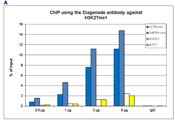
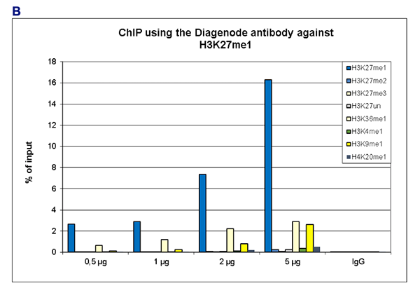
Figure 1. ChIP results obtained with the Diagenode antibody directed against H3K27me1
ChIP was performed with the Diagenode antibody against H3K27me1 (Cat. No. C15410045) on sheared chromatin from 500,000 K562 cells using the “iDeal ChIP-seq” kit (Cat. No. C01010051). The chromatin was spiked with a panel of in vitro assembled nucleosomes, each containing a specific lysine methylation (SNAP-ChIP K-MetStat Panel, Epicypher). A titration of the antibody consisting of 0.5, 1, 2 and 5 µg per ChIP experiment was analysed. IgG (2 µg/IP) was used as negative IP control.
Figure 1A. Quantitative PCR was performed with primers for the coding sequence of the active GAPDH and ACTB genes, used as positive controls, and for the inactive MYOD1 and MYT1 genes, used as negative controls. The graph shows the recovery, expressed as a % of input (the relative amount of immunoprecipitated DNA compared to input DNA after qPCR analysis).
Figure 1B. Recovery of the nucleosomes carrying the H3K27me1, H3K27me2, H3K27me3, H3K4me1, H3K9me1, H3K36me1, H4K20me1 and the unmodified H3K4 as determined by qPCR. The figure clearly shows the antibody is specific in ChIP for the H3K27me1 modification with some slight cross reaction with H3K36me1 and H3K9me1 at higher concentrations.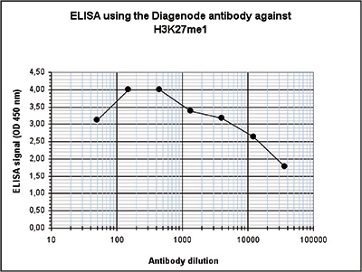
Figure 2. Determination of the antibody titer
To determine the titer of the antibody, an ELISA was performed using a serial dilution of the Diagenode antibody directed against H3K27me1 (Cat. No. C15410045). The antigen used was a peptide containing the histone modification of interest. By plotting the absorbance against the antibody dilution (Figure 1), the titer of the purified antibody was estimated to be 1:32,900.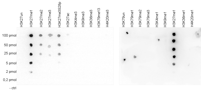
Figure 3. Cross reactivity tests using the Diagenode antibody directed against H3K27me1
A Dot Blot analysis was performed to test the cross reactivity of the Diagenode antibody against H3K27me1 (Cat. No. C15410045) with peptides containing other modifications and unmodified sequences of histone H3 and H4. One hundred to 0.2 pmol of the peptide containing the respective histone modification were spotted on a membrane. The antibody was used at a dilution of 1:20,000. Figure 3 shows a high specificity of the antibody for the modification of interest.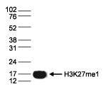
Figure 4. Western blot analysis using the Diagenode antibody directed against H3K27me1
Histone extracts (15 µg) from HeLa cells were analysed by Western blot using the Diagenode antibody against H3K27me1 (Cat. No. C15410045) diluted 1:1,000 in TBS-Tween containing 5% skimmed milk. The position of the protein of interest is indicated on the right; the marker (in kDa) is shown on the left.A.
B.
C.
D.
E.
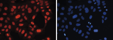
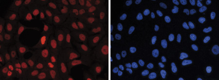
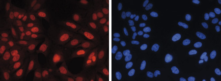
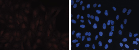
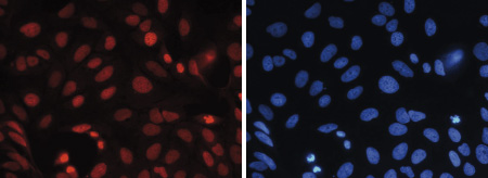
Figure 5. Immunofluorescence using the Diagenode antibody directed against H3K27me1
Human osteosarcoma (U2OS) cells were stained with the Diagenode antibody against H3K27me1 (Cat. No. C15410045) and with DAPI. Cells were fixed with 4% formaldehyde for 20’ and blocked with PBS/TX-100 containing 5% normal goat serum. Figure 5A: cells were immunofluorescently labeled with the H3K27me1 antibody (left) diluted 1:1,000 in blocking solution followed by an anti-rabbit antibody conjugated to Alexa568 or with DAPI (right), which specifically labels DNA. Figure 5B, C, D and E: staining of the cells with the H3K27me1 antibody after incubation of the antibody with 2 ng/µl blocking peptide containing the unmodified and the mono-, di- and trimethylated H3K27, respectively. - 出版物

How to properly cite our product/service in your work
We strongly recommend using this: H3K27me1 polyclonal antibody (sample size) (Hologic Diagenode Cat# C15410045-10 Lot# A932-00234P). Click here to copy to clipboard.
Using our products or services in your publication? Let us know!
Motif distribution and DNA methylation underlie distinct Cdx2 binding during development and homeostasis
Alireza Lorzadeh et al.
Transcription factors guide tissue development by binding to developmental stage-specific targets and establishing an appropriate enhancer landscape. In turn, DNA and chromatin modifications direct the genomic binding of transcription factors. However, how transcription factors navigate chromatin features to selecti...The Polycomb-Dependent Epigenome Controls β Cell Dysfunction, Dedifferentiation, and Diabetes.
Lu TT, Heyne S, Dror E, Casas E, Leonhardt L, Boenke T, Yang CH, Sagar , Arrigoni L, Dalgaard K, Teperino R, Enders L, Selvaraj M, Ruf M, Raja SJ, Xie H, Boenisch U, Orkin SH, Lynn FC, Hoffman BG, Grün D, Vavouri T, Lempradl AM, Pospisilik JA
To date, it remains largely unclear to what extent chromatin machinery contributes to the susceptibility and progression of complex diseases. Here, we combine deep epigenome mapping with single-cell transcriptomics to mine for evidence of chromatin dysregulation in type 2 diabetes. We find two chromatin-state signat...Applying the INTACT method to purify endosperm nuclei and to generate parental-specific epigenome profiles.
Moreno-Romero J. et al.
The early endosperm tissue of dicot species is very difficult to isolate by manual dissection. This protocol details how to apply the INTACT (isolation of nuclei tagged in specific cell types) system for isolating early endosperm nuclei of Arabidopsis at high purity and how to generate parental-specific epigenome pr...