Notice (8): Undefined variable: solution_of_interest [APP/View/Products/view.ctp, line 755]Code Context<!-- BEGIN: REQUEST_FORM MODAL -->
<div id="request_formModal" class="reveal-modal medium" data-reveal aria-labelledby="modalTitle" aria-hidden="true" role="dialog">
<?= $this->element('Forms/simple_form', array('solution_of_interest' => $solution_of_interest, 'header' => $header, 'message' => $message, 'campaign_id' => $campaign_id)) ?>
$viewFile = '/home/website-server/www/app/View/Products/view.ctp'
$dataForView = array(
'language' => 'jp',
'meta_keywords' => 'iDeal ChIP-qPCR kit ,ChIP-qPCR assays, ChIP-grade antibodies,diagenode',
'meta_description' => 'Diagenode’s iDeal ChIP-qPCR kit is a highly optimized solution for ChIP-qPCR assays. The kit provides high yields with excellent specificity and sensitivity. The iDeal ChIP-qPCR kit used with our highly validated ChIP-grade antibodies provides you with excellent, reproducible results from each experiment.',
'meta_title' => 'iDeal ChIP-qPCR kit | Diagenode',
'product' => array(
'Product' => array(
'id' => '2899',
'antibody_id' => null,
'name' => 'iDeal ChIP-qPCR kit',
'description' => '<p>DiagenodeのiDealChIP-qPCR kitは、優れた特異性と感度で高い収量を可能にするChIP-qPCR アッセイ用に最適化されたソリューションです。高度に検証されたChIPグレード抗体と一緒に使用することで、優れた再現性のある結果を提供します。</p>',
'label1' => '特徴',
'info1' => '<ul>
<li><strong><span style="font-weight: 400;"><strong>Fast</strong> and <strong>highly optimized</strong> protocol for ChIP-qPCR from cells and tissues</span></strong></li>
<li><strong><span style="font-weight: 400;"><strong>Easy ChIP</strong> made faster and more <strong>reproducible</strong> with magnetic beads</span></strong></li>
<li><strong><span style="font-weight: 400;"><strong>High yields</strong> with excellent <strong>specificity</strong> and <strong>sensitivity</strong> due to combination of Diagenode ChIP-grade antibodies<br /></span></strong></li>
<li><strong><span style="font-weight: 400;">Eluted DNA suitable for <strong>qPCR analysis</strong></span></strong></li>
</ul>
<h3><strong>Optimized</strong> protocol for <strong>reproducible ChIP-qPCR</strong> results</h3>
<p>Ideal for <strong>histone</strong> and <strong>non-histone proteins</strong> – Use 1 million cells for histone marks or 4 million cells for transcription factors. The protocol allows the use of fewer cells per IP. The minimum number of cells will depend on the abundance of the protein in your sample.</p>
<p>Suitable for <strong>cells</strong> and for <strong>tissues</strong> samples – For tissues, use 7 mg per IP for histone marks or 30 mg per IP for transcription factors. The protocol allows the use of less tissue per IP. The minimum amount of tissue will depend on the abundance of the protein in your sample.</p>
<center><img src="https://www.diagenode.com/img/categories/chip-qpcr/chip-qpcr-figure.jpg" /></center>
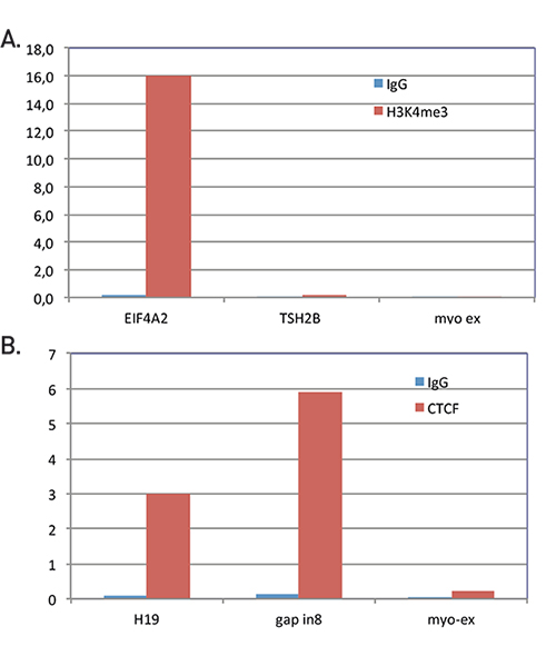
<p><small><strong>Chromatin immunoprecipitation analysis using H3K4me3 (A) and CTCF antibodies (B)</strong><br /> ChIP was performed on human HeLa cells using the H3K4me3 (Cat. No. C15410003) and CTCF (Cat. No. C15410210) antibodies. IgG was used as a negative control. The IP’d DNA was analyzed by qPCR with the following primer sets: EIF4A2, used as a positive control, and THS2B and Myoglobin exon 2, used as negative controls for H3K4me3. H19 imprinting control region and GAPDH intron 8, used as positive controls, and Myoglobin exon 2, used as a negative control for CTCF. The figure shows the recovery, expressed as a % of input (the relative amount of immunoprecipitated DNA compared to input DNA after qPCR analysis).</small></p>',
'label2' => '',
'info2' => '<p><span style="font-weight: 400;">The</span> <a href="https://www.diagenode.com/en/p/chromatin-shearing-optimization-kit-low-sds-for-tfs-25-rxns"><span style="font-weight: 400;">Chromatin shearing optimization kit – Low SDS (iDeal Kit for TFs)</span></a><span style="font-weight: 400;"> is the kit compatible with the iDeal ChIP-qPCR kit, recommended for the optimization of chromatin shearing, a critical step for ChIP.</span></p>
<p><a href="https://www.diagenode.com/en/p/chip-cross-link-gold-600-ul"><span style="font-weight: 400;">ChIP Cross-link Gold</span></a> <span style="font-weight: 400;">should be used in combination with formaldehyde when working with higher order and/or dynamic interactions, for efficient protein-protein fixation.</span></p>
<p><span style="font-weight: 400;">Highly specific </span><a href="https://www.diagenode.com/en/categories/chip-grade-antibodies"><span style="font-weight: 400;">ChIP-grade antibodies</span></a><span style="font-weight: 400;">, provided with batch-specific validation data.</span></p>
<p><span style="font-weight: 400;">Check out the list of available </span><a href="https://www.diagenode.com/en/categories/primer-pairs"><span style="font-weight: 400;">Primer pairs</span></a><span style="font-weight: 400;"> designed for high specificity to specific genomic regions.</span></p>
<p><span style="font-weight: 400;">Using our IP-Star Compact, please check out the <a href="https://www.diagenode.com/en/p/auto-ideal-chip-qpcr-kit">automated version</a> of the kit.</span></p>',
'label3' => '',
'info3' => '',
'format' => '24 rxns',
'catalog_number' => 'C01010180',
'old_catalog_number' => '',
'sf_code' => 'C01010180-',
'type' => 'RFR',
'search_order' => '',
'price_EUR' => '560',
'price_USD' => '560',
'price_GBP' => '510',
'price_JPY' => '87725',
'price_CNY' => '',
'price_AUD' => '1400',
'country' => 'ALL',
'except_countries' => 'None',
'quote' => false,
'in_stock' => false,
'featured' => true,
'no_promo' => false,
'online' => true,
'master' => true,
'last_datasheet_update' => '',
'slug' => 'ideal-chip-qpcr-kit',
'meta_title' => 'iDeal ChIP-qPCR kit | Diagenode',
'meta_keywords' => 'iDeal ChIP-qPCR kit ,ChIP-qPCR assays, ChIP-grade antibodies,diagenode',
'meta_description' => 'Diagenode’s iDeal ChIP-qPCR kit is a highly optimized solution for ChIP-qPCR assays. The kit provides high yields with excellent specificity and sensitivity. The iDeal ChIP-qPCR kit used with our highly validated ChIP-grade antibodies provides you with excellent, reproducible results from each experiment.',
'modified' => '2023-04-20 16:07:26',
'created' => '2017-06-07 15:38:54',
'locale' => 'jpn'
),
'Antibody' => array(
'host' => '*****',
'id' => null,
'name' => null,
'description' => null,
'clonality' => null,
'isotype' => null,
'lot' => null,
'concentration' => null,
'reactivity' => null,
'type' => null,
'purity' => null,
'classification' => null,
'application_table' => null,
'storage_conditions' => null,
'storage_buffer' => null,
'precautions' => null,
'uniprot_acc' => null,
'slug' => null,
'meta_keywords' => null,
'meta_description' => null,
'modified' => null,
'created' => null,
'select_label' => null
),
'Slave' => array(),
'Group' => array(),
'Related' => array(),
'Application' => array(
(int) 0 => array(
[maximum depth reached]
),
(int) 1 => array(
[maximum depth reached]
)
),
'Category' => array(
(int) 0 => array(
[maximum depth reached]
)
),
'Document' => array(
(int) 0 => array(
[maximum depth reached]
)
),
'Feature' => array(),
'Image' => array(
(int) 0 => array(
[maximum depth reached]
)
),
'Promotion' => array(),
'Protocol' => array(),
'Publication' => array(
(int) 0 => array(
[maximum depth reached]
),
(int) 1 => array(
[maximum depth reached]
),
(int) 2 => array(
[maximum depth reached]
),
(int) 3 => array(
[maximum depth reached]
),
(int) 4 => array(
[maximum depth reached]
),
(int) 5 => array(
[maximum depth reached]
),
(int) 6 => array(
[maximum depth reached]
),
(int) 7 => array(
[maximum depth reached]
),
(int) 8 => array(
[maximum depth reached]
),
(int) 9 => array(
[maximum depth reached]
),
(int) 10 => array(
[maximum depth reached]
),
(int) 11 => array(
[maximum depth reached]
),
(int) 12 => array(
[maximum depth reached]
),
(int) 13 => array(
[maximum depth reached]
),
(int) 14 => array(
[maximum depth reached]
),
(int) 15 => array(
[maximum depth reached]
),
(int) 16 => array(
[maximum depth reached]
),
(int) 17 => array(
[maximum depth reached]
),
(int) 18 => array(
[maximum depth reached]
),
(int) 19 => array(
[maximum depth reached]
),
(int) 20 => array(
[maximum depth reached]
),
(int) 21 => array(
[maximum depth reached]
),
(int) 22 => array(
[maximum depth reached]
),
(int) 23 => array(
[maximum depth reached]
),
(int) 24 => array(
[maximum depth reached]
),
(int) 25 => array(
[maximum depth reached]
)
),
'Testimonial' => array(),
'Area' => array(),
'SafetySheet' => array(
(int) 0 => array(
[maximum depth reached]
),
(int) 1 => array(
[maximum depth reached]
),
(int) 2 => array(
[maximum depth reached]
),
(int) 3 => array(
[maximum depth reached]
),
(int) 4 => array(
[maximum depth reached]
),
(int) 5 => array(
[maximum depth reached]
),
(int) 6 => array(
[maximum depth reached]
),
(int) 7 => array(
[maximum depth reached]
)
)
)
)
$language = 'jp'
$meta_keywords = 'iDeal ChIP-qPCR kit ,ChIP-qPCR assays, ChIP-grade antibodies,diagenode'
$meta_description = 'Diagenode’s iDeal ChIP-qPCR kit is a highly optimized solution for ChIP-qPCR assays. The kit provides high yields with excellent specificity and sensitivity. The iDeal ChIP-qPCR kit used with our highly validated ChIP-grade antibodies provides you with excellent, reproducible results from each experiment.'
$meta_title = 'iDeal ChIP-qPCR kit | Diagenode'
$product = array(
'Product' => array(
'id' => '2899',
'antibody_id' => null,
'name' => 'iDeal ChIP-qPCR kit',
'description' => '<p>DiagenodeのiDealChIP-qPCR kitは、優れた特異性と感度で高い収量を可能にするChIP-qPCR アッセイ用に最適化されたソリューションです。高度に検証されたChIPグレード抗体と一緒に使用することで、優れた再現性のある結果を提供します。</p>',
'label1' => '特徴',
'info1' => '<ul>
<li><strong><span style="font-weight: 400;"><strong>Fast</strong> and <strong>highly optimized</strong> protocol for ChIP-qPCR from cells and tissues</span></strong></li>
<li><strong><span style="font-weight: 400;"><strong>Easy ChIP</strong> made faster and more <strong>reproducible</strong> with magnetic beads</span></strong></li>
<li><strong><span style="font-weight: 400;"><strong>High yields</strong> with excellent <strong>specificity</strong> and <strong>sensitivity</strong> due to combination of Diagenode ChIP-grade antibodies<br /></span></strong></li>
<li><strong><span style="font-weight: 400;">Eluted DNA suitable for <strong>qPCR analysis</strong></span></strong></li>
</ul>
<h3><strong>Optimized</strong> protocol for <strong>reproducible ChIP-qPCR</strong> results</h3>
<p>Ideal for <strong>histone</strong> and <strong>non-histone proteins</strong> – Use 1 million cells for histone marks or 4 million cells for transcription factors. The protocol allows the use of fewer cells per IP. The minimum number of cells will depend on the abundance of the protein in your sample.</p>
<p>Suitable for <strong>cells</strong> and for <strong>tissues</strong> samples – For tissues, use 7 mg per IP for histone marks or 30 mg per IP for transcription factors. The protocol allows the use of less tissue per IP. The minimum amount of tissue will depend on the abundance of the protein in your sample.</p>
<center><img src="https://www.diagenode.com/img/categories/chip-qpcr/chip-qpcr-figure.jpg" /></center>
<p><small><strong>Chromatin immunoprecipitation analysis using H3K4me3 (A) and CTCF antibodies (B)</strong><br /> ChIP was performed on human HeLa cells using the H3K4me3 (Cat. No. C15410003) and CTCF (Cat. No. C15410210) antibodies. IgG was used as a negative control. The IP’d DNA was analyzed by qPCR with the following primer sets: EIF4A2, used as a positive control, and THS2B and Myoglobin exon 2, used as negative controls for H3K4me3. H19 imprinting control region and GAPDH intron 8, used as positive controls, and Myoglobin exon 2, used as a negative control for CTCF. The figure shows the recovery, expressed as a % of input (the relative amount of immunoprecipitated DNA compared to input DNA after qPCR analysis).</small></p>',
'label2' => '',
'info2' => '<p><span style="font-weight: 400;">The</span> <a href="https://www.diagenode.com/en/p/chromatin-shearing-optimization-kit-low-sds-for-tfs-25-rxns"><span style="font-weight: 400;">Chromatin shearing optimization kit – Low SDS (iDeal Kit for TFs)</span></a><span style="font-weight: 400;"> is the kit compatible with the iDeal ChIP-qPCR kit, recommended for the optimization of chromatin shearing, a critical step for ChIP.</span></p>
<p><a href="https://www.diagenode.com/en/p/chip-cross-link-gold-600-ul"><span style="font-weight: 400;">ChIP Cross-link Gold</span></a> <span style="font-weight: 400;">should be used in combination with formaldehyde when working with higher order and/or dynamic interactions, for efficient protein-protein fixation.</span></p>
<p><span style="font-weight: 400;">Highly specific </span><a href="https://www.diagenode.com/en/categories/chip-grade-antibodies"><span style="font-weight: 400;">ChIP-grade antibodies</span></a><span style="font-weight: 400;">, provided with batch-specific validation data.</span></p>
<p><span style="font-weight: 400;">Check out the list of available </span><a href="https://www.diagenode.com/en/categories/primer-pairs"><span style="font-weight: 400;">Primer pairs</span></a><span style="font-weight: 400;"> designed for high specificity to specific genomic regions.</span></p>
<p><span style="font-weight: 400;">Using our IP-Star Compact, please check out the <a href="https://www.diagenode.com/en/p/auto-ideal-chip-qpcr-kit">automated version</a> of the kit.</span></p>',
'label3' => '',
'info3' => '',
'format' => '24 rxns',
'catalog_number' => 'C01010180',
'old_catalog_number' => '',
'sf_code' => 'C01010180-',
'type' => 'RFR',
'search_order' => '',
'price_EUR' => '560',
'price_USD' => '560',
'price_GBP' => '510',
'price_JPY' => '87725',
'price_CNY' => '',
'price_AUD' => '1400',
'country' => 'ALL',
'except_countries' => 'None',
'quote' => false,
'in_stock' => false,
'featured' => true,
'no_promo' => false,
'online' => true,
'master' => true,
'last_datasheet_update' => '',
'slug' => 'ideal-chip-qpcr-kit',
'meta_title' => 'iDeal ChIP-qPCR kit | Diagenode',
'meta_keywords' => 'iDeal ChIP-qPCR kit ,ChIP-qPCR assays, ChIP-grade antibodies,diagenode',
'meta_description' => 'Diagenode’s iDeal ChIP-qPCR kit is a highly optimized solution for ChIP-qPCR assays. The kit provides high yields with excellent specificity and sensitivity. The iDeal ChIP-qPCR kit used with our highly validated ChIP-grade antibodies provides you with excellent, reproducible results from each experiment.',
'modified' => '2023-04-20 16:07:26',
'created' => '2017-06-07 15:38:54',
'locale' => 'jpn'
),
'Antibody' => array(
'host' => '*****',
'id' => null,
'name' => null,
'description' => null,
'clonality' => null,
'isotype' => null,
'lot' => null,
'concentration' => null,
'reactivity' => null,
'type' => null,
'purity' => null,
'classification' => null,
'application_table' => null,
'storage_conditions' => null,
'storage_buffer' => null,
'precautions' => null,
'uniprot_acc' => null,
'slug' => null,
'meta_keywords' => null,
'meta_description' => null,
'modified' => null,
'created' => null,
'select_label' => null
),
'Slave' => array(),
'Group' => array(),
'Related' => array(),
'Application' => array(
(int) 0 => array(
'id' => '10',
'position' => '10',
'parent_id' => '2',
'name' => 'ChIP-qPCR',
'description' => '<div class="row">
<div class="small-12 medium-12 large-12 columns text-justify">
<p class="text-justify">Chromatin Immunoprecipitation (ChIP) coupled with quantitative PCR can be used to investigate protein-DNA interaction at known genomic binding sites. if sites are not known, qPCR primers can also be designed against potential regulatory regions such as promoters. ChIP-qPCR is advantageous in studies that focus on specific genes and potential regulatory regions across differing experimental conditions as the cost of performing real-time PCR is minimal. This technique is now used in a variety of life science disciplines including cellular differentiation, tumor suppressor gene silencing, and the effect of histone modifications on gene expression.</p>
<p class="text-justify"><strong>The ChIP-qPCR workflow</strong></p>
</div>
<div class="small-12 medium-12 large-12 columns text-center"><br /> <img src="https://www.diagenode.com/img/chip-qpcr-diagram.png" /></div>
<div class="small-12 medium-12 large-12 columns"><br />
<ol>
<li class="large-12 columns"><strong>Chromatin preparation: </strong>cell fixation (cross-linking) of chromatin-bound proteins such as histones or transcription factors to DNA followed by cell lysis.</li>
<li class="large-12 columns"><strong>Chromatin shearing: </strong>fragmentation of chromatin<strong> </strong>by sonication down to desired fragment size (100-500 bp)</li>
<li class="large-12 columns"><strong>Chromatin IP</strong>: protein-DNA complexe capture using<strong> <a href="https://www.diagenode.com/en/categories/chip-grade-antibodies">specific ChIP-grade antibodies</a></strong> against the histone or transcription factor of interest</li>
<li class="large-12 columns"><strong>DNA purification</strong>: chromatin reverse cross-linking and elution followed by purification<strong> </strong></li>
<li class="large-12 columns"><strong>qPCR and analysis</strong>: using previously designed primers to amplify IP'd material at specific loci</li>
</ol>
</div>
</div>
<div class="row" style="margin-top: 32px;">
<div class="small-12 medium-10 large-9 small-centered columns">
<div class="radius panel" style="background-color: #fff;">
<h3 class="text-center" style="color: #b21329;">Need guidance?</h3>
<p class="text-justify">Choose our full ChIP kits or simply choose what you need from antibodies, buffers, beads, chromatin shearing and purification reagents. With the ChIP Kit Customizer, you have complete flexibility on which components you want from our validated ChIP kits.</p>
<div class="row">
<div class="small-6 medium-6 large-6 columns"><a href="https://www.diagenode.com/pages/which-kit-to-choose"><img src="https://www.diagenode.com/img/banners/banner-decide.png" alt="" /></a></div>
<div class="small-6 medium-6 large-6 columns"><a href="https://www.diagenode.com/pages/chip-kit-customizer-1"><img src="https://www.diagenode.com/img/banners/banner-customizer.png" alt="" /></a></div>
</div>
</div>
</div>
</div>',
'in_footer' => false,
'in_menu' => true,
'online' => true,
'tabular' => true,
'slug' => 'chip-qpcr',
'meta_keywords' => 'Chromatin immunoprecipitation,ChIP Quantitative PCR,polymerase chain reaction (PCR)',
'meta_description' => 'Diagenode's ChIP qPCR kits can be used to quantify enriched DNA after chromatin immunoprecipitation. ChIP-qPCR is advantageous in studies that focus on specific genes and potential regulatory regions across differing experimental conditions as the cost of',
'meta_title' => 'ChIP Quantitative PCR (ChIP-qPCR) | Diagenode',
'modified' => '2018-01-09 16:46:56',
'created' => '2014-12-11 00:22:08',
'ProductsApplication' => array(
[maximum depth reached]
)
),
(int) 1 => array(
'id' => '2',
'position' => '10',
'parent_id' => null,
'name' => 'エピジェネティクス・クロマチン解析',
'description' => '<div class="row">
<div class="large-12 columns">エピジェネティクス研究は、異なる転写パターン、遺伝子発現およびサイレンシングを引き起こすクロマチンの変化に対処します。<br /><br />クロマチンの主成分はDNA<span>およびヒストン蛋白質です。<span> </span></span>各ヒストンコア蛋白質(H2A<span>、</span>H2B<span>、</span>H3<span>および</span>H4<span>)の</span>2<span>つのコピーを</span>8<span>量体に組み込み、</span>DNA<span>で包んでヌクレオソームコアを形成させます。<span> </span></span>ヌクレオソームは、転写機械のDNA<span>への接近可能性および</span>クロマチン再構成因子を制御します。</div>
<div class="large-12 columns">
<p></p>
<p>クロマチン免疫沈降(ChIP<span>)は、関心対象の特定の蛋白質に対するゲノム結合部位の位置を解明するために使用される方法であり、遺伝子発現の制御に関する非常に貴重な洞察を提供します。<span> </span></span>ChIPは特定の抗原を含むクロマチン断片の選択的富化に関与します。 特定の蛋白質または蛋白質修飾を認識する抗体を使用して、特定の遺伝子座における抗原の相対存在量を決定します。</p>
<p>ChIP-seq<span>および</span>ChIP-qPCR<span>は、蛋白質</span>-DNA<span>結合部位の同定を可能にする技術です。</span></p>
<p> </p>
</div>
</div>',
'in_footer' => true,
'in_menu' => true,
'online' => true,
'tabular' => true,
'slug' => 'epigenetics-chromatin-study',
'meta_keywords' => 'Chromatin Immunoprecipitation Sequencing,ChIP-Seq,ChIP-seq grade antibodies,DNA purification,qPCR,Shearing of chromatin',
'meta_description' => 'Diagenodeは、ChIP-Seqおよびクロマチン免疫沈降(ChIP)専用キットと抗体を定量PCRアプリケーションと組み合わせて幅広く提供しています。',
'meta_title' => 'エピジェネティクス - クロマチン免疫沈降(ChIP)| Diagenode',
'modified' => '2018-01-11 09:58:47',
'created' => '2015-02-20 16:10:25',
'ProductsApplication' => array(
[maximum depth reached]
)
)
),
'Category' => array(
(int) 0 => array(
'id' => '131',
'position' => '2',
'parent_id' => '59',
'name' => 'ChIP-qPCR kits',
'description' => '',
'no_promo' => false,
'in_menu' => false,
'online' => true,
'tabular' => true,
'hide' => false,
'all_format' => false,
'is_antibody' => false,
'slug' => 'chip-qpcr-kits',
'cookies_tag_id' => null,
'meta_keywords' => '',
'meta_description' => '',
'meta_title' => '',
'modified' => '2017-10-19 12:32:20',
'created' => '2017-06-07 15:43:16',
'ProductsCategory' => array(
[maximum depth reached]
),
'CookiesTag' => array([maximum depth reached])
)
),
'Document' => array(
(int) 0 => array(
'id' => '962',
'name' => 'iDeal ChIP qPCR Kit manual',
'description' => '<p>Diagenode’s iDeal ChIP-qPCR Kit is a highly optimized solution for ChIP-qPCR assays. Two versions of the protocol (manual and automated) are described in this manual. The kit provides high yields with excellent specificity and sensitivity. The iDeal ChIP-qPCR kit used with our highly validated ChIP-grade antibodies provides you with excellent, reproducible results from each experiment.</p>',
'image_id' => null,
'type' => 'Manual',
'url' => 'files/products/kits/ideal-chip-qpcr-manual.pdf',
'slug' => 'ideal-chip-qpcr-manual',
'meta_keywords' => '',
'meta_description' => '',
'modified' => '2018-09-18 11:04:26',
'created' => '2017-06-07 15:53:23',
'ProductsDocument' => array(
[maximum depth reached]
)
)
),
'Feature' => array(),
'Image' => array(
(int) 0 => array(
'id' => '1775',
'name' => 'product/kits/chip-kit-icon.png',
'alt' => 'ChIP kit icon',
'modified' => '2018-04-17 11:52:29',
'created' => '2018-03-15 15:50:34',
'ProductsImage' => array(
[maximum depth reached]
)
)
),
'Promotion' => array(),
'Protocol' => array(),
'Publication' => array(
(int) 0 => array(
'id' => '4915',
'name' => 'MYB/LINC00092 regulatory axis promotes the progression of papillary thyroid carcinoma',
'authors' => 'Cheng L. et al.',
'description' => '<p><strong>Introduction:</strong>Thyroid carcinoma is the most frequent malignancy in different endocrine-related tumours. In this study, we demonstrated a long non-coding RNA LINC00092-associated molecular mechanism in promoting the progression of papillary thyroid carcinoma (PTC).</p>
<p><strong>Material and methods:</strong>The expression of LINC00092 was analysed in the The Cancer Genome Atlas Thyroid Cancer (TCGA-THCA) patient cohorts and further determined by q-PCR. (3-[4,5-dimethylthiazol-2-yl]-2,5 diphenyl tetrazolium bromide) (MTT) assay, and wound healing assay confirmed the function of LINC00092 in migration and proliferation. Q-ChIP validated the transcriptional target. Luciferase reporter assay validated the miRNA-mRNA target.</p>
<p><strong>Results:</strong>The analysis in patient cohorts and in PTC TPC-1 cells showed that the expression of LINC00092 was repressed in thyroid carcinoma. In addition, the expression of LINC00092 was negatively associated with the advanced thyroid TNM stages. LINC00092 repressed epithelial-mesenchymal transition (EMT), migration, and proliferation of TPC-1 cells. Interestingly, we identified that MYB, a well-studied tumour promoter, is a transcription factor of LINC00092, thereby the expression of LINC00092 was directly repressed by MYB. Furthermore, miR-4741 was also validated as a direct target of MYB and was induced by MYB. Notably, LINC00092 was repressed by miR-4741 through the direct 3’-untranslational region (3’-UTR) target. Therefore, MYB induced EMT of TPC-1 cells by repressing LINC00092 directly or indirectly via miR-4741.</p>
<p><strong>Conclusions:</strong>Our study validated that LINC00092 is a tumour suppressor lncRNA in PTC. MYB directly or indirectly represses LINC00092, which contributes to the PTC progression. MYB, LINC00092, and miR-4741 form a coherent feed forward loop. The axis of MYB-LINC00092 promotes progression of PTC.</p>',
'date' => '2024-02-21',
'pmid' => 'https://journals.viamedica.pl/endokrynologia_polska/article/view/98120',
'doi' => '',
'modified' => '2024-02-26 13:36:09',
'created' => '2024-02-26 13:36:09',
'ProductsPublication' => array(
[maximum depth reached]
)
),
(int) 1 => array(
'id' => '4891',
'name' => 'Substrate stiffness promotes vascular smooth muscle cell calcification by reducing the levels of nuclear actin monomers',
'authors' => 'McNeill M.C. et al. ',
'description' => '<p><strong class="sub-title">Background:<span> </span></strong>Vascular calcification (VC) is a prevalent independent risk factor for adverse cardiovascular events and is associated with diabetes, hypertension, chronic kidney disease, and atherosclerosis. However, the mechanisms regulating the osteogenic differentiation of vascular smooth muscle cells (VSMC) are not fully understood.</p>
<p><strong class="sub-title">Methods:<span> </span></strong>Using hydrogels of tuneable stiffness and lysyl oxidase-mediated stiffening of human saphenous vein ex vivo, we investigated the role of substrate stiffness in the regulation of VSMC calcification.</p>
<p><strong class="sub-title">Results:<span> </span></strong>We demonstrate that increased substrate stiffness enhances VSMC osteogenic differentiation and VSMC calcification. We show that the effects of substrate stiffness are mediated via a reduction in the level of actin monomer within the nucleus. We show that in cells interacting with soft substrate, elevated levels of nuclear actin monomer repress osteogenic differentiation and calcification by repressing YAP-mediated activation of both TEA Domain transcription factor (TEAD) and RUNX Family Transcription factor 2 (RUNX2).</p>
<p><strong class="sub-title">Conclusion:<span> </span></strong>This work highlights for the first time the role of nuclear actin in mediating substrate stiffness-dependent VSMC calcification and the dual role of YAP-TEAD and YAP-RUNX2 transcriptional complexes.</p>',
'date' => '2024-01-04',
'pmid' => 'https://pubmed.ncbi.nlm.nih.gov/38181546/',
'doi' => '10.1016/j.yjmcc.2023.12.005',
'modified' => '2024-01-09 09:02:46',
'created' => '2024-01-09 09:02:46',
'ProductsPublication' => array(
[maximum depth reached]
)
),
(int) 2 => array(
'id' => '4886',
'name' => 'DAXX promotes centromeric stability independently of ATRX by preventing the accumulation of R-loop-induced DNA double-stranded breaks',
'authors' => 'Pinto L.M. et al.',
'description' => '<p><span>Maintaining chromatin integrity at the repetitive non-coding DNA sequences underlying centromeres is crucial to prevent replicative stress, DNA breaks and genomic instability. The concerted action of transcriptional repressors, chromatin remodelling complexes and epigenetic factors controls transcription and chromatin structure in these regions. The histone chaperone complex ATRX/DAXX is involved in the establishment and maintenance of centromeric chromatin through the deposition of the histone variant H3.3. ATRX and DAXX have also evolved mutually-independent functions in transcription and chromatin dynamics. Here, using paediatric glioma and pancreatic neuroendocrine tumor cell lines, we identify a novel ATRX-independent function for DAXX in promoting genome stability by preventing transcription-associated R-loop accumulation and DNA double-strand break formation at centromeres. This function of DAXX required its interaction with histone H3.3 but was independent of H3.3 deposition and did not reflect a role in the repression of centromeric transcription. DAXX depletion mobilized BRCA1 at centromeres, in line with BRCA1 role in counteracting centromeric R-loop accumulation. Our results provide novel insights into the mechanisms protecting the human genome from chromosomal instability, as well as potential perspectives in the treatment of cancers with DAXX alterations.</span></p>',
'date' => '2023-12-01',
'pmid' => 'https://academic.oup.com/nar/advance-article/doi/10.1093/nar/gkad1141/7457013#428428433',
'doi' => '10.1093/nar/gkad1141',
'modified' => '2023-12-05 08:48:55',
'created' => '2023-12-05 08:48:55',
'ProductsPublication' => array(
[maximum depth reached]
)
),
(int) 3 => array(
'id' => '4829',
'name' => 'CRISPR/Cas9-mediated inactivation of miR-34a and miR-34b/c inHCT116 colorectal cancer cells: comprehensive characterization afterexposure to 5-FU reveals EMT and autophagy as key processes regulatedby miR-34.',
'authors' => 'Huang Z. et al.',
'description' => '<p>The miR-34a and miR-34b/c encoding genes represent direct targets of the p53 transcription factor, and presumably mediate part of the tumor suppressive effects of p53. Here, we sought to determine their functional relevance by inactivating miR-34a and/or miR-34b/c using a CRISPR/Cas9 approach in the colorectal cancer (CRC) cell line HCT116. Concomitant deletion of miR-34a and miR-34b/c resulted in significantly reduced suppression of proliferation after p53 activation, enhanced migration, invasion and EMT, as well as reduced sensitivity to chemotherapeutics, increased stress-induced autophagic flux, decreased apoptosis and upregulation of autophagy-related genes after 5-FU treatment. However, inactivation of singular miR-34a or miR-34b/c had little effects on the aforementioned processes. RNA-Seq analysis revealed that concomitant deletion of miR-34a/b/c caused EMT signature enrichment, impaired gene repression by the p53-DREAM pathway and elevated autophagy after 5-FU treatment. A gene signature comprised of mRNAs significantly upregulated after combined inactivation of miR-34a and miR-34b/c showed a significant association with the invasive colon cancer subtype CMS4 and poor overall survival in two CRC patient cohorts, and with 5-FU resistance in CRC cell lines. In miR-34a/b/c-deficient cells the upregulated miR-34 target FOXM1 directly induced p62 and ATG9A, which increased autophagy and consequently attenuated apoptosis and rendered the miR-34a/b/c-KO cells more resistant to 5-FU. Inhibition of autophagy by depletion of ATG9A or chloroquine re-sensitized miR-34a/b/c-deficient HCT116 cells to 5-FU. In summary, our findings show a complementary role of miR-34a and miR-34b/c in the regulation of EMT and autophagy which may be relevant for CRC therapy in the future.</p>',
'date' => '2023-07-01',
'pmid' => 'https://www.ncbi.nlm.nih.gov/pubmed/37488217',
'doi' => '10.1038/s41418-023-01193-2',
'modified' => '2023-08-01 13:38:31',
'created' => '2023-08-01 15:59:38',
'ProductsPublication' => array(
[maximum depth reached]
)
),
(int) 4 => array(
'id' => '4805',
'name' => 'Curcumin activates a ROS/KEAP1/NRF2/miR-34a/b/c cascade tosuppress colorectal cancer metastasis.',
'authors' => 'Liu C. et al.',
'description' => '<p><span>Curcumin, a natural phytochemical isolated from tumeric roots, represents a candidate for prevention and therapy of colorectal cancer/CRC. However, the exact mechanism of action and the downstream mediators of curcumin's tumor suppressive effects have remained largely unknown. Here we used a genetic approach to determine the role of the p53/miR-34 pathway as mediator of the effects of curcumin. Three isogenic CRC cell lines rendered deficient for the p53, miR-34a and/or miR-34b/c genes were exposed to curcumin and subjected to cell biological analyses. siRNA-mediated inhibition and ectopic expression of NRF2, as well as Western blot, qPCR and qChIP analyses of its target genes were performed. CRC cells were i.v. injected into NOD/SCID mice and lung-metastases formation was determined by longitudinal, non-invasive imaging. In CRC cells curcumin induced apoptosis and senescence, and suppressed migration and invasion in a p53-independent manner. Curcumin activated the KEAP1/NRF2/ARE pathway by inducing ROS. Notably, curcumin induced miR-34a and miR-34b/c expression in a ROS/NRF2-dependent and p53-independent manner. NRF2 directly induced miR-34a and miR-34b/c via occupying multiple ARE motifs in their promoter regions. Curcumin reverted repression of miR-34a and miR-34b/c induced by IL6 and hypoxia. Deletion of miR-34a and miR-34b/c significantly reduced curcumin-induced apoptosis and senescence, and prevented the inhibition of migration and invasion by curcumin or ectopic NRF2. In CRC cells curcumin induced MET and prevented the formation of lung-metastases in mice in a miR-34a-dependent manner. In addition, we found that curcumin may enhance the therapeutic effects of 5-FU on CRC cells deficient for p53 and miR-34a/b/c. Activation of the KEAP1/NRF2/miR-34a/b/c axis mediates the tumor suppressive activity of curcumin and suggests a new approach for activating miR-34 genes in tumors for therapeutic purposes.</span></p>',
'date' => '2023-05-01',
'pmid' => 'https://www.ncbi.nlm.nih.gov/pubmed/37210578',
'doi' => '10.1038/s41418-023-01178-1',
'modified' => '2023-06-15 08:47:50',
'created' => '2023-06-13 21:11:31',
'ProductsPublication' => array(
[maximum depth reached]
)
),
(int) 5 => array(
'id' => '4681',
'name' => 'miR-34a and IRE1A/XBP-1(S) Form a Double-NegativeFeedback Loop to Regulate Hypoxia-Induced EMT, Metastasis,Chemo-Resistance and Autophagy',
'authors' => 'Bouznad N. et al.',
'description' => '<p>Tumor-associated hypoxia, i.e., decreased availability of oxygen, results in a poor clinical outcome since it promotes EMT, metastasis, and chemotherapy-resistance. We have previously identified p53 and its target miR-34a, as critical determinants of the effect of hypoxia on colorectal cancer (CRC). Here, we aimed to characterize mechanisms that contribute to the selective advantage of cells with loss of p53/miR-34a function in a hypoxic environment. Using in silico prediction, we identified XBP-1 and IRE1A as potential miR-34a targets. IRE1A and XBP-1 are central components of the unfolded protein response that is activated by ER stress, which is also induced in tumor cells as a response to harsh conditions surrounding tumors such as hypoxia and a limited supply of nutrients. Here we characterized the XBP-1(S) transcription factor and its regulator IRE1A as direct, conserved miR-34a targets in CRC cells. After hypoxia and DNA damage, IRE1A and XBP-1 were repressed by p53 in a miR-34a-dependent manner, whereas p53-deficient cells showed induction of IRE1A and XBP-1(S). Furthermore, miR-34a expression was directly suppressed by XBP-1(S). In p53-deficient CRC cells, hypoxia-induced EMT, migration, invasion, metastases formation, and resistance to 5-FU were dependent on IRE1A/XBP-1(S) activation. Hypoxia-induced autophagy was identified as an XBP-1(S)-dependent mediator of 5-FU resistance and was reversed by ectopic miR-34a expression. The HIF1A/IRE1A/XBP-1(S)/p53/miR-34a feedback loop described here represents a central regulator of the response to hypoxia and ER stress that maintains cellular homeostasis. In tumors, the inactivation of p53 and miR-34a may result in IRE1A/XPB-1(S)-mediated EMT and autophagy, which ultimately promotes metastasis and chemoresistance.</p>',
'date' => '2023-02-01',
'pmid' => 'https://doi.org/10.3390%2Fcancers15041143',
'doi' => '10.3390/cancers15041143',
'modified' => '2023-04-14 09:30:16',
'created' => '2023-02-28 12:19:11',
'ProductsPublication' => array(
[maximum depth reached]
)
),
(int) 6 => array(
'id' => '4670',
'name' => 'Epigenetic regulation of plastin 3 expression by the macrosatelliteDXZ4 and the transcriptional regulator CHD4.',
'authors' => 'Strathmann E. A. et al.',
'description' => '<p>Dysregulated Plastin 3 (PLS3) levels associate with a wide range of skeletal and neuromuscular disorders and the most common types of solid and hematopoietic cancer. Most importantly, PLS3 overexpression protects against spinal muscular atrophy. Despite its crucial role in F-actin dynamics in healthy cells and its involvement in many diseases, the mechanisms that regulate PLS3 expression are unknown. Interestingly, PLS3 is an X-linked gene and all asymptomatic SMN1-deleted individuals in SMA-discordant families who exhibit PLS3 upregulation are female, suggesting that PLS3 may escape X chromosome inactivation. To elucidate mechanisms contributing to PLS3 regulation, we performed a multi-omics analysis in two SMA-discordant families using lymphoblastoid cell lines and iPSC-derived spinal motor neurons originated from fibroblasts. We show that PLS3 tissue-specifically escapes X-inactivation. PLS3 is located ∼500 kb proximal to the DXZ4 macrosatellite, which is essential for X chromosome inactivation. By applying molecular combing in a total of 25 lymphoblastoid cell lines (asymptomatic individuals, individuals with SMA, control subjects) with variable PLS3 expression, we found a significant correlation between the copy number of DXZ4 monomers and PLS3 levels. Additionally, we identified chromodomain helicase DNA binding protein 4 (CHD4) as an epigenetic transcriptional regulator of PLS3 and validated co-regulation of the two genes by siRNA-mediated knock-down and overexpression of CHD4. We show that CHD4 binds the PLS3 promoter by performing chromatin immunoprecipitation and that CHD4/NuRD activates the transcription of PLS3 by dual-luciferase promoter assays. Thus, we provide evidence for a multilevel epigenetic regulation of PLS3 that may help to understand the protective or disease-associated PLS3 dysregulation.</p>',
'date' => '2023-02-01',
'pmid' => 'https://doi.org/10.1016%2Fj.ajhg.2023.02.004',
'doi' => '10.1016/j.ajhg.2023.02.004',
'modified' => '2023-04-14 09:36:04',
'created' => '2023-02-28 12:19:11',
'ProductsPublication' => array(
[maximum depth reached]
)
),
(int) 7 => array(
'id' => '4495',
'name' => 'Exploration of nuclear body-enhanced sumoylation reveals that PMLrepresses 2-cell features of embryonic stem cells.',
'authors' => 'Tessier S. et al.',
'description' => '<p>Membrane-less organelles are condensates formed by phase separation whose functions often remain enigmatic. Upon oxidative stress, PML scaffolds Nuclear Bodies (NBs) to regulate senescence or metabolic adaptation. PML NBs recruit many partner proteins, but the actual biochemical mechanism underlying their pleiotropic functions remains elusive. Similarly, PML role in embryonic stem cell (ESC) and retro-element biology is unsettled. Here we demonstrate that PML is essential for oxidative stress-driven partner SUMO2/3 conjugation in mouse ESCs (mESCs) or leukemia, a process often followed by their poly-ubiquitination and degradation. Functionally, PML is required for stress responses in mESCs. Differential proteomics unravel the KAP1 complex as a PML NB-dependent SUMO2-target in arsenic-treated APL mice or mESCs. PML-driven KAP1 sumoylation enables activation of this key epigenetic repressor implicated in retro-element silencing. Accordingly, Pml mESCs re-express transposable elements and display 2-Cell-Like features, the latter enforced by PML-controlled SUMO2-conjugation of DPPA2. Thus, PML orchestrates mESC state by coordinating SUMO2-conjugation of different transcriptional regulators, raising new hypotheses about PML roles in cancer.</p>',
'date' => '2022-09-01',
'pmid' => 'https://www.ncbi.nlm.nih.gov/pubmed/36175410',
'doi' => '10.1038/s41467-022-33147-6',
'modified' => '2022-11-21 10:21:48',
'created' => '2022-11-15 09:26:20',
'ProductsPublication' => array(
[maximum depth reached]
)
),
(int) 8 => array(
'id' => '4449',
'name' => 'RAD51 protects human cells from transcription-replication conflicts.',
'authors' => 'Bhowmick R. et al.',
'description' => '<p>Oncogene activation during tumorigenesis promotes DNA replication stress (RS), which subsequently drives the formation of cancer-associated chromosomal rearrangements. Many episodes of physiological RS likely arise due to conflicts between the DNA replication and transcription machineries operating simultaneously at the same loci. One role of the RAD51 recombinase in human cells is to protect replication forks undergoing RS. Here, we have identified a key role for RAD51 in preventing transcription-replication conflicts (TRCs) from triggering replication fork breakage. The genomic regions most affected by RAD51 deficiency are characterized by being replicated and transcribed in early S-phase and show significant overlap with loci prone to cancer-associated amplification. Consistent with a role for RAD51 in protecting against transcription-replication conflicts, many of the adverse effects of RAD51 depletion are ameliorated by inhibiting early S-phase transcription. We propose a model whereby RAD51 suppresses fork breakage and subsequent inadvertent amplification of genomic loci prone to experiencing TRCs.</p>',
'date' => '2022-08-01',
'pmid' => 'https://www.ncbi.nlm.nih.gov/pubmed/36002000',
'doi' => '10.1016/j.molcel.2022.07.010',
'modified' => '2022-10-14 16:44:54',
'created' => '2022-09-28 09:53:13',
'ProductsPublication' => array(
[maximum depth reached]
)
),
(int) 9 => array(
'id' => '4515',
'name' => 'Epigenetic remodeling of downstream enhancer regions is linked toselective expression of the IL1F10 gene in differentiated humankeratinocytes.',
'authors' => 'Talabot-Ayer D. et al.',
'description' => '<p>Interleukin (IL)-38, encoded by the IL1F10 gene, is a member of the IL-1 family of cytokines. IL-38 is constitutively expressed in epithelia in healthy humans, and in particular in epidermal keratinocytes in the skin. IL-38 expression is closely correlated with keratinocyte differentiation. The aim of this study was to further characterize the regulation of IL1F10 expression and the mechanisms involved in its selective induction in differentiated human keratinocytes. We observed coordinated expression of two IL1F10 transcripts, transcribed from two different promoters, upon differentiation of primary human keratinocytes. Using ENCODE datasets and ChIP-qPCR on ex vivo isolated normal human epidermis, we identified regulatory regions located downstream of the IL1F10 gene, which displayed features of differentiated keratinocyte-specific enhancers. Expression of the IL1F10 gene was linked to changes in the epigenetic landscape at these downstream enhancer regions in human epidermis. Overexpression of the transcription factors KLF4 and TAp63β in an immortalized normal human keratinocyte (iNHK) cell line promoted the expression of mRNA encoding the differentiation markers keratin 10 and involucrin, and of IL1F10. ChIP-qPCR experiments indicated that KLF4 and TAp63β overexpression also modified the chromatin state of the proximal downstream enhancer region, suggesting a role for KLF4 and TAp63β in directly or indirectly regulating IL1F10 transcription. In conclusion, expression of the IL1F10 gene in differentiated keratinocytes in normal human epidermis involves coordinated transcription from two promoters and is linked to epigenetic remodeling of enhancer regions located downstream of the gene.</p>',
'date' => '2022-08-01',
'pmid' => 'https://www.ncbi.nlm.nih.gov/pubmed/35961432',
'doi' => '10.1016/j.gene.2022.146800',
'modified' => '2022-11-24 08:49:31',
'created' => '2022-11-15 09:26:20',
'ProductsPublication' => array(
[maximum depth reached]
)
),
(int) 10 => array(
'id' => '4552',
'name' => 'Prolonged FOS activity disrupts a global myogenic transcriptionalprogram by altering 3D chromatin architecture in primary muscleprogenitor cells.',
'authors' => 'Barutcu A Rasim et al.',
'description' => '<p>BACKGROUND: The AP-1 transcription factor, FBJ osteosarcoma oncogene (FOS), is induced in adult muscle satellite cells (SCs) within hours following muscle damage and is required for effective stem cell activation and muscle repair. However, why FOS is rapidly downregulated before SCs enter cell cycle as progenitor cells (i.e., transiently expressed) remains unclear. Further, whether boosting FOS levels in the proliferating progeny of SCs can enhance their myogenic properties needs further evaluation. METHODS: We established an inducible, FOS expression system to evaluate the impact of persistent FOS activity in muscle progenitor cells ex vivo. We performed various assays to measure cellular proliferation and differentiation, as well as uncover changes in RNA levels and three-dimensional (3D) chromatin interactions. RESULTS: Persistent FOS activity in primary muscle progenitor cells severely antagonizes their ability to differentiate and form myotubes within the first 2 weeks in culture. RNA-seq analysis revealed that ectopic FOS activity in muscle progenitor cells suppressed a global pro-myogenic transcriptional program, while activating a stress-induced, mitogen-activated protein kinase (MAPK) transcriptional signature. Additionally, we observed various FOS-dependent, chromosomal re-organization events in A/B compartments, topologically associated domains (TADs), and genomic loops near FOS-regulated genes. CONCLUSIONS: Our results suggest that elevated FOS activity in recently activated muscle progenitor cells perturbs cellular differentiation by altering the 3D chromosome organization near critical pro-myogenic genes. This work highlights the crucial importance of tightly controlling FOS expression in the muscle lineage and suggests that in states of chronic stress or disease, persistent FOS activity in muscle precursor cells may disrupt the muscle-forming process.</p>',
'date' => '2022-08-01',
'pmid' => 'https://www.ncbi.nlm.nih.gov/pubmed/35971133',
'doi' => '10.1186/s13395-022-00303-x',
'modified' => '2022-11-24 10:11:55',
'created' => '2022-11-24 08:49:52',
'ProductsPublication' => array(
[maximum depth reached]
)
),
(int) 11 => array(
'id' => '4519',
'name' => 'PARP1-SNAI2 transcription axis drives resistance to PARP inhibitor,Talazoparib.',
'authors' => 'Ding X. et al.',
'description' => '<p>The synthetic lethal association between BRCA deficiency and poly (ADP-ribose) polymerase (PARP) inhibition supports PARP inhibitor (PARPi) clinical efficacy in BRCA-mutated tumors. PARPis also demonstrate activity in non-BRCA mutated tumors presumably through induction of PARP1-DNA trapping. Despite pronounced clinical response, therapeutic resistance to PARPis inevitably develops. An abundance of knowledge has been built around resistance mechanisms in BRCA-mutated tumors, however, parallel understanding in non-BRCA mutated settings remains insufficient. In this study, we find a strong correlation between the epithelial-mesenchymal transition (EMT) signature and resistance to a clinical PARPi, Talazoparib, in non-BRCA mutated tumor cells. Genetic profiling demonstrates that SNAI2, a master EMT transcription factor, is transcriptionally induced by Talazoparib treatment or PARP1 depletion and this induction is partially responsible for the emerging resistance. Mechanistically, we find that the PARP1 protein directly binds to SNAI2 gene promoter and suppresses its transcription. Talazoparib treatment or PARP1 depletion lifts PARP1-mediated suppression and increases chromatin accessibility around SNAI2 promoters, thus driving SNAI2 transcription and drug resistance. We also find that depletion of the chromatin remodeler CHD1L suppresses SNAI2 expression and reverts acquired resistance to Talazoparib. The PARP1/CHD1L/SNAI2 transcription axis might be therapeutically targeted to re-sensitize Talazoparib in non-BRCA mutated tumors.</p>',
'date' => '2022-07-01',
'pmid' => 'https://www.ncbi.nlm.nih.gov/pubmed/35864202',
'doi' => '10.1038/s41598-022-16623-3',
'modified' => '2022-11-24 08:54:20',
'created' => '2022-11-15 09:26:20',
'ProductsPublication' => array(
[maximum depth reached]
)
),
(int) 12 => array(
'id' => '4397',
'name' => 'AP4 suppresses DNA damage, chromosomal instability and senescence viainducing and repressing miR-22-3p',
'authors' => 'Chou Jinjiang et al.',
'description' => '<p>Background AP4 (TFAP4) encodes a basic helix-loop-helix leucine zipper (bHLH-LZ) transcription factor and is a direct target gene of the oncogenic transcription factor c-MYC. Here, we set out to determine the relevance of AP4 in human colorectal cancer (CRC) cells. Methods A CRISPR/Cas9 approach was employed to generate AP4-deficient CRC cell lines with inducible expression of c-MYC. Colony formation, β-gal staining, immunofluorescence, comet and homologous recombination (HR) assays and RNA-Seq analysis were used to determine the effects of AP4 inactivation. qPCR and qChIP analyses was performed to validate differentially expressed AP4 targets. Expression data from CRC cohorts was subjected to bioinformatics analyses. Immunohistochemistry was used to evaluate AP4 targets in vivo. Ap4-deficient APCmin/+ mice were analyzed to determine conservation. Immunofluorescence, chromosome and micronuclei enumeration, MTT and colony formation assays were used to determine the effects of AP4 inactivation and target gene regulation on chromosomal instability (CIN) and drug sensitivity. Results Inactivation of AP4 in CRC cell lines resulted in increased spontaneous and c-MYC-induced DNA damage, chromosomal instability (CIN) and cellular senescence. AP4-deficient cells displayed increased expression of the long non-coding RNA MIR22HG, which encodes miR-22-3p and was directly repressed by AP4. Furthermore, Mediator of DNA damage Checkpoint 1 (MDC1), a central component of the DNA damage response and a known target of miR-22-3p, displayed decreased expression in AP4-deficient cells. Accordingly, MDC1 was directly induced by AP4 and indirectly by AP4-mediated repression of miR-22-3p. Adenomas and organoids from Ap4-deficient APCmin/+ mice displayed conservation of these regulations. Inhibition of miR-22-3p or ectopic MDC1 expression reversed the increased senescence, DNA damage, CIN and defective HR observed in AP4-deficient CRC cells. AP4-deficiency also sensitized CRC cells to 5-FU treatment, whereas ectopic AP4 conferred resistance to 5-FU in a miR-22-3p and MDC1-dependent manner. Conclusions In summary, AP4, miR-22-3p and MDC1 form a conserved and coherent, regulatory feed-forward loop to promote DNA repair, which suppresses DNA damage, senescence and CIN, and contributes to 5-FU resistance. These findings explain how elevated AP4 expression contributes to development and chemo-resistance of colorectal cancer after c-MYC activation. Graphical abstract Supplementary Information The online version contains supplementary material available at 10.1186/s12943-022-01581-1.</p>',
'date' => '2022-05-01',
'pmid' => 'https://www.ncbi.nlm.nih.gov/pubmed/35624466',
'doi' => '10.1186/s12943-022-01581-1',
'modified' => '2022-08-11 14:30:54',
'created' => '2022-08-11 12:14:50',
'ProductsPublication' => array(
[maximum depth reached]
)
),
(int) 13 => array(
'id' => '4266',
'name' => 'Sevoflurane induces inflammation in primary hippocampal neurons byregulating Hoxa5/Gm5106/miR-27b-3p positive feedback loop.',
'authors' => 'Zhu, Zifu and Ma, Li',
'description' => '<p>Postoperative cognitive dysfunction (POCD) is a normal condition that develops after surgery with anesthesia, leading to deterioration of cognitive functions. However, the mechanism of POCD still remains unknown. To elucidate the POCD molecular mechanism, sevoflurane was employed in the present study to generate neuroinflammation mice model. Sevoflurane treatment caused inflammatory markers IL6, IL-10 and TNF-α high expression in primary hippocampal neurons and blood samples. Long non-coding RNA Gm5106 was found to be increased after being stimulated with sevoflurane. Silencing Gm5106 inhibited neuron inflammation. In the meanwhile, Gm5106 was identified as a direct target of miR-27b-3p that was inhibited by sevoflurane and related to inflammation suppression. In addition, transcription factor (TF) Hoxa5 was validated to activate Gm5106 through two binding motifs in the promoter region after sevoflurane exposure. Furthermore, miR-27b-3p also directly targeted Hoxa5 3'UTR, which affected nuclear Hoxa5 protein served as TF. Hoxa5 protein instead of 3'UTR reduced miR-27b-3p, in which Gm5106 knocking down abrogated this effect. In conclusion, sevoflurane induces neuroinflammation through increasing long non-coding RNA Gm5106, which is transcriptionally activated by Hoxa5 and directly targeted by miR-27-3p. Apart from that, Hoxa5, Gm5106, and miR-27b-3p form a positive feedback loop in sevoflurane stimulation.</p>',
'date' => '2021-12-01',
'pmid' => 'https://doi.org/10.1080%2F21655979.2021.2005927',
'doi' => '10.1080/21655979.2021.2005927',
'modified' => '2022-05-23 09:41:39',
'created' => '2022-05-19 10:41:50',
'ProductsPublication' => array(
[maximum depth reached]
)
),
(int) 14 => array(
'id' => '4319',
'name' => 'Regulatory interplay between Vav1, Syk and β-catenin occurs in lungcancer cells.',
'authors' => 'Boudria Rofia et al. ',
'description' => '<p>Vav1 exhibits two signal transducing properties as an adaptor protein and a regulator of cytoskeleton organization through its Guanine nucleotide Exchange Factor module. Although the expression of Vav1 is restricted to the hematopoietic lineage, its ectopic expression has been unraveled in a number of solid tumors. In this study, we show that in lung cancer cells, as such in hematopoietic cells, Vav1 interacts with the Spleen Tyrosine Kinase, Syk. Likewise, Syk interacts with β-catenin and, together with Vav1, regulates the phosphorylation status of β-catenin. Depletion of Vav1, Syk or β-catenin inhibits Rac1 activity and decreases cell migration suggesting the interplay of the three effectors to a common signaling pathway. This model is further supported by the finding that in turn, β-catenin regulates the transcription of Syk gene expression. This study highlights the elaborated connection between Vav1, Syk and β-catenin and the contribution of the trio to cell migration.</p>',
'date' => '2021-10-01',
'pmid' => 'https://doi.org/10.1016%2Fj.cellsig.2021.110079',
'doi' => '10.1016/j.cellsig.2021.110079',
'modified' => '2022-06-20 09:32:21',
'created' => '2022-05-19 10:41:50',
'ProductsPublication' => array(
[maximum depth reached]
)
),
(int) 15 => array(
'id' => '4329',
'name' => 'Epigenetic remodelling of enhancers in response to estrogen deprivationand re-stimulation.',
'authors' => 'Sklias Athena et al.',
'description' => '<p>Estrogen hormones are implicated in a majority of breast cancers and estrogen receptor alpha (ER), the main nuclear factor mediating estrogen signaling, orchestrates a complex molecular circuitry that is not yet fully elucidated. Here, we investigated genome-wide DNA methylation, histone acetylation and transcription after estradiol (E2) deprivation and re-stimulation to better characterize the ability of ER to coordinate gene regulation. We found that E2 deprivation mostly resulted in DNA hypermethylation and histone deacetylation in enhancers. Transcriptome analysis revealed that E2 deprivation leads to a global down-regulation in gene expression, and more specifically of TET2 demethylase that may be involved in the DNA hypermethylation following short-term E2 deprivation. Further enrichment analysis of transcription factor (TF) binding and motif occurrence highlights the importance of ER connection mainly with two partner TF families, AP-1 and FOX. These interactions take place in the proximity of E2 deprivation-mediated differentially methylated and histone acetylated enhancers. Finally, while most deprivation-dependent epigenetic changes were reversed following E2 re-stimulation, DNA hypermethylation and H3K27 deacetylation at certain enhancers were partially retained. Overall, these results show that inactivation of ER mediates rapid and mostly reversible epigenetic changes at enhancers, and bring new insight into early events, which may ultimately lead to endocrine resistance.</p>',
'date' => '2021-09-01',
'pmid' => 'https://doi.org/10.1093%2Fnar%2Fgkab697',
'doi' => '10.1093/nar/gkab697',
'modified' => '2022-06-22 09:25:09',
'created' => '2022-05-19 10:41:50',
'ProductsPublication' => array(
[maximum depth reached]
)
),
(int) 16 => array(
'id' => '4282',
'name' => 'Enhanced targeted DNA methylation of the CMV and endogenous promoterswith dCas9-DNMT3A3L entails distinct subsequent histonemodification changes in CHO cells.',
'authors' => 'Marx Nicolas et al. ',
'description' => '<p>With the emergence of new CRISPR/dCas9 tools that enable site specific modulation of DNA methylation and histone modifications, more detailed investigations of the contribution of epigenetic regulation to the precise phenotype of cells in culture, including recombinant production subclones, is now possible. These also allow a wide range of applications in metabolic engineering once the impact of such epigenetic modifications on the chromatin state is available. In this study, enhanced DNA methylation tools were targeted to a recombinant viral promoter (CMV), an endogenous promoter that is silenced in its native state in CHO cells, but had been reactivated previously (β-galactoside α-2,6-sialyltransferase 1) and an active endogenous promoter (α-1,6-fucosyltransferase), respectively. Comparative ChIP-analysis of histone modifications revealed a general loss of active promoter histone marks and the acquisition of distinct repressive heterochromatin marks after targeted methylation. On the other hand, targeted demethylation resulted in autologous acquisition of active promoter histone marks and loss of repressive heterochromatin marks. These data suggest that DNA methylation directs the removal or deposition of specific histone marks associated with either active, poised or silenced chromatin. Moreover, we show that de novo methylation of the CMV promoter results in reduced transgene expression in CHO cells. Although targeted DNA methylation is not efficient, the transgene is repressed, thus offering an explanation for seemingly conflicting reports about the source of CMV promoter instability in CHO cells. Importantly, modulation of epigenetic marks enables to nudge the cell into a specific gene expression pattern or phenotype, which is stabilized in the cell by autologous addition of further epigenetic marks. Such engineering strategies have the added advantage of being reversible and potentially tunable to not only turn on or off a targeted gene, but also to achieve the setting of a desirable expression level.</p>',
'date' => '2021-07-01',
'pmid' => 'https://doi.org/10.1016%2Fj.ymben.2021.04.014',
'doi' => '10.1016/j.ymben.2021.04.014',
'modified' => '2022-05-23 10:09:24',
'created' => '2022-05-19 10:41:50',
'ProductsPublication' => array(
[maximum depth reached]
)
),
(int) 17 => array(
'id' => '4315',
'name' => 'Atg7 deficiency in microglia drives an altered transcriptomic profileassociated with an impaired neuroinflammatory response',
'authors' => 'Friess L. et al.',
'description' => '<p>Microglia, resident immunocompetent cells of the central nervous system, can display a range of reaction states and thereby exhibit distinct biological functions across development, adulthood and under disease conditions. Distinct gene expression profiles are reported to define each of these microglial reaction states. Hence, the identification of modulators of selective microglial transcriptomic signature, which have the potential to regulate unique microglial function has gained interest. Here, we report the identification of ATG7 (Autophagy-related 7) as a selective modulator of an NF-κB-dependent transcriptional program controlling the pro-inflammatory response of microglia. We also uncover that microglial Atg7-deficiency was associated with reduced microglia-mediated neurotoxicity, and thus a loss of biological function associated with the pro-inflammatory microglial reactive state. Further, we show that Atg7-deficiency in microglia did not impact on their ability to respond to alternative stimulus, such as one driving them towards an anti-inflammatory/tumor supportive phenotype. The identification of distinct regulators, such as Atg7, controlling specific microglial transcriptional programs could lead to developing novel therapeutic strategies aiming to manipulate selected microglial phenotypes, instead of the whole microglial population with is associated with several pitfalls. Supplementary Information The online version contains supplementary material available at 10.1186/s13041-021-00794-7.</p>',
'date' => '2021-06-01',
'pmid' => 'https://www.ncbi.nlm.nih.gov/pubmed/34082793',
'doi' => '10.1186/s13041-021-00794-7',
'modified' => '2022-08-02 16:47:13',
'created' => '2022-05-19 10:41:50',
'ProductsPublication' => array(
[maximum depth reached]
)
),
(int) 18 => array(
'id' => '4167',
'name' => 'FOS licenses early events in stem cell activation driving skeletal muscleregeneration.',
'authors' => 'Almada, Albert E. et al.',
'description' => '<p>Muscle satellite cells (SCs) are a quiescent (non-proliferative) stem cell population in uninjured skeletal muscle. Although SCs have been investigated for nearly 60 years, the molecular drivers that transform quiescent SCs into the rapidly dividing (activated) stem/progenitor cells that mediate muscle repair after injury remain largely unknown. Here we identify a prominent FBJ osteosarcoma oncogene (Fos) mRNA and protein signature in recently activated SCs that is rapidly, heterogeneously, and transiently induced by muscle damage. We further reveal a requirement for FOS to efficiently initiate key stem cell functions, including cell cycle entry, proliferative expansion, and muscle regeneration, via induction of "pro-regenerative" target genes that stimulate cell migration, division, and differentiation. Disruption of one of these Fos/AP-1 targets, NAD(+)-consuming mono-ADP-ribosyl-transferase 1 (Art1), in SCs delays cell cycle entry and impedes progenitor cell expansion and muscle regeneration. This work uncovers an early-activated FOS/ART1/mono-ADP-ribosylation (MARylation) pathway that is essential for stem cell-regenerative responses.</p>',
'date' => '2021-01-01',
'pmid' => 'https://www.ncbi.nlm.nih.gov/pubmed/33503437',
'doi' => '10.1016/j.celrep.2020.108656',
'modified' => '2021-12-21 15:46:42',
'created' => '2021-12-06 15:53:19',
'ProductsPublication' => array(
[maximum depth reached]
)
),
(int) 19 => array(
'id' => '4313',
'name' => 'CREB3 Transactivates lncRNA ZFAS1 to Promote PapillaryThyroid Carcinoma Metastasis by Modulating miR-373-3/MMP3Regulatory Axis',
'authors' => 'Wang G. et al.',
'description' => '<p>The incidence rate of thyroid carcinoma ranks ninth among human malignancies, and it accounts for the most frequent malignancy in endocrine-related tumors. This study aimed to investigate the role of long noncoding RNA (lncRNA) ZFAS1 in the metastasis of papillary thyroid carcinoma (PTC) and the potential molecular mechanisms. Both ZFAS1 and MMP3 were highly expressed in thyroid carcinoma and PTC cell, as measured by the q-PCR and TCGA database. In addition, ZFAS1 induced TPC-1 metastasis through inducing the epithelial–mesenchymal transition (EMT) process. Besides, ZFAS1 knockdown by siRNA induced miR-373-3p expression and reduced MMP3 expression, as quantified by q-PCR and Western blotting. According to the luciferase assay, both ZFAS1 and MMP3 were classified as the direct targets of miR-373-3p. However, MMP3 itself did not affect ZFAS1. Using the online prediction tool, CREB3 was predicted as the transcription factor (TF) of ZFAS1 that contained two binding sites on its promoter region, and CREB3 was positively correlated with ZFAS1 in thyroid carcinoma cohorts. Results from the dual-luciferase assay and ChIP-qPCR indicated that both the two binding sites were essential for the transcription of ZFAS1. In conclusion, CREB3 activated lncRNA ZFAS1 at the transcriptional level to promote PTC metastasis by modulating miR-373-3p/MMP3.</p>',
'date' => '2021-01-01',
'pmid' => 'https://www.ncbi.nlm.nih.gov/pubmed/34249125',
'doi' => '10.1155/2021/9981683',
'modified' => '2022-08-02 16:44:03',
'created' => '2022-05-19 10:41:50',
'ProductsPublication' => array(
[maximum depth reached]
)
),
(int) 20 => array(
'id' => '4029',
'name' => 'The transcription factor scleraxis differentially regulates gene expressionin tenocytes isolated at different developmental stages.',
'authors' => 'Paterson, YZ and Evans, N and Kan, S and Cribbs, A and Henson, FMD andGuest, DJ',
'description' => '<p>The transcription factor scleraxis (SCX) is expressed throughout tendon development and plays a key role in directing tendon wound healing. However, little is known regarding its role in fetal or young postnatal tendons, stages in development that are known for their enhanced regenerative capabilities. Here we used RNA-sequencing to compare the transcriptome of adult and fetal tenocytes following SCX knockdown. SCX knockdown had a larger effect on gene expression in fetal tenocytes, effecting 477 genes in comparison to the 183 genes effected in adult tenocytes, indicating that scleraxis-dependent processes may differ in these two developmental stages. Gene ontology, network and pathway analysis revealed an overrepresentation of extracellular matrix (ECM) remodelling processes within both comparisons. These included several matrix metalloproteinases, proteoglycans and collagens, some of which were also investigated in SCX knockdown tenocytes from young postnatal foals. Using chromatin immunoprecipitation, we also identified novel genes that SCX differentially interacts with in adult and fetal tenocytes. These results indicate a role for SCX in modulating ECM synthesis and breakdown and provides a useful dataset for further study into SCX gene regulation.</p>',
'date' => '2020-08-11',
'pmid' => 'http://www.pubmed.gov/32795590',
'doi' => '10.1016/j.mod.2020.103635',
'modified' => '2020-12-16 17:57:29',
'created' => '2020-10-12 14:54:59',
'ProductsPublication' => array(
[maximum depth reached]
)
),
(int) 21 => array(
'id' => '3926',
'name' => 'TET-Mediated Hypermethylation Primes SDH-Deficient Cells for HIF2α-Driven Mesenchymal Transition.',
'authors' => 'Morin A, Goncalves J, Moog S, Castro-Vega LJ, Job S, Buffet A, Fontenille MJ, Woszczyk J, Gimenez-Roqueplo AP, Letouzé E, Favier J',
'description' => '<p>Loss-of-function mutations in the SDHB subunit of succinate dehydrogenase predispose patients to aggressive tumors characterized by pseudohypoxic and hypermethylator phenotypes. The mechanisms leading to DNA hypermethylation and its contribution to SDH-deficient cancers remain undemonstrated. We examine the genome-wide distribution of 5-methylcytosine and 5-hydroxymethylcytosine and their correlation with RNA expression in SDHB-deficient tumors and murine Sdhb cells. We report that DNA hypermethylation results from TET inhibition. Although it preferentially affects PRC2 targets and known developmental genes, PRC2 activity does not contribute to the DNA hypermethylator phenotype. We also prove, in vitro and in vivo, that TET silencing, although recapitulating the methylation profile of Sdhb cells, is not sufficient to drive their EMT-like phenotype, which requires additional HIF2α activation. Altogether, our findings reveal synergistic roles of TET repression and pseudohypoxia in the acquisition of metastatic traits, providing a rationale for targeting HIF2α and DNA methylation in SDH-associated malignancies.</p>',
'date' => '2020-03-31',
'pmid' => 'http://www.pubmed.gov/32234487',
'doi' => '10.1016/j.celrep.2020.03.022',
'modified' => '2020-08-17 10:50:11',
'created' => '2020-08-10 12:12:25',
'ProductsPublication' => array(
[maximum depth reached]
)
),
(int) 22 => array(
'id' => '3764',
'name' => 'The nuclear hypoxia-regulated NLUCAT1 long non-coding RNA contributes to an aggressive phenotype in lung adenocarcinoma through regulation of oxidative stress.',
'authors' => 'Moreno Leon L, Gautier M, Allan R, Ilié M, Nottet N, Pons N, Paquet A, Lebrigand K, Truchi M, Fassy J, Magnone V, Kinnebrew G, Radovich M, Cheok MH, Barbry P, Vassaux G, Marquette CH, Ponzio G, Ivan M, Pottier N, Hofman P, Mari B, Rezzonico R',
'description' => '<p>Lung cancer is the leading cause of cancer death worldwide, with poor prognosis and a high rate of recurrence despite early surgical removal. Hypoxic regions within tumors represent sources of aggressiveness and resistance to therapy. Although long non-coding RNAs (lncRNAs) are increasingly recognized as major gene expression regulators, their regulation and function following hypoxic stress are still largely unexplored. Combining profiling studies on early-stage lung adenocarcinoma (LUAD) biopsies and on A549 LUAD cell lines cultured in normoxic or hypoxic conditions, we identified a subset of lncRNAs that are both correlated with the hypoxic status of tumors and regulated by hypoxia in vitro. We focused on a new transcript, NLUCAT1, which is strongly upregulated by hypoxia in vitro and correlated with hypoxic markers and poor prognosis in LUADs. Full molecular characterization showed that NLUCAT1 is a large nuclear transcript composed of six exons and mainly regulated by NF-κB and NRF2 transcription factors. CRISPR-Cas9-mediated invalidation of NLUCAT1 revealed a decrease in proliferative and invasive properties, an increase in oxidative stress and a higher sensitivity to cisplatin-induced apoptosis. Transcriptome analysis of NLUCAT1-deficient cells showed repressed genes within the antioxidant and/or cisplatin-response networks. We demonstrated that the concomitant knockdown of four of these genes products, GPX2, GLRX, ALDH3A1, and PDK4, significantly increased ROS-dependent caspase activation, thus partially mimicking the consequences of NLUCAT1 inactivation in LUAD cells. Overall, we demonstrate that NLUCAT1 contributes to an aggressive phenotype in early-stage hypoxic tumors, suggesting it may represent a new potential therapeutic target in LUADs.</p>',
'date' => '2019-08-15',
'pmid' => 'http://www.pubmed.gov/31417181',
'doi' => '10.1038/s41388-019-0935-y',
'modified' => '2019-10-03 10:00:42',
'created' => '2019-10-02 16:16:55',
'ProductsPublication' => array(
[maximum depth reached]
)
),
(int) 23 => array(
'id' => '3745',
'name' => 'Elevated cyclic-AMP represses expression of exchange protein activated by cAMP (EPAC1) by inhibiting YAP-TEAD activity and HDAC-mediated histone deacetylation.',
'authors' => 'Ebrahimighaei R, McNeill MC, Smith SA, Wray JP, Ford KL, Newby AC, Bond M',
'description' => '<p>Ligand-induced activation of Exchange Protein Activated by cAMP-1 (EPAC1) is implicated in numerous physiological and pathological processes, including cardiac fibrosis where changes in EPAC1 expression have been detected. However, little is known about how EPAC1 expression is regulated. Therefore, we investigated regulation of EPAC1 expression by cAMP in cardiac fibroblasts. Elevation of cAMP using forskolin, cAMP-analogues or adenosine A2B-receptor activation significantly reduced EPAC1 mRNA and protein levels and inhibited formation of F-actin stress fibres. Inhibition of actin polymerisation with cytochalasin-D, latrunculin-B or the ROCK inhibitor, Y-27632, mimicked effects of cAMP on EPAC1 mRNA and protein levels. Elevated cAMP also inhibited activity of an EPAC1 promoter-reporter gene, which contained a consensus binding element for TEAD, which is a target for inhibition by cAMP. Inhibition of TEAD activity using siRNA-silencing of its co-factors YAP and TAZ, expression of dominant-negative TEAD or treatment with YAP-TEAD inhibitors, significantly inhibited EPAC1 expression. However, whereas expression of constitutively-active YAP completely reversed forskolin inhibition of EPAC1-promoter activity it did not rescue EPAC1 mRNA levels. Chromatin-immunoprecipitation detected a significant reduction in histone3-lysine27-acetylation at the EPAC1 proximal promoter in response to forskolin stimulation. HDAC1/3 inhibition partially reversed forskolin inhibition of EPAC1 expression, which was completely rescued by simultaneously expressing constitutively active YAP. Taken together, these data demonstrate that cAMP downregulates EPAC1 gene expression via disrupting the actin cytoskeleton, which inhibits YAP/TAZ-TEAD activity in concert with HDAC-mediated histone deacetylation at the EPAC1 proximal promoter. This represents a novel negative feedback mechanism controlling EPAC1 levels in response to cAMP elevation.</p>',
'date' => '2019-06-27',
'pmid' => 'http://www.pubmed.gov/31255721',
'doi' => '10.1016/j.bbamcr.2019.06.013',
'modified' => '2019-08-06 16:34:40',
'created' => '2019-07-31 13:35:50',
'ProductsPublication' => array(
[maximum depth reached]
)
),
(int) 24 => array(
'id' => '3667',
'name' => 'Adolescent social isolation affects parvalbumin expression in the medial prefrontal cortex in the MAM-E17 model of schizophrenia.',
'authors' => 'Maćkowiak M, Latusz J, Głowacka U, Bator E, Bilecki W',
'description' => '<p>Altered parvalbumin (PV) expression is observed in the prefrontal cortex of subjects with schizophrenia. Environmental context, particularly during adolescence, might regulate PV expression. In the present study, we investigated the effect of adolescent social isolation (SI) on PV expression in the medial prefrontal cortex in a neurodevelopmental model (MAM-E17) of schizophrenia. SI exposure occurred from postnatal day 30 to 40, followed by resocialization until late adolescence or early adulthood. PV mRNA and protein levels, as well as the number of PV cells, were analysed at these ages. Moreover, epigenetic regulation of PV expression by histone methylation was examined by measuring the total and PV gene-bound H3K4me3 levels. MAM only decreased levels of the PV mRNA and protein in adulthood. Decreases in total H3K4me3 levels and its level at the PV gene were also observed at this age. In contrast, in late adolescence, SI induced a decrease in the expression of the PV mRNA in the MAM group that was related to the reduction in total and PV gene-bound H3K4me3 levels. However, at this age, SI increased the levels of the PV protein in both the control and MAM groups. In adulthood, SI did not affect PV mRNA or H3K4me3 levels but decreased levels of the PV protein in both groups. Both MAM and SI failed to change the number of PV cells at any age. The results indicate that adolescent SI accelerated epigenetic impairments of PV expression in MAM-E17 rats; however, subsequent resocialization abolished this dysfunction, but failed to prevent alterations in PV protein.</p>',
'date' => '2019-02-01',
'pmid' => 'http://www.pubmed.gov/30519836',
'doi' => '10.1007/s11011-018-0359-3',
'modified' => '2019-07-01 11:35:31',
'created' => '2019-06-21 14:55:31',
'ProductsPublication' => array(
[maximum depth reached]
)
),
(int) 25 => array(
'id' => '3669',
'name' => 'Enhancers in the Peril lincRNA locus regulate distant but not local genes.',
'authors' => 'Groff AF, Barutcu AR, Lewandowski JP, Rinn JL',
'description' => '<p>BACKGROUND: Recently, it has become clear that some promoters function as long-range regulators of gene expression. However, direct and quantitative assessment of enhancer activity at long intergenic noncoding RNA (lincRNA) or mRNA gene bodies has not been performed. To unbiasedly assess the enhancer capacity across lincRNA and mRNA loci, we performed a massively parallel reporter assay (MPRA) on six lincRNA loci and their closest protein-coding neighbors. RESULTS: For both gene classes, we find significantly more MPRA activity in promoter regions than in gene bodies. However, three lincRNA loci, Lincp21, LincEnc1, and Peril, and one mRNA locus, Morc2a, display significant enhancer activity within their gene bodies. We hypothesize that such peaks may mark long-range enhancers, and test this in vivo using RNA sequencing from a knockout mouse model and high-throughput chromosome conformation capture (Hi-C). We find that ablation of a high-activity MPRA peak in the Peril gene body leads to consistent dysregulation of Mccc1 and Exosc9 in the neighboring topologically associated domain (TAD). This occurs irrespective of Peril lincRNA expression, demonstrating this regulation is DNA-dependent. Hi-C confirms long-range contacts with the neighboring TAD, and these interactions are altered upon Peril knockout. Surprisingly, we do not observe consistent regulation of genes within the local TAD. Together, these data suggest a long-range enhancer-like function for the Peril gene body. CONCLUSIONS: A multi-faceted approach combining high-throughput enhancer discovery with genetic models can connect enhancers to their gene targets and provides evidence of inter-TAD gene regulation.</p>',
'date' => '2018-12-11',
'pmid' => 'http://www.pubmed.gov/30537984',
'doi' => '10.1186/s13059-018-1589-8',
'modified' => '2019-07-01 11:33:17',
'created' => '2019-06-21 14:55:31',
'ProductsPublication' => array(
[maximum depth reached]
)
)
),
'Testimonial' => array(),
'Area' => array(),
'SafetySheet' => array(
(int) 0 => array(
'id' => '678',
'name' => 'iDeal ChIP-qPCR Kit SDS GB en',
'language' => 'en',
'url' => 'files/SDS/iDeal_ChIP-qPCR/SDS-C01010180_C01010181-iDeal_ChIP-qPCR_Kit-GB-en-1_0.pdf',
'countries' => 'GB',
'modified' => '2020-07-02 09:19:14',
'created' => '2020-07-02 09:19:14',
'ProductsSafetySheet' => array(
[maximum depth reached]
)
),
(int) 1 => array(
'id' => '680',
'name' => 'iDeal ChIP-qPCR Kit SDS US en',
'language' => 'en',
'url' => 'files/SDS/iDeal_ChIP-qPCR/SDS-C01010180_C01010181-iDeal_ChIP-qPCR_Kit-US-en-1_0.pdf',
'countries' => 'US',
'modified' => '2020-07-02 09:21:27',
'created' => '2020-07-02 09:21:27',
'ProductsSafetySheet' => array(
[maximum depth reached]
)
),
(int) 2 => array(
'id' => '675',
'name' => 'iDeal ChIP-qPCR Kit SDS DE de',
'language' => 'de',
'url' => 'files/SDS/iDeal_ChIP-qPCR/SDS-C01010180_C01010181-iDeal_ChIP-qPCR_Kit-DE-de-1_0.pdf',
'countries' => 'DE',
'modified' => '2020-07-02 09:17:44',
'created' => '2020-07-02 09:17:44',
'ProductsSafetySheet' => array(
[maximum depth reached]
)
),
(int) 3 => array(
'id' => '679',
'name' => 'iDeal ChIP-qPCR Kit SDS JP ja',
'language' => 'ja',
'url' => 'files/SDS/iDeal_ChIP-qPCR/SDS-C01010180_C01010181-iDeal_ChIP-qPCR_Kit-JP-ja-1_1.pdf',
'countries' => 'JP',
'modified' => '2020-07-02 09:20:57',
'created' => '2020-07-02 09:20:57',
'ProductsSafetySheet' => array(
[maximum depth reached]
)
),
(int) 4 => array(
'id' => '674',
'name' => 'iDeal ChIP-qPCR Kit SDS BE nl',
'language' => 'nl',
'url' => 'files/SDS/iDeal_ChIP-qPCR/SDS-C01010180_C01010181-iDeal_ChIP-qPCR_Kit-BE-nl-1_0.pdf',
'countries' => 'BE',
'modified' => '2020-07-02 09:17:12',
'created' => '2020-07-02 09:17:12',
'ProductsSafetySheet' => array(
[maximum depth reached]
)
),
(int) 5 => array(
'id' => '673',
'name' => 'iDeal ChIP-qPCR Kit SDS BE fr',
'language' => 'fr',
'url' => 'files/SDS/iDeal_ChIP-qPCR/SDS-C01010180_C01010181-iDeal_ChIP-qPCR_Kit-BE-fr-1_0.pdf',
'countries' => 'BE',
'modified' => '2020-07-02 09:16:46',
'created' => '2020-07-02 09:16:46',
'ProductsSafetySheet' => array(
[maximum depth reached]
)
),
(int) 6 => array(
'id' => '677',
'name' => 'iDeal ChIP-qPCR Kit SDS FR fr',
'language' => 'fr',
'url' => 'files/SDS/iDeal_ChIP-qPCR/SDS-C01010180_C01010181-iDeal_ChIP-qPCR_Kit-FR-fr-1_0.pdf',
'countries' => 'FR',
'modified' => '2020-07-02 09:18:42',
'created' => '2020-07-02 09:18:42',
'ProductsSafetySheet' => array(
[maximum depth reached]
)
),
(int) 7 => array(
'id' => '676',
'name' => 'iDeal ChIP-qPCR Kit SDS ES es',
'language' => 'es',
'url' => 'files/SDS/iDeal_ChIP-qPCR/SDS-C01010180_C01010181-iDeal_ChIP-qPCR_Kit-ES-es-1_0.pdf',
'countries' => 'ES',
'modified' => '2020-07-02 09:18:11',
'created' => '2020-07-02 09:18:11',
'ProductsSafetySheet' => array(
[maximum depth reached]
)
)
)
)
$country = 'US'
$countries_allowed = array(
(int) 0 => 'CA',
(int) 1 => 'US',
(int) 2 => 'IE',
(int) 3 => 'GB',
(int) 4 => 'DK',
(int) 5 => 'NO',
(int) 6 => 'SE',
(int) 7 => 'FI',
(int) 8 => 'NL',
(int) 9 => 'BE',
(int) 10 => 'LU',
(int) 11 => 'FR',
(int) 12 => 'DE',
(int) 13 => 'CH',
(int) 14 => 'AT',
(int) 15 => 'ES',
(int) 16 => 'IT',
(int) 17 => 'PT'
)
$outsource = false
$other_formats = array()
$edit = ''
$testimonials = ''
$featured_testimonials = ''
$related_products = ''
$rrbs_service = array(
(int) 0 => (int) 1894,
(int) 1 => (int) 1895
)
$chipseq_service = array(
(int) 0 => (int) 2683,
(int) 1 => (int) 1835,
(int) 2 => (int) 1836,
(int) 3 => (int) 2684,
(int) 4 => (int) 1838,
(int) 5 => (int) 1839,
(int) 6 => (int) 1856
)
$labelize = object(Closure) {
}
$old_catalog_number = ''
$country_code = 'US'
$label = '<img src="/img/banners/banner-customizer-back.png" alt=""/>'
$document = array(
'id' => '962',
'name' => 'iDeal ChIP qPCR Kit manual',
'description' => '<p>Diagenode’s iDeal ChIP-qPCR Kit is a highly optimized solution for ChIP-qPCR assays. Two versions of the protocol (manual and automated) are described in this manual. The kit provides high yields with excellent specificity and sensitivity. The iDeal ChIP-qPCR kit used with our highly validated ChIP-grade antibodies provides you with excellent, reproducible results from each experiment.</p>',
'image_id' => null,
'type' => 'Manual',
'url' => 'files/products/kits/ideal-chip-qpcr-manual.pdf',
'slug' => 'ideal-chip-qpcr-manual',
'meta_keywords' => '',
'meta_description' => '',
'modified' => '2018-09-18 11:04:26',
'created' => '2017-06-07 15:53:23',
'ProductsDocument' => array(
'id' => '2374',
'product_id' => '2899',
'document_id' => '962'
)
)
$sds = array(
'id' => '676',
'name' => 'iDeal ChIP-qPCR Kit SDS ES es',
'language' => 'es',
'url' => 'files/SDS/iDeal_ChIP-qPCR/SDS-C01010180_C01010181-iDeal_ChIP-qPCR_Kit-ES-es-1_0.pdf',
'countries' => 'ES',
'modified' => '2020-07-02 09:18:11',
'created' => '2020-07-02 09:18:11',
'ProductsSafetySheet' => array(
'id' => '1246',
'product_id' => '2899',
'safety_sheet_id' => '676'
)
)
$publication = array(
'id' => '3669',
'name' => 'Enhancers in the Peril lincRNA locus regulate distant but not local genes.',
'authors' => 'Groff AF, Barutcu AR, Lewandowski JP, Rinn JL',
'description' => '<p>BACKGROUND: Recently, it has become clear that some promoters function as long-range regulators of gene expression. However, direct and quantitative assessment of enhancer activity at long intergenic noncoding RNA (lincRNA) or mRNA gene bodies has not been performed. To unbiasedly assess the enhancer capacity across lincRNA and mRNA loci, we performed a massively parallel reporter assay (MPRA) on six lincRNA loci and their closest protein-coding neighbors. RESULTS: For both gene classes, we find significantly more MPRA activity in promoter regions than in gene bodies. However, three lincRNA loci, Lincp21, LincEnc1, and Peril, and one mRNA locus, Morc2a, display significant enhancer activity within their gene bodies. We hypothesize that such peaks may mark long-range enhancers, and test this in vivo using RNA sequencing from a knockout mouse model and high-throughput chromosome conformation capture (Hi-C). We find that ablation of a high-activity MPRA peak in the Peril gene body leads to consistent dysregulation of Mccc1 and Exosc9 in the neighboring topologically associated domain (TAD). This occurs irrespective of Peril lincRNA expression, demonstrating this regulation is DNA-dependent. Hi-C confirms long-range contacts with the neighboring TAD, and these interactions are altered upon Peril knockout. Surprisingly, we do not observe consistent regulation of genes within the local TAD. Together, these data suggest a long-range enhancer-like function for the Peril gene body. CONCLUSIONS: A multi-faceted approach combining high-throughput enhancer discovery with genetic models can connect enhancers to their gene targets and provides evidence of inter-TAD gene regulation.</p>',
'date' => '2018-12-11',
'pmid' => 'http://www.pubmed.gov/30537984',
'doi' => '10.1186/s13059-018-1589-8',
'modified' => '2019-07-01 11:33:17',
'created' => '2019-06-21 14:55:31',
'ProductsPublication' => array(
'id' => '3805',
'product_id' => '2899',
'publication_id' => '3669'
)
)
$externalLink = ' <a href="http://www.pubmed.gov/30537984" target="_blank"><i class="fa fa-external-link"></i></a>'
include - APP/View/Products/view.ctp, line 755
View::_evaluate() - CORE/Cake/View/View.php, line 971
View::_render() - CORE/Cake/View/View.php, line 933
View::render() - CORE/Cake/View/View.php, line 473
Controller::render() - CORE/Cake/Controller/Controller.php, line 963
ProductsController::slug() - APP/Controller/ProductsController.php, line 1052
ReflectionMethod::invokeArgs() - [internal], line ??
Controller::invokeAction() - CORE/Cake/Controller/Controller.php, line 491
Dispatcher::_invoke() - CORE/Cake/Routing/Dispatcher.php, line 193
Dispatcher::dispatch() - CORE/Cake/Routing/Dispatcher.php, line 167
[main] - APP/webroot/index.php, line 118
Notice (8): Undefined variable: header [APP/View/Products/view.ctp, line 755]Code Context<!-- BEGIN: REQUEST_FORM MODAL -->
<div id="request_formModal" class="reveal-modal medium" data-reveal aria-labelledby="modalTitle" aria-hidden="true" role="dialog">
<?= $this->element('Forms/simple_form', array('solution_of_interest' => $solution_of_interest, 'header' => $header, 'message' => $message, 'campaign_id' => $campaign_id)) ?>
$viewFile = '/home/website-server/www/app/View/Products/view.ctp'
$dataForView = array(
'language' => 'jp',
'meta_keywords' => 'iDeal ChIP-qPCR kit ,ChIP-qPCR assays, ChIP-grade antibodies,diagenode',
'meta_description' => 'Diagenode’s iDeal ChIP-qPCR kit is a highly optimized solution for ChIP-qPCR assays. The kit provides high yields with excellent specificity and sensitivity. The iDeal ChIP-qPCR kit used with our highly validated ChIP-grade antibodies provides you with excellent, reproducible results from each experiment.',
'meta_title' => 'iDeal ChIP-qPCR kit | Diagenode',
'product' => array(
'Product' => array(
'id' => '2899',
'antibody_id' => null,
'name' => 'iDeal ChIP-qPCR kit',
'description' => '<p>DiagenodeのiDealChIP-qPCR kitは、優れた特異性と感度で高い収量を可能にするChIP-qPCR アッセイ用に最適化されたソリューションです。高度に検証されたChIPグレード抗体と一緒に使用することで、優れた再現性のある結果を提供します。</p>',
'label1' => '特徴',
'info1' => '<ul>
<li><strong><span style="font-weight: 400;"><strong>Fast</strong> and <strong>highly optimized</strong> protocol for ChIP-qPCR from cells and tissues</span></strong></li>
<li><strong><span style="font-weight: 400;"><strong>Easy ChIP</strong> made faster and more <strong>reproducible</strong> with magnetic beads</span></strong></li>
<li><strong><span style="font-weight: 400;"><strong>High yields</strong> with excellent <strong>specificity</strong> and <strong>sensitivity</strong> due to combination of Diagenode ChIP-grade antibodies<br /></span></strong></li>
<li><strong><span style="font-weight: 400;">Eluted DNA suitable for <strong>qPCR analysis</strong></span></strong></li>
</ul>
<h3><strong>Optimized</strong> protocol for <strong>reproducible ChIP-qPCR</strong> results</h3>
<p>Ideal for <strong>histone</strong> and <strong>non-histone proteins</strong> – Use 1 million cells for histone marks or 4 million cells for transcription factors. The protocol allows the use of fewer cells per IP. The minimum number of cells will depend on the abundance of the protein in your sample.</p>
<p>Suitable for <strong>cells</strong> and for <strong>tissues</strong> samples – For tissues, use 7 mg per IP for histone marks or 30 mg per IP for transcription factors. The protocol allows the use of less tissue per IP. The minimum amount of tissue will depend on the abundance of the protein in your sample.</p>
<center><img src="https://www.diagenode.com/img/categories/chip-qpcr/chip-qpcr-figure.jpg" /></center>
<p><small><strong>Chromatin immunoprecipitation analysis using H3K4me3 (A) and CTCF antibodies (B)</strong><br /> ChIP was performed on human HeLa cells using the H3K4me3 (Cat. No. C15410003) and CTCF (Cat. No. C15410210) antibodies. IgG was used as a negative control. The IP’d DNA was analyzed by qPCR with the following primer sets: EIF4A2, used as a positive control, and THS2B and Myoglobin exon 2, used as negative controls for H3K4me3. H19 imprinting control region and GAPDH intron 8, used as positive controls, and Myoglobin exon 2, used as a negative control for CTCF. The figure shows the recovery, expressed as a % of input (the relative amount of immunoprecipitated DNA compared to input DNA after qPCR analysis).</small></p>',
'label2' => '',
'info2' => '<p><span style="font-weight: 400;">The</span> <a href="https://www.diagenode.com/en/p/chromatin-shearing-optimization-kit-low-sds-for-tfs-25-rxns"><span style="font-weight: 400;">Chromatin shearing optimization kit – Low SDS (iDeal Kit for TFs)</span></a><span style="font-weight: 400;"> is the kit compatible with the iDeal ChIP-qPCR kit, recommended for the optimization of chromatin shearing, a critical step for ChIP.</span></p>
<p><a href="https://www.diagenode.com/en/p/chip-cross-link-gold-600-ul"><span style="font-weight: 400;">ChIP Cross-link Gold</span></a> <span style="font-weight: 400;">should be used in combination with formaldehyde when working with higher order and/or dynamic interactions, for efficient protein-protein fixation.</span></p>
<p><span style="font-weight: 400;">Highly specific </span><a href="https://www.diagenode.com/en/categories/chip-grade-antibodies"><span style="font-weight: 400;">ChIP-grade antibodies</span></a><span style="font-weight: 400;">, provided with batch-specific validation data.</span></p>
<p><span style="font-weight: 400;">Check out the list of available </span><a href="https://www.diagenode.com/en/categories/primer-pairs"><span style="font-weight: 400;">Primer pairs</span></a><span style="font-weight: 400;"> designed for high specificity to specific genomic regions.</span></p>
<p><span style="font-weight: 400;">Using our IP-Star Compact, please check out the <a href="https://www.diagenode.com/en/p/auto-ideal-chip-qpcr-kit">automated version</a> of the kit.</span></p>',
'label3' => '',
'info3' => '',
'format' => '24 rxns',
'catalog_number' => 'C01010180',
'old_catalog_number' => '',
'sf_code' => 'C01010180-',
'type' => 'RFR',
'search_order' => '',
'price_EUR' => '560',
'price_USD' => '560',
'price_GBP' => '510',
'price_JPY' => '87725',
'price_CNY' => '',
'price_AUD' => '1400',
'country' => 'ALL',
'except_countries' => 'None',
'quote' => false,
'in_stock' => false,
'featured' => true,
'no_promo' => false,
'online' => true,
'master' => true,
'last_datasheet_update' => '',
'slug' => 'ideal-chip-qpcr-kit',
'meta_title' => 'iDeal ChIP-qPCR kit | Diagenode',
'meta_keywords' => 'iDeal ChIP-qPCR kit ,ChIP-qPCR assays, ChIP-grade antibodies,diagenode',
'meta_description' => 'Diagenode’s iDeal ChIP-qPCR kit is a highly optimized solution for ChIP-qPCR assays. The kit provides high yields with excellent specificity and sensitivity. The iDeal ChIP-qPCR kit used with our highly validated ChIP-grade antibodies provides you with excellent, reproducible results from each experiment.',
'modified' => '2023-04-20 16:07:26',
'created' => '2017-06-07 15:38:54',
'locale' => 'jpn'
),
'Antibody' => array(
'host' => '*****',
'id' => null,
'name' => null,
'description' => null,
'clonality' => null,
'isotype' => null,
'lot' => null,
'concentration' => null,
'reactivity' => null,
'type' => null,
'purity' => null,
'classification' => null,
'application_table' => null,
'storage_conditions' => null,
'storage_buffer' => null,
'precautions' => null,
'uniprot_acc' => null,
'slug' => null,
'meta_keywords' => null,
'meta_description' => null,
'modified' => null,
'created' => null,
'select_label' => null
),
'Slave' => array(),
'Group' => array(),
'Related' => array(),
'Application' => array(
(int) 0 => array(
[maximum depth reached]
),
(int) 1 => array(
[maximum depth reached]
)
),
'Category' => array(
(int) 0 => array(
[maximum depth reached]
)
),
'Document' => array(
(int) 0 => array(
[maximum depth reached]
)
),
'Feature' => array(),
'Image' => array(
(int) 0 => array(
[maximum depth reached]
)
),
'Promotion' => array(),
'Protocol' => array(),
'Publication' => array(
(int) 0 => array(
[maximum depth reached]
),
(int) 1 => array(
[maximum depth reached]
),
(int) 2 => array(
[maximum depth reached]
),
(int) 3 => array(
[maximum depth reached]
),
(int) 4 => array(
[maximum depth reached]
),
(int) 5 => array(
[maximum depth reached]
),
(int) 6 => array(
[maximum depth reached]
),
(int) 7 => array(
[maximum depth reached]
),
(int) 8 => array(
[maximum depth reached]
),
(int) 9 => array(
[maximum depth reached]
),
(int) 10 => array(
[maximum depth reached]
),
(int) 11 => array(
[maximum depth reached]
),
(int) 12 => array(
[maximum depth reached]
),
(int) 13 => array(
[maximum depth reached]
),
(int) 14 => array(
[maximum depth reached]
),
(int) 15 => array(
[maximum depth reached]
),
(int) 16 => array(
[maximum depth reached]
),
(int) 17 => array(
[maximum depth reached]
),
(int) 18 => array(
[maximum depth reached]
),
(int) 19 => array(
[maximum depth reached]
),
(int) 20 => array(
[maximum depth reached]
),
(int) 21 => array(
[maximum depth reached]
),
(int) 22 => array(
[maximum depth reached]
),
(int) 23 => array(
[maximum depth reached]
),
(int) 24 => array(
[maximum depth reached]
),
(int) 25 => array(
[maximum depth reached]
)
),
'Testimonial' => array(),
'Area' => array(),
'SafetySheet' => array(
(int) 0 => array(
[maximum depth reached]
),
(int) 1 => array(
[maximum depth reached]
),
(int) 2 => array(
[maximum depth reached]
),
(int) 3 => array(
[maximum depth reached]
),
(int) 4 => array(
[maximum depth reached]
),
(int) 5 => array(
[maximum depth reached]
),
(int) 6 => array(
[maximum depth reached]
),
(int) 7 => array(
[maximum depth reached]
)
)
)
)
$language = 'jp'
$meta_keywords = 'iDeal ChIP-qPCR kit ,ChIP-qPCR assays, ChIP-grade antibodies,diagenode'
$meta_description = 'Diagenode’s iDeal ChIP-qPCR kit is a highly optimized solution for ChIP-qPCR assays. The kit provides high yields with excellent specificity and sensitivity. The iDeal ChIP-qPCR kit used with our highly validated ChIP-grade antibodies provides you with excellent, reproducible results from each experiment.'
$meta_title = 'iDeal ChIP-qPCR kit | Diagenode'
$product = array(
'Product' => array(
'id' => '2899',
'antibody_id' => null,
'name' => 'iDeal ChIP-qPCR kit',
'description' => '<p>DiagenodeのiDealChIP-qPCR kitは、優れた特異性と感度で高い収量を可能にするChIP-qPCR アッセイ用に最適化されたソリューションです。高度に検証されたChIPグレード抗体と一緒に使用することで、優れた再現性のある結果を提供します。</p>',
'label1' => '特徴',
'info1' => '<ul>
<li><strong><span style="font-weight: 400;"><strong>Fast</strong> and <strong>highly optimized</strong> protocol for ChIP-qPCR from cells and tissues</span></strong></li>
<li><strong><span style="font-weight: 400;"><strong>Easy ChIP</strong> made faster and more <strong>reproducible</strong> with magnetic beads</span></strong></li>
<li><strong><span style="font-weight: 400;"><strong>High yields</strong> with excellent <strong>specificity</strong> and <strong>sensitivity</strong> due to combination of Diagenode ChIP-grade antibodies<br /></span></strong></li>
<li><strong><span style="font-weight: 400;">Eluted DNA suitable for <strong>qPCR analysis</strong></span></strong></li>
</ul>
<h3><strong>Optimized</strong> protocol for <strong>reproducible ChIP-qPCR</strong> results</h3>
<p>Ideal for <strong>histone</strong> and <strong>non-histone proteins</strong> – Use 1 million cells for histone marks or 4 million cells for transcription factors. The protocol allows the use of fewer cells per IP. The minimum number of cells will depend on the abundance of the protein in your sample.</p>
<p>Suitable for <strong>cells</strong> and for <strong>tissues</strong> samples – For tissues, use 7 mg per IP for histone marks or 30 mg per IP for transcription factors. The protocol allows the use of less tissue per IP. The minimum amount of tissue will depend on the abundance of the protein in your sample.</p>
<center><img src="https://www.diagenode.com/img/categories/chip-qpcr/chip-qpcr-figure.jpg" /></center>
<p><small><strong>Chromatin immunoprecipitation analysis using H3K4me3 (A) and CTCF antibodies (B)</strong><br /> ChIP was performed on human HeLa cells using the H3K4me3 (Cat. No. C15410003) and CTCF (Cat. No. C15410210) antibodies. IgG was used as a negative control. The IP’d DNA was analyzed by qPCR with the following primer sets: EIF4A2, used as a positive control, and THS2B and Myoglobin exon 2, used as negative controls for H3K4me3. H19 imprinting control region and GAPDH intron 8, used as positive controls, and Myoglobin exon 2, used as a negative control for CTCF. The figure shows the recovery, expressed as a % of input (the relative amount of immunoprecipitated DNA compared to input DNA after qPCR analysis).</small></p>',
'label2' => '',
'info2' => '<p><span style="font-weight: 400;">The</span> <a href="https://www.diagenode.com/en/p/chromatin-shearing-optimization-kit-low-sds-for-tfs-25-rxns"><span style="font-weight: 400;">Chromatin shearing optimization kit – Low SDS (iDeal Kit for TFs)</span></a><span style="font-weight: 400;"> is the kit compatible with the iDeal ChIP-qPCR kit, recommended for the optimization of chromatin shearing, a critical step for ChIP.</span></p>
<p><a href="https://www.diagenode.com/en/p/chip-cross-link-gold-600-ul"><span style="font-weight: 400;">ChIP Cross-link Gold</span></a> <span style="font-weight: 400;">should be used in combination with formaldehyde when working with higher order and/or dynamic interactions, for efficient protein-protein fixation.</span></p>
<p><span style="font-weight: 400;">Highly specific </span><a href="https://www.diagenode.com/en/categories/chip-grade-antibodies"><span style="font-weight: 400;">ChIP-grade antibodies</span></a><span style="font-weight: 400;">, provided with batch-specific validation data.</span></p>
<p><span style="font-weight: 400;">Check out the list of available </span><a href="https://www.diagenode.com/en/categories/primer-pairs"><span style="font-weight: 400;">Primer pairs</span></a><span style="font-weight: 400;"> designed for high specificity to specific genomic regions.</span></p>
<p><span style="font-weight: 400;">Using our IP-Star Compact, please check out the <a href="https://www.diagenode.com/en/p/auto-ideal-chip-qpcr-kit">automated version</a> of the kit.</span></p>',
'label3' => '',
'info3' => '',
'format' => '24 rxns',
'catalog_number' => 'C01010180',
'old_catalog_number' => '',
'sf_code' => 'C01010180-',
'type' => 'RFR',
'search_order' => '',
'price_EUR' => '560',
'price_USD' => '560',
'price_GBP' => '510',
'price_JPY' => '87725',
'price_CNY' => '',
'price_AUD' => '1400',
'country' => 'ALL',
'except_countries' => 'None',
'quote' => false,
'in_stock' => false,
'featured' => true,
'no_promo' => false,
'online' => true,
'master' => true,
'last_datasheet_update' => '',
'slug' => 'ideal-chip-qpcr-kit',
'meta_title' => 'iDeal ChIP-qPCR kit | Diagenode',
'meta_keywords' => 'iDeal ChIP-qPCR kit ,ChIP-qPCR assays, ChIP-grade antibodies,diagenode',
'meta_description' => 'Diagenode’s iDeal ChIP-qPCR kit is a highly optimized solution for ChIP-qPCR assays. The kit provides high yields with excellent specificity and sensitivity. The iDeal ChIP-qPCR kit used with our highly validated ChIP-grade antibodies provides you with excellent, reproducible results from each experiment.',
'modified' => '2023-04-20 16:07:26',
'created' => '2017-06-07 15:38:54',
'locale' => 'jpn'
),
'Antibody' => array(
'host' => '*****',
'id' => null,
'name' => null,
'description' => null,
'clonality' => null,
'isotype' => null,
'lot' => null,
'concentration' => null,
'reactivity' => null,
'type' => null,
'purity' => null,
'classification' => null,
'application_table' => null,
'storage_conditions' => null,
'storage_buffer' => null,
'precautions' => null,
'uniprot_acc' => null,
'slug' => null,
'meta_keywords' => null,
'meta_description' => null,
'modified' => null,
'created' => null,
'select_label' => null
),
'Slave' => array(),
'Group' => array(),
'Related' => array(),
'Application' => array(
(int) 0 => array(
'id' => '10',
'position' => '10',
'parent_id' => '2',
'name' => 'ChIP-qPCR',
'description' => '<div class="row">
<div class="small-12 medium-12 large-12 columns text-justify">
<p class="text-justify">Chromatin Immunoprecipitation (ChIP) coupled with quantitative PCR can be used to investigate protein-DNA interaction at known genomic binding sites. if sites are not known, qPCR primers can also be designed against potential regulatory regions such as promoters. ChIP-qPCR is advantageous in studies that focus on specific genes and potential regulatory regions across differing experimental conditions as the cost of performing real-time PCR is minimal. This technique is now used in a variety of life science disciplines including cellular differentiation, tumor suppressor gene silencing, and the effect of histone modifications on gene expression.</p>
<p class="text-justify"><strong>The ChIP-qPCR workflow</strong></p>
</div>
<div class="small-12 medium-12 large-12 columns text-center"><br /> <img src="https://www.diagenode.com/img/chip-qpcr-diagram.png" /></div>
<div class="small-12 medium-12 large-12 columns"><br />
<ol>
<li class="large-12 columns"><strong>Chromatin preparation: </strong>cell fixation (cross-linking) of chromatin-bound proteins such as histones or transcription factors to DNA followed by cell lysis.</li>
<li class="large-12 columns"><strong>Chromatin shearing: </strong>fragmentation of chromatin<strong> </strong>by sonication down to desired fragment size (100-500 bp)</li>
<li class="large-12 columns"><strong>Chromatin IP</strong>: protein-DNA complexe capture using<strong> <a href="https://www.diagenode.com/en/categories/chip-grade-antibodies">specific ChIP-grade antibodies</a></strong> against the histone or transcription factor of interest</li>
<li class="large-12 columns"><strong>DNA purification</strong>: chromatin reverse cross-linking and elution followed by purification<strong> </strong></li>
<li class="large-12 columns"><strong>qPCR and analysis</strong>: using previously designed primers to amplify IP'd material at specific loci</li>
</ol>
</div>
</div>
<div class="row" style="margin-top: 32px;">
<div class="small-12 medium-10 large-9 small-centered columns">
<div class="radius panel" style="background-color: #fff;">
<h3 class="text-center" style="color: #b21329;">Need guidance?</h3>
<p class="text-justify">Choose our full ChIP kits or simply choose what you need from antibodies, buffers, beads, chromatin shearing and purification reagents. With the ChIP Kit Customizer, you have complete flexibility on which components you want from our validated ChIP kits.</p>
<div class="row">
<div class="small-6 medium-6 large-6 columns"><a href="https://www.diagenode.com/pages/which-kit-to-choose"><img src="https://www.diagenode.com/img/banners/banner-decide.png" alt="" /></a></div>
<div class="small-6 medium-6 large-6 columns"><a href="https://www.diagenode.com/pages/chip-kit-customizer-1"><img src="https://www.diagenode.com/img/banners/banner-customizer.png" alt="" /></a></div>
</div>
</div>
</div>
</div>',
'in_footer' => false,
'in_menu' => true,
'online' => true,
'tabular' => true,
'slug' => 'chip-qpcr',
'meta_keywords' => 'Chromatin immunoprecipitation,ChIP Quantitative PCR,polymerase chain reaction (PCR)',
'meta_description' => 'Diagenode's ChIP qPCR kits can be used to quantify enriched DNA after chromatin immunoprecipitation. ChIP-qPCR is advantageous in studies that focus on specific genes and potential regulatory regions across differing experimental conditions as the cost of',
'meta_title' => 'ChIP Quantitative PCR (ChIP-qPCR) | Diagenode',
'modified' => '2018-01-09 16:46:56',
'created' => '2014-12-11 00:22:08',
'ProductsApplication' => array(
[maximum depth reached]
)
),
(int) 1 => array(
'id' => '2',
'position' => '10',
'parent_id' => null,
'name' => 'エピジェネティクス・クロマチン解析',
'description' => '<div class="row">
<div class="large-12 columns">エピジェネティクス研究は、異なる転写パターン、遺伝子発現およびサイレンシングを引き起こすクロマチンの変化に対処します。<br /><br />クロマチンの主成分はDNA<span>およびヒストン蛋白質です。<span> </span></span>各ヒストンコア蛋白質(H2A<span>、</span>H2B<span>、</span>H3<span>および</span>H4<span>)の</span>2<span>つのコピーを</span>8<span>量体に組み込み、</span>DNA<span>で包んでヌクレオソームコアを形成させます。<span> </span></span>ヌクレオソームは、転写機械のDNA<span>への接近可能性および</span>クロマチン再構成因子を制御します。</div>
<div class="large-12 columns">
<p></p>
<p>クロマチン免疫沈降(ChIP<span>)は、関心対象の特定の蛋白質に対するゲノム結合部位の位置を解明するために使用される方法であり、遺伝子発現の制御に関する非常に貴重な洞察を提供します。<span> </span></span>ChIPは特定の抗原を含むクロマチン断片の選択的富化に関与します。 特定の蛋白質または蛋白質修飾を認識する抗体を使用して、特定の遺伝子座における抗原の相対存在量を決定します。</p>
<p>ChIP-seq<span>および</span>ChIP-qPCR<span>は、蛋白質</span>-DNA<span>結合部位の同定を可能にする技術です。</span></p>
<p> </p>
</div>
</div>',
'in_footer' => true,
'in_menu' => true,
'online' => true,
'tabular' => true,
'slug' => 'epigenetics-chromatin-study',
'meta_keywords' => 'Chromatin Immunoprecipitation Sequencing,ChIP-Seq,ChIP-seq grade antibodies,DNA purification,qPCR,Shearing of chromatin',
'meta_description' => 'Diagenodeは、ChIP-Seqおよびクロマチン免疫沈降(ChIP)専用キットと抗体を定量PCRアプリケーションと組み合わせて幅広く提供しています。',
'meta_title' => 'エピジェネティクス - クロマチン免疫沈降(ChIP)| Diagenode',
'modified' => '2018-01-11 09:58:47',
'created' => '2015-02-20 16:10:25',
'ProductsApplication' => array(
[maximum depth reached]
)
)
),
'Category' => array(
(int) 0 => array(
'id' => '131',
'position' => '2',
'parent_id' => '59',
'name' => 'ChIP-qPCR kits',
'description' => '',
'no_promo' => false,
'in_menu' => false,
'online' => true,
'tabular' => true,
'hide' => false,
'all_format' => false,
'is_antibody' => false,
'slug' => 'chip-qpcr-kits',
'cookies_tag_id' => null,
'meta_keywords' => '',
'meta_description' => '',
'meta_title' => '',
'modified' => '2017-10-19 12:32:20',
'created' => '2017-06-07 15:43:16',
'ProductsCategory' => array(
[maximum depth reached]
),
'CookiesTag' => array([maximum depth reached])
)
),
'Document' => array(
(int) 0 => array(
'id' => '962',
'name' => 'iDeal ChIP qPCR Kit manual',
'description' => '<p>Diagenode’s iDeal ChIP-qPCR Kit is a highly optimized solution for ChIP-qPCR assays. Two versions of the protocol (manual and automated) are described in this manual. The kit provides high yields with excellent specificity and sensitivity. The iDeal ChIP-qPCR kit used with our highly validated ChIP-grade antibodies provides you with excellent, reproducible results from each experiment.</p>',
'image_id' => null,
'type' => 'Manual',
'url' => 'files/products/kits/ideal-chip-qpcr-manual.pdf',
'slug' => 'ideal-chip-qpcr-manual',
'meta_keywords' => '',
'meta_description' => '',
'modified' => '2018-09-18 11:04:26',
'created' => '2017-06-07 15:53:23',
'ProductsDocument' => array(
[maximum depth reached]
)
)
),
'Feature' => array(),
'Image' => array(
(int) 0 => array(
'id' => '1775',
'name' => 'product/kits/chip-kit-icon.png',
'alt' => 'ChIP kit icon',
'modified' => '2018-04-17 11:52:29',
'created' => '2018-03-15 15:50:34',
'ProductsImage' => array(
[maximum depth reached]
)
)
),
'Promotion' => array(),
'Protocol' => array(),
'Publication' => array(
(int) 0 => array(
'id' => '4915',
'name' => 'MYB/LINC00092 regulatory axis promotes the progression of papillary thyroid carcinoma',
'authors' => 'Cheng L. et al.',
'description' => '<p><strong>Introduction:</strong>Thyroid carcinoma is the most frequent malignancy in different endocrine-related tumours. In this study, we demonstrated a long non-coding RNA LINC00092-associated molecular mechanism in promoting the progression of papillary thyroid carcinoma (PTC).</p>
<p><strong>Material and methods:</strong>The expression of LINC00092 was analysed in the The Cancer Genome Atlas Thyroid Cancer (TCGA-THCA) patient cohorts and further determined by q-PCR. (3-[4,5-dimethylthiazol-2-yl]-2,5 diphenyl tetrazolium bromide) (MTT) assay, and wound healing assay confirmed the function of LINC00092 in migration and proliferation. Q-ChIP validated the transcriptional target. Luciferase reporter assay validated the miRNA-mRNA target.</p>
<p><strong>Results:</strong>The analysis in patient cohorts and in PTC TPC-1 cells showed that the expression of LINC00092 was repressed in thyroid carcinoma. In addition, the expression of LINC00092 was negatively associated with the advanced thyroid TNM stages. LINC00092 repressed epithelial-mesenchymal transition (EMT), migration, and proliferation of TPC-1 cells. Interestingly, we identified that MYB, a well-studied tumour promoter, is a transcription factor of LINC00092, thereby the expression of LINC00092 was directly repressed by MYB. Furthermore, miR-4741 was also validated as a direct target of MYB and was induced by MYB. Notably, LINC00092 was repressed by miR-4741 through the direct 3’-untranslational region (3’-UTR) target. Therefore, MYB induced EMT of TPC-1 cells by repressing LINC00092 directly or indirectly via miR-4741.</p>
<p><strong>Conclusions:</strong>Our study validated that LINC00092 is a tumour suppressor lncRNA in PTC. MYB directly or indirectly represses LINC00092, which contributes to the PTC progression. MYB, LINC00092, and miR-4741 form a coherent feed forward loop. The axis of MYB-LINC00092 promotes progression of PTC.</p>',
'date' => '2024-02-21',
'pmid' => 'https://journals.viamedica.pl/endokrynologia_polska/article/view/98120',
'doi' => '',
'modified' => '2024-02-26 13:36:09',
'created' => '2024-02-26 13:36:09',
'ProductsPublication' => array(
[maximum depth reached]
)
),
(int) 1 => array(
'id' => '4891',
'name' => 'Substrate stiffness promotes vascular smooth muscle cell calcification by reducing the levels of nuclear actin monomers',
'authors' => 'McNeill M.C. et al. ',
'description' => '<p><strong class="sub-title">Background:<span> </span></strong>Vascular calcification (VC) is a prevalent independent risk factor for adverse cardiovascular events and is associated with diabetes, hypertension, chronic kidney disease, and atherosclerosis. However, the mechanisms regulating the osteogenic differentiation of vascular smooth muscle cells (VSMC) are not fully understood.</p>
<p><strong class="sub-title">Methods:<span> </span></strong>Using hydrogels of tuneable stiffness and lysyl oxidase-mediated stiffening of human saphenous vein ex vivo, we investigated the role of substrate stiffness in the regulation of VSMC calcification.</p>
<p><strong class="sub-title">Results:<span> </span></strong>We demonstrate that increased substrate stiffness enhances VSMC osteogenic differentiation and VSMC calcification. We show that the effects of substrate stiffness are mediated via a reduction in the level of actin monomer within the nucleus. We show that in cells interacting with soft substrate, elevated levels of nuclear actin monomer repress osteogenic differentiation and calcification by repressing YAP-mediated activation of both TEA Domain transcription factor (TEAD) and RUNX Family Transcription factor 2 (RUNX2).</p>
<p><strong class="sub-title">Conclusion:<span> </span></strong>This work highlights for the first time the role of nuclear actin in mediating substrate stiffness-dependent VSMC calcification and the dual role of YAP-TEAD and YAP-RUNX2 transcriptional complexes.</p>',
'date' => '2024-01-04',
'pmid' => 'https://pubmed.ncbi.nlm.nih.gov/38181546/',
'doi' => '10.1016/j.yjmcc.2023.12.005',
'modified' => '2024-01-09 09:02:46',
'created' => '2024-01-09 09:02:46',
'ProductsPublication' => array(
[maximum depth reached]
)
),
(int) 2 => array(
'id' => '4886',
'name' => 'DAXX promotes centromeric stability independently of ATRX by preventing the accumulation of R-loop-induced DNA double-stranded breaks',
'authors' => 'Pinto L.M. et al.',
'description' => '<p><span>Maintaining chromatin integrity at the repetitive non-coding DNA sequences underlying centromeres is crucial to prevent replicative stress, DNA breaks and genomic instability. The concerted action of transcriptional repressors, chromatin remodelling complexes and epigenetic factors controls transcription and chromatin structure in these regions. The histone chaperone complex ATRX/DAXX is involved in the establishment and maintenance of centromeric chromatin through the deposition of the histone variant H3.3. ATRX and DAXX have also evolved mutually-independent functions in transcription and chromatin dynamics. Here, using paediatric glioma and pancreatic neuroendocrine tumor cell lines, we identify a novel ATRX-independent function for DAXX in promoting genome stability by preventing transcription-associated R-loop accumulation and DNA double-strand break formation at centromeres. This function of DAXX required its interaction with histone H3.3 but was independent of H3.3 deposition and did not reflect a role in the repression of centromeric transcription. DAXX depletion mobilized BRCA1 at centromeres, in line with BRCA1 role in counteracting centromeric R-loop accumulation. Our results provide novel insights into the mechanisms protecting the human genome from chromosomal instability, as well as potential perspectives in the treatment of cancers with DAXX alterations.</span></p>',
'date' => '2023-12-01',
'pmid' => 'https://academic.oup.com/nar/advance-article/doi/10.1093/nar/gkad1141/7457013#428428433',
'doi' => '10.1093/nar/gkad1141',
'modified' => '2023-12-05 08:48:55',
'created' => '2023-12-05 08:48:55',
'ProductsPublication' => array(
[maximum depth reached]
)
),
(int) 3 => array(
'id' => '4829',
'name' => 'CRISPR/Cas9-mediated inactivation of miR-34a and miR-34b/c inHCT116 colorectal cancer cells: comprehensive characterization afterexposure to 5-FU reveals EMT and autophagy as key processes regulatedby miR-34.',
'authors' => 'Huang Z. et al.',
'description' => '<p>The miR-34a and miR-34b/c encoding genes represent direct targets of the p53 transcription factor, and presumably mediate part of the tumor suppressive effects of p53. Here, we sought to determine their functional relevance by inactivating miR-34a and/or miR-34b/c using a CRISPR/Cas9 approach in the colorectal cancer (CRC) cell line HCT116. Concomitant deletion of miR-34a and miR-34b/c resulted in significantly reduced suppression of proliferation after p53 activation, enhanced migration, invasion and EMT, as well as reduced sensitivity to chemotherapeutics, increased stress-induced autophagic flux, decreased apoptosis and upregulation of autophagy-related genes after 5-FU treatment. However, inactivation of singular miR-34a or miR-34b/c had little effects on the aforementioned processes. RNA-Seq analysis revealed that concomitant deletion of miR-34a/b/c caused EMT signature enrichment, impaired gene repression by the p53-DREAM pathway and elevated autophagy after 5-FU treatment. A gene signature comprised of mRNAs significantly upregulated after combined inactivation of miR-34a and miR-34b/c showed a significant association with the invasive colon cancer subtype CMS4 and poor overall survival in two CRC patient cohorts, and with 5-FU resistance in CRC cell lines. In miR-34a/b/c-deficient cells the upregulated miR-34 target FOXM1 directly induced p62 and ATG9A, which increased autophagy and consequently attenuated apoptosis and rendered the miR-34a/b/c-KO cells more resistant to 5-FU. Inhibition of autophagy by depletion of ATG9A or chloroquine re-sensitized miR-34a/b/c-deficient HCT116 cells to 5-FU. In summary, our findings show a complementary role of miR-34a and miR-34b/c in the regulation of EMT and autophagy which may be relevant for CRC therapy in the future.</p>',
'date' => '2023-07-01',
'pmid' => 'https://www.ncbi.nlm.nih.gov/pubmed/37488217',
'doi' => '10.1038/s41418-023-01193-2',
'modified' => '2023-08-01 13:38:31',
'created' => '2023-08-01 15:59:38',
'ProductsPublication' => array(
[maximum depth reached]
)
),
(int) 4 => array(
'id' => '4805',
'name' => 'Curcumin activates a ROS/KEAP1/NRF2/miR-34a/b/c cascade tosuppress colorectal cancer metastasis.',
'authors' => 'Liu C. et al.',
'description' => '<p><span>Curcumin, a natural phytochemical isolated from tumeric roots, represents a candidate for prevention and therapy of colorectal cancer/CRC. However, the exact mechanism of action and the downstream mediators of curcumin's tumor suppressive effects have remained largely unknown. Here we used a genetic approach to determine the role of the p53/miR-34 pathway as mediator of the effects of curcumin. Three isogenic CRC cell lines rendered deficient for the p53, miR-34a and/or miR-34b/c genes were exposed to curcumin and subjected to cell biological analyses. siRNA-mediated inhibition and ectopic expression of NRF2, as well as Western blot, qPCR and qChIP analyses of its target genes were performed. CRC cells were i.v. injected into NOD/SCID mice and lung-metastases formation was determined by longitudinal, non-invasive imaging. In CRC cells curcumin induced apoptosis and senescence, and suppressed migration and invasion in a p53-independent manner. Curcumin activated the KEAP1/NRF2/ARE pathway by inducing ROS. Notably, curcumin induced miR-34a and miR-34b/c expression in a ROS/NRF2-dependent and p53-independent manner. NRF2 directly induced miR-34a and miR-34b/c via occupying multiple ARE motifs in their promoter regions. Curcumin reverted repression of miR-34a and miR-34b/c induced by IL6 and hypoxia. Deletion of miR-34a and miR-34b/c significantly reduced curcumin-induced apoptosis and senescence, and prevented the inhibition of migration and invasion by curcumin or ectopic NRF2. In CRC cells curcumin induced MET and prevented the formation of lung-metastases in mice in a miR-34a-dependent manner. In addition, we found that curcumin may enhance the therapeutic effects of 5-FU on CRC cells deficient for p53 and miR-34a/b/c. Activation of the KEAP1/NRF2/miR-34a/b/c axis mediates the tumor suppressive activity of curcumin and suggests a new approach for activating miR-34 genes in tumors for therapeutic purposes.</span></p>',
'date' => '2023-05-01',
'pmid' => 'https://www.ncbi.nlm.nih.gov/pubmed/37210578',
'doi' => '10.1038/s41418-023-01178-1',
'modified' => '2023-06-15 08:47:50',
'created' => '2023-06-13 21:11:31',
'ProductsPublication' => array(
[maximum depth reached]
)
),
(int) 5 => array(
'id' => '4681',
'name' => 'miR-34a and IRE1A/XBP-1(S) Form a Double-NegativeFeedback Loop to Regulate Hypoxia-Induced EMT, Metastasis,Chemo-Resistance and Autophagy',
'authors' => 'Bouznad N. et al.',
'description' => '<p>Tumor-associated hypoxia, i.e., decreased availability of oxygen, results in a poor clinical outcome since it promotes EMT, metastasis, and chemotherapy-resistance. We have previously identified p53 and its target miR-34a, as critical determinants of the effect of hypoxia on colorectal cancer (CRC). Here, we aimed to characterize mechanisms that contribute to the selective advantage of cells with loss of p53/miR-34a function in a hypoxic environment. Using in silico prediction, we identified XBP-1 and IRE1A as potential miR-34a targets. IRE1A and XBP-1 are central components of the unfolded protein response that is activated by ER stress, which is also induced in tumor cells as a response to harsh conditions surrounding tumors such as hypoxia and a limited supply of nutrients. Here we characterized the XBP-1(S) transcription factor and its regulator IRE1A as direct, conserved miR-34a targets in CRC cells. After hypoxia and DNA damage, IRE1A and XBP-1 were repressed by p53 in a miR-34a-dependent manner, whereas p53-deficient cells showed induction of IRE1A and XBP-1(S). Furthermore, miR-34a expression was directly suppressed by XBP-1(S). In p53-deficient CRC cells, hypoxia-induced EMT, migration, invasion, metastases formation, and resistance to 5-FU were dependent on IRE1A/XBP-1(S) activation. Hypoxia-induced autophagy was identified as an XBP-1(S)-dependent mediator of 5-FU resistance and was reversed by ectopic miR-34a expression. The HIF1A/IRE1A/XBP-1(S)/p53/miR-34a feedback loop described here represents a central regulator of the response to hypoxia and ER stress that maintains cellular homeostasis. In tumors, the inactivation of p53 and miR-34a may result in IRE1A/XPB-1(S)-mediated EMT and autophagy, which ultimately promotes metastasis and chemoresistance.</p>',
'date' => '2023-02-01',
'pmid' => 'https://doi.org/10.3390%2Fcancers15041143',
'doi' => '10.3390/cancers15041143',
'modified' => '2023-04-14 09:30:16',
'created' => '2023-02-28 12:19:11',
'ProductsPublication' => array(
[maximum depth reached]
)
),
(int) 6 => array(
'id' => '4670',
'name' => 'Epigenetic regulation of plastin 3 expression by the macrosatelliteDXZ4 and the transcriptional regulator CHD4.',
'authors' => 'Strathmann E. A. et al.',
'description' => '<p>Dysregulated Plastin 3 (PLS3) levels associate with a wide range of skeletal and neuromuscular disorders and the most common types of solid and hematopoietic cancer. Most importantly, PLS3 overexpression protects against spinal muscular atrophy. Despite its crucial role in F-actin dynamics in healthy cells and its involvement in many diseases, the mechanisms that regulate PLS3 expression are unknown. Interestingly, PLS3 is an X-linked gene and all asymptomatic SMN1-deleted individuals in SMA-discordant families who exhibit PLS3 upregulation are female, suggesting that PLS3 may escape X chromosome inactivation. To elucidate mechanisms contributing to PLS3 regulation, we performed a multi-omics analysis in two SMA-discordant families using lymphoblastoid cell lines and iPSC-derived spinal motor neurons originated from fibroblasts. We show that PLS3 tissue-specifically escapes X-inactivation. PLS3 is located ∼500 kb proximal to the DXZ4 macrosatellite, which is essential for X chromosome inactivation. By applying molecular combing in a total of 25 lymphoblastoid cell lines (asymptomatic individuals, individuals with SMA, control subjects) with variable PLS3 expression, we found a significant correlation between the copy number of DXZ4 monomers and PLS3 levels. Additionally, we identified chromodomain helicase DNA binding protein 4 (CHD4) as an epigenetic transcriptional regulator of PLS3 and validated co-regulation of the two genes by siRNA-mediated knock-down and overexpression of CHD4. We show that CHD4 binds the PLS3 promoter by performing chromatin immunoprecipitation and that CHD4/NuRD activates the transcription of PLS3 by dual-luciferase promoter assays. Thus, we provide evidence for a multilevel epigenetic regulation of PLS3 that may help to understand the protective or disease-associated PLS3 dysregulation.</p>',
'date' => '2023-02-01',
'pmid' => 'https://doi.org/10.1016%2Fj.ajhg.2023.02.004',
'doi' => '10.1016/j.ajhg.2023.02.004',
'modified' => '2023-04-14 09:36:04',
'created' => '2023-02-28 12:19:11',
'ProductsPublication' => array(
[maximum depth reached]
)
),
(int) 7 => array(
'id' => '4495',
'name' => 'Exploration of nuclear body-enhanced sumoylation reveals that PMLrepresses 2-cell features of embryonic stem cells.',
'authors' => 'Tessier S. et al.',
'description' => '<p>Membrane-less organelles are condensates formed by phase separation whose functions often remain enigmatic. Upon oxidative stress, PML scaffolds Nuclear Bodies (NBs) to regulate senescence or metabolic adaptation. PML NBs recruit many partner proteins, but the actual biochemical mechanism underlying their pleiotropic functions remains elusive. Similarly, PML role in embryonic stem cell (ESC) and retro-element biology is unsettled. Here we demonstrate that PML is essential for oxidative stress-driven partner SUMO2/3 conjugation in mouse ESCs (mESCs) or leukemia, a process often followed by their poly-ubiquitination and degradation. Functionally, PML is required for stress responses in mESCs. Differential proteomics unravel the KAP1 complex as a PML NB-dependent SUMO2-target in arsenic-treated APL mice or mESCs. PML-driven KAP1 sumoylation enables activation of this key epigenetic repressor implicated in retro-element silencing. Accordingly, Pml mESCs re-express transposable elements and display 2-Cell-Like features, the latter enforced by PML-controlled SUMO2-conjugation of DPPA2. Thus, PML orchestrates mESC state by coordinating SUMO2-conjugation of different transcriptional regulators, raising new hypotheses about PML roles in cancer.</p>',
'date' => '2022-09-01',
'pmid' => 'https://www.ncbi.nlm.nih.gov/pubmed/36175410',
'doi' => '10.1038/s41467-022-33147-6',
'modified' => '2022-11-21 10:21:48',
'created' => '2022-11-15 09:26:20',
'ProductsPublication' => array(
[maximum depth reached]
)
),
(int) 8 => array(
'id' => '4449',
'name' => 'RAD51 protects human cells from transcription-replication conflicts.',
'authors' => 'Bhowmick R. et al.',
'description' => '<p>Oncogene activation during tumorigenesis promotes DNA replication stress (RS), which subsequently drives the formation of cancer-associated chromosomal rearrangements. Many episodes of physiological RS likely arise due to conflicts between the DNA replication and transcription machineries operating simultaneously at the same loci. One role of the RAD51 recombinase in human cells is to protect replication forks undergoing RS. Here, we have identified a key role for RAD51 in preventing transcription-replication conflicts (TRCs) from triggering replication fork breakage. The genomic regions most affected by RAD51 deficiency are characterized by being replicated and transcribed in early S-phase and show significant overlap with loci prone to cancer-associated amplification. Consistent with a role for RAD51 in protecting against transcription-replication conflicts, many of the adverse effects of RAD51 depletion are ameliorated by inhibiting early S-phase transcription. We propose a model whereby RAD51 suppresses fork breakage and subsequent inadvertent amplification of genomic loci prone to experiencing TRCs.</p>',
'date' => '2022-08-01',
'pmid' => 'https://www.ncbi.nlm.nih.gov/pubmed/36002000',
'doi' => '10.1016/j.molcel.2022.07.010',
'modified' => '2022-10-14 16:44:54',
'created' => '2022-09-28 09:53:13',
'ProductsPublication' => array(
[maximum depth reached]
)
),
(int) 9 => array(
'id' => '4515',
'name' => 'Epigenetic remodeling of downstream enhancer regions is linked toselective expression of the IL1F10 gene in differentiated humankeratinocytes.',
'authors' => 'Talabot-Ayer D. et al.',
'description' => '<p>Interleukin (IL)-38, encoded by the IL1F10 gene, is a member of the IL-1 family of cytokines. IL-38 is constitutively expressed in epithelia in healthy humans, and in particular in epidermal keratinocytes in the skin. IL-38 expression is closely correlated with keratinocyte differentiation. The aim of this study was to further characterize the regulation of IL1F10 expression and the mechanisms involved in its selective induction in differentiated human keratinocytes. We observed coordinated expression of two IL1F10 transcripts, transcribed from two different promoters, upon differentiation of primary human keratinocytes. Using ENCODE datasets and ChIP-qPCR on ex vivo isolated normal human epidermis, we identified regulatory regions located downstream of the IL1F10 gene, which displayed features of differentiated keratinocyte-specific enhancers. Expression of the IL1F10 gene was linked to changes in the epigenetic landscape at these downstream enhancer regions in human epidermis. Overexpression of the transcription factors KLF4 and TAp63β in an immortalized normal human keratinocyte (iNHK) cell line promoted the expression of mRNA encoding the differentiation markers keratin 10 and involucrin, and of IL1F10. ChIP-qPCR experiments indicated that KLF4 and TAp63β overexpression also modified the chromatin state of the proximal downstream enhancer region, suggesting a role for KLF4 and TAp63β in directly or indirectly regulating IL1F10 transcription. In conclusion, expression of the IL1F10 gene in differentiated keratinocytes in normal human epidermis involves coordinated transcription from two promoters and is linked to epigenetic remodeling of enhancer regions located downstream of the gene.</p>',
'date' => '2022-08-01',
'pmid' => 'https://www.ncbi.nlm.nih.gov/pubmed/35961432',
'doi' => '10.1016/j.gene.2022.146800',
'modified' => '2022-11-24 08:49:31',
'created' => '2022-11-15 09:26:20',
'ProductsPublication' => array(
[maximum depth reached]
)
),
(int) 10 => array(
'id' => '4552',
'name' => 'Prolonged FOS activity disrupts a global myogenic transcriptionalprogram by altering 3D chromatin architecture in primary muscleprogenitor cells.',
'authors' => 'Barutcu A Rasim et al.',
'description' => '<p>BACKGROUND: The AP-1 transcription factor, FBJ osteosarcoma oncogene (FOS), is induced in adult muscle satellite cells (SCs) within hours following muscle damage and is required for effective stem cell activation and muscle repair. However, why FOS is rapidly downregulated before SCs enter cell cycle as progenitor cells (i.e., transiently expressed) remains unclear. Further, whether boosting FOS levels in the proliferating progeny of SCs can enhance their myogenic properties needs further evaluation. METHODS: We established an inducible, FOS expression system to evaluate the impact of persistent FOS activity in muscle progenitor cells ex vivo. We performed various assays to measure cellular proliferation and differentiation, as well as uncover changes in RNA levels and three-dimensional (3D) chromatin interactions. RESULTS: Persistent FOS activity in primary muscle progenitor cells severely antagonizes their ability to differentiate and form myotubes within the first 2 weeks in culture. RNA-seq analysis revealed that ectopic FOS activity in muscle progenitor cells suppressed a global pro-myogenic transcriptional program, while activating a stress-induced, mitogen-activated protein kinase (MAPK) transcriptional signature. Additionally, we observed various FOS-dependent, chromosomal re-organization events in A/B compartments, topologically associated domains (TADs), and genomic loops near FOS-regulated genes. CONCLUSIONS: Our results suggest that elevated FOS activity in recently activated muscle progenitor cells perturbs cellular differentiation by altering the 3D chromosome organization near critical pro-myogenic genes. This work highlights the crucial importance of tightly controlling FOS expression in the muscle lineage and suggests that in states of chronic stress or disease, persistent FOS activity in muscle precursor cells may disrupt the muscle-forming process.</p>',
'date' => '2022-08-01',
'pmid' => 'https://www.ncbi.nlm.nih.gov/pubmed/35971133',
'doi' => '10.1186/s13395-022-00303-x',
'modified' => '2022-11-24 10:11:55',
'created' => '2022-11-24 08:49:52',
'ProductsPublication' => array(
[maximum depth reached]
)
),
(int) 11 => array(
'id' => '4519',
'name' => 'PARP1-SNAI2 transcription axis drives resistance to PARP inhibitor,Talazoparib.',
'authors' => 'Ding X. et al.',
'description' => '<p>The synthetic lethal association between BRCA deficiency and poly (ADP-ribose) polymerase (PARP) inhibition supports PARP inhibitor (PARPi) clinical efficacy in BRCA-mutated tumors. PARPis also demonstrate activity in non-BRCA mutated tumors presumably through induction of PARP1-DNA trapping. Despite pronounced clinical response, therapeutic resistance to PARPis inevitably develops. An abundance of knowledge has been built around resistance mechanisms in BRCA-mutated tumors, however, parallel understanding in non-BRCA mutated settings remains insufficient. In this study, we find a strong correlation between the epithelial-mesenchymal transition (EMT) signature and resistance to a clinical PARPi, Talazoparib, in non-BRCA mutated tumor cells. Genetic profiling demonstrates that SNAI2, a master EMT transcription factor, is transcriptionally induced by Talazoparib treatment or PARP1 depletion and this induction is partially responsible for the emerging resistance. Mechanistically, we find that the PARP1 protein directly binds to SNAI2 gene promoter and suppresses its transcription. Talazoparib treatment or PARP1 depletion lifts PARP1-mediated suppression and increases chromatin accessibility around SNAI2 promoters, thus driving SNAI2 transcription and drug resistance. We also find that depletion of the chromatin remodeler CHD1L suppresses SNAI2 expression and reverts acquired resistance to Talazoparib. The PARP1/CHD1L/SNAI2 transcription axis might be therapeutically targeted to re-sensitize Talazoparib in non-BRCA mutated tumors.</p>',
'date' => '2022-07-01',
'pmid' => 'https://www.ncbi.nlm.nih.gov/pubmed/35864202',
'doi' => '10.1038/s41598-022-16623-3',
'modified' => '2022-11-24 08:54:20',
'created' => '2022-11-15 09:26:20',
'ProductsPublication' => array(
[maximum depth reached]
)
),
(int) 12 => array(
'id' => '4397',
'name' => 'AP4 suppresses DNA damage, chromosomal instability and senescence viainducing and repressing miR-22-3p',
'authors' => 'Chou Jinjiang et al.',
'description' => '<p>Background AP4 (TFAP4) encodes a basic helix-loop-helix leucine zipper (bHLH-LZ) transcription factor and is a direct target gene of the oncogenic transcription factor c-MYC. Here, we set out to determine the relevance of AP4 in human colorectal cancer (CRC) cells. Methods A CRISPR/Cas9 approach was employed to generate AP4-deficient CRC cell lines with inducible expression of c-MYC. Colony formation, β-gal staining, immunofluorescence, comet and homologous recombination (HR) assays and RNA-Seq analysis were used to determine the effects of AP4 inactivation. qPCR and qChIP analyses was performed to validate differentially expressed AP4 targets. Expression data from CRC cohorts was subjected to bioinformatics analyses. Immunohistochemistry was used to evaluate AP4 targets in vivo. Ap4-deficient APCmin/+ mice were analyzed to determine conservation. Immunofluorescence, chromosome and micronuclei enumeration, MTT and colony formation assays were used to determine the effects of AP4 inactivation and target gene regulation on chromosomal instability (CIN) and drug sensitivity. Results Inactivation of AP4 in CRC cell lines resulted in increased spontaneous and c-MYC-induced DNA damage, chromosomal instability (CIN) and cellular senescence. AP4-deficient cells displayed increased expression of the long non-coding RNA MIR22HG, which encodes miR-22-3p and was directly repressed by AP4. Furthermore, Mediator of DNA damage Checkpoint 1 (MDC1), a central component of the DNA damage response and a known target of miR-22-3p, displayed decreased expression in AP4-deficient cells. Accordingly, MDC1 was directly induced by AP4 and indirectly by AP4-mediated repression of miR-22-3p. Adenomas and organoids from Ap4-deficient APCmin/+ mice displayed conservation of these regulations. Inhibition of miR-22-3p or ectopic MDC1 expression reversed the increased senescence, DNA damage, CIN and defective HR observed in AP4-deficient CRC cells. AP4-deficiency also sensitized CRC cells to 5-FU treatment, whereas ectopic AP4 conferred resistance to 5-FU in a miR-22-3p and MDC1-dependent manner. Conclusions In summary, AP4, miR-22-3p and MDC1 form a conserved and coherent, regulatory feed-forward loop to promote DNA repair, which suppresses DNA damage, senescence and CIN, and contributes to 5-FU resistance. These findings explain how elevated AP4 expression contributes to development and chemo-resistance of colorectal cancer after c-MYC activation. Graphical abstract Supplementary Information The online version contains supplementary material available at 10.1186/s12943-022-01581-1.</p>',
'date' => '2022-05-01',
'pmid' => 'https://www.ncbi.nlm.nih.gov/pubmed/35624466',
'doi' => '10.1186/s12943-022-01581-1',
'modified' => '2022-08-11 14:30:54',
'created' => '2022-08-11 12:14:50',
'ProductsPublication' => array(
[maximum depth reached]
)
),
(int) 13 => array(
'id' => '4266',
'name' => 'Sevoflurane induces inflammation in primary hippocampal neurons byregulating Hoxa5/Gm5106/miR-27b-3p positive feedback loop.',
'authors' => 'Zhu, Zifu and Ma, Li',
'description' => '<p>Postoperative cognitive dysfunction (POCD) is a normal condition that develops after surgery with anesthesia, leading to deterioration of cognitive functions. However, the mechanism of POCD still remains unknown. To elucidate the POCD molecular mechanism, sevoflurane was employed in the present study to generate neuroinflammation mice model. Sevoflurane treatment caused inflammatory markers IL6, IL-10 and TNF-α high expression in primary hippocampal neurons and blood samples. Long non-coding RNA Gm5106 was found to be increased after being stimulated with sevoflurane. Silencing Gm5106 inhibited neuron inflammation. In the meanwhile, Gm5106 was identified as a direct target of miR-27b-3p that was inhibited by sevoflurane and related to inflammation suppression. In addition, transcription factor (TF) Hoxa5 was validated to activate Gm5106 through two binding motifs in the promoter region after sevoflurane exposure. Furthermore, miR-27b-3p also directly targeted Hoxa5 3'UTR, which affected nuclear Hoxa5 protein served as TF. Hoxa5 protein instead of 3'UTR reduced miR-27b-3p, in which Gm5106 knocking down abrogated this effect. In conclusion, sevoflurane induces neuroinflammation through increasing long non-coding RNA Gm5106, which is transcriptionally activated by Hoxa5 and directly targeted by miR-27-3p. Apart from that, Hoxa5, Gm5106, and miR-27b-3p form a positive feedback loop in sevoflurane stimulation.</p>',
'date' => '2021-12-01',
'pmid' => 'https://doi.org/10.1080%2F21655979.2021.2005927',
'doi' => '10.1080/21655979.2021.2005927',
'modified' => '2022-05-23 09:41:39',
'created' => '2022-05-19 10:41:50',
'ProductsPublication' => array(
[maximum depth reached]
)
),
(int) 14 => array(
'id' => '4319',
'name' => 'Regulatory interplay between Vav1, Syk and β-catenin occurs in lungcancer cells.',
'authors' => 'Boudria Rofia et al. ',
'description' => '<p>Vav1 exhibits two signal transducing properties as an adaptor protein and a regulator of cytoskeleton organization through its Guanine nucleotide Exchange Factor module. Although the expression of Vav1 is restricted to the hematopoietic lineage, its ectopic expression has been unraveled in a number of solid tumors. In this study, we show that in lung cancer cells, as such in hematopoietic cells, Vav1 interacts with the Spleen Tyrosine Kinase, Syk. Likewise, Syk interacts with β-catenin and, together with Vav1, regulates the phosphorylation status of β-catenin. Depletion of Vav1, Syk or β-catenin inhibits Rac1 activity and decreases cell migration suggesting the interplay of the three effectors to a common signaling pathway. This model is further supported by the finding that in turn, β-catenin regulates the transcription of Syk gene expression. This study highlights the elaborated connection between Vav1, Syk and β-catenin and the contribution of the trio to cell migration.</p>',
'date' => '2021-10-01',
'pmid' => 'https://doi.org/10.1016%2Fj.cellsig.2021.110079',
'doi' => '10.1016/j.cellsig.2021.110079',
'modified' => '2022-06-20 09:32:21',
'created' => '2022-05-19 10:41:50',
'ProductsPublication' => array(
[maximum depth reached]
)
),
(int) 15 => array(
'id' => '4329',
'name' => 'Epigenetic remodelling of enhancers in response to estrogen deprivationand re-stimulation.',
'authors' => 'Sklias Athena et al.',
'description' => '<p>Estrogen hormones are implicated in a majority of breast cancers and estrogen receptor alpha (ER), the main nuclear factor mediating estrogen signaling, orchestrates a complex molecular circuitry that is not yet fully elucidated. Here, we investigated genome-wide DNA methylation, histone acetylation and transcription after estradiol (E2) deprivation and re-stimulation to better characterize the ability of ER to coordinate gene regulation. We found that E2 deprivation mostly resulted in DNA hypermethylation and histone deacetylation in enhancers. Transcriptome analysis revealed that E2 deprivation leads to a global down-regulation in gene expression, and more specifically of TET2 demethylase that may be involved in the DNA hypermethylation following short-term E2 deprivation. Further enrichment analysis of transcription factor (TF) binding and motif occurrence highlights the importance of ER connection mainly with two partner TF families, AP-1 and FOX. These interactions take place in the proximity of E2 deprivation-mediated differentially methylated and histone acetylated enhancers. Finally, while most deprivation-dependent epigenetic changes were reversed following E2 re-stimulation, DNA hypermethylation and H3K27 deacetylation at certain enhancers were partially retained. Overall, these results show that inactivation of ER mediates rapid and mostly reversible epigenetic changes at enhancers, and bring new insight into early events, which may ultimately lead to endocrine resistance.</p>',
'date' => '2021-09-01',
'pmid' => 'https://doi.org/10.1093%2Fnar%2Fgkab697',
'doi' => '10.1093/nar/gkab697',
'modified' => '2022-06-22 09:25:09',
'created' => '2022-05-19 10:41:50',
'ProductsPublication' => array(
[maximum depth reached]
)
),
(int) 16 => array(
'id' => '4282',
'name' => 'Enhanced targeted DNA methylation of the CMV and endogenous promoterswith dCas9-DNMT3A3L entails distinct subsequent histonemodification changes in CHO cells.',
'authors' => 'Marx Nicolas et al. ',
'description' => '<p>With the emergence of new CRISPR/dCas9 tools that enable site specific modulation of DNA methylation and histone modifications, more detailed investigations of the contribution of epigenetic regulation to the precise phenotype of cells in culture, including recombinant production subclones, is now possible. These also allow a wide range of applications in metabolic engineering once the impact of such epigenetic modifications on the chromatin state is available. In this study, enhanced DNA methylation tools were targeted to a recombinant viral promoter (CMV), an endogenous promoter that is silenced in its native state in CHO cells, but had been reactivated previously (β-galactoside α-2,6-sialyltransferase 1) and an active endogenous promoter (α-1,6-fucosyltransferase), respectively. Comparative ChIP-analysis of histone modifications revealed a general loss of active promoter histone marks and the acquisition of distinct repressive heterochromatin marks after targeted methylation. On the other hand, targeted demethylation resulted in autologous acquisition of active promoter histone marks and loss of repressive heterochromatin marks. These data suggest that DNA methylation directs the removal or deposition of specific histone marks associated with either active, poised or silenced chromatin. Moreover, we show that de novo methylation of the CMV promoter results in reduced transgene expression in CHO cells. Although targeted DNA methylation is not efficient, the transgene is repressed, thus offering an explanation for seemingly conflicting reports about the source of CMV promoter instability in CHO cells. Importantly, modulation of epigenetic marks enables to nudge the cell into a specific gene expression pattern or phenotype, which is stabilized in the cell by autologous addition of further epigenetic marks. Such engineering strategies have the added advantage of being reversible and potentially tunable to not only turn on or off a targeted gene, but also to achieve the setting of a desirable expression level.</p>',
'date' => '2021-07-01',
'pmid' => 'https://doi.org/10.1016%2Fj.ymben.2021.04.014',
'doi' => '10.1016/j.ymben.2021.04.014',
'modified' => '2022-05-23 10:09:24',
'created' => '2022-05-19 10:41:50',
'ProductsPublication' => array(
[maximum depth reached]
)
),
(int) 17 => array(
'id' => '4315',
'name' => 'Atg7 deficiency in microglia drives an altered transcriptomic profileassociated with an impaired neuroinflammatory response',
'authors' => 'Friess L. et al.',
'description' => '<p>Microglia, resident immunocompetent cells of the central nervous system, can display a range of reaction states and thereby exhibit distinct biological functions across development, adulthood and under disease conditions. Distinct gene expression profiles are reported to define each of these microglial reaction states. Hence, the identification of modulators of selective microglial transcriptomic signature, which have the potential to regulate unique microglial function has gained interest. Here, we report the identification of ATG7 (Autophagy-related 7) as a selective modulator of an NF-κB-dependent transcriptional program controlling the pro-inflammatory response of microglia. We also uncover that microglial Atg7-deficiency was associated with reduced microglia-mediated neurotoxicity, and thus a loss of biological function associated with the pro-inflammatory microglial reactive state. Further, we show that Atg7-deficiency in microglia did not impact on their ability to respond to alternative stimulus, such as one driving them towards an anti-inflammatory/tumor supportive phenotype. The identification of distinct regulators, such as Atg7, controlling specific microglial transcriptional programs could lead to developing novel therapeutic strategies aiming to manipulate selected microglial phenotypes, instead of the whole microglial population with is associated with several pitfalls. Supplementary Information The online version contains supplementary material available at 10.1186/s13041-021-00794-7.</p>',
'date' => '2021-06-01',
'pmid' => 'https://www.ncbi.nlm.nih.gov/pubmed/34082793',
'doi' => '10.1186/s13041-021-00794-7',
'modified' => '2022-08-02 16:47:13',
'created' => '2022-05-19 10:41:50',
'ProductsPublication' => array(
[maximum depth reached]
)
),
(int) 18 => array(
'id' => '4167',
'name' => 'FOS licenses early events in stem cell activation driving skeletal muscleregeneration.',
'authors' => 'Almada, Albert E. et al.',
'description' => '<p>Muscle satellite cells (SCs) are a quiescent (non-proliferative) stem cell population in uninjured skeletal muscle. Although SCs have been investigated for nearly 60 years, the molecular drivers that transform quiescent SCs into the rapidly dividing (activated) stem/progenitor cells that mediate muscle repair after injury remain largely unknown. Here we identify a prominent FBJ osteosarcoma oncogene (Fos) mRNA and protein signature in recently activated SCs that is rapidly, heterogeneously, and transiently induced by muscle damage. We further reveal a requirement for FOS to efficiently initiate key stem cell functions, including cell cycle entry, proliferative expansion, and muscle regeneration, via induction of "pro-regenerative" target genes that stimulate cell migration, division, and differentiation. Disruption of one of these Fos/AP-1 targets, NAD(+)-consuming mono-ADP-ribosyl-transferase 1 (Art1), in SCs delays cell cycle entry and impedes progenitor cell expansion and muscle regeneration. This work uncovers an early-activated FOS/ART1/mono-ADP-ribosylation (MARylation) pathway that is essential for stem cell-regenerative responses.</p>',
'date' => '2021-01-01',
'pmid' => 'https://www.ncbi.nlm.nih.gov/pubmed/33503437',
'doi' => '10.1016/j.celrep.2020.108656',
'modified' => '2021-12-21 15:46:42',
'created' => '2021-12-06 15:53:19',
'ProductsPublication' => array(
[maximum depth reached]
)
),
(int) 19 => array(
'id' => '4313',
'name' => 'CREB3 Transactivates lncRNA ZFAS1 to Promote PapillaryThyroid Carcinoma Metastasis by Modulating miR-373-3/MMP3Regulatory Axis',
'authors' => 'Wang G. et al.',
'description' => '<p>The incidence rate of thyroid carcinoma ranks ninth among human malignancies, and it accounts for the most frequent malignancy in endocrine-related tumors. This study aimed to investigate the role of long noncoding RNA (lncRNA) ZFAS1 in the metastasis of papillary thyroid carcinoma (PTC) and the potential molecular mechanisms. Both ZFAS1 and MMP3 were highly expressed in thyroid carcinoma and PTC cell, as measured by the q-PCR and TCGA database. In addition, ZFAS1 induced TPC-1 metastasis through inducing the epithelial–mesenchymal transition (EMT) process. Besides, ZFAS1 knockdown by siRNA induced miR-373-3p expression and reduced MMP3 expression, as quantified by q-PCR and Western blotting. According to the luciferase assay, both ZFAS1 and MMP3 were classified as the direct targets of miR-373-3p. However, MMP3 itself did not affect ZFAS1. Using the online prediction tool, CREB3 was predicted as the transcription factor (TF) of ZFAS1 that contained two binding sites on its promoter region, and CREB3 was positively correlated with ZFAS1 in thyroid carcinoma cohorts. Results from the dual-luciferase assay and ChIP-qPCR indicated that both the two binding sites were essential for the transcription of ZFAS1. In conclusion, CREB3 activated lncRNA ZFAS1 at the transcriptional level to promote PTC metastasis by modulating miR-373-3p/MMP3.</p>',
'date' => '2021-01-01',
'pmid' => 'https://www.ncbi.nlm.nih.gov/pubmed/34249125',
'doi' => '10.1155/2021/9981683',
'modified' => '2022-08-02 16:44:03',
'created' => '2022-05-19 10:41:50',
'ProductsPublication' => array(
[maximum depth reached]
)
),
(int) 20 => array(
'id' => '4029',
'name' => 'The transcription factor scleraxis differentially regulates gene expressionin tenocytes isolated at different developmental stages.',
'authors' => 'Paterson, YZ and Evans, N and Kan, S and Cribbs, A and Henson, FMD andGuest, DJ',
'description' => '<p>The transcription factor scleraxis (SCX) is expressed throughout tendon development and plays a key role in directing tendon wound healing. However, little is known regarding its role in fetal or young postnatal tendons, stages in development that are known for their enhanced regenerative capabilities. Here we used RNA-sequencing to compare the transcriptome of adult and fetal tenocytes following SCX knockdown. SCX knockdown had a larger effect on gene expression in fetal tenocytes, effecting 477 genes in comparison to the 183 genes effected in adult tenocytes, indicating that scleraxis-dependent processes may differ in these two developmental stages. Gene ontology, network and pathway analysis revealed an overrepresentation of extracellular matrix (ECM) remodelling processes within both comparisons. These included several matrix metalloproteinases, proteoglycans and collagens, some of which were also investigated in SCX knockdown tenocytes from young postnatal foals. Using chromatin immunoprecipitation, we also identified novel genes that SCX differentially interacts with in adult and fetal tenocytes. These results indicate a role for SCX in modulating ECM synthesis and breakdown and provides a useful dataset for further study into SCX gene regulation.</p>',
'date' => '2020-08-11',
'pmid' => 'http://www.pubmed.gov/32795590',
'doi' => '10.1016/j.mod.2020.103635',
'modified' => '2020-12-16 17:57:29',
'created' => '2020-10-12 14:54:59',
'ProductsPublication' => array(
[maximum depth reached]
)
),
(int) 21 => array(
'id' => '3926',
'name' => 'TET-Mediated Hypermethylation Primes SDH-Deficient Cells for HIF2α-Driven Mesenchymal Transition.',
'authors' => 'Morin A, Goncalves J, Moog S, Castro-Vega LJ, Job S, Buffet A, Fontenille MJ, Woszczyk J, Gimenez-Roqueplo AP, Letouzé E, Favier J',
'description' => '<p>Loss-of-function mutations in the SDHB subunit of succinate dehydrogenase predispose patients to aggressive tumors characterized by pseudohypoxic and hypermethylator phenotypes. The mechanisms leading to DNA hypermethylation and its contribution to SDH-deficient cancers remain undemonstrated. We examine the genome-wide distribution of 5-methylcytosine and 5-hydroxymethylcytosine and their correlation with RNA expression in SDHB-deficient tumors and murine Sdhb cells. We report that DNA hypermethylation results from TET inhibition. Although it preferentially affects PRC2 targets and known developmental genes, PRC2 activity does not contribute to the DNA hypermethylator phenotype. We also prove, in vitro and in vivo, that TET silencing, although recapitulating the methylation profile of Sdhb cells, is not sufficient to drive their EMT-like phenotype, which requires additional HIF2α activation. Altogether, our findings reveal synergistic roles of TET repression and pseudohypoxia in the acquisition of metastatic traits, providing a rationale for targeting HIF2α and DNA methylation in SDH-associated malignancies.</p>',
'date' => '2020-03-31',
'pmid' => 'http://www.pubmed.gov/32234487',
'doi' => '10.1016/j.celrep.2020.03.022',
'modified' => '2020-08-17 10:50:11',
'created' => '2020-08-10 12:12:25',
'ProductsPublication' => array(
[maximum depth reached]
)
),
(int) 22 => array(
'id' => '3764',
'name' => 'The nuclear hypoxia-regulated NLUCAT1 long non-coding RNA contributes to an aggressive phenotype in lung adenocarcinoma through regulation of oxidative stress.',
'authors' => 'Moreno Leon L, Gautier M, Allan R, Ilié M, Nottet N, Pons N, Paquet A, Lebrigand K, Truchi M, Fassy J, Magnone V, Kinnebrew G, Radovich M, Cheok MH, Barbry P, Vassaux G, Marquette CH, Ponzio G, Ivan M, Pottier N, Hofman P, Mari B, Rezzonico R',
'description' => '<p>Lung cancer is the leading cause of cancer death worldwide, with poor prognosis and a high rate of recurrence despite early surgical removal. Hypoxic regions within tumors represent sources of aggressiveness and resistance to therapy. Although long non-coding RNAs (lncRNAs) are increasingly recognized as major gene expression regulators, their regulation and function following hypoxic stress are still largely unexplored. Combining profiling studies on early-stage lung adenocarcinoma (LUAD) biopsies and on A549 LUAD cell lines cultured in normoxic or hypoxic conditions, we identified a subset of lncRNAs that are both correlated with the hypoxic status of tumors and regulated by hypoxia in vitro. We focused on a new transcript, NLUCAT1, which is strongly upregulated by hypoxia in vitro and correlated with hypoxic markers and poor prognosis in LUADs. Full molecular characterization showed that NLUCAT1 is a large nuclear transcript composed of six exons and mainly regulated by NF-κB and NRF2 transcription factors. CRISPR-Cas9-mediated invalidation of NLUCAT1 revealed a decrease in proliferative and invasive properties, an increase in oxidative stress and a higher sensitivity to cisplatin-induced apoptosis. Transcriptome analysis of NLUCAT1-deficient cells showed repressed genes within the antioxidant and/or cisplatin-response networks. We demonstrated that the concomitant knockdown of four of these genes products, GPX2, GLRX, ALDH3A1, and PDK4, significantly increased ROS-dependent caspase activation, thus partially mimicking the consequences of NLUCAT1 inactivation in LUAD cells. Overall, we demonstrate that NLUCAT1 contributes to an aggressive phenotype in early-stage hypoxic tumors, suggesting it may represent a new potential therapeutic target in LUADs.</p>',
'date' => '2019-08-15',
'pmid' => 'http://www.pubmed.gov/31417181',
'doi' => '10.1038/s41388-019-0935-y',
'modified' => '2019-10-03 10:00:42',
'created' => '2019-10-02 16:16:55',
'ProductsPublication' => array(
[maximum depth reached]
)
),
(int) 23 => array(
'id' => '3745',
'name' => 'Elevated cyclic-AMP represses expression of exchange protein activated by cAMP (EPAC1) by inhibiting YAP-TEAD activity and HDAC-mediated histone deacetylation.',
'authors' => 'Ebrahimighaei R, McNeill MC, Smith SA, Wray JP, Ford KL, Newby AC, Bond M',
'description' => '<p>Ligand-induced activation of Exchange Protein Activated by cAMP-1 (EPAC1) is implicated in numerous physiological and pathological processes, including cardiac fibrosis where changes in EPAC1 expression have been detected. However, little is known about how EPAC1 expression is regulated. Therefore, we investigated regulation of EPAC1 expression by cAMP in cardiac fibroblasts. Elevation of cAMP using forskolin, cAMP-analogues or adenosine A2B-receptor activation significantly reduced EPAC1 mRNA and protein levels and inhibited formation of F-actin stress fibres. Inhibition of actin polymerisation with cytochalasin-D, latrunculin-B or the ROCK inhibitor, Y-27632, mimicked effects of cAMP on EPAC1 mRNA and protein levels. Elevated cAMP also inhibited activity of an EPAC1 promoter-reporter gene, which contained a consensus binding element for TEAD, which is a target for inhibition by cAMP. Inhibition of TEAD activity using siRNA-silencing of its co-factors YAP and TAZ, expression of dominant-negative TEAD or treatment with YAP-TEAD inhibitors, significantly inhibited EPAC1 expression. However, whereas expression of constitutively-active YAP completely reversed forskolin inhibition of EPAC1-promoter activity it did not rescue EPAC1 mRNA levels. Chromatin-immunoprecipitation detected a significant reduction in histone3-lysine27-acetylation at the EPAC1 proximal promoter in response to forskolin stimulation. HDAC1/3 inhibition partially reversed forskolin inhibition of EPAC1 expression, which was completely rescued by simultaneously expressing constitutively active YAP. Taken together, these data demonstrate that cAMP downregulates EPAC1 gene expression via disrupting the actin cytoskeleton, which inhibits YAP/TAZ-TEAD activity in concert with HDAC-mediated histone deacetylation at the EPAC1 proximal promoter. This represents a novel negative feedback mechanism controlling EPAC1 levels in response to cAMP elevation.</p>',
'date' => '2019-06-27',
'pmid' => 'http://www.pubmed.gov/31255721',
'doi' => '10.1016/j.bbamcr.2019.06.013',
'modified' => '2019-08-06 16:34:40',
'created' => '2019-07-31 13:35:50',
'ProductsPublication' => array(
[maximum depth reached]
)
),
(int) 24 => array(
'id' => '3667',
'name' => 'Adolescent social isolation affects parvalbumin expression in the medial prefrontal cortex in the MAM-E17 model of schizophrenia.',
'authors' => 'Maćkowiak M, Latusz J, Głowacka U, Bator E, Bilecki W',
'description' => '<p>Altered parvalbumin (PV) expression is observed in the prefrontal cortex of subjects with schizophrenia. Environmental context, particularly during adolescence, might regulate PV expression. In the present study, we investigated the effect of adolescent social isolation (SI) on PV expression in the medial prefrontal cortex in a neurodevelopmental model (MAM-E17) of schizophrenia. SI exposure occurred from postnatal day 30 to 40, followed by resocialization until late adolescence or early adulthood. PV mRNA and protein levels, as well as the number of PV cells, were analysed at these ages. Moreover, epigenetic regulation of PV expression by histone methylation was examined by measuring the total and PV gene-bound H3K4me3 levels. MAM only decreased levels of the PV mRNA and protein in adulthood. Decreases in total H3K4me3 levels and its level at the PV gene were also observed at this age. In contrast, in late adolescence, SI induced a decrease in the expression of the PV mRNA in the MAM group that was related to the reduction in total and PV gene-bound H3K4me3 levels. However, at this age, SI increased the levels of the PV protein in both the control and MAM groups. In adulthood, SI did not affect PV mRNA or H3K4me3 levels but decreased levels of the PV protein in both groups. Both MAM and SI failed to change the number of PV cells at any age. The results indicate that adolescent SI accelerated epigenetic impairments of PV expression in MAM-E17 rats; however, subsequent resocialization abolished this dysfunction, but failed to prevent alterations in PV protein.</p>',
'date' => '2019-02-01',
'pmid' => 'http://www.pubmed.gov/30519836',
'doi' => '10.1007/s11011-018-0359-3',
'modified' => '2019-07-01 11:35:31',
'created' => '2019-06-21 14:55:31',
'ProductsPublication' => array(
[maximum depth reached]
)
),
(int) 25 => array(
'id' => '3669',
'name' => 'Enhancers in the Peril lincRNA locus regulate distant but not local genes.',
'authors' => 'Groff AF, Barutcu AR, Lewandowski JP, Rinn JL',
'description' => '<p>BACKGROUND: Recently, it has become clear that some promoters function as long-range regulators of gene expression. However, direct and quantitative assessment of enhancer activity at long intergenic noncoding RNA (lincRNA) or mRNA gene bodies has not been performed. To unbiasedly assess the enhancer capacity across lincRNA and mRNA loci, we performed a massively parallel reporter assay (MPRA) on six lincRNA loci and their closest protein-coding neighbors. RESULTS: For both gene classes, we find significantly more MPRA activity in promoter regions than in gene bodies. However, three lincRNA loci, Lincp21, LincEnc1, and Peril, and one mRNA locus, Morc2a, display significant enhancer activity within their gene bodies. We hypothesize that such peaks may mark long-range enhancers, and test this in vivo using RNA sequencing from a knockout mouse model and high-throughput chromosome conformation capture (Hi-C). We find that ablation of a high-activity MPRA peak in the Peril gene body leads to consistent dysregulation of Mccc1 and Exosc9 in the neighboring topologically associated domain (TAD). This occurs irrespective of Peril lincRNA expression, demonstrating this regulation is DNA-dependent. Hi-C confirms long-range contacts with the neighboring TAD, and these interactions are altered upon Peril knockout. Surprisingly, we do not observe consistent regulation of genes within the local TAD. Together, these data suggest a long-range enhancer-like function for the Peril gene body. CONCLUSIONS: A multi-faceted approach combining high-throughput enhancer discovery with genetic models can connect enhancers to their gene targets and provides evidence of inter-TAD gene regulation.</p>',
'date' => '2018-12-11',
'pmid' => 'http://www.pubmed.gov/30537984',
'doi' => '10.1186/s13059-018-1589-8',
'modified' => '2019-07-01 11:33:17',
'created' => '2019-06-21 14:55:31',
'ProductsPublication' => array(
[maximum depth reached]
)
)
),
'Testimonial' => array(),
'Area' => array(),
'SafetySheet' => array(
(int) 0 => array(
'id' => '678',
'name' => 'iDeal ChIP-qPCR Kit SDS GB en',
'language' => 'en',
'url' => 'files/SDS/iDeal_ChIP-qPCR/SDS-C01010180_C01010181-iDeal_ChIP-qPCR_Kit-GB-en-1_0.pdf',
'countries' => 'GB',
'modified' => '2020-07-02 09:19:14',
'created' => '2020-07-02 09:19:14',
'ProductsSafetySheet' => array(
[maximum depth reached]
)
),
(int) 1 => array(
'id' => '680',
'name' => 'iDeal ChIP-qPCR Kit SDS US en',
'language' => 'en',
'url' => 'files/SDS/iDeal_ChIP-qPCR/SDS-C01010180_C01010181-iDeal_ChIP-qPCR_Kit-US-en-1_0.pdf',
'countries' => 'US',
'modified' => '2020-07-02 09:21:27',
'created' => '2020-07-02 09:21:27',
'ProductsSafetySheet' => array(
[maximum depth reached]
)
),
(int) 2 => array(
'id' => '675',
'name' => 'iDeal ChIP-qPCR Kit SDS DE de',
'language' => 'de',
'url' => 'files/SDS/iDeal_ChIP-qPCR/SDS-C01010180_C01010181-iDeal_ChIP-qPCR_Kit-DE-de-1_0.pdf',
'countries' => 'DE',
'modified' => '2020-07-02 09:17:44',
'created' => '2020-07-02 09:17:44',
'ProductsSafetySheet' => array(
[maximum depth reached]
)
),
(int) 3 => array(
'id' => '679',
'name' => 'iDeal ChIP-qPCR Kit SDS JP ja',
'language' => 'ja',
'url' => 'files/SDS/iDeal_ChIP-qPCR/SDS-C01010180_C01010181-iDeal_ChIP-qPCR_Kit-JP-ja-1_1.pdf',
'countries' => 'JP',
'modified' => '2020-07-02 09:20:57',
'created' => '2020-07-02 09:20:57',
'ProductsSafetySheet' => array(
[maximum depth reached]
)
),
(int) 4 => array(
'id' => '674',
'name' => 'iDeal ChIP-qPCR Kit SDS BE nl',
'language' => 'nl',
'url' => 'files/SDS/iDeal_ChIP-qPCR/SDS-C01010180_C01010181-iDeal_ChIP-qPCR_Kit-BE-nl-1_0.pdf',
'countries' => 'BE',
'modified' => '2020-07-02 09:17:12',
'created' => '2020-07-02 09:17:12',
'ProductsSafetySheet' => array(
[maximum depth reached]
)
),
(int) 5 => array(
'id' => '673',
'name' => 'iDeal ChIP-qPCR Kit SDS BE fr',
'language' => 'fr',
'url' => 'files/SDS/iDeal_ChIP-qPCR/SDS-C01010180_C01010181-iDeal_ChIP-qPCR_Kit-BE-fr-1_0.pdf',
'countries' => 'BE',
'modified' => '2020-07-02 09:16:46',
'created' => '2020-07-02 09:16:46',
'ProductsSafetySheet' => array(
[maximum depth reached]
)
),
(int) 6 => array(
'id' => '677',
'name' => 'iDeal ChIP-qPCR Kit SDS FR fr',
'language' => 'fr',
'url' => 'files/SDS/iDeal_ChIP-qPCR/SDS-C01010180_C01010181-iDeal_ChIP-qPCR_Kit-FR-fr-1_0.pdf',
'countries' => 'FR',
'modified' => '2020-07-02 09:18:42',
'created' => '2020-07-02 09:18:42',
'ProductsSafetySheet' => array(
[maximum depth reached]
)
),
(int) 7 => array(
'id' => '676',
'name' => 'iDeal ChIP-qPCR Kit SDS ES es',
'language' => 'es',
'url' => 'files/SDS/iDeal_ChIP-qPCR/SDS-C01010180_C01010181-iDeal_ChIP-qPCR_Kit-ES-es-1_0.pdf',
'countries' => 'ES',
'modified' => '2020-07-02 09:18:11',
'created' => '2020-07-02 09:18:11',
'ProductsSafetySheet' => array(
[maximum depth reached]
)
)
)
)
$country = 'US'
$countries_allowed = array(
(int) 0 => 'CA',
(int) 1 => 'US',
(int) 2 => 'IE',
(int) 3 => 'GB',
(int) 4 => 'DK',
(int) 5 => 'NO',
(int) 6 => 'SE',
(int) 7 => 'FI',
(int) 8 => 'NL',
(int) 9 => 'BE',
(int) 10 => 'LU',
(int) 11 => 'FR',
(int) 12 => 'DE',
(int) 13 => 'CH',
(int) 14 => 'AT',
(int) 15 => 'ES',
(int) 16 => 'IT',
(int) 17 => 'PT'
)
$outsource = false
$other_formats = array()
$edit = ''
$testimonials = ''
$featured_testimonials = ''
$related_products = ''
$rrbs_service = array(
(int) 0 => (int) 1894,
(int) 1 => (int) 1895
)
$chipseq_service = array(
(int) 0 => (int) 2683,
(int) 1 => (int) 1835,
(int) 2 => (int) 1836,
(int) 3 => (int) 2684,
(int) 4 => (int) 1838,
(int) 5 => (int) 1839,
(int) 6 => (int) 1856
)
$labelize = object(Closure) {
}
$old_catalog_number = ''
$country_code = 'US'
$label = '<img src="/img/banners/banner-customizer-back.png" alt=""/>'
$document = array(
'id' => '962',
'name' => 'iDeal ChIP qPCR Kit manual',
'description' => '<p>Diagenode’s iDeal ChIP-qPCR Kit is a highly optimized solution for ChIP-qPCR assays. Two versions of the protocol (manual and automated) are described in this manual. The kit provides high yields with excellent specificity and sensitivity. The iDeal ChIP-qPCR kit used with our highly validated ChIP-grade antibodies provides you with excellent, reproducible results from each experiment.</p>',
'image_id' => null,
'type' => 'Manual',
'url' => 'files/products/kits/ideal-chip-qpcr-manual.pdf',
'slug' => 'ideal-chip-qpcr-manual',
'meta_keywords' => '',
'meta_description' => '',
'modified' => '2018-09-18 11:04:26',
'created' => '2017-06-07 15:53:23',
'ProductsDocument' => array(
'id' => '2374',
'product_id' => '2899',
'document_id' => '962'
)
)
$sds = array(
'id' => '676',
'name' => 'iDeal ChIP-qPCR Kit SDS ES es',
'language' => 'es',
'url' => 'files/SDS/iDeal_ChIP-qPCR/SDS-C01010180_C01010181-iDeal_ChIP-qPCR_Kit-ES-es-1_0.pdf',
'countries' => 'ES',
'modified' => '2020-07-02 09:18:11',
'created' => '2020-07-02 09:18:11',
'ProductsSafetySheet' => array(
'id' => '1246',
'product_id' => '2899',
'safety_sheet_id' => '676'
)
)
$publication = array(
'id' => '3669',
'name' => 'Enhancers in the Peril lincRNA locus regulate distant but not local genes.',
'authors' => 'Groff AF, Barutcu AR, Lewandowski JP, Rinn JL',
'description' => '<p>BACKGROUND: Recently, it has become clear that some promoters function as long-range regulators of gene expression. However, direct and quantitative assessment of enhancer activity at long intergenic noncoding RNA (lincRNA) or mRNA gene bodies has not been performed. To unbiasedly assess the enhancer capacity across lincRNA and mRNA loci, we performed a massively parallel reporter assay (MPRA) on six lincRNA loci and their closest protein-coding neighbors. RESULTS: For both gene classes, we find significantly more MPRA activity in promoter regions than in gene bodies. However, three lincRNA loci, Lincp21, LincEnc1, and Peril, and one mRNA locus, Morc2a, display significant enhancer activity within their gene bodies. We hypothesize that such peaks may mark long-range enhancers, and test this in vivo using RNA sequencing from a knockout mouse model and high-throughput chromosome conformation capture (Hi-C). We find that ablation of a high-activity MPRA peak in the Peril gene body leads to consistent dysregulation of Mccc1 and Exosc9 in the neighboring topologically associated domain (TAD). This occurs irrespective of Peril lincRNA expression, demonstrating this regulation is DNA-dependent. Hi-C confirms long-range contacts with the neighboring TAD, and these interactions are altered upon Peril knockout. Surprisingly, we do not observe consistent regulation of genes within the local TAD. Together, these data suggest a long-range enhancer-like function for the Peril gene body. CONCLUSIONS: A multi-faceted approach combining high-throughput enhancer discovery with genetic models can connect enhancers to their gene targets and provides evidence of inter-TAD gene regulation.</p>',
'date' => '2018-12-11',
'pmid' => 'http://www.pubmed.gov/30537984',
'doi' => '10.1186/s13059-018-1589-8',
'modified' => '2019-07-01 11:33:17',
'created' => '2019-06-21 14:55:31',
'ProductsPublication' => array(
'id' => '3805',
'product_id' => '2899',
'publication_id' => '3669'
)
)
$externalLink = ' <a href="http://www.pubmed.gov/30537984" target="_blank"><i class="fa fa-external-link"></i></a>'
include - APP/View/Products/view.ctp, line 755
View::_evaluate() - CORE/Cake/View/View.php, line 971
View::_render() - CORE/Cake/View/View.php, line 933
View::render() - CORE/Cake/View/View.php, line 473
Controller::render() - CORE/Cake/Controller/Controller.php, line 963
ProductsController::slug() - APP/Controller/ProductsController.php, line 1052
ReflectionMethod::invokeArgs() - [internal], line ??
Controller::invokeAction() - CORE/Cake/Controller/Controller.php, line 491
Dispatcher::_invoke() - CORE/Cake/Routing/Dispatcher.php, line 193
Dispatcher::dispatch() - CORE/Cake/Routing/Dispatcher.php, line 167
[main] - APP/webroot/index.php, line 118
Notice (8): Undefined variable: message [APP/View/Products/view.ctp, line 755]Code Context<!-- BEGIN: REQUEST_FORM MODAL -->
<div id="request_formModal" class="reveal-modal medium" data-reveal aria-labelledby="modalTitle" aria-hidden="true" role="dialog">
<?= $this->element('Forms/simple_form', array('solution_of_interest' => $solution_of_interest, 'header' => $header, 'message' => $message, 'campaign_id' => $campaign_id)) ?>
$viewFile = '/home/website-server/www/app/View/Products/view.ctp'
$dataForView = array(
'language' => 'jp',
'meta_keywords' => 'iDeal ChIP-qPCR kit ,ChIP-qPCR assays, ChIP-grade antibodies,diagenode',
'meta_description' => 'Diagenode’s iDeal ChIP-qPCR kit is a highly optimized solution for ChIP-qPCR assays. The kit provides high yields with excellent specificity and sensitivity. The iDeal ChIP-qPCR kit used with our highly validated ChIP-grade antibodies provides you with excellent, reproducible results from each experiment.',
'meta_title' => 'iDeal ChIP-qPCR kit | Diagenode',
'product' => array(
'Product' => array(
'id' => '2899',
'antibody_id' => null,
'name' => 'iDeal ChIP-qPCR kit',
'description' => '<p>DiagenodeのiDealChIP-qPCR kitは、優れた特異性と感度で高い収量を可能にするChIP-qPCR アッセイ用に最適化されたソリューションです。高度に検証されたChIPグレード抗体と一緒に使用することで、優れた再現性のある結果を提供します。</p>',
'label1' => '特徴',
'info1' => '<ul>
<li><strong><span style="font-weight: 400;"><strong>Fast</strong> and <strong>highly optimized</strong> protocol for ChIP-qPCR from cells and tissues</span></strong></li>
<li><strong><span style="font-weight: 400;"><strong>Easy ChIP</strong> made faster and more <strong>reproducible</strong> with magnetic beads</span></strong></li>
<li><strong><span style="font-weight: 400;"><strong>High yields</strong> with excellent <strong>specificity</strong> and <strong>sensitivity</strong> due to combination of Diagenode ChIP-grade antibodies<br /></span></strong></li>
<li><strong><span style="font-weight: 400;">Eluted DNA suitable for <strong>qPCR analysis</strong></span></strong></li>
</ul>
<h3><strong>Optimized</strong> protocol for <strong>reproducible ChIP-qPCR</strong> results</h3>
<p>Ideal for <strong>histone</strong> and <strong>non-histone proteins</strong> – Use 1 million cells for histone marks or 4 million cells for transcription factors. The protocol allows the use of fewer cells per IP. The minimum number of cells will depend on the abundance of the protein in your sample.</p>
<p>Suitable for <strong>cells</strong> and for <strong>tissues</strong> samples – For tissues, use 7 mg per IP for histone marks or 30 mg per IP for transcription factors. The protocol allows the use of less tissue per IP. The minimum amount of tissue will depend on the abundance of the protein in your sample.</p>
<center><img src="https://www.diagenode.com/img/categories/chip-qpcr/chip-qpcr-figure.jpg" /></center>
<p><small><strong>Chromatin immunoprecipitation analysis using H3K4me3 (A) and CTCF antibodies (B)</strong><br /> ChIP was performed on human HeLa cells using the H3K4me3 (Cat. No. C15410003) and CTCF (Cat. No. C15410210) antibodies. IgG was used as a negative control. The IP’d DNA was analyzed by qPCR with the following primer sets: EIF4A2, used as a positive control, and THS2B and Myoglobin exon 2, used as negative controls for H3K4me3. H19 imprinting control region and GAPDH intron 8, used as positive controls, and Myoglobin exon 2, used as a negative control for CTCF. The figure shows the recovery, expressed as a % of input (the relative amount of immunoprecipitated DNA compared to input DNA after qPCR analysis).</small></p>',
'label2' => '',
'info2' => '<p><span style="font-weight: 400;">The</span> <a href="https://www.diagenode.com/en/p/chromatin-shearing-optimization-kit-low-sds-for-tfs-25-rxns"><span style="font-weight: 400;">Chromatin shearing optimization kit – Low SDS (iDeal Kit for TFs)</span></a><span style="font-weight: 400;"> is the kit compatible with the iDeal ChIP-qPCR kit, recommended for the optimization of chromatin shearing, a critical step for ChIP.</span></p>
<p><a href="https://www.diagenode.com/en/p/chip-cross-link-gold-600-ul"><span style="font-weight: 400;">ChIP Cross-link Gold</span></a> <span style="font-weight: 400;">should be used in combination with formaldehyde when working with higher order and/or dynamic interactions, for efficient protein-protein fixation.</span></p>
<p><span style="font-weight: 400;">Highly specific </span><a href="https://www.diagenode.com/en/categories/chip-grade-antibodies"><span style="font-weight: 400;">ChIP-grade antibodies</span></a><span style="font-weight: 400;">, provided with batch-specific validation data.</span></p>
<p><span style="font-weight: 400;">Check out the list of available </span><a href="https://www.diagenode.com/en/categories/primer-pairs"><span style="font-weight: 400;">Primer pairs</span></a><span style="font-weight: 400;"> designed for high specificity to specific genomic regions.</span></p>
<p><span style="font-weight: 400;">Using our IP-Star Compact, please check out the <a href="https://www.diagenode.com/en/p/auto-ideal-chip-qpcr-kit">automated version</a> of the kit.</span></p>',
'label3' => '',
'info3' => '',
'format' => '24 rxns',
'catalog_number' => 'C01010180',
'old_catalog_number' => '',
'sf_code' => 'C01010180-',
'type' => 'RFR',
'search_order' => '',
'price_EUR' => '560',
'price_USD' => '560',
'price_GBP' => '510',
'price_JPY' => '87725',
'price_CNY' => '',
'price_AUD' => '1400',
'country' => 'ALL',
'except_countries' => 'None',
'quote' => false,
'in_stock' => false,
'featured' => true,
'no_promo' => false,
'online' => true,
'master' => true,
'last_datasheet_update' => '',
'slug' => 'ideal-chip-qpcr-kit',
'meta_title' => 'iDeal ChIP-qPCR kit | Diagenode',
'meta_keywords' => 'iDeal ChIP-qPCR kit ,ChIP-qPCR assays, ChIP-grade antibodies,diagenode',
'meta_description' => 'Diagenode’s iDeal ChIP-qPCR kit is a highly optimized solution for ChIP-qPCR assays. The kit provides high yields with excellent specificity and sensitivity. The iDeal ChIP-qPCR kit used with our highly validated ChIP-grade antibodies provides you with excellent, reproducible results from each experiment.',
'modified' => '2023-04-20 16:07:26',
'created' => '2017-06-07 15:38:54',
'locale' => 'jpn'
),
'Antibody' => array(
'host' => '*****',
'id' => null,
'name' => null,
'description' => null,
'clonality' => null,
'isotype' => null,
'lot' => null,
'concentration' => null,
'reactivity' => null,
'type' => null,
'purity' => null,
'classification' => null,
'application_table' => null,
'storage_conditions' => null,
'storage_buffer' => null,
'precautions' => null,
'uniprot_acc' => null,
'slug' => null,
'meta_keywords' => null,
'meta_description' => null,
'modified' => null,
'created' => null,
'select_label' => null
),
'Slave' => array(),
'Group' => array(),
'Related' => array(),
'Application' => array(
(int) 0 => array(
[maximum depth reached]
),
(int) 1 => array(
[maximum depth reached]
)
),
'Category' => array(
(int) 0 => array(
[maximum depth reached]
)
),
'Document' => array(
(int) 0 => array(
[maximum depth reached]
)
),
'Feature' => array(),
'Image' => array(
(int) 0 => array(
[maximum depth reached]
)
),
'Promotion' => array(),
'Protocol' => array(),
'Publication' => array(
(int) 0 => array(
[maximum depth reached]
),
(int) 1 => array(
[maximum depth reached]
),
(int) 2 => array(
[maximum depth reached]
),
(int) 3 => array(
[maximum depth reached]
),
(int) 4 => array(
[maximum depth reached]
),
(int) 5 => array(
[maximum depth reached]
),
(int) 6 => array(
[maximum depth reached]
),
(int) 7 => array(
[maximum depth reached]
),
(int) 8 => array(
[maximum depth reached]
),
(int) 9 => array(
[maximum depth reached]
),
(int) 10 => array(
[maximum depth reached]
),
(int) 11 => array(
[maximum depth reached]
),
(int) 12 => array(
[maximum depth reached]
),
(int) 13 => array(
[maximum depth reached]
),
(int) 14 => array(
[maximum depth reached]
),
(int) 15 => array(
[maximum depth reached]
),
(int) 16 => array(
[maximum depth reached]
),
(int) 17 => array(
[maximum depth reached]
),
(int) 18 => array(
[maximum depth reached]
),
(int) 19 => array(
[maximum depth reached]
),
(int) 20 => array(
[maximum depth reached]
),
(int) 21 => array(
[maximum depth reached]
),
(int) 22 => array(
[maximum depth reached]
),
(int) 23 => array(
[maximum depth reached]
),
(int) 24 => array(
[maximum depth reached]
),
(int) 25 => array(
[maximum depth reached]
)
),
'Testimonial' => array(),
'Area' => array(),
'SafetySheet' => array(
(int) 0 => array(
[maximum depth reached]
),
(int) 1 => array(
[maximum depth reached]
),
(int) 2 => array(
[maximum depth reached]
),
(int) 3 => array(
[maximum depth reached]
),
(int) 4 => array(
[maximum depth reached]
),
(int) 5 => array(
[maximum depth reached]
),
(int) 6 => array(
[maximum depth reached]
),
(int) 7 => array(
[maximum depth reached]
)
)
)
)
$language = 'jp'
$meta_keywords = 'iDeal ChIP-qPCR kit ,ChIP-qPCR assays, ChIP-grade antibodies,diagenode'
$meta_description = 'Diagenode’s iDeal ChIP-qPCR kit is a highly optimized solution for ChIP-qPCR assays. The kit provides high yields with excellent specificity and sensitivity. The iDeal ChIP-qPCR kit used with our highly validated ChIP-grade antibodies provides you with excellent, reproducible results from each experiment.'
$meta_title = 'iDeal ChIP-qPCR kit | Diagenode'
$product = array(
'Product' => array(
'id' => '2899',
'antibody_id' => null,
'name' => 'iDeal ChIP-qPCR kit',
'description' => '<p>DiagenodeのiDealChIP-qPCR kitは、優れた特異性と感度で高い収量を可能にするChIP-qPCR アッセイ用に最適化されたソリューションです。高度に検証されたChIPグレード抗体と一緒に使用することで、優れた再現性のある結果を提供します。</p>',
'label1' => '特徴',
'info1' => '<ul>
<li><strong><span style="font-weight: 400;"><strong>Fast</strong> and <strong>highly optimized</strong> protocol for ChIP-qPCR from cells and tissues</span></strong></li>
<li><strong><span style="font-weight: 400;"><strong>Easy ChIP</strong> made faster and more <strong>reproducible</strong> with magnetic beads</span></strong></li>
<li><strong><span style="font-weight: 400;"><strong>High yields</strong> with excellent <strong>specificity</strong> and <strong>sensitivity</strong> due to combination of Diagenode ChIP-grade antibodies<br /></span></strong></li>
<li><strong><span style="font-weight: 400;">Eluted DNA suitable for <strong>qPCR analysis</strong></span></strong></li>
</ul>
<h3><strong>Optimized</strong> protocol for <strong>reproducible ChIP-qPCR</strong> results</h3>
<p>Ideal for <strong>histone</strong> and <strong>non-histone proteins</strong> – Use 1 million cells for histone marks or 4 million cells for transcription factors. The protocol allows the use of fewer cells per IP. The minimum number of cells will depend on the abundance of the protein in your sample.</p>
<p>Suitable for <strong>cells</strong> and for <strong>tissues</strong> samples – For tissues, use 7 mg per IP for histone marks or 30 mg per IP for transcription factors. The protocol allows the use of less tissue per IP. The minimum amount of tissue will depend on the abundance of the protein in your sample.</p>
<center><img src="https://www.diagenode.com/img/categories/chip-qpcr/chip-qpcr-figure.jpg" /></center>
<p><small><strong>Chromatin immunoprecipitation analysis using H3K4me3 (A) and CTCF antibodies (B)</strong><br /> ChIP was performed on human HeLa cells using the H3K4me3 (Cat. No. C15410003) and CTCF (Cat. No. C15410210) antibodies. IgG was used as a negative control. The IP’d DNA was analyzed by qPCR with the following primer sets: EIF4A2, used as a positive control, and THS2B and Myoglobin exon 2, used as negative controls for H3K4me3. H19 imprinting control region and GAPDH intron 8, used as positive controls, and Myoglobin exon 2, used as a negative control for CTCF. The figure shows the recovery, expressed as a % of input (the relative amount of immunoprecipitated DNA compared to input DNA after qPCR analysis).</small></p>',
'label2' => '',
'info2' => '<p><span style="font-weight: 400;">The</span> <a href="https://www.diagenode.com/en/p/chromatin-shearing-optimization-kit-low-sds-for-tfs-25-rxns"><span style="font-weight: 400;">Chromatin shearing optimization kit – Low SDS (iDeal Kit for TFs)</span></a><span style="font-weight: 400;"> is the kit compatible with the iDeal ChIP-qPCR kit, recommended for the optimization of chromatin shearing, a critical step for ChIP.</span></p>
<p><a href="https://www.diagenode.com/en/p/chip-cross-link-gold-600-ul"><span style="font-weight: 400;">ChIP Cross-link Gold</span></a> <span style="font-weight: 400;">should be used in combination with formaldehyde when working with higher order and/or dynamic interactions, for efficient protein-protein fixation.</span></p>
<p><span style="font-weight: 400;">Highly specific </span><a href="https://www.diagenode.com/en/categories/chip-grade-antibodies"><span style="font-weight: 400;">ChIP-grade antibodies</span></a><span style="font-weight: 400;">, provided with batch-specific validation data.</span></p>
<p><span style="font-weight: 400;">Check out the list of available </span><a href="https://www.diagenode.com/en/categories/primer-pairs"><span style="font-weight: 400;">Primer pairs</span></a><span style="font-weight: 400;"> designed for high specificity to specific genomic regions.</span></p>
<p><span style="font-weight: 400;">Using our IP-Star Compact, please check out the <a href="https://www.diagenode.com/en/p/auto-ideal-chip-qpcr-kit">automated version</a> of the kit.</span></p>',
'label3' => '',
'info3' => '',
'format' => '24 rxns',
'catalog_number' => 'C01010180',
'old_catalog_number' => '',
'sf_code' => 'C01010180-',
'type' => 'RFR',
'search_order' => '',
'price_EUR' => '560',
'price_USD' => '560',
'price_GBP' => '510',
'price_JPY' => '87725',
'price_CNY' => '',
'price_AUD' => '1400',
'country' => 'ALL',
'except_countries' => 'None',
'quote' => false,
'in_stock' => false,
'featured' => true,
'no_promo' => false,
'online' => true,
'master' => true,
'last_datasheet_update' => '',
'slug' => 'ideal-chip-qpcr-kit',
'meta_title' => 'iDeal ChIP-qPCR kit | Diagenode',
'meta_keywords' => 'iDeal ChIP-qPCR kit ,ChIP-qPCR assays, ChIP-grade antibodies,diagenode',
'meta_description' => 'Diagenode’s iDeal ChIP-qPCR kit is a highly optimized solution for ChIP-qPCR assays. The kit provides high yields with excellent specificity and sensitivity. The iDeal ChIP-qPCR kit used with our highly validated ChIP-grade antibodies provides you with excellent, reproducible results from each experiment.',
'modified' => '2023-04-20 16:07:26',
'created' => '2017-06-07 15:38:54',
'locale' => 'jpn'
),
'Antibody' => array(
'host' => '*****',
'id' => null,
'name' => null,
'description' => null,
'clonality' => null,
'isotype' => null,
'lot' => null,
'concentration' => null,
'reactivity' => null,
'type' => null,
'purity' => null,
'classification' => null,
'application_table' => null,
'storage_conditions' => null,
'storage_buffer' => null,
'precautions' => null,
'uniprot_acc' => null,
'slug' => null,
'meta_keywords' => null,
'meta_description' => null,
'modified' => null,
'created' => null,
'select_label' => null
),
'Slave' => array(),
'Group' => array(),
'Related' => array(),
'Application' => array(
(int) 0 => array(
'id' => '10',
'position' => '10',
'parent_id' => '2',
'name' => 'ChIP-qPCR',
'description' => '<div class="row">
<div class="small-12 medium-12 large-12 columns text-justify">
<p class="text-justify">Chromatin Immunoprecipitation (ChIP) coupled with quantitative PCR can be used to investigate protein-DNA interaction at known genomic binding sites. if sites are not known, qPCR primers can also be designed against potential regulatory regions such as promoters. ChIP-qPCR is advantageous in studies that focus on specific genes and potential regulatory regions across differing experimental conditions as the cost of performing real-time PCR is minimal. This technique is now used in a variety of life science disciplines including cellular differentiation, tumor suppressor gene silencing, and the effect of histone modifications on gene expression.</p>
<p class="text-justify"><strong>The ChIP-qPCR workflow</strong></p>
</div>
<div class="small-12 medium-12 large-12 columns text-center"><br /> <img src="https://www.diagenode.com/img/chip-qpcr-diagram.png" /></div>
<div class="small-12 medium-12 large-12 columns"><br />
<ol>
<li class="large-12 columns"><strong>Chromatin preparation: </strong>cell fixation (cross-linking) of chromatin-bound proteins such as histones or transcription factors to DNA followed by cell lysis.</li>
<li class="large-12 columns"><strong>Chromatin shearing: </strong>fragmentation of chromatin<strong> </strong>by sonication down to desired fragment size (100-500 bp)</li>
<li class="large-12 columns"><strong>Chromatin IP</strong>: protein-DNA complexe capture using<strong> <a href="https://www.diagenode.com/en/categories/chip-grade-antibodies">specific ChIP-grade antibodies</a></strong> against the histone or transcription factor of interest</li>
<li class="large-12 columns"><strong>DNA purification</strong>: chromatin reverse cross-linking and elution followed by purification<strong> </strong></li>
<li class="large-12 columns"><strong>qPCR and analysis</strong>: using previously designed primers to amplify IP'd material at specific loci</li>
</ol>
</div>
</div>
<div class="row" style="margin-top: 32px;">
<div class="small-12 medium-10 large-9 small-centered columns">
<div class="radius panel" style="background-color: #fff;">
<h3 class="text-center" style="color: #b21329;">Need guidance?</h3>
<p class="text-justify">Choose our full ChIP kits or simply choose what you need from antibodies, buffers, beads, chromatin shearing and purification reagents. With the ChIP Kit Customizer, you have complete flexibility on which components you want from our validated ChIP kits.</p>
<div class="row">
<div class="small-6 medium-6 large-6 columns"><a href="https://www.diagenode.com/pages/which-kit-to-choose"><img src="https://www.diagenode.com/img/banners/banner-decide.png" alt="" /></a></div>
<div class="small-6 medium-6 large-6 columns"><a href="https://www.diagenode.com/pages/chip-kit-customizer-1"><img src="https://www.diagenode.com/img/banners/banner-customizer.png" alt="" /></a></div>
</div>
</div>
</div>
</div>',
'in_footer' => false,
'in_menu' => true,
'online' => true,
'tabular' => true,
'slug' => 'chip-qpcr',
'meta_keywords' => 'Chromatin immunoprecipitation,ChIP Quantitative PCR,polymerase chain reaction (PCR)',
'meta_description' => 'Diagenode's ChIP qPCR kits can be used to quantify enriched DNA after chromatin immunoprecipitation. ChIP-qPCR is advantageous in studies that focus on specific genes and potential regulatory regions across differing experimental conditions as the cost of',
'meta_title' => 'ChIP Quantitative PCR (ChIP-qPCR) | Diagenode',
'modified' => '2018-01-09 16:46:56',
'created' => '2014-12-11 00:22:08',
'ProductsApplication' => array(
[maximum depth reached]
)
),
(int) 1 => array(
'id' => '2',
'position' => '10',
'parent_id' => null,
'name' => 'エピジェネティクス・クロマチン解析',
'description' => '<div class="row">
<div class="large-12 columns">エピジェネティクス研究は、異なる転写パターン、遺伝子発現およびサイレンシングを引き起こすクロマチンの変化に対処します。<br /><br />クロマチンの主成分はDNA<span>およびヒストン蛋白質です。<span> </span></span>各ヒストンコア蛋白質(H2A<span>、</span>H2B<span>、</span>H3<span>および</span>H4<span>)の</span>2<span>つのコピーを</span>8<span>量体に組み込み、</span>DNA<span>で包んでヌクレオソームコアを形成させます。<span> </span></span>ヌクレオソームは、転写機械のDNA<span>への接近可能性および</span>クロマチン再構成因子を制御します。</div>
<div class="large-12 columns">
<p></p>
<p>クロマチン免疫沈降(ChIP<span>)は、関心対象の特定の蛋白質に対するゲノム結合部位の位置を解明するために使用される方法であり、遺伝子発現の制御に関する非常に貴重な洞察を提供します。<span> </span></span>ChIPは特定の抗原を含むクロマチン断片の選択的富化に関与します。 特定の蛋白質または蛋白質修飾を認識する抗体を使用して、特定の遺伝子座における抗原の相対存在量を決定します。</p>
<p>ChIP-seq<span>および</span>ChIP-qPCR<span>は、蛋白質</span>-DNA<span>結合部位の同定を可能にする技術です。</span></p>
<p> </p>
</div>
</div>',
'in_footer' => true,
'in_menu' => true,
'online' => true,
'tabular' => true,
'slug' => 'epigenetics-chromatin-study',
'meta_keywords' => 'Chromatin Immunoprecipitation Sequencing,ChIP-Seq,ChIP-seq grade antibodies,DNA purification,qPCR,Shearing of chromatin',
'meta_description' => 'Diagenodeは、ChIP-Seqおよびクロマチン免疫沈降(ChIP)専用キットと抗体を定量PCRアプリケーションと組み合わせて幅広く提供しています。',
'meta_title' => 'エピジェネティクス - クロマチン免疫沈降(ChIP)| Diagenode',
'modified' => '2018-01-11 09:58:47',
'created' => '2015-02-20 16:10:25',
'ProductsApplication' => array(
[maximum depth reached]
)
)
),
'Category' => array(
(int) 0 => array(
'id' => '131',
'position' => '2',
'parent_id' => '59',
'name' => 'ChIP-qPCR kits',
'description' => '',
'no_promo' => false,
'in_menu' => false,
'online' => true,
'tabular' => true,
'hide' => false,
'all_format' => false,
'is_antibody' => false,
'slug' => 'chip-qpcr-kits',
'cookies_tag_id' => null,
'meta_keywords' => '',
'meta_description' => '',
'meta_title' => '',
'modified' => '2017-10-19 12:32:20',
'created' => '2017-06-07 15:43:16',
'ProductsCategory' => array(
[maximum depth reached]
),
'CookiesTag' => array([maximum depth reached])
)
),
'Document' => array(
(int) 0 => array(
'id' => '962',
'name' => 'iDeal ChIP qPCR Kit manual',
'description' => '<p>Diagenode’s iDeal ChIP-qPCR Kit is a highly optimized solution for ChIP-qPCR assays. Two versions of the protocol (manual and automated) are described in this manual. The kit provides high yields with excellent specificity and sensitivity. The iDeal ChIP-qPCR kit used with our highly validated ChIP-grade antibodies provides you with excellent, reproducible results from each experiment.</p>',
'image_id' => null,
'type' => 'Manual',
'url' => 'files/products/kits/ideal-chip-qpcr-manual.pdf',
'slug' => 'ideal-chip-qpcr-manual',
'meta_keywords' => '',
'meta_description' => '',
'modified' => '2018-09-18 11:04:26',
'created' => '2017-06-07 15:53:23',
'ProductsDocument' => array(
[maximum depth reached]
)
)
),
'Feature' => array(),
'Image' => array(
(int) 0 => array(
'id' => '1775',
'name' => 'product/kits/chip-kit-icon.png',
'alt' => 'ChIP kit icon',
'modified' => '2018-04-17 11:52:29',
'created' => '2018-03-15 15:50:34',
'ProductsImage' => array(
[maximum depth reached]
)
)
),
'Promotion' => array(),
'Protocol' => array(),
'Publication' => array(
(int) 0 => array(
'id' => '4915',
'name' => 'MYB/LINC00092 regulatory axis promotes the progression of papillary thyroid carcinoma',
'authors' => 'Cheng L. et al.',
'description' => '<p><strong>Introduction:</strong>Thyroid carcinoma is the most frequent malignancy in different endocrine-related tumours. In this study, we demonstrated a long non-coding RNA LINC00092-associated molecular mechanism in promoting the progression of papillary thyroid carcinoma (PTC).</p>
<p><strong>Material and methods:</strong>The expression of LINC00092 was analysed in the The Cancer Genome Atlas Thyroid Cancer (TCGA-THCA) patient cohorts and further determined by q-PCR. (3-[4,5-dimethylthiazol-2-yl]-2,5 diphenyl tetrazolium bromide) (MTT) assay, and wound healing assay confirmed the function of LINC00092 in migration and proliferation. Q-ChIP validated the transcriptional target. Luciferase reporter assay validated the miRNA-mRNA target.</p>
<p><strong>Results:</strong>The analysis in patient cohorts and in PTC TPC-1 cells showed that the expression of LINC00092 was repressed in thyroid carcinoma. In addition, the expression of LINC00092 was negatively associated with the advanced thyroid TNM stages. LINC00092 repressed epithelial-mesenchymal transition (EMT), migration, and proliferation of TPC-1 cells. Interestingly, we identified that MYB, a well-studied tumour promoter, is a transcription factor of LINC00092, thereby the expression of LINC00092 was directly repressed by MYB. Furthermore, miR-4741 was also validated as a direct target of MYB and was induced by MYB. Notably, LINC00092 was repressed by miR-4741 through the direct 3’-untranslational region (3’-UTR) target. Therefore, MYB induced EMT of TPC-1 cells by repressing LINC00092 directly or indirectly via miR-4741.</p>
<p><strong>Conclusions:</strong>Our study validated that LINC00092 is a tumour suppressor lncRNA in PTC. MYB directly or indirectly represses LINC00092, which contributes to the PTC progression. MYB, LINC00092, and miR-4741 form a coherent feed forward loop. The axis of MYB-LINC00092 promotes progression of PTC.</p>',
'date' => '2024-02-21',
'pmid' => 'https://journals.viamedica.pl/endokrynologia_polska/article/view/98120',
'doi' => '',
'modified' => '2024-02-26 13:36:09',
'created' => '2024-02-26 13:36:09',
'ProductsPublication' => array(
[maximum depth reached]
)
),
(int) 1 => array(
'id' => '4891',
'name' => 'Substrate stiffness promotes vascular smooth muscle cell calcification by reducing the levels of nuclear actin monomers',
'authors' => 'McNeill M.C. et al. ',
'description' => '<p><strong class="sub-title">Background:<span> </span></strong>Vascular calcification (VC) is a prevalent independent risk factor for adverse cardiovascular events and is associated with diabetes, hypertension, chronic kidney disease, and atherosclerosis. However, the mechanisms regulating the osteogenic differentiation of vascular smooth muscle cells (VSMC) are not fully understood.</p>
<p><strong class="sub-title">Methods:<span> </span></strong>Using hydrogels of tuneable stiffness and lysyl oxidase-mediated stiffening of human saphenous vein ex vivo, we investigated the role of substrate stiffness in the regulation of VSMC calcification.</p>
<p><strong class="sub-title">Results:<span> </span></strong>We demonstrate that increased substrate stiffness enhances VSMC osteogenic differentiation and VSMC calcification. We show that the effects of substrate stiffness are mediated via a reduction in the level of actin monomer within the nucleus. We show that in cells interacting with soft substrate, elevated levels of nuclear actin monomer repress osteogenic differentiation and calcification by repressing YAP-mediated activation of both TEA Domain transcription factor (TEAD) and RUNX Family Transcription factor 2 (RUNX2).</p>
<p><strong class="sub-title">Conclusion:<span> </span></strong>This work highlights for the first time the role of nuclear actin in mediating substrate stiffness-dependent VSMC calcification and the dual role of YAP-TEAD and YAP-RUNX2 transcriptional complexes.</p>',
'date' => '2024-01-04',
'pmid' => 'https://pubmed.ncbi.nlm.nih.gov/38181546/',
'doi' => '10.1016/j.yjmcc.2023.12.005',
'modified' => '2024-01-09 09:02:46',
'created' => '2024-01-09 09:02:46',
'ProductsPublication' => array(
[maximum depth reached]
)
),
(int) 2 => array(
'id' => '4886',
'name' => 'DAXX promotes centromeric stability independently of ATRX by preventing the accumulation of R-loop-induced DNA double-stranded breaks',
'authors' => 'Pinto L.M. et al.',
'description' => '<p><span>Maintaining chromatin integrity at the repetitive non-coding DNA sequences underlying centromeres is crucial to prevent replicative stress, DNA breaks and genomic instability. The concerted action of transcriptional repressors, chromatin remodelling complexes and epigenetic factors controls transcription and chromatin structure in these regions. The histone chaperone complex ATRX/DAXX is involved in the establishment and maintenance of centromeric chromatin through the deposition of the histone variant H3.3. ATRX and DAXX have also evolved mutually-independent functions in transcription and chromatin dynamics. Here, using paediatric glioma and pancreatic neuroendocrine tumor cell lines, we identify a novel ATRX-independent function for DAXX in promoting genome stability by preventing transcription-associated R-loop accumulation and DNA double-strand break formation at centromeres. This function of DAXX required its interaction with histone H3.3 but was independent of H3.3 deposition and did not reflect a role in the repression of centromeric transcription. DAXX depletion mobilized BRCA1 at centromeres, in line with BRCA1 role in counteracting centromeric R-loop accumulation. Our results provide novel insights into the mechanisms protecting the human genome from chromosomal instability, as well as potential perspectives in the treatment of cancers with DAXX alterations.</span></p>',
'date' => '2023-12-01',
'pmid' => 'https://academic.oup.com/nar/advance-article/doi/10.1093/nar/gkad1141/7457013#428428433',
'doi' => '10.1093/nar/gkad1141',
'modified' => '2023-12-05 08:48:55',
'created' => '2023-12-05 08:48:55',
'ProductsPublication' => array(
[maximum depth reached]
)
),
(int) 3 => array(
'id' => '4829',
'name' => 'CRISPR/Cas9-mediated inactivation of miR-34a and miR-34b/c inHCT116 colorectal cancer cells: comprehensive characterization afterexposure to 5-FU reveals EMT and autophagy as key processes regulatedby miR-34.',
'authors' => 'Huang Z. et al.',
'description' => '<p>The miR-34a and miR-34b/c encoding genes represent direct targets of the p53 transcription factor, and presumably mediate part of the tumor suppressive effects of p53. Here, we sought to determine their functional relevance by inactivating miR-34a and/or miR-34b/c using a CRISPR/Cas9 approach in the colorectal cancer (CRC) cell line HCT116. Concomitant deletion of miR-34a and miR-34b/c resulted in significantly reduced suppression of proliferation after p53 activation, enhanced migration, invasion and EMT, as well as reduced sensitivity to chemotherapeutics, increased stress-induced autophagic flux, decreased apoptosis and upregulation of autophagy-related genes after 5-FU treatment. However, inactivation of singular miR-34a or miR-34b/c had little effects on the aforementioned processes. RNA-Seq analysis revealed that concomitant deletion of miR-34a/b/c caused EMT signature enrichment, impaired gene repression by the p53-DREAM pathway and elevated autophagy after 5-FU treatment. A gene signature comprised of mRNAs significantly upregulated after combined inactivation of miR-34a and miR-34b/c showed a significant association with the invasive colon cancer subtype CMS4 and poor overall survival in two CRC patient cohorts, and with 5-FU resistance in CRC cell lines. In miR-34a/b/c-deficient cells the upregulated miR-34 target FOXM1 directly induced p62 and ATG9A, which increased autophagy and consequently attenuated apoptosis and rendered the miR-34a/b/c-KO cells more resistant to 5-FU. Inhibition of autophagy by depletion of ATG9A or chloroquine re-sensitized miR-34a/b/c-deficient HCT116 cells to 5-FU. In summary, our findings show a complementary role of miR-34a and miR-34b/c in the regulation of EMT and autophagy which may be relevant for CRC therapy in the future.</p>',
'date' => '2023-07-01',
'pmid' => 'https://www.ncbi.nlm.nih.gov/pubmed/37488217',
'doi' => '10.1038/s41418-023-01193-2',
'modified' => '2023-08-01 13:38:31',
'created' => '2023-08-01 15:59:38',
'ProductsPublication' => array(
[maximum depth reached]
)
),
(int) 4 => array(
'id' => '4805',
'name' => 'Curcumin activates a ROS/KEAP1/NRF2/miR-34a/b/c cascade tosuppress colorectal cancer metastasis.',
'authors' => 'Liu C. et al.',
'description' => '<p><span>Curcumin, a natural phytochemical isolated from tumeric roots, represents a candidate for prevention and therapy of colorectal cancer/CRC. However, the exact mechanism of action and the downstream mediators of curcumin's tumor suppressive effects have remained largely unknown. Here we used a genetic approach to determine the role of the p53/miR-34 pathway as mediator of the effects of curcumin. Three isogenic CRC cell lines rendered deficient for the p53, miR-34a and/or miR-34b/c genes were exposed to curcumin and subjected to cell biological analyses. siRNA-mediated inhibition and ectopic expression of NRF2, as well as Western blot, qPCR and qChIP analyses of its target genes were performed. CRC cells were i.v. injected into NOD/SCID mice and lung-metastases formation was determined by longitudinal, non-invasive imaging. In CRC cells curcumin induced apoptosis and senescence, and suppressed migration and invasion in a p53-independent manner. Curcumin activated the KEAP1/NRF2/ARE pathway by inducing ROS. Notably, curcumin induced miR-34a and miR-34b/c expression in a ROS/NRF2-dependent and p53-independent manner. NRF2 directly induced miR-34a and miR-34b/c via occupying multiple ARE motifs in their promoter regions. Curcumin reverted repression of miR-34a and miR-34b/c induced by IL6 and hypoxia. Deletion of miR-34a and miR-34b/c significantly reduced curcumin-induced apoptosis and senescence, and prevented the inhibition of migration and invasion by curcumin or ectopic NRF2. In CRC cells curcumin induced MET and prevented the formation of lung-metastases in mice in a miR-34a-dependent manner. In addition, we found that curcumin may enhance the therapeutic effects of 5-FU on CRC cells deficient for p53 and miR-34a/b/c. Activation of the KEAP1/NRF2/miR-34a/b/c axis mediates the tumor suppressive activity of curcumin and suggests a new approach for activating miR-34 genes in tumors for therapeutic purposes.</span></p>',
'date' => '2023-05-01',
'pmid' => 'https://www.ncbi.nlm.nih.gov/pubmed/37210578',
'doi' => '10.1038/s41418-023-01178-1',
'modified' => '2023-06-15 08:47:50',
'created' => '2023-06-13 21:11:31',
'ProductsPublication' => array(
[maximum depth reached]
)
),
(int) 5 => array(
'id' => '4681',
'name' => 'miR-34a and IRE1A/XBP-1(S) Form a Double-NegativeFeedback Loop to Regulate Hypoxia-Induced EMT, Metastasis,Chemo-Resistance and Autophagy',
'authors' => 'Bouznad N. et al.',
'description' => '<p>Tumor-associated hypoxia, i.e., decreased availability of oxygen, results in a poor clinical outcome since it promotes EMT, metastasis, and chemotherapy-resistance. We have previously identified p53 and its target miR-34a, as critical determinants of the effect of hypoxia on colorectal cancer (CRC). Here, we aimed to characterize mechanisms that contribute to the selective advantage of cells with loss of p53/miR-34a function in a hypoxic environment. Using in silico prediction, we identified XBP-1 and IRE1A as potential miR-34a targets. IRE1A and XBP-1 are central components of the unfolded protein response that is activated by ER stress, which is also induced in tumor cells as a response to harsh conditions surrounding tumors such as hypoxia and a limited supply of nutrients. Here we characterized the XBP-1(S) transcription factor and its regulator IRE1A as direct, conserved miR-34a targets in CRC cells. After hypoxia and DNA damage, IRE1A and XBP-1 were repressed by p53 in a miR-34a-dependent manner, whereas p53-deficient cells showed induction of IRE1A and XBP-1(S). Furthermore, miR-34a expression was directly suppressed by XBP-1(S). In p53-deficient CRC cells, hypoxia-induced EMT, migration, invasion, metastases formation, and resistance to 5-FU were dependent on IRE1A/XBP-1(S) activation. Hypoxia-induced autophagy was identified as an XBP-1(S)-dependent mediator of 5-FU resistance and was reversed by ectopic miR-34a expression. The HIF1A/IRE1A/XBP-1(S)/p53/miR-34a feedback loop described here represents a central regulator of the response to hypoxia and ER stress that maintains cellular homeostasis. In tumors, the inactivation of p53 and miR-34a may result in IRE1A/XPB-1(S)-mediated EMT and autophagy, which ultimately promotes metastasis and chemoresistance.</p>',
'date' => '2023-02-01',
'pmid' => 'https://doi.org/10.3390%2Fcancers15041143',
'doi' => '10.3390/cancers15041143',
'modified' => '2023-04-14 09:30:16',
'created' => '2023-02-28 12:19:11',
'ProductsPublication' => array(
[maximum depth reached]
)
),
(int) 6 => array(
'id' => '4670',
'name' => 'Epigenetic regulation of plastin 3 expression by the macrosatelliteDXZ4 and the transcriptional regulator CHD4.',
'authors' => 'Strathmann E. A. et al.',
'description' => '<p>Dysregulated Plastin 3 (PLS3) levels associate with a wide range of skeletal and neuromuscular disorders and the most common types of solid and hematopoietic cancer. Most importantly, PLS3 overexpression protects against spinal muscular atrophy. Despite its crucial role in F-actin dynamics in healthy cells and its involvement in many diseases, the mechanisms that regulate PLS3 expression are unknown. Interestingly, PLS3 is an X-linked gene and all asymptomatic SMN1-deleted individuals in SMA-discordant families who exhibit PLS3 upregulation are female, suggesting that PLS3 may escape X chromosome inactivation. To elucidate mechanisms contributing to PLS3 regulation, we performed a multi-omics analysis in two SMA-discordant families using lymphoblastoid cell lines and iPSC-derived spinal motor neurons originated from fibroblasts. We show that PLS3 tissue-specifically escapes X-inactivation. PLS3 is located ∼500 kb proximal to the DXZ4 macrosatellite, which is essential for X chromosome inactivation. By applying molecular combing in a total of 25 lymphoblastoid cell lines (asymptomatic individuals, individuals with SMA, control subjects) with variable PLS3 expression, we found a significant correlation between the copy number of DXZ4 monomers and PLS3 levels. Additionally, we identified chromodomain helicase DNA binding protein 4 (CHD4) as an epigenetic transcriptional regulator of PLS3 and validated co-regulation of the two genes by siRNA-mediated knock-down and overexpression of CHD4. We show that CHD4 binds the PLS3 promoter by performing chromatin immunoprecipitation and that CHD4/NuRD activates the transcription of PLS3 by dual-luciferase promoter assays. Thus, we provide evidence for a multilevel epigenetic regulation of PLS3 that may help to understand the protective or disease-associated PLS3 dysregulation.</p>',
'date' => '2023-02-01',
'pmid' => 'https://doi.org/10.1016%2Fj.ajhg.2023.02.004',
'doi' => '10.1016/j.ajhg.2023.02.004',
'modified' => '2023-04-14 09:36:04',
'created' => '2023-02-28 12:19:11',
'ProductsPublication' => array(
[maximum depth reached]
)
),
(int) 7 => array(
'id' => '4495',
'name' => 'Exploration of nuclear body-enhanced sumoylation reveals that PMLrepresses 2-cell features of embryonic stem cells.',
'authors' => 'Tessier S. et al.',
'description' => '<p>Membrane-less organelles are condensates formed by phase separation whose functions often remain enigmatic. Upon oxidative stress, PML scaffolds Nuclear Bodies (NBs) to regulate senescence or metabolic adaptation. PML NBs recruit many partner proteins, but the actual biochemical mechanism underlying their pleiotropic functions remains elusive. Similarly, PML role in embryonic stem cell (ESC) and retro-element biology is unsettled. Here we demonstrate that PML is essential for oxidative stress-driven partner SUMO2/3 conjugation in mouse ESCs (mESCs) or leukemia, a process often followed by their poly-ubiquitination and degradation. Functionally, PML is required for stress responses in mESCs. Differential proteomics unravel the KAP1 complex as a PML NB-dependent SUMO2-target in arsenic-treated APL mice or mESCs. PML-driven KAP1 sumoylation enables activation of this key epigenetic repressor implicated in retro-element silencing. Accordingly, Pml mESCs re-express transposable elements and display 2-Cell-Like features, the latter enforced by PML-controlled SUMO2-conjugation of DPPA2. Thus, PML orchestrates mESC state by coordinating SUMO2-conjugation of different transcriptional regulators, raising new hypotheses about PML roles in cancer.</p>',
'date' => '2022-09-01',
'pmid' => 'https://www.ncbi.nlm.nih.gov/pubmed/36175410',
'doi' => '10.1038/s41467-022-33147-6',
'modified' => '2022-11-21 10:21:48',
'created' => '2022-11-15 09:26:20',
'ProductsPublication' => array(
[maximum depth reached]
)
),
(int) 8 => array(
'id' => '4449',
'name' => 'RAD51 protects human cells from transcription-replication conflicts.',
'authors' => 'Bhowmick R. et al.',
'description' => '<p>Oncogene activation during tumorigenesis promotes DNA replication stress (RS), which subsequently drives the formation of cancer-associated chromosomal rearrangements. Many episodes of physiological RS likely arise due to conflicts between the DNA replication and transcription machineries operating simultaneously at the same loci. One role of the RAD51 recombinase in human cells is to protect replication forks undergoing RS. Here, we have identified a key role for RAD51 in preventing transcription-replication conflicts (TRCs) from triggering replication fork breakage. The genomic regions most affected by RAD51 deficiency are characterized by being replicated and transcribed in early S-phase and show significant overlap with loci prone to cancer-associated amplification. Consistent with a role for RAD51 in protecting against transcription-replication conflicts, many of the adverse effects of RAD51 depletion are ameliorated by inhibiting early S-phase transcription. We propose a model whereby RAD51 suppresses fork breakage and subsequent inadvertent amplification of genomic loci prone to experiencing TRCs.</p>',
'date' => '2022-08-01',
'pmid' => 'https://www.ncbi.nlm.nih.gov/pubmed/36002000',
'doi' => '10.1016/j.molcel.2022.07.010',
'modified' => '2022-10-14 16:44:54',
'created' => '2022-09-28 09:53:13',
'ProductsPublication' => array(
[maximum depth reached]
)
),
(int) 9 => array(
'id' => '4515',
'name' => 'Epigenetic remodeling of downstream enhancer regions is linked toselective expression of the IL1F10 gene in differentiated humankeratinocytes.',
'authors' => 'Talabot-Ayer D. et al.',
'description' => '<p>Interleukin (IL)-38, encoded by the IL1F10 gene, is a member of the IL-1 family of cytokines. IL-38 is constitutively expressed in epithelia in healthy humans, and in particular in epidermal keratinocytes in the skin. IL-38 expression is closely correlated with keratinocyte differentiation. The aim of this study was to further characterize the regulation of IL1F10 expression and the mechanisms involved in its selective induction in differentiated human keratinocytes. We observed coordinated expression of two IL1F10 transcripts, transcribed from two different promoters, upon differentiation of primary human keratinocytes. Using ENCODE datasets and ChIP-qPCR on ex vivo isolated normal human epidermis, we identified regulatory regions located downstream of the IL1F10 gene, which displayed features of differentiated keratinocyte-specific enhancers. Expression of the IL1F10 gene was linked to changes in the epigenetic landscape at these downstream enhancer regions in human epidermis. Overexpression of the transcription factors KLF4 and TAp63β in an immortalized normal human keratinocyte (iNHK) cell line promoted the expression of mRNA encoding the differentiation markers keratin 10 and involucrin, and of IL1F10. ChIP-qPCR experiments indicated that KLF4 and TAp63β overexpression also modified the chromatin state of the proximal downstream enhancer region, suggesting a role for KLF4 and TAp63β in directly or indirectly regulating IL1F10 transcription. In conclusion, expression of the IL1F10 gene in differentiated keratinocytes in normal human epidermis involves coordinated transcription from two promoters and is linked to epigenetic remodeling of enhancer regions located downstream of the gene.</p>',
'date' => '2022-08-01',
'pmid' => 'https://www.ncbi.nlm.nih.gov/pubmed/35961432',
'doi' => '10.1016/j.gene.2022.146800',
'modified' => '2022-11-24 08:49:31',
'created' => '2022-11-15 09:26:20',
'ProductsPublication' => array(
[maximum depth reached]
)
),
(int) 10 => array(
'id' => '4552',
'name' => 'Prolonged FOS activity disrupts a global myogenic transcriptionalprogram by altering 3D chromatin architecture in primary muscleprogenitor cells.',
'authors' => 'Barutcu A Rasim et al.',
'description' => '<p>BACKGROUND: The AP-1 transcription factor, FBJ osteosarcoma oncogene (FOS), is induced in adult muscle satellite cells (SCs) within hours following muscle damage and is required for effective stem cell activation and muscle repair. However, why FOS is rapidly downregulated before SCs enter cell cycle as progenitor cells (i.e., transiently expressed) remains unclear. Further, whether boosting FOS levels in the proliferating progeny of SCs can enhance their myogenic properties needs further evaluation. METHODS: We established an inducible, FOS expression system to evaluate the impact of persistent FOS activity in muscle progenitor cells ex vivo. We performed various assays to measure cellular proliferation and differentiation, as well as uncover changes in RNA levels and three-dimensional (3D) chromatin interactions. RESULTS: Persistent FOS activity in primary muscle progenitor cells severely antagonizes their ability to differentiate and form myotubes within the first 2 weeks in culture. RNA-seq analysis revealed that ectopic FOS activity in muscle progenitor cells suppressed a global pro-myogenic transcriptional program, while activating a stress-induced, mitogen-activated protein kinase (MAPK) transcriptional signature. Additionally, we observed various FOS-dependent, chromosomal re-organization events in A/B compartments, topologically associated domains (TADs), and genomic loops near FOS-regulated genes. CONCLUSIONS: Our results suggest that elevated FOS activity in recently activated muscle progenitor cells perturbs cellular differentiation by altering the 3D chromosome organization near critical pro-myogenic genes. This work highlights the crucial importance of tightly controlling FOS expression in the muscle lineage and suggests that in states of chronic stress or disease, persistent FOS activity in muscle precursor cells may disrupt the muscle-forming process.</p>',
'date' => '2022-08-01',
'pmid' => 'https://www.ncbi.nlm.nih.gov/pubmed/35971133',
'doi' => '10.1186/s13395-022-00303-x',
'modified' => '2022-11-24 10:11:55',
'created' => '2022-11-24 08:49:52',
'ProductsPublication' => array(
[maximum depth reached]
)
),
(int) 11 => array(
'id' => '4519',
'name' => 'PARP1-SNAI2 transcription axis drives resistance to PARP inhibitor,Talazoparib.',
'authors' => 'Ding X. et al.',
'description' => '<p>The synthetic lethal association between BRCA deficiency and poly (ADP-ribose) polymerase (PARP) inhibition supports PARP inhibitor (PARPi) clinical efficacy in BRCA-mutated tumors. PARPis also demonstrate activity in non-BRCA mutated tumors presumably through induction of PARP1-DNA trapping. Despite pronounced clinical response, therapeutic resistance to PARPis inevitably develops. An abundance of knowledge has been built around resistance mechanisms in BRCA-mutated tumors, however, parallel understanding in non-BRCA mutated settings remains insufficient. In this study, we find a strong correlation between the epithelial-mesenchymal transition (EMT) signature and resistance to a clinical PARPi, Talazoparib, in non-BRCA mutated tumor cells. Genetic profiling demonstrates that SNAI2, a master EMT transcription factor, is transcriptionally induced by Talazoparib treatment or PARP1 depletion and this induction is partially responsible for the emerging resistance. Mechanistically, we find that the PARP1 protein directly binds to SNAI2 gene promoter and suppresses its transcription. Talazoparib treatment or PARP1 depletion lifts PARP1-mediated suppression and increases chromatin accessibility around SNAI2 promoters, thus driving SNAI2 transcription and drug resistance. We also find that depletion of the chromatin remodeler CHD1L suppresses SNAI2 expression and reverts acquired resistance to Talazoparib. The PARP1/CHD1L/SNAI2 transcription axis might be therapeutically targeted to re-sensitize Talazoparib in non-BRCA mutated tumors.</p>',
'date' => '2022-07-01',
'pmid' => 'https://www.ncbi.nlm.nih.gov/pubmed/35864202',
'doi' => '10.1038/s41598-022-16623-3',
'modified' => '2022-11-24 08:54:20',
'created' => '2022-11-15 09:26:20',
'ProductsPublication' => array(
[maximum depth reached]
)
),
(int) 12 => array(
'id' => '4397',
'name' => 'AP4 suppresses DNA damage, chromosomal instability and senescence viainducing and repressing miR-22-3p',
'authors' => 'Chou Jinjiang et al.',
'description' => '<p>Background AP4 (TFAP4) encodes a basic helix-loop-helix leucine zipper (bHLH-LZ) transcription factor and is a direct target gene of the oncogenic transcription factor c-MYC. Here, we set out to determine the relevance of AP4 in human colorectal cancer (CRC) cells. Methods A CRISPR/Cas9 approach was employed to generate AP4-deficient CRC cell lines with inducible expression of c-MYC. Colony formation, β-gal staining, immunofluorescence, comet and homologous recombination (HR) assays and RNA-Seq analysis were used to determine the effects of AP4 inactivation. qPCR and qChIP analyses was performed to validate differentially expressed AP4 targets. Expression data from CRC cohorts was subjected to bioinformatics analyses. Immunohistochemistry was used to evaluate AP4 targets in vivo. Ap4-deficient APCmin/+ mice were analyzed to determine conservation. Immunofluorescence, chromosome and micronuclei enumeration, MTT and colony formation assays were used to determine the effects of AP4 inactivation and target gene regulation on chromosomal instability (CIN) and drug sensitivity. Results Inactivation of AP4 in CRC cell lines resulted in increased spontaneous and c-MYC-induced DNA damage, chromosomal instability (CIN) and cellular senescence. AP4-deficient cells displayed increased expression of the long non-coding RNA MIR22HG, which encodes miR-22-3p and was directly repressed by AP4. Furthermore, Mediator of DNA damage Checkpoint 1 (MDC1), a central component of the DNA damage response and a known target of miR-22-3p, displayed decreased expression in AP4-deficient cells. Accordingly, MDC1 was directly induced by AP4 and indirectly by AP4-mediated repression of miR-22-3p. Adenomas and organoids from Ap4-deficient APCmin/+ mice displayed conservation of these regulations. Inhibition of miR-22-3p or ectopic MDC1 expression reversed the increased senescence, DNA damage, CIN and defective HR observed in AP4-deficient CRC cells. AP4-deficiency also sensitized CRC cells to 5-FU treatment, whereas ectopic AP4 conferred resistance to 5-FU in a miR-22-3p and MDC1-dependent manner. Conclusions In summary, AP4, miR-22-3p and MDC1 form a conserved and coherent, regulatory feed-forward loop to promote DNA repair, which suppresses DNA damage, senescence and CIN, and contributes to 5-FU resistance. These findings explain how elevated AP4 expression contributes to development and chemo-resistance of colorectal cancer after c-MYC activation. Graphical abstract Supplementary Information The online version contains supplementary material available at 10.1186/s12943-022-01581-1.</p>',
'date' => '2022-05-01',
'pmid' => 'https://www.ncbi.nlm.nih.gov/pubmed/35624466',
'doi' => '10.1186/s12943-022-01581-1',
'modified' => '2022-08-11 14:30:54',
'created' => '2022-08-11 12:14:50',
'ProductsPublication' => array(
[maximum depth reached]
)
),
(int) 13 => array(
'id' => '4266',
'name' => 'Sevoflurane induces inflammation in primary hippocampal neurons byregulating Hoxa5/Gm5106/miR-27b-3p positive feedback loop.',
'authors' => 'Zhu, Zifu and Ma, Li',
'description' => '<p>Postoperative cognitive dysfunction (POCD) is a normal condition that develops after surgery with anesthesia, leading to deterioration of cognitive functions. However, the mechanism of POCD still remains unknown. To elucidate the POCD molecular mechanism, sevoflurane was employed in the present study to generate neuroinflammation mice model. Sevoflurane treatment caused inflammatory markers IL6, IL-10 and TNF-α high expression in primary hippocampal neurons and blood samples. Long non-coding RNA Gm5106 was found to be increased after being stimulated with sevoflurane. Silencing Gm5106 inhibited neuron inflammation. In the meanwhile, Gm5106 was identified as a direct target of miR-27b-3p that was inhibited by sevoflurane and related to inflammation suppression. In addition, transcription factor (TF) Hoxa5 was validated to activate Gm5106 through two binding motifs in the promoter region after sevoflurane exposure. Furthermore, miR-27b-3p also directly targeted Hoxa5 3'UTR, which affected nuclear Hoxa5 protein served as TF. Hoxa5 protein instead of 3'UTR reduced miR-27b-3p, in which Gm5106 knocking down abrogated this effect. In conclusion, sevoflurane induces neuroinflammation through increasing long non-coding RNA Gm5106, which is transcriptionally activated by Hoxa5 and directly targeted by miR-27-3p. Apart from that, Hoxa5, Gm5106, and miR-27b-3p form a positive feedback loop in sevoflurane stimulation.</p>',
'date' => '2021-12-01',
'pmid' => 'https://doi.org/10.1080%2F21655979.2021.2005927',
'doi' => '10.1080/21655979.2021.2005927',
'modified' => '2022-05-23 09:41:39',
'created' => '2022-05-19 10:41:50',
'ProductsPublication' => array(
[maximum depth reached]
)
),
(int) 14 => array(
'id' => '4319',
'name' => 'Regulatory interplay between Vav1, Syk and β-catenin occurs in lungcancer cells.',
'authors' => 'Boudria Rofia et al. ',
'description' => '<p>Vav1 exhibits two signal transducing properties as an adaptor protein and a regulator of cytoskeleton organization through its Guanine nucleotide Exchange Factor module. Although the expression of Vav1 is restricted to the hematopoietic lineage, its ectopic expression has been unraveled in a number of solid tumors. In this study, we show that in lung cancer cells, as such in hematopoietic cells, Vav1 interacts with the Spleen Tyrosine Kinase, Syk. Likewise, Syk interacts with β-catenin and, together with Vav1, regulates the phosphorylation status of β-catenin. Depletion of Vav1, Syk or β-catenin inhibits Rac1 activity and decreases cell migration suggesting the interplay of the three effectors to a common signaling pathway. This model is further supported by the finding that in turn, β-catenin regulates the transcription of Syk gene expression. This study highlights the elaborated connection between Vav1, Syk and β-catenin and the contribution of the trio to cell migration.</p>',
'date' => '2021-10-01',
'pmid' => 'https://doi.org/10.1016%2Fj.cellsig.2021.110079',
'doi' => '10.1016/j.cellsig.2021.110079',
'modified' => '2022-06-20 09:32:21',
'created' => '2022-05-19 10:41:50',
'ProductsPublication' => array(
[maximum depth reached]
)
),
(int) 15 => array(
'id' => '4329',
'name' => 'Epigenetic remodelling of enhancers in response to estrogen deprivationand re-stimulation.',
'authors' => 'Sklias Athena et al.',
'description' => '<p>Estrogen hormones are implicated in a majority of breast cancers and estrogen receptor alpha (ER), the main nuclear factor mediating estrogen signaling, orchestrates a complex molecular circuitry that is not yet fully elucidated. Here, we investigated genome-wide DNA methylation, histone acetylation and transcription after estradiol (E2) deprivation and re-stimulation to better characterize the ability of ER to coordinate gene regulation. We found that E2 deprivation mostly resulted in DNA hypermethylation and histone deacetylation in enhancers. Transcriptome analysis revealed that E2 deprivation leads to a global down-regulation in gene expression, and more specifically of TET2 demethylase that may be involved in the DNA hypermethylation following short-term E2 deprivation. Further enrichment analysis of transcription factor (TF) binding and motif occurrence highlights the importance of ER connection mainly with two partner TF families, AP-1 and FOX. These interactions take place in the proximity of E2 deprivation-mediated differentially methylated and histone acetylated enhancers. Finally, while most deprivation-dependent epigenetic changes were reversed following E2 re-stimulation, DNA hypermethylation and H3K27 deacetylation at certain enhancers were partially retained. Overall, these results show that inactivation of ER mediates rapid and mostly reversible epigenetic changes at enhancers, and bring new insight into early events, which may ultimately lead to endocrine resistance.</p>',
'date' => '2021-09-01',
'pmid' => 'https://doi.org/10.1093%2Fnar%2Fgkab697',
'doi' => '10.1093/nar/gkab697',
'modified' => '2022-06-22 09:25:09',
'created' => '2022-05-19 10:41:50',
'ProductsPublication' => array(
[maximum depth reached]
)
),
(int) 16 => array(
'id' => '4282',
'name' => 'Enhanced targeted DNA methylation of the CMV and endogenous promoterswith dCas9-DNMT3A3L entails distinct subsequent histonemodification changes in CHO cells.',
'authors' => 'Marx Nicolas et al. ',
'description' => '<p>With the emergence of new CRISPR/dCas9 tools that enable site specific modulation of DNA methylation and histone modifications, more detailed investigations of the contribution of epigenetic regulation to the precise phenotype of cells in culture, including recombinant production subclones, is now possible. These also allow a wide range of applications in metabolic engineering once the impact of such epigenetic modifications on the chromatin state is available. In this study, enhanced DNA methylation tools were targeted to a recombinant viral promoter (CMV), an endogenous promoter that is silenced in its native state in CHO cells, but had been reactivated previously (β-galactoside α-2,6-sialyltransferase 1) and an active endogenous promoter (α-1,6-fucosyltransferase), respectively. Comparative ChIP-analysis of histone modifications revealed a general loss of active promoter histone marks and the acquisition of distinct repressive heterochromatin marks after targeted methylation. On the other hand, targeted demethylation resulted in autologous acquisition of active promoter histone marks and loss of repressive heterochromatin marks. These data suggest that DNA methylation directs the removal or deposition of specific histone marks associated with either active, poised or silenced chromatin. Moreover, we show that de novo methylation of the CMV promoter results in reduced transgene expression in CHO cells. Although targeted DNA methylation is not efficient, the transgene is repressed, thus offering an explanation for seemingly conflicting reports about the source of CMV promoter instability in CHO cells. Importantly, modulation of epigenetic marks enables to nudge the cell into a specific gene expression pattern or phenotype, which is stabilized in the cell by autologous addition of further epigenetic marks. Such engineering strategies have the added advantage of being reversible and potentially tunable to not only turn on or off a targeted gene, but also to achieve the setting of a desirable expression level.</p>',
'date' => '2021-07-01',
'pmid' => 'https://doi.org/10.1016%2Fj.ymben.2021.04.014',
'doi' => '10.1016/j.ymben.2021.04.014',
'modified' => '2022-05-23 10:09:24',
'created' => '2022-05-19 10:41:50',
'ProductsPublication' => array(
[maximum depth reached]
)
),
(int) 17 => array(
'id' => '4315',
'name' => 'Atg7 deficiency in microglia drives an altered transcriptomic profileassociated with an impaired neuroinflammatory response',
'authors' => 'Friess L. et al.',
'description' => '<p>Microglia, resident immunocompetent cells of the central nervous system, can display a range of reaction states and thereby exhibit distinct biological functions across development, adulthood and under disease conditions. Distinct gene expression profiles are reported to define each of these microglial reaction states. Hence, the identification of modulators of selective microglial transcriptomic signature, which have the potential to regulate unique microglial function has gained interest. Here, we report the identification of ATG7 (Autophagy-related 7) as a selective modulator of an NF-κB-dependent transcriptional program controlling the pro-inflammatory response of microglia. We also uncover that microglial Atg7-deficiency was associated with reduced microglia-mediated neurotoxicity, and thus a loss of biological function associated with the pro-inflammatory microglial reactive state. Further, we show that Atg7-deficiency in microglia did not impact on their ability to respond to alternative stimulus, such as one driving them towards an anti-inflammatory/tumor supportive phenotype. The identification of distinct regulators, such as Atg7, controlling specific microglial transcriptional programs could lead to developing novel therapeutic strategies aiming to manipulate selected microglial phenotypes, instead of the whole microglial population with is associated with several pitfalls. Supplementary Information The online version contains supplementary material available at 10.1186/s13041-021-00794-7.</p>',
'date' => '2021-06-01',
'pmid' => 'https://www.ncbi.nlm.nih.gov/pubmed/34082793',
'doi' => '10.1186/s13041-021-00794-7',
'modified' => '2022-08-02 16:47:13',
'created' => '2022-05-19 10:41:50',
'ProductsPublication' => array(
[maximum depth reached]
)
),
(int) 18 => array(
'id' => '4167',
'name' => 'FOS licenses early events in stem cell activation driving skeletal muscleregeneration.',
'authors' => 'Almada, Albert E. et al.',
'description' => '<p>Muscle satellite cells (SCs) are a quiescent (non-proliferative) stem cell population in uninjured skeletal muscle. Although SCs have been investigated for nearly 60 years, the molecular drivers that transform quiescent SCs into the rapidly dividing (activated) stem/progenitor cells that mediate muscle repair after injury remain largely unknown. Here we identify a prominent FBJ osteosarcoma oncogene (Fos) mRNA and protein signature in recently activated SCs that is rapidly, heterogeneously, and transiently induced by muscle damage. We further reveal a requirement for FOS to efficiently initiate key stem cell functions, including cell cycle entry, proliferative expansion, and muscle regeneration, via induction of "pro-regenerative" target genes that stimulate cell migration, division, and differentiation. Disruption of one of these Fos/AP-1 targets, NAD(+)-consuming mono-ADP-ribosyl-transferase 1 (Art1), in SCs delays cell cycle entry and impedes progenitor cell expansion and muscle regeneration. This work uncovers an early-activated FOS/ART1/mono-ADP-ribosylation (MARylation) pathway that is essential for stem cell-regenerative responses.</p>',
'date' => '2021-01-01',
'pmid' => 'https://www.ncbi.nlm.nih.gov/pubmed/33503437',
'doi' => '10.1016/j.celrep.2020.108656',
'modified' => '2021-12-21 15:46:42',
'created' => '2021-12-06 15:53:19',
'ProductsPublication' => array(
[maximum depth reached]
)
),
(int) 19 => array(
'id' => '4313',
'name' => 'CREB3 Transactivates lncRNA ZFAS1 to Promote PapillaryThyroid Carcinoma Metastasis by Modulating miR-373-3/MMP3Regulatory Axis',
'authors' => 'Wang G. et al.',
'description' => '<p>The incidence rate of thyroid carcinoma ranks ninth among human malignancies, and it accounts for the most frequent malignancy in endocrine-related tumors. This study aimed to investigate the role of long noncoding RNA (lncRNA) ZFAS1 in the metastasis of papillary thyroid carcinoma (PTC) and the potential molecular mechanisms. Both ZFAS1 and MMP3 were highly expressed in thyroid carcinoma and PTC cell, as measured by the q-PCR and TCGA database. In addition, ZFAS1 induced TPC-1 metastasis through inducing the epithelial–mesenchymal transition (EMT) process. Besides, ZFAS1 knockdown by siRNA induced miR-373-3p expression and reduced MMP3 expression, as quantified by q-PCR and Western blotting. According to the luciferase assay, both ZFAS1 and MMP3 were classified as the direct targets of miR-373-3p. However, MMP3 itself did not affect ZFAS1. Using the online prediction tool, CREB3 was predicted as the transcription factor (TF) of ZFAS1 that contained two binding sites on its promoter region, and CREB3 was positively correlated with ZFAS1 in thyroid carcinoma cohorts. Results from the dual-luciferase assay and ChIP-qPCR indicated that both the two binding sites were essential for the transcription of ZFAS1. In conclusion, CREB3 activated lncRNA ZFAS1 at the transcriptional level to promote PTC metastasis by modulating miR-373-3p/MMP3.</p>',
'date' => '2021-01-01',
'pmid' => 'https://www.ncbi.nlm.nih.gov/pubmed/34249125',
'doi' => '10.1155/2021/9981683',
'modified' => '2022-08-02 16:44:03',
'created' => '2022-05-19 10:41:50',
'ProductsPublication' => array(
[maximum depth reached]
)
),
(int) 20 => array(
'id' => '4029',
'name' => 'The transcription factor scleraxis differentially regulates gene expressionin tenocytes isolated at different developmental stages.',
'authors' => 'Paterson, YZ and Evans, N and Kan, S and Cribbs, A and Henson, FMD andGuest, DJ',
'description' => '<p>The transcription factor scleraxis (SCX) is expressed throughout tendon development and plays a key role in directing tendon wound healing. However, little is known regarding its role in fetal or young postnatal tendons, stages in development that are known for their enhanced regenerative capabilities. Here we used RNA-sequencing to compare the transcriptome of adult and fetal tenocytes following SCX knockdown. SCX knockdown had a larger effect on gene expression in fetal tenocytes, effecting 477 genes in comparison to the 183 genes effected in adult tenocytes, indicating that scleraxis-dependent processes may differ in these two developmental stages. Gene ontology, network and pathway analysis revealed an overrepresentation of extracellular matrix (ECM) remodelling processes within both comparisons. These included several matrix metalloproteinases, proteoglycans and collagens, some of which were also investigated in SCX knockdown tenocytes from young postnatal foals. Using chromatin immunoprecipitation, we also identified novel genes that SCX differentially interacts with in adult and fetal tenocytes. These results indicate a role for SCX in modulating ECM synthesis and breakdown and provides a useful dataset for further study into SCX gene regulation.</p>',
'date' => '2020-08-11',
'pmid' => 'http://www.pubmed.gov/32795590',
'doi' => '10.1016/j.mod.2020.103635',
'modified' => '2020-12-16 17:57:29',
'created' => '2020-10-12 14:54:59',
'ProductsPublication' => array(
[maximum depth reached]
)
),
(int) 21 => array(
'id' => '3926',
'name' => 'TET-Mediated Hypermethylation Primes SDH-Deficient Cells for HIF2α-Driven Mesenchymal Transition.',
'authors' => 'Morin A, Goncalves J, Moog S, Castro-Vega LJ, Job S, Buffet A, Fontenille MJ, Woszczyk J, Gimenez-Roqueplo AP, Letouzé E, Favier J',
'description' => '<p>Loss-of-function mutations in the SDHB subunit of succinate dehydrogenase predispose patients to aggressive tumors characterized by pseudohypoxic and hypermethylator phenotypes. The mechanisms leading to DNA hypermethylation and its contribution to SDH-deficient cancers remain undemonstrated. We examine the genome-wide distribution of 5-methylcytosine and 5-hydroxymethylcytosine and their correlation with RNA expression in SDHB-deficient tumors and murine Sdhb cells. We report that DNA hypermethylation results from TET inhibition. Although it preferentially affects PRC2 targets and known developmental genes, PRC2 activity does not contribute to the DNA hypermethylator phenotype. We also prove, in vitro and in vivo, that TET silencing, although recapitulating the methylation profile of Sdhb cells, is not sufficient to drive their EMT-like phenotype, which requires additional HIF2α activation. Altogether, our findings reveal synergistic roles of TET repression and pseudohypoxia in the acquisition of metastatic traits, providing a rationale for targeting HIF2α and DNA methylation in SDH-associated malignancies.</p>',
'date' => '2020-03-31',
'pmid' => 'http://www.pubmed.gov/32234487',
'doi' => '10.1016/j.celrep.2020.03.022',
'modified' => '2020-08-17 10:50:11',
'created' => '2020-08-10 12:12:25',
'ProductsPublication' => array(
[maximum depth reached]
)
),
(int) 22 => array(
'id' => '3764',
'name' => 'The nuclear hypoxia-regulated NLUCAT1 long non-coding RNA contributes to an aggressive phenotype in lung adenocarcinoma through regulation of oxidative stress.',
'authors' => 'Moreno Leon L, Gautier M, Allan R, Ilié M, Nottet N, Pons N, Paquet A, Lebrigand K, Truchi M, Fassy J, Magnone V, Kinnebrew G, Radovich M, Cheok MH, Barbry P, Vassaux G, Marquette CH, Ponzio G, Ivan M, Pottier N, Hofman P, Mari B, Rezzonico R',
'description' => '<p>Lung cancer is the leading cause of cancer death worldwide, with poor prognosis and a high rate of recurrence despite early surgical removal. Hypoxic regions within tumors represent sources of aggressiveness and resistance to therapy. Although long non-coding RNAs (lncRNAs) are increasingly recognized as major gene expression regulators, their regulation and function following hypoxic stress are still largely unexplored. Combining profiling studies on early-stage lung adenocarcinoma (LUAD) biopsies and on A549 LUAD cell lines cultured in normoxic or hypoxic conditions, we identified a subset of lncRNAs that are both correlated with the hypoxic status of tumors and regulated by hypoxia in vitro. We focused on a new transcript, NLUCAT1, which is strongly upregulated by hypoxia in vitro and correlated with hypoxic markers and poor prognosis in LUADs. Full molecular characterization showed that NLUCAT1 is a large nuclear transcript composed of six exons and mainly regulated by NF-κB and NRF2 transcription factors. CRISPR-Cas9-mediated invalidation of NLUCAT1 revealed a decrease in proliferative and invasive properties, an increase in oxidative stress and a higher sensitivity to cisplatin-induced apoptosis. Transcriptome analysis of NLUCAT1-deficient cells showed repressed genes within the antioxidant and/or cisplatin-response networks. We demonstrated that the concomitant knockdown of four of these genes products, GPX2, GLRX, ALDH3A1, and PDK4, significantly increased ROS-dependent caspase activation, thus partially mimicking the consequences of NLUCAT1 inactivation in LUAD cells. Overall, we demonstrate that NLUCAT1 contributes to an aggressive phenotype in early-stage hypoxic tumors, suggesting it may represent a new potential therapeutic target in LUADs.</p>',
'date' => '2019-08-15',
'pmid' => 'http://www.pubmed.gov/31417181',
'doi' => '10.1038/s41388-019-0935-y',
'modified' => '2019-10-03 10:00:42',
'created' => '2019-10-02 16:16:55',
'ProductsPublication' => array(
[maximum depth reached]
)
),
(int) 23 => array(
'id' => '3745',
'name' => 'Elevated cyclic-AMP represses expression of exchange protein activated by cAMP (EPAC1) by inhibiting YAP-TEAD activity and HDAC-mediated histone deacetylation.',
'authors' => 'Ebrahimighaei R, McNeill MC, Smith SA, Wray JP, Ford KL, Newby AC, Bond M',
'description' => '<p>Ligand-induced activation of Exchange Protein Activated by cAMP-1 (EPAC1) is implicated in numerous physiological and pathological processes, including cardiac fibrosis where changes in EPAC1 expression have been detected. However, little is known about how EPAC1 expression is regulated. Therefore, we investigated regulation of EPAC1 expression by cAMP in cardiac fibroblasts. Elevation of cAMP using forskolin, cAMP-analogues or adenosine A2B-receptor activation significantly reduced EPAC1 mRNA and protein levels and inhibited formation of F-actin stress fibres. Inhibition of actin polymerisation with cytochalasin-D, latrunculin-B or the ROCK inhibitor, Y-27632, mimicked effects of cAMP on EPAC1 mRNA and protein levels. Elevated cAMP also inhibited activity of an EPAC1 promoter-reporter gene, which contained a consensus binding element for TEAD, which is a target for inhibition by cAMP. Inhibition of TEAD activity using siRNA-silencing of its co-factors YAP and TAZ, expression of dominant-negative TEAD or treatment with YAP-TEAD inhibitors, significantly inhibited EPAC1 expression. However, whereas expression of constitutively-active YAP completely reversed forskolin inhibition of EPAC1-promoter activity it did not rescue EPAC1 mRNA levels. Chromatin-immunoprecipitation detected a significant reduction in histone3-lysine27-acetylation at the EPAC1 proximal promoter in response to forskolin stimulation. HDAC1/3 inhibition partially reversed forskolin inhibition of EPAC1 expression, which was completely rescued by simultaneously expressing constitutively active YAP. Taken together, these data demonstrate that cAMP downregulates EPAC1 gene expression via disrupting the actin cytoskeleton, which inhibits YAP/TAZ-TEAD activity in concert with HDAC-mediated histone deacetylation at the EPAC1 proximal promoter. This represents a novel negative feedback mechanism controlling EPAC1 levels in response to cAMP elevation.</p>',
'date' => '2019-06-27',
'pmid' => 'http://www.pubmed.gov/31255721',
'doi' => '10.1016/j.bbamcr.2019.06.013',
'modified' => '2019-08-06 16:34:40',
'created' => '2019-07-31 13:35:50',
'ProductsPublication' => array(
[maximum depth reached]
)
),
(int) 24 => array(
'id' => '3667',
'name' => 'Adolescent social isolation affects parvalbumin expression in the medial prefrontal cortex in the MAM-E17 model of schizophrenia.',
'authors' => 'Maćkowiak M, Latusz J, Głowacka U, Bator E, Bilecki W',
'description' => '<p>Altered parvalbumin (PV) expression is observed in the prefrontal cortex of subjects with schizophrenia. Environmental context, particularly during adolescence, might regulate PV expression. In the present study, we investigated the effect of adolescent social isolation (SI) on PV expression in the medial prefrontal cortex in a neurodevelopmental model (MAM-E17) of schizophrenia. SI exposure occurred from postnatal day 30 to 40, followed by resocialization until late adolescence or early adulthood. PV mRNA and protein levels, as well as the number of PV cells, were analysed at these ages. Moreover, epigenetic regulation of PV expression by histone methylation was examined by measuring the total and PV gene-bound H3K4me3 levels. MAM only decreased levels of the PV mRNA and protein in adulthood. Decreases in total H3K4me3 levels and its level at the PV gene were also observed at this age. In contrast, in late adolescence, SI induced a decrease in the expression of the PV mRNA in the MAM group that was related to the reduction in total and PV gene-bound H3K4me3 levels. However, at this age, SI increased the levels of the PV protein in both the control and MAM groups. In adulthood, SI did not affect PV mRNA or H3K4me3 levels but decreased levels of the PV protein in both groups. Both MAM and SI failed to change the number of PV cells at any age. The results indicate that adolescent SI accelerated epigenetic impairments of PV expression in MAM-E17 rats; however, subsequent resocialization abolished this dysfunction, but failed to prevent alterations in PV protein.</p>',
'date' => '2019-02-01',
'pmid' => 'http://www.pubmed.gov/30519836',
'doi' => '10.1007/s11011-018-0359-3',
'modified' => '2019-07-01 11:35:31',
'created' => '2019-06-21 14:55:31',
'ProductsPublication' => array(
[maximum depth reached]
)
),
(int) 25 => array(
'id' => '3669',
'name' => 'Enhancers in the Peril lincRNA locus regulate distant but not local genes.',
'authors' => 'Groff AF, Barutcu AR, Lewandowski JP, Rinn JL',
'description' => '<p>BACKGROUND: Recently, it has become clear that some promoters function as long-range regulators of gene expression. However, direct and quantitative assessment of enhancer activity at long intergenic noncoding RNA (lincRNA) or mRNA gene bodies has not been performed. To unbiasedly assess the enhancer capacity across lincRNA and mRNA loci, we performed a massively parallel reporter assay (MPRA) on six lincRNA loci and their closest protein-coding neighbors. RESULTS: For both gene classes, we find significantly more MPRA activity in promoter regions than in gene bodies. However, three lincRNA loci, Lincp21, LincEnc1, and Peril, and one mRNA locus, Morc2a, display significant enhancer activity within their gene bodies. We hypothesize that such peaks may mark long-range enhancers, and test this in vivo using RNA sequencing from a knockout mouse model and high-throughput chromosome conformation capture (Hi-C). We find that ablation of a high-activity MPRA peak in the Peril gene body leads to consistent dysregulation of Mccc1 and Exosc9 in the neighboring topologically associated domain (TAD). This occurs irrespective of Peril lincRNA expression, demonstrating this regulation is DNA-dependent. Hi-C confirms long-range contacts with the neighboring TAD, and these interactions are altered upon Peril knockout. Surprisingly, we do not observe consistent regulation of genes within the local TAD. Together, these data suggest a long-range enhancer-like function for the Peril gene body. CONCLUSIONS: A multi-faceted approach combining high-throughput enhancer discovery with genetic models can connect enhancers to their gene targets and provides evidence of inter-TAD gene regulation.</p>',
'date' => '2018-12-11',
'pmid' => 'http://www.pubmed.gov/30537984',
'doi' => '10.1186/s13059-018-1589-8',
'modified' => '2019-07-01 11:33:17',
'created' => '2019-06-21 14:55:31',
'ProductsPublication' => array(
[maximum depth reached]
)
)
),
'Testimonial' => array(),
'Area' => array(),
'SafetySheet' => array(
(int) 0 => array(
'id' => '678',
'name' => 'iDeal ChIP-qPCR Kit SDS GB en',
'language' => 'en',
'url' => 'files/SDS/iDeal_ChIP-qPCR/SDS-C01010180_C01010181-iDeal_ChIP-qPCR_Kit-GB-en-1_0.pdf',
'countries' => 'GB',
'modified' => '2020-07-02 09:19:14',
'created' => '2020-07-02 09:19:14',
'ProductsSafetySheet' => array(
[maximum depth reached]
)
),
(int) 1 => array(
'id' => '680',
'name' => 'iDeal ChIP-qPCR Kit SDS US en',
'language' => 'en',
'url' => 'files/SDS/iDeal_ChIP-qPCR/SDS-C01010180_C01010181-iDeal_ChIP-qPCR_Kit-US-en-1_0.pdf',
'countries' => 'US',
'modified' => '2020-07-02 09:21:27',
'created' => '2020-07-02 09:21:27',
'ProductsSafetySheet' => array(
[maximum depth reached]
)
),
(int) 2 => array(
'id' => '675',
'name' => 'iDeal ChIP-qPCR Kit SDS DE de',
'language' => 'de',
'url' => 'files/SDS/iDeal_ChIP-qPCR/SDS-C01010180_C01010181-iDeal_ChIP-qPCR_Kit-DE-de-1_0.pdf',
'countries' => 'DE',
'modified' => '2020-07-02 09:17:44',
'created' => '2020-07-02 09:17:44',
'ProductsSafetySheet' => array(
[maximum depth reached]
)
),
(int) 3 => array(
'id' => '679',
'name' => 'iDeal ChIP-qPCR Kit SDS JP ja',
'language' => 'ja',
'url' => 'files/SDS/iDeal_ChIP-qPCR/SDS-C01010180_C01010181-iDeal_ChIP-qPCR_Kit-JP-ja-1_1.pdf',
'countries' => 'JP',
'modified' => '2020-07-02 09:20:57',
'created' => '2020-07-02 09:20:57',
'ProductsSafetySheet' => array(
[maximum depth reached]
)
),
(int) 4 => array(
'id' => '674',
'name' => 'iDeal ChIP-qPCR Kit SDS BE nl',
'language' => 'nl',
'url' => 'files/SDS/iDeal_ChIP-qPCR/SDS-C01010180_C01010181-iDeal_ChIP-qPCR_Kit-BE-nl-1_0.pdf',
'countries' => 'BE',
'modified' => '2020-07-02 09:17:12',
'created' => '2020-07-02 09:17:12',
'ProductsSafetySheet' => array(
[maximum depth reached]
)
),
(int) 5 => array(
'id' => '673',
'name' => 'iDeal ChIP-qPCR Kit SDS BE fr',
'language' => 'fr',
'url' => 'files/SDS/iDeal_ChIP-qPCR/SDS-C01010180_C01010181-iDeal_ChIP-qPCR_Kit-BE-fr-1_0.pdf',
'countries' => 'BE',
'modified' => '2020-07-02 09:16:46',
'created' => '2020-07-02 09:16:46',
'ProductsSafetySheet' => array(
[maximum depth reached]
)
),
(int) 6 => array(
'id' => '677',
'name' => 'iDeal ChIP-qPCR Kit SDS FR fr',
'language' => 'fr',
'url' => 'files/SDS/iDeal_ChIP-qPCR/SDS-C01010180_C01010181-iDeal_ChIP-qPCR_Kit-FR-fr-1_0.pdf',
'countries' => 'FR',
'modified' => '2020-07-02 09:18:42',
'created' => '2020-07-02 09:18:42',
'ProductsSafetySheet' => array(
[maximum depth reached]
)
),
(int) 7 => array(
'id' => '676',
'name' => 'iDeal ChIP-qPCR Kit SDS ES es',
'language' => 'es',
'url' => 'files/SDS/iDeal_ChIP-qPCR/SDS-C01010180_C01010181-iDeal_ChIP-qPCR_Kit-ES-es-1_0.pdf',
'countries' => 'ES',
'modified' => '2020-07-02 09:18:11',
'created' => '2020-07-02 09:18:11',
'ProductsSafetySheet' => array(
[maximum depth reached]
)
)
)
)
$country = 'US'
$countries_allowed = array(
(int) 0 => 'CA',
(int) 1 => 'US',
(int) 2 => 'IE',
(int) 3 => 'GB',
(int) 4 => 'DK',
(int) 5 => 'NO',
(int) 6 => 'SE',
(int) 7 => 'FI',
(int) 8 => 'NL',
(int) 9 => 'BE',
(int) 10 => 'LU',
(int) 11 => 'FR',
(int) 12 => 'DE',
(int) 13 => 'CH',
(int) 14 => 'AT',
(int) 15 => 'ES',
(int) 16 => 'IT',
(int) 17 => 'PT'
)
$outsource = false
$other_formats = array()
$edit = ''
$testimonials = ''
$featured_testimonials = ''
$related_products = ''
$rrbs_service = array(
(int) 0 => (int) 1894,
(int) 1 => (int) 1895
)
$chipseq_service = array(
(int) 0 => (int) 2683,
(int) 1 => (int) 1835,
(int) 2 => (int) 1836,
(int) 3 => (int) 2684,
(int) 4 => (int) 1838,
(int) 5 => (int) 1839,
(int) 6 => (int) 1856
)
$labelize = object(Closure) {
}
$old_catalog_number = ''
$country_code = 'US'
$label = '<img src="/img/banners/banner-customizer-back.png" alt=""/>'
$document = array(
'id' => '962',
'name' => 'iDeal ChIP qPCR Kit manual',
'description' => '<p>Diagenode’s iDeal ChIP-qPCR Kit is a highly optimized solution for ChIP-qPCR assays. Two versions of the protocol (manual and automated) are described in this manual. The kit provides high yields with excellent specificity and sensitivity. The iDeal ChIP-qPCR kit used with our highly validated ChIP-grade antibodies provides you with excellent, reproducible results from each experiment.</p>',
'image_id' => null,
'type' => 'Manual',
'url' => 'files/products/kits/ideal-chip-qpcr-manual.pdf',
'slug' => 'ideal-chip-qpcr-manual',
'meta_keywords' => '',
'meta_description' => '',
'modified' => '2018-09-18 11:04:26',
'created' => '2017-06-07 15:53:23',
'ProductsDocument' => array(
'id' => '2374',
'product_id' => '2899',
'document_id' => '962'
)
)
$sds = array(
'id' => '676',
'name' => 'iDeal ChIP-qPCR Kit SDS ES es',
'language' => 'es',
'url' => 'files/SDS/iDeal_ChIP-qPCR/SDS-C01010180_C01010181-iDeal_ChIP-qPCR_Kit-ES-es-1_0.pdf',
'countries' => 'ES',
'modified' => '2020-07-02 09:18:11',
'created' => '2020-07-02 09:18:11',
'ProductsSafetySheet' => array(
'id' => '1246',
'product_id' => '2899',
'safety_sheet_id' => '676'
)
)
$publication = array(
'id' => '3669',
'name' => 'Enhancers in the Peril lincRNA locus regulate distant but not local genes.',
'authors' => 'Groff AF, Barutcu AR, Lewandowski JP, Rinn JL',
'description' => '<p>BACKGROUND: Recently, it has become clear that some promoters function as long-range regulators of gene expression. However, direct and quantitative assessment of enhancer activity at long intergenic noncoding RNA (lincRNA) or mRNA gene bodies has not been performed. To unbiasedly assess the enhancer capacity across lincRNA and mRNA loci, we performed a massively parallel reporter assay (MPRA) on six lincRNA loci and their closest protein-coding neighbors. RESULTS: For both gene classes, we find significantly more MPRA activity in promoter regions than in gene bodies. However, three lincRNA loci, Lincp21, LincEnc1, and Peril, and one mRNA locus, Morc2a, display significant enhancer activity within their gene bodies. We hypothesize that such peaks may mark long-range enhancers, and test this in vivo using RNA sequencing from a knockout mouse model and high-throughput chromosome conformation capture (Hi-C). We find that ablation of a high-activity MPRA peak in the Peril gene body leads to consistent dysregulation of Mccc1 and Exosc9 in the neighboring topologically associated domain (TAD). This occurs irrespective of Peril lincRNA expression, demonstrating this regulation is DNA-dependent. Hi-C confirms long-range contacts with the neighboring TAD, and these interactions are altered upon Peril knockout. Surprisingly, we do not observe consistent regulation of genes within the local TAD. Together, these data suggest a long-range enhancer-like function for the Peril gene body. CONCLUSIONS: A multi-faceted approach combining high-throughput enhancer discovery with genetic models can connect enhancers to their gene targets and provides evidence of inter-TAD gene regulation.</p>',
'date' => '2018-12-11',
'pmid' => 'http://www.pubmed.gov/30537984',
'doi' => '10.1186/s13059-018-1589-8',
'modified' => '2019-07-01 11:33:17',
'created' => '2019-06-21 14:55:31',
'ProductsPublication' => array(
'id' => '3805',
'product_id' => '2899',
'publication_id' => '3669'
)
)
$externalLink = ' <a href="http://www.pubmed.gov/30537984" target="_blank"><i class="fa fa-external-link"></i></a>'
include - APP/View/Products/view.ctp, line 755
View::_evaluate() - CORE/Cake/View/View.php, line 971
View::_render() - CORE/Cake/View/View.php, line 933
View::render() - CORE/Cake/View/View.php, line 473
Controller::render() - CORE/Cake/Controller/Controller.php, line 963
ProductsController::slug() - APP/Controller/ProductsController.php, line 1052
ReflectionMethod::invokeArgs() - [internal], line ??
Controller::invokeAction() - CORE/Cake/Controller/Controller.php, line 491
Dispatcher::_invoke() - CORE/Cake/Routing/Dispatcher.php, line 193
Dispatcher::dispatch() - CORE/Cake/Routing/Dispatcher.php, line 167
[main] - APP/webroot/index.php, line 118
Notice (8): Undefined variable: campaign_id [APP/View/Products/view.ctp, line 755]Code Context<!-- BEGIN: REQUEST_FORM MODAL -->
<div id="request_formModal" class="reveal-modal medium" data-reveal aria-labelledby="modalTitle" aria-hidden="true" role="dialog">
<?= $this->element('Forms/simple_form', array('solution_of_interest' => $solution_of_interest, 'header' => $header, 'message' => $message, 'campaign_id' => $campaign_id)) ?>
$viewFile = '/home/website-server/www/app/View/Products/view.ctp'
$dataForView = array(
'language' => 'jp',
'meta_keywords' => 'iDeal ChIP-qPCR kit ,ChIP-qPCR assays, ChIP-grade antibodies,diagenode',
'meta_description' => 'Diagenode’s iDeal ChIP-qPCR kit is a highly optimized solution for ChIP-qPCR assays. The kit provides high yields with excellent specificity and sensitivity. The iDeal ChIP-qPCR kit used with our highly validated ChIP-grade antibodies provides you with excellent, reproducible results from each experiment.',
'meta_title' => 'iDeal ChIP-qPCR kit | Diagenode',
'product' => array(
'Product' => array(
'id' => '2899',
'antibody_id' => null,
'name' => 'iDeal ChIP-qPCR kit',
'description' => '<p>DiagenodeのiDealChIP-qPCR kitは、優れた特異性と感度で高い収量を可能にするChIP-qPCR アッセイ用に最適化されたソリューションです。高度に検証されたChIPグレード抗体と一緒に使用することで、優れた再現性のある結果を提供します。</p>',
'label1' => '特徴',
'info1' => '<ul>
<li><strong><span style="font-weight: 400;"><strong>Fast</strong> and <strong>highly optimized</strong> protocol for ChIP-qPCR from cells and tissues</span></strong></li>
<li><strong><span style="font-weight: 400;"><strong>Easy ChIP</strong> made faster and more <strong>reproducible</strong> with magnetic beads</span></strong></li>
<li><strong><span style="font-weight: 400;"><strong>High yields</strong> with excellent <strong>specificity</strong> and <strong>sensitivity</strong> due to combination of Diagenode ChIP-grade antibodies<br /></span></strong></li>
<li><strong><span style="font-weight: 400;">Eluted DNA suitable for <strong>qPCR analysis</strong></span></strong></li>
</ul>
<h3><strong>Optimized</strong> protocol for <strong>reproducible ChIP-qPCR</strong> results</h3>
<p>Ideal for <strong>histone</strong> and <strong>non-histone proteins</strong> – Use 1 million cells for histone marks or 4 million cells for transcription factors. The protocol allows the use of fewer cells per IP. The minimum number of cells will depend on the abundance of the protein in your sample.</p>
<p>Suitable for <strong>cells</strong> and for <strong>tissues</strong> samples – For tissues, use 7 mg per IP for histone marks or 30 mg per IP for transcription factors. The protocol allows the use of less tissue per IP. The minimum amount of tissue will depend on the abundance of the protein in your sample.</p>
<center><img src="https://www.diagenode.com/img/categories/chip-qpcr/chip-qpcr-figure.jpg" /></center>
<p><small><strong>Chromatin immunoprecipitation analysis using H3K4me3 (A) and CTCF antibodies (B)</strong><br /> ChIP was performed on human HeLa cells using the H3K4me3 (Cat. No. C15410003) and CTCF (Cat. No. C15410210) antibodies. IgG was used as a negative control. The IP’d DNA was analyzed by qPCR with the following primer sets: EIF4A2, used as a positive control, and THS2B and Myoglobin exon 2, used as negative controls for H3K4me3. H19 imprinting control region and GAPDH intron 8, used as positive controls, and Myoglobin exon 2, used as a negative control for CTCF. The figure shows the recovery, expressed as a % of input (the relative amount of immunoprecipitated DNA compared to input DNA after qPCR analysis).</small></p>',
'label2' => '',
'info2' => '<p><span style="font-weight: 400;">The</span> <a href="https://www.diagenode.com/en/p/chromatin-shearing-optimization-kit-low-sds-for-tfs-25-rxns"><span style="font-weight: 400;">Chromatin shearing optimization kit – Low SDS (iDeal Kit for TFs)</span></a><span style="font-weight: 400;"> is the kit compatible with the iDeal ChIP-qPCR kit, recommended for the optimization of chromatin shearing, a critical step for ChIP.</span></p>
<p><a href="https://www.diagenode.com/en/p/chip-cross-link-gold-600-ul"><span style="font-weight: 400;">ChIP Cross-link Gold</span></a> <span style="font-weight: 400;">should be used in combination with formaldehyde when working with higher order and/or dynamic interactions, for efficient protein-protein fixation.</span></p>
<p><span style="font-weight: 400;">Highly specific </span><a href="https://www.diagenode.com/en/categories/chip-grade-antibodies"><span style="font-weight: 400;">ChIP-grade antibodies</span></a><span style="font-weight: 400;">, provided with batch-specific validation data.</span></p>
<p><span style="font-weight: 400;">Check out the list of available </span><a href="https://www.diagenode.com/en/categories/primer-pairs"><span style="font-weight: 400;">Primer pairs</span></a><span style="font-weight: 400;"> designed for high specificity to specific genomic regions.</span></p>
<p><span style="font-weight: 400;">Using our IP-Star Compact, please check out the <a href="https://www.diagenode.com/en/p/auto-ideal-chip-qpcr-kit">automated version</a> of the kit.</span></p>',
'label3' => '',
'info3' => '',
'format' => '24 rxns',
'catalog_number' => 'C01010180',
'old_catalog_number' => '',
'sf_code' => 'C01010180-',
'type' => 'RFR',
'search_order' => '',
'price_EUR' => '560',
'price_USD' => '560',
'price_GBP' => '510',
'price_JPY' => '87725',
'price_CNY' => '',
'price_AUD' => '1400',
'country' => 'ALL',
'except_countries' => 'None',
'quote' => false,
'in_stock' => false,
'featured' => true,
'no_promo' => false,
'online' => true,
'master' => true,
'last_datasheet_update' => '',
'slug' => 'ideal-chip-qpcr-kit',
'meta_title' => 'iDeal ChIP-qPCR kit | Diagenode',
'meta_keywords' => 'iDeal ChIP-qPCR kit ,ChIP-qPCR assays, ChIP-grade antibodies,diagenode',
'meta_description' => 'Diagenode’s iDeal ChIP-qPCR kit is a highly optimized solution for ChIP-qPCR assays. The kit provides high yields with excellent specificity and sensitivity. The iDeal ChIP-qPCR kit used with our highly validated ChIP-grade antibodies provides you with excellent, reproducible results from each experiment.',
'modified' => '2023-04-20 16:07:26',
'created' => '2017-06-07 15:38:54',
'locale' => 'jpn'
),
'Antibody' => array(
'host' => '*****',
'id' => null,
'name' => null,
'description' => null,
'clonality' => null,
'isotype' => null,
'lot' => null,
'concentration' => null,
'reactivity' => null,
'type' => null,
'purity' => null,
'classification' => null,
'application_table' => null,
'storage_conditions' => null,
'storage_buffer' => null,
'precautions' => null,
'uniprot_acc' => null,
'slug' => null,
'meta_keywords' => null,
'meta_description' => null,
'modified' => null,
'created' => null,
'select_label' => null
),
'Slave' => array(),
'Group' => array(),
'Related' => array(),
'Application' => array(
(int) 0 => array(
[maximum depth reached]
),
(int) 1 => array(
[maximum depth reached]
)
),
'Category' => array(
(int) 0 => array(
[maximum depth reached]
)
),
'Document' => array(
(int) 0 => array(
[maximum depth reached]
)
),
'Feature' => array(),
'Image' => array(
(int) 0 => array(
[maximum depth reached]
)
),
'Promotion' => array(),
'Protocol' => array(),
'Publication' => array(
(int) 0 => array(
[maximum depth reached]
),
(int) 1 => array(
[maximum depth reached]
),
(int) 2 => array(
[maximum depth reached]
),
(int) 3 => array(
[maximum depth reached]
),
(int) 4 => array(
[maximum depth reached]
),
(int) 5 => array(
[maximum depth reached]
),
(int) 6 => array(
[maximum depth reached]
),
(int) 7 => array(
[maximum depth reached]
),
(int) 8 => array(
[maximum depth reached]
),
(int) 9 => array(
[maximum depth reached]
),
(int) 10 => array(
[maximum depth reached]
),
(int) 11 => array(
[maximum depth reached]
),
(int) 12 => array(
[maximum depth reached]
),
(int) 13 => array(
[maximum depth reached]
),
(int) 14 => array(
[maximum depth reached]
),
(int) 15 => array(
[maximum depth reached]
),
(int) 16 => array(
[maximum depth reached]
),
(int) 17 => array(
[maximum depth reached]
),
(int) 18 => array(
[maximum depth reached]
),
(int) 19 => array(
[maximum depth reached]
),
(int) 20 => array(
[maximum depth reached]
),
(int) 21 => array(
[maximum depth reached]
),
(int) 22 => array(
[maximum depth reached]
),
(int) 23 => array(
[maximum depth reached]
),
(int) 24 => array(
[maximum depth reached]
),
(int) 25 => array(
[maximum depth reached]
)
),
'Testimonial' => array(),
'Area' => array(),
'SafetySheet' => array(
(int) 0 => array(
[maximum depth reached]
),
(int) 1 => array(
[maximum depth reached]
),
(int) 2 => array(
[maximum depth reached]
),
(int) 3 => array(
[maximum depth reached]
),
(int) 4 => array(
[maximum depth reached]
),
(int) 5 => array(
[maximum depth reached]
),
(int) 6 => array(
[maximum depth reached]
),
(int) 7 => array(
[maximum depth reached]
)
)
)
)
$language = 'jp'
$meta_keywords = 'iDeal ChIP-qPCR kit ,ChIP-qPCR assays, ChIP-grade antibodies,diagenode'
$meta_description = 'Diagenode’s iDeal ChIP-qPCR kit is a highly optimized solution for ChIP-qPCR assays. The kit provides high yields with excellent specificity and sensitivity. The iDeal ChIP-qPCR kit used with our highly validated ChIP-grade antibodies provides you with excellent, reproducible results from each experiment.'
$meta_title = 'iDeal ChIP-qPCR kit | Diagenode'
$product = array(
'Product' => array(
'id' => '2899',
'antibody_id' => null,
'name' => 'iDeal ChIP-qPCR kit',
'description' => '<p>DiagenodeのiDealChIP-qPCR kitは、優れた特異性と感度で高い収量を可能にするChIP-qPCR アッセイ用に最適化されたソリューションです。高度に検証されたChIPグレード抗体と一緒に使用することで、優れた再現性のある結果を提供します。</p>',
'label1' => '特徴',
'info1' => '<ul>
<li><strong><span style="font-weight: 400;"><strong>Fast</strong> and <strong>highly optimized</strong> protocol for ChIP-qPCR from cells and tissues</span></strong></li>
<li><strong><span style="font-weight: 400;"><strong>Easy ChIP</strong> made faster and more <strong>reproducible</strong> with magnetic beads</span></strong></li>
<li><strong><span style="font-weight: 400;"><strong>High yields</strong> with excellent <strong>specificity</strong> and <strong>sensitivity</strong> due to combination of Diagenode ChIP-grade antibodies<br /></span></strong></li>
<li><strong><span style="font-weight: 400;">Eluted DNA suitable for <strong>qPCR analysis</strong></span></strong></li>
</ul>
<h3><strong>Optimized</strong> protocol for <strong>reproducible ChIP-qPCR</strong> results</h3>
<p>Ideal for <strong>histone</strong> and <strong>non-histone proteins</strong> – Use 1 million cells for histone marks or 4 million cells for transcription factors. The protocol allows the use of fewer cells per IP. The minimum number of cells will depend on the abundance of the protein in your sample.</p>
<p>Suitable for <strong>cells</strong> and for <strong>tissues</strong> samples – For tissues, use 7 mg per IP for histone marks or 30 mg per IP for transcription factors. The protocol allows the use of less tissue per IP. The minimum amount of tissue will depend on the abundance of the protein in your sample.</p>
<center><img src="https://www.diagenode.com/img/categories/chip-qpcr/chip-qpcr-figure.jpg" /></center>
<p><small><strong>Chromatin immunoprecipitation analysis using H3K4me3 (A) and CTCF antibodies (B)</strong><br /> ChIP was performed on human HeLa cells using the H3K4me3 (Cat. No. C15410003) and CTCF (Cat. No. C15410210) antibodies. IgG was used as a negative control. The IP’d DNA was analyzed by qPCR with the following primer sets: EIF4A2, used as a positive control, and THS2B and Myoglobin exon 2, used as negative controls for H3K4me3. H19 imprinting control region and GAPDH intron 8, used as positive controls, and Myoglobin exon 2, used as a negative control for CTCF. The figure shows the recovery, expressed as a % of input (the relative amount of immunoprecipitated DNA compared to input DNA after qPCR analysis).</small></p>',
'label2' => '',
'info2' => '<p><span style="font-weight: 400;">The</span> <a href="https://www.diagenode.com/en/p/chromatin-shearing-optimization-kit-low-sds-for-tfs-25-rxns"><span style="font-weight: 400;">Chromatin shearing optimization kit – Low SDS (iDeal Kit for TFs)</span></a><span style="font-weight: 400;"> is the kit compatible with the iDeal ChIP-qPCR kit, recommended for the optimization of chromatin shearing, a critical step for ChIP.</span></p>
<p><a href="https://www.diagenode.com/en/p/chip-cross-link-gold-600-ul"><span style="font-weight: 400;">ChIP Cross-link Gold</span></a> <span style="font-weight: 400;">should be used in combination with formaldehyde when working with higher order and/or dynamic interactions, for efficient protein-protein fixation.</span></p>
<p><span style="font-weight: 400;">Highly specific </span><a href="https://www.diagenode.com/en/categories/chip-grade-antibodies"><span style="font-weight: 400;">ChIP-grade antibodies</span></a><span style="font-weight: 400;">, provided with batch-specific validation data.</span></p>
<p><span style="font-weight: 400;">Check out the list of available </span><a href="https://www.diagenode.com/en/categories/primer-pairs"><span style="font-weight: 400;">Primer pairs</span></a><span style="font-weight: 400;"> designed for high specificity to specific genomic regions.</span></p>
<p><span style="font-weight: 400;">Using our IP-Star Compact, please check out the <a href="https://www.diagenode.com/en/p/auto-ideal-chip-qpcr-kit">automated version</a> of the kit.</span></p>',
'label3' => '',
'info3' => '',
'format' => '24 rxns',
'catalog_number' => 'C01010180',
'old_catalog_number' => '',
'sf_code' => 'C01010180-',
'type' => 'RFR',
'search_order' => '',
'price_EUR' => '560',
'price_USD' => '560',
'price_GBP' => '510',
'price_JPY' => '87725',
'price_CNY' => '',
'price_AUD' => '1400',
'country' => 'ALL',
'except_countries' => 'None',
'quote' => false,
'in_stock' => false,
'featured' => true,
'no_promo' => false,
'online' => true,
'master' => true,
'last_datasheet_update' => '',
'slug' => 'ideal-chip-qpcr-kit',
'meta_title' => 'iDeal ChIP-qPCR kit | Diagenode',
'meta_keywords' => 'iDeal ChIP-qPCR kit ,ChIP-qPCR assays, ChIP-grade antibodies,diagenode',
'meta_description' => 'Diagenode’s iDeal ChIP-qPCR kit is a highly optimized solution for ChIP-qPCR assays. The kit provides high yields with excellent specificity and sensitivity. The iDeal ChIP-qPCR kit used with our highly validated ChIP-grade antibodies provides you with excellent, reproducible results from each experiment.',
'modified' => '2023-04-20 16:07:26',
'created' => '2017-06-07 15:38:54',
'locale' => 'jpn'
),
'Antibody' => array(
'host' => '*****',
'id' => null,
'name' => null,
'description' => null,
'clonality' => null,
'isotype' => null,
'lot' => null,
'concentration' => null,
'reactivity' => null,
'type' => null,
'purity' => null,
'classification' => null,
'application_table' => null,
'storage_conditions' => null,
'storage_buffer' => null,
'precautions' => null,
'uniprot_acc' => null,
'slug' => null,
'meta_keywords' => null,
'meta_description' => null,
'modified' => null,
'created' => null,
'select_label' => null
),
'Slave' => array(),
'Group' => array(),
'Related' => array(),
'Application' => array(
(int) 0 => array(
'id' => '10',
'position' => '10',
'parent_id' => '2',
'name' => 'ChIP-qPCR',
'description' => '<div class="row">
<div class="small-12 medium-12 large-12 columns text-justify">
<p class="text-justify">Chromatin Immunoprecipitation (ChIP) coupled with quantitative PCR can be used to investigate protein-DNA interaction at known genomic binding sites. if sites are not known, qPCR primers can also be designed against potential regulatory regions such as promoters. ChIP-qPCR is advantageous in studies that focus on specific genes and potential regulatory regions across differing experimental conditions as the cost of performing real-time PCR is minimal. This technique is now used in a variety of life science disciplines including cellular differentiation, tumor suppressor gene silencing, and the effect of histone modifications on gene expression.</p>
<p class="text-justify"><strong>The ChIP-qPCR workflow</strong></p>
</div>
<div class="small-12 medium-12 large-12 columns text-center"><br /> <img src="https://www.diagenode.com/img/chip-qpcr-diagram.png" /></div>
<div class="small-12 medium-12 large-12 columns"><br />
<ol>
<li class="large-12 columns"><strong>Chromatin preparation: </strong>cell fixation (cross-linking) of chromatin-bound proteins such as histones or transcription factors to DNA followed by cell lysis.</li>
<li class="large-12 columns"><strong>Chromatin shearing: </strong>fragmentation of chromatin<strong> </strong>by sonication down to desired fragment size (100-500 bp)</li>
<li class="large-12 columns"><strong>Chromatin IP</strong>: protein-DNA complexe capture using<strong> <a href="https://www.diagenode.com/en/categories/chip-grade-antibodies">specific ChIP-grade antibodies</a></strong> against the histone or transcription factor of interest</li>
<li class="large-12 columns"><strong>DNA purification</strong>: chromatin reverse cross-linking and elution followed by purification<strong> </strong></li>
<li class="large-12 columns"><strong>qPCR and analysis</strong>: using previously designed primers to amplify IP'd material at specific loci</li>
</ol>
</div>
</div>
<div class="row" style="margin-top: 32px;">
<div class="small-12 medium-10 large-9 small-centered columns">
<div class="radius panel" style="background-color: #fff;">
<h3 class="text-center" style="color: #b21329;">Need guidance?</h3>
<p class="text-justify">Choose our full ChIP kits or simply choose what you need from antibodies, buffers, beads, chromatin shearing and purification reagents. With the ChIP Kit Customizer, you have complete flexibility on which components you want from our validated ChIP kits.</p>
<div class="row">
<div class="small-6 medium-6 large-6 columns"><a href="https://www.diagenode.com/pages/which-kit-to-choose"><img src="https://www.diagenode.com/img/banners/banner-decide.png" alt="" /></a></div>
<div class="small-6 medium-6 large-6 columns"><a href="https://www.diagenode.com/pages/chip-kit-customizer-1"><img src="https://www.diagenode.com/img/banners/banner-customizer.png" alt="" /></a></div>
</div>
</div>
</div>
</div>',
'in_footer' => false,
'in_menu' => true,
'online' => true,
'tabular' => true,
'slug' => 'chip-qpcr',
'meta_keywords' => 'Chromatin immunoprecipitation,ChIP Quantitative PCR,polymerase chain reaction (PCR)',
'meta_description' => 'Diagenode's ChIP qPCR kits can be used to quantify enriched DNA after chromatin immunoprecipitation. ChIP-qPCR is advantageous in studies that focus on specific genes and potential regulatory regions across differing experimental conditions as the cost of',
'meta_title' => 'ChIP Quantitative PCR (ChIP-qPCR) | Diagenode',
'modified' => '2018-01-09 16:46:56',
'created' => '2014-12-11 00:22:08',
'ProductsApplication' => array(
[maximum depth reached]
)
),
(int) 1 => array(
'id' => '2',
'position' => '10',
'parent_id' => null,
'name' => 'エピジェネティクス・クロマチン解析',
'description' => '<div class="row">
<div class="large-12 columns">エピジェネティクス研究は、異なる転写パターン、遺伝子発現およびサイレンシングを引き起こすクロマチンの変化に対処します。<br /><br />クロマチンの主成分はDNA<span>およびヒストン蛋白質です。<span> </span></span>各ヒストンコア蛋白質(H2A<span>、</span>H2B<span>、</span>H3<span>および</span>H4<span>)の</span>2<span>つのコピーを</span>8<span>量体に組み込み、</span>DNA<span>で包んでヌクレオソームコアを形成させます。<span> </span></span>ヌクレオソームは、転写機械のDNA<span>への接近可能性および</span>クロマチン再構成因子を制御します。</div>
<div class="large-12 columns">
<p></p>
<p>クロマチン免疫沈降(ChIP<span>)は、関心対象の特定の蛋白質に対するゲノム結合部位の位置を解明するために使用される方法であり、遺伝子発現の制御に関する非常に貴重な洞察を提供します。<span> </span></span>ChIPは特定の抗原を含むクロマチン断片の選択的富化に関与します。 特定の蛋白質または蛋白質修飾を認識する抗体を使用して、特定の遺伝子座における抗原の相対存在量を決定します。</p>
<p>ChIP-seq<span>および</span>ChIP-qPCR<span>は、蛋白質</span>-DNA<span>結合部位の同定を可能にする技術です。</span></p>
<p> </p>
</div>
</div>',
'in_footer' => true,
'in_menu' => true,
'online' => true,
'tabular' => true,
'slug' => 'epigenetics-chromatin-study',
'meta_keywords' => 'Chromatin Immunoprecipitation Sequencing,ChIP-Seq,ChIP-seq grade antibodies,DNA purification,qPCR,Shearing of chromatin',
'meta_description' => 'Diagenodeは、ChIP-Seqおよびクロマチン免疫沈降(ChIP)専用キットと抗体を定量PCRアプリケーションと組み合わせて幅広く提供しています。',
'meta_title' => 'エピジェネティクス - クロマチン免疫沈降(ChIP)| Diagenode',
'modified' => '2018-01-11 09:58:47',
'created' => '2015-02-20 16:10:25',
'ProductsApplication' => array(
[maximum depth reached]
)
)
),
'Category' => array(
(int) 0 => array(
'id' => '131',
'position' => '2',
'parent_id' => '59',
'name' => 'ChIP-qPCR kits',
'description' => '',
'no_promo' => false,
'in_menu' => false,
'online' => true,
'tabular' => true,
'hide' => false,
'all_format' => false,
'is_antibody' => false,
'slug' => 'chip-qpcr-kits',
'cookies_tag_id' => null,
'meta_keywords' => '',
'meta_description' => '',
'meta_title' => '',
'modified' => '2017-10-19 12:32:20',
'created' => '2017-06-07 15:43:16',
'ProductsCategory' => array(
[maximum depth reached]
),
'CookiesTag' => array([maximum depth reached])
)
),
'Document' => array(
(int) 0 => array(
'id' => '962',
'name' => 'iDeal ChIP qPCR Kit manual',
'description' => '<p>Diagenode’s iDeal ChIP-qPCR Kit is a highly optimized solution for ChIP-qPCR assays. Two versions of the protocol (manual and automated) are described in this manual. The kit provides high yields with excellent specificity and sensitivity. The iDeal ChIP-qPCR kit used with our highly validated ChIP-grade antibodies provides you with excellent, reproducible results from each experiment.</p>',
'image_id' => null,
'type' => 'Manual',
'url' => 'files/products/kits/ideal-chip-qpcr-manual.pdf',
'slug' => 'ideal-chip-qpcr-manual',
'meta_keywords' => '',
'meta_description' => '',
'modified' => '2018-09-18 11:04:26',
'created' => '2017-06-07 15:53:23',
'ProductsDocument' => array(
[maximum depth reached]
)
)
),
'Feature' => array(),
'Image' => array(
(int) 0 => array(
'id' => '1775',
'name' => 'product/kits/chip-kit-icon.png',
'alt' => 'ChIP kit icon',
'modified' => '2018-04-17 11:52:29',
'created' => '2018-03-15 15:50:34',
'ProductsImage' => array(
[maximum depth reached]
)
)
),
'Promotion' => array(),
'Protocol' => array(),
'Publication' => array(
(int) 0 => array(
'id' => '4915',
'name' => 'MYB/LINC00092 regulatory axis promotes the progression of papillary thyroid carcinoma',
'authors' => 'Cheng L. et al.',
'description' => '<p><strong>Introduction:</strong>Thyroid carcinoma is the most frequent malignancy in different endocrine-related tumours. In this study, we demonstrated a long non-coding RNA LINC00092-associated molecular mechanism in promoting the progression of papillary thyroid carcinoma (PTC).</p>
<p><strong>Material and methods:</strong>The expression of LINC00092 was analysed in the The Cancer Genome Atlas Thyroid Cancer (TCGA-THCA) patient cohorts and further determined by q-PCR. (3-[4,5-dimethylthiazol-2-yl]-2,5 diphenyl tetrazolium bromide) (MTT) assay, and wound healing assay confirmed the function of LINC00092 in migration and proliferation. Q-ChIP validated the transcriptional target. Luciferase reporter assay validated the miRNA-mRNA target.</p>
<p><strong>Results:</strong>The analysis in patient cohorts and in PTC TPC-1 cells showed that the expression of LINC00092 was repressed in thyroid carcinoma. In addition, the expression of LINC00092 was negatively associated with the advanced thyroid TNM stages. LINC00092 repressed epithelial-mesenchymal transition (EMT), migration, and proliferation of TPC-1 cells. Interestingly, we identified that MYB, a well-studied tumour promoter, is a transcription factor of LINC00092, thereby the expression of LINC00092 was directly repressed by MYB. Furthermore, miR-4741 was also validated as a direct target of MYB and was induced by MYB. Notably, LINC00092 was repressed by miR-4741 through the direct 3’-untranslational region (3’-UTR) target. Therefore, MYB induced EMT of TPC-1 cells by repressing LINC00092 directly or indirectly via miR-4741.</p>
<p><strong>Conclusions:</strong>Our study validated that LINC00092 is a tumour suppressor lncRNA in PTC. MYB directly or indirectly represses LINC00092, which contributes to the PTC progression. MYB, LINC00092, and miR-4741 form a coherent feed forward loop. The axis of MYB-LINC00092 promotes progression of PTC.</p>',
'date' => '2024-02-21',
'pmid' => 'https://journals.viamedica.pl/endokrynologia_polska/article/view/98120',
'doi' => '',
'modified' => '2024-02-26 13:36:09',
'created' => '2024-02-26 13:36:09',
'ProductsPublication' => array(
[maximum depth reached]
)
),
(int) 1 => array(
'id' => '4891',
'name' => 'Substrate stiffness promotes vascular smooth muscle cell calcification by reducing the levels of nuclear actin monomers',
'authors' => 'McNeill M.C. et al. ',
'description' => '<p><strong class="sub-title">Background:<span> </span></strong>Vascular calcification (VC) is a prevalent independent risk factor for adverse cardiovascular events and is associated with diabetes, hypertension, chronic kidney disease, and atherosclerosis. However, the mechanisms regulating the osteogenic differentiation of vascular smooth muscle cells (VSMC) are not fully understood.</p>
<p><strong class="sub-title">Methods:<span> </span></strong>Using hydrogels of tuneable stiffness and lysyl oxidase-mediated stiffening of human saphenous vein ex vivo, we investigated the role of substrate stiffness in the regulation of VSMC calcification.</p>
<p><strong class="sub-title">Results:<span> </span></strong>We demonstrate that increased substrate stiffness enhances VSMC osteogenic differentiation and VSMC calcification. We show that the effects of substrate stiffness are mediated via a reduction in the level of actin monomer within the nucleus. We show that in cells interacting with soft substrate, elevated levels of nuclear actin monomer repress osteogenic differentiation and calcification by repressing YAP-mediated activation of both TEA Domain transcription factor (TEAD) and RUNX Family Transcription factor 2 (RUNX2).</p>
<p><strong class="sub-title">Conclusion:<span> </span></strong>This work highlights for the first time the role of nuclear actin in mediating substrate stiffness-dependent VSMC calcification and the dual role of YAP-TEAD and YAP-RUNX2 transcriptional complexes.</p>',
'date' => '2024-01-04',
'pmid' => 'https://pubmed.ncbi.nlm.nih.gov/38181546/',
'doi' => '10.1016/j.yjmcc.2023.12.005',
'modified' => '2024-01-09 09:02:46',
'created' => '2024-01-09 09:02:46',
'ProductsPublication' => array(
[maximum depth reached]
)
),
(int) 2 => array(
'id' => '4886',
'name' => 'DAXX promotes centromeric stability independently of ATRX by preventing the accumulation of R-loop-induced DNA double-stranded breaks',
'authors' => 'Pinto L.M. et al.',
'description' => '<p><span>Maintaining chromatin integrity at the repetitive non-coding DNA sequences underlying centromeres is crucial to prevent replicative stress, DNA breaks and genomic instability. The concerted action of transcriptional repressors, chromatin remodelling complexes and epigenetic factors controls transcription and chromatin structure in these regions. The histone chaperone complex ATRX/DAXX is involved in the establishment and maintenance of centromeric chromatin through the deposition of the histone variant H3.3. ATRX and DAXX have also evolved mutually-independent functions in transcription and chromatin dynamics. Here, using paediatric glioma and pancreatic neuroendocrine tumor cell lines, we identify a novel ATRX-independent function for DAXX in promoting genome stability by preventing transcription-associated R-loop accumulation and DNA double-strand break formation at centromeres. This function of DAXX required its interaction with histone H3.3 but was independent of H3.3 deposition and did not reflect a role in the repression of centromeric transcription. DAXX depletion mobilized BRCA1 at centromeres, in line with BRCA1 role in counteracting centromeric R-loop accumulation. Our results provide novel insights into the mechanisms protecting the human genome from chromosomal instability, as well as potential perspectives in the treatment of cancers with DAXX alterations.</span></p>',
'date' => '2023-12-01',
'pmid' => 'https://academic.oup.com/nar/advance-article/doi/10.1093/nar/gkad1141/7457013#428428433',
'doi' => '10.1093/nar/gkad1141',
'modified' => '2023-12-05 08:48:55',
'created' => '2023-12-05 08:48:55',
'ProductsPublication' => array(
[maximum depth reached]
)
),
(int) 3 => array(
'id' => '4829',
'name' => 'CRISPR/Cas9-mediated inactivation of miR-34a and miR-34b/c inHCT116 colorectal cancer cells: comprehensive characterization afterexposure to 5-FU reveals EMT and autophagy as key processes regulatedby miR-34.',
'authors' => 'Huang Z. et al.',
'description' => '<p>The miR-34a and miR-34b/c encoding genes represent direct targets of the p53 transcription factor, and presumably mediate part of the tumor suppressive effects of p53. Here, we sought to determine their functional relevance by inactivating miR-34a and/or miR-34b/c using a CRISPR/Cas9 approach in the colorectal cancer (CRC) cell line HCT116. Concomitant deletion of miR-34a and miR-34b/c resulted in significantly reduced suppression of proliferation after p53 activation, enhanced migration, invasion and EMT, as well as reduced sensitivity to chemotherapeutics, increased stress-induced autophagic flux, decreased apoptosis and upregulation of autophagy-related genes after 5-FU treatment. However, inactivation of singular miR-34a or miR-34b/c had little effects on the aforementioned processes. RNA-Seq analysis revealed that concomitant deletion of miR-34a/b/c caused EMT signature enrichment, impaired gene repression by the p53-DREAM pathway and elevated autophagy after 5-FU treatment. A gene signature comprised of mRNAs significantly upregulated after combined inactivation of miR-34a and miR-34b/c showed a significant association with the invasive colon cancer subtype CMS4 and poor overall survival in two CRC patient cohorts, and with 5-FU resistance in CRC cell lines. In miR-34a/b/c-deficient cells the upregulated miR-34 target FOXM1 directly induced p62 and ATG9A, which increased autophagy and consequently attenuated apoptosis and rendered the miR-34a/b/c-KO cells more resistant to 5-FU. Inhibition of autophagy by depletion of ATG9A or chloroquine re-sensitized miR-34a/b/c-deficient HCT116 cells to 5-FU. In summary, our findings show a complementary role of miR-34a and miR-34b/c in the regulation of EMT and autophagy which may be relevant for CRC therapy in the future.</p>',
'date' => '2023-07-01',
'pmid' => 'https://www.ncbi.nlm.nih.gov/pubmed/37488217',
'doi' => '10.1038/s41418-023-01193-2',
'modified' => '2023-08-01 13:38:31',
'created' => '2023-08-01 15:59:38',
'ProductsPublication' => array(
[maximum depth reached]
)
),
(int) 4 => array(
'id' => '4805',
'name' => 'Curcumin activates a ROS/KEAP1/NRF2/miR-34a/b/c cascade tosuppress colorectal cancer metastasis.',
'authors' => 'Liu C. et al.',
'description' => '<p><span>Curcumin, a natural phytochemical isolated from tumeric roots, represents a candidate for prevention and therapy of colorectal cancer/CRC. However, the exact mechanism of action and the downstream mediators of curcumin's tumor suppressive effects have remained largely unknown. Here we used a genetic approach to determine the role of the p53/miR-34 pathway as mediator of the effects of curcumin. Three isogenic CRC cell lines rendered deficient for the p53, miR-34a and/or miR-34b/c genes were exposed to curcumin and subjected to cell biological analyses. siRNA-mediated inhibition and ectopic expression of NRF2, as well as Western blot, qPCR and qChIP analyses of its target genes were performed. CRC cells were i.v. injected into NOD/SCID mice and lung-metastases formation was determined by longitudinal, non-invasive imaging. In CRC cells curcumin induced apoptosis and senescence, and suppressed migration and invasion in a p53-independent manner. Curcumin activated the KEAP1/NRF2/ARE pathway by inducing ROS. Notably, curcumin induced miR-34a and miR-34b/c expression in a ROS/NRF2-dependent and p53-independent manner. NRF2 directly induced miR-34a and miR-34b/c via occupying multiple ARE motifs in their promoter regions. Curcumin reverted repression of miR-34a and miR-34b/c induced by IL6 and hypoxia. Deletion of miR-34a and miR-34b/c significantly reduced curcumin-induced apoptosis and senescence, and prevented the inhibition of migration and invasion by curcumin or ectopic NRF2. In CRC cells curcumin induced MET and prevented the formation of lung-metastases in mice in a miR-34a-dependent manner. In addition, we found that curcumin may enhance the therapeutic effects of 5-FU on CRC cells deficient for p53 and miR-34a/b/c. Activation of the KEAP1/NRF2/miR-34a/b/c axis mediates the tumor suppressive activity of curcumin and suggests a new approach for activating miR-34 genes in tumors for therapeutic purposes.</span></p>',
'date' => '2023-05-01',
'pmid' => 'https://www.ncbi.nlm.nih.gov/pubmed/37210578',
'doi' => '10.1038/s41418-023-01178-1',
'modified' => '2023-06-15 08:47:50',
'created' => '2023-06-13 21:11:31',
'ProductsPublication' => array(
[maximum depth reached]
)
),
(int) 5 => array(
'id' => '4681',
'name' => 'miR-34a and IRE1A/XBP-1(S) Form a Double-NegativeFeedback Loop to Regulate Hypoxia-Induced EMT, Metastasis,Chemo-Resistance and Autophagy',
'authors' => 'Bouznad N. et al.',
'description' => '<p>Tumor-associated hypoxia, i.e., decreased availability of oxygen, results in a poor clinical outcome since it promotes EMT, metastasis, and chemotherapy-resistance. We have previously identified p53 and its target miR-34a, as critical determinants of the effect of hypoxia on colorectal cancer (CRC). Here, we aimed to characterize mechanisms that contribute to the selective advantage of cells with loss of p53/miR-34a function in a hypoxic environment. Using in silico prediction, we identified XBP-1 and IRE1A as potential miR-34a targets. IRE1A and XBP-1 are central components of the unfolded protein response that is activated by ER stress, which is also induced in tumor cells as a response to harsh conditions surrounding tumors such as hypoxia and a limited supply of nutrients. Here we characterized the XBP-1(S) transcription factor and its regulator IRE1A as direct, conserved miR-34a targets in CRC cells. After hypoxia and DNA damage, IRE1A and XBP-1 were repressed by p53 in a miR-34a-dependent manner, whereas p53-deficient cells showed induction of IRE1A and XBP-1(S). Furthermore, miR-34a expression was directly suppressed by XBP-1(S). In p53-deficient CRC cells, hypoxia-induced EMT, migration, invasion, metastases formation, and resistance to 5-FU were dependent on IRE1A/XBP-1(S) activation. Hypoxia-induced autophagy was identified as an XBP-1(S)-dependent mediator of 5-FU resistance and was reversed by ectopic miR-34a expression. The HIF1A/IRE1A/XBP-1(S)/p53/miR-34a feedback loop described here represents a central regulator of the response to hypoxia and ER stress that maintains cellular homeostasis. In tumors, the inactivation of p53 and miR-34a may result in IRE1A/XPB-1(S)-mediated EMT and autophagy, which ultimately promotes metastasis and chemoresistance.</p>',
'date' => '2023-02-01',
'pmid' => 'https://doi.org/10.3390%2Fcancers15041143',
'doi' => '10.3390/cancers15041143',
'modified' => '2023-04-14 09:30:16',
'created' => '2023-02-28 12:19:11',
'ProductsPublication' => array(
[maximum depth reached]
)
),
(int) 6 => array(
'id' => '4670',
'name' => 'Epigenetic regulation of plastin 3 expression by the macrosatelliteDXZ4 and the transcriptional regulator CHD4.',
'authors' => 'Strathmann E. A. et al.',
'description' => '<p>Dysregulated Plastin 3 (PLS3) levels associate with a wide range of skeletal and neuromuscular disorders and the most common types of solid and hematopoietic cancer. Most importantly, PLS3 overexpression protects against spinal muscular atrophy. Despite its crucial role in F-actin dynamics in healthy cells and its involvement in many diseases, the mechanisms that regulate PLS3 expression are unknown. Interestingly, PLS3 is an X-linked gene and all asymptomatic SMN1-deleted individuals in SMA-discordant families who exhibit PLS3 upregulation are female, suggesting that PLS3 may escape X chromosome inactivation. To elucidate mechanisms contributing to PLS3 regulation, we performed a multi-omics analysis in two SMA-discordant families using lymphoblastoid cell lines and iPSC-derived spinal motor neurons originated from fibroblasts. We show that PLS3 tissue-specifically escapes X-inactivation. PLS3 is located ∼500 kb proximal to the DXZ4 macrosatellite, which is essential for X chromosome inactivation. By applying molecular combing in a total of 25 lymphoblastoid cell lines (asymptomatic individuals, individuals with SMA, control subjects) with variable PLS3 expression, we found a significant correlation between the copy number of DXZ4 monomers and PLS3 levels. Additionally, we identified chromodomain helicase DNA binding protein 4 (CHD4) as an epigenetic transcriptional regulator of PLS3 and validated co-regulation of the two genes by siRNA-mediated knock-down and overexpression of CHD4. We show that CHD4 binds the PLS3 promoter by performing chromatin immunoprecipitation and that CHD4/NuRD activates the transcription of PLS3 by dual-luciferase promoter assays. Thus, we provide evidence for a multilevel epigenetic regulation of PLS3 that may help to understand the protective or disease-associated PLS3 dysregulation.</p>',
'date' => '2023-02-01',
'pmid' => 'https://doi.org/10.1016%2Fj.ajhg.2023.02.004',
'doi' => '10.1016/j.ajhg.2023.02.004',
'modified' => '2023-04-14 09:36:04',
'created' => '2023-02-28 12:19:11',
'ProductsPublication' => array(
[maximum depth reached]
)
),
(int) 7 => array(
'id' => '4495',
'name' => 'Exploration of nuclear body-enhanced sumoylation reveals that PMLrepresses 2-cell features of embryonic stem cells.',
'authors' => 'Tessier S. et al.',
'description' => '<p>Membrane-less organelles are condensates formed by phase separation whose functions often remain enigmatic. Upon oxidative stress, PML scaffolds Nuclear Bodies (NBs) to regulate senescence or metabolic adaptation. PML NBs recruit many partner proteins, but the actual biochemical mechanism underlying their pleiotropic functions remains elusive. Similarly, PML role in embryonic stem cell (ESC) and retro-element biology is unsettled. Here we demonstrate that PML is essential for oxidative stress-driven partner SUMO2/3 conjugation in mouse ESCs (mESCs) or leukemia, a process often followed by their poly-ubiquitination and degradation. Functionally, PML is required for stress responses in mESCs. Differential proteomics unravel the KAP1 complex as a PML NB-dependent SUMO2-target in arsenic-treated APL mice or mESCs. PML-driven KAP1 sumoylation enables activation of this key epigenetic repressor implicated in retro-element silencing. Accordingly, Pml mESCs re-express transposable elements and display 2-Cell-Like features, the latter enforced by PML-controlled SUMO2-conjugation of DPPA2. Thus, PML orchestrates mESC state by coordinating SUMO2-conjugation of different transcriptional regulators, raising new hypotheses about PML roles in cancer.</p>',
'date' => '2022-09-01',
'pmid' => 'https://www.ncbi.nlm.nih.gov/pubmed/36175410',
'doi' => '10.1038/s41467-022-33147-6',
'modified' => '2022-11-21 10:21:48',
'created' => '2022-11-15 09:26:20',
'ProductsPublication' => array(
[maximum depth reached]
)
),
(int) 8 => array(
'id' => '4449',
'name' => 'RAD51 protects human cells from transcription-replication conflicts.',
'authors' => 'Bhowmick R. et al.',
'description' => '<p>Oncogene activation during tumorigenesis promotes DNA replication stress (RS), which subsequently drives the formation of cancer-associated chromosomal rearrangements. Many episodes of physiological RS likely arise due to conflicts between the DNA replication and transcription machineries operating simultaneously at the same loci. One role of the RAD51 recombinase in human cells is to protect replication forks undergoing RS. Here, we have identified a key role for RAD51 in preventing transcription-replication conflicts (TRCs) from triggering replication fork breakage. The genomic regions most affected by RAD51 deficiency are characterized by being replicated and transcribed in early S-phase and show significant overlap with loci prone to cancer-associated amplification. Consistent with a role for RAD51 in protecting against transcription-replication conflicts, many of the adverse effects of RAD51 depletion are ameliorated by inhibiting early S-phase transcription. We propose a model whereby RAD51 suppresses fork breakage and subsequent inadvertent amplification of genomic loci prone to experiencing TRCs.</p>',
'date' => '2022-08-01',
'pmid' => 'https://www.ncbi.nlm.nih.gov/pubmed/36002000',
'doi' => '10.1016/j.molcel.2022.07.010',
'modified' => '2022-10-14 16:44:54',
'created' => '2022-09-28 09:53:13',
'ProductsPublication' => array(
[maximum depth reached]
)
),
(int) 9 => array(
'id' => '4515',
'name' => 'Epigenetic remodeling of downstream enhancer regions is linked toselective expression of the IL1F10 gene in differentiated humankeratinocytes.',
'authors' => 'Talabot-Ayer D. et al.',
'description' => '<p>Interleukin (IL)-38, encoded by the IL1F10 gene, is a member of the IL-1 family of cytokines. IL-38 is constitutively expressed in epithelia in healthy humans, and in particular in epidermal keratinocytes in the skin. IL-38 expression is closely correlated with keratinocyte differentiation. The aim of this study was to further characterize the regulation of IL1F10 expression and the mechanisms involved in its selective induction in differentiated human keratinocytes. We observed coordinated expression of two IL1F10 transcripts, transcribed from two different promoters, upon differentiation of primary human keratinocytes. Using ENCODE datasets and ChIP-qPCR on ex vivo isolated normal human epidermis, we identified regulatory regions located downstream of the IL1F10 gene, which displayed features of differentiated keratinocyte-specific enhancers. Expression of the IL1F10 gene was linked to changes in the epigenetic landscape at these downstream enhancer regions in human epidermis. Overexpression of the transcription factors KLF4 and TAp63β in an immortalized normal human keratinocyte (iNHK) cell line promoted the expression of mRNA encoding the differentiation markers keratin 10 and involucrin, and of IL1F10. ChIP-qPCR experiments indicated that KLF4 and TAp63β overexpression also modified the chromatin state of the proximal downstream enhancer region, suggesting a role for KLF4 and TAp63β in directly or indirectly regulating IL1F10 transcription. In conclusion, expression of the IL1F10 gene in differentiated keratinocytes in normal human epidermis involves coordinated transcription from two promoters and is linked to epigenetic remodeling of enhancer regions located downstream of the gene.</p>',
'date' => '2022-08-01',
'pmid' => 'https://www.ncbi.nlm.nih.gov/pubmed/35961432',
'doi' => '10.1016/j.gene.2022.146800',
'modified' => '2022-11-24 08:49:31',
'created' => '2022-11-15 09:26:20',
'ProductsPublication' => array(
[maximum depth reached]
)
),
(int) 10 => array(
'id' => '4552',
'name' => 'Prolonged FOS activity disrupts a global myogenic transcriptionalprogram by altering 3D chromatin architecture in primary muscleprogenitor cells.',
'authors' => 'Barutcu A Rasim et al.',
'description' => '<p>BACKGROUND: The AP-1 transcription factor, FBJ osteosarcoma oncogene (FOS), is induced in adult muscle satellite cells (SCs) within hours following muscle damage and is required for effective stem cell activation and muscle repair. However, why FOS is rapidly downregulated before SCs enter cell cycle as progenitor cells (i.e., transiently expressed) remains unclear. Further, whether boosting FOS levels in the proliferating progeny of SCs can enhance their myogenic properties needs further evaluation. METHODS: We established an inducible, FOS expression system to evaluate the impact of persistent FOS activity in muscle progenitor cells ex vivo. We performed various assays to measure cellular proliferation and differentiation, as well as uncover changes in RNA levels and three-dimensional (3D) chromatin interactions. RESULTS: Persistent FOS activity in primary muscle progenitor cells severely antagonizes their ability to differentiate and form myotubes within the first 2 weeks in culture. RNA-seq analysis revealed that ectopic FOS activity in muscle progenitor cells suppressed a global pro-myogenic transcriptional program, while activating a stress-induced, mitogen-activated protein kinase (MAPK) transcriptional signature. Additionally, we observed various FOS-dependent, chromosomal re-organization events in A/B compartments, topologically associated domains (TADs), and genomic loops near FOS-regulated genes. CONCLUSIONS: Our results suggest that elevated FOS activity in recently activated muscle progenitor cells perturbs cellular differentiation by altering the 3D chromosome organization near critical pro-myogenic genes. This work highlights the crucial importance of tightly controlling FOS expression in the muscle lineage and suggests that in states of chronic stress or disease, persistent FOS activity in muscle precursor cells may disrupt the muscle-forming process.</p>',
'date' => '2022-08-01',
'pmid' => 'https://www.ncbi.nlm.nih.gov/pubmed/35971133',
'doi' => '10.1186/s13395-022-00303-x',
'modified' => '2022-11-24 10:11:55',
'created' => '2022-11-24 08:49:52',
'ProductsPublication' => array(
[maximum depth reached]
)
),
(int) 11 => array(
'id' => '4519',
'name' => 'PARP1-SNAI2 transcription axis drives resistance to PARP inhibitor,Talazoparib.',
'authors' => 'Ding X. et al.',
'description' => '<p>The synthetic lethal association between BRCA deficiency and poly (ADP-ribose) polymerase (PARP) inhibition supports PARP inhibitor (PARPi) clinical efficacy in BRCA-mutated tumors. PARPis also demonstrate activity in non-BRCA mutated tumors presumably through induction of PARP1-DNA trapping. Despite pronounced clinical response, therapeutic resistance to PARPis inevitably develops. An abundance of knowledge has been built around resistance mechanisms in BRCA-mutated tumors, however, parallel understanding in non-BRCA mutated settings remains insufficient. In this study, we find a strong correlation between the epithelial-mesenchymal transition (EMT) signature and resistance to a clinical PARPi, Talazoparib, in non-BRCA mutated tumor cells. Genetic profiling demonstrates that SNAI2, a master EMT transcription factor, is transcriptionally induced by Talazoparib treatment or PARP1 depletion and this induction is partially responsible for the emerging resistance. Mechanistically, we find that the PARP1 protein directly binds to SNAI2 gene promoter and suppresses its transcription. Talazoparib treatment or PARP1 depletion lifts PARP1-mediated suppression and increases chromatin accessibility around SNAI2 promoters, thus driving SNAI2 transcription and drug resistance. We also find that depletion of the chromatin remodeler CHD1L suppresses SNAI2 expression and reverts acquired resistance to Talazoparib. The PARP1/CHD1L/SNAI2 transcription axis might be therapeutically targeted to re-sensitize Talazoparib in non-BRCA mutated tumors.</p>',
'date' => '2022-07-01',
'pmid' => 'https://www.ncbi.nlm.nih.gov/pubmed/35864202',
'doi' => '10.1038/s41598-022-16623-3',
'modified' => '2022-11-24 08:54:20',
'created' => '2022-11-15 09:26:20',
'ProductsPublication' => array(
[maximum depth reached]
)
),
(int) 12 => array(
'id' => '4397',
'name' => 'AP4 suppresses DNA damage, chromosomal instability and senescence viainducing and repressing miR-22-3p',
'authors' => 'Chou Jinjiang et al.',
'description' => '<p>Background AP4 (TFAP4) encodes a basic helix-loop-helix leucine zipper (bHLH-LZ) transcription factor and is a direct target gene of the oncogenic transcription factor c-MYC. Here, we set out to determine the relevance of AP4 in human colorectal cancer (CRC) cells. Methods A CRISPR/Cas9 approach was employed to generate AP4-deficient CRC cell lines with inducible expression of c-MYC. Colony formation, β-gal staining, immunofluorescence, comet and homologous recombination (HR) assays and RNA-Seq analysis were used to determine the effects of AP4 inactivation. qPCR and qChIP analyses was performed to validate differentially expressed AP4 targets. Expression data from CRC cohorts was subjected to bioinformatics analyses. Immunohistochemistry was used to evaluate AP4 targets in vivo. Ap4-deficient APCmin/+ mice were analyzed to determine conservation. Immunofluorescence, chromosome and micronuclei enumeration, MTT and colony formation assays were used to determine the effects of AP4 inactivation and target gene regulation on chromosomal instability (CIN) and drug sensitivity. Results Inactivation of AP4 in CRC cell lines resulted in increased spontaneous and c-MYC-induced DNA damage, chromosomal instability (CIN) and cellular senescence. AP4-deficient cells displayed increased expression of the long non-coding RNA MIR22HG, which encodes miR-22-3p and was directly repressed by AP4. Furthermore, Mediator of DNA damage Checkpoint 1 (MDC1), a central component of the DNA damage response and a known target of miR-22-3p, displayed decreased expression in AP4-deficient cells. Accordingly, MDC1 was directly induced by AP4 and indirectly by AP4-mediated repression of miR-22-3p. Adenomas and organoids from Ap4-deficient APCmin/+ mice displayed conservation of these regulations. Inhibition of miR-22-3p or ectopic MDC1 expression reversed the increased senescence, DNA damage, CIN and defective HR observed in AP4-deficient CRC cells. AP4-deficiency also sensitized CRC cells to 5-FU treatment, whereas ectopic AP4 conferred resistance to 5-FU in a miR-22-3p and MDC1-dependent manner. Conclusions In summary, AP4, miR-22-3p and MDC1 form a conserved and coherent, regulatory feed-forward loop to promote DNA repair, which suppresses DNA damage, senescence and CIN, and contributes to 5-FU resistance. These findings explain how elevated AP4 expression contributes to development and chemo-resistance of colorectal cancer after c-MYC activation. Graphical abstract Supplementary Information The online version contains supplementary material available at 10.1186/s12943-022-01581-1.</p>',
'date' => '2022-05-01',
'pmid' => 'https://www.ncbi.nlm.nih.gov/pubmed/35624466',
'doi' => '10.1186/s12943-022-01581-1',
'modified' => '2022-08-11 14:30:54',
'created' => '2022-08-11 12:14:50',
'ProductsPublication' => array(
[maximum depth reached]
)
),
(int) 13 => array(
'id' => '4266',
'name' => 'Sevoflurane induces inflammation in primary hippocampal neurons byregulating Hoxa5/Gm5106/miR-27b-3p positive feedback loop.',
'authors' => 'Zhu, Zifu and Ma, Li',
'description' => '<p>Postoperative cognitive dysfunction (POCD) is a normal condition that develops after surgery with anesthesia, leading to deterioration of cognitive functions. However, the mechanism of POCD still remains unknown. To elucidate the POCD molecular mechanism, sevoflurane was employed in the present study to generate neuroinflammation mice model. Sevoflurane treatment caused inflammatory markers IL6, IL-10 and TNF-α high expression in primary hippocampal neurons and blood samples. Long non-coding RNA Gm5106 was found to be increased after being stimulated with sevoflurane. Silencing Gm5106 inhibited neuron inflammation. In the meanwhile, Gm5106 was identified as a direct target of miR-27b-3p that was inhibited by sevoflurane and related to inflammation suppression. In addition, transcription factor (TF) Hoxa5 was validated to activate Gm5106 through two binding motifs in the promoter region after sevoflurane exposure. Furthermore, miR-27b-3p also directly targeted Hoxa5 3'UTR, which affected nuclear Hoxa5 protein served as TF. Hoxa5 protein instead of 3'UTR reduced miR-27b-3p, in which Gm5106 knocking down abrogated this effect. In conclusion, sevoflurane induces neuroinflammation through increasing long non-coding RNA Gm5106, which is transcriptionally activated by Hoxa5 and directly targeted by miR-27-3p. Apart from that, Hoxa5, Gm5106, and miR-27b-3p form a positive feedback loop in sevoflurane stimulation.</p>',
'date' => '2021-12-01',
'pmid' => 'https://doi.org/10.1080%2F21655979.2021.2005927',
'doi' => '10.1080/21655979.2021.2005927',
'modified' => '2022-05-23 09:41:39',
'created' => '2022-05-19 10:41:50',
'ProductsPublication' => array(
[maximum depth reached]
)
),
(int) 14 => array(
'id' => '4319',
'name' => 'Regulatory interplay between Vav1, Syk and β-catenin occurs in lungcancer cells.',
'authors' => 'Boudria Rofia et al. ',
'description' => '<p>Vav1 exhibits two signal transducing properties as an adaptor protein and a regulator of cytoskeleton organization through its Guanine nucleotide Exchange Factor module. Although the expression of Vav1 is restricted to the hematopoietic lineage, its ectopic expression has been unraveled in a number of solid tumors. In this study, we show that in lung cancer cells, as such in hematopoietic cells, Vav1 interacts with the Spleen Tyrosine Kinase, Syk. Likewise, Syk interacts with β-catenin and, together with Vav1, regulates the phosphorylation status of β-catenin. Depletion of Vav1, Syk or β-catenin inhibits Rac1 activity and decreases cell migration suggesting the interplay of the three effectors to a common signaling pathway. This model is further supported by the finding that in turn, β-catenin regulates the transcription of Syk gene expression. This study highlights the elaborated connection between Vav1, Syk and β-catenin and the contribution of the trio to cell migration.</p>',
'date' => '2021-10-01',
'pmid' => 'https://doi.org/10.1016%2Fj.cellsig.2021.110079',
'doi' => '10.1016/j.cellsig.2021.110079',
'modified' => '2022-06-20 09:32:21',
'created' => '2022-05-19 10:41:50',
'ProductsPublication' => array(
[maximum depth reached]
)
),
(int) 15 => array(
'id' => '4329',
'name' => 'Epigenetic remodelling of enhancers in response to estrogen deprivationand re-stimulation.',
'authors' => 'Sklias Athena et al.',
'description' => '<p>Estrogen hormones are implicated in a majority of breast cancers and estrogen receptor alpha (ER), the main nuclear factor mediating estrogen signaling, orchestrates a complex molecular circuitry that is not yet fully elucidated. Here, we investigated genome-wide DNA methylation, histone acetylation and transcription after estradiol (E2) deprivation and re-stimulation to better characterize the ability of ER to coordinate gene regulation. We found that E2 deprivation mostly resulted in DNA hypermethylation and histone deacetylation in enhancers. Transcriptome analysis revealed that E2 deprivation leads to a global down-regulation in gene expression, and more specifically of TET2 demethylase that may be involved in the DNA hypermethylation following short-term E2 deprivation. Further enrichment analysis of transcription factor (TF) binding and motif occurrence highlights the importance of ER connection mainly with two partner TF families, AP-1 and FOX. These interactions take place in the proximity of E2 deprivation-mediated differentially methylated and histone acetylated enhancers. Finally, while most deprivation-dependent epigenetic changes were reversed following E2 re-stimulation, DNA hypermethylation and H3K27 deacetylation at certain enhancers were partially retained. Overall, these results show that inactivation of ER mediates rapid and mostly reversible epigenetic changes at enhancers, and bring new insight into early events, which may ultimately lead to endocrine resistance.</p>',
'date' => '2021-09-01',
'pmid' => 'https://doi.org/10.1093%2Fnar%2Fgkab697',
'doi' => '10.1093/nar/gkab697',
'modified' => '2022-06-22 09:25:09',
'created' => '2022-05-19 10:41:50',
'ProductsPublication' => array(
[maximum depth reached]
)
),
(int) 16 => array(
'id' => '4282',
'name' => 'Enhanced targeted DNA methylation of the CMV and endogenous promoterswith dCas9-DNMT3A3L entails distinct subsequent histonemodification changes in CHO cells.',
'authors' => 'Marx Nicolas et al. ',
'description' => '<p>With the emergence of new CRISPR/dCas9 tools that enable site specific modulation of DNA methylation and histone modifications, more detailed investigations of the contribution of epigenetic regulation to the precise phenotype of cells in culture, including recombinant production subclones, is now possible. These also allow a wide range of applications in metabolic engineering once the impact of such epigenetic modifications on the chromatin state is available. In this study, enhanced DNA methylation tools were targeted to a recombinant viral promoter (CMV), an endogenous promoter that is silenced in its native state in CHO cells, but had been reactivated previously (β-galactoside α-2,6-sialyltransferase 1) and an active endogenous promoter (α-1,6-fucosyltransferase), respectively. Comparative ChIP-analysis of histone modifications revealed a general loss of active promoter histone marks and the acquisition of distinct repressive heterochromatin marks after targeted methylation. On the other hand, targeted demethylation resulted in autologous acquisition of active promoter histone marks and loss of repressive heterochromatin marks. These data suggest that DNA methylation directs the removal or deposition of specific histone marks associated with either active, poised or silenced chromatin. Moreover, we show that de novo methylation of the CMV promoter results in reduced transgene expression in CHO cells. Although targeted DNA methylation is not efficient, the transgene is repressed, thus offering an explanation for seemingly conflicting reports about the source of CMV promoter instability in CHO cells. Importantly, modulation of epigenetic marks enables to nudge the cell into a specific gene expression pattern or phenotype, which is stabilized in the cell by autologous addition of further epigenetic marks. Such engineering strategies have the added advantage of being reversible and potentially tunable to not only turn on or off a targeted gene, but also to achieve the setting of a desirable expression level.</p>',
'date' => '2021-07-01',
'pmid' => 'https://doi.org/10.1016%2Fj.ymben.2021.04.014',
'doi' => '10.1016/j.ymben.2021.04.014',
'modified' => '2022-05-23 10:09:24',
'created' => '2022-05-19 10:41:50',
'ProductsPublication' => array(
[maximum depth reached]
)
),
(int) 17 => array(
'id' => '4315',
'name' => 'Atg7 deficiency in microglia drives an altered transcriptomic profileassociated with an impaired neuroinflammatory response',
'authors' => 'Friess L. et al.',
'description' => '<p>Microglia, resident immunocompetent cells of the central nervous system, can display a range of reaction states and thereby exhibit distinct biological functions across development, adulthood and under disease conditions. Distinct gene expression profiles are reported to define each of these microglial reaction states. Hence, the identification of modulators of selective microglial transcriptomic signature, which have the potential to regulate unique microglial function has gained interest. Here, we report the identification of ATG7 (Autophagy-related 7) as a selective modulator of an NF-κB-dependent transcriptional program controlling the pro-inflammatory response of microglia. We also uncover that microglial Atg7-deficiency was associated with reduced microglia-mediated neurotoxicity, and thus a loss of biological function associated with the pro-inflammatory microglial reactive state. Further, we show that Atg7-deficiency in microglia did not impact on their ability to respond to alternative stimulus, such as one driving them towards an anti-inflammatory/tumor supportive phenotype. The identification of distinct regulators, such as Atg7, controlling specific microglial transcriptional programs could lead to developing novel therapeutic strategies aiming to manipulate selected microglial phenotypes, instead of the whole microglial population with is associated with several pitfalls. Supplementary Information The online version contains supplementary material available at 10.1186/s13041-021-00794-7.</p>',
'date' => '2021-06-01',
'pmid' => 'https://www.ncbi.nlm.nih.gov/pubmed/34082793',
'doi' => '10.1186/s13041-021-00794-7',
'modified' => '2022-08-02 16:47:13',
'created' => '2022-05-19 10:41:50',
'ProductsPublication' => array(
[maximum depth reached]
)
),
(int) 18 => array(
'id' => '4167',
'name' => 'FOS licenses early events in stem cell activation driving skeletal muscleregeneration.',
'authors' => 'Almada, Albert E. et al.',
'description' => '<p>Muscle satellite cells (SCs) are a quiescent (non-proliferative) stem cell population in uninjured skeletal muscle. Although SCs have been investigated for nearly 60 years, the molecular drivers that transform quiescent SCs into the rapidly dividing (activated) stem/progenitor cells that mediate muscle repair after injury remain largely unknown. Here we identify a prominent FBJ osteosarcoma oncogene (Fos) mRNA and protein signature in recently activated SCs that is rapidly, heterogeneously, and transiently induced by muscle damage. We further reveal a requirement for FOS to efficiently initiate key stem cell functions, including cell cycle entry, proliferative expansion, and muscle regeneration, via induction of "pro-regenerative" target genes that stimulate cell migration, division, and differentiation. Disruption of one of these Fos/AP-1 targets, NAD(+)-consuming mono-ADP-ribosyl-transferase 1 (Art1), in SCs delays cell cycle entry and impedes progenitor cell expansion and muscle regeneration. This work uncovers an early-activated FOS/ART1/mono-ADP-ribosylation (MARylation) pathway that is essential for stem cell-regenerative responses.</p>',
'date' => '2021-01-01',
'pmid' => 'https://www.ncbi.nlm.nih.gov/pubmed/33503437',
'doi' => '10.1016/j.celrep.2020.108656',
'modified' => '2021-12-21 15:46:42',
'created' => '2021-12-06 15:53:19',
'ProductsPublication' => array(
[maximum depth reached]
)
),
(int) 19 => array(
'id' => '4313',
'name' => 'CREB3 Transactivates lncRNA ZFAS1 to Promote PapillaryThyroid Carcinoma Metastasis by Modulating miR-373-3/MMP3Regulatory Axis',
'authors' => 'Wang G. et al.',
'description' => '<p>The incidence rate of thyroid carcinoma ranks ninth among human malignancies, and it accounts for the most frequent malignancy in endocrine-related tumors. This study aimed to investigate the role of long noncoding RNA (lncRNA) ZFAS1 in the metastasis of papillary thyroid carcinoma (PTC) and the potential molecular mechanisms. Both ZFAS1 and MMP3 were highly expressed in thyroid carcinoma and PTC cell, as measured by the q-PCR and TCGA database. In addition, ZFAS1 induced TPC-1 metastasis through inducing the epithelial–mesenchymal transition (EMT) process. Besides, ZFAS1 knockdown by siRNA induced miR-373-3p expression and reduced MMP3 expression, as quantified by q-PCR and Western blotting. According to the luciferase assay, both ZFAS1 and MMP3 were classified as the direct targets of miR-373-3p. However, MMP3 itself did not affect ZFAS1. Using the online prediction tool, CREB3 was predicted as the transcription factor (TF) of ZFAS1 that contained two binding sites on its promoter region, and CREB3 was positively correlated with ZFAS1 in thyroid carcinoma cohorts. Results from the dual-luciferase assay and ChIP-qPCR indicated that both the two binding sites were essential for the transcription of ZFAS1. In conclusion, CREB3 activated lncRNA ZFAS1 at the transcriptional level to promote PTC metastasis by modulating miR-373-3p/MMP3.</p>',
'date' => '2021-01-01',
'pmid' => 'https://www.ncbi.nlm.nih.gov/pubmed/34249125',
'doi' => '10.1155/2021/9981683',
'modified' => '2022-08-02 16:44:03',
'created' => '2022-05-19 10:41:50',
'ProductsPublication' => array(
[maximum depth reached]
)
),
(int) 20 => array(
'id' => '4029',
'name' => 'The transcription factor scleraxis differentially regulates gene expressionin tenocytes isolated at different developmental stages.',
'authors' => 'Paterson, YZ and Evans, N and Kan, S and Cribbs, A and Henson, FMD andGuest, DJ',
'description' => '<p>The transcription factor scleraxis (SCX) is expressed throughout tendon development and plays a key role in directing tendon wound healing. However, little is known regarding its role in fetal or young postnatal tendons, stages in development that are known for their enhanced regenerative capabilities. Here we used RNA-sequencing to compare the transcriptome of adult and fetal tenocytes following SCX knockdown. SCX knockdown had a larger effect on gene expression in fetal tenocytes, effecting 477 genes in comparison to the 183 genes effected in adult tenocytes, indicating that scleraxis-dependent processes may differ in these two developmental stages. Gene ontology, network and pathway analysis revealed an overrepresentation of extracellular matrix (ECM) remodelling processes within both comparisons. These included several matrix metalloproteinases, proteoglycans and collagens, some of which were also investigated in SCX knockdown tenocytes from young postnatal foals. Using chromatin immunoprecipitation, we also identified novel genes that SCX differentially interacts with in adult and fetal tenocytes. These results indicate a role for SCX in modulating ECM synthesis and breakdown and provides a useful dataset for further study into SCX gene regulation.</p>',
'date' => '2020-08-11',
'pmid' => 'http://www.pubmed.gov/32795590',
'doi' => '10.1016/j.mod.2020.103635',
'modified' => '2020-12-16 17:57:29',
'created' => '2020-10-12 14:54:59',
'ProductsPublication' => array(
[maximum depth reached]
)
),
(int) 21 => array(
'id' => '3926',
'name' => 'TET-Mediated Hypermethylation Primes SDH-Deficient Cells for HIF2α-Driven Mesenchymal Transition.',
'authors' => 'Morin A, Goncalves J, Moog S, Castro-Vega LJ, Job S, Buffet A, Fontenille MJ, Woszczyk J, Gimenez-Roqueplo AP, Letouzé E, Favier J',
'description' => '<p>Loss-of-function mutations in the SDHB subunit of succinate dehydrogenase predispose patients to aggressive tumors characterized by pseudohypoxic and hypermethylator phenotypes. The mechanisms leading to DNA hypermethylation and its contribution to SDH-deficient cancers remain undemonstrated. We examine the genome-wide distribution of 5-methylcytosine and 5-hydroxymethylcytosine and their correlation with RNA expression in SDHB-deficient tumors and murine Sdhb cells. We report that DNA hypermethylation results from TET inhibition. Although it preferentially affects PRC2 targets and known developmental genes, PRC2 activity does not contribute to the DNA hypermethylator phenotype. We also prove, in vitro and in vivo, that TET silencing, although recapitulating the methylation profile of Sdhb cells, is not sufficient to drive their EMT-like phenotype, which requires additional HIF2α activation. Altogether, our findings reveal synergistic roles of TET repression and pseudohypoxia in the acquisition of metastatic traits, providing a rationale for targeting HIF2α and DNA methylation in SDH-associated malignancies.</p>',
'date' => '2020-03-31',
'pmid' => 'http://www.pubmed.gov/32234487',
'doi' => '10.1016/j.celrep.2020.03.022',
'modified' => '2020-08-17 10:50:11',
'created' => '2020-08-10 12:12:25',
'ProductsPublication' => array(
[maximum depth reached]
)
),
(int) 22 => array(
'id' => '3764',
'name' => 'The nuclear hypoxia-regulated NLUCAT1 long non-coding RNA contributes to an aggressive phenotype in lung adenocarcinoma through regulation of oxidative stress.',
'authors' => 'Moreno Leon L, Gautier M, Allan R, Ilié M, Nottet N, Pons N, Paquet A, Lebrigand K, Truchi M, Fassy J, Magnone V, Kinnebrew G, Radovich M, Cheok MH, Barbry P, Vassaux G, Marquette CH, Ponzio G, Ivan M, Pottier N, Hofman P, Mari B, Rezzonico R',
'description' => '<p>Lung cancer is the leading cause of cancer death worldwide, with poor prognosis and a high rate of recurrence despite early surgical removal. Hypoxic regions within tumors represent sources of aggressiveness and resistance to therapy. Although long non-coding RNAs (lncRNAs) are increasingly recognized as major gene expression regulators, their regulation and function following hypoxic stress are still largely unexplored. Combining profiling studies on early-stage lung adenocarcinoma (LUAD) biopsies and on A549 LUAD cell lines cultured in normoxic or hypoxic conditions, we identified a subset of lncRNAs that are both correlated with the hypoxic status of tumors and regulated by hypoxia in vitro. We focused on a new transcript, NLUCAT1, which is strongly upregulated by hypoxia in vitro and correlated with hypoxic markers and poor prognosis in LUADs. Full molecular characterization showed that NLUCAT1 is a large nuclear transcript composed of six exons and mainly regulated by NF-κB and NRF2 transcription factors. CRISPR-Cas9-mediated invalidation of NLUCAT1 revealed a decrease in proliferative and invasive properties, an increase in oxidative stress and a higher sensitivity to cisplatin-induced apoptosis. Transcriptome analysis of NLUCAT1-deficient cells showed repressed genes within the antioxidant and/or cisplatin-response networks. We demonstrated that the concomitant knockdown of four of these genes products, GPX2, GLRX, ALDH3A1, and PDK4, significantly increased ROS-dependent caspase activation, thus partially mimicking the consequences of NLUCAT1 inactivation in LUAD cells. Overall, we demonstrate that NLUCAT1 contributes to an aggressive phenotype in early-stage hypoxic tumors, suggesting it may represent a new potential therapeutic target in LUADs.</p>',
'date' => '2019-08-15',
'pmid' => 'http://www.pubmed.gov/31417181',
'doi' => '10.1038/s41388-019-0935-y',
'modified' => '2019-10-03 10:00:42',
'created' => '2019-10-02 16:16:55',
'ProductsPublication' => array(
[maximum depth reached]
)
),
(int) 23 => array(
'id' => '3745',
'name' => 'Elevated cyclic-AMP represses expression of exchange protein activated by cAMP (EPAC1) by inhibiting YAP-TEAD activity and HDAC-mediated histone deacetylation.',
'authors' => 'Ebrahimighaei R, McNeill MC, Smith SA, Wray JP, Ford KL, Newby AC, Bond M',
'description' => '<p>Ligand-induced activation of Exchange Protein Activated by cAMP-1 (EPAC1) is implicated in numerous physiological and pathological processes, including cardiac fibrosis where changes in EPAC1 expression have been detected. However, little is known about how EPAC1 expression is regulated. Therefore, we investigated regulation of EPAC1 expression by cAMP in cardiac fibroblasts. Elevation of cAMP using forskolin, cAMP-analogues or adenosine A2B-receptor activation significantly reduced EPAC1 mRNA and protein levels and inhibited formation of F-actin stress fibres. Inhibition of actin polymerisation with cytochalasin-D, latrunculin-B or the ROCK inhibitor, Y-27632, mimicked effects of cAMP on EPAC1 mRNA and protein levels. Elevated cAMP also inhibited activity of an EPAC1 promoter-reporter gene, which contained a consensus binding element for TEAD, which is a target for inhibition by cAMP. Inhibition of TEAD activity using siRNA-silencing of its co-factors YAP and TAZ, expression of dominant-negative TEAD or treatment with YAP-TEAD inhibitors, significantly inhibited EPAC1 expression. However, whereas expression of constitutively-active YAP completely reversed forskolin inhibition of EPAC1-promoter activity it did not rescue EPAC1 mRNA levels. Chromatin-immunoprecipitation detected a significant reduction in histone3-lysine27-acetylation at the EPAC1 proximal promoter in response to forskolin stimulation. HDAC1/3 inhibition partially reversed forskolin inhibition of EPAC1 expression, which was completely rescued by simultaneously expressing constitutively active YAP. Taken together, these data demonstrate that cAMP downregulates EPAC1 gene expression via disrupting the actin cytoskeleton, which inhibits YAP/TAZ-TEAD activity in concert with HDAC-mediated histone deacetylation at the EPAC1 proximal promoter. This represents a novel negative feedback mechanism controlling EPAC1 levels in response to cAMP elevation.</p>',
'date' => '2019-06-27',
'pmid' => 'http://www.pubmed.gov/31255721',
'doi' => '10.1016/j.bbamcr.2019.06.013',
'modified' => '2019-08-06 16:34:40',
'created' => '2019-07-31 13:35:50',
'ProductsPublication' => array(
[maximum depth reached]
)
),
(int) 24 => array(
'id' => '3667',
'name' => 'Adolescent social isolation affects parvalbumin expression in the medial prefrontal cortex in the MAM-E17 model of schizophrenia.',
'authors' => 'Maćkowiak M, Latusz J, Głowacka U, Bator E, Bilecki W',
'description' => '<p>Altered parvalbumin (PV) expression is observed in the prefrontal cortex of subjects with schizophrenia. Environmental context, particularly during adolescence, might regulate PV expression. In the present study, we investigated the effect of adolescent social isolation (SI) on PV expression in the medial prefrontal cortex in a neurodevelopmental model (MAM-E17) of schizophrenia. SI exposure occurred from postnatal day 30 to 40, followed by resocialization until late adolescence or early adulthood. PV mRNA and protein levels, as well as the number of PV cells, were analysed at these ages. Moreover, epigenetic regulation of PV expression by histone methylation was examined by measuring the total and PV gene-bound H3K4me3 levels. MAM only decreased levels of the PV mRNA and protein in adulthood. Decreases in total H3K4me3 levels and its level at the PV gene were also observed at this age. In contrast, in late adolescence, SI induced a decrease in the expression of the PV mRNA in the MAM group that was related to the reduction in total and PV gene-bound H3K4me3 levels. However, at this age, SI increased the levels of the PV protein in both the control and MAM groups. In adulthood, SI did not affect PV mRNA or H3K4me3 levels but decreased levels of the PV protein in both groups. Both MAM and SI failed to change the number of PV cells at any age. The results indicate that adolescent SI accelerated epigenetic impairments of PV expression in MAM-E17 rats; however, subsequent resocialization abolished this dysfunction, but failed to prevent alterations in PV protein.</p>',
'date' => '2019-02-01',
'pmid' => 'http://www.pubmed.gov/30519836',
'doi' => '10.1007/s11011-018-0359-3',
'modified' => '2019-07-01 11:35:31',
'created' => '2019-06-21 14:55:31',
'ProductsPublication' => array(
[maximum depth reached]
)
),
(int) 25 => array(
'id' => '3669',
'name' => 'Enhancers in the Peril lincRNA locus regulate distant but not local genes.',
'authors' => 'Groff AF, Barutcu AR, Lewandowski JP, Rinn JL',
'description' => '<p>BACKGROUND: Recently, it has become clear that some promoters function as long-range regulators of gene expression. However, direct and quantitative assessment of enhancer activity at long intergenic noncoding RNA (lincRNA) or mRNA gene bodies has not been performed. To unbiasedly assess the enhancer capacity across lincRNA and mRNA loci, we performed a massively parallel reporter assay (MPRA) on six lincRNA loci and their closest protein-coding neighbors. RESULTS: For both gene classes, we find significantly more MPRA activity in promoter regions than in gene bodies. However, three lincRNA loci, Lincp21, LincEnc1, and Peril, and one mRNA locus, Morc2a, display significant enhancer activity within their gene bodies. We hypothesize that such peaks may mark long-range enhancers, and test this in vivo using RNA sequencing from a knockout mouse model and high-throughput chromosome conformation capture (Hi-C). We find that ablation of a high-activity MPRA peak in the Peril gene body leads to consistent dysregulation of Mccc1 and Exosc9 in the neighboring topologically associated domain (TAD). This occurs irrespective of Peril lincRNA expression, demonstrating this regulation is DNA-dependent. Hi-C confirms long-range contacts with the neighboring TAD, and these interactions are altered upon Peril knockout. Surprisingly, we do not observe consistent regulation of genes within the local TAD. Together, these data suggest a long-range enhancer-like function for the Peril gene body. CONCLUSIONS: A multi-faceted approach combining high-throughput enhancer discovery with genetic models can connect enhancers to their gene targets and provides evidence of inter-TAD gene regulation.</p>',
'date' => '2018-12-11',
'pmid' => 'http://www.pubmed.gov/30537984',
'doi' => '10.1186/s13059-018-1589-8',
'modified' => '2019-07-01 11:33:17',
'created' => '2019-06-21 14:55:31',
'ProductsPublication' => array(
[maximum depth reached]
)
)
),
'Testimonial' => array(),
'Area' => array(),
'SafetySheet' => array(
(int) 0 => array(
'id' => '678',
'name' => 'iDeal ChIP-qPCR Kit SDS GB en',
'language' => 'en',
'url' => 'files/SDS/iDeal_ChIP-qPCR/SDS-C01010180_C01010181-iDeal_ChIP-qPCR_Kit-GB-en-1_0.pdf',
'countries' => 'GB',
'modified' => '2020-07-02 09:19:14',
'created' => '2020-07-02 09:19:14',
'ProductsSafetySheet' => array(
[maximum depth reached]
)
),
(int) 1 => array(
'id' => '680',
'name' => 'iDeal ChIP-qPCR Kit SDS US en',
'language' => 'en',
'url' => 'files/SDS/iDeal_ChIP-qPCR/SDS-C01010180_C01010181-iDeal_ChIP-qPCR_Kit-US-en-1_0.pdf',
'countries' => 'US',
'modified' => '2020-07-02 09:21:27',
'created' => '2020-07-02 09:21:27',
'ProductsSafetySheet' => array(
[maximum depth reached]
)
),
(int) 2 => array(
'id' => '675',
'name' => 'iDeal ChIP-qPCR Kit SDS DE de',
'language' => 'de',
'url' => 'files/SDS/iDeal_ChIP-qPCR/SDS-C01010180_C01010181-iDeal_ChIP-qPCR_Kit-DE-de-1_0.pdf',
'countries' => 'DE',
'modified' => '2020-07-02 09:17:44',
'created' => '2020-07-02 09:17:44',
'ProductsSafetySheet' => array(
[maximum depth reached]
)
),
(int) 3 => array(
'id' => '679',
'name' => 'iDeal ChIP-qPCR Kit SDS JP ja',
'language' => 'ja',
'url' => 'files/SDS/iDeal_ChIP-qPCR/SDS-C01010180_C01010181-iDeal_ChIP-qPCR_Kit-JP-ja-1_1.pdf',
'countries' => 'JP',
'modified' => '2020-07-02 09:20:57',
'created' => '2020-07-02 09:20:57',
'ProductsSafetySheet' => array(
[maximum depth reached]
)
),
(int) 4 => array(
'id' => '674',
'name' => 'iDeal ChIP-qPCR Kit SDS BE nl',
'language' => 'nl',
'url' => 'files/SDS/iDeal_ChIP-qPCR/SDS-C01010180_C01010181-iDeal_ChIP-qPCR_Kit-BE-nl-1_0.pdf',
'countries' => 'BE',
'modified' => '2020-07-02 09:17:12',
'created' => '2020-07-02 09:17:12',
'ProductsSafetySheet' => array(
[maximum depth reached]
)
),
(int) 5 => array(
'id' => '673',
'name' => 'iDeal ChIP-qPCR Kit SDS BE fr',
'language' => 'fr',
'url' => 'files/SDS/iDeal_ChIP-qPCR/SDS-C01010180_C01010181-iDeal_ChIP-qPCR_Kit-BE-fr-1_0.pdf',
'countries' => 'BE',
'modified' => '2020-07-02 09:16:46',
'created' => '2020-07-02 09:16:46',
'ProductsSafetySheet' => array(
[maximum depth reached]
)
),
(int) 6 => array(
'id' => '677',
'name' => 'iDeal ChIP-qPCR Kit SDS FR fr',
'language' => 'fr',
'url' => 'files/SDS/iDeal_ChIP-qPCR/SDS-C01010180_C01010181-iDeal_ChIP-qPCR_Kit-FR-fr-1_0.pdf',
'countries' => 'FR',
'modified' => '2020-07-02 09:18:42',
'created' => '2020-07-02 09:18:42',
'ProductsSafetySheet' => array(
[maximum depth reached]
)
),
(int) 7 => array(
'id' => '676',
'name' => 'iDeal ChIP-qPCR Kit SDS ES es',
'language' => 'es',
'url' => 'files/SDS/iDeal_ChIP-qPCR/SDS-C01010180_C01010181-iDeal_ChIP-qPCR_Kit-ES-es-1_0.pdf',
'countries' => 'ES',
'modified' => '2020-07-02 09:18:11',
'created' => '2020-07-02 09:18:11',
'ProductsSafetySheet' => array(
[maximum depth reached]
)
)
)
)
$country = 'US'
$countries_allowed = array(
(int) 0 => 'CA',
(int) 1 => 'US',
(int) 2 => 'IE',
(int) 3 => 'GB',
(int) 4 => 'DK',
(int) 5 => 'NO',
(int) 6 => 'SE',
(int) 7 => 'FI',
(int) 8 => 'NL',
(int) 9 => 'BE',
(int) 10 => 'LU',
(int) 11 => 'FR',
(int) 12 => 'DE',
(int) 13 => 'CH',
(int) 14 => 'AT',
(int) 15 => 'ES',
(int) 16 => 'IT',
(int) 17 => 'PT'
)
$outsource = false
$other_formats = array()
$edit = ''
$testimonials = ''
$featured_testimonials = ''
$related_products = ''
$rrbs_service = array(
(int) 0 => (int) 1894,
(int) 1 => (int) 1895
)
$chipseq_service = array(
(int) 0 => (int) 2683,
(int) 1 => (int) 1835,
(int) 2 => (int) 1836,
(int) 3 => (int) 2684,
(int) 4 => (int) 1838,
(int) 5 => (int) 1839,
(int) 6 => (int) 1856
)
$labelize = object(Closure) {
}
$old_catalog_number = ''
$country_code = 'US'
$label = '<img src="/img/banners/banner-customizer-back.png" alt=""/>'
$document = array(
'id' => '962',
'name' => 'iDeal ChIP qPCR Kit manual',
'description' => '<p>Diagenode’s iDeal ChIP-qPCR Kit is a highly optimized solution for ChIP-qPCR assays. Two versions of the protocol (manual and automated) are described in this manual. The kit provides high yields with excellent specificity and sensitivity. The iDeal ChIP-qPCR kit used with our highly validated ChIP-grade antibodies provides you with excellent, reproducible results from each experiment.</p>',
'image_id' => null,
'type' => 'Manual',
'url' => 'files/products/kits/ideal-chip-qpcr-manual.pdf',
'slug' => 'ideal-chip-qpcr-manual',
'meta_keywords' => '',
'meta_description' => '',
'modified' => '2018-09-18 11:04:26',
'created' => '2017-06-07 15:53:23',
'ProductsDocument' => array(
'id' => '2374',
'product_id' => '2899',
'document_id' => '962'
)
)
$sds = array(
'id' => '676',
'name' => 'iDeal ChIP-qPCR Kit SDS ES es',
'language' => 'es',
'url' => 'files/SDS/iDeal_ChIP-qPCR/SDS-C01010180_C01010181-iDeal_ChIP-qPCR_Kit-ES-es-1_0.pdf',
'countries' => 'ES',
'modified' => '2020-07-02 09:18:11',
'created' => '2020-07-02 09:18:11',
'ProductsSafetySheet' => array(
'id' => '1246',
'product_id' => '2899',
'safety_sheet_id' => '676'
)
)
$publication = array(
'id' => '3669',
'name' => 'Enhancers in the Peril lincRNA locus regulate distant but not local genes.',
'authors' => 'Groff AF, Barutcu AR, Lewandowski JP, Rinn JL',
'description' => '<p>BACKGROUND: Recently, it has become clear that some promoters function as long-range regulators of gene expression. However, direct and quantitative assessment of enhancer activity at long intergenic noncoding RNA (lincRNA) or mRNA gene bodies has not been performed. To unbiasedly assess the enhancer capacity across lincRNA and mRNA loci, we performed a massively parallel reporter assay (MPRA) on six lincRNA loci and their closest protein-coding neighbors. RESULTS: For both gene classes, we find significantly more MPRA activity in promoter regions than in gene bodies. However, three lincRNA loci, Lincp21, LincEnc1, and Peril, and one mRNA locus, Morc2a, display significant enhancer activity within their gene bodies. We hypothesize that such peaks may mark long-range enhancers, and test this in vivo using RNA sequencing from a knockout mouse model and high-throughput chromosome conformation capture (Hi-C). We find that ablation of a high-activity MPRA peak in the Peril gene body leads to consistent dysregulation of Mccc1 and Exosc9 in the neighboring topologically associated domain (TAD). This occurs irrespective of Peril lincRNA expression, demonstrating this regulation is DNA-dependent. Hi-C confirms long-range contacts with the neighboring TAD, and these interactions are altered upon Peril knockout. Surprisingly, we do not observe consistent regulation of genes within the local TAD. Together, these data suggest a long-range enhancer-like function for the Peril gene body. CONCLUSIONS: A multi-faceted approach combining high-throughput enhancer discovery with genetic models can connect enhancers to their gene targets and provides evidence of inter-TAD gene regulation.</p>',
'date' => '2018-12-11',
'pmid' => 'http://www.pubmed.gov/30537984',
'doi' => '10.1186/s13059-018-1589-8',
'modified' => '2019-07-01 11:33:17',
'created' => '2019-06-21 14:55:31',
'ProductsPublication' => array(
'id' => '3805',
'product_id' => '2899',
'publication_id' => '3669'
)
)
$externalLink = ' <a href="http://www.pubmed.gov/30537984" target="_blank"><i class="fa fa-external-link"></i></a>'
include - APP/View/Products/view.ctp, line 755
View::_evaluate() - CORE/Cake/View/View.php, line 971
View::_render() - CORE/Cake/View/View.php, line 933
View::render() - CORE/Cake/View/View.php, line 473
Controller::render() - CORE/Cake/Controller/Controller.php, line 963
ProductsController::slug() - APP/Controller/ProductsController.php, line 1052
ReflectionMethod::invokeArgs() - [internal], line ??
Controller::invokeAction() - CORE/Cake/Controller/Controller.php, line 491
Dispatcher::_invoke() - CORE/Cake/Routing/Dispatcher.php, line 193
Dispatcher::dispatch() - CORE/Cake/Routing/Dispatcher.php, line 167
[main] - APP/webroot/index.php, line 118
×