Histones are the main constituents of the protein part of chromosomes of eukaryotic cells. They are rich in the amino acids arginine and lysine and have been greatly conserved during evolution. Histones pack the DNA into tight masses of chromatin. Two core histones of each class H2A, H2B, H3 and H4 assemble and are wrapped by 146 base pairs of DNA to form one octameric nucleosome. Histone tails undergo numerous post-translational modifications, which either directly or indirectly alter chromatin structure to facilitate transcriptional activation or repression or other nuclear processes. In addition to the genetic code, combinations of the different histone modifications reveal the so-called “histone code”. Histone methylation and demethylation is dynamically regulated by respectively histone methyl transferases and histone demethylases.
H3K79me2 Antibody - ChIP-seq Grade (sample size)

Polyclonal antibody raised in rabbit against the region of histone H3 containing the dimethylated lysine 79 (H3K79me2), using a KLH-conjugated synthetic peptide.
| Lot | A1193D |
|---|---|
| Concentration | 1.1 µg/µl |
| Species reactivity | Human |
| Type | Polyclonal |
| Purity | Affinity purified |
| Host | Rabbit |
| Precautions | This product is for research use only. Not for use in diagnostic or therapeutic procedures. |
| Applications | Suggested dilution | References |
|---|---|---|
| ChIP/ChIP-seq * | 1-2 μg/ChIP | Fig 1, 2 |
| ELISA | 1:500 | Fig 3 |
| Dot Blotting | 1:5,000 | Fig 4 |
| Western Blotting | 1:200 | Fig 5 |
- Validation data
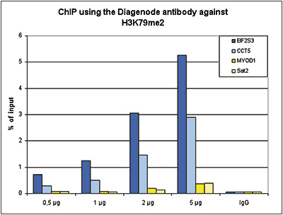
Figure 1. ChIP results obtained with the Diagenode antibody directed against H3K79me2
ChIP assays were performed using human HeLa cells, the Diagenode antibody against H3K79me2 (Cat. No. C15410051) and optimized PCR primer pairs for qPCR. ChIP was performed with the “Auto Histone ChIP-seq” kit (cat. No. C01010020), using sheared chromatin from 1 million cells. A titration consisting of 0.5, 1, 2 and 5 μg of antibody per ChIP experiment was analyzed. IgG (1 μg/IP) was used as a negative IP control. Quantitative PCR was performed with primers specific for the coding regions of the active EIF2S3 and CCT5 genes, used as positive controls, and for the inactive MYOD1) gene and the Sat2 satellite repeat, used as negative controls. Figure 1 shows the recovery, expressed as a % of input (the relative amount of immunoprecipitated DNA compared to input DNA after qPCR analysis).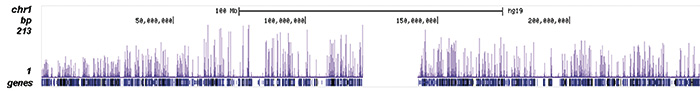
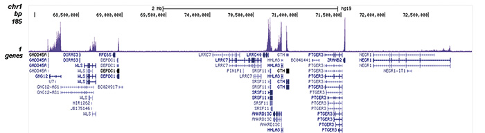
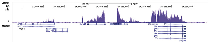
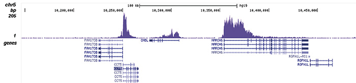
Figure 2. ChIP-seq results obtained with the Diagenode antibody directed against H3K79me2
ChIP was performed with 1 μg of the Diagenode antibody against H3K79me2 (Cat. No. C15410051) as described above. The IP’d DNA was subsequently analysed on an Illumina HiSeq 2000. Library preparation, cluster generation and sequencing were performed according to the manufacturer’s instructions. The 50 bp tags were aligned to the human genome using the BWA algorithm. Figure 2 shows the peak distribution along the complete sequence and a 5 Mb region of chromosome 1 (figure 2A and B) and in two 300 kb regions surrounding the EIF2S3 and CCT5 positive control genes (figure 2C and D).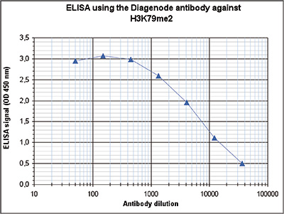
Figure 3. Determination of the antibody titer
To determine the titer of the antibody, an ELISA was performed using a serial dilution of the Diagenode antibody directed against H3K79me2 (Cat. No. C15410051). The antigen used was a peptide containing the histone modification of interest. By plotting the absorbance against the antibody dilution (Figure 3), the titer of the antibody was estimated to be 1:6,600.
Figure 4. Cross reactivity tests using the Diagenode antibody directed against H3K79me2
A Dot Blot analysis was performed to test the cross reactivity of the Diagenode antibody against H3K79me2 (Cat. No. C15410051) with peptides containing other modifications and unmodified sequences of histone H3. One hundred to 0.2 pmol of peptide containing the respective histone modification were spotted on a membrane. The antibody was used at a dilution of 1:5,000. Figure 4 shows a high specificity of the antibody for the modification of interest.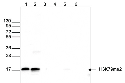
Figure 5. Western blot analysis using the Diagenode antibody directed against H3K79me2
Western blot was performed on whole cell (25 μg, lane 1) and histone extracts (15 μg, lane 2) from HeLa cells, and on 1 μg of recombinant histone H2A, H2B, H3 and H4 (lane 3, 4, 5 and 6, respectively) using the Diagenode antibody against H3K79me2 (Cat. No. C15410051) diluted 1:200 in TBS-Tween containing 5% skimmed milk. The position of the protein of interest is indicated on the right; the marker (in kDa) is shown on the left. - 出版物

How to properly cite our product/service in your work
We strongly recommend using this: H3K79me2 Antibody - ChIP-seq Grade (sample size) (Hologic Diagenode Cat# C15410051-10 Lot# A1193D). Click here to copy to clipboard.
Using our products or services in your publication? Let us know!
Chromatin environment-dependent effects of DOT1L on gene expression in male germ cells
Manon Coulée et al.
The H3K79 methyltransferase DOT1L is essential for multiple aspects of mammalian development where it has been shown to regulate gene expression. Here, by producing and integrating epigenomic and spike-in RNA-seq data, we decipher the molecular role of DOT1L during mouse spermatogenesis and show that it has opposite...DOT1L O-GlcNAcylation promotes its protein stability andMLL-fusion leukemia cell proliferation.
Song Tanjing et al.
Histone lysine methylation functions at the interface of the extracellular environment and intracellular gene expression. DOT1L is a versatile histone H3K79 methyltransferase with a prominent role in MLL-fusion leukemia, yet little is known about how DOT1L responds to extracellular stimuli. Here, we report that DOT1...Environmental enrichment induces epigenomic and genome organization changesrelevant for cognitive function
Espeso-Gil, S. et al.
In early development, the environment triggers mnemonic epigenomic programs resulting in memory and learning experiences to confer cognitive phenotypes into adulthood. To uncover how environmental stimulation impacts the epigenome and genome organization, we used the paradigm of environmental enrichment (EE) in youn...DOT1L Activity Promotes Proliferation and Protects Cortical Neural Stem Cells from Activation of ATF4-DDIT3-Mediated ER Stress In Vitro
Roidl D, Hellbach N, Bovio PP, Villarreal A, Heidrich S, Nestel S, Grüning BA, Boenisch U, Vogel T
Growing evidence suggests that the lysine methyltransferase DOT1L/KMT4 has important roles in proliferation, survival, and differentiation of stem cells in development and in disease. We investigated the function of DOT1L in neural stem cells (NSCs) of the cerebral cortex. The pharmacological inhibition and shRNA-me...Anticheckpoint pathways at telomeres in yeast
Ribeyre Cyril, Shore David
Telomeres hide (or ‘cap’) chromosome ends from DNA-damage surveillance mechanisms that arrest the cell cycle and promote repair, but the checkpoint status of telomeres is not well understood. Here we characterize the response in Saccharomyces cerevisiae to DNA double-strand breaks (DSBs) flanked by varying amounts o...