Notice (8): Undefined variable: solution_of_interest [APP/View/Products/view.ctp, line 755]Code Context<!-- BEGIN: REQUEST_FORM MODAL -->
<div id="request_formModal" class="reveal-modal medium" data-reveal aria-labelledby="modalTitle" aria-hidden="true" role="dialog">
<?= $this->element('Forms/simple_form', array('solution_of_interest' => $solution_of_interest, 'header' => $header, 'message' => $message, 'campaign_id' => $campaign_id)) ?>
$viewFile = '/home/website-server/www/app/View/Products/view.ctp'
$dataForView = array(
'language' => 'jp',
'meta_keywords' => '',
'meta_description' => 'H3K4me2 polyclonal antibody - Classic (sample size)',
'meta_title' => 'H3K4me2 polyclonal antibody - Classic (sample size)',
'product' => array(
'Product' => array(
'id' => '2200',
'antibody_id' => '112',
'name' => 'H3K4me2 polyclonal antibody (sample size)',
'description' => '<p><span>Polyclonal antibody raised in rabbit against histone H3 containing the dimethylated lysine 4 (H3K4me2), using a KLH-conjugated synthetic peptide.</span></p>',
'label1' => 'Validation Data',
'info1' => '',
'label2' => '',
'info2' => '',
'label3' => '',
'info3' => '',
'format' => '10 µg',
'catalog_number' => 'C15410035-10',
'old_catalog_number' => 'pAb-035-050',
'sf_code' => 'C15410035-D001-000582',
'type' => 'FRE',
'search_order' => '03-Antibody',
'price_EUR' => '105',
'price_USD' => '115',
'price_GBP' => '100',
'price_JPY' => '16450',
'price_CNY' => '',
'price_AUD' => '288',
'country' => 'ALL',
'except_countries' => 'None',
'quote' => false,
'in_stock' => false,
'featured' => false,
'no_promo' => false,
'online' => true,
'master' => false,
'last_datasheet_update' => '0000-00-00',
'slug' => 'h3k4me2-polyclonal-antibody-classic-sample-size-10-ug',
'meta_title' => 'H3K4me2 polyclonal antibody - Classic (sample size)',
'meta_keywords' => '',
'meta_description' => 'H3K4me2 polyclonal antibody - Classic (sample size)',
'modified' => '2022-04-08 11:04:59',
'created' => '2015-06-29 14:08:20',
'locale' => 'jpn'
),
'Antibody' => array(
'host' => '*****',
'id' => '112',
'name' => 'H3K4me2 polyclonal antibody',
'description' => 'Histones are the main constituents of the protein part of chromosomes of eukaryotic cells. They are rich in the amino acids arginine and lysine and have been greatly conserved during evolution. Histones pack the DNA into tight masses of chromatin. Two core histones of each class H2A, H2B, H3 and H4 assemble and are wrapped by 146 base pairs of DNA to form one octameric nucleosome. Histone tails undergo numerous post-translational modifications, which either directly or indirectly alter chromatin structure to facilitate transcriptional activation or repression or other nuclear processes. In addition to the genetic code, combinations of the different histone modifications reveal the so-called “histone code”. Histone methylation and demethylation is dynamically regulated by respectively histone methyl transferases and histone demethylases.',
'clonality' => '',
'isotype' => '',
'lot' => 'A936-0023',
'concentration' => '1.1 µg/µl',
'reactivity' => 'Human, Arabidopsis: positive. Other species: not tested.',
'type' => 'Polyclonal',
'purity' => 'Affinity purified polyclonal antibody.',
'classification' => 'Classic',
'application_table' => '<table>
<thead>
<tr>
<th>Applications</th>
<th>Suggested dilution</th>
<th>References</th>
</tr>
</thead>
<tbody>
<tr>
<td>ChIP/ChIP-seq <sup>*</sup></td>
<td>0.5 - 1 µg/ChIP</td>
<td>Fig 1, 2</td>
</tr>
<tr>
<td>CUT&Tag</td>
<td>0.5 µg</td>
<td>Fig 3</td>
</tr>
<tr>
<td>ELISA</td>
<td>1:500</td>
<td>Fig 4</td>
</tr>
<tr>
<td>Dot Blotting</td>
<td>1:20,000</td>
<td>Fig 5</td>
</tr>
<tr>
<td>Western Blotting</td>
<td>1:1,000</td>
<td>Fig 6</td>
</tr>
</tbody>
</table>
<p></p>
<p><small><sup>*</sup> Please note that of the optimal antibody amount per IP should be determined by the end-user. We recommend testing 0.5-5 µg per IP.</small></p>',
'storage_conditions' => 'Store at -20°C; for long storage, store at -80°C. Avoid multiple freeze-thaw cycles.',
'storage_buffer' => 'PBS containing 0.05% azide',
'precautions' => 'This product is for research use only. Not for use in diagnostic or therapeutic procedures.',
'uniprot_acc' => '',
'slug' => '',
'meta_keywords' => '',
'meta_description' => '',
'modified' => '2022-09-02 11:23:23',
'created' => '0000-00-00 00:00:00',
'select_label' => '112 - H3K4me2 polyclonal antibody (A936-0023 - 1.1 µg/µl - Human, Arabidopsis: positive. Other species: not tested. - Affinity purified polyclonal antibody. - Rabbit)'
),
'Slave' => array(),
'Group' => array(
'Group' => array(
[maximum depth reached]
),
'Master' => array(
[maximum depth reached]
),
'Product' => array(
[maximum depth reached]
)
),
'Related' => array(
(int) 0 => array(
[maximum depth reached]
)
),
'Application' => array(
(int) 0 => array(
[maximum depth reached]
),
(int) 1 => array(
[maximum depth reached]
),
(int) 2 => array(
[maximum depth reached]
),
(int) 3 => array(
[maximum depth reached]
),
(int) 4 => array(
[maximum depth reached]
),
(int) 5 => array(
[maximum depth reached]
),
(int) 6 => array(
[maximum depth reached]
)
),
'Category' => array(
(int) 0 => array(
[maximum depth reached]
),
(int) 1 => array(
[maximum depth reached]
),
(int) 2 => array(
[maximum depth reached]
),
(int) 3 => array(
[maximum depth reached]
),
(int) 4 => array(
[maximum depth reached]
),
(int) 5 => array(
[maximum depth reached]
)
),
'Document' => array(
(int) 0 => array(
[maximum depth reached]
),
(int) 1 => array(
[maximum depth reached]
),
(int) 2 => array(
[maximum depth reached]
)
),
'Feature' => array(),
'Image' => array(
(int) 0 => array(
[maximum depth reached]
)
),
'Promotion' => array(),
'Protocol' => array(),
'Publication' => array(
(int) 0 => array(
[maximum depth reached]
),
(int) 1 => array(
[maximum depth reached]
),
(int) 2 => array(
[maximum depth reached]
),
(int) 3 => array(
[maximum depth reached]
),
(int) 4 => array(
[maximum depth reached]
),
(int) 5 => array(
[maximum depth reached]
),
(int) 6 => array(
[maximum depth reached]
),
(int) 7 => array(
[maximum depth reached]
),
(int) 8 => array(
[maximum depth reached]
),
(int) 9 => array(
[maximum depth reached]
),
(int) 10 => array(
[maximum depth reached]
),
(int) 11 => array(
[maximum depth reached]
),
(int) 12 => array(
[maximum depth reached]
),
(int) 13 => array(
[maximum depth reached]
),
(int) 14 => array(
[maximum depth reached]
),
(int) 15 => array(
[maximum depth reached]
),
(int) 16 => array(
[maximum depth reached]
)
),
'Testimonial' => array(),
'Area' => array(),
'SafetySheet' => array(
(int) 0 => array(
[maximum depth reached]
),
(int) 1 => array(
[maximum depth reached]
),
(int) 2 => array(
[maximum depth reached]
),
(int) 3 => array(
[maximum depth reached]
),
(int) 4 => array(
[maximum depth reached]
),
(int) 5 => array(
[maximum depth reached]
),
(int) 6 => array(
[maximum depth reached]
),
(int) 7 => array(
[maximum depth reached]
)
)
),
'meta_canonical' => 'https://www.diagenode.com/jp/p/h3k4me2-polyclonal-antibody-classic-50-mg'
)
$language = 'jp'
$meta_keywords = ''
$meta_description = 'H3K4me2 polyclonal antibody - Classic (sample size)'
$meta_title = 'H3K4me2 polyclonal antibody - Classic (sample size)'
$product = array(
'Product' => array(
'id' => '2200',
'antibody_id' => '112',
'name' => 'H3K4me2 polyclonal antibody (sample size)',
'description' => '<p><span>Polyclonal antibody raised in rabbit against histone H3 containing the dimethylated lysine 4 (H3K4me2), using a KLH-conjugated synthetic peptide.</span></p>',
'label1' => 'Validation Data',
'info1' => '<div class="row">
<div class="small-6 columns">
<p><img src="https://www.diagenode.com/img/product/antibodies/C15410035-chip.png" alt="H3K4me2 Antibody ChIP Grade" caption="false" width="432" height="303" /></p>
</div>
<div class="small-6 columns">
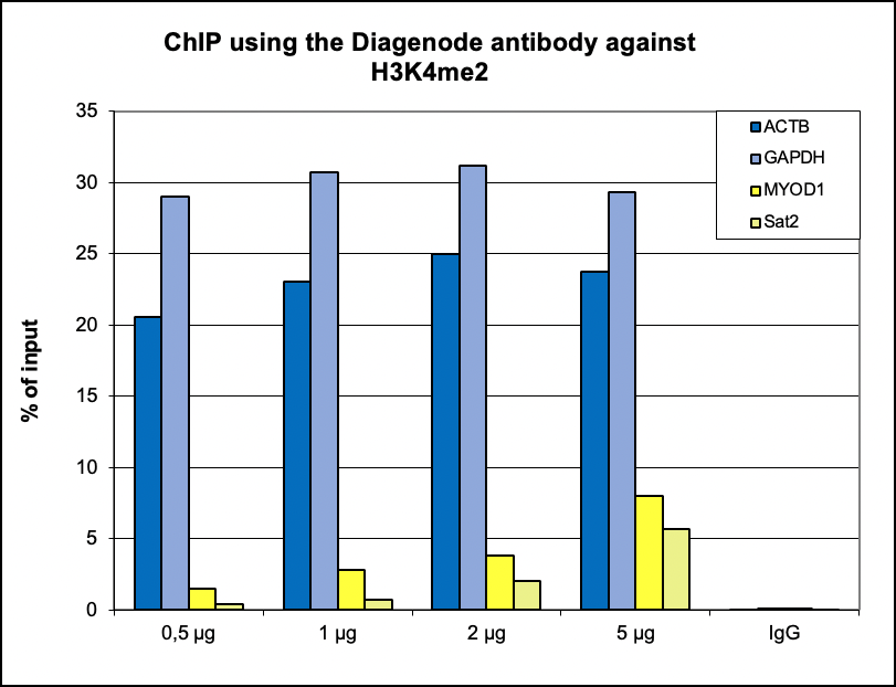
<p><small><strong>Figure 1. ChIP results obtained with the Diagenode antibody directed against H3K4me2</strong><br /> ChIP was performed with the Diagenode antibody against H3K4me2 (cat. No. C15410035) on sheared chromatin from 500,000 K562 cells using the “iDeal ChIP-seq” kit (cat. No. C01010051). A titration of the antibody consisting of 0.5, 1, 2 and 5 µg per ChIP experiment was analysed. IgG (1 µg/IP) was used as negative IP control. Quantitative PCR was performed with primers for a region upstream of the ACTB and GAPDH promoters, used as positive controls, and for the MYOD1 gene and the Sat2 satellite repeat, used as negative controls. The graph shows the recovery, expressed as a % of input (the relative amount of immunoprecipitated DNA compared to input DNA after qPCR analysis).</small></p>
</div>
</div>
<div class="row">
<div class="small-12 columns">
<p class="text-center">A. <img src="https://www.diagenode.com/img/product/antibodies/c15410035-chipseq-a.jpg" alt="H3K4me2 Antibody Cut &" caption="false" width="629" height="101" /></p>
<p class="text-center">B. <img src="https://www.diagenode.com/img/product/antibodies/c15410035-chipseq-b.jpg" alt="H3K4me2 Antibody for ChIP-seq" caption="false" width="626" height="116" /></p>
</div>
</div>
<div class="extra-spaced"></div>
<div class="row">
<div class="small-12 columns">
<p class="text-center">C. <img src="https://www.diagenode.com/img/product/antibodies/c15410035-chipseq-c.jpg" alt="H3K4me2 Antibody for ChIP-seq assay" caption="false" width="626" height="97" /></p>
<p class="text-center">D. <img src="https://www.diagenode.com/img/product/antibodies/c15410035-chipseq-d.jpg" alt="H3K4me2 Antibody validated in ChIP-seq" caption="false" width="626" height="89" /></p>
</div>
</div>
<div class="row">
<div class="small-12 columns">
<p><small><strong>Figure 2. ChIP-seq results obtained with the Diagenode antibody directed against H3K4me2</strong><br />ChIP was performed on HeLa cells using 0.5 µg of the Diagenode antibody against H3K4me2 (cat. No. C15410035). The IP'd DNA was analysed on an Illumina Hiseq. Library preparation, cluster generation and sequencing were performed according to the manufacturer's instructions. The 51 bp tags were aligned to the human genome using the BWA algorithm. Figure 2 shows the peak distribution along the complete sequence and a 1.5 Mb region of the human X-chromosome (figure 2A and 2B) and in 2 chromosomal regions surrounding the ACTB and GAPDH positive control genes (figure 2C and D, respectively).</small></p>
</div>
</div>
<div class="row">
<div class="small-12 columns">
<p class="text-center">A. <img src="https://www.diagenode.com/img/product/antibodies/C15410035-cuttag-a.png" alt="H3K4me2 Antibody Cut&Tag" caption="false" width="629" height="101" /></p>
<p class="text-center">B. <img src="https://www.diagenode.com/img/product/antibodies/C15410035-cuttag-b.png" alt="H3K4me2 Antibody Cut&Tag" caption="false" width="626" height="116" /></p>
</div>
</div>
<div class="row">
<div class="small-12 columns">
<p><small><strong>Figure 3. Cut&Tag results obtained with the Diagenode antibody directed against H3K4me2</strong><br />CUT&TAG (Kaya-Okur, H.S., Nat Commun 10, 1930, 2019) was performed on 50,000 K562 cells using 0.5 µg of the Diagenode antibody against H3K4me2 (cat. No. C15410035) and the Diagenode pA-Tn5 transposase (C01070001). The libraries were subsequently analysed on an Illumina NextSeq 500 sequencer (2x75 paired-end reads) according to the manufacturer's instructions. The tags were aligned to the human genome (hg19) using the BWA algorithm. Figure 3 shows the peak distribution in 2 genomic regions surrounding the FOS gene on chromosome 14 and the EIF2S3 gene on the X-chromosome (figure 3A and B, respectively).</small></p>
</div>
</div>
<div class="row">
<div class="small-6 columns">
<p><img src="https://www.diagenode.com/img/product/antibodies/C15410035-ELISA.jpg" alt="H3K4me2 Antibody ELISA validation" caption="false" width="432" height="348" /></p>
</div>
<div class="small-6 columns">
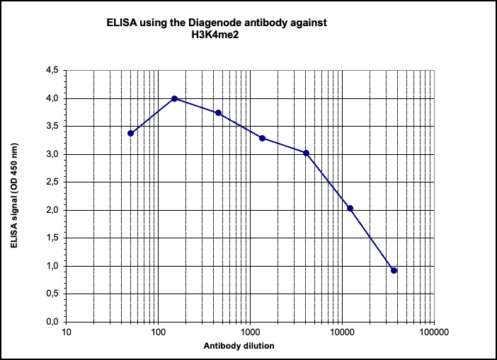
<p><small><strong>Figure 4. Determination of the titer</strong><br />To determine the titer, an ELISA was performed using a serial dilution of the Diagenode antibody directed against H3K4me2 (cat. No. C15410035) in antigen coated wells. The antigen used was a peptide containing the histone modification of interest. By plotting the absorbance against the antibody dilution (Figure 4), the titer of the antibody was estimated to be 1:12,000.</small></p>
</div>
</div>
<div class="row">
<div class="small-4 columns">
<p><img src="https://www.diagenode.com/img/product/antibodies/C15410035-fig4-dotblot.png" alt="H3K4me2 Antibody validated in Dot Blot" caption="false" width="278" height="196" /></p>
</div>
<div class="small-8 columns">
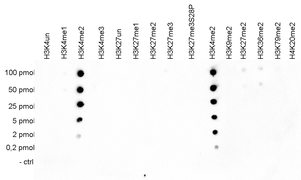
<p><small><strong>Figure 5. Cross reactivity test using the Diagenode antibody directed against H3K4me2</strong> <br />A Dot Blot analysis was performed to test the cross reactivity of the Diagenode antibody against H3K4me2 (cat. No. C15410035) with peptides containing other modifications of histone H3 and H4 and the unmodified H3K4 sequence. One hundred to 0.2 pmol of the respective peptides were spotted on a membrane. The antibody was used at a dilution of 1:2,000. Figure 5 shows a high specificity of the antibody for the modification of interest.</small></p>
</div>
</div>
<div class="extra-spaced"></div>
<div class="row">
<div class="small-4 columns">
<p><img src="https://www.diagenode.com/img/product/antibodies/C15410035-fig5-WB.png" alt="H3K4me2 Antibody validated in Western Blot" caption="false" width="142" height="139" /></p>
</div>
<div class="small-8 columns">
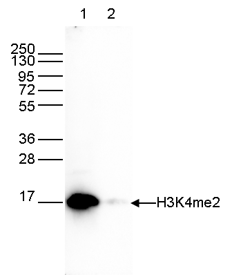
<p><small><strong>Figure 6. Western blot analysis using the Diagenode antibody directed against H3K4me2</strong><br /> Western blot was performed on whole cell extracts (25 µg, lane 1) and on 1 µg of recombinant histone H3 (lane 2) using the Diagenode antibody against H3K4me2 (cat. No. C15410035) diluted 1:1,000 in TBS-Tween containing 5% skimmed milk. The position of the protein of interest is indicated on the right; the marker (in kDa) is shown on the left.</small></p>
</div>
</div>',
'label2' => 'Target Description',
'info2' => '<p>Histones are the main constituents of the protein part of chromosomes of eukaryotic cells. They are rich in the amino acids arginine and lysine and have been greatly conserved during evolution. Histones pack the DNA into tight masses of chromatin. Two core histones of each class H2A, H2B, H3 and H4 assemble and are wrapped by 146 base pairs of DNA to form one octameric nucleosome. Histone tails undergo numerous post-translational modifications, which either directly or indirectly alter chromatin structure to facilitate transcriptional activation or repression or other nuclear processes. In addition to the genetic code, combinations of the different histone modifications reveal the so-called “histone code”. Histone methylation and demethylation is dynamically regulated by respectively histone methyl transferases and histone demethylases.</p>',
'label3' => '',
'info3' => '',
'format' => '10 µg',
'catalog_number' => 'C15410035-10',
'old_catalog_number' => 'pAb-035-050',
'sf_code' => 'C15410035-D001-000582',
'type' => 'FRE',
'search_order' => '03-Antibody',
'price_EUR' => '105',
'price_USD' => '115',
'price_GBP' => '100',
'price_JPY' => '16450',
'price_CNY' => '',
'price_AUD' => '288',
'country' => 'ALL',
'except_countries' => 'None',
'quote' => false,
'in_stock' => false,
'featured' => false,
'no_promo' => false,
'online' => true,
'master' => false,
'last_datasheet_update' => '0000-00-00',
'slug' => 'h3k4me2-polyclonal-antibody-classic-sample-size-10-ug',
'meta_title' => 'H3K4me2 polyclonal antibody - Classic (sample size)',
'meta_keywords' => '',
'meta_description' => 'H3K4me2 polyclonal antibody - Classic (sample size)',
'modified' => '2022-04-08 11:04:59',
'created' => '2015-06-29 14:08:20',
'locale' => 'jpn'
),
'Antibody' => array(
'host' => '*****',
'id' => '112',
'name' => 'H3K4me2 polyclonal antibody',
'description' => 'Histones are the main constituents of the protein part of chromosomes of eukaryotic cells. They are rich in the amino acids arginine and lysine and have been greatly conserved during evolution. Histones pack the DNA into tight masses of chromatin. Two core histones of each class H2A, H2B, H3 and H4 assemble and are wrapped by 146 base pairs of DNA to form one octameric nucleosome. Histone tails undergo numerous post-translational modifications, which either directly or indirectly alter chromatin structure to facilitate transcriptional activation or repression or other nuclear processes. In addition to the genetic code, combinations of the different histone modifications reveal the so-called “histone code”. Histone methylation and demethylation is dynamically regulated by respectively histone methyl transferases and histone demethylases.',
'clonality' => '',
'isotype' => '',
'lot' => 'A936-0023',
'concentration' => '1.1 µg/µl',
'reactivity' => 'Human, Arabidopsis: positive. Other species: not tested.',
'type' => 'Polyclonal',
'purity' => 'Affinity purified polyclonal antibody.',
'classification' => 'Classic',
'application_table' => '<table>
<thead>
<tr>
<th>Applications</th>
<th>Suggested dilution</th>
<th>References</th>
</tr>
</thead>
<tbody>
<tr>
<td>ChIP/ChIP-seq <sup>*</sup></td>
<td>0.5 - 1 µg/ChIP</td>
<td>Fig 1, 2</td>
</tr>
<tr>
<td>CUT&Tag</td>
<td>0.5 µg</td>
<td>Fig 3</td>
</tr>
<tr>
<td>ELISA</td>
<td>1:500</td>
<td>Fig 4</td>
</tr>
<tr>
<td>Dot Blotting</td>
<td>1:20,000</td>
<td>Fig 5</td>
</tr>
<tr>
<td>Western Blotting</td>
<td>1:1,000</td>
<td>Fig 6</td>
</tr>
</tbody>
</table>
<p></p>
<p><small><sup>*</sup> Please note that of the optimal antibody amount per IP should be determined by the end-user. We recommend testing 0.5-5 µg per IP.</small></p>',
'storage_conditions' => 'Store at -20°C; for long storage, store at -80°C. Avoid multiple freeze-thaw cycles.',
'storage_buffer' => 'PBS containing 0.05% azide',
'precautions' => 'This product is for research use only. Not for use in diagnostic or therapeutic procedures.',
'uniprot_acc' => '',
'slug' => '',
'meta_keywords' => '',
'meta_description' => '',
'modified' => '2022-09-02 11:23:23',
'created' => '0000-00-00 00:00:00',
'select_label' => '112 - H3K4me2 polyclonal antibody (A936-0023 - 1.1 µg/µl - Human, Arabidopsis: positive. Other species: not tested. - Affinity purified polyclonal antibody. - Rabbit)'
),
'Slave' => array(),
'Group' => array(
'Group' => array(
'id' => '46',
'name' => 'C15410035',
'product_id' => '2199',
'modified' => '2016-02-18 20:49:59',
'created' => '2016-02-18 20:49:59'
),
'Master' => array(
'id' => '2199',
'antibody_id' => '112',
'name' => 'H3K4me2 Antibody',
'description' => '<p><span>Polyclonal antibody raised in rabbit against histone <strong>H3 containing the dimethylated lysine 4 (H3K4me2),</strong> using a KLH-conjugated synthetic peptide.</span></p>',
'label1' => 'Validation Data',
'info1' => '<div class="row">
<div class="small-6 columns">
<p><img src="https://www.diagenode.com/img/product/antibodies/C15410035-chip.png" alt="H3K4me2 Antibody ChIP Grade" caption="false" width="432" height="303" /></p>
</div>
<div class="small-6 columns">
<p><small><strong>Figure 1. ChIP results obtained with the Diagenode antibody directed against H3K4me2</strong><br /> ChIP was performed with the Diagenode antibody against H3K4me2 (cat. No. C15410035) on sheared chromatin from 500,000 K562 cells using the “iDeal ChIP-seq” kit (cat. No. C01010051). A titration of the antibody consisting of 0.5, 1, 2 and 5 µg per ChIP experiment was analysed. IgG (1 µg/IP) was used as negative IP control. Quantitative PCR was performed with primers for a region upstream of the ACTB and GAPDH promoters, used as positive controls, and for the MYOD1 gene and the Sat2 satellite repeat, used as negative controls. The graph shows the recovery, expressed as a % of input (the relative amount of immunoprecipitated DNA compared to input DNA after qPCR analysis).</small></p>
</div>
</div>
<div class="row">
<div class="small-12 columns">
<p class="text-center">A. <img src="https://www.diagenode.com/img/product/antibodies/c15410035-chipseq-a.jpg" alt="H3K4me2 Antibody Cut &" caption="false" width="629" height="101" /></p>
<p class="text-center">B. <img src="https://www.diagenode.com/img/product/antibodies/c15410035-chipseq-b.jpg" alt="H3K4me2 Antibody for ChIP-seq" caption="false" width="626" height="116" /></p>
</div>
</div>
<div class="extra-spaced"></div>
<div class="row">
<div class="small-12 columns">
<p class="text-center">C. <img src="https://www.diagenode.com/img/product/antibodies/c15410035-chipseq-c.jpg" alt="H3K4me2 Antibody for ChIP-seq assay" caption="false" width="626" height="97" /></p>
<p class="text-center">D. <img src="https://www.diagenode.com/img/product/antibodies/c15410035-chipseq-d.jpg" alt="H3K4me2 Antibody validated in ChIP-seq" caption="false" width="626" height="89" /></p>
</div>
</div>
<div class="row">
<div class="small-12 columns">
<p><small><strong>Figure 2. ChIP-seq results obtained with the Diagenode antibody directed against H3K4me2</strong><br />ChIP was performed on HeLa cells using 0.5 µg of the Diagenode antibody against H3K4me2 (cat. No. C15410035). The IP'd DNA was analysed on an Illumina Hiseq. Library preparation, cluster generation and sequencing were performed according to the manufacturer's instructions. The 51 bp tags were aligned to the human genome using the BWA algorithm. Figure 2 shows the peak distribution along the complete sequence and a 1.5 Mb region of the human X-chromosome (figure 2A and 2B) and in 2 chromosomal regions surrounding the ACTB and GAPDH positive control genes (figure 2C and D, respectively).</small></p>
</div>
</div>
<div class="row">
<div class="small-12 columns">
<p class="text-center">A. <img src="https://www.diagenode.com/img/product/antibodies/C15410035-cuttag-a.png" alt="H3K4me2 Antibody Cut&Tag" caption="false" width="629" height="101" /></p>
<p class="text-center">B. <img src="https://www.diagenode.com/img/product/antibodies/C15410035-cuttag-b.png" alt="H3K4me2 Antibody Cut&Tag" caption="false" width="626" height="116" /></p>
</div>
</div>
<div class="row">
<div class="small-12 columns">
<p><small><strong>Figure 3. Cut&Tag results obtained with the Diagenode antibody directed against H3K4me2</strong><br />CUT&TAG (Kaya-Okur, H.S., Nat Commun 10, 1930, 2019) was performed on 50,000 K562 cells using 0.5 µg of the Diagenode antibody against H3K4me2 (cat. No. C15410035) and the Diagenode pA-Tn5 transposase (C01070001). The libraries were subsequently analysed on an Illumina NextSeq 500 sequencer (2x75 paired-end reads) according to the manufacturer's instructions. The tags were aligned to the human genome (hg19) using the BWA algorithm. Figure 3 shows the peak distribution in 2 genomic regions surrounding the FOS gene on chromosome 14 and the EIF2S3 gene on the X-chromosome (figure 3A and B, respectively).</small></p>
</div>
</div>
<div class="row">
<div class="small-6 columns">
<p><img src="https://www.diagenode.com/img/product/antibodies/C15410035-ELISA.jpg" alt="H3K4me2 Antibody ELISA validation" caption="false" width="432" height="348" /></p>
</div>
<div class="small-6 columns">
<p><small><strong>Figure 4. Determination of the titer</strong><br />To determine the titer, an ELISA was performed using a serial dilution of the Diagenode antibody directed against H3K4me2 (cat. No. C15410035) in antigen coated wells. The antigen used was a peptide containing the histone modification of interest. By plotting the absorbance against the antibody dilution (Figure 4), the titer of the antibody was estimated to be 1:12,000.</small></p>
</div>
</div>
<div class="row">
<div class="small-4 columns">
<p><img src="https://www.diagenode.com/img/product/antibodies/C15410035-fig4-dotblot.png" alt="H3K4me2 Antibody validated in Dot Blot" caption="false" width="278" height="196" /></p>
</div>
<div class="small-8 columns">
<p><small><strong>Figure 5. Cross reactivity test using the Diagenode antibody directed against H3K4me2</strong> <br />A Dot Blot analysis was performed to test the cross reactivity of the Diagenode antibody against H3K4me2 (cat. No. C15410035) with peptides containing other modifications of histone H3 and H4 and the unmodified H3K4 sequence. One hundred to 0.2 pmol of the respective peptides were spotted on a membrane. The antibody was used at a dilution of 1:2,000. Figure 5 shows a high specificity of the antibody for the modification of interest.</small></p>
</div>
</div>
<div class="extra-spaced"></div>
<div class="row">
<div class="small-4 columns">
<p><img src="https://www.diagenode.com/img/product/antibodies/C15410035-fig5-WB.png" alt="H3K4me2 Antibody validated in Western Blot" caption="false" width="142" height="139" /></p>
</div>
<div class="small-8 columns">
<p><small><strong>Figure 6. Western blot analysis using the Diagenode antibody directed against H3K4me2</strong><br /> Western blot was performed on whole cell extracts (25 µg, lane 1) and on 1 µg of recombinant histone H3 (lane 2) using the Diagenode antibody against H3K4me2 (cat. No. C15410035) diluted 1:1,000 in TBS-Tween containing 5% skimmed milk. The position of the protein of interest is indicated on the right; the marker (in kDa) is shown on the left.</small></p>
</div>
</div>',
'label2' => 'Target Description',
'info2' => '<p>Histones are the main constituents of the protein part of chromosomes of eukaryotic cells. They are rich in the amino acids arginine and lysine and have been greatly conserved during evolution. Histones pack the DNA into tight masses of chromatin. Two core histones of each class H2A, H2B, H3 and H4 assemble and are wrapped by 146 base pairs of DNA to form one octameric nucleosome. Histone tails undergo numerous post-translational modifications, which either directly or indirectly alter chromatin structure to facilitate transcriptional activation or repression or other nuclear processes. In addition to the genetic code, combinations of the different histone modifications reveal the so-called “histone code”. Histone methylation and demethylation is dynamically regulated by respectively histone methyl transferases and histone demethylases.</p>',
'label3' => '',
'info3' => '',
'format' => '50 μg',
'catalog_number' => 'C15410035',
'old_catalog_number' => 'pAb-035-050',
'sf_code' => 'C15410035-D001-000581',
'type' => 'FRE',
'search_order' => '03-Antibody',
'price_EUR' => '380',
'price_USD' => '380',
'price_GBP' => '340',
'price_JPY' => '59525',
'price_CNY' => '',
'price_AUD' => '950',
'country' => 'ALL',
'except_countries' => 'None',
'quote' => false,
'in_stock' => false,
'featured' => false,
'no_promo' => false,
'online' => true,
'master' => true,
'last_datasheet_update' => 'January 17, 2020',
'slug' => 'h3k4me2-polyclonal-antibody-classic-50-mg',
'meta_title' => 'H3K4me2 Antibody - ChIP-seq Grade (C15410035) | Diagenode',
'meta_keywords' => '',
'meta_description' => 'H3K4me2 (Histone H3 dimethylated at lysine 4) Polyclonal Antibody validated in ChIP-Seq, ChIP-qPCR, ELISA, DB, WB and IF. Batch-specific data available on the website. Sample size available.',
'modified' => '2024-01-28 11:10:10',
'created' => '2015-06-29 14:08:20'
),
'Product' => array(
(int) 0 => array(
[maximum depth reached]
)
)
),
'Related' => array(
(int) 0 => array(
'id' => '2199',
'antibody_id' => '112',
'name' => 'H3K4me2 Antibody',
'description' => '<p><span>Polyclonal antibody raised in rabbit against histone <strong>H3 containing the dimethylated lysine 4 (H3K4me2),</strong> using a KLH-conjugated synthetic peptide.</span></p>',
'label1' => 'Validation Data',
'info1' => '<div class="row">
<div class="small-6 columns">
<p><img src="https://www.diagenode.com/img/product/antibodies/C15410035-chip.png" alt="H3K4me2 Antibody ChIP Grade" caption="false" width="432" height="303" /></p>
</div>
<div class="small-6 columns">
<p><small><strong>Figure 1. ChIP results obtained with the Diagenode antibody directed against H3K4me2</strong><br /> ChIP was performed with the Diagenode antibody against H3K4me2 (cat. No. C15410035) on sheared chromatin from 500,000 K562 cells using the “iDeal ChIP-seq” kit (cat. No. C01010051). A titration of the antibody consisting of 0.5, 1, 2 and 5 µg per ChIP experiment was analysed. IgG (1 µg/IP) was used as negative IP control. Quantitative PCR was performed with primers for a region upstream of the ACTB and GAPDH promoters, used as positive controls, and for the MYOD1 gene and the Sat2 satellite repeat, used as negative controls. The graph shows the recovery, expressed as a % of input (the relative amount of immunoprecipitated DNA compared to input DNA after qPCR analysis).</small></p>
</div>
</div>
<div class="row">
<div class="small-12 columns">
<p class="text-center">A. <img src="https://www.diagenode.com/img/product/antibodies/c15410035-chipseq-a.jpg" alt="H3K4me2 Antibody Cut &" caption="false" width="629" height="101" /></p>
<p class="text-center">B. <img src="https://www.diagenode.com/img/product/antibodies/c15410035-chipseq-b.jpg" alt="H3K4me2 Antibody for ChIP-seq" caption="false" width="626" height="116" /></p>
</div>
</div>
<div class="extra-spaced"></div>
<div class="row">
<div class="small-12 columns">
<p class="text-center">C. <img src="https://www.diagenode.com/img/product/antibodies/c15410035-chipseq-c.jpg" alt="H3K4me2 Antibody for ChIP-seq assay" caption="false" width="626" height="97" /></p>
<p class="text-center">D. <img src="https://www.diagenode.com/img/product/antibodies/c15410035-chipseq-d.jpg" alt="H3K4me2 Antibody validated in ChIP-seq" caption="false" width="626" height="89" /></p>
</div>
</div>
<div class="row">
<div class="small-12 columns">
<p><small><strong>Figure 2. ChIP-seq results obtained with the Diagenode antibody directed against H3K4me2</strong><br />ChIP was performed on HeLa cells using 0.5 µg of the Diagenode antibody against H3K4me2 (cat. No. C15410035). The IP'd DNA was analysed on an Illumina Hiseq. Library preparation, cluster generation and sequencing were performed according to the manufacturer's instructions. The 51 bp tags were aligned to the human genome using the BWA algorithm. Figure 2 shows the peak distribution along the complete sequence and a 1.5 Mb region of the human X-chromosome (figure 2A and 2B) and in 2 chromosomal regions surrounding the ACTB and GAPDH positive control genes (figure 2C and D, respectively).</small></p>
</div>
</div>
<div class="row">
<div class="small-12 columns">
<p class="text-center">A. <img src="https://www.diagenode.com/img/product/antibodies/C15410035-cuttag-a.png" alt="H3K4me2 Antibody Cut&Tag" caption="false" width="629" height="101" /></p>
<p class="text-center">B. <img src="https://www.diagenode.com/img/product/antibodies/C15410035-cuttag-b.png" alt="H3K4me2 Antibody Cut&Tag" caption="false" width="626" height="116" /></p>
</div>
</div>
<div class="row">
<div class="small-12 columns">
<p><small><strong>Figure 3. Cut&Tag results obtained with the Diagenode antibody directed against H3K4me2</strong><br />CUT&TAG (Kaya-Okur, H.S., Nat Commun 10, 1930, 2019) was performed on 50,000 K562 cells using 0.5 µg of the Diagenode antibody against H3K4me2 (cat. No. C15410035) and the Diagenode pA-Tn5 transposase (C01070001). The libraries were subsequently analysed on an Illumina NextSeq 500 sequencer (2x75 paired-end reads) according to the manufacturer's instructions. The tags were aligned to the human genome (hg19) using the BWA algorithm. Figure 3 shows the peak distribution in 2 genomic regions surrounding the FOS gene on chromosome 14 and the EIF2S3 gene on the X-chromosome (figure 3A and B, respectively).</small></p>
</div>
</div>
<div class="row">
<div class="small-6 columns">
<p><img src="https://www.diagenode.com/img/product/antibodies/C15410035-ELISA.jpg" alt="H3K4me2 Antibody ELISA validation" caption="false" width="432" height="348" /></p>
</div>
<div class="small-6 columns">
<p><small><strong>Figure 4. Determination of the titer</strong><br />To determine the titer, an ELISA was performed using a serial dilution of the Diagenode antibody directed against H3K4me2 (cat. No. C15410035) in antigen coated wells. The antigen used was a peptide containing the histone modification of interest. By plotting the absorbance against the antibody dilution (Figure 4), the titer of the antibody was estimated to be 1:12,000.</small></p>
</div>
</div>
<div class="row">
<div class="small-4 columns">
<p><img src="https://www.diagenode.com/img/product/antibodies/C15410035-fig4-dotblot.png" alt="H3K4me2 Antibody validated in Dot Blot" caption="false" width="278" height="196" /></p>
</div>
<div class="small-8 columns">
<p><small><strong>Figure 5. Cross reactivity test using the Diagenode antibody directed against H3K4me2</strong> <br />A Dot Blot analysis was performed to test the cross reactivity of the Diagenode antibody against H3K4me2 (cat. No. C15410035) with peptides containing other modifications of histone H3 and H4 and the unmodified H3K4 sequence. One hundred to 0.2 pmol of the respective peptides were spotted on a membrane. The antibody was used at a dilution of 1:2,000. Figure 5 shows a high specificity of the antibody for the modification of interest.</small></p>
</div>
</div>
<div class="extra-spaced"></div>
<div class="row">
<div class="small-4 columns">
<p><img src="https://www.diagenode.com/img/product/antibodies/C15410035-fig5-WB.png" alt="H3K4me2 Antibody validated in Western Blot" caption="false" width="142" height="139" /></p>
</div>
<div class="small-8 columns">
<p><small><strong>Figure 6. Western blot analysis using the Diagenode antibody directed against H3K4me2</strong><br /> Western blot was performed on whole cell extracts (25 µg, lane 1) and on 1 µg of recombinant histone H3 (lane 2) using the Diagenode antibody against H3K4me2 (cat. No. C15410035) diluted 1:1,000 in TBS-Tween containing 5% skimmed milk. The position of the protein of interest is indicated on the right; the marker (in kDa) is shown on the left.</small></p>
</div>
</div>',
'label2' => 'Target Description',
'info2' => '<p>Histones are the main constituents of the protein part of chromosomes of eukaryotic cells. They are rich in the amino acids arginine and lysine and have been greatly conserved during evolution. Histones pack the DNA into tight masses of chromatin. Two core histones of each class H2A, H2B, H3 and H4 assemble and are wrapped by 146 base pairs of DNA to form one octameric nucleosome. Histone tails undergo numerous post-translational modifications, which either directly or indirectly alter chromatin structure to facilitate transcriptional activation or repression or other nuclear processes. In addition to the genetic code, combinations of the different histone modifications reveal the so-called “histone code”. Histone methylation and demethylation is dynamically regulated by respectively histone methyl transferases and histone demethylases.</p>',
'label3' => '',
'info3' => '',
'format' => '50 μg',
'catalog_number' => 'C15410035',
'old_catalog_number' => 'pAb-035-050',
'sf_code' => 'C15410035-D001-000581',
'type' => 'FRE',
'search_order' => '03-Antibody',
'price_EUR' => '380',
'price_USD' => '380',
'price_GBP' => '340',
'price_JPY' => '59525',
'price_CNY' => '',
'price_AUD' => '950',
'country' => 'ALL',
'except_countries' => 'None',
'quote' => false,
'in_stock' => false,
'featured' => false,
'no_promo' => false,
'online' => true,
'master' => true,
'last_datasheet_update' => 'January 17, 2020',
'slug' => 'h3k4me2-polyclonal-antibody-classic-50-mg',
'meta_title' => 'H3K4me2 Antibody - ChIP-seq Grade (C15410035) | Diagenode',
'meta_keywords' => '',
'meta_description' => 'H3K4me2 (Histone H3 dimethylated at lysine 4) Polyclonal Antibody validated in ChIP-Seq, ChIP-qPCR, ELISA, DB, WB and IF. Batch-specific data available on the website. Sample size available.',
'modified' => '2024-01-28 11:10:10',
'created' => '2015-06-29 14:08:20',
'ProductsRelated' => array(
[maximum depth reached]
),
'Image' => array(
[maximum depth reached]
)
)
),
'Application' => array(
(int) 0 => array(
'id' => '20',
'position' => '10',
'parent_id' => '40',
'name' => 'ELISA',
'description' => '<div class="row">
<div class="small-12 medium-12 large-12 columns">Enzyme-linked immunosorbent assay.</div>
</div>',
'in_footer' => false,
'in_menu' => false,
'online' => true,
'tabular' => true,
'slug' => 'elisa-antibodies',
'meta_keywords' => ' ELISA Antibodies,Monoclonal antibody, Polyclonal antibody',
'meta_description' => 'Diagenode offers Monoclonal & Polyclonal antibodies for ELISA applications',
'meta_title' => 'ELISA Antibodies - Monoclonal & Polyclonal antibody | Diagenode',
'modified' => '2016-01-13 12:21:41',
'created' => '2014-07-08 08:13:28',
'ProductsApplication' => array(
[maximum depth reached]
)
),
(int) 1 => array(
'id' => '28',
'position' => '10',
'parent_id' => '40',
'name' => 'DB',
'description' => '<p>Dot blotting</p>',
'in_footer' => false,
'in_menu' => false,
'online' => true,
'tabular' => true,
'slug' => 'dot-blotting',
'meta_keywords' => 'Dot blotting,Monoclonal & Polyclonal antibody,',
'meta_description' => 'Diagenode offers Monoclonal & Polyclonal antibodies for Dot blotting applications',
'meta_title' => 'Dot blotting Antibodies - Monoclonal & Polyclonal antibody | Diagenode',
'modified' => '2016-01-13 14:40:49',
'created' => '2015-07-08 13:45:05',
'ProductsApplication' => array(
[maximum depth reached]
)
),
(int) 2 => array(
'id' => '19',
'position' => '10',
'parent_id' => '40',
'name' => 'WB',
'description' => '<p><strong>Western blot</strong> : The quality of antibodies used in this technique is crucial for correct and specific protein identification. Diagenode offers huge selection of highly sensitive and specific western blot-validated antibodies.</p>
<p>Learn more about: <a href="https://www.diagenode.com/applications/western-blot">Loading control, MW marker visualization</a><em>. <br /></em></p>
<p><em></em>Check our selection of antibodies validated in Western blot.</p>',
'in_footer' => false,
'in_menu' => false,
'online' => true,
'tabular' => true,
'slug' => 'western-blot-antibodies',
'meta_keywords' => ' Western Blot Antibodies ,western blot protocol,Western Blotting Products,Polyclonal antibodies ,monoclonal antibodies ',
'meta_description' => 'Diagenode offers a wide range of antibodies and technical support for western blot applications',
'meta_title' => ' Western Blot - Monoclonal antibody - Polyclonal antibody | Diagenode',
'modified' => '2016-04-26 12:44:51',
'created' => '2015-01-07 09:20:00',
'ProductsApplication' => array(
[maximum depth reached]
)
),
(int) 3 => array(
'id' => '29',
'position' => '10',
'parent_id' => '40',
'name' => 'IF',
'description' => '<p><strong>Immunofluorescence</strong>:</p>
<p>Diagenode offers huge selection of highly sensitive antibodies validated in IF.</p>
<p><img src="https://www.diagenode.com/img/product/antibodies/C15200229-IF.jpg" alt="" height="245" width="256" /></p>
<p><sup><strong>Immunofluorescence using the Diagenode monoclonal antibody directed against CRISPR/Cas9</strong></sup></p>
<p><sup>HeLa cells transfected with a Cas9 expression vector (left) or untransfected cells (right) were fixed in methanol at -20°C, permeabilized with acetone at -20°C and blocked with PBS containing 2% BSA. The cells were stained with the Cas9 C-terminal antibody (Cat. No. C15200229) diluted 1:400, followed by incubation with an anti-mouse secondary antibody coupled to AF488. The bottom images show counter-staining of the nuclei with Hoechst 33342.</sup></p>
<h5><sup>Check our selection of antibodies validated in IF.</sup></h5>',
'in_footer' => false,
'in_menu' => false,
'online' => true,
'tabular' => true,
'slug' => 'immunofluorescence',
'meta_keywords' => 'Immunofluorescence,Monoclonal antibody,Polyclonal antibody',
'meta_description' => 'Diagenode offers a wide range of antibodies and technical support for Immunofluorescence applications',
'meta_title' => 'Immunofluorescence - Monoclonal antibody - Polyclonal antibody | Diagenode',
'modified' => '2016-04-27 16:23:10',
'created' => '2015-07-08 13:46:02',
'ProductsApplication' => array(
[maximum depth reached]
)
),
(int) 4 => array(
'id' => '42',
'position' => '10',
'parent_id' => '40',
'name' => 'ChIP-seq (ab)',
'description' => '',
'in_footer' => false,
'in_menu' => false,
'online' => true,
'tabular' => true,
'slug' => 'chip-seq-antibodies',
'meta_keywords' => 'Chromatin Immunoprecipitation Sequencing,ChIP-Seq,ChIP-seq grade antibodies,DNA purification,qPCR,Shearing of chromatin',
'meta_description' => 'Diagenode offers a wide range of antibodies and technical support for ChIP Sequencing applications',
'meta_title' => 'ChIP Sequencing Antibodies (ChIP-Seq) | Diagenode',
'modified' => '2016-01-20 11:06:19',
'created' => '2015-10-20 11:44:45',
'ProductsApplication' => array(
[maximum depth reached]
)
),
(int) 5 => array(
'id' => '43',
'position' => '10',
'parent_id' => '40',
'name' => 'ChIP-qPCR (ab)',
'description' => '',
'in_footer' => false,
'in_menu' => false,
'online' => true,
'tabular' => true,
'slug' => 'chip-qpcr-antibodies',
'meta_keywords' => 'Chromatin Immunoprecipitation Sequencing,ChIP-Seq,ChIP-seq grade antibodies,DNA purification,qPCR,Shearing of chromatin',
'meta_description' => 'Diagenode offers a wide range of antibodies and technical support for ChIP-qPCR applications',
'meta_title' => 'ChIP Quantitative PCR Antibodies (ChIP-qPCR) | Diagenode',
'modified' => '2016-01-20 11:30:24',
'created' => '2015-10-20 11:45:36',
'ProductsApplication' => array(
[maximum depth reached]
)
),
(int) 6 => array(
'id' => '55',
'position' => '10',
'parent_id' => '40',
'name' => 'CUT&Tag',
'description' => '<p>CUT&Tagアッセイを成功させるための重要な要素の1つは使用される抗体の品質です。 特異性高い抗体は、目的のタンパク質のみをターゲットとした確実な結果を可能にします。 CUT&Tagで検証済みの抗体のセレクションはこちらからご覧ください。</p>
<p>Read more:</p>
<p><a href="https://www.diagenode.com/en/categories/cutandtag">Products for CUT&Tag assay</a></p>
<p><a href="https://www.diagenode.com/en/pages/cut-and-tag">Performance of Diagenode's antibodies in CUT&Tag</a></p>
<script src="chrome-extension://hhojmcideegachlhfgfdhailpfhgknjm/web_accessible_resources/index.js"></script>',
'in_footer' => false,
'in_menu' => false,
'online' => true,
'tabular' => true,
'slug' => 'cut-and-tag',
'meta_keywords' => 'CUT&Tag',
'meta_description' => 'CUT&Tag',
'meta_title' => 'CUT&Tag',
'modified' => '2021-04-27 05:17:46',
'created' => '2020-08-20 10:13:47',
'ProductsApplication' => array(
[maximum depth reached]
)
)
),
'Category' => array(
(int) 0 => array(
'id' => '17',
'position' => '10',
'parent_id' => '4',
'name' => 'ChIP-seq grade antibodies',
'description' => '<p><b>Unparalleled ChIP-Seq results with the most rigorously validated antibodies</b></p>
<p><span style="font-weight: 400;">Diagenode provides leading solutions for epigenetic research. Because ChIP-seq is a widely-used technique, we validate our antibodies in ChIP and ChIP-seq experiments (in addition to conventional methods like Western blot, Dot blot, ELISA, and immunofluorescence) to provide the highest quality antibody. We standardize our validation and production to guarantee high product quality without technical bias. Diagenode guarantees ChIP-seq grade antibody performance under our suggested conditions.</span></p>
<div class="row">
<div class="small-12 medium-9 large-9 columns">
<p><strong>ChIP-seq profile</strong> of active (H3K4me3 and H3K36me3) and inactive (H3K27me3) marks using Diagenode antibodies.</p>
<img src="https://www.diagenode.com/img/categories/antibodies/chip-seq-grade-antibodies.png" /></div>
<div class="small-12 medium-3 large-3 columns">
<p><small> ChIP was performed on sheared chromatin from 100,000 K562 cells using iDeal ChIP-seq kit for Histones (cat. No. C01010051) with 1 µg of the Diagenode antibodies against H3K27me3 (cat. No. C15410195) and H3K4me3 (cat. No. C15410003), and 0.5 µg of the antibody against H3K36me3 (cat. No. C15410192). The IP'd DNA was subsequently analysed on an Illumina Genome Analyzer. Library preparation, cluster generation and sequencing were performed according to the manufacturer's instructions. The 36 bp tags were aligned to the human genome using the ELAND algorithm. The figure shows the signal distribution along the complete sequence of human chromosome 3, a zoomin to a 10 Mb region and a further zoomin to a 1.5 Mb region. </small></p>
</div>
</div>
<p>Diagenode’s highly validated antibodies:</p>
<ul>
<li>Highly sensitive and specific</li>
<li>Cost-effective (requires less antibody per reaction)</li>
<li>Batch-specific data is available on the website</li>
<li>Expert technical support</li>
<li>Sample sizes available</li>
<li>100% satisfaction guarantee</li>
</ul>',
'no_promo' => false,
'in_menu' => true,
'online' => true,
'tabular' => false,
'hide' => true,
'all_format' => false,
'is_antibody' => true,
'slug' => 'chip-seq-grade-antibodies',
'cookies_tag_id' => null,
'meta_keywords' => 'ChIP-seq grade antibodies,polyclonal antibody,WB, ELISA, ChIP-seq (ab), ChIP-qPCR (ab)',
'meta_description' => 'Diagenode Offers Wide Range of Validated ChIP-Seq Grade Antibodies for Unparalleled ChIP-Seq Results',
'meta_title' => 'Chromatin Immunoprecipitation ChIP-Seq Grade Antibodies | Diagenode',
'modified' => '2019-07-03 10:57:22',
'created' => '2015-02-16 02:24:01',
'ProductsCategory' => array(
[maximum depth reached]
),
'CookiesTag' => array([maximum depth reached])
),
(int) 1 => array(
'id' => '111',
'position' => '40',
'parent_id' => '4',
'name' => 'Histone antibodies',
'description' => '<p>Histones are the main protein components of chromatin involved in the compaction of DNA into nucleosomes, the basic units of chromatin. A <strong>nucleosome</strong> consists of one pair of each of the core histones (<strong>H2A</strong>, <strong>H2B</strong>, <strong>H3</strong> and <strong>H4</strong>) forming an octameric structure wrapped by 146 base pairs of DNA. The different nucleosomes are linked by the linker histone<strong> H1, </strong>allowing for further condensation of chromatin.</p>
<p>The core histones have a globular structure with large unstructured N-terminal tails protruding from the nucleosome. They can undergo to multiple post-translational modifications (PTM), mainly at the N-terminal tails. These <strong>post-translational modifications </strong>include methylation, acetylation, phosphorylation, ubiquitinylation, citrullination, sumoylation, deamination and crotonylation. The most well characterized PTMs are <strong>methylation,</strong> <strong>acetylation and phosphorylation</strong>. Histone methylation occurs mainly on lysine (K) residues, which can be mono-, di- or tri-methylated, and on arginines (R), which can be mono-methylated and symmetrically or asymmetrically di-methylated. Histone acetylation occurs on lysines and histone phosphorylation mainly on serines (S), threonines (T) and tyrosines (Y).</p>
<p>The PTMs of the different residues are involved in numerous processes such as DNA repair, DNA replication and chromosome condensation. They influence the chromatin organization and can be positively or negatively associated with gene expression. Trimethylation of H3K4, H3K36 and H3K79, and lysine acetylation generally result in an open chromatin configuration (figure below) and are therefore associated with <strong>euchromatin</strong> and gene activation. Trimethylation of H3K9, K3K27 and H4K20, on the other hand, is enriched in <strong>heterochromatin </strong>and associated with gene silencing. The combination of different histone modifications is called the "<strong>histone code</strong>”, analogous to the genetic code.</p>
<p><img src="https://www.diagenode.com/img/categories/antibodies/histone-marks-illustration.png" /></p>
<p>Diagenode is proud to offer a large range of antibodies against histones and histone modifications. Our antibodies are highly specific and have been validated in many applications, including <strong>ChIP</strong> and <strong>ChIP-seq</strong>.</p>
<p>Diagenode’s collection includes antibodies recognizing:</p>
<ul>
<li><strong>Histone H1 variants</strong></li>
<li><strong>Histone H2A, H2A variants and histone H2A</strong> <strong>modifications</strong> (serine phosphorylation, lysine acetylation, lysine ubiquitinylation)</li>
<li><strong>Histone H2B and H2B</strong> <strong>modifications </strong>(serine phosphorylation, lysine acetylation)</li>
<li><strong>Histone H3 and H3 modifications </strong>(lysine methylation (mono-, di- and tri-methylated), lysine acetylation, serine phosphorylation, threonine phosphorylation, arginine methylation (mono-methylated, symmetrically and asymmetrically di-methylated))</li>
<li><strong>Histone H4 and H4 modifications (</strong>lysine methylation (mono-, di- and tri-methylated), lysine acetylation, arginine methylation (mono-methylated and symmetrically di-methylated), serine phosphorylation )</li>
</ul>
<p><span style="font-weight: 400;"><strong>HDAC's HAT's, HMT's and other</strong> <strong>enzymes</strong> which modify histones can be found in the category <a href="../categories/chromatin-modifying-proteins-histone-transferase">Histone modifying enzymes</a><br /></span></p>
<p><span style="font-weight: 400;"> Diagenode’s highly validated antibodies:</span></p>
<ul>
<li><span style="font-weight: 400;"> Highly sensitive and specific</span></li>
<li><span style="font-weight: 400;"> Cost-effective (requires less antibody per reaction)</span></li>
<li><span style="font-weight: 400;"> Batch-specific data is available on the website</span></li>
<li><span style="font-weight: 400;"> Expert technical support</span></li>
<li><span style="font-weight: 400;"> Sample sizes available</span></li>
<li><span style="font-weight: 400;"> 100% satisfaction guarantee</span></li>
</ul>',
'no_promo' => false,
'in_menu' => false,
'online' => true,
'tabular' => false,
'hide' => true,
'all_format' => false,
'is_antibody' => true,
'slug' => 'histone-antibodies',
'cookies_tag_id' => null,
'meta_keywords' => 'Histone, antibody, histone h1, histone h2, histone h3, histone h4',
'meta_description' => 'Polyclonal and Monoclonal Antibodies against Histones and their modifications validated for many applications, including Chromatin Immunoprecipitation (ChIP) and ChIP-Sequencing (ChIP-seq)',
'meta_title' => 'Histone and Modified Histone Antibodies | Diagenode',
'modified' => '2020-09-17 13:34:56',
'created' => '2016-04-01 16:01:32',
'ProductsCategory' => array(
[maximum depth reached]
),
'CookiesTag' => array([maximum depth reached])
),
(int) 2 => array(
'id' => '102',
'position' => '1',
'parent_id' => '4',
'name' => 'Sample size antibodies',
'description' => '<h1><strong>Validated epigenetics antibodies</strong> – care for a sample?<br /> </h1>
<p>Diagenode has partnered with leading epigenetics experts and numerous epigenetics consortiums to bring to you a validated and comprehensive collection of epigenetic antibodies. As an expert in epigenetics, we are committed to offering highly-specific antibodies validated for ChIP/ChIP-seq and many other applications. All batch-specific validation data is available on our website.<br /><a href="../categories/antibodies">Read about our expertise in antibody production</a>.</p>
<ul>
<li><strong>Focused</strong> - Diagenode's selection of antibodies is exclusively dedicated for epigenetic research. <a title="See the full collection." href="../categories/all-antibodies">See the full collection.</a></li>
<li><strong>Strict quality standards</strong> with rigorous QC and validation</li>
<li><strong>Classified</strong> based on level of validation for flexibility of application</li>
</ul>
<p>Existing sample sizes are listed below. We will soon expand our collection. Are you looking for a sample size of another antibody? Just <a href="mailto:agnieszka.zelisko@diagenode.com?Subject=Sample%20Size%20Request" target="_top">Contact us</a>.</p>',
'no_promo' => false,
'in_menu' => true,
'online' => true,
'tabular' => false,
'hide' => true,
'all_format' => true,
'is_antibody' => true,
'slug' => 'sample-size-antibodies',
'cookies_tag_id' => null,
'meta_keywords' => '5-hmC monoclonal antibody,CRISPR/Cas9 polyclonal antibody ,H3K36me3 polyclonal antibody,diagenode',
'meta_description' => 'Diagenode offers sample volume on selected antibodies for researchers to test, validate and provide confidence and flexibility in choosing from our wide range of antibodies ',
'meta_title' => 'Sample-size Antibodies | Diagenode',
'modified' => '2019-07-03 10:57:05',
'created' => '2015-10-27 12:13:34',
'ProductsCategory' => array(
[maximum depth reached]
),
'CookiesTag' => array([maximum depth reached])
),
(int) 3 => array(
'id' => '103',
'position' => '0',
'parent_id' => '4',
'name' => 'All antibodies',
'description' => '<p><span style="font-weight: 400;">All Diagenode’s antibodies are listed below. Please, use our Quick search field to find the antibody of interest by target name, application, purity.</span></p>
<p><span style="font-weight: 400;">Diagenode’s highly validated antibodies:</span></p>
<ul>
<li>Highly sensitive and specific</li>
<li>Cost-effective (requires less antibody per reaction)</li>
<li>Batch-specific data is available on the website</li>
<li>Expert technical support</li>
<li>Sample sizes available</li>
<li>100% satisfaction guarantee</li>
</ul>',
'no_promo' => false,
'in_menu' => true,
'online' => true,
'tabular' => false,
'hide' => true,
'all_format' => false,
'is_antibody' => true,
'slug' => 'all-antibodies',
'cookies_tag_id' => null,
'meta_keywords' => 'Antibodies,Premium Antibodies,Classic,Pioneer',
'meta_description' => 'Diagenode Offers Strict quality standards with Rigorous QC and validated Antibodies. Classified based on level of validation for flexibility of Application. Comprehensive selection of histone and non-histone Antibodies',
'meta_title' => 'Diagenode's selection of Antibodies is exclusively dedicated for Epigenetic Research | Diagenode',
'modified' => '2019-07-03 10:55:44',
'created' => '2015-11-02 14:49:22',
'ProductsCategory' => array(
[maximum depth reached]
),
'CookiesTag' => array([maximum depth reached])
),
(int) 4 => array(
'id' => '127',
'position' => '10',
'parent_id' => '4',
'name' => 'ChIP-grade antibodies',
'description' => '<div class="row">
<div class="small-12 columns"><center></center>
<p><br />Chromatin immunoprecipitation (<b>ChIP</b>) is a technique to study the associations of proteins with the specific genomic regions in intact cells. One of the most important steps of this protocol is the immunoprecipitation of targeted protein using the antibody specifically recognizing it. The quality of antibodies used in ChIP is essential for the success of the experiment. Diagenode offers extensively validated ChIP-grade antibodies, confirmed for their specificity, and high level of performance in ChIP. Each batch is validated, and batch-specific data are available on the website.</p>
<p></p>
</div>
</div>
<p><strong>ChIP results</strong> obtained with the antibody directed against H3K4me3 (Cat. No. <a href="../p/h3k4me3-polyclonal-antibody-premium-50-ug-50-ul">C15410003</a>). </p>
<div class="row">
<div class="small-12 medium-6 large-6 columns"><img src="https://www.diagenode.com/img/product/antibodies/C15410003-fig1-ChIP.jpg" alt="" width="400" height="315" /> </div>
<div class="small-12 medium-6 large-6 columns">
<p></p>
<p></p>
<p></p>
</div>
</div>
<p></p>
<p>Our aim at Diagenode is to offer the largest collection of highly specific <strong>ChIP-grade antibodies</strong>. We add new antibodies monthly. Find your ChIP-grade antibody in the list below and check more information about tested applications, extensive validation data, and product information.</p>',
'no_promo' => false,
'in_menu' => true,
'online' => true,
'tabular' => false,
'hide' => true,
'all_format' => false,
'is_antibody' => true,
'slug' => 'chip-grade-antibodies',
'cookies_tag_id' => null,
'meta_keywords' => 'ChIP-grade antibodies, polyclonal antibody, monoclonal antibody, Diagenode',
'meta_description' => 'Diagenode Offers Extensively Validated ChIP-Grade Antibodies, Confirmed for their Specificity, and high level of Performance in Chromatin Immunoprecipitation ChIP',
'meta_title' => 'Chromatin immunoprecipitation ChIP-grade antibodies | Diagenode',
'modified' => '2024-11-19 17:27:07',
'created' => '2017-02-14 11:16:04',
'ProductsCategory' => array(
[maximum depth reached]
),
'CookiesTag' => array([maximum depth reached])
),
(int) 5 => array(
'id' => '149',
'position' => '42',
'parent_id' => '4',
'name' => 'CUT&Tag Antibodies',
'description' => '<p> </p>',
'no_promo' => false,
'in_menu' => true,
'online' => true,
'tabular' => false,
'hide' => false,
'all_format' => false,
'is_antibody' => false,
'slug' => 'cut-and-tag-antibodies',
'cookies_tag_id' => null,
'meta_keywords' => '',
'meta_description' => '',
'meta_title' => '',
'modified' => '2021-07-14 15:30:21',
'created' => '2021-06-17 16:37:44',
'ProductsCategory' => array(
[maximum depth reached]
),
'CookiesTag' => array([maximum depth reached])
)
),
'Document' => array(
(int) 0 => array(
'id' => '450',
'name' => 'Datasheet H3K4me2 pAb-035-050',
'description' => 'Datasheet description',
'image_id' => null,
'type' => 'Datasheet',
'url' => 'files/products/antibodies/Datasheet_H3K4me2_pAb-035-050.pdf',
'slug' => 'datasheet-h3k4me2-pab-035-050',
'meta_keywords' => null,
'meta_description' => null,
'modified' => '2015-07-07 11:47:44',
'created' => '2015-07-07 11:47:44',
'ProductsDocument' => array(
[maximum depth reached]
)
),
(int) 1 => array(
'id' => '11',
'name' => 'Antibodies you can trust',
'description' => '<p style="text-align: justify;"><span>Epigenetic research tools have evolved over time from endpoint PCR to qPCR to the analyses of large sets of genome-wide sequencing data. ChIP sequencing (ChIP-seq) has now become the gold standard method for chromatin studies, given the accuracy and coverage scale of the approach over other methods. Successful ChIP-seq, however, requires a higher level of experimental accuracy and consistency in all steps of ChIP than ever before. Particularly crucial is the quality of ChIP antibodies. </span></p>',
'image_id' => null,
'type' => 'Poster',
'url' => 'files/posters/Antibodies_you_can_trust_Poster.pdf',
'slug' => 'antibodies-you-can-trust-poster',
'meta_keywords' => '',
'meta_description' => '',
'modified' => '2015-10-01 20:18:31',
'created' => '2015-07-03 16:05:15',
'ProductsDocument' => array(
[maximum depth reached]
)
),
(int) 2 => array(
'id' => '38',
'name' => 'Epigenetic Antibodies Brochure',
'description' => '<p>More than in any other immuoprecipitation assays, quality antibodies are critical tools in many epigenetics experiments. Since 10 years, Diagenode has developed the most stringent quality production available on the market for antibodies exclusively focused on epigenetic uses. All our antibodies have been qualified to work in epigenetic applications.</p>',
'image_id' => null,
'type' => 'Brochure',
'url' => 'files/brochures/Epigenetic_Antibodies_Brochure.pdf',
'slug' => 'epigenetic-antibodies-brochure',
'meta_keywords' => '',
'meta_description' => '',
'modified' => '2016-06-15 11:24:06',
'created' => '2015-07-03 16:05:27',
'ProductsDocument' => array(
[maximum depth reached]
)
)
),
'Feature' => array(),
'Image' => array(
(int) 0 => array(
'id' => '1815',
'name' => 'product/antibodies/ab-cuttag-icon.png',
'alt' => 'cut and tag antibody icon',
'modified' => '2021-02-11 12:45:34',
'created' => '2021-02-11 12:45:34',
'ProductsImage' => array(
[maximum depth reached]
)
)
),
'Promotion' => array(),
'Protocol' => array(),
'Publication' => array(
(int) 0 => array(
'id' => '4942',
'name' => 'Epigenomic signatures of sarcomatoid differentiation to guide the treatment of renal cell carcinoma',
'authors' => 'Talal El Zarif et al.',
'description' => '<p><span>Renal cell carcinoma with sarcomatoid differentiation (sRCC) is associated with poor survival and a heightened response to immune checkpoint inhibitors (ICIs). Two major barriers to improving outcomes for sRCC are the limited understanding of its gene regulatory programs and the low diagnostic yield of tumor biopsies due to spatial heterogeneity. Herein, we characterized the epigenomic landscape of sRCC by profiling 107 epigenomic libraries from tissue and plasma samples from 50 patients with RCC and healthy volunteers. By profiling histone modifications and DNA methylation, we identified highly recurrent epigenomic reprogramming enriched in sRCC. Furthermore, CRISPRa experiments implicated the transcription factor FOSL1 in activating sRCC-associated gene regulatory programs, and </span><em>FOSL1</em><span><span> </span>expression was associated with the response to ICIs in RCC in two randomized clinical trials. Finally, we established a blood-based diagnostic approach using detectable sRCC epigenomic signatures in patient plasma, providing a framework for discovering epigenomic correlates of tumor histology via liquid biopsy.</span></p>',
'date' => '2024-06-25',
'pmid' => 'https://www.cell.com/cell-reports/fulltext/S2211-1247(24)00678-8',
'doi' => 'https://doi.org/10.1016/j.celrep.2024.114350',
'modified' => '2024-06-24 10:33:29',
'created' => '2024-06-24 10:33:29',
'ProductsPublication' => array(
[maximum depth reached]
)
),
(int) 1 => array(
'id' => '4712',
'name' => 'Epigenomic charting and functional annotation of risk loci in renal cellcarcinoma.',
'authors' => 'Nassar A. H. et al.',
'description' => '<p>While the mutational and transcriptional landscapes of renal cell carcinoma (RCC) are well-known, the epigenome is poorly understood. We characterize the epigenome of clear cell (ccRCC), papillary (pRCC), and chromophobe RCC (chRCC) by using ChIP-seq, ATAC-Seq, RNA-seq, and SNP arrays. We integrate 153 individual data sets from 42 patients and nominate 50 histology-specific master transcription factors (MTF) to define RCC histologic subtypes, including EPAS1 and ETS-1 in ccRCC, HNF1B in pRCC, and FOXI1 in chRCC. We confirm histology-specific MTFs via immunohistochemistry including a ccRCC-specific TF, BHLHE41. FOXI1 overexpression with knock-down of EPAS1 in the 786-O ccRCC cell line induces transcriptional upregulation of chRCC-specific genes, TFCP2L1, ATP6V0D2, KIT, and INSRR, implicating FOXI1 as a MTF for chRCC. Integrating RCC GWAS risk SNPs with H3K27ac ChIP-seq and ATAC-seq data reveals that risk-variants are significantly enriched in allelically-imbalanced peaks. This epigenomic atlas in primary human samples provides a resource for future investigation.</p>',
'date' => '2023-01-01',
'pmid' => 'https://www.ncbi.nlm.nih.gov/pubmed/36681680',
'doi' => '10.1038/s41467-023-35833-5',
'modified' => '2023-04-05 08:45:30',
'created' => '2023-02-28 12:19:11',
'ProductsPublication' => array(
[maximum depth reached]
)
),
(int) 2 => array(
'id' => '4526',
'name' => 'Heterocycle-containing tranylcypromine derivatives endowed with highanti-LSD1 activity.',
'authors' => 'Fioravanti R. et al.',
'description' => '<p>As regioisomers/bioisosteres of , a 4-phenylbenzamide tranylcypromine (TCP) derivative previously disclosed by us, we report here the synthesis and biological evaluation of some (hetero)arylbenzoylamino TCP derivatives -, in which the 4-phenyl moiety of was shifted at the benzamide C3 position or replaced by 2- or 3-furyl, 2- or 3-thienyl, or 4-pyridyl group, all at the benzamide C4 or C3 position. In anti-LSD1-CoREST assay, all the derivatives were more effective than the analogues, with the thienyl analogs and being the most potent (IC values = 0.015 and 0.005 μM) and the most selective over MAO-B (selectivity indexes: 24.4 and 164). When tested in U937 AML and prostate cancer LNCaP cells, selected compounds , , , , and displayed cell growth arrest mainly in LNCaP cells. Western blot analyses showed increased levels of H3K4me2 and/or H3K9me2 confirming the involvement of LSD1 inhibition in these assays.</p>',
'date' => '2022-12-01',
'pmid' => 'https://www.ncbi.nlm.nih.gov/pubmed/35317680',
'doi' => '10.1080/14756366.2022.2052869',
'modified' => '2022-11-24 09:19:45',
'created' => '2022-11-15 09:26:20',
'ProductsPublication' => array(
[maximum depth reached]
)
),
(int) 3 => array(
'id' => '4474',
'name' => 'DNA sequence and chromatin modifiers cooperate to confer epigeneticbistability at imprinting control regions.',
'authors' => 'Butz S. et al.',
'description' => '<p>Genomic imprinting is regulated by parental-specific DNA methylation of imprinting control regions (ICRs). Despite an identical DNA sequence, ICRs can exist in two distinct epigenetic states that are memorized throughout unlimited cell divisions and reset during germline formation. Here, we systematically study the genetic and epigenetic determinants of this epigenetic bistability. By iterative integration of ICRs and related DNA sequences to an ectopic location in the mouse genome, we first identify the DNA sequence features required for maintenance of epigenetic states in embryonic stem cells. The autonomous regulatory properties of ICRs further enabled us to create DNA-methylation-sensitive reporters and to screen for key components involved in regulating their epigenetic memory. Besides DNMT1, UHRF1 and ZFP57, we identify factors that prevent switching from methylated to unmethylated states and show that two of these candidates, ATF7IP and ZMYM2, are important for the stability of DNA and H3K9 methylation at ICRs in embryonic stem cells.</p>',
'date' => '2022-11-01',
'pmid' => 'https://www.ncbi.nlm.nih.gov/pubmed/36333500',
'doi' => '10.1038/s41588-022-01210-z',
'modified' => '2022-11-18 12:20:16',
'created' => '2022-11-15 09:26:20',
'ProductsPublication' => array(
[maximum depth reached]
)
),
(int) 4 => array(
'id' => '4454',
'name' => 'Histone lysine demethylase inhibition reprograms prostate cancermetabolism and mechanics.',
'authors' => 'Chianese Ugo and Papulino Chiara and Passaro Eugenia andEvers Tom Mj and Babaei Mehrad and Toraldo Antonella andDe Marchi Tommaso and Niméus Emma and Carafa Vincenzo andNicoletti Maria Maddalena and Del Gaudio Nunzio andIaccarino Nunzia an',
'description' => '<p>OBJECTIVE: Aberrant activity of androgen receptor (AR) is the primary cause underlying development and progression of prostate cancer (PCa) and castration-resistant PCa (CRPC). Androgen signaling regulates gene transcription and lipid metabolism, facilitating tumor growth and therapy resistance in early and advanced PCa. Although direct AR signaling inhibitors exist, AR expression and function can also be epigenetically regulated. Specifically, lysine (K)-specific demethylases (KDMs), which are often overexpressed in PCa and CRPC phenotypes, regulate the AR transcriptional program. METHODS: We investigated LSD1/UTX inhibition, two KDMs, in PCa and CRPC using a multi-omics approach. We first performed a mitochondrial stress test to evaluate respiratory capacity after treatment with MC3324, a dual KDM-inhibitor, and then carried out lipidomic, proteomic, and metabolic analyses. We also investigated mechanical cellular properties with acoustic force spectroscopy. RESULTS: MC3324 induced a global increase in H3K4me2 and H3K27me3 accompanied by significant growth arrest and apoptosis in androgen-responsive and -unresponsive PCa systems. LSD1/UTX inhibition downregulated AR at both transcriptional and non-transcriptional level, showing cancer selectivity, indicating its potential use in resistance to androgen deprivation therapy. Since MC3324 impaired metabolic activity, by modifying the protein and lipid content in PCa and CRPC cell lines. Epigenetic inhibition of LSD1/UTX disrupted mitochondrial ATP production and mediated lipid plasticity, which affected the phosphocholine class, an important structural element for the cell membrane in PCa and CRPC associated with changes in physical and mechanical properties of cancer cells. CONCLUSIONS: Our data suggest a network in which epigenetics, hormone signaling, metabolite availability, lipid content, and mechano-metabolic process are closely related. This network may be able to identify additional hotspots for pharmacological intervention and underscores the key role of KDM-mediated epigenetic modulation in PCa and CRPC.</p>',
'date' => '2022-08-01',
'pmid' => 'https://www.ncbi.nlm.nih.gov/pubmed/35944897',
'doi' => '10.1016/j.molmet.2022.101561',
'modified' => '2022-10-21 09:37:56',
'created' => '2022-09-28 09:53:13',
'ProductsPublication' => array(
[maximum depth reached]
)
),
(int) 5 => array(
'id' => '4402',
'name' => 'The CpG Island-Binding Protein SAMD1 Contributes to anUnfavorable Gene Signature in HepG2 Hepatocellular CarcinomaCells.',
'authors' => 'Simon C. et al.',
'description' => '<p>The unmethylated CpG island-binding protein SAMD1 is upregulated in many human cancer types, but its cancer-related role has not yet been investigated. Here, we used the hepatocellular carcinoma cell line HepG2 as a cancer model and investigated the cellular and transcriptional roles of SAMD1 using ChIP-Seq and RNA-Seq. SAMD1 targets several thousand gene promoters, where it acts predominantly as a transcriptional repressor. HepG2 cells with SAMD1 deletion showed slightly reduced proliferation, but strongly impaired clonogenicity. This phenotype was accompanied by the decreased expression of pro-proliferative genes, including MYC target genes. Consistently, we observed a decrease in the active H3K4me2 histone mark at most promoters, irrespective of SAMD1 binding. Conversely, we noticed an increase in interferon response pathways and a gain of H3K4me2 at a subset of enhancers that were enriched for IFN-stimulated response elements (ISREs). We identified key transcription factor genes, such as , , and , that were directly repressed by SAMD1. Moreover, SAMD1 deletion also led to the derepression of the PI3K-inhibitor , contributing to diminished mTOR signaling and ribosome biogenesis pathways. Our work suggests that SAMD1 is involved in establishing a pro-proliferative setting in hepatocellular carcinoma cells. Inhibiting SAMD1's function in liver cancer cells may therefore lead to a more favorable gene signature.</p>',
'date' => '2022-04-01',
'pmid' => 'https://www.ncbi.nlm.nih.gov/pubmed/35453756',
'doi' => '10.3390/biology11040557',
'modified' => '2022-08-11 14:45:43',
'created' => '2022-08-11 12:14:50',
'ProductsPublication' => array(
[maximum depth reached]
)
),
(int) 6 => array(
'id' => '4343',
'name' => 'The SAM domain-containing protein 1 (SAMD1) acts as a repressivechromatin regulator at unmethylated CpG islands',
'authors' => 'Stielow B. et al. ',
'description' => '<p>CpG islands (CGIs) are key regulatory DNA elements at most promoters, but how they influence the chromatin status and transcription remains elusive. Here, we identify and characterize SAMD1 (SAM domain-containing protein 1) as an unmethylated CGI-binding protein. SAMD1 has an atypical winged-helix domain that directly recognizes unmethylated CpG-containing DNA via simultaneous interactions with both the major and the minor groove. The SAM domain interacts with L3MBTL3, but it can also homopolymerize into a closed pentameric ring. At a genome-wide level, SAMD1 localizes to H3K4me3-decorated CGIs, where it acts as a repressor. SAMD1 tethers L3MBTL3 to chromatin and interacts with the KDM1A histone demethylase complex to modulate H3K4me2 and H3K4me3 levels at CGIs, thereby providing a mechanism for SAMD1-mediated transcriptional repression. The absence of SAMD1 impairs ES cell differentiation processes, leading to misregulation of key biological pathways. Together, our work establishes SAMD1 as a newly identified chromatin regulator acting at unmethylated CGIs.</p>',
'date' => '2021-05-01',
'pmid' => 'https://www.ncbi.nlm.nih.gov/pubmed/33980486',
'doi' => '10.1126/sciadv.abf2229',
'modified' => '2022-08-03 16:34:24',
'created' => '2022-05-19 10:41:50',
'ProductsPublication' => array(
[maximum depth reached]
)
),
(int) 7 => array(
'id' => '3841',
'name' => 'Inhibition of Histone Demethylases LSD1 and UTX Regulates ERα Signaling in Breast Cancer.',
'authors' => 'Benedetti R, Dell'Aversana C, De Marchi T, Rotili D, Liu NQ, Novakovic B, Boccella S, Di Maro S, Cosconati S, Baldi A, Niméus E, Schultz J, Höglund U, Maione S, Papulino C, Chianese U, Iovino F, Federico A, Mai A, Stunnenberg HG, Nebbioso A, Altucci L',
'description' => '<p>In breast cancer, Lysine-specific demethylase-1 (LSD1) and other lysine demethylases (KDMs), such as Lysine-specific demethylase 6A also known as Ubiquitously transcribed tetratricopeptide repeat, X chromosome (UTX), are co-expressed and co-localize with estrogen receptors (ERs), suggesting the potential use of hybrid (epi)molecules to target histone methylation and therefore regulate/redirect hormone receptor signaling. Here, we report on the biological activity of a dual-KDM inhibitor (MC3324), obtained by coupling the chemical properties of tranylcypromine, a known LSD1 inhibitor, with the 2OG competitive moiety developed for JmjC inhibition. MC3324 displays unique features not exhibited by the single moieties and well-characterized mono-pharmacological inhibitors. Inhibiting LSD1 and UTX, MC3324 induces significant growth arrest and apoptosis in hormone-responsive breast cancer model accompanied by a robust increase in H3K4me2 and H3K27me3. MC3324 down-regulates ERα in breast cancer at both transcriptional and non-transcriptional levels, mimicking the action of a selective endocrine receptor disruptor. MC3324 alters the histone methylation of ERα-regulated promoters, thereby affecting the transcription of genes involved in cell surveillance, hormone response, and death. MC3324 reduces cell proliferation in ex vivo breast cancers, as well as in breast models with acquired resistance to endocrine therapies. Similarly, MC3324 displays tumor-selective potential in vivo, in both xenograft mice and chicken embryo models, with no toxicity and good oral efficacy. This epigenetic multi-target approach is effective and may overcome potential mechanism(s) of resistance in breast cancer.</p>',
'date' => '2019-12-16',
'pmid' => 'http://www.pubmed.gov/31888209',
'doi' => '10.3390/cancers11122027',
'modified' => '2020-02-20 11:15:48',
'created' => '2020-02-13 10:02:44',
'ProductsPublication' => array(
[maximum depth reached]
)
),
(int) 8 => array(
'id' => '4039',
'name' => 'ChIP-seq of plasma cell-free nucleosomes identifies cell-of-origin geneexpression programs',
'authors' => 'Sadeh, Ronen and Sharkia, Israa and Fialkoff, Gavriel and Rahat, Ayelet andGutin, Jenia and Chappleboim, Alon and Nitzan, Mor and Fox-Fisher, Ilanaand Neiman, Daniel and Meler, Guy and Kamari, Zahala and Yaish, Dayana andPeretz, Tamar and Hubert, Ayala',
'description' => '<p>Blood cell-free DNA (cfDNA) is derived from fragmented chromatin in dying cells. As such, it remains associated with histones that may retain the covalent modifications present in the cell of origin. Until now this rich epigenetic information carried by cell-free nucleosomes has not been explored at the genome level. Here, we perform ChIP-seq of cell free nucleosomes (cfChIP-seq) directly from human blood plasma to sequence DNA fragments from nucleosomes carrying specific chromatin marks. We assay a cohort of healthy subjects and patients and use cfChIP-seq to generate rich sequencing libraries from low volumes of blood. We find that cfChIP-seq of chromatin marks associated with active transcription recapitulates ChIP-seq profiles of the same marks in the tissue of origin, and reflects gene activity in these cells of origin. We demonstrate that cfChIP-seq detects changes in expression programs in patients with heart and liver injury or cancer. cfChIP-seq opens a new window into normal and pathologic tissue dynamics with far-reaching implications for biology and medicine.</p>',
'date' => '2019-05-01',
'pmid' => 'https://www.biorxiv.org/content/10.1101/638643v1.full',
'doi' => '10.1101/638643',
'modified' => '2021-02-19 13:49:32',
'created' => '2021-02-18 10:21:53',
'ProductsPublication' => array(
[maximum depth reached]
)
),
(int) 9 => array(
'id' => '3658',
'name' => 'The Wnt-Driven Mll1 Epigenome Regulates Salivary Gland and Head and Neck Cancer.',
'authors' => 'Zhu Q, Fang L, Heuberger J, Kranz A, Schipper J, Scheckenbach K, Vidal RO, Sunaga-Franze DY, Müller M, Wulf-Goldenberg A, Sauer S, Birchmeier W',
'description' => '<p>We identified a regulatory system that acts downstream of Wnt/β-catenin signaling in salivary gland and head and neck carcinomas. We show in a mouse tumor model of K14-Cre-induced Wnt/β-catenin gain-of-function and Bmpr1a loss-of-function mutations that tumor-propagating cells exhibit increased Mll1 activity and genome-wide increased H3K4 tri-methylation at promoters. Null mutations of Mll1 in tumor mice and in xenotransplanted human head and neck tumors resulted in loss of self-renewal of tumor-propagating cells and in block of tumor formation but did not alter normal tissue homeostasis. CRISPR/Cas9 mutagenesis and pharmacological interference of Mll1 at sequences that inhibit essential protein-protein interactions or the SET enzyme active site also blocked the self-renewal of mouse and human tumor-propagating cells. Our work provides strong genetic evidence for a crucial role of Mll1 in solid tumors. Moreover, inhibitors targeting specific Mll1 interactions might offer additional directions for therapies to treat these aggressive tumors.</p>',
'date' => '2019-01-08',
'pmid' => 'http://www.pubmed.gov/30625324',
'doi' => '10.1016/j.celrep.2018.12.059',
'modified' => '2019-06-07 09:00:14',
'created' => '2019-06-06 12:11:18',
'ProductsPublication' => array(
[maximum depth reached]
)
),
(int) 10 => array(
'id' => '3451',
'name' => 'Histone variant H2A.Z deposition and acetylation directs the canonical Notch signaling response.',
'authors' => 'Giaimo BD, Ferrante F, Vallejo DM, Hein K, Gutierrez-Perez I, Nist A, Stiewe T, Mittler G, Herold S, Zimmermann T, Bartkuhn M, Schwarz P, Oswald F, Dominguez M, Borggrefe T',
'description' => '<p>A fundamental as yet incompletely understood feature of Notch signal transduction is a transcriptional shift from repression to activation that depends on chromatin regulation mediated by transcription factor RBP-J and associated cofactors. Incorporation of histone variants alter the functional properties of chromatin and are implicated in the regulation of gene expression. Here, we show that depletion of histone variant H2A.Z leads to upregulation of canonical Notch target genes and that the H2A.Z-chaperone TRRAP/p400/Tip60 complex physically associates with RBP-J at Notch-dependent enhancers. When targeted to RBP-J-bound enhancers, the acetyltransferase Tip60 acetylates H2A.Z and upregulates Notch target gene expression. Importantly, the Drosophila homologs of Tip60, p400 and H2A.Z modulate Notch signaling response and growth in vivo. Together, our data reveal that loading and acetylation of H2A.Z are required to assure tight control of canonical Notch activation.</p>',
'date' => '2018-09-19',
'pmid' => 'http://www.pubmed.gov/29986055',
'doi' => '10.1093/nar/gky551',
'modified' => '2019-02-15 20:44:16',
'created' => '2019-02-14 15:01:22',
'ProductsPublication' => array(
[maximum depth reached]
)
),
(int) 11 => array(
'id' => '3523',
'name' => 'The Arabidopsis LDL1/2-HDA6 histone modification complex is functionally associated with CCA1/LHY in regulation of circadian clock genes.',
'authors' => 'Hung FY, Chen FF, Li C, Chen C, Lai YC, Chen JH, Cui Y, Wu K',
'description' => '<p>In Arabidopsis, the circadian clock central oscillator genes are important cellular components to generate and maintain circadian rhythms. There is a negative feedback loop between the morning expressed CCA1 (CIRCADIAN CLOCK ASSOCIATED 1)/LHY (LATE ELONGATED HYPOCOTYL) and evening expressed TOC1 (TIMING OF CAB EXPRESSION 1). CCA1 and LHY negatively regulate the expression of TOC1, while TOC1 also binds to the promoters of CCA1 and LHY to repress their expression. Recent studies indicate that histone modifications play an important role in the regulation of the central oscillators. However, the regulatory relationship between histone modifications and the circadian clock genes remains largely unclear. In this study, we found that the Lysine-Specific Demethylase 1 (LSD1)-like histone demethylases, LDL1 and LDL2, can interact with CCA1/LHY to repress the expression of TOC1. ChIP-Seq analysis indicated that LDL1 targets a subset of genes involved in the circadian rhythm regulated by CCA1. Furthermore, LDL1 and LDL2 interact with the histone deacetylase HDA6 and co-regulate TOC1 by histone demetylation and deacetylaion. These results provide new insight into the molecular mechanism of how the circadian clock central oscillator genes are regulated through histone modifications.</p>',
'date' => '2018-08-07',
'pmid' => 'http://www.pubmed.gov/30124938',
'doi' => '10.1093/nar/gky749',
'modified' => '2019-02-28 10:16:36',
'created' => '2019-02-27 12:54:44',
'ProductsPublication' => array(
[maximum depth reached]
)
),
(int) 12 => array(
'id' => '3627',
'name' => 'SETBP1 induces transcription of a network of development genes by acting as an epigenetic hub.',
'authors' => 'Piazza R, Magistroni V, Redaelli S, Mauri M, Massimino L, Sessa A, Peronaci M, Lalowski M, Soliymani R, Mezzatesta C, Pirola A, Banfi F, Rubio A, Rea D, Stagno F, Usala E, Martino B, Campiotti L, Merli M, Passamonti F, Onida F, Morotti A, Pavesi F, Bregni',
'description' => '<p>SETBP1 variants occur as somatic mutations in several hematological malignancies such as atypical chronic myeloid leukemia and as de novo germline mutations in the Schinzel-Giedion syndrome. Here we show that SETBP1 binds to gDNA in AT-rich promoter regions, causing activation of gene expression through recruitment of a HCF1/KMT2A/PHF8 epigenetic complex. Deletion of two AT-hooks abrogates the binding of SETBP1 to gDNA and impairs target gene upregulation. Genes controlled by SETBP1 such as MECOM are significantly upregulated in leukemias containing SETBP1 mutations. Gene ontology analysis of deregulated SETBP1 target genes indicates that they are also key controllers of visceral organ development and brain morphogenesis. In line with these findings, in utero brain electroporation of mutated SETBP1 causes impairment of mouse neurogenesis with a profound delay in neuronal migration. In summary, this work unveils a SETBP1 function that directly affects gene transcription and clarifies the mechanism operating in myeloid malignancies and in the Schinzel-Giedion syndrome caused by SETBP1 mutations.</p>',
'date' => '2018-06-06',
'pmid' => 'http://www.pubmed.gov/29875417',
'doi' => '10.1038/s41467-018-04462-8',
'modified' => '2019-05-16 11:15:03',
'created' => '2019-04-25 11:11:44',
'ProductsPublication' => array(
[maximum depth reached]
)
),
(int) 13 => array(
'id' => '3526',
'name' => 'A CLK3-HMGA2 Alternative Splicing Axis Impacts Human Hematopoietic Stem Cell Molecular Identity throughout Development.',
'authors' => 'Cesana M, Guo MH, Cacchiarelli D, Wahlster L, Barragan J, Doulatov S, Vo LT, Salvatori B, Trapnell C, Clement K, Cahan P, Tsanov KM, Sousa PM, Tazon-Vega B, Bolondi A, Giorgi FM, Califano A, Rinn JL, Meissner A, Hirschhorn JN, Daley GQ',
'description' => '<p>While gene expression dynamics have been extensively cataloged during hematopoietic differentiation in the adult, less is known about transcriptome diversity of human hematopoietic stem cells (HSCs) during development. To characterize transcriptional and post-transcriptional changes in HSCs during development, we leveraged high-throughput genomic approaches to profile miRNAs, lincRNAs, and mRNAs. Our findings indicate that HSCs manifest distinct alternative splicing patterns in key hematopoietic regulators. Detailed analysis of the splicing dynamics and function of one such regulator, HMGA2, identified an alternative isoform that escapes miRNA-mediated targeting. We further identified the splicing kinase CLK3 that, by regulating HMGA2 splicing, preserves HMGA2 function in the setting of an increase in let-7 miRNA levels, delineating how CLK3 and HMGA2 form a functional axis that influences HSC properties during development. Collectively, our study highlights molecular mechanisms by which alternative splicing and miRNA-mediated post-transcriptional regulation impact the molecular identity and stage-specific developmental features of human HSCs.</p>',
'date' => '2018-04-05',
'pmid' => 'http://www.pubmed.gov/29625070',
'doi' => '10.1016/j.stem.2018.03.012',
'modified' => '2019-02-28 10:44:10',
'created' => '2019-02-27 12:54:44',
'ProductsPublication' => array(
[maximum depth reached]
)
),
(int) 14 => array(
'id' => '3054',
'name' => 'Overexpression of histone demethylase Fbxl10 leads to enhanced migration in mouse embryonic fibroblasts.',
'authors' => 'Rohde M. et al.',
'description' => '<p>Cell migration is a central process in the development and maintenance of multicellular organisms. Tissue formation during embryonic development, wound healing, immune responses and invasive tumors all require the orchestrated movement of cells to specific locations. Histone demethylase proteins alter transcription by regulating the chromatin state at specific gene loci. FBXL10 is a conserved and ubiquitously expressed member of the JmjC domain-containing histone demethylase family and is implicated in the demethylation of H3K4me3 and H3K36me2 and thereby removing active chromatin marks. However, the physiological role of FBXL10 in vivo remains largely unknown. Therefore, we established an inducible gain of function model to analyze the role of Fbxl10 and compared wild-type with Fbxl10 overexpressing mouse embryonic fibroblasts (MEFs). Our study shows that overexpression of Fbxl10 in MEFs doesn't influence the proliferation capability but leads to an enhanced migration capacity in comparison to wild-type MEFs. Transcriptome and ChIP-seq experiments demonstrated that Fbxl10 binds to genes involved in migration like Areg, Mdk, Lmnb1, Thbs1, Mgp and Cxcl12. Taken together, our results strongly suggest that Fbxl10 plays a critical role in migration by binding to the promoter region of migration-associated genes and thereby might influences cell behaviour to a possibly more aggressive phenotype.</p>',
'date' => '2016-09-17',
'pmid' => 'https://www.ncbi.nlm.nih.gov/pubmed/27646113',
'doi' => '',
'modified' => '2016-10-24 14:35:45',
'created' => '2016-10-24 14:35:45',
'ProductsPublication' => array(
[maximum depth reached]
)
),
(int) 15 => array(
'id' => '1793',
'name' => 'A novel microscopy-based high-throughput screening method to identify proteins that regulate global histone modification levels.',
'authors' => 'Baas R, Lelieveld D, van Teeffelen H, Lijnzaad P, Castelijns B, van Schaik FM, Vermeulen M, Egan DA, Timmers HT, de Graaf P',
'description' => '<p>Posttranslational modifications of histones play an important role in the regulation of gene expression and chromatin structure in eukaryotes. The balance between chromatin factors depositing (writers) and removing (erasers) histone marks regulates the steady-state levels of chromatin modifications. Here we describe a novel microscopy-based screening method to identify proteins that regulate histone modification levels in a high-throughput fashion. We named our method CROSS, for Chromatin Regulation Ontology SiRNA Screening. CROSS is based on an siRNA library targeting the expression of 529 proteins involved in chromatin regulation. As a proof of principle, we used CROSS to identify chromatin factors involved in histone H3 methylation on either lysine-4 or lysine-27. Furthermore, we show that CROSS can be used to identify chromatin factors that affect growth in cancer cell lines. Taken together, CROSS is a powerful method to identify the writers and erasers of novel and known chromatin marks and facilitates the identification of drugs targeting epigenetic modifications.</p>',
'date' => '2014-02-01',
'pmid' => 'https://www.ncbi.nlm.nih.gov/pubmed/24334265',
'doi' => '',
'modified' => '2016-04-12 09:46:40',
'created' => '2015-07-24 15:39:01',
'ProductsPublication' => array(
[maximum depth reached]
)
),
(int) 16 => array(
'id' => '930',
'name' => 'The H3K4me3 histone demethylase Fbxl10 is a regulator of chemokine expression, cellular morphology and the metabolome of fibroblasts',
'authors' => 'Janzer A, Stamm K, Becker A, Zimmer A, Buettner R, Kirfel J',
'description' => 'Fbxl10 (Jhdm1b/Kdm2b) is a conserved and ubiquitously expressed member of the JHDM (JmjC-domain-containing histone demethy-lase) family. Fbxl10 was implicated in the demethylation of H3K4me3 or H3K36me2 thereby removing active chromatin marks and inhibiting gene transcription. Apart from the JmjC domain, Fbxl10 consists of a CxxC domain, a PHD domain and a Fbox domain. By purifying the JmjC and the PHD domain of Fbxl10 and using different approaches we were able to characterize the properties of these domains in vitro. Our results suggest that Fbxl10 is rather a H3K4me3 than a H3K36me2 histone demethylase. The PHD domain exerts a dual function in binding H3K4me3 and H3K36me2 and exhibiting E3 ubiquitin ligase activity. We generated mouse embryonic fibroblasts (MEFs) stably over-expressing Fbxl10. These cells reveal an increase in cell size but no changes in proliferation, mitosis or apoptosis. Using a microarray approach we were able to identify potentially new target genes for Fbxl10 including chemokines, the non-coding RNA Xist, and proteins involved in metabolic processes. Additionally, we found that Fbxl10 is recruited to the promoters of Ccl7, Xist, Crabp2 and RipK3. Promoter occupancy by Fbxl10 was accompanied by reduced levels of H3K4me3 but unchanged levels of H3K36me2. Furthermore, knockdown of Fbxl10 using small interfering RNA approaches, showed inverse regulation of Fbxl10 target genes. In summary, our data reveal a regulatory role of Fbxl10 in cell morphology, chemokine expression and the metabolic control of fibroblasts. ',
'date' => '2012-07-23',
'pmid' => 'http://www.ncbi.nlm.nih.gov/pubmed/22825849',
'doi' => '',
'modified' => '2015-07-24 15:38:58',
'created' => '2015-07-24 15:38:58',
'ProductsPublication' => array(
[maximum depth reached]
)
)
),
'Testimonial' => array(),
'Area' => array(),
'SafetySheet' => array(
(int) 0 => array(
'id' => '1662',
'name' => 'H3K4me2 Antibody SDS GB en',
'language' => 'en',
'url' => 'files/SDS/H3K4me2/SDS-C15410035-H3K4me2_Antibody-GB-en-GHS_2_0.pdf',
'countries' => 'GB',
'modified' => '2021-08-31 10:36:09',
'created' => '2021-08-31 10:36:09',
'ProductsSafetySheet' => array(
[maximum depth reached]
)
),
(int) 1 => array(
'id' => '1664',
'name' => 'H3K4me2 Antibody SDS US en',
'language' => 'en',
'url' => 'files/SDS/H3K4me2/SDS-C15410035-H3K4me2_Antibody-US-en-GHS_2_0.pdf',
'countries' => 'US',
'modified' => '2021-08-31 10:36:09',
'created' => '2021-08-31 10:36:09',
'ProductsSafetySheet' => array(
[maximum depth reached]
)
),
(int) 2 => array(
'id' => '1659',
'name' => 'H3K4me2 Antibody SDS DE de',
'language' => 'de',
'url' => 'files/SDS/H3K4me2/SDS-C15410035-H3K4me2_Antibody-DE-de-GHS_2_0.pdf',
'countries' => 'DE',
'modified' => '2021-08-31 10:36:09',
'created' => '2021-08-31 10:36:09',
'ProductsSafetySheet' => array(
[maximum depth reached]
)
),
(int) 3 => array(
'id' => '1663',
'name' => 'H3K4me2 Antibody SDS JP ja',
'language' => 'ja',
'url' => 'files/SDS/H3K4me2/SDS-C15410035-H3K4me2_Antibody-JP-ja-GHS_3_0.pdf',
'countries' => 'JP',
'modified' => '2021-08-31 10:36:09',
'created' => '2021-08-31 10:36:09',
'ProductsSafetySheet' => array(
[maximum depth reached]
)
),
(int) 4 => array(
'id' => '1658',
'name' => 'H3K4me2 Antibody SDS BE nl',
'language' => 'nl',
'url' => 'files/SDS/H3K4me2/SDS-C15410035-H3K4me2_Antibody-BE-nl-GHS_2_0.pdf',
'countries' => 'BE',
'modified' => '2021-08-31 10:36:09',
'created' => '2021-08-31 10:36:09',
'ProductsSafetySheet' => array(
[maximum depth reached]
)
),
(int) 5 => array(
'id' => '1657',
'name' => 'H3K4me2 Antibody SDS BE fr',
'language' => 'fr',
'url' => 'files/SDS/H3K4me2/SDS-C15410035-H3K4me2_Antibody-BE-fr-GHS_2_0.pdf',
'countries' => 'BE',
'modified' => '2021-08-31 10:36:09',
'created' => '2021-08-31 10:36:09',
'ProductsSafetySheet' => array(
[maximum depth reached]
)
),
(int) 6 => array(
'id' => '1661',
'name' => 'H3K4me2 Antibody SDS FR fr',
'language' => 'fr',
'url' => 'files/SDS/H3K4me2/SDS-C15410035-H3K4me2_Antibody-FR-fr-GHS_2_0.pdf',
'countries' => 'FR',
'modified' => '2021-08-31 10:36:09',
'created' => '2021-08-31 10:36:09',
'ProductsSafetySheet' => array(
[maximum depth reached]
)
),
(int) 7 => array(
'id' => '1660',
'name' => 'H3K4me2 Antibody SDS ES es',
'language' => 'es',
'url' => 'files/SDS/H3K4me2/SDS-C15410035-H3K4me2_Antibody-ES-es-GHS_2_0.pdf',
'countries' => 'ES',
'modified' => '2021-08-31 10:36:09',
'created' => '2021-08-31 10:36:09',
'ProductsSafetySheet' => array(
[maximum depth reached]
)
)
)
)
$meta_canonical = 'https://www.diagenode.com/jp/p/h3k4me2-polyclonal-antibody-classic-50-mg'
$country = 'US'
$countries_allowed = array(
(int) 0 => 'CA',
(int) 1 => 'US',
(int) 2 => 'IE',
(int) 3 => 'GB',
(int) 4 => 'DK',
(int) 5 => 'NO',
(int) 6 => 'SE',
(int) 7 => 'FI',
(int) 8 => 'NL',
(int) 9 => 'BE',
(int) 10 => 'LU',
(int) 11 => 'FR',
(int) 12 => 'DE',
(int) 13 => 'CH',
(int) 14 => 'AT',
(int) 15 => 'ES',
(int) 16 => 'IT',
(int) 17 => 'PT'
)
$outsource = true
$other_formats = array(
(int) 0 => array(
'id' => '2199',
'antibody_id' => '112',
'name' => 'H3K4me2 Antibody',
'description' => '<p><span>Polyclonal antibody raised in rabbit against histone <strong>H3 containing the dimethylated lysine 4 (H3K4me2),</strong> using a KLH-conjugated synthetic peptide.</span></p>',
'label1' => 'Validation Data',
'info1' => '<div class="row">
<div class="small-6 columns">
<p><img src="https://www.diagenode.com/img/product/antibodies/C15410035-chip.png" alt="H3K4me2 Antibody ChIP Grade" caption="false" width="432" height="303" /></p>
</div>
<div class="small-6 columns">
<p><small><strong>Figure 1. ChIP results obtained with the Diagenode antibody directed against H3K4me2</strong><br /> ChIP was performed with the Diagenode antibody against H3K4me2 (cat. No. C15410035) on sheared chromatin from 500,000 K562 cells using the “iDeal ChIP-seq” kit (cat. No. C01010051). A titration of the antibody consisting of 0.5, 1, 2 and 5 µg per ChIP experiment was analysed. IgG (1 µg/IP) was used as negative IP control. Quantitative PCR was performed with primers for a region upstream of the ACTB and GAPDH promoters, used as positive controls, and for the MYOD1 gene and the Sat2 satellite repeat, used as negative controls. The graph shows the recovery, expressed as a % of input (the relative amount of immunoprecipitated DNA compared to input DNA after qPCR analysis).</small></p>
</div>
</div>
<div class="row">
<div class="small-12 columns">
<p class="text-center">A. <img src="https://www.diagenode.com/img/product/antibodies/c15410035-chipseq-a.jpg" alt="H3K4me2 Antibody Cut &" caption="false" width="629" height="101" /></p>
<p class="text-center">B. <img src="https://www.diagenode.com/img/product/antibodies/c15410035-chipseq-b.jpg" alt="H3K4me2 Antibody for ChIP-seq" caption="false" width="626" height="116" /></p>
</div>
</div>
<div class="extra-spaced"></div>
<div class="row">
<div class="small-12 columns">
<p class="text-center">C. <img src="https://www.diagenode.com/img/product/antibodies/c15410035-chipseq-c.jpg" alt="H3K4me2 Antibody for ChIP-seq assay" caption="false" width="626" height="97" /></p>
<p class="text-center">D. <img src="https://www.diagenode.com/img/product/antibodies/c15410035-chipseq-d.jpg" alt="H3K4me2 Antibody validated in ChIP-seq" caption="false" width="626" height="89" /></p>
</div>
</div>
<div class="row">
<div class="small-12 columns">
<p><small><strong>Figure 2. ChIP-seq results obtained with the Diagenode antibody directed against H3K4me2</strong><br />ChIP was performed on HeLa cells using 0.5 µg of the Diagenode antibody against H3K4me2 (cat. No. C15410035). The IP'd DNA was analysed on an Illumina Hiseq. Library preparation, cluster generation and sequencing were performed according to the manufacturer's instructions. The 51 bp tags were aligned to the human genome using the BWA algorithm. Figure 2 shows the peak distribution along the complete sequence and a 1.5 Mb region of the human X-chromosome (figure 2A and 2B) and in 2 chromosomal regions surrounding the ACTB and GAPDH positive control genes (figure 2C and D, respectively).</small></p>
</div>
</div>
<div class="row">
<div class="small-12 columns">
<p class="text-center">A. <img src="https://www.diagenode.com/img/product/antibodies/C15410035-cuttag-a.png" alt="H3K4me2 Antibody Cut&Tag" caption="false" width="629" height="101" /></p>
<p class="text-center">B. <img src="https://www.diagenode.com/img/product/antibodies/C15410035-cuttag-b.png" alt="H3K4me2 Antibody Cut&Tag" caption="false" width="626" height="116" /></p>
</div>
</div>
<div class="row">
<div class="small-12 columns">
<p><small><strong>Figure 3. Cut&Tag results obtained with the Diagenode antibody directed against H3K4me2</strong><br />CUT&TAG (Kaya-Okur, H.S., Nat Commun 10, 1930, 2019) was performed on 50,000 K562 cells using 0.5 µg of the Diagenode antibody against H3K4me2 (cat. No. C15410035) and the Diagenode pA-Tn5 transposase (C01070001). The libraries were subsequently analysed on an Illumina NextSeq 500 sequencer (2x75 paired-end reads) according to the manufacturer's instructions. The tags were aligned to the human genome (hg19) using the BWA algorithm. Figure 3 shows the peak distribution in 2 genomic regions surrounding the FOS gene on chromosome 14 and the EIF2S3 gene on the X-chromosome (figure 3A and B, respectively).</small></p>
</div>
</div>
<div class="row">
<div class="small-6 columns">
<p><img src="https://www.diagenode.com/img/product/antibodies/C15410035-ELISA.jpg" alt="H3K4me2 Antibody ELISA validation" caption="false" width="432" height="348" /></p>
</div>
<div class="small-6 columns">
<p><small><strong>Figure 4. Determination of the titer</strong><br />To determine the titer, an ELISA was performed using a serial dilution of the Diagenode antibody directed against H3K4me2 (cat. No. C15410035) in antigen coated wells. The antigen used was a peptide containing the histone modification of interest. By plotting the absorbance against the antibody dilution (Figure 4), the titer of the antibody was estimated to be 1:12,000.</small></p>
</div>
</div>
<div class="row">
<div class="small-4 columns">
<p><img src="https://www.diagenode.com/img/product/antibodies/C15410035-fig4-dotblot.png" alt="H3K4me2 Antibody validated in Dot Blot" caption="false" width="278" height="196" /></p>
</div>
<div class="small-8 columns">
<p><small><strong>Figure 5. Cross reactivity test using the Diagenode antibody directed against H3K4me2</strong> <br />A Dot Blot analysis was performed to test the cross reactivity of the Diagenode antibody against H3K4me2 (cat. No. C15410035) with peptides containing other modifications of histone H3 and H4 and the unmodified H3K4 sequence. One hundred to 0.2 pmol of the respective peptides were spotted on a membrane. The antibody was used at a dilution of 1:2,000. Figure 5 shows a high specificity of the antibody for the modification of interest.</small></p>
</div>
</div>
<div class="extra-spaced"></div>
<div class="row">
<div class="small-4 columns">
<p><img src="https://www.diagenode.com/img/product/antibodies/C15410035-fig5-WB.png" alt="H3K4me2 Antibody validated in Western Blot" caption="false" width="142" height="139" /></p>
</div>
<div class="small-8 columns">
<p><small><strong>Figure 6. Western blot analysis using the Diagenode antibody directed against H3K4me2</strong><br /> Western blot was performed on whole cell extracts (25 µg, lane 1) and on 1 µg of recombinant histone H3 (lane 2) using the Diagenode antibody against H3K4me2 (cat. No. C15410035) diluted 1:1,000 in TBS-Tween containing 5% skimmed milk. The position of the protein of interest is indicated on the right; the marker (in kDa) is shown on the left.</small></p>
</div>
</div>',
'label2' => 'Target Description',
'info2' => '<p>Histones are the main constituents of the protein part of chromosomes of eukaryotic cells. They are rich in the amino acids arginine and lysine and have been greatly conserved during evolution. Histones pack the DNA into tight masses of chromatin. Two core histones of each class H2A, H2B, H3 and H4 assemble and are wrapped by 146 base pairs of DNA to form one octameric nucleosome. Histone tails undergo numerous post-translational modifications, which either directly or indirectly alter chromatin structure to facilitate transcriptional activation or repression or other nuclear processes. In addition to the genetic code, combinations of the different histone modifications reveal the so-called “histone code”. Histone methylation and demethylation is dynamically regulated by respectively histone methyl transferases and histone demethylases.</p>',
'label3' => '',
'info3' => '',
'format' => '50 μg',
'catalog_number' => 'C15410035',
'old_catalog_number' => 'pAb-035-050',
'sf_code' => 'C15410035-D001-000581',
'type' => 'FRE',
'search_order' => '03-Antibody',
'price_EUR' => '380',
'price_USD' => '380',
'price_GBP' => '340',
'price_JPY' => '59525',
'price_CNY' => '',
'price_AUD' => '950',
'country' => 'ALL',
'except_countries' => 'None',
'quote' => false,
'in_stock' => false,
'featured' => false,
'no_promo' => false,
'online' => true,
'master' => true,
'last_datasheet_update' => 'January 17, 2020',
'slug' => 'h3k4me2-polyclonal-antibody-classic-50-mg',
'meta_title' => 'H3K4me2 Antibody - ChIP-seq Grade (C15410035) | Diagenode',
'meta_keywords' => '',
'meta_description' => 'H3K4me2 (Histone H3 dimethylated at lysine 4) Polyclonal Antibody validated in ChIP-Seq, ChIP-qPCR, ELISA, DB, WB and IF. Batch-specific data available on the website. Sample size available.',
'modified' => '2024-01-28 11:10:10',
'created' => '2015-06-29 14:08:20'
)
)
$pro = array(
'id' => '2200',
'antibody_id' => '112',
'name' => 'H3K4me2 Antibody (sample size)',
'description' => '<p><span>Polyclonal antibody raised in rabbit against histone <strong>H3 containing the dimethylated lysine 4 (H3K4me2)</strong>, using a KLH-conjugated synthetic peptide.</span></p>',
'label1' => '',
'info1' => '',
'label2' => '',
'info2' => '',
'label3' => '',
'info3' => '',
'format' => '10 µg',
'catalog_number' => 'C15410035-10',
'old_catalog_number' => 'pAb-035-050',
'sf_code' => 'C15410035-D001-000582',
'type' => 'FRE',
'search_order' => '03-Antibody',
'price_EUR' => '105',
'price_USD' => '115',
'price_GBP' => '100',
'price_JPY' => '16450',
'price_CNY' => '',
'price_AUD' => '288',
'country' => 'ALL',
'except_countries' => 'None',
'quote' => false,
'in_stock' => false,
'featured' => false,
'no_promo' => false,
'online' => true,
'master' => false,
'last_datasheet_update' => '0000-00-00',
'slug' => 'h3k4me2-polyclonal-antibody-classic-sample-size-10-ug',
'meta_title' => 'H3K4me2 Antibody - ChIP-seq Grade (C15410035) | Diagenode',
'meta_keywords' => '',
'meta_description' => 'H3K4me2 (Histone H3 dimethylated at lysine 4) Polyclonal Antibody validated in ChIP-seq, ChIP-qPCR, DB, WB, ELISA and IF. Batch-specific data available on the website. Sample size available.',
'modified' => '2022-04-08 11:04:59',
'created' => '2015-06-29 14:08:20',
'ProductsGroup' => array(
'id' => '53',
'product_id' => '2200',
'group_id' => '46'
)
)
$edit = ''
$testimonials = ''
$featured_testimonials = ''
$related_products = '<li>
<div class="row">
<div class="small-12 columns">
<a href="/jp/p/h3k4me2-polyclonal-antibody-classic-50-mg"><img src="/img/product/antibodies/ab-cuttag-icon.png" alt="cut and tag antibody icon" class="th"/></a> </div>
<div class="small-12 columns">
<div class="small-6 columns" style="padding-left:0px;padding-right:0px;margin-top:-6px;margin-left:-1px">
<span class="success label" style="">C15410035</span>
</div>
<div class="small-6 columns text-right" style="padding-left:0px;padding-right:0px;margin-top:-6px">
<!--a href="#" style="color:#B21329"><i class="fa fa-info-circle"></i></a-->
<!-- BEGIN: ADD TO CART MODAL --><div id="cartModal-2199" class="reveal-modal small" data-reveal aria-labelledby="modalTitle" aria-hidden="true" role="dialog">
<form action="/jp/carts/add/2199" id="CartAdd/2199Form" method="post" accept-charset="utf-8"><div style="display:none;"><input type="hidden" name="_method" value="POST"/></div><input type="hidden" name="data[Cart][product_id]" value="2199" id="CartProductId"/>
<div class="row">
<div class="small-12 medium-12 large-12 columns">
<p><strong><input name="data[Cart][quantity]" placeholder="1" value="1" min="1" style="width:60px;display:inline" type="number" id="CartQuantity" required="required"/></strong>H3K4me2 Antibody個カートに追加。</p>
<div class="row">
<div class="small-6 medium-6 large-6 columns">
<button class="alert small button expand" onclick="$(this).addToCart('H3K4me2 Antibody',
'C15410035',
'380',
$('#CartQuantity').val());" name="checkout" id="checkout" value="checkout" type="submit">お会計</button> </div>
<div class="small-6 medium-6 large-6 columns">
<button class="alert small button expand" onclick="$(this).addToCart('H3K4me2 Antibody',
'C15410035',
'380',
$('#CartQuantity').val());" name="keepshop" id="keepshop" type="submit">お買い物を続ける</button> </div>
</div>
</div>
</div>
</form><a class="close-reveal-modal" aria-label="Close">×</a></div><!-- END: ADD TO CART MODAL --><a href="#" id="h3k4me2-polyclonal-antibody-classic-50-mg" data-reveal-id="cartModal-2199" class="" style="color:#B21329"><i class="fa fa-cart-plus"></i></a>
</div>
</div>
<div class="small-12 columns" >
<h6 style="height:60px">H3K4me2 polyclonal antibody </h6>
</div>
</div>
</li>
'
$related = array(
'id' => '2199',
'antibody_id' => '112',
'name' => 'H3K4me2 Antibody',
'description' => '<p><span>Polyclonal antibody raised in rabbit against histone <strong>H3 containing the dimethylated lysine 4 (H3K4me2),</strong> using a KLH-conjugated synthetic peptide.</span></p>',
'label1' => 'Validation Data',
'info1' => '<div class="row">
<div class="small-6 columns">
<p><img src="https://www.diagenode.com/img/product/antibodies/C15410035-chip.png" alt="H3K4me2 Antibody ChIP Grade" caption="false" width="432" height="303" /></p>
</div>
<div class="small-6 columns">
<p><small><strong>Figure 1. ChIP results obtained with the Diagenode antibody directed against H3K4me2</strong><br /> ChIP was performed with the Diagenode antibody against H3K4me2 (cat. No. C15410035) on sheared chromatin from 500,000 K562 cells using the “iDeal ChIP-seq” kit (cat. No. C01010051). A titration of the antibody consisting of 0.5, 1, 2 and 5 µg per ChIP experiment was analysed. IgG (1 µg/IP) was used as negative IP control. Quantitative PCR was performed with primers for a region upstream of the ACTB and GAPDH promoters, used as positive controls, and for the MYOD1 gene and the Sat2 satellite repeat, used as negative controls. The graph shows the recovery, expressed as a % of input (the relative amount of immunoprecipitated DNA compared to input DNA after qPCR analysis).</small></p>
</div>
</div>
<div class="row">
<div class="small-12 columns">
<p class="text-center">A. <img src="https://www.diagenode.com/img/product/antibodies/c15410035-chipseq-a.jpg" alt="H3K4me2 Antibody Cut &" caption="false" width="629" height="101" /></p>
<p class="text-center">B. <img src="https://www.diagenode.com/img/product/antibodies/c15410035-chipseq-b.jpg" alt="H3K4me2 Antibody for ChIP-seq" caption="false" width="626" height="116" /></p>
</div>
</div>
<div class="extra-spaced"></div>
<div class="row">
<div class="small-12 columns">
<p class="text-center">C. <img src="https://www.diagenode.com/img/product/antibodies/c15410035-chipseq-c.jpg" alt="H3K4me2 Antibody for ChIP-seq assay" caption="false" width="626" height="97" /></p>
<p class="text-center">D. <img src="https://www.diagenode.com/img/product/antibodies/c15410035-chipseq-d.jpg" alt="H3K4me2 Antibody validated in ChIP-seq" caption="false" width="626" height="89" /></p>
</div>
</div>
<div class="row">
<div class="small-12 columns">
<p><small><strong>Figure 2. ChIP-seq results obtained with the Diagenode antibody directed against H3K4me2</strong><br />ChIP was performed on HeLa cells using 0.5 µg of the Diagenode antibody against H3K4me2 (cat. No. C15410035). The IP'd DNA was analysed on an Illumina Hiseq. Library preparation, cluster generation and sequencing were performed according to the manufacturer's instructions. The 51 bp tags were aligned to the human genome using the BWA algorithm. Figure 2 shows the peak distribution along the complete sequence and a 1.5 Mb region of the human X-chromosome (figure 2A and 2B) and in 2 chromosomal regions surrounding the ACTB and GAPDH positive control genes (figure 2C and D, respectively).</small></p>
</div>
</div>
<div class="row">
<div class="small-12 columns">
<p class="text-center">A. <img src="https://www.diagenode.com/img/product/antibodies/C15410035-cuttag-a.png" alt="H3K4me2 Antibody Cut&Tag" caption="false" width="629" height="101" /></p>
<p class="text-center">B. <img src="https://www.diagenode.com/img/product/antibodies/C15410035-cuttag-b.png" alt="H3K4me2 Antibody Cut&Tag" caption="false" width="626" height="116" /></p>
</div>
</div>
<div class="row">
<div class="small-12 columns">
<p><small><strong>Figure 3. Cut&Tag results obtained with the Diagenode antibody directed against H3K4me2</strong><br />CUT&TAG (Kaya-Okur, H.S., Nat Commun 10, 1930, 2019) was performed on 50,000 K562 cells using 0.5 µg of the Diagenode antibody against H3K4me2 (cat. No. C15410035) and the Diagenode pA-Tn5 transposase (C01070001). The libraries were subsequently analysed on an Illumina NextSeq 500 sequencer (2x75 paired-end reads) according to the manufacturer's instructions. The tags were aligned to the human genome (hg19) using the BWA algorithm. Figure 3 shows the peak distribution in 2 genomic regions surrounding the FOS gene on chromosome 14 and the EIF2S3 gene on the X-chromosome (figure 3A and B, respectively).</small></p>
</div>
</div>
<div class="row">
<div class="small-6 columns">
<p><img src="https://www.diagenode.com/img/product/antibodies/C15410035-ELISA.jpg" alt="H3K4me2 Antibody ELISA validation" caption="false" width="432" height="348" /></p>
</div>
<div class="small-6 columns">
<p><small><strong>Figure 4. Determination of the titer</strong><br />To determine the titer, an ELISA was performed using a serial dilution of the Diagenode antibody directed against H3K4me2 (cat. No. C15410035) in antigen coated wells. The antigen used was a peptide containing the histone modification of interest. By plotting the absorbance against the antibody dilution (Figure 4), the titer of the antibody was estimated to be 1:12,000.</small></p>
</div>
</div>
<div class="row">
<div class="small-4 columns">
<p><img src="https://www.diagenode.com/img/product/antibodies/C15410035-fig4-dotblot.png" alt="H3K4me2 Antibody validated in Dot Blot" caption="false" width="278" height="196" /></p>
</div>
<div class="small-8 columns">
<p><small><strong>Figure 5. Cross reactivity test using the Diagenode antibody directed against H3K4me2</strong> <br />A Dot Blot analysis was performed to test the cross reactivity of the Diagenode antibody against H3K4me2 (cat. No. C15410035) with peptides containing other modifications of histone H3 and H4 and the unmodified H3K4 sequence. One hundred to 0.2 pmol of the respective peptides were spotted on a membrane. The antibody was used at a dilution of 1:2,000. Figure 5 shows a high specificity of the antibody for the modification of interest.</small></p>
</div>
</div>
<div class="extra-spaced"></div>
<div class="row">
<div class="small-4 columns">
<p><img src="https://www.diagenode.com/img/product/antibodies/C15410035-fig5-WB.png" alt="H3K4me2 Antibody validated in Western Blot" caption="false" width="142" height="139" /></p>
</div>
<div class="small-8 columns">
<p><small><strong>Figure 6. Western blot analysis using the Diagenode antibody directed against H3K4me2</strong><br /> Western blot was performed on whole cell extracts (25 µg, lane 1) and on 1 µg of recombinant histone H3 (lane 2) using the Diagenode antibody against H3K4me2 (cat. No. C15410035) diluted 1:1,000 in TBS-Tween containing 5% skimmed milk. The position of the protein of interest is indicated on the right; the marker (in kDa) is shown on the left.</small></p>
</div>
</div>',
'label2' => 'Target Description',
'info2' => '<p>Histones are the main constituents of the protein part of chromosomes of eukaryotic cells. They are rich in the amino acids arginine and lysine and have been greatly conserved during evolution. Histones pack the DNA into tight masses of chromatin. Two core histones of each class H2A, H2B, H3 and H4 assemble and are wrapped by 146 base pairs of DNA to form one octameric nucleosome. Histone tails undergo numerous post-translational modifications, which either directly or indirectly alter chromatin structure to facilitate transcriptional activation or repression or other nuclear processes. In addition to the genetic code, combinations of the different histone modifications reveal the so-called “histone code”. Histone methylation and demethylation is dynamically regulated by respectively histone methyl transferases and histone demethylases.</p>',
'label3' => '',
'info3' => '',
'format' => '50 μg',
'catalog_number' => 'C15410035',
'old_catalog_number' => 'pAb-035-050',
'sf_code' => 'C15410035-D001-000581',
'type' => 'FRE',
'search_order' => '03-Antibody',
'price_EUR' => '380',
'price_USD' => '380',
'price_GBP' => '340',
'price_JPY' => '59525',
'price_CNY' => '',
'price_AUD' => '950',
'country' => 'ALL',
'except_countries' => 'None',
'quote' => false,
'in_stock' => false,
'featured' => false,
'no_promo' => false,
'online' => true,
'master' => true,
'last_datasheet_update' => 'January 17, 2020',
'slug' => 'h3k4me2-polyclonal-antibody-classic-50-mg',
'meta_title' => 'H3K4me2 Antibody - ChIP-seq Grade (C15410035) | Diagenode',
'meta_keywords' => '',
'meta_description' => 'H3K4me2 (Histone H3 dimethylated at lysine 4) Polyclonal Antibody validated in ChIP-Seq, ChIP-qPCR, ELISA, DB, WB and IF. Batch-specific data available on the website. Sample size available.',
'modified' => '2024-01-28 11:10:10',
'created' => '2015-06-29 14:08:20',
'ProductsRelated' => array(
'id' => '3191',
'product_id' => '2200',
'related_id' => '2199'
),
'Image' => array(
(int) 0 => array(
'id' => '1815',
'name' => 'product/antibodies/ab-cuttag-icon.png',
'alt' => 'cut and tag antibody icon',
'modified' => '2021-02-11 12:45:34',
'created' => '2021-02-11 12:45:34',
'ProductsImage' => array(
[maximum depth reached]
)
)
)
)
$rrbs_service = array(
(int) 0 => (int) 1894,
(int) 1 => (int) 1895
)
$chipseq_service = array(
(int) 0 => (int) 2683,
(int) 1 => (int) 1835,
(int) 2 => (int) 1836,
(int) 3 => (int) 2684,
(int) 4 => (int) 1838,
(int) 5 => (int) 1839,
(int) 6 => (int) 1856
)
$labelize = object(Closure) {
}
$old_catalog_number = ' <span style="color:#CCC">(pAb-035-050)</span>'
$country_code = 'US'
$other_format = array(
'id' => '2199',
'antibody_id' => '112',
'name' => 'H3K4me2 Antibody',
'description' => '<p><span>Polyclonal antibody raised in rabbit against histone <strong>H3 containing the dimethylated lysine 4 (H3K4me2),</strong> using a KLH-conjugated synthetic peptide.</span></p>',
'label1' => 'Validation Data',
'info1' => '<div class="row">
<div class="small-6 columns">
<p><img src="https://www.diagenode.com/img/product/antibodies/C15410035-chip.png" alt="H3K4me2 Antibody ChIP Grade" caption="false" width="432" height="303" /></p>
</div>
<div class="small-6 columns">
<p><small><strong>Figure 1. ChIP results obtained with the Diagenode antibody directed against H3K4me2</strong><br /> ChIP was performed with the Diagenode antibody against H3K4me2 (cat. No. C15410035) on sheared chromatin from 500,000 K562 cells using the “iDeal ChIP-seq” kit (cat. No. C01010051). A titration of the antibody consisting of 0.5, 1, 2 and 5 µg per ChIP experiment was analysed. IgG (1 µg/IP) was used as negative IP control. Quantitative PCR was performed with primers for a region upstream of the ACTB and GAPDH promoters, used as positive controls, and for the MYOD1 gene and the Sat2 satellite repeat, used as negative controls. The graph shows the recovery, expressed as a % of input (the relative amount of immunoprecipitated DNA compared to input DNA after qPCR analysis).</small></p>
</div>
</div>
<div class="row">
<div class="small-12 columns">
<p class="text-center">A. <img src="https://www.diagenode.com/img/product/antibodies/c15410035-chipseq-a.jpg" alt="H3K4me2 Antibody Cut &" caption="false" width="629" height="101" /></p>
<p class="text-center">B. <img src="https://www.diagenode.com/img/product/antibodies/c15410035-chipseq-b.jpg" alt="H3K4me2 Antibody for ChIP-seq" caption="false" width="626" height="116" /></p>
</div>
</div>
<div class="extra-spaced"></div>
<div class="row">
<div class="small-12 columns">
<p class="text-center">C. <img src="https://www.diagenode.com/img/product/antibodies/c15410035-chipseq-c.jpg" alt="H3K4me2 Antibody for ChIP-seq assay" caption="false" width="626" height="97" /></p>
<p class="text-center">D. <img src="https://www.diagenode.com/img/product/antibodies/c15410035-chipseq-d.jpg" alt="H3K4me2 Antibody validated in ChIP-seq" caption="false" width="626" height="89" /></p>
</div>
</div>
<div class="row">
<div class="small-12 columns">
<p><small><strong>Figure 2. ChIP-seq results obtained with the Diagenode antibody directed against H3K4me2</strong><br />ChIP was performed on HeLa cells using 0.5 µg of the Diagenode antibody against H3K4me2 (cat. No. C15410035). The IP'd DNA was analysed on an Illumina Hiseq. Library preparation, cluster generation and sequencing were performed according to the manufacturer's instructions. The 51 bp tags were aligned to the human genome using the BWA algorithm. Figure 2 shows the peak distribution along the complete sequence and a 1.5 Mb region of the human X-chromosome (figure 2A and 2B) and in 2 chromosomal regions surrounding the ACTB and GAPDH positive control genes (figure 2C and D, respectively).</small></p>
</div>
</div>
<div class="row">
<div class="small-12 columns">
<p class="text-center">A. <img src="https://www.diagenode.com/img/product/antibodies/C15410035-cuttag-a.png" alt="H3K4me2 Antibody Cut&Tag" caption="false" width="629" height="101" /></p>
<p class="text-center">B. <img src="https://www.diagenode.com/img/product/antibodies/C15410035-cuttag-b.png" alt="H3K4me2 Antibody Cut&Tag" caption="false" width="626" height="116" /></p>
</div>
</div>
<div class="row">
<div class="small-12 columns">
<p><small><strong>Figure 3. Cut&Tag results obtained with the Diagenode antibody directed against H3K4me2</strong><br />CUT&TAG (Kaya-Okur, H.S., Nat Commun 10, 1930, 2019) was performed on 50,000 K562 cells using 0.5 µg of the Diagenode antibody against H3K4me2 (cat. No. C15410035) and the Diagenode pA-Tn5 transposase (C01070001). The libraries were subsequently analysed on an Illumina NextSeq 500 sequencer (2x75 paired-end reads) according to the manufacturer's instructions. The tags were aligned to the human genome (hg19) using the BWA algorithm. Figure 3 shows the peak distribution in 2 genomic regions surrounding the FOS gene on chromosome 14 and the EIF2S3 gene on the X-chromosome (figure 3A and B, respectively).</small></p>
</div>
</div>
<div class="row">
<div class="small-6 columns">
<p><img src="https://www.diagenode.com/img/product/antibodies/C15410035-ELISA.jpg" alt="H3K4me2 Antibody ELISA validation" caption="false" width="432" height="348" /></p>
</div>
<div class="small-6 columns">
<p><small><strong>Figure 4. Determination of the titer</strong><br />To determine the titer, an ELISA was performed using a serial dilution of the Diagenode antibody directed against H3K4me2 (cat. No. C15410035) in antigen coated wells. The antigen used was a peptide containing the histone modification of interest. By plotting the absorbance against the antibody dilution (Figure 4), the titer of the antibody was estimated to be 1:12,000.</small></p>
</div>
</div>
<div class="row">
<div class="small-4 columns">
<p><img src="https://www.diagenode.com/img/product/antibodies/C15410035-fig4-dotblot.png" alt="H3K4me2 Antibody validated in Dot Blot" caption="false" width="278" height="196" /></p>
</div>
<div class="small-8 columns">
<p><small><strong>Figure 5. Cross reactivity test using the Diagenode antibody directed against H3K4me2</strong> <br />A Dot Blot analysis was performed to test the cross reactivity of the Diagenode antibody against H3K4me2 (cat. No. C15410035) with peptides containing other modifications of histone H3 and H4 and the unmodified H3K4 sequence. One hundred to 0.2 pmol of the respective peptides were spotted on a membrane. The antibody was used at a dilution of 1:2,000. Figure 5 shows a high specificity of the antibody for the modification of interest.</small></p>
</div>
</div>
<div class="extra-spaced"></div>
<div class="row">
<div class="small-4 columns">
<p><img src="https://www.diagenode.com/img/product/antibodies/C15410035-fig5-WB.png" alt="H3K4me2 Antibody validated in Western Blot" caption="false" width="142" height="139" /></p>
</div>
<div class="small-8 columns">
<p><small><strong>Figure 6. Western blot analysis using the Diagenode antibody directed against H3K4me2</strong><br /> Western blot was performed on whole cell extracts (25 µg, lane 1) and on 1 µg of recombinant histone H3 (lane 2) using the Diagenode antibody against H3K4me2 (cat. No. C15410035) diluted 1:1,000 in TBS-Tween containing 5% skimmed milk. The position of the protein of interest is indicated on the right; the marker (in kDa) is shown on the left.</small></p>
</div>
</div>',
'label2' => 'Target Description',
'info2' => '<p>Histones are the main constituents of the protein part of chromosomes of eukaryotic cells. They are rich in the amino acids arginine and lysine and have been greatly conserved during evolution. Histones pack the DNA into tight masses of chromatin. Two core histones of each class H2A, H2B, H3 and H4 assemble and are wrapped by 146 base pairs of DNA to form one octameric nucleosome. Histone tails undergo numerous post-translational modifications, which either directly or indirectly alter chromatin structure to facilitate transcriptional activation or repression or other nuclear processes. In addition to the genetic code, combinations of the different histone modifications reveal the so-called “histone code”. Histone methylation and demethylation is dynamically regulated by respectively histone methyl transferases and histone demethylases.</p>',
'label3' => '',
'info3' => '',
'format' => '50 μg',
'catalog_number' => 'C15410035',
'old_catalog_number' => 'pAb-035-050',
'sf_code' => 'C15410035-D001-000581',
'type' => 'FRE',
'search_order' => '03-Antibody',
'price_EUR' => '380',
'price_USD' => '380',
'price_GBP' => '340',
'price_JPY' => '59525',
'price_CNY' => '',
'price_AUD' => '950',
'country' => 'ALL',
'except_countries' => 'None',
'quote' => false,
'in_stock' => false,
'featured' => false,
'no_promo' => false,
'online' => true,
'master' => true,
'last_datasheet_update' => 'January 17, 2020',
'slug' => 'h3k4me2-polyclonal-antibody-classic-50-mg',
'meta_title' => 'H3K4me2 Antibody - ChIP-seq Grade (C15410035) | Diagenode',
'meta_keywords' => '',
'meta_description' => 'H3K4me2 (Histone H3 dimethylated at lysine 4) Polyclonal Antibody validated in ChIP-Seq, ChIP-qPCR, ELISA, DB, WB and IF. Batch-specific data available on the website. Sample size available.',
'modified' => '2024-01-28 11:10:10',
'created' => '2015-06-29 14:08:20'
)
$img = 'banners/banner-cut_tag-chipmentation-500.jpg'
$label = '<img src="/img/banners/banner-customizer-back.png" alt=""/>'
$application = array(
'id' => '55',
'position' => '10',
'parent_id' => '40',
'name' => 'CUT&Tag',
'description' => '<p>CUT&Tagアッセイを成功させるための重要な要素の1つは使用される抗体の品質です。 特異性高い抗体は、目的のタンパク質のみをターゲットとした確実な結果を可能にします。 CUT&Tagで検証済みの抗体のセレクションはこちらからご覧ください。</p>
<p>Read more:</p>
<p><a href="https://www.diagenode.com/en/categories/cutandtag">Products for CUT&Tag assay</a></p>
<p><a href="https://www.diagenode.com/en/pages/cut-and-tag">Performance of Diagenode's antibodies in CUT&Tag</a></p>
<script src="chrome-extension://hhojmcideegachlhfgfdhailpfhgknjm/web_accessible_resources/index.js"></script>',
'in_footer' => false,
'in_menu' => false,
'online' => true,
'tabular' => true,
'slug' => 'cut-and-tag',
'meta_keywords' => 'CUT&Tag',
'meta_description' => 'CUT&Tag',
'meta_title' => 'CUT&Tag',
'modified' => '2021-04-27 05:17:46',
'created' => '2020-08-20 10:13:47',
'ProductsApplication' => array(
'id' => '5512',
'product_id' => '2200',
'application_id' => '55'
)
)
$slugs = array(
(int) 0 => 'cut-and-tag'
)
$applications = array(
'id' => '55',
'position' => '10',
'parent_id' => '40',
'name' => 'CUT&Tag',
'description' => '<p>CUT&Tagアッセイを成功させるための重要な要素の1つは使用される抗体の品質です。 特異性高い抗体は、目的のタンパク質のみをターゲットとした確実な結果を可能にします。 CUT&Tagで検証済みの抗体のセレクションはこちらからご覧ください。</p>
<p>Read more:</p>
<p><a href="https://www.diagenode.com/en/categories/cutandtag">Products for CUT&Tag assay</a></p>
<p><a href="https://www.diagenode.com/en/pages/cut-and-tag">Performance of Diagenode's antibodies in CUT&Tag</a></p>
<script src="chrome-extension://hhojmcideegachlhfgfdhailpfhgknjm/web_accessible_resources/index.js"></script>',
'in_footer' => false,
'in_menu' => false,
'online' => true,
'tabular' => true,
'slug' => 'cut-and-tag',
'meta_keywords' => 'CUT&Tag',
'meta_description' => 'CUT&Tag',
'meta_title' => 'CUT&Tag',
'modified' => '2021-04-27 05:17:46',
'created' => '2020-08-20 10:13:47',
'locale' => 'jpn'
)
$description = '<p>CUT&Tagアッセイを成功させるための重要な要素の1つは使用される抗体の品質です。 特異性高い抗体は、目的のタンパク質のみをターゲットとした確実な結果を可能にします。 CUT&Tagで検証済みの抗体のセレクションはこちらからご覧ください。</p>
<p>Read more:</p>
<p><a href="https://www.diagenode.com/en/categories/cutandtag">Products for CUT&Tag assay</a></p>
<p><a href="https://www.diagenode.com/en/pages/cut-and-tag">Performance of Diagenode's antibodies in CUT&Tag</a></p>
<script src="chrome-extension://hhojmcideegachlhfgfdhailpfhgknjm/web_accessible_resources/index.js"></script>'
$name = 'CUT&Tag'
$document = array(
'id' => '38',
'name' => 'Epigenetic Antibodies Brochure',
'description' => '<p>More than in any other immuoprecipitation assays, quality antibodies are critical tools in many epigenetics experiments. Since 10 years, Diagenode has developed the most stringent quality production available on the market for antibodies exclusively focused on epigenetic uses. All our antibodies have been qualified to work in epigenetic applications.</p>',
'image_id' => null,
'type' => 'Brochure',
'url' => 'files/brochures/Epigenetic_Antibodies_Brochure.pdf',
'slug' => 'epigenetic-antibodies-brochure',
'meta_keywords' => '',
'meta_description' => '',
'modified' => '2016-06-15 11:24:06',
'created' => '2015-07-03 16:05:27',
'ProductsDocument' => array(
'id' => '1354',
'product_id' => '2200',
'document_id' => '38'
)
)
$sds = array(
'id' => '1660',
'name' => 'H3K4me2 Antibody SDS ES es',
'language' => 'es',
'url' => 'files/SDS/H3K4me2/SDS-C15410035-H3K4me2_Antibody-ES-es-GHS_2_0.pdf',
'countries' => 'ES',
'modified' => '2021-08-31 10:36:09',
'created' => '2021-08-31 10:36:09',
'ProductsSafetySheet' => array(
'id' => '2911',
'product_id' => '2200',
'safety_sheet_id' => '1660'
)
)
$publication = array(
'id' => '930',
'name' => 'The H3K4me3 histone demethylase Fbxl10 is a regulator of chemokine expression, cellular morphology and the metabolome of fibroblasts',
'authors' => 'Janzer A, Stamm K, Becker A, Zimmer A, Buettner R, Kirfel J',
'description' => 'Fbxl10 (Jhdm1b/Kdm2b) is a conserved and ubiquitously expressed member of the JHDM (JmjC-domain-containing histone demethy-lase) family. Fbxl10 was implicated in the demethylation of H3K4me3 or H3K36me2 thereby removing active chromatin marks and inhibiting gene transcription. Apart from the JmjC domain, Fbxl10 consists of a CxxC domain, a PHD domain and a Fbox domain. By purifying the JmjC and the PHD domain of Fbxl10 and using different approaches we were able to characterize the properties of these domains in vitro. Our results suggest that Fbxl10 is rather a H3K4me3 than a H3K36me2 histone demethylase. The PHD domain exerts a dual function in binding H3K4me3 and H3K36me2 and exhibiting E3 ubiquitin ligase activity. We generated mouse embryonic fibroblasts (MEFs) stably over-expressing Fbxl10. These cells reveal an increase in cell size but no changes in proliferation, mitosis or apoptosis. Using a microarray approach we were able to identify potentially new target genes for Fbxl10 including chemokines, the non-coding RNA Xist, and proteins involved in metabolic processes. Additionally, we found that Fbxl10 is recruited to the promoters of Ccl7, Xist, Crabp2 and RipK3. Promoter occupancy by Fbxl10 was accompanied by reduced levels of H3K4me3 but unchanged levels of H3K36me2. Furthermore, knockdown of Fbxl10 using small interfering RNA approaches, showed inverse regulation of Fbxl10 target genes. In summary, our data reveal a regulatory role of Fbxl10 in cell morphology, chemokine expression and the metabolic control of fibroblasts. ',
'date' => '2012-07-23',
'pmid' => 'http://www.ncbi.nlm.nih.gov/pubmed/22825849',
'doi' => '',
'modified' => '2015-07-24 15:38:58',
'created' => '2015-07-24 15:38:58',
'ProductsPublication' => array(
'id' => '292',
'product_id' => '2200',
'publication_id' => '930'
)
)
$externalLink = ' <a href="http://www.ncbi.nlm.nih.gov/pubmed/22825849" target="_blank"><i class="fa fa-external-link"></i></a>'
include - APP/View/Products/view.ctp, line 755
View::_evaluate() - CORE/Cake/View/View.php, line 971
View::_render() - CORE/Cake/View/View.php, line 933
View::render() - CORE/Cake/View/View.php, line 473
Controller::render() - CORE/Cake/Controller/Controller.php, line 963
ProductsController::slug() - APP/Controller/ProductsController.php, line 1052
ReflectionMethod::invokeArgs() - [internal], line ??
Controller::invokeAction() - CORE/Cake/Controller/Controller.php, line 491
Dispatcher::_invoke() - CORE/Cake/Routing/Dispatcher.php, line 193
Dispatcher::dispatch() - CORE/Cake/Routing/Dispatcher.php, line 167
[main] - APP/webroot/index.php, line 118
Notice (8): Undefined variable: header [APP/View/Products/view.ctp, line 755]Code Context<!-- BEGIN: REQUEST_FORM MODAL -->
<div id="request_formModal" class="reveal-modal medium" data-reveal aria-labelledby="modalTitle" aria-hidden="true" role="dialog">
<?= $this->element('Forms/simple_form', array('solution_of_interest' => $solution_of_interest, 'header' => $header, 'message' => $message, 'campaign_id' => $campaign_id)) ?>
$viewFile = '/home/website-server/www/app/View/Products/view.ctp'
$dataForView = array(
'language' => 'jp',
'meta_keywords' => '',
'meta_description' => 'H3K4me2 polyclonal antibody - Classic (sample size)',
'meta_title' => 'H3K4me2 polyclonal antibody - Classic (sample size)',
'product' => array(
'Product' => array(
'id' => '2200',
'antibody_id' => '112',
'name' => 'H3K4me2 polyclonal antibody (sample size)',
'description' => '<p><span>Polyclonal antibody raised in rabbit against histone H3 containing the dimethylated lysine 4 (H3K4me2), using a KLH-conjugated synthetic peptide.</span></p>',
'label1' => 'Validation Data',
'info1' => '',
'label2' => '',
'info2' => '',
'label3' => '',
'info3' => '',
'format' => '10 µg',
'catalog_number' => 'C15410035-10',
'old_catalog_number' => 'pAb-035-050',
'sf_code' => 'C15410035-D001-000582',
'type' => 'FRE',
'search_order' => '03-Antibody',
'price_EUR' => '105',
'price_USD' => '115',
'price_GBP' => '100',
'price_JPY' => '16450',
'price_CNY' => '',
'price_AUD' => '288',
'country' => 'ALL',
'except_countries' => 'None',
'quote' => false,
'in_stock' => false,
'featured' => false,
'no_promo' => false,
'online' => true,
'master' => false,
'last_datasheet_update' => '0000-00-00',
'slug' => 'h3k4me2-polyclonal-antibody-classic-sample-size-10-ug',
'meta_title' => 'H3K4me2 polyclonal antibody - Classic (sample size)',
'meta_keywords' => '',
'meta_description' => 'H3K4me2 polyclonal antibody - Classic (sample size)',
'modified' => '2022-04-08 11:04:59',
'created' => '2015-06-29 14:08:20',
'locale' => 'jpn'
),
'Antibody' => array(
'host' => '*****',
'id' => '112',
'name' => 'H3K4me2 polyclonal antibody',
'description' => 'Histones are the main constituents of the protein part of chromosomes of eukaryotic cells. They are rich in the amino acids arginine and lysine and have been greatly conserved during evolution. Histones pack the DNA into tight masses of chromatin. Two core histones of each class H2A, H2B, H3 and H4 assemble and are wrapped by 146 base pairs of DNA to form one octameric nucleosome. Histone tails undergo numerous post-translational modifications, which either directly or indirectly alter chromatin structure to facilitate transcriptional activation or repression or other nuclear processes. In addition to the genetic code, combinations of the different histone modifications reveal the so-called “histone code”. Histone methylation and demethylation is dynamically regulated by respectively histone methyl transferases and histone demethylases.',
'clonality' => '',
'isotype' => '',
'lot' => 'A936-0023',
'concentration' => '1.1 µg/µl',
'reactivity' => 'Human, Arabidopsis: positive. Other species: not tested.',
'type' => 'Polyclonal',
'purity' => 'Affinity purified polyclonal antibody.',
'classification' => 'Classic',
'application_table' => '<table>
<thead>
<tr>
<th>Applications</th>
<th>Suggested dilution</th>
<th>References</th>
</tr>
</thead>
<tbody>
<tr>
<td>ChIP/ChIP-seq <sup>*</sup></td>
<td>0.5 - 1 µg/ChIP</td>
<td>Fig 1, 2</td>
</tr>
<tr>
<td>CUT&Tag</td>
<td>0.5 µg</td>
<td>Fig 3</td>
</tr>
<tr>
<td>ELISA</td>
<td>1:500</td>
<td>Fig 4</td>
</tr>
<tr>
<td>Dot Blotting</td>
<td>1:20,000</td>
<td>Fig 5</td>
</tr>
<tr>
<td>Western Blotting</td>
<td>1:1,000</td>
<td>Fig 6</td>
</tr>
</tbody>
</table>
<p></p>
<p><small><sup>*</sup> Please note that of the optimal antibody amount per IP should be determined by the end-user. We recommend testing 0.5-5 µg per IP.</small></p>',
'storage_conditions' => 'Store at -20°C; for long storage, store at -80°C. Avoid multiple freeze-thaw cycles.',
'storage_buffer' => 'PBS containing 0.05% azide',
'precautions' => 'This product is for research use only. Not for use in diagnostic or therapeutic procedures.',
'uniprot_acc' => '',
'slug' => '',
'meta_keywords' => '',
'meta_description' => '',
'modified' => '2022-09-02 11:23:23',
'created' => '0000-00-00 00:00:00',
'select_label' => '112 - H3K4me2 polyclonal antibody (A936-0023 - 1.1 µg/µl - Human, Arabidopsis: positive. Other species: not tested. - Affinity purified polyclonal antibody. - Rabbit)'
),
'Slave' => array(),
'Group' => array(
'Group' => array(
[maximum depth reached]
),
'Master' => array(
[maximum depth reached]
),
'Product' => array(
[maximum depth reached]
)
),
'Related' => array(
(int) 0 => array(
[maximum depth reached]
)
),
'Application' => array(
(int) 0 => array(
[maximum depth reached]
),
(int) 1 => array(
[maximum depth reached]
),
(int) 2 => array(
[maximum depth reached]
),
(int) 3 => array(
[maximum depth reached]
),
(int) 4 => array(
[maximum depth reached]
),
(int) 5 => array(
[maximum depth reached]
),
(int) 6 => array(
[maximum depth reached]
)
),
'Category' => array(
(int) 0 => array(
[maximum depth reached]
),
(int) 1 => array(
[maximum depth reached]
),
(int) 2 => array(
[maximum depth reached]
),
(int) 3 => array(
[maximum depth reached]
),
(int) 4 => array(
[maximum depth reached]
),
(int) 5 => array(
[maximum depth reached]
)
),
'Document' => array(
(int) 0 => array(
[maximum depth reached]
),
(int) 1 => array(
[maximum depth reached]
),
(int) 2 => array(
[maximum depth reached]
)
),
'Feature' => array(),
'Image' => array(
(int) 0 => array(
[maximum depth reached]
)
),
'Promotion' => array(),
'Protocol' => array(),
'Publication' => array(
(int) 0 => array(
[maximum depth reached]
),
(int) 1 => array(
[maximum depth reached]
),
(int) 2 => array(
[maximum depth reached]
),
(int) 3 => array(
[maximum depth reached]
),
(int) 4 => array(
[maximum depth reached]
),
(int) 5 => array(
[maximum depth reached]
),
(int) 6 => array(
[maximum depth reached]
),
(int) 7 => array(
[maximum depth reached]
),
(int) 8 => array(
[maximum depth reached]
),
(int) 9 => array(
[maximum depth reached]
),
(int) 10 => array(
[maximum depth reached]
),
(int) 11 => array(
[maximum depth reached]
),
(int) 12 => array(
[maximum depth reached]
),
(int) 13 => array(
[maximum depth reached]
),
(int) 14 => array(
[maximum depth reached]
),
(int) 15 => array(
[maximum depth reached]
),
(int) 16 => array(
[maximum depth reached]
)
),
'Testimonial' => array(),
'Area' => array(),
'SafetySheet' => array(
(int) 0 => array(
[maximum depth reached]
),
(int) 1 => array(
[maximum depth reached]
),
(int) 2 => array(
[maximum depth reached]
),
(int) 3 => array(
[maximum depth reached]
),
(int) 4 => array(
[maximum depth reached]
),
(int) 5 => array(
[maximum depth reached]
),
(int) 6 => array(
[maximum depth reached]
),
(int) 7 => array(
[maximum depth reached]
)
)
),
'meta_canonical' => 'https://www.diagenode.com/jp/p/h3k4me2-polyclonal-antibody-classic-50-mg'
)
$language = 'jp'
$meta_keywords = ''
$meta_description = 'H3K4me2 polyclonal antibody - Classic (sample size)'
$meta_title = 'H3K4me2 polyclonal antibody - Classic (sample size)'
$product = array(
'Product' => array(
'id' => '2200',
'antibody_id' => '112',
'name' => 'H3K4me2 polyclonal antibody (sample size)',
'description' => '<p><span>Polyclonal antibody raised in rabbit against histone H3 containing the dimethylated lysine 4 (H3K4me2), using a KLH-conjugated synthetic peptide.</span></p>',
'label1' => 'Validation Data',
'info1' => '<div class="row">
<div class="small-6 columns">
<p><img src="https://www.diagenode.com/img/product/antibodies/C15410035-chip.png" alt="H3K4me2 Antibody ChIP Grade" caption="false" width="432" height="303" /></p>
</div>
<div class="small-6 columns">
<p><small><strong>Figure 1. ChIP results obtained with the Diagenode antibody directed against H3K4me2</strong><br /> ChIP was performed with the Diagenode antibody against H3K4me2 (cat. No. C15410035) on sheared chromatin from 500,000 K562 cells using the “iDeal ChIP-seq” kit (cat. No. C01010051). A titration of the antibody consisting of 0.5, 1, 2 and 5 µg per ChIP experiment was analysed. IgG (1 µg/IP) was used as negative IP control. Quantitative PCR was performed with primers for a region upstream of the ACTB and GAPDH promoters, used as positive controls, and for the MYOD1 gene and the Sat2 satellite repeat, used as negative controls. The graph shows the recovery, expressed as a % of input (the relative amount of immunoprecipitated DNA compared to input DNA after qPCR analysis).</small></p>
</div>
</div>
<div class="row">
<div class="small-12 columns">
<p class="text-center">A. <img src="https://www.diagenode.com/img/product/antibodies/c15410035-chipseq-a.jpg" alt="H3K4me2 Antibody Cut &" caption="false" width="629" height="101" /></p>
<p class="text-center">B. <img src="https://www.diagenode.com/img/product/antibodies/c15410035-chipseq-b.jpg" alt="H3K4me2 Antibody for ChIP-seq" caption="false" width="626" height="116" /></p>
</div>
</div>
<div class="extra-spaced"></div>
<div class="row">
<div class="small-12 columns">
<p class="text-center">C. <img src="https://www.diagenode.com/img/product/antibodies/c15410035-chipseq-c.jpg" alt="H3K4me2 Antibody for ChIP-seq assay" caption="false" width="626" height="97" /></p>
<p class="text-center">D. <img src="https://www.diagenode.com/img/product/antibodies/c15410035-chipseq-d.jpg" alt="H3K4me2 Antibody validated in ChIP-seq" caption="false" width="626" height="89" /></p>
</div>
</div>
<div class="row">
<div class="small-12 columns">
<p><small><strong>Figure 2. ChIP-seq results obtained with the Diagenode antibody directed against H3K4me2</strong><br />ChIP was performed on HeLa cells using 0.5 µg of the Diagenode antibody against H3K4me2 (cat. No. C15410035). The IP'd DNA was analysed on an Illumina Hiseq. Library preparation, cluster generation and sequencing were performed according to the manufacturer's instructions. The 51 bp tags were aligned to the human genome using the BWA algorithm. Figure 2 shows the peak distribution along the complete sequence and a 1.5 Mb region of the human X-chromosome (figure 2A and 2B) and in 2 chromosomal regions surrounding the ACTB and GAPDH positive control genes (figure 2C and D, respectively).</small></p>
</div>
</div>
<div class="row">
<div class="small-12 columns">
<p class="text-center">A. <img src="https://www.diagenode.com/img/product/antibodies/C15410035-cuttag-a.png" alt="H3K4me2 Antibody Cut&Tag" caption="false" width="629" height="101" /></p>
<p class="text-center">B. <img src="https://www.diagenode.com/img/product/antibodies/C15410035-cuttag-b.png" alt="H3K4me2 Antibody Cut&Tag" caption="false" width="626" height="116" /></p>
</div>
</div>
<div class="row">
<div class="small-12 columns">
<p><small><strong>Figure 3. Cut&Tag results obtained with the Diagenode antibody directed against H3K4me2</strong><br />CUT&TAG (Kaya-Okur, H.S., Nat Commun 10, 1930, 2019) was performed on 50,000 K562 cells using 0.5 µg of the Diagenode antibody against H3K4me2 (cat. No. C15410035) and the Diagenode pA-Tn5 transposase (C01070001). The libraries were subsequently analysed on an Illumina NextSeq 500 sequencer (2x75 paired-end reads) according to the manufacturer's instructions. The tags were aligned to the human genome (hg19) using the BWA algorithm. Figure 3 shows the peak distribution in 2 genomic regions surrounding the FOS gene on chromosome 14 and the EIF2S3 gene on the X-chromosome (figure 3A and B, respectively).</small></p>
</div>
</div>
<div class="row">
<div class="small-6 columns">
<p><img src="https://www.diagenode.com/img/product/antibodies/C15410035-ELISA.jpg" alt="H3K4me2 Antibody ELISA validation" caption="false" width="432" height="348" /></p>
</div>
<div class="small-6 columns">
<p><small><strong>Figure 4. Determination of the titer</strong><br />To determine the titer, an ELISA was performed using a serial dilution of the Diagenode antibody directed against H3K4me2 (cat. No. C15410035) in antigen coated wells. The antigen used was a peptide containing the histone modification of interest. By plotting the absorbance against the antibody dilution (Figure 4), the titer of the antibody was estimated to be 1:12,000.</small></p>
</div>
</div>
<div class="row">
<div class="small-4 columns">
<p><img src="https://www.diagenode.com/img/product/antibodies/C15410035-fig4-dotblot.png" alt="H3K4me2 Antibody validated in Dot Blot" caption="false" width="278" height="196" /></p>
</div>
<div class="small-8 columns">
<p><small><strong>Figure 5. Cross reactivity test using the Diagenode antibody directed against H3K4me2</strong> <br />A Dot Blot analysis was performed to test the cross reactivity of the Diagenode antibody against H3K4me2 (cat. No. C15410035) with peptides containing other modifications of histone H3 and H4 and the unmodified H3K4 sequence. One hundred to 0.2 pmol of the respective peptides were spotted on a membrane. The antibody was used at a dilution of 1:2,000. Figure 5 shows a high specificity of the antibody for the modification of interest.</small></p>
</div>
</div>
<div class="extra-spaced"></div>
<div class="row">
<div class="small-4 columns">
<p><img src="https://www.diagenode.com/img/product/antibodies/C15410035-fig5-WB.png" alt="H3K4me2 Antibody validated in Western Blot" caption="false" width="142" height="139" /></p>
</div>
<div class="small-8 columns">
<p><small><strong>Figure 6. Western blot analysis using the Diagenode antibody directed against H3K4me2</strong><br /> Western blot was performed on whole cell extracts (25 µg, lane 1) and on 1 µg of recombinant histone H3 (lane 2) using the Diagenode antibody against H3K4me2 (cat. No. C15410035) diluted 1:1,000 in TBS-Tween containing 5% skimmed milk. The position of the protein of interest is indicated on the right; the marker (in kDa) is shown on the left.</small></p>
</div>
</div>',
'label2' => 'Target Description',
'info2' => '<p>Histones are the main constituents of the protein part of chromosomes of eukaryotic cells. They are rich in the amino acids arginine and lysine and have been greatly conserved during evolution. Histones pack the DNA into tight masses of chromatin. Two core histones of each class H2A, H2B, H3 and H4 assemble and are wrapped by 146 base pairs of DNA to form one octameric nucleosome. Histone tails undergo numerous post-translational modifications, which either directly or indirectly alter chromatin structure to facilitate transcriptional activation or repression or other nuclear processes. In addition to the genetic code, combinations of the different histone modifications reveal the so-called “histone code”. Histone methylation and demethylation is dynamically regulated by respectively histone methyl transferases and histone demethylases.</p>',
'label3' => '',
'info3' => '',
'format' => '10 µg',
'catalog_number' => 'C15410035-10',
'old_catalog_number' => 'pAb-035-050',
'sf_code' => 'C15410035-D001-000582',
'type' => 'FRE',
'search_order' => '03-Antibody',
'price_EUR' => '105',
'price_USD' => '115',
'price_GBP' => '100',
'price_JPY' => '16450',
'price_CNY' => '',
'price_AUD' => '288',
'country' => 'ALL',
'except_countries' => 'None',
'quote' => false,
'in_stock' => false,
'featured' => false,
'no_promo' => false,
'online' => true,
'master' => false,
'last_datasheet_update' => '0000-00-00',
'slug' => 'h3k4me2-polyclonal-antibody-classic-sample-size-10-ug',
'meta_title' => 'H3K4me2 polyclonal antibody - Classic (sample size)',
'meta_keywords' => '',
'meta_description' => 'H3K4me2 polyclonal antibody - Classic (sample size)',
'modified' => '2022-04-08 11:04:59',
'created' => '2015-06-29 14:08:20',
'locale' => 'jpn'
),
'Antibody' => array(
'host' => '*****',
'id' => '112',
'name' => 'H3K4me2 polyclonal antibody',
'description' => 'Histones are the main constituents of the protein part of chromosomes of eukaryotic cells. They are rich in the amino acids arginine and lysine and have been greatly conserved during evolution. Histones pack the DNA into tight masses of chromatin. Two core histones of each class H2A, H2B, H3 and H4 assemble and are wrapped by 146 base pairs of DNA to form one octameric nucleosome. Histone tails undergo numerous post-translational modifications, which either directly or indirectly alter chromatin structure to facilitate transcriptional activation or repression or other nuclear processes. In addition to the genetic code, combinations of the different histone modifications reveal the so-called “histone code”. Histone methylation and demethylation is dynamically regulated by respectively histone methyl transferases and histone demethylases.',
'clonality' => '',
'isotype' => '',
'lot' => 'A936-0023',
'concentration' => '1.1 µg/µl',
'reactivity' => 'Human, Arabidopsis: positive. Other species: not tested.',
'type' => 'Polyclonal',
'purity' => 'Affinity purified polyclonal antibody.',
'classification' => 'Classic',
'application_table' => '<table>
<thead>
<tr>
<th>Applications</th>
<th>Suggested dilution</th>
<th>References</th>
</tr>
</thead>
<tbody>
<tr>
<td>ChIP/ChIP-seq <sup>*</sup></td>
<td>0.5 - 1 µg/ChIP</td>
<td>Fig 1, 2</td>
</tr>
<tr>
<td>CUT&Tag</td>
<td>0.5 µg</td>
<td>Fig 3</td>
</tr>
<tr>
<td>ELISA</td>
<td>1:500</td>
<td>Fig 4</td>
</tr>
<tr>
<td>Dot Blotting</td>
<td>1:20,000</td>
<td>Fig 5</td>
</tr>
<tr>
<td>Western Blotting</td>
<td>1:1,000</td>
<td>Fig 6</td>
</tr>
</tbody>
</table>
<p></p>
<p><small><sup>*</sup> Please note that of the optimal antibody amount per IP should be determined by the end-user. We recommend testing 0.5-5 µg per IP.</small></p>',
'storage_conditions' => 'Store at -20°C; for long storage, store at -80°C. Avoid multiple freeze-thaw cycles.',
'storage_buffer' => 'PBS containing 0.05% azide',
'precautions' => 'This product is for research use only. Not for use in diagnostic or therapeutic procedures.',
'uniprot_acc' => '',
'slug' => '',
'meta_keywords' => '',
'meta_description' => '',
'modified' => '2022-09-02 11:23:23',
'created' => '0000-00-00 00:00:00',
'select_label' => '112 - H3K4me2 polyclonal antibody (A936-0023 - 1.1 µg/µl - Human, Arabidopsis: positive. Other species: not tested. - Affinity purified polyclonal antibody. - Rabbit)'
),
'Slave' => array(),
'Group' => array(
'Group' => array(
'id' => '46',
'name' => 'C15410035',
'product_id' => '2199',
'modified' => '2016-02-18 20:49:59',
'created' => '2016-02-18 20:49:59'
),
'Master' => array(
'id' => '2199',
'antibody_id' => '112',
'name' => 'H3K4me2 Antibody',
'description' => '<p><span>Polyclonal antibody raised in rabbit against histone <strong>H3 containing the dimethylated lysine 4 (H3K4me2),</strong> using a KLH-conjugated synthetic peptide.</span></p>',
'label1' => 'Validation Data',
'info1' => '<div class="row">
<div class="small-6 columns">
<p><img src="https://www.diagenode.com/img/product/antibodies/C15410035-chip.png" alt="H3K4me2 Antibody ChIP Grade" caption="false" width="432" height="303" /></p>
</div>
<div class="small-6 columns">
<p><small><strong>Figure 1. ChIP results obtained with the Diagenode antibody directed against H3K4me2</strong><br /> ChIP was performed with the Diagenode antibody against H3K4me2 (cat. No. C15410035) on sheared chromatin from 500,000 K562 cells using the “iDeal ChIP-seq” kit (cat. No. C01010051). A titration of the antibody consisting of 0.5, 1, 2 and 5 µg per ChIP experiment was analysed. IgG (1 µg/IP) was used as negative IP control. Quantitative PCR was performed with primers for a region upstream of the ACTB and GAPDH promoters, used as positive controls, and for the MYOD1 gene and the Sat2 satellite repeat, used as negative controls. The graph shows the recovery, expressed as a % of input (the relative amount of immunoprecipitated DNA compared to input DNA after qPCR analysis).</small></p>
</div>
</div>
<div class="row">
<div class="small-12 columns">
<p class="text-center">A. <img src="https://www.diagenode.com/img/product/antibodies/c15410035-chipseq-a.jpg" alt="H3K4me2 Antibody Cut &" caption="false" width="629" height="101" /></p>
<p class="text-center">B. <img src="https://www.diagenode.com/img/product/antibodies/c15410035-chipseq-b.jpg" alt="H3K4me2 Antibody for ChIP-seq" caption="false" width="626" height="116" /></p>
</div>
</div>
<div class="extra-spaced"></div>
<div class="row">
<div class="small-12 columns">
<p class="text-center">C. <img src="https://www.diagenode.com/img/product/antibodies/c15410035-chipseq-c.jpg" alt="H3K4me2 Antibody for ChIP-seq assay" caption="false" width="626" height="97" /></p>
<p class="text-center">D. <img src="https://www.diagenode.com/img/product/antibodies/c15410035-chipseq-d.jpg" alt="H3K4me2 Antibody validated in ChIP-seq" caption="false" width="626" height="89" /></p>
</div>
</div>
<div class="row">
<div class="small-12 columns">
<p><small><strong>Figure 2. ChIP-seq results obtained with the Diagenode antibody directed against H3K4me2</strong><br />ChIP was performed on HeLa cells using 0.5 µg of the Diagenode antibody against H3K4me2 (cat. No. C15410035). The IP'd DNA was analysed on an Illumina Hiseq. Library preparation, cluster generation and sequencing were performed according to the manufacturer's instructions. The 51 bp tags were aligned to the human genome using the BWA algorithm. Figure 2 shows the peak distribution along the complete sequence and a 1.5 Mb region of the human X-chromosome (figure 2A and 2B) and in 2 chromosomal regions surrounding the ACTB and GAPDH positive control genes (figure 2C and D, respectively).</small></p>
</div>
</div>
<div class="row">
<div class="small-12 columns">
<p class="text-center">A. <img src="https://www.diagenode.com/img/product/antibodies/C15410035-cuttag-a.png" alt="H3K4me2 Antibody Cut&Tag" caption="false" width="629" height="101" /></p>
<p class="text-center">B. <img src="https://www.diagenode.com/img/product/antibodies/C15410035-cuttag-b.png" alt="H3K4me2 Antibody Cut&Tag" caption="false" width="626" height="116" /></p>
</div>
</div>
<div class="row">
<div class="small-12 columns">
<p><small><strong>Figure 3. Cut&Tag results obtained with the Diagenode antibody directed against H3K4me2</strong><br />CUT&TAG (Kaya-Okur, H.S., Nat Commun 10, 1930, 2019) was performed on 50,000 K562 cells using 0.5 µg of the Diagenode antibody against H3K4me2 (cat. No. C15410035) and the Diagenode pA-Tn5 transposase (C01070001). The libraries were subsequently analysed on an Illumina NextSeq 500 sequencer (2x75 paired-end reads) according to the manufacturer's instructions. The tags were aligned to the human genome (hg19) using the BWA algorithm. Figure 3 shows the peak distribution in 2 genomic regions surrounding the FOS gene on chromosome 14 and the EIF2S3 gene on the X-chromosome (figure 3A and B, respectively).</small></p>
</div>
</div>
<div class="row">
<div class="small-6 columns">
<p><img src="https://www.diagenode.com/img/product/antibodies/C15410035-ELISA.jpg" alt="H3K4me2 Antibody ELISA validation" caption="false" width="432" height="348" /></p>
</div>
<div class="small-6 columns">
<p><small><strong>Figure 4. Determination of the titer</strong><br />To determine the titer, an ELISA was performed using a serial dilution of the Diagenode antibody directed against H3K4me2 (cat. No. C15410035) in antigen coated wells. The antigen used was a peptide containing the histone modification of interest. By plotting the absorbance against the antibody dilution (Figure 4), the titer of the antibody was estimated to be 1:12,000.</small></p>
</div>
</div>
<div class="row">
<div class="small-4 columns">
<p><img src="https://www.diagenode.com/img/product/antibodies/C15410035-fig4-dotblot.png" alt="H3K4me2 Antibody validated in Dot Blot" caption="false" width="278" height="196" /></p>
</div>
<div class="small-8 columns">
<p><small><strong>Figure 5. Cross reactivity test using the Diagenode antibody directed against H3K4me2</strong> <br />A Dot Blot analysis was performed to test the cross reactivity of the Diagenode antibody against H3K4me2 (cat. No. C15410035) with peptides containing other modifications of histone H3 and H4 and the unmodified H3K4 sequence. One hundred to 0.2 pmol of the respective peptides were spotted on a membrane. The antibody was used at a dilution of 1:2,000. Figure 5 shows a high specificity of the antibody for the modification of interest.</small></p>
</div>
</div>
<div class="extra-spaced"></div>
<div class="row">
<div class="small-4 columns">
<p><img src="https://www.diagenode.com/img/product/antibodies/C15410035-fig5-WB.png" alt="H3K4me2 Antibody validated in Western Blot" caption="false" width="142" height="139" /></p>
</div>
<div class="small-8 columns">
<p><small><strong>Figure 6. Western blot analysis using the Diagenode antibody directed against H3K4me2</strong><br /> Western blot was performed on whole cell extracts (25 µg, lane 1) and on 1 µg of recombinant histone H3 (lane 2) using the Diagenode antibody against H3K4me2 (cat. No. C15410035) diluted 1:1,000 in TBS-Tween containing 5% skimmed milk. The position of the protein of interest is indicated on the right; the marker (in kDa) is shown on the left.</small></p>
</div>
</div>',
'label2' => 'Target Description',
'info2' => '<p>Histones are the main constituents of the protein part of chromosomes of eukaryotic cells. They are rich in the amino acids arginine and lysine and have been greatly conserved during evolution. Histones pack the DNA into tight masses of chromatin. Two core histones of each class H2A, H2B, H3 and H4 assemble and are wrapped by 146 base pairs of DNA to form one octameric nucleosome. Histone tails undergo numerous post-translational modifications, which either directly or indirectly alter chromatin structure to facilitate transcriptional activation or repression or other nuclear processes. In addition to the genetic code, combinations of the different histone modifications reveal the so-called “histone code”. Histone methylation and demethylation is dynamically regulated by respectively histone methyl transferases and histone demethylases.</p>',
'label3' => '',
'info3' => '',
'format' => '50 μg',
'catalog_number' => 'C15410035',
'old_catalog_number' => 'pAb-035-050',
'sf_code' => 'C15410035-D001-000581',
'type' => 'FRE',
'search_order' => '03-Antibody',
'price_EUR' => '380',
'price_USD' => '380',
'price_GBP' => '340',
'price_JPY' => '59525',
'price_CNY' => '',
'price_AUD' => '950',
'country' => 'ALL',
'except_countries' => 'None',
'quote' => false,
'in_stock' => false,
'featured' => false,
'no_promo' => false,
'online' => true,
'master' => true,
'last_datasheet_update' => 'January 17, 2020',
'slug' => 'h3k4me2-polyclonal-antibody-classic-50-mg',
'meta_title' => 'H3K4me2 Antibody - ChIP-seq Grade (C15410035) | Diagenode',
'meta_keywords' => '',
'meta_description' => 'H3K4me2 (Histone H3 dimethylated at lysine 4) Polyclonal Antibody validated in ChIP-Seq, ChIP-qPCR, ELISA, DB, WB and IF. Batch-specific data available on the website. Sample size available.',
'modified' => '2024-01-28 11:10:10',
'created' => '2015-06-29 14:08:20'
),
'Product' => array(
(int) 0 => array(
[maximum depth reached]
)
)
),
'Related' => array(
(int) 0 => array(
'id' => '2199',
'antibody_id' => '112',
'name' => 'H3K4me2 Antibody',
'description' => '<p><span>Polyclonal antibody raised in rabbit against histone <strong>H3 containing the dimethylated lysine 4 (H3K4me2),</strong> using a KLH-conjugated synthetic peptide.</span></p>',
'label1' => 'Validation Data',
'info1' => '<div class="row">
<div class="small-6 columns">
<p><img src="https://www.diagenode.com/img/product/antibodies/C15410035-chip.png" alt="H3K4me2 Antibody ChIP Grade" caption="false" width="432" height="303" /></p>
</div>
<div class="small-6 columns">
<p><small><strong>Figure 1. ChIP results obtained with the Diagenode antibody directed against H3K4me2</strong><br /> ChIP was performed with the Diagenode antibody against H3K4me2 (cat. No. C15410035) on sheared chromatin from 500,000 K562 cells using the “iDeal ChIP-seq” kit (cat. No. C01010051). A titration of the antibody consisting of 0.5, 1, 2 and 5 µg per ChIP experiment was analysed. IgG (1 µg/IP) was used as negative IP control. Quantitative PCR was performed with primers for a region upstream of the ACTB and GAPDH promoters, used as positive controls, and for the MYOD1 gene and the Sat2 satellite repeat, used as negative controls. The graph shows the recovery, expressed as a % of input (the relative amount of immunoprecipitated DNA compared to input DNA after qPCR analysis).</small></p>
</div>
</div>
<div class="row">
<div class="small-12 columns">
<p class="text-center">A. <img src="https://www.diagenode.com/img/product/antibodies/c15410035-chipseq-a.jpg" alt="H3K4me2 Antibody Cut &" caption="false" width="629" height="101" /></p>
<p class="text-center">B. <img src="https://www.diagenode.com/img/product/antibodies/c15410035-chipseq-b.jpg" alt="H3K4me2 Antibody for ChIP-seq" caption="false" width="626" height="116" /></p>
</div>
</div>
<div class="extra-spaced"></div>
<div class="row">
<div class="small-12 columns">
<p class="text-center">C. <img src="https://www.diagenode.com/img/product/antibodies/c15410035-chipseq-c.jpg" alt="H3K4me2 Antibody for ChIP-seq assay" caption="false" width="626" height="97" /></p>
<p class="text-center">D. <img src="https://www.diagenode.com/img/product/antibodies/c15410035-chipseq-d.jpg" alt="H3K4me2 Antibody validated in ChIP-seq" caption="false" width="626" height="89" /></p>
</div>
</div>
<div class="row">
<div class="small-12 columns">
<p><small><strong>Figure 2. ChIP-seq results obtained with the Diagenode antibody directed against H3K4me2</strong><br />ChIP was performed on HeLa cells using 0.5 µg of the Diagenode antibody against H3K4me2 (cat. No. C15410035). The IP'd DNA was analysed on an Illumina Hiseq. Library preparation, cluster generation and sequencing were performed according to the manufacturer's instructions. The 51 bp tags were aligned to the human genome using the BWA algorithm. Figure 2 shows the peak distribution along the complete sequence and a 1.5 Mb region of the human X-chromosome (figure 2A and 2B) and in 2 chromosomal regions surrounding the ACTB and GAPDH positive control genes (figure 2C and D, respectively).</small></p>
</div>
</div>
<div class="row">
<div class="small-12 columns">
<p class="text-center">A. <img src="https://www.diagenode.com/img/product/antibodies/C15410035-cuttag-a.png" alt="H3K4me2 Antibody Cut&Tag" caption="false" width="629" height="101" /></p>
<p class="text-center">B. <img src="https://www.diagenode.com/img/product/antibodies/C15410035-cuttag-b.png" alt="H3K4me2 Antibody Cut&Tag" caption="false" width="626" height="116" /></p>
</div>
</div>
<div class="row">
<div class="small-12 columns">
<p><small><strong>Figure 3. Cut&Tag results obtained with the Diagenode antibody directed against H3K4me2</strong><br />CUT&TAG (Kaya-Okur, H.S., Nat Commun 10, 1930, 2019) was performed on 50,000 K562 cells using 0.5 µg of the Diagenode antibody against H3K4me2 (cat. No. C15410035) and the Diagenode pA-Tn5 transposase (C01070001). The libraries were subsequently analysed on an Illumina NextSeq 500 sequencer (2x75 paired-end reads) according to the manufacturer's instructions. The tags were aligned to the human genome (hg19) using the BWA algorithm. Figure 3 shows the peak distribution in 2 genomic regions surrounding the FOS gene on chromosome 14 and the EIF2S3 gene on the X-chromosome (figure 3A and B, respectively).</small></p>
</div>
</div>
<div class="row">
<div class="small-6 columns">
<p><img src="https://www.diagenode.com/img/product/antibodies/C15410035-ELISA.jpg" alt="H3K4me2 Antibody ELISA validation" caption="false" width="432" height="348" /></p>
</div>
<div class="small-6 columns">
<p><small><strong>Figure 4. Determination of the titer</strong><br />To determine the titer, an ELISA was performed using a serial dilution of the Diagenode antibody directed against H3K4me2 (cat. No. C15410035) in antigen coated wells. The antigen used was a peptide containing the histone modification of interest. By plotting the absorbance against the antibody dilution (Figure 4), the titer of the antibody was estimated to be 1:12,000.</small></p>
</div>
</div>
<div class="row">
<div class="small-4 columns">
<p><img src="https://www.diagenode.com/img/product/antibodies/C15410035-fig4-dotblot.png" alt="H3K4me2 Antibody validated in Dot Blot" caption="false" width="278" height="196" /></p>
</div>
<div class="small-8 columns">
<p><small><strong>Figure 5. Cross reactivity test using the Diagenode antibody directed against H3K4me2</strong> <br />A Dot Blot analysis was performed to test the cross reactivity of the Diagenode antibody against H3K4me2 (cat. No. C15410035) with peptides containing other modifications of histone H3 and H4 and the unmodified H3K4 sequence. One hundred to 0.2 pmol of the respective peptides were spotted on a membrane. The antibody was used at a dilution of 1:2,000. Figure 5 shows a high specificity of the antibody for the modification of interest.</small></p>
</div>
</div>
<div class="extra-spaced"></div>
<div class="row">
<div class="small-4 columns">
<p><img src="https://www.diagenode.com/img/product/antibodies/C15410035-fig5-WB.png" alt="H3K4me2 Antibody validated in Western Blot" caption="false" width="142" height="139" /></p>
</div>
<div class="small-8 columns">
<p><small><strong>Figure 6. Western blot analysis using the Diagenode antibody directed against H3K4me2</strong><br /> Western blot was performed on whole cell extracts (25 µg, lane 1) and on 1 µg of recombinant histone H3 (lane 2) using the Diagenode antibody against H3K4me2 (cat. No. C15410035) diluted 1:1,000 in TBS-Tween containing 5% skimmed milk. The position of the protein of interest is indicated on the right; the marker (in kDa) is shown on the left.</small></p>
</div>
</div>',
'label2' => 'Target Description',
'info2' => '<p>Histones are the main constituents of the protein part of chromosomes of eukaryotic cells. They are rich in the amino acids arginine and lysine and have been greatly conserved during evolution. Histones pack the DNA into tight masses of chromatin. Two core histones of each class H2A, H2B, H3 and H4 assemble and are wrapped by 146 base pairs of DNA to form one octameric nucleosome. Histone tails undergo numerous post-translational modifications, which either directly or indirectly alter chromatin structure to facilitate transcriptional activation or repression or other nuclear processes. In addition to the genetic code, combinations of the different histone modifications reveal the so-called “histone code”. Histone methylation and demethylation is dynamically regulated by respectively histone methyl transferases and histone demethylases.</p>',
'label3' => '',
'info3' => '',
'format' => '50 μg',
'catalog_number' => 'C15410035',
'old_catalog_number' => 'pAb-035-050',
'sf_code' => 'C15410035-D001-000581',
'type' => 'FRE',
'search_order' => '03-Antibody',
'price_EUR' => '380',
'price_USD' => '380',
'price_GBP' => '340',
'price_JPY' => '59525',
'price_CNY' => '',
'price_AUD' => '950',
'country' => 'ALL',
'except_countries' => 'None',
'quote' => false,
'in_stock' => false,
'featured' => false,
'no_promo' => false,
'online' => true,
'master' => true,
'last_datasheet_update' => 'January 17, 2020',
'slug' => 'h3k4me2-polyclonal-antibody-classic-50-mg',
'meta_title' => 'H3K4me2 Antibody - ChIP-seq Grade (C15410035) | Diagenode',
'meta_keywords' => '',
'meta_description' => 'H3K4me2 (Histone H3 dimethylated at lysine 4) Polyclonal Antibody validated in ChIP-Seq, ChIP-qPCR, ELISA, DB, WB and IF. Batch-specific data available on the website. Sample size available.',
'modified' => '2024-01-28 11:10:10',
'created' => '2015-06-29 14:08:20',
'ProductsRelated' => array(
[maximum depth reached]
),
'Image' => array(
[maximum depth reached]
)
)
),
'Application' => array(
(int) 0 => array(
'id' => '20',
'position' => '10',
'parent_id' => '40',
'name' => 'ELISA',
'description' => '<div class="row">
<div class="small-12 medium-12 large-12 columns">Enzyme-linked immunosorbent assay.</div>
</div>',
'in_footer' => false,
'in_menu' => false,
'online' => true,
'tabular' => true,
'slug' => 'elisa-antibodies',
'meta_keywords' => ' ELISA Antibodies,Monoclonal antibody, Polyclonal antibody',
'meta_description' => 'Diagenode offers Monoclonal & Polyclonal antibodies for ELISA applications',
'meta_title' => 'ELISA Antibodies - Monoclonal & Polyclonal antibody | Diagenode',
'modified' => '2016-01-13 12:21:41',
'created' => '2014-07-08 08:13:28',
'ProductsApplication' => array(
[maximum depth reached]
)
),
(int) 1 => array(
'id' => '28',
'position' => '10',
'parent_id' => '40',
'name' => 'DB',
'description' => '<p>Dot blotting</p>',
'in_footer' => false,
'in_menu' => false,
'online' => true,
'tabular' => true,
'slug' => 'dot-blotting',
'meta_keywords' => 'Dot blotting,Monoclonal & Polyclonal antibody,',
'meta_description' => 'Diagenode offers Monoclonal & Polyclonal antibodies for Dot blotting applications',
'meta_title' => 'Dot blotting Antibodies - Monoclonal & Polyclonal antibody | Diagenode',
'modified' => '2016-01-13 14:40:49',
'created' => '2015-07-08 13:45:05',
'ProductsApplication' => array(
[maximum depth reached]
)
),
(int) 2 => array(
'id' => '19',
'position' => '10',
'parent_id' => '40',
'name' => 'WB',
'description' => '<p><strong>Western blot</strong> : The quality of antibodies used in this technique is crucial for correct and specific protein identification. Diagenode offers huge selection of highly sensitive and specific western blot-validated antibodies.</p>
<p>Learn more about: <a href="https://www.diagenode.com/applications/western-blot">Loading control, MW marker visualization</a><em>. <br /></em></p>
<p><em></em>Check our selection of antibodies validated in Western blot.</p>',
'in_footer' => false,
'in_menu' => false,
'online' => true,
'tabular' => true,
'slug' => 'western-blot-antibodies',
'meta_keywords' => ' Western Blot Antibodies ,western blot protocol,Western Blotting Products,Polyclonal antibodies ,monoclonal antibodies ',
'meta_description' => 'Diagenode offers a wide range of antibodies and technical support for western blot applications',
'meta_title' => ' Western Blot - Monoclonal antibody - Polyclonal antibody | Diagenode',
'modified' => '2016-04-26 12:44:51',
'created' => '2015-01-07 09:20:00',
'ProductsApplication' => array(
[maximum depth reached]
)
),
(int) 3 => array(
'id' => '29',
'position' => '10',
'parent_id' => '40',
'name' => 'IF',
'description' => '<p><strong>Immunofluorescence</strong>:</p>
<p>Diagenode offers huge selection of highly sensitive antibodies validated in IF.</p>
<p><img src="https://www.diagenode.com/img/product/antibodies/C15200229-IF.jpg" alt="" height="245" width="256" /></p>
<p><sup><strong>Immunofluorescence using the Diagenode monoclonal antibody directed against CRISPR/Cas9</strong></sup></p>
<p><sup>HeLa cells transfected with a Cas9 expression vector (left) or untransfected cells (right) were fixed in methanol at -20°C, permeabilized with acetone at -20°C and blocked with PBS containing 2% BSA. The cells were stained with the Cas9 C-terminal antibody (Cat. No. C15200229) diluted 1:400, followed by incubation with an anti-mouse secondary antibody coupled to AF488. The bottom images show counter-staining of the nuclei with Hoechst 33342.</sup></p>
<h5><sup>Check our selection of antibodies validated in IF.</sup></h5>',
'in_footer' => false,
'in_menu' => false,
'online' => true,
'tabular' => true,
'slug' => 'immunofluorescence',
'meta_keywords' => 'Immunofluorescence,Monoclonal antibody,Polyclonal antibody',
'meta_description' => 'Diagenode offers a wide range of antibodies and technical support for Immunofluorescence applications',
'meta_title' => 'Immunofluorescence - Monoclonal antibody - Polyclonal antibody | Diagenode',
'modified' => '2016-04-27 16:23:10',
'created' => '2015-07-08 13:46:02',
'ProductsApplication' => array(
[maximum depth reached]
)
),
(int) 4 => array(
'id' => '42',
'position' => '10',
'parent_id' => '40',
'name' => 'ChIP-seq (ab)',
'description' => '',
'in_footer' => false,
'in_menu' => false,
'online' => true,
'tabular' => true,
'slug' => 'chip-seq-antibodies',
'meta_keywords' => 'Chromatin Immunoprecipitation Sequencing,ChIP-Seq,ChIP-seq grade antibodies,DNA purification,qPCR,Shearing of chromatin',
'meta_description' => 'Diagenode offers a wide range of antibodies and technical support for ChIP Sequencing applications',
'meta_title' => 'ChIP Sequencing Antibodies (ChIP-Seq) | Diagenode',
'modified' => '2016-01-20 11:06:19',
'created' => '2015-10-20 11:44:45',
'ProductsApplication' => array(
[maximum depth reached]
)
),
(int) 5 => array(
'id' => '43',
'position' => '10',
'parent_id' => '40',
'name' => 'ChIP-qPCR (ab)',
'description' => '',
'in_footer' => false,
'in_menu' => false,
'online' => true,
'tabular' => true,
'slug' => 'chip-qpcr-antibodies',
'meta_keywords' => 'Chromatin Immunoprecipitation Sequencing,ChIP-Seq,ChIP-seq grade antibodies,DNA purification,qPCR,Shearing of chromatin',
'meta_description' => 'Diagenode offers a wide range of antibodies and technical support for ChIP-qPCR applications',
'meta_title' => 'ChIP Quantitative PCR Antibodies (ChIP-qPCR) | Diagenode',
'modified' => '2016-01-20 11:30:24',
'created' => '2015-10-20 11:45:36',
'ProductsApplication' => array(
[maximum depth reached]
)
),
(int) 6 => array(
'id' => '55',
'position' => '10',
'parent_id' => '40',
'name' => 'CUT&Tag',
'description' => '<p>CUT&Tagアッセイを成功させるための重要な要素の1つは使用される抗体の品質です。 特異性高い抗体は、目的のタンパク質のみをターゲットとした確実な結果を可能にします。 CUT&Tagで検証済みの抗体のセレクションはこちらからご覧ください。</p>
<p>Read more:</p>
<p><a href="https://www.diagenode.com/en/categories/cutandtag">Products for CUT&Tag assay</a></p>
<p><a href="https://www.diagenode.com/en/pages/cut-and-tag">Performance of Diagenode's antibodies in CUT&Tag</a></p>
<script src="chrome-extension://hhojmcideegachlhfgfdhailpfhgknjm/web_accessible_resources/index.js"></script>',
'in_footer' => false,
'in_menu' => false,
'online' => true,
'tabular' => true,
'slug' => 'cut-and-tag',
'meta_keywords' => 'CUT&Tag',
'meta_description' => 'CUT&Tag',
'meta_title' => 'CUT&Tag',
'modified' => '2021-04-27 05:17:46',
'created' => '2020-08-20 10:13:47',
'ProductsApplication' => array(
[maximum depth reached]
)
)
),
'Category' => array(
(int) 0 => array(
'id' => '17',
'position' => '10',
'parent_id' => '4',
'name' => 'ChIP-seq grade antibodies',
'description' => '<p><b>Unparalleled ChIP-Seq results with the most rigorously validated antibodies</b></p>
<p><span style="font-weight: 400;">Diagenode provides leading solutions for epigenetic research. Because ChIP-seq is a widely-used technique, we validate our antibodies in ChIP and ChIP-seq experiments (in addition to conventional methods like Western blot, Dot blot, ELISA, and immunofluorescence) to provide the highest quality antibody. We standardize our validation and production to guarantee high product quality without technical bias. Diagenode guarantees ChIP-seq grade antibody performance under our suggested conditions.</span></p>
<div class="row">
<div class="small-12 medium-9 large-9 columns">
<p><strong>ChIP-seq profile</strong> of active (H3K4me3 and H3K36me3) and inactive (H3K27me3) marks using Diagenode antibodies.</p>
<img src="https://www.diagenode.com/img/categories/antibodies/chip-seq-grade-antibodies.png" /></div>
<div class="small-12 medium-3 large-3 columns">
<p><small> ChIP was performed on sheared chromatin from 100,000 K562 cells using iDeal ChIP-seq kit for Histones (cat. No. C01010051) with 1 µg of the Diagenode antibodies against H3K27me3 (cat. No. C15410195) and H3K4me3 (cat. No. C15410003), and 0.5 µg of the antibody against H3K36me3 (cat. No. C15410192). The IP'd DNA was subsequently analysed on an Illumina Genome Analyzer. Library preparation, cluster generation and sequencing were performed according to the manufacturer's instructions. The 36 bp tags were aligned to the human genome using the ELAND algorithm. The figure shows the signal distribution along the complete sequence of human chromosome 3, a zoomin to a 10 Mb region and a further zoomin to a 1.5 Mb region. </small></p>
</div>
</div>
<p>Diagenode’s highly validated antibodies:</p>
<ul>
<li>Highly sensitive and specific</li>
<li>Cost-effective (requires less antibody per reaction)</li>
<li>Batch-specific data is available on the website</li>
<li>Expert technical support</li>
<li>Sample sizes available</li>
<li>100% satisfaction guarantee</li>
</ul>',
'no_promo' => false,
'in_menu' => true,
'online' => true,
'tabular' => false,
'hide' => true,
'all_format' => false,
'is_antibody' => true,
'slug' => 'chip-seq-grade-antibodies',
'cookies_tag_id' => null,
'meta_keywords' => 'ChIP-seq grade antibodies,polyclonal antibody,WB, ELISA, ChIP-seq (ab), ChIP-qPCR (ab)',
'meta_description' => 'Diagenode Offers Wide Range of Validated ChIP-Seq Grade Antibodies for Unparalleled ChIP-Seq Results',
'meta_title' => 'Chromatin Immunoprecipitation ChIP-Seq Grade Antibodies | Diagenode',
'modified' => '2019-07-03 10:57:22',
'created' => '2015-02-16 02:24:01',
'ProductsCategory' => array(
[maximum depth reached]
),
'CookiesTag' => array([maximum depth reached])
),
(int) 1 => array(
'id' => '111',
'position' => '40',
'parent_id' => '4',
'name' => 'Histone antibodies',
'description' => '<p>Histones are the main protein components of chromatin involved in the compaction of DNA into nucleosomes, the basic units of chromatin. A <strong>nucleosome</strong> consists of one pair of each of the core histones (<strong>H2A</strong>, <strong>H2B</strong>, <strong>H3</strong> and <strong>H4</strong>) forming an octameric structure wrapped by 146 base pairs of DNA. The different nucleosomes are linked by the linker histone<strong> H1, </strong>allowing for further condensation of chromatin.</p>
<p>The core histones have a globular structure with large unstructured N-terminal tails protruding from the nucleosome. They can undergo to multiple post-translational modifications (PTM), mainly at the N-terminal tails. These <strong>post-translational modifications </strong>include methylation, acetylation, phosphorylation, ubiquitinylation, citrullination, sumoylation, deamination and crotonylation. The most well characterized PTMs are <strong>methylation,</strong> <strong>acetylation and phosphorylation</strong>. Histone methylation occurs mainly on lysine (K) residues, which can be mono-, di- or tri-methylated, and on arginines (R), which can be mono-methylated and symmetrically or asymmetrically di-methylated. Histone acetylation occurs on lysines and histone phosphorylation mainly on serines (S), threonines (T) and tyrosines (Y).</p>
<p>The PTMs of the different residues are involved in numerous processes such as DNA repair, DNA replication and chromosome condensation. They influence the chromatin organization and can be positively or negatively associated with gene expression. Trimethylation of H3K4, H3K36 and H3K79, and lysine acetylation generally result in an open chromatin configuration (figure below) and are therefore associated with <strong>euchromatin</strong> and gene activation. Trimethylation of H3K9, K3K27 and H4K20, on the other hand, is enriched in <strong>heterochromatin </strong>and associated with gene silencing. The combination of different histone modifications is called the "<strong>histone code</strong>”, analogous to the genetic code.</p>
<p><img src="https://www.diagenode.com/img/categories/antibodies/histone-marks-illustration.png" /></p>
<p>Diagenode is proud to offer a large range of antibodies against histones and histone modifications. Our antibodies are highly specific and have been validated in many applications, including <strong>ChIP</strong> and <strong>ChIP-seq</strong>.</p>
<p>Diagenode’s collection includes antibodies recognizing:</p>
<ul>
<li><strong>Histone H1 variants</strong></li>
<li><strong>Histone H2A, H2A variants and histone H2A</strong> <strong>modifications</strong> (serine phosphorylation, lysine acetylation, lysine ubiquitinylation)</li>
<li><strong>Histone H2B and H2B</strong> <strong>modifications </strong>(serine phosphorylation, lysine acetylation)</li>
<li><strong>Histone H3 and H3 modifications </strong>(lysine methylation (mono-, di- and tri-methylated), lysine acetylation, serine phosphorylation, threonine phosphorylation, arginine methylation (mono-methylated, symmetrically and asymmetrically di-methylated))</li>
<li><strong>Histone H4 and H4 modifications (</strong>lysine methylation (mono-, di- and tri-methylated), lysine acetylation, arginine methylation (mono-methylated and symmetrically di-methylated), serine phosphorylation )</li>
</ul>
<p><span style="font-weight: 400;"><strong>HDAC's HAT's, HMT's and other</strong> <strong>enzymes</strong> which modify histones can be found in the category <a href="../categories/chromatin-modifying-proteins-histone-transferase">Histone modifying enzymes</a><br /></span></p>
<p><span style="font-weight: 400;"> Diagenode’s highly validated antibodies:</span></p>
<ul>
<li><span style="font-weight: 400;"> Highly sensitive and specific</span></li>
<li><span style="font-weight: 400;"> Cost-effective (requires less antibody per reaction)</span></li>
<li><span style="font-weight: 400;"> Batch-specific data is available on the website</span></li>
<li><span style="font-weight: 400;"> Expert technical support</span></li>
<li><span style="font-weight: 400;"> Sample sizes available</span></li>
<li><span style="font-weight: 400;"> 100% satisfaction guarantee</span></li>
</ul>',
'no_promo' => false,
'in_menu' => false,
'online' => true,
'tabular' => false,
'hide' => true,
'all_format' => false,
'is_antibody' => true,
'slug' => 'histone-antibodies',
'cookies_tag_id' => null,
'meta_keywords' => 'Histone, antibody, histone h1, histone h2, histone h3, histone h4',
'meta_description' => 'Polyclonal and Monoclonal Antibodies against Histones and their modifications validated for many applications, including Chromatin Immunoprecipitation (ChIP) and ChIP-Sequencing (ChIP-seq)',
'meta_title' => 'Histone and Modified Histone Antibodies | Diagenode',
'modified' => '2020-09-17 13:34:56',
'created' => '2016-04-01 16:01:32',
'ProductsCategory' => array(
[maximum depth reached]
),
'CookiesTag' => array([maximum depth reached])
),
(int) 2 => array(
'id' => '102',
'position' => '1',
'parent_id' => '4',
'name' => 'Sample size antibodies',
'description' => '<h1><strong>Validated epigenetics antibodies</strong> – care for a sample?<br /> </h1>
<p>Diagenode has partnered with leading epigenetics experts and numerous epigenetics consortiums to bring to you a validated and comprehensive collection of epigenetic antibodies. As an expert in epigenetics, we are committed to offering highly-specific antibodies validated for ChIP/ChIP-seq and many other applications. All batch-specific validation data is available on our website.<br /><a href="../categories/antibodies">Read about our expertise in antibody production</a>.</p>
<ul>
<li><strong>Focused</strong> - Diagenode's selection of antibodies is exclusively dedicated for epigenetic research. <a title="See the full collection." href="../categories/all-antibodies">See the full collection.</a></li>
<li><strong>Strict quality standards</strong> with rigorous QC and validation</li>
<li><strong>Classified</strong> based on level of validation for flexibility of application</li>
</ul>
<p>Existing sample sizes are listed below. We will soon expand our collection. Are you looking for a sample size of another antibody? Just <a href="mailto:agnieszka.zelisko@diagenode.com?Subject=Sample%20Size%20Request" target="_top">Contact us</a>.</p>',
'no_promo' => false,
'in_menu' => true,
'online' => true,
'tabular' => false,
'hide' => true,
'all_format' => true,
'is_antibody' => true,
'slug' => 'sample-size-antibodies',
'cookies_tag_id' => null,
'meta_keywords' => '5-hmC monoclonal antibody,CRISPR/Cas9 polyclonal antibody ,H3K36me3 polyclonal antibody,diagenode',
'meta_description' => 'Diagenode offers sample volume on selected antibodies for researchers to test, validate and provide confidence and flexibility in choosing from our wide range of antibodies ',
'meta_title' => 'Sample-size Antibodies | Diagenode',
'modified' => '2019-07-03 10:57:05',
'created' => '2015-10-27 12:13:34',
'ProductsCategory' => array(
[maximum depth reached]
),
'CookiesTag' => array([maximum depth reached])
),
(int) 3 => array(
'id' => '103',
'position' => '0',
'parent_id' => '4',
'name' => 'All antibodies',
'description' => '<p><span style="font-weight: 400;">All Diagenode’s antibodies are listed below. Please, use our Quick search field to find the antibody of interest by target name, application, purity.</span></p>
<p><span style="font-weight: 400;">Diagenode’s highly validated antibodies:</span></p>
<ul>
<li>Highly sensitive and specific</li>
<li>Cost-effective (requires less antibody per reaction)</li>
<li>Batch-specific data is available on the website</li>
<li>Expert technical support</li>
<li>Sample sizes available</li>
<li>100% satisfaction guarantee</li>
</ul>',
'no_promo' => false,
'in_menu' => true,
'online' => true,
'tabular' => false,
'hide' => true,
'all_format' => false,
'is_antibody' => true,
'slug' => 'all-antibodies',
'cookies_tag_id' => null,
'meta_keywords' => 'Antibodies,Premium Antibodies,Classic,Pioneer',
'meta_description' => 'Diagenode Offers Strict quality standards with Rigorous QC and validated Antibodies. Classified based on level of validation for flexibility of Application. Comprehensive selection of histone and non-histone Antibodies',
'meta_title' => 'Diagenode's selection of Antibodies is exclusively dedicated for Epigenetic Research | Diagenode',
'modified' => '2019-07-03 10:55:44',
'created' => '2015-11-02 14:49:22',
'ProductsCategory' => array(
[maximum depth reached]
),
'CookiesTag' => array([maximum depth reached])
),
(int) 4 => array(
'id' => '127',
'position' => '10',
'parent_id' => '4',
'name' => 'ChIP-grade antibodies',
'description' => '<div class="row">
<div class="small-12 columns"><center></center>
<p><br />Chromatin immunoprecipitation (<b>ChIP</b>) is a technique to study the associations of proteins with the specific genomic regions in intact cells. One of the most important steps of this protocol is the immunoprecipitation of targeted protein using the antibody specifically recognizing it. The quality of antibodies used in ChIP is essential for the success of the experiment. Diagenode offers extensively validated ChIP-grade antibodies, confirmed for their specificity, and high level of performance in ChIP. Each batch is validated, and batch-specific data are available on the website.</p>
<p></p>
</div>
</div>
<p><strong>ChIP results</strong> obtained with the antibody directed against H3K4me3 (Cat. No. <a href="../p/h3k4me3-polyclonal-antibody-premium-50-ug-50-ul">C15410003</a>). </p>
<div class="row">
<div class="small-12 medium-6 large-6 columns"><img src="https://www.diagenode.com/img/product/antibodies/C15410003-fig1-ChIP.jpg" alt="" width="400" height="315" /> </div>
<div class="small-12 medium-6 large-6 columns">
<p></p>
<p></p>
<p></p>
</div>
</div>
<p></p>
<p>Our aim at Diagenode is to offer the largest collection of highly specific <strong>ChIP-grade antibodies</strong>. We add new antibodies monthly. Find your ChIP-grade antibody in the list below and check more information about tested applications, extensive validation data, and product information.</p>',
'no_promo' => false,
'in_menu' => true,
'online' => true,
'tabular' => false,
'hide' => true,
'all_format' => false,
'is_antibody' => true,
'slug' => 'chip-grade-antibodies',
'cookies_tag_id' => null,
'meta_keywords' => 'ChIP-grade antibodies, polyclonal antibody, monoclonal antibody, Diagenode',
'meta_description' => 'Diagenode Offers Extensively Validated ChIP-Grade Antibodies, Confirmed for their Specificity, and high level of Performance in Chromatin Immunoprecipitation ChIP',
'meta_title' => 'Chromatin immunoprecipitation ChIP-grade antibodies | Diagenode',
'modified' => '2024-11-19 17:27:07',
'created' => '2017-02-14 11:16:04',
'ProductsCategory' => array(
[maximum depth reached]
),
'CookiesTag' => array([maximum depth reached])
),
(int) 5 => array(
'id' => '149',
'position' => '42',
'parent_id' => '4',
'name' => 'CUT&Tag Antibodies',
'description' => '<p> </p>',
'no_promo' => false,
'in_menu' => true,
'online' => true,
'tabular' => false,
'hide' => false,
'all_format' => false,
'is_antibody' => false,
'slug' => 'cut-and-tag-antibodies',
'cookies_tag_id' => null,
'meta_keywords' => '',
'meta_description' => '',
'meta_title' => '',
'modified' => '2021-07-14 15:30:21',
'created' => '2021-06-17 16:37:44',
'ProductsCategory' => array(
[maximum depth reached]
),
'CookiesTag' => array([maximum depth reached])
)
),
'Document' => array(
(int) 0 => array(
'id' => '450',
'name' => 'Datasheet H3K4me2 pAb-035-050',
'description' => 'Datasheet description',
'image_id' => null,
'type' => 'Datasheet',
'url' => 'files/products/antibodies/Datasheet_H3K4me2_pAb-035-050.pdf',
'slug' => 'datasheet-h3k4me2-pab-035-050',
'meta_keywords' => null,
'meta_description' => null,
'modified' => '2015-07-07 11:47:44',
'created' => '2015-07-07 11:47:44',
'ProductsDocument' => array(
[maximum depth reached]
)
),
(int) 1 => array(
'id' => '11',
'name' => 'Antibodies you can trust',
'description' => '<p style="text-align: justify;"><span>Epigenetic research tools have evolved over time from endpoint PCR to qPCR to the analyses of large sets of genome-wide sequencing data. ChIP sequencing (ChIP-seq) has now become the gold standard method for chromatin studies, given the accuracy and coverage scale of the approach over other methods. Successful ChIP-seq, however, requires a higher level of experimental accuracy and consistency in all steps of ChIP than ever before. Particularly crucial is the quality of ChIP antibodies. </span></p>',
'image_id' => null,
'type' => 'Poster',
'url' => 'files/posters/Antibodies_you_can_trust_Poster.pdf',
'slug' => 'antibodies-you-can-trust-poster',
'meta_keywords' => '',
'meta_description' => '',
'modified' => '2015-10-01 20:18:31',
'created' => '2015-07-03 16:05:15',
'ProductsDocument' => array(
[maximum depth reached]
)
),
(int) 2 => array(
'id' => '38',
'name' => 'Epigenetic Antibodies Brochure',
'description' => '<p>More than in any other immuoprecipitation assays, quality antibodies are critical tools in many epigenetics experiments. Since 10 years, Diagenode has developed the most stringent quality production available on the market for antibodies exclusively focused on epigenetic uses. All our antibodies have been qualified to work in epigenetic applications.</p>',
'image_id' => null,
'type' => 'Brochure',
'url' => 'files/brochures/Epigenetic_Antibodies_Brochure.pdf',
'slug' => 'epigenetic-antibodies-brochure',
'meta_keywords' => '',
'meta_description' => '',
'modified' => '2016-06-15 11:24:06',
'created' => '2015-07-03 16:05:27',
'ProductsDocument' => array(
[maximum depth reached]
)
)
),
'Feature' => array(),
'Image' => array(
(int) 0 => array(
'id' => '1815',
'name' => 'product/antibodies/ab-cuttag-icon.png',
'alt' => 'cut and tag antibody icon',
'modified' => '2021-02-11 12:45:34',
'created' => '2021-02-11 12:45:34',
'ProductsImage' => array(
[maximum depth reached]
)
)
),
'Promotion' => array(),
'Protocol' => array(),
'Publication' => array(
(int) 0 => array(
'id' => '4942',
'name' => 'Epigenomic signatures of sarcomatoid differentiation to guide the treatment of renal cell carcinoma',
'authors' => 'Talal El Zarif et al.',
'description' => '<p><span>Renal cell carcinoma with sarcomatoid differentiation (sRCC) is associated with poor survival and a heightened response to immune checkpoint inhibitors (ICIs). Two major barriers to improving outcomes for sRCC are the limited understanding of its gene regulatory programs and the low diagnostic yield of tumor biopsies due to spatial heterogeneity. Herein, we characterized the epigenomic landscape of sRCC by profiling 107 epigenomic libraries from tissue and plasma samples from 50 patients with RCC and healthy volunteers. By profiling histone modifications and DNA methylation, we identified highly recurrent epigenomic reprogramming enriched in sRCC. Furthermore, CRISPRa experiments implicated the transcription factor FOSL1 in activating sRCC-associated gene regulatory programs, and </span><em>FOSL1</em><span><span> </span>expression was associated with the response to ICIs in RCC in two randomized clinical trials. Finally, we established a blood-based diagnostic approach using detectable sRCC epigenomic signatures in patient plasma, providing a framework for discovering epigenomic correlates of tumor histology via liquid biopsy.</span></p>',
'date' => '2024-06-25',
'pmid' => 'https://www.cell.com/cell-reports/fulltext/S2211-1247(24)00678-8',
'doi' => 'https://doi.org/10.1016/j.celrep.2024.114350',
'modified' => '2024-06-24 10:33:29',
'created' => '2024-06-24 10:33:29',
'ProductsPublication' => array(
[maximum depth reached]
)
),
(int) 1 => array(
'id' => '4712',
'name' => 'Epigenomic charting and functional annotation of risk loci in renal cellcarcinoma.',
'authors' => 'Nassar A. H. et al.',
'description' => '<p>While the mutational and transcriptional landscapes of renal cell carcinoma (RCC) are well-known, the epigenome is poorly understood. We characterize the epigenome of clear cell (ccRCC), papillary (pRCC), and chromophobe RCC (chRCC) by using ChIP-seq, ATAC-Seq, RNA-seq, and SNP arrays. We integrate 153 individual data sets from 42 patients and nominate 50 histology-specific master transcription factors (MTF) to define RCC histologic subtypes, including EPAS1 and ETS-1 in ccRCC, HNF1B in pRCC, and FOXI1 in chRCC. We confirm histology-specific MTFs via immunohistochemistry including a ccRCC-specific TF, BHLHE41. FOXI1 overexpression with knock-down of EPAS1 in the 786-O ccRCC cell line induces transcriptional upregulation of chRCC-specific genes, TFCP2L1, ATP6V0D2, KIT, and INSRR, implicating FOXI1 as a MTF for chRCC. Integrating RCC GWAS risk SNPs with H3K27ac ChIP-seq and ATAC-seq data reveals that risk-variants are significantly enriched in allelically-imbalanced peaks. This epigenomic atlas in primary human samples provides a resource for future investigation.</p>',
'date' => '2023-01-01',
'pmid' => 'https://www.ncbi.nlm.nih.gov/pubmed/36681680',
'doi' => '10.1038/s41467-023-35833-5',
'modified' => '2023-04-05 08:45:30',
'created' => '2023-02-28 12:19:11',
'ProductsPublication' => array(
[maximum depth reached]
)
),
(int) 2 => array(
'id' => '4526',
'name' => 'Heterocycle-containing tranylcypromine derivatives endowed with highanti-LSD1 activity.',
'authors' => 'Fioravanti R. et al.',
'description' => '<p>As regioisomers/bioisosteres of , a 4-phenylbenzamide tranylcypromine (TCP) derivative previously disclosed by us, we report here the synthesis and biological evaluation of some (hetero)arylbenzoylamino TCP derivatives -, in which the 4-phenyl moiety of was shifted at the benzamide C3 position or replaced by 2- or 3-furyl, 2- or 3-thienyl, or 4-pyridyl group, all at the benzamide C4 or C3 position. In anti-LSD1-CoREST assay, all the derivatives were more effective than the analogues, with the thienyl analogs and being the most potent (IC values = 0.015 and 0.005 μM) and the most selective over MAO-B (selectivity indexes: 24.4 and 164). When tested in U937 AML and prostate cancer LNCaP cells, selected compounds , , , , and displayed cell growth arrest mainly in LNCaP cells. Western blot analyses showed increased levels of H3K4me2 and/or H3K9me2 confirming the involvement of LSD1 inhibition in these assays.</p>',
'date' => '2022-12-01',
'pmid' => 'https://www.ncbi.nlm.nih.gov/pubmed/35317680',
'doi' => '10.1080/14756366.2022.2052869',
'modified' => '2022-11-24 09:19:45',
'created' => '2022-11-15 09:26:20',
'ProductsPublication' => array(
[maximum depth reached]
)
),
(int) 3 => array(
'id' => '4474',
'name' => 'DNA sequence and chromatin modifiers cooperate to confer epigeneticbistability at imprinting control regions.',
'authors' => 'Butz S. et al.',
'description' => '<p>Genomic imprinting is regulated by parental-specific DNA methylation of imprinting control regions (ICRs). Despite an identical DNA sequence, ICRs can exist in two distinct epigenetic states that are memorized throughout unlimited cell divisions and reset during germline formation. Here, we systematically study the genetic and epigenetic determinants of this epigenetic bistability. By iterative integration of ICRs and related DNA sequences to an ectopic location in the mouse genome, we first identify the DNA sequence features required for maintenance of epigenetic states in embryonic stem cells. The autonomous regulatory properties of ICRs further enabled us to create DNA-methylation-sensitive reporters and to screen for key components involved in regulating their epigenetic memory. Besides DNMT1, UHRF1 and ZFP57, we identify factors that prevent switching from methylated to unmethylated states and show that two of these candidates, ATF7IP and ZMYM2, are important for the stability of DNA and H3K9 methylation at ICRs in embryonic stem cells.</p>',
'date' => '2022-11-01',
'pmid' => 'https://www.ncbi.nlm.nih.gov/pubmed/36333500',
'doi' => '10.1038/s41588-022-01210-z',
'modified' => '2022-11-18 12:20:16',
'created' => '2022-11-15 09:26:20',
'ProductsPublication' => array(
[maximum depth reached]
)
),
(int) 4 => array(
'id' => '4454',
'name' => 'Histone lysine demethylase inhibition reprograms prostate cancermetabolism and mechanics.',
'authors' => 'Chianese Ugo and Papulino Chiara and Passaro Eugenia andEvers Tom Mj and Babaei Mehrad and Toraldo Antonella andDe Marchi Tommaso and Niméus Emma and Carafa Vincenzo andNicoletti Maria Maddalena and Del Gaudio Nunzio andIaccarino Nunzia an',
'description' => '<p>OBJECTIVE: Aberrant activity of androgen receptor (AR) is the primary cause underlying development and progression of prostate cancer (PCa) and castration-resistant PCa (CRPC). Androgen signaling regulates gene transcription and lipid metabolism, facilitating tumor growth and therapy resistance in early and advanced PCa. Although direct AR signaling inhibitors exist, AR expression and function can also be epigenetically regulated. Specifically, lysine (K)-specific demethylases (KDMs), which are often overexpressed in PCa and CRPC phenotypes, regulate the AR transcriptional program. METHODS: We investigated LSD1/UTX inhibition, two KDMs, in PCa and CRPC using a multi-omics approach. We first performed a mitochondrial stress test to evaluate respiratory capacity after treatment with MC3324, a dual KDM-inhibitor, and then carried out lipidomic, proteomic, and metabolic analyses. We also investigated mechanical cellular properties with acoustic force spectroscopy. RESULTS: MC3324 induced a global increase in H3K4me2 and H3K27me3 accompanied by significant growth arrest and apoptosis in androgen-responsive and -unresponsive PCa systems. LSD1/UTX inhibition downregulated AR at both transcriptional and non-transcriptional level, showing cancer selectivity, indicating its potential use in resistance to androgen deprivation therapy. Since MC3324 impaired metabolic activity, by modifying the protein and lipid content in PCa and CRPC cell lines. Epigenetic inhibition of LSD1/UTX disrupted mitochondrial ATP production and mediated lipid plasticity, which affected the phosphocholine class, an important structural element for the cell membrane in PCa and CRPC associated with changes in physical and mechanical properties of cancer cells. CONCLUSIONS: Our data suggest a network in which epigenetics, hormone signaling, metabolite availability, lipid content, and mechano-metabolic process are closely related. This network may be able to identify additional hotspots for pharmacological intervention and underscores the key role of KDM-mediated epigenetic modulation in PCa and CRPC.</p>',
'date' => '2022-08-01',
'pmid' => 'https://www.ncbi.nlm.nih.gov/pubmed/35944897',
'doi' => '10.1016/j.molmet.2022.101561',
'modified' => '2022-10-21 09:37:56',
'created' => '2022-09-28 09:53:13',
'ProductsPublication' => array(
[maximum depth reached]
)
),
(int) 5 => array(
'id' => '4402',
'name' => 'The CpG Island-Binding Protein SAMD1 Contributes to anUnfavorable Gene Signature in HepG2 Hepatocellular CarcinomaCells.',
'authors' => 'Simon C. et al.',
'description' => '<p>The unmethylated CpG island-binding protein SAMD1 is upregulated in many human cancer types, but its cancer-related role has not yet been investigated. Here, we used the hepatocellular carcinoma cell line HepG2 as a cancer model and investigated the cellular and transcriptional roles of SAMD1 using ChIP-Seq and RNA-Seq. SAMD1 targets several thousand gene promoters, where it acts predominantly as a transcriptional repressor. HepG2 cells with SAMD1 deletion showed slightly reduced proliferation, but strongly impaired clonogenicity. This phenotype was accompanied by the decreased expression of pro-proliferative genes, including MYC target genes. Consistently, we observed a decrease in the active H3K4me2 histone mark at most promoters, irrespective of SAMD1 binding. Conversely, we noticed an increase in interferon response pathways and a gain of H3K4me2 at a subset of enhancers that were enriched for IFN-stimulated response elements (ISREs). We identified key transcription factor genes, such as , , and , that were directly repressed by SAMD1. Moreover, SAMD1 deletion also led to the derepression of the PI3K-inhibitor , contributing to diminished mTOR signaling and ribosome biogenesis pathways. Our work suggests that SAMD1 is involved in establishing a pro-proliferative setting in hepatocellular carcinoma cells. Inhibiting SAMD1's function in liver cancer cells may therefore lead to a more favorable gene signature.</p>',
'date' => '2022-04-01',
'pmid' => 'https://www.ncbi.nlm.nih.gov/pubmed/35453756',
'doi' => '10.3390/biology11040557',
'modified' => '2022-08-11 14:45:43',
'created' => '2022-08-11 12:14:50',
'ProductsPublication' => array(
[maximum depth reached]
)
),
(int) 6 => array(
'id' => '4343',
'name' => 'The SAM domain-containing protein 1 (SAMD1) acts as a repressivechromatin regulator at unmethylated CpG islands',
'authors' => 'Stielow B. et al. ',
'description' => '<p>CpG islands (CGIs) are key regulatory DNA elements at most promoters, but how they influence the chromatin status and transcription remains elusive. Here, we identify and characterize SAMD1 (SAM domain-containing protein 1) as an unmethylated CGI-binding protein. SAMD1 has an atypical winged-helix domain that directly recognizes unmethylated CpG-containing DNA via simultaneous interactions with both the major and the minor groove. The SAM domain interacts with L3MBTL3, but it can also homopolymerize into a closed pentameric ring. At a genome-wide level, SAMD1 localizes to H3K4me3-decorated CGIs, where it acts as a repressor. SAMD1 tethers L3MBTL3 to chromatin and interacts with the KDM1A histone demethylase complex to modulate H3K4me2 and H3K4me3 levels at CGIs, thereby providing a mechanism for SAMD1-mediated transcriptional repression. The absence of SAMD1 impairs ES cell differentiation processes, leading to misregulation of key biological pathways. Together, our work establishes SAMD1 as a newly identified chromatin regulator acting at unmethylated CGIs.</p>',
'date' => '2021-05-01',
'pmid' => 'https://www.ncbi.nlm.nih.gov/pubmed/33980486',
'doi' => '10.1126/sciadv.abf2229',
'modified' => '2022-08-03 16:34:24',
'created' => '2022-05-19 10:41:50',
'ProductsPublication' => array(
[maximum depth reached]
)
),
(int) 7 => array(
'id' => '3841',
'name' => 'Inhibition of Histone Demethylases LSD1 and UTX Regulates ERα Signaling in Breast Cancer.',
'authors' => 'Benedetti R, Dell'Aversana C, De Marchi T, Rotili D, Liu NQ, Novakovic B, Boccella S, Di Maro S, Cosconati S, Baldi A, Niméus E, Schultz J, Höglund U, Maione S, Papulino C, Chianese U, Iovino F, Federico A, Mai A, Stunnenberg HG, Nebbioso A, Altucci L',
'description' => '<p>In breast cancer, Lysine-specific demethylase-1 (LSD1) and other lysine demethylases (KDMs), such as Lysine-specific demethylase 6A also known as Ubiquitously transcribed tetratricopeptide repeat, X chromosome (UTX), are co-expressed and co-localize with estrogen receptors (ERs), suggesting the potential use of hybrid (epi)molecules to target histone methylation and therefore regulate/redirect hormone receptor signaling. Here, we report on the biological activity of a dual-KDM inhibitor (MC3324), obtained by coupling the chemical properties of tranylcypromine, a known LSD1 inhibitor, with the 2OG competitive moiety developed for JmjC inhibition. MC3324 displays unique features not exhibited by the single moieties and well-characterized mono-pharmacological inhibitors. Inhibiting LSD1 and UTX, MC3324 induces significant growth arrest and apoptosis in hormone-responsive breast cancer model accompanied by a robust increase in H3K4me2 and H3K27me3. MC3324 down-regulates ERα in breast cancer at both transcriptional and non-transcriptional levels, mimicking the action of a selective endocrine receptor disruptor. MC3324 alters the histone methylation of ERα-regulated promoters, thereby affecting the transcription of genes involved in cell surveillance, hormone response, and death. MC3324 reduces cell proliferation in ex vivo breast cancers, as well as in breast models with acquired resistance to endocrine therapies. Similarly, MC3324 displays tumor-selective potential in vivo, in both xenograft mice and chicken embryo models, with no toxicity and good oral efficacy. This epigenetic multi-target approach is effective and may overcome potential mechanism(s) of resistance in breast cancer.</p>',
'date' => '2019-12-16',
'pmid' => 'http://www.pubmed.gov/31888209',
'doi' => '10.3390/cancers11122027',
'modified' => '2020-02-20 11:15:48',
'created' => '2020-02-13 10:02:44',
'ProductsPublication' => array(
[maximum depth reached]
)
),
(int) 8 => array(
'id' => '4039',
'name' => 'ChIP-seq of plasma cell-free nucleosomes identifies cell-of-origin geneexpression programs',
'authors' => 'Sadeh, Ronen and Sharkia, Israa and Fialkoff, Gavriel and Rahat, Ayelet andGutin, Jenia and Chappleboim, Alon and Nitzan, Mor and Fox-Fisher, Ilanaand Neiman, Daniel and Meler, Guy and Kamari, Zahala and Yaish, Dayana andPeretz, Tamar and Hubert, Ayala',
'description' => '<p>Blood cell-free DNA (cfDNA) is derived from fragmented chromatin in dying cells. As such, it remains associated with histones that may retain the covalent modifications present in the cell of origin. Until now this rich epigenetic information carried by cell-free nucleosomes has not been explored at the genome level. Here, we perform ChIP-seq of cell free nucleosomes (cfChIP-seq) directly from human blood plasma to sequence DNA fragments from nucleosomes carrying specific chromatin marks. We assay a cohort of healthy subjects and patients and use cfChIP-seq to generate rich sequencing libraries from low volumes of blood. We find that cfChIP-seq of chromatin marks associated with active transcription recapitulates ChIP-seq profiles of the same marks in the tissue of origin, and reflects gene activity in these cells of origin. We demonstrate that cfChIP-seq detects changes in expression programs in patients with heart and liver injury or cancer. cfChIP-seq opens a new window into normal and pathologic tissue dynamics with far-reaching implications for biology and medicine.</p>',
'date' => '2019-05-01',
'pmid' => 'https://www.biorxiv.org/content/10.1101/638643v1.full',
'doi' => '10.1101/638643',
'modified' => '2021-02-19 13:49:32',
'created' => '2021-02-18 10:21:53',
'ProductsPublication' => array(
[maximum depth reached]
)
),
(int) 9 => array(
'id' => '3658',
'name' => 'The Wnt-Driven Mll1 Epigenome Regulates Salivary Gland and Head and Neck Cancer.',
'authors' => 'Zhu Q, Fang L, Heuberger J, Kranz A, Schipper J, Scheckenbach K, Vidal RO, Sunaga-Franze DY, Müller M, Wulf-Goldenberg A, Sauer S, Birchmeier W',
'description' => '<p>We identified a regulatory system that acts downstream of Wnt/β-catenin signaling in salivary gland and head and neck carcinomas. We show in a mouse tumor model of K14-Cre-induced Wnt/β-catenin gain-of-function and Bmpr1a loss-of-function mutations that tumor-propagating cells exhibit increased Mll1 activity and genome-wide increased H3K4 tri-methylation at promoters. Null mutations of Mll1 in tumor mice and in xenotransplanted human head and neck tumors resulted in loss of self-renewal of tumor-propagating cells and in block of tumor formation but did not alter normal tissue homeostasis. CRISPR/Cas9 mutagenesis and pharmacological interference of Mll1 at sequences that inhibit essential protein-protein interactions or the SET enzyme active site also blocked the self-renewal of mouse and human tumor-propagating cells. Our work provides strong genetic evidence for a crucial role of Mll1 in solid tumors. Moreover, inhibitors targeting specific Mll1 interactions might offer additional directions for therapies to treat these aggressive tumors.</p>',
'date' => '2019-01-08',
'pmid' => 'http://www.pubmed.gov/30625324',
'doi' => '10.1016/j.celrep.2018.12.059',
'modified' => '2019-06-07 09:00:14',
'created' => '2019-06-06 12:11:18',
'ProductsPublication' => array(
[maximum depth reached]
)
),
(int) 10 => array(
'id' => '3451',
'name' => 'Histone variant H2A.Z deposition and acetylation directs the canonical Notch signaling response.',
'authors' => 'Giaimo BD, Ferrante F, Vallejo DM, Hein K, Gutierrez-Perez I, Nist A, Stiewe T, Mittler G, Herold S, Zimmermann T, Bartkuhn M, Schwarz P, Oswald F, Dominguez M, Borggrefe T',
'description' => '<p>A fundamental as yet incompletely understood feature of Notch signal transduction is a transcriptional shift from repression to activation that depends on chromatin regulation mediated by transcription factor RBP-J and associated cofactors. Incorporation of histone variants alter the functional properties of chromatin and are implicated in the regulation of gene expression. Here, we show that depletion of histone variant H2A.Z leads to upregulation of canonical Notch target genes and that the H2A.Z-chaperone TRRAP/p400/Tip60 complex physically associates with RBP-J at Notch-dependent enhancers. When targeted to RBP-J-bound enhancers, the acetyltransferase Tip60 acetylates H2A.Z and upregulates Notch target gene expression. Importantly, the Drosophila homologs of Tip60, p400 and H2A.Z modulate Notch signaling response and growth in vivo. Together, our data reveal that loading and acetylation of H2A.Z are required to assure tight control of canonical Notch activation.</p>',
'date' => '2018-09-19',
'pmid' => 'http://www.pubmed.gov/29986055',
'doi' => '10.1093/nar/gky551',
'modified' => '2019-02-15 20:44:16',
'created' => '2019-02-14 15:01:22',
'ProductsPublication' => array(
[maximum depth reached]
)
),
(int) 11 => array(
'id' => '3523',
'name' => 'The Arabidopsis LDL1/2-HDA6 histone modification complex is functionally associated with CCA1/LHY in regulation of circadian clock genes.',
'authors' => 'Hung FY, Chen FF, Li C, Chen C, Lai YC, Chen JH, Cui Y, Wu K',
'description' => '<p>In Arabidopsis, the circadian clock central oscillator genes are important cellular components to generate and maintain circadian rhythms. There is a negative feedback loop between the morning expressed CCA1 (CIRCADIAN CLOCK ASSOCIATED 1)/LHY (LATE ELONGATED HYPOCOTYL) and evening expressed TOC1 (TIMING OF CAB EXPRESSION 1). CCA1 and LHY negatively regulate the expression of TOC1, while TOC1 also binds to the promoters of CCA1 and LHY to repress their expression. Recent studies indicate that histone modifications play an important role in the regulation of the central oscillators. However, the regulatory relationship between histone modifications and the circadian clock genes remains largely unclear. In this study, we found that the Lysine-Specific Demethylase 1 (LSD1)-like histone demethylases, LDL1 and LDL2, can interact with CCA1/LHY to repress the expression of TOC1. ChIP-Seq analysis indicated that LDL1 targets a subset of genes involved in the circadian rhythm regulated by CCA1. Furthermore, LDL1 and LDL2 interact with the histone deacetylase HDA6 and co-regulate TOC1 by histone demetylation and deacetylaion. These results provide new insight into the molecular mechanism of how the circadian clock central oscillator genes are regulated through histone modifications.</p>',
'date' => '2018-08-07',
'pmid' => 'http://www.pubmed.gov/30124938',
'doi' => '10.1093/nar/gky749',
'modified' => '2019-02-28 10:16:36',
'created' => '2019-02-27 12:54:44',
'ProductsPublication' => array(
[maximum depth reached]
)
),
(int) 12 => array(
'id' => '3627',
'name' => 'SETBP1 induces transcription of a network of development genes by acting as an epigenetic hub.',
'authors' => 'Piazza R, Magistroni V, Redaelli S, Mauri M, Massimino L, Sessa A, Peronaci M, Lalowski M, Soliymani R, Mezzatesta C, Pirola A, Banfi F, Rubio A, Rea D, Stagno F, Usala E, Martino B, Campiotti L, Merli M, Passamonti F, Onida F, Morotti A, Pavesi F, Bregni',
'description' => '<p>SETBP1 variants occur as somatic mutations in several hematological malignancies such as atypical chronic myeloid leukemia and as de novo germline mutations in the Schinzel-Giedion syndrome. Here we show that SETBP1 binds to gDNA in AT-rich promoter regions, causing activation of gene expression through recruitment of a HCF1/KMT2A/PHF8 epigenetic complex. Deletion of two AT-hooks abrogates the binding of SETBP1 to gDNA and impairs target gene upregulation. Genes controlled by SETBP1 such as MECOM are significantly upregulated in leukemias containing SETBP1 mutations. Gene ontology analysis of deregulated SETBP1 target genes indicates that they are also key controllers of visceral organ development and brain morphogenesis. In line with these findings, in utero brain electroporation of mutated SETBP1 causes impairment of mouse neurogenesis with a profound delay in neuronal migration. In summary, this work unveils a SETBP1 function that directly affects gene transcription and clarifies the mechanism operating in myeloid malignancies and in the Schinzel-Giedion syndrome caused by SETBP1 mutations.</p>',
'date' => '2018-06-06',
'pmid' => 'http://www.pubmed.gov/29875417',
'doi' => '10.1038/s41467-018-04462-8',
'modified' => '2019-05-16 11:15:03',
'created' => '2019-04-25 11:11:44',
'ProductsPublication' => array(
[maximum depth reached]
)
),
(int) 13 => array(
'id' => '3526',
'name' => 'A CLK3-HMGA2 Alternative Splicing Axis Impacts Human Hematopoietic Stem Cell Molecular Identity throughout Development.',
'authors' => 'Cesana M, Guo MH, Cacchiarelli D, Wahlster L, Barragan J, Doulatov S, Vo LT, Salvatori B, Trapnell C, Clement K, Cahan P, Tsanov KM, Sousa PM, Tazon-Vega B, Bolondi A, Giorgi FM, Califano A, Rinn JL, Meissner A, Hirschhorn JN, Daley GQ',
'description' => '<p>While gene expression dynamics have been extensively cataloged during hematopoietic differentiation in the adult, less is known about transcriptome diversity of human hematopoietic stem cells (HSCs) during development. To characterize transcriptional and post-transcriptional changes in HSCs during development, we leveraged high-throughput genomic approaches to profile miRNAs, lincRNAs, and mRNAs. Our findings indicate that HSCs manifest distinct alternative splicing patterns in key hematopoietic regulators. Detailed analysis of the splicing dynamics and function of one such regulator, HMGA2, identified an alternative isoform that escapes miRNA-mediated targeting. We further identified the splicing kinase CLK3 that, by regulating HMGA2 splicing, preserves HMGA2 function in the setting of an increase in let-7 miRNA levels, delineating how CLK3 and HMGA2 form a functional axis that influences HSC properties during development. Collectively, our study highlights molecular mechanisms by which alternative splicing and miRNA-mediated post-transcriptional regulation impact the molecular identity and stage-specific developmental features of human HSCs.</p>',
'date' => '2018-04-05',
'pmid' => 'http://www.pubmed.gov/29625070',
'doi' => '10.1016/j.stem.2018.03.012',
'modified' => '2019-02-28 10:44:10',
'created' => '2019-02-27 12:54:44',
'ProductsPublication' => array(
[maximum depth reached]
)
),
(int) 14 => array(
'id' => '3054',
'name' => 'Overexpression of histone demethylase Fbxl10 leads to enhanced migration in mouse embryonic fibroblasts.',
'authors' => 'Rohde M. et al.',
'description' => '<p>Cell migration is a central process in the development and maintenance of multicellular organisms. Tissue formation during embryonic development, wound healing, immune responses and invasive tumors all require the orchestrated movement of cells to specific locations. Histone demethylase proteins alter transcription by regulating the chromatin state at specific gene loci. FBXL10 is a conserved and ubiquitously expressed member of the JmjC domain-containing histone demethylase family and is implicated in the demethylation of H3K4me3 and H3K36me2 and thereby removing active chromatin marks. However, the physiological role of FBXL10 in vivo remains largely unknown. Therefore, we established an inducible gain of function model to analyze the role of Fbxl10 and compared wild-type with Fbxl10 overexpressing mouse embryonic fibroblasts (MEFs). Our study shows that overexpression of Fbxl10 in MEFs doesn't influence the proliferation capability but leads to an enhanced migration capacity in comparison to wild-type MEFs. Transcriptome and ChIP-seq experiments demonstrated that Fbxl10 binds to genes involved in migration like Areg, Mdk, Lmnb1, Thbs1, Mgp and Cxcl12. Taken together, our results strongly suggest that Fbxl10 plays a critical role in migration by binding to the promoter region of migration-associated genes and thereby might influences cell behaviour to a possibly more aggressive phenotype.</p>',
'date' => '2016-09-17',
'pmid' => 'https://www.ncbi.nlm.nih.gov/pubmed/27646113',
'doi' => '',
'modified' => '2016-10-24 14:35:45',
'created' => '2016-10-24 14:35:45',
'ProductsPublication' => array(
[maximum depth reached]
)
),
(int) 15 => array(
'id' => '1793',
'name' => 'A novel microscopy-based high-throughput screening method to identify proteins that regulate global histone modification levels.',
'authors' => 'Baas R, Lelieveld D, van Teeffelen H, Lijnzaad P, Castelijns B, van Schaik FM, Vermeulen M, Egan DA, Timmers HT, de Graaf P',
'description' => '<p>Posttranslational modifications of histones play an important role in the regulation of gene expression and chromatin structure in eukaryotes. The balance between chromatin factors depositing (writers) and removing (erasers) histone marks regulates the steady-state levels of chromatin modifications. Here we describe a novel microscopy-based screening method to identify proteins that regulate histone modification levels in a high-throughput fashion. We named our method CROSS, for Chromatin Regulation Ontology SiRNA Screening. CROSS is based on an siRNA library targeting the expression of 529 proteins involved in chromatin regulation. As a proof of principle, we used CROSS to identify chromatin factors involved in histone H3 methylation on either lysine-4 or lysine-27. Furthermore, we show that CROSS can be used to identify chromatin factors that affect growth in cancer cell lines. Taken together, CROSS is a powerful method to identify the writers and erasers of novel and known chromatin marks and facilitates the identification of drugs targeting epigenetic modifications.</p>',
'date' => '2014-02-01',
'pmid' => 'https://www.ncbi.nlm.nih.gov/pubmed/24334265',
'doi' => '',
'modified' => '2016-04-12 09:46:40',
'created' => '2015-07-24 15:39:01',
'ProductsPublication' => array(
[maximum depth reached]
)
),
(int) 16 => array(
'id' => '930',
'name' => 'The H3K4me3 histone demethylase Fbxl10 is a regulator of chemokine expression, cellular morphology and the metabolome of fibroblasts',
'authors' => 'Janzer A, Stamm K, Becker A, Zimmer A, Buettner R, Kirfel J',
'description' => 'Fbxl10 (Jhdm1b/Kdm2b) is a conserved and ubiquitously expressed member of the JHDM (JmjC-domain-containing histone demethy-lase) family. Fbxl10 was implicated in the demethylation of H3K4me3 or H3K36me2 thereby removing active chromatin marks and inhibiting gene transcription. Apart from the JmjC domain, Fbxl10 consists of a CxxC domain, a PHD domain and a Fbox domain. By purifying the JmjC and the PHD domain of Fbxl10 and using different approaches we were able to characterize the properties of these domains in vitro. Our results suggest that Fbxl10 is rather a H3K4me3 than a H3K36me2 histone demethylase. The PHD domain exerts a dual function in binding H3K4me3 and H3K36me2 and exhibiting E3 ubiquitin ligase activity. We generated mouse embryonic fibroblasts (MEFs) stably over-expressing Fbxl10. These cells reveal an increase in cell size but no changes in proliferation, mitosis or apoptosis. Using a microarray approach we were able to identify potentially new target genes for Fbxl10 including chemokines, the non-coding RNA Xist, and proteins involved in metabolic processes. Additionally, we found that Fbxl10 is recruited to the promoters of Ccl7, Xist, Crabp2 and RipK3. Promoter occupancy by Fbxl10 was accompanied by reduced levels of H3K4me3 but unchanged levels of H3K36me2. Furthermore, knockdown of Fbxl10 using small interfering RNA approaches, showed inverse regulation of Fbxl10 target genes. In summary, our data reveal a regulatory role of Fbxl10 in cell morphology, chemokine expression and the metabolic control of fibroblasts. ',
'date' => '2012-07-23',
'pmid' => 'http://www.ncbi.nlm.nih.gov/pubmed/22825849',
'doi' => '',
'modified' => '2015-07-24 15:38:58',
'created' => '2015-07-24 15:38:58',
'ProductsPublication' => array(
[maximum depth reached]
)
)
),
'Testimonial' => array(),
'Area' => array(),
'SafetySheet' => array(
(int) 0 => array(
'id' => '1662',
'name' => 'H3K4me2 Antibody SDS GB en',
'language' => 'en',
'url' => 'files/SDS/H3K4me2/SDS-C15410035-H3K4me2_Antibody-GB-en-GHS_2_0.pdf',
'countries' => 'GB',
'modified' => '2021-08-31 10:36:09',
'created' => '2021-08-31 10:36:09',
'ProductsSafetySheet' => array(
[maximum depth reached]
)
),
(int) 1 => array(
'id' => '1664',
'name' => 'H3K4me2 Antibody SDS US en',
'language' => 'en',
'url' => 'files/SDS/H3K4me2/SDS-C15410035-H3K4me2_Antibody-US-en-GHS_2_0.pdf',
'countries' => 'US',
'modified' => '2021-08-31 10:36:09',
'created' => '2021-08-31 10:36:09',
'ProductsSafetySheet' => array(
[maximum depth reached]
)
),
(int) 2 => array(
'id' => '1659',
'name' => 'H3K4me2 Antibody SDS DE de',
'language' => 'de',
'url' => 'files/SDS/H3K4me2/SDS-C15410035-H3K4me2_Antibody-DE-de-GHS_2_0.pdf',
'countries' => 'DE',
'modified' => '2021-08-31 10:36:09',
'created' => '2021-08-31 10:36:09',
'ProductsSafetySheet' => array(
[maximum depth reached]
)
),
(int) 3 => array(
'id' => '1663',
'name' => 'H3K4me2 Antibody SDS JP ja',
'language' => 'ja',
'url' => 'files/SDS/H3K4me2/SDS-C15410035-H3K4me2_Antibody-JP-ja-GHS_3_0.pdf',
'countries' => 'JP',
'modified' => '2021-08-31 10:36:09',
'created' => '2021-08-31 10:36:09',
'ProductsSafetySheet' => array(
[maximum depth reached]
)
),
(int) 4 => array(
'id' => '1658',
'name' => 'H3K4me2 Antibody SDS BE nl',
'language' => 'nl',
'url' => 'files/SDS/H3K4me2/SDS-C15410035-H3K4me2_Antibody-BE-nl-GHS_2_0.pdf',
'countries' => 'BE',
'modified' => '2021-08-31 10:36:09',
'created' => '2021-08-31 10:36:09',
'ProductsSafetySheet' => array(
[maximum depth reached]
)
),
(int) 5 => array(
'id' => '1657',
'name' => 'H3K4me2 Antibody SDS BE fr',
'language' => 'fr',
'url' => 'files/SDS/H3K4me2/SDS-C15410035-H3K4me2_Antibody-BE-fr-GHS_2_0.pdf',
'countries' => 'BE',
'modified' => '2021-08-31 10:36:09',
'created' => '2021-08-31 10:36:09',
'ProductsSafetySheet' => array(
[maximum depth reached]
)
),
(int) 6 => array(
'id' => '1661',
'name' => 'H3K4me2 Antibody SDS FR fr',
'language' => 'fr',
'url' => 'files/SDS/H3K4me2/SDS-C15410035-H3K4me2_Antibody-FR-fr-GHS_2_0.pdf',
'countries' => 'FR',
'modified' => '2021-08-31 10:36:09',
'created' => '2021-08-31 10:36:09',
'ProductsSafetySheet' => array(
[maximum depth reached]
)
),
(int) 7 => array(
'id' => '1660',
'name' => 'H3K4me2 Antibody SDS ES es',
'language' => 'es',
'url' => 'files/SDS/H3K4me2/SDS-C15410035-H3K4me2_Antibody-ES-es-GHS_2_0.pdf',
'countries' => 'ES',
'modified' => '2021-08-31 10:36:09',
'created' => '2021-08-31 10:36:09',
'ProductsSafetySheet' => array(
[maximum depth reached]
)
)
)
)
$meta_canonical = 'https://www.diagenode.com/jp/p/h3k4me2-polyclonal-antibody-classic-50-mg'
$country = 'US'
$countries_allowed = array(
(int) 0 => 'CA',
(int) 1 => 'US',
(int) 2 => 'IE',
(int) 3 => 'GB',
(int) 4 => 'DK',
(int) 5 => 'NO',
(int) 6 => 'SE',
(int) 7 => 'FI',
(int) 8 => 'NL',
(int) 9 => 'BE',
(int) 10 => 'LU',
(int) 11 => 'FR',
(int) 12 => 'DE',
(int) 13 => 'CH',
(int) 14 => 'AT',
(int) 15 => 'ES',
(int) 16 => 'IT',
(int) 17 => 'PT'
)
$outsource = true
$other_formats = array(
(int) 0 => array(
'id' => '2199',
'antibody_id' => '112',
'name' => 'H3K4me2 Antibody',
'description' => '<p><span>Polyclonal antibody raised in rabbit against histone <strong>H3 containing the dimethylated lysine 4 (H3K4me2),</strong> using a KLH-conjugated synthetic peptide.</span></p>',
'label1' => 'Validation Data',
'info1' => '<div class="row">
<div class="small-6 columns">
<p><img src="https://www.diagenode.com/img/product/antibodies/C15410035-chip.png" alt="H3K4me2 Antibody ChIP Grade" caption="false" width="432" height="303" /></p>
</div>
<div class="small-6 columns">
<p><small><strong>Figure 1. ChIP results obtained with the Diagenode antibody directed against H3K4me2</strong><br /> ChIP was performed with the Diagenode antibody against H3K4me2 (cat. No. C15410035) on sheared chromatin from 500,000 K562 cells using the “iDeal ChIP-seq” kit (cat. No. C01010051). A titration of the antibody consisting of 0.5, 1, 2 and 5 µg per ChIP experiment was analysed. IgG (1 µg/IP) was used as negative IP control. Quantitative PCR was performed with primers for a region upstream of the ACTB and GAPDH promoters, used as positive controls, and for the MYOD1 gene and the Sat2 satellite repeat, used as negative controls. The graph shows the recovery, expressed as a % of input (the relative amount of immunoprecipitated DNA compared to input DNA after qPCR analysis).</small></p>
</div>
</div>
<div class="row">
<div class="small-12 columns">
<p class="text-center">A. <img src="https://www.diagenode.com/img/product/antibodies/c15410035-chipseq-a.jpg" alt="H3K4me2 Antibody Cut &" caption="false" width="629" height="101" /></p>
<p class="text-center">B. <img src="https://www.diagenode.com/img/product/antibodies/c15410035-chipseq-b.jpg" alt="H3K4me2 Antibody for ChIP-seq" caption="false" width="626" height="116" /></p>
</div>
</div>
<div class="extra-spaced"></div>
<div class="row">
<div class="small-12 columns">
<p class="text-center">C. <img src="https://www.diagenode.com/img/product/antibodies/c15410035-chipseq-c.jpg" alt="H3K4me2 Antibody for ChIP-seq assay" caption="false" width="626" height="97" /></p>
<p class="text-center">D. <img src="https://www.diagenode.com/img/product/antibodies/c15410035-chipseq-d.jpg" alt="H3K4me2 Antibody validated in ChIP-seq" caption="false" width="626" height="89" /></p>
</div>
</div>
<div class="row">
<div class="small-12 columns">
<p><small><strong>Figure 2. ChIP-seq results obtained with the Diagenode antibody directed against H3K4me2</strong><br />ChIP was performed on HeLa cells using 0.5 µg of the Diagenode antibody against H3K4me2 (cat. No. C15410035). The IP'd DNA was analysed on an Illumina Hiseq. Library preparation, cluster generation and sequencing were performed according to the manufacturer's instructions. The 51 bp tags were aligned to the human genome using the BWA algorithm. Figure 2 shows the peak distribution along the complete sequence and a 1.5 Mb region of the human X-chromosome (figure 2A and 2B) and in 2 chromosomal regions surrounding the ACTB and GAPDH positive control genes (figure 2C and D, respectively).</small></p>
</div>
</div>
<div class="row">
<div class="small-12 columns">
<p class="text-center">A. <img src="https://www.diagenode.com/img/product/antibodies/C15410035-cuttag-a.png" alt="H3K4me2 Antibody Cut&Tag" caption="false" width="629" height="101" /></p>
<p class="text-center">B. <img src="https://www.diagenode.com/img/product/antibodies/C15410035-cuttag-b.png" alt="H3K4me2 Antibody Cut&Tag" caption="false" width="626" height="116" /></p>
</div>
</div>
<div class="row">
<div class="small-12 columns">
<p><small><strong>Figure 3. Cut&Tag results obtained with the Diagenode antibody directed against H3K4me2</strong><br />CUT&TAG (Kaya-Okur, H.S., Nat Commun 10, 1930, 2019) was performed on 50,000 K562 cells using 0.5 µg of the Diagenode antibody against H3K4me2 (cat. No. C15410035) and the Diagenode pA-Tn5 transposase (C01070001). The libraries were subsequently analysed on an Illumina NextSeq 500 sequencer (2x75 paired-end reads) according to the manufacturer's instructions. The tags were aligned to the human genome (hg19) using the BWA algorithm. Figure 3 shows the peak distribution in 2 genomic regions surrounding the FOS gene on chromosome 14 and the EIF2S3 gene on the X-chromosome (figure 3A and B, respectively).</small></p>
</div>
</div>
<div class="row">
<div class="small-6 columns">
<p><img src="https://www.diagenode.com/img/product/antibodies/C15410035-ELISA.jpg" alt="H3K4me2 Antibody ELISA validation" caption="false" width="432" height="348" /></p>
</div>
<div class="small-6 columns">
<p><small><strong>Figure 4. Determination of the titer</strong><br />To determine the titer, an ELISA was performed using a serial dilution of the Diagenode antibody directed against H3K4me2 (cat. No. C15410035) in antigen coated wells. The antigen used was a peptide containing the histone modification of interest. By plotting the absorbance against the antibody dilution (Figure 4), the titer of the antibody was estimated to be 1:12,000.</small></p>
</div>
</div>
<div class="row">
<div class="small-4 columns">
<p><img src="https://www.diagenode.com/img/product/antibodies/C15410035-fig4-dotblot.png" alt="H3K4me2 Antibody validated in Dot Blot" caption="false" width="278" height="196" /></p>
</div>
<div class="small-8 columns">
<p><small><strong>Figure 5. Cross reactivity test using the Diagenode antibody directed against H3K4me2</strong> <br />A Dot Blot analysis was performed to test the cross reactivity of the Diagenode antibody against H3K4me2 (cat. No. C15410035) with peptides containing other modifications of histone H3 and H4 and the unmodified H3K4 sequence. One hundred to 0.2 pmol of the respective peptides were spotted on a membrane. The antibody was used at a dilution of 1:2,000. Figure 5 shows a high specificity of the antibody for the modification of interest.</small></p>
</div>
</div>
<div class="extra-spaced"></div>
<div class="row">
<div class="small-4 columns">
<p><img src="https://www.diagenode.com/img/product/antibodies/C15410035-fig5-WB.png" alt="H3K4me2 Antibody validated in Western Blot" caption="false" width="142" height="139" /></p>
</div>
<div class="small-8 columns">
<p><small><strong>Figure 6. Western blot analysis using the Diagenode antibody directed against H3K4me2</strong><br /> Western blot was performed on whole cell extracts (25 µg, lane 1) and on 1 µg of recombinant histone H3 (lane 2) using the Diagenode antibody against H3K4me2 (cat. No. C15410035) diluted 1:1,000 in TBS-Tween containing 5% skimmed milk. The position of the protein of interest is indicated on the right; the marker (in kDa) is shown on the left.</small></p>
</div>
</div>',
'label2' => 'Target Description',
'info2' => '<p>Histones are the main constituents of the protein part of chromosomes of eukaryotic cells. They are rich in the amino acids arginine and lysine and have been greatly conserved during evolution. Histones pack the DNA into tight masses of chromatin. Two core histones of each class H2A, H2B, H3 and H4 assemble and are wrapped by 146 base pairs of DNA to form one octameric nucleosome. Histone tails undergo numerous post-translational modifications, which either directly or indirectly alter chromatin structure to facilitate transcriptional activation or repression or other nuclear processes. In addition to the genetic code, combinations of the different histone modifications reveal the so-called “histone code”. Histone methylation and demethylation is dynamically regulated by respectively histone methyl transferases and histone demethylases.</p>',
'label3' => '',
'info3' => '',
'format' => '50 μg',
'catalog_number' => 'C15410035',
'old_catalog_number' => 'pAb-035-050',
'sf_code' => 'C15410035-D001-000581',
'type' => 'FRE',
'search_order' => '03-Antibody',
'price_EUR' => '380',
'price_USD' => '380',
'price_GBP' => '340',
'price_JPY' => '59525',
'price_CNY' => '',
'price_AUD' => '950',
'country' => 'ALL',
'except_countries' => 'None',
'quote' => false,
'in_stock' => false,
'featured' => false,
'no_promo' => false,
'online' => true,
'master' => true,
'last_datasheet_update' => 'January 17, 2020',
'slug' => 'h3k4me2-polyclonal-antibody-classic-50-mg',
'meta_title' => 'H3K4me2 Antibody - ChIP-seq Grade (C15410035) | Diagenode',
'meta_keywords' => '',
'meta_description' => 'H3K4me2 (Histone H3 dimethylated at lysine 4) Polyclonal Antibody validated in ChIP-Seq, ChIP-qPCR, ELISA, DB, WB and IF. Batch-specific data available on the website. Sample size available.',
'modified' => '2024-01-28 11:10:10',
'created' => '2015-06-29 14:08:20'
)
)
$pro = array(
'id' => '2200',
'antibody_id' => '112',
'name' => 'H3K4me2 Antibody (sample size)',
'description' => '<p><span>Polyclonal antibody raised in rabbit against histone <strong>H3 containing the dimethylated lysine 4 (H3K4me2)</strong>, using a KLH-conjugated synthetic peptide.</span></p>',
'label1' => '',
'info1' => '',
'label2' => '',
'info2' => '',
'label3' => '',
'info3' => '',
'format' => '10 µg',
'catalog_number' => 'C15410035-10',
'old_catalog_number' => 'pAb-035-050',
'sf_code' => 'C15410035-D001-000582',
'type' => 'FRE',
'search_order' => '03-Antibody',
'price_EUR' => '105',
'price_USD' => '115',
'price_GBP' => '100',
'price_JPY' => '16450',
'price_CNY' => '',
'price_AUD' => '288',
'country' => 'ALL',
'except_countries' => 'None',
'quote' => false,
'in_stock' => false,
'featured' => false,
'no_promo' => false,
'online' => true,
'master' => false,
'last_datasheet_update' => '0000-00-00',
'slug' => 'h3k4me2-polyclonal-antibody-classic-sample-size-10-ug',
'meta_title' => 'H3K4me2 Antibody - ChIP-seq Grade (C15410035) | Diagenode',
'meta_keywords' => '',
'meta_description' => 'H3K4me2 (Histone H3 dimethylated at lysine 4) Polyclonal Antibody validated in ChIP-seq, ChIP-qPCR, DB, WB, ELISA and IF. Batch-specific data available on the website. Sample size available.',
'modified' => '2022-04-08 11:04:59',
'created' => '2015-06-29 14:08:20',
'ProductsGroup' => array(
'id' => '53',
'product_id' => '2200',
'group_id' => '46'
)
)
$edit = ''
$testimonials = ''
$featured_testimonials = ''
$related_products = '<li>
<div class="row">
<div class="small-12 columns">
<a href="/jp/p/h3k4me2-polyclonal-antibody-classic-50-mg"><img src="/img/product/antibodies/ab-cuttag-icon.png" alt="cut and tag antibody icon" class="th"/></a> </div>
<div class="small-12 columns">
<div class="small-6 columns" style="padding-left:0px;padding-right:0px;margin-top:-6px;margin-left:-1px">
<span class="success label" style="">C15410035</span>
</div>
<div class="small-6 columns text-right" style="padding-left:0px;padding-right:0px;margin-top:-6px">
<!--a href="#" style="color:#B21329"><i class="fa fa-info-circle"></i></a-->
<!-- BEGIN: ADD TO CART MODAL --><div id="cartModal-2199" class="reveal-modal small" data-reveal aria-labelledby="modalTitle" aria-hidden="true" role="dialog">
<form action="/jp/carts/add/2199" id="CartAdd/2199Form" method="post" accept-charset="utf-8"><div style="display:none;"><input type="hidden" name="_method" value="POST"/></div><input type="hidden" name="data[Cart][product_id]" value="2199" id="CartProductId"/>
<div class="row">
<div class="small-12 medium-12 large-12 columns">
<p><strong><input name="data[Cart][quantity]" placeholder="1" value="1" min="1" style="width:60px;display:inline" type="number" id="CartQuantity" required="required"/></strong>H3K4me2 Antibody個カートに追加。</p>
<div class="row">
<div class="small-6 medium-6 large-6 columns">
<button class="alert small button expand" onclick="$(this).addToCart('H3K4me2 Antibody',
'C15410035',
'380',
$('#CartQuantity').val());" name="checkout" id="checkout" value="checkout" type="submit">お会計</button> </div>
<div class="small-6 medium-6 large-6 columns">
<button class="alert small button expand" onclick="$(this).addToCart('H3K4me2 Antibody',
'C15410035',
'380',
$('#CartQuantity').val());" name="keepshop" id="keepshop" type="submit">お買い物を続ける</button> </div>
</div>
</div>
</div>
</form><a class="close-reveal-modal" aria-label="Close">×</a></div><!-- END: ADD TO CART MODAL --><a href="#" id="h3k4me2-polyclonal-antibody-classic-50-mg" data-reveal-id="cartModal-2199" class="" style="color:#B21329"><i class="fa fa-cart-plus"></i></a>
</div>
</div>
<div class="small-12 columns" >
<h6 style="height:60px">H3K4me2 polyclonal antibody </h6>
</div>
</div>
</li>
'
$related = array(
'id' => '2199',
'antibody_id' => '112',
'name' => 'H3K4me2 Antibody',
'description' => '<p><span>Polyclonal antibody raised in rabbit against histone <strong>H3 containing the dimethylated lysine 4 (H3K4me2),</strong> using a KLH-conjugated synthetic peptide.</span></p>',
'label1' => 'Validation Data',
'info1' => '<div class="row">
<div class="small-6 columns">
<p><img src="https://www.diagenode.com/img/product/antibodies/C15410035-chip.png" alt="H3K4me2 Antibody ChIP Grade" caption="false" width="432" height="303" /></p>
</div>
<div class="small-6 columns">
<p><small><strong>Figure 1. ChIP results obtained with the Diagenode antibody directed against H3K4me2</strong><br /> ChIP was performed with the Diagenode antibody against H3K4me2 (cat. No. C15410035) on sheared chromatin from 500,000 K562 cells using the “iDeal ChIP-seq” kit (cat. No. C01010051). A titration of the antibody consisting of 0.5, 1, 2 and 5 µg per ChIP experiment was analysed. IgG (1 µg/IP) was used as negative IP control. Quantitative PCR was performed with primers for a region upstream of the ACTB and GAPDH promoters, used as positive controls, and for the MYOD1 gene and the Sat2 satellite repeat, used as negative controls. The graph shows the recovery, expressed as a % of input (the relative amount of immunoprecipitated DNA compared to input DNA after qPCR analysis).</small></p>
</div>
</div>
<div class="row">
<div class="small-12 columns">
<p class="text-center">A. <img src="https://www.diagenode.com/img/product/antibodies/c15410035-chipseq-a.jpg" alt="H3K4me2 Antibody Cut &" caption="false" width="629" height="101" /></p>
<p class="text-center">B. <img src="https://www.diagenode.com/img/product/antibodies/c15410035-chipseq-b.jpg" alt="H3K4me2 Antibody for ChIP-seq" caption="false" width="626" height="116" /></p>
</div>
</div>
<div class="extra-spaced"></div>
<div class="row">
<div class="small-12 columns">
<p class="text-center">C. <img src="https://www.diagenode.com/img/product/antibodies/c15410035-chipseq-c.jpg" alt="H3K4me2 Antibody for ChIP-seq assay" caption="false" width="626" height="97" /></p>
<p class="text-center">D. <img src="https://www.diagenode.com/img/product/antibodies/c15410035-chipseq-d.jpg" alt="H3K4me2 Antibody validated in ChIP-seq" caption="false" width="626" height="89" /></p>
</div>
</div>
<div class="row">
<div class="small-12 columns">
<p><small><strong>Figure 2. ChIP-seq results obtained with the Diagenode antibody directed against H3K4me2</strong><br />ChIP was performed on HeLa cells using 0.5 µg of the Diagenode antibody against H3K4me2 (cat. No. C15410035). The IP'd DNA was analysed on an Illumina Hiseq. Library preparation, cluster generation and sequencing were performed according to the manufacturer's instructions. The 51 bp tags were aligned to the human genome using the BWA algorithm. Figure 2 shows the peak distribution along the complete sequence and a 1.5 Mb region of the human X-chromosome (figure 2A and 2B) and in 2 chromosomal regions surrounding the ACTB and GAPDH positive control genes (figure 2C and D, respectively).</small></p>
</div>
</div>
<div class="row">
<div class="small-12 columns">
<p class="text-center">A. <img src="https://www.diagenode.com/img/product/antibodies/C15410035-cuttag-a.png" alt="H3K4me2 Antibody Cut&Tag" caption="false" width="629" height="101" /></p>
<p class="text-center">B. <img src="https://www.diagenode.com/img/product/antibodies/C15410035-cuttag-b.png" alt="H3K4me2 Antibody Cut&Tag" caption="false" width="626" height="116" /></p>
</div>
</div>
<div class="row">
<div class="small-12 columns">
<p><small><strong>Figure 3. Cut&Tag results obtained with the Diagenode antibody directed against H3K4me2</strong><br />CUT&TAG (Kaya-Okur, H.S., Nat Commun 10, 1930, 2019) was performed on 50,000 K562 cells using 0.5 µg of the Diagenode antibody against H3K4me2 (cat. No. C15410035) and the Diagenode pA-Tn5 transposase (C01070001). The libraries were subsequently analysed on an Illumina NextSeq 500 sequencer (2x75 paired-end reads) according to the manufacturer's instructions. The tags were aligned to the human genome (hg19) using the BWA algorithm. Figure 3 shows the peak distribution in 2 genomic regions surrounding the FOS gene on chromosome 14 and the EIF2S3 gene on the X-chromosome (figure 3A and B, respectively).</small></p>
</div>
</div>
<div class="row">
<div class="small-6 columns">
<p><img src="https://www.diagenode.com/img/product/antibodies/C15410035-ELISA.jpg" alt="H3K4me2 Antibody ELISA validation" caption="false" width="432" height="348" /></p>
</div>
<div class="small-6 columns">
<p><small><strong>Figure 4. Determination of the titer</strong><br />To determine the titer, an ELISA was performed using a serial dilution of the Diagenode antibody directed against H3K4me2 (cat. No. C15410035) in antigen coated wells. The antigen used was a peptide containing the histone modification of interest. By plotting the absorbance against the antibody dilution (Figure 4), the titer of the antibody was estimated to be 1:12,000.</small></p>
</div>
</div>
<div class="row">
<div class="small-4 columns">
<p><img src="https://www.diagenode.com/img/product/antibodies/C15410035-fig4-dotblot.png" alt="H3K4me2 Antibody validated in Dot Blot" caption="false" width="278" height="196" /></p>
</div>
<div class="small-8 columns">
<p><small><strong>Figure 5. Cross reactivity test using the Diagenode antibody directed against H3K4me2</strong> <br />A Dot Blot analysis was performed to test the cross reactivity of the Diagenode antibody against H3K4me2 (cat. No. C15410035) with peptides containing other modifications of histone H3 and H4 and the unmodified H3K4 sequence. One hundred to 0.2 pmol of the respective peptides were spotted on a membrane. The antibody was used at a dilution of 1:2,000. Figure 5 shows a high specificity of the antibody for the modification of interest.</small></p>
</div>
</div>
<div class="extra-spaced"></div>
<div class="row">
<div class="small-4 columns">
<p><img src="https://www.diagenode.com/img/product/antibodies/C15410035-fig5-WB.png" alt="H3K4me2 Antibody validated in Western Blot" caption="false" width="142" height="139" /></p>
</div>
<div class="small-8 columns">
<p><small><strong>Figure 6. Western blot analysis using the Diagenode antibody directed against H3K4me2</strong><br /> Western blot was performed on whole cell extracts (25 µg, lane 1) and on 1 µg of recombinant histone H3 (lane 2) using the Diagenode antibody against H3K4me2 (cat. No. C15410035) diluted 1:1,000 in TBS-Tween containing 5% skimmed milk. The position of the protein of interest is indicated on the right; the marker (in kDa) is shown on the left.</small></p>
</div>
</div>',
'label2' => 'Target Description',
'info2' => '<p>Histones are the main constituents of the protein part of chromosomes of eukaryotic cells. They are rich in the amino acids arginine and lysine and have been greatly conserved during evolution. Histones pack the DNA into tight masses of chromatin. Two core histones of each class H2A, H2B, H3 and H4 assemble and are wrapped by 146 base pairs of DNA to form one octameric nucleosome. Histone tails undergo numerous post-translational modifications, which either directly or indirectly alter chromatin structure to facilitate transcriptional activation or repression or other nuclear processes. In addition to the genetic code, combinations of the different histone modifications reveal the so-called “histone code”. Histone methylation and demethylation is dynamically regulated by respectively histone methyl transferases and histone demethylases.</p>',
'label3' => '',
'info3' => '',
'format' => '50 μg',
'catalog_number' => 'C15410035',
'old_catalog_number' => 'pAb-035-050',
'sf_code' => 'C15410035-D001-000581',
'type' => 'FRE',
'search_order' => '03-Antibody',
'price_EUR' => '380',
'price_USD' => '380',
'price_GBP' => '340',
'price_JPY' => '59525',
'price_CNY' => '',
'price_AUD' => '950',
'country' => 'ALL',
'except_countries' => 'None',
'quote' => false,
'in_stock' => false,
'featured' => false,
'no_promo' => false,
'online' => true,
'master' => true,
'last_datasheet_update' => 'January 17, 2020',
'slug' => 'h3k4me2-polyclonal-antibody-classic-50-mg',
'meta_title' => 'H3K4me2 Antibody - ChIP-seq Grade (C15410035) | Diagenode',
'meta_keywords' => '',
'meta_description' => 'H3K4me2 (Histone H3 dimethylated at lysine 4) Polyclonal Antibody validated in ChIP-Seq, ChIP-qPCR, ELISA, DB, WB and IF. Batch-specific data available on the website. Sample size available.',
'modified' => '2024-01-28 11:10:10',
'created' => '2015-06-29 14:08:20',
'ProductsRelated' => array(
'id' => '3191',
'product_id' => '2200',
'related_id' => '2199'
),
'Image' => array(
(int) 0 => array(
'id' => '1815',
'name' => 'product/antibodies/ab-cuttag-icon.png',
'alt' => 'cut and tag antibody icon',
'modified' => '2021-02-11 12:45:34',
'created' => '2021-02-11 12:45:34',
'ProductsImage' => array(
[maximum depth reached]
)
)
)
)
$rrbs_service = array(
(int) 0 => (int) 1894,
(int) 1 => (int) 1895
)
$chipseq_service = array(
(int) 0 => (int) 2683,
(int) 1 => (int) 1835,
(int) 2 => (int) 1836,
(int) 3 => (int) 2684,
(int) 4 => (int) 1838,
(int) 5 => (int) 1839,
(int) 6 => (int) 1856
)
$labelize = object(Closure) {
}
$old_catalog_number = ' <span style="color:#CCC">(pAb-035-050)</span>'
$country_code = 'US'
$other_format = array(
'id' => '2199',
'antibody_id' => '112',
'name' => 'H3K4me2 Antibody',
'description' => '<p><span>Polyclonal antibody raised in rabbit against histone <strong>H3 containing the dimethylated lysine 4 (H3K4me2),</strong> using a KLH-conjugated synthetic peptide.</span></p>',
'label1' => 'Validation Data',
'info1' => '<div class="row">
<div class="small-6 columns">
<p><img src="https://www.diagenode.com/img/product/antibodies/C15410035-chip.png" alt="H3K4me2 Antibody ChIP Grade" caption="false" width="432" height="303" /></p>
</div>
<div class="small-6 columns">
<p><small><strong>Figure 1. ChIP results obtained with the Diagenode antibody directed against H3K4me2</strong><br /> ChIP was performed with the Diagenode antibody against H3K4me2 (cat. No. C15410035) on sheared chromatin from 500,000 K562 cells using the “iDeal ChIP-seq” kit (cat. No. C01010051). A titration of the antibody consisting of 0.5, 1, 2 and 5 µg per ChIP experiment was analysed. IgG (1 µg/IP) was used as negative IP control. Quantitative PCR was performed with primers for a region upstream of the ACTB and GAPDH promoters, used as positive controls, and for the MYOD1 gene and the Sat2 satellite repeat, used as negative controls. The graph shows the recovery, expressed as a % of input (the relative amount of immunoprecipitated DNA compared to input DNA after qPCR analysis).</small></p>
</div>
</div>
<div class="row">
<div class="small-12 columns">
<p class="text-center">A. <img src="https://www.diagenode.com/img/product/antibodies/c15410035-chipseq-a.jpg" alt="H3K4me2 Antibody Cut &" caption="false" width="629" height="101" /></p>
<p class="text-center">B. <img src="https://www.diagenode.com/img/product/antibodies/c15410035-chipseq-b.jpg" alt="H3K4me2 Antibody for ChIP-seq" caption="false" width="626" height="116" /></p>
</div>
</div>
<div class="extra-spaced"></div>
<div class="row">
<div class="small-12 columns">
<p class="text-center">C. <img src="https://www.diagenode.com/img/product/antibodies/c15410035-chipseq-c.jpg" alt="H3K4me2 Antibody for ChIP-seq assay" caption="false" width="626" height="97" /></p>
<p class="text-center">D. <img src="https://www.diagenode.com/img/product/antibodies/c15410035-chipseq-d.jpg" alt="H3K4me2 Antibody validated in ChIP-seq" caption="false" width="626" height="89" /></p>
</div>
</div>
<div class="row">
<div class="small-12 columns">
<p><small><strong>Figure 2. ChIP-seq results obtained with the Diagenode antibody directed against H3K4me2</strong><br />ChIP was performed on HeLa cells using 0.5 µg of the Diagenode antibody against H3K4me2 (cat. No. C15410035). The IP'd DNA was analysed on an Illumina Hiseq. Library preparation, cluster generation and sequencing were performed according to the manufacturer's instructions. The 51 bp tags were aligned to the human genome using the BWA algorithm. Figure 2 shows the peak distribution along the complete sequence and a 1.5 Mb region of the human X-chromosome (figure 2A and 2B) and in 2 chromosomal regions surrounding the ACTB and GAPDH positive control genes (figure 2C and D, respectively).</small></p>
</div>
</div>
<div class="row">
<div class="small-12 columns">
<p class="text-center">A. <img src="https://www.diagenode.com/img/product/antibodies/C15410035-cuttag-a.png" alt="H3K4me2 Antibody Cut&Tag" caption="false" width="629" height="101" /></p>
<p class="text-center">B. <img src="https://www.diagenode.com/img/product/antibodies/C15410035-cuttag-b.png" alt="H3K4me2 Antibody Cut&Tag" caption="false" width="626" height="116" /></p>
</div>
</div>
<div class="row">
<div class="small-12 columns">
<p><small><strong>Figure 3. Cut&Tag results obtained with the Diagenode antibody directed against H3K4me2</strong><br />CUT&TAG (Kaya-Okur, H.S., Nat Commun 10, 1930, 2019) was performed on 50,000 K562 cells using 0.5 µg of the Diagenode antibody against H3K4me2 (cat. No. C15410035) and the Diagenode pA-Tn5 transposase (C01070001). The libraries were subsequently analysed on an Illumina NextSeq 500 sequencer (2x75 paired-end reads) according to the manufacturer's instructions. The tags were aligned to the human genome (hg19) using the BWA algorithm. Figure 3 shows the peak distribution in 2 genomic regions surrounding the FOS gene on chromosome 14 and the EIF2S3 gene on the X-chromosome (figure 3A and B, respectively).</small></p>
</div>
</div>
<div class="row">
<div class="small-6 columns">
<p><img src="https://www.diagenode.com/img/product/antibodies/C15410035-ELISA.jpg" alt="H3K4me2 Antibody ELISA validation" caption="false" width="432" height="348" /></p>
</div>
<div class="small-6 columns">
<p><small><strong>Figure 4. Determination of the titer</strong><br />To determine the titer, an ELISA was performed using a serial dilution of the Diagenode antibody directed against H3K4me2 (cat. No. C15410035) in antigen coated wells. The antigen used was a peptide containing the histone modification of interest. By plotting the absorbance against the antibody dilution (Figure 4), the titer of the antibody was estimated to be 1:12,000.</small></p>
</div>
</div>
<div class="row">
<div class="small-4 columns">
<p><img src="https://www.diagenode.com/img/product/antibodies/C15410035-fig4-dotblot.png" alt="H3K4me2 Antibody validated in Dot Blot" caption="false" width="278" height="196" /></p>
</div>
<div class="small-8 columns">
<p><small><strong>Figure 5. Cross reactivity test using the Diagenode antibody directed against H3K4me2</strong> <br />A Dot Blot analysis was performed to test the cross reactivity of the Diagenode antibody against H3K4me2 (cat. No. C15410035) with peptides containing other modifications of histone H3 and H4 and the unmodified H3K4 sequence. One hundred to 0.2 pmol of the respective peptides were spotted on a membrane. The antibody was used at a dilution of 1:2,000. Figure 5 shows a high specificity of the antibody for the modification of interest.</small></p>
</div>
</div>
<div class="extra-spaced"></div>
<div class="row">
<div class="small-4 columns">
<p><img src="https://www.diagenode.com/img/product/antibodies/C15410035-fig5-WB.png" alt="H3K4me2 Antibody validated in Western Blot" caption="false" width="142" height="139" /></p>
</div>
<div class="small-8 columns">
<p><small><strong>Figure 6. Western blot analysis using the Diagenode antibody directed against H3K4me2</strong><br /> Western blot was performed on whole cell extracts (25 µg, lane 1) and on 1 µg of recombinant histone H3 (lane 2) using the Diagenode antibody against H3K4me2 (cat. No. C15410035) diluted 1:1,000 in TBS-Tween containing 5% skimmed milk. The position of the protein of interest is indicated on the right; the marker (in kDa) is shown on the left.</small></p>
</div>
</div>',
'label2' => 'Target Description',
'info2' => '<p>Histones are the main constituents of the protein part of chromosomes of eukaryotic cells. They are rich in the amino acids arginine and lysine and have been greatly conserved during evolution. Histones pack the DNA into tight masses of chromatin. Two core histones of each class H2A, H2B, H3 and H4 assemble and are wrapped by 146 base pairs of DNA to form one octameric nucleosome. Histone tails undergo numerous post-translational modifications, which either directly or indirectly alter chromatin structure to facilitate transcriptional activation or repression or other nuclear processes. In addition to the genetic code, combinations of the different histone modifications reveal the so-called “histone code”. Histone methylation and demethylation is dynamically regulated by respectively histone methyl transferases and histone demethylases.</p>',
'label3' => '',
'info3' => '',
'format' => '50 μg',
'catalog_number' => 'C15410035',
'old_catalog_number' => 'pAb-035-050',
'sf_code' => 'C15410035-D001-000581',
'type' => 'FRE',
'search_order' => '03-Antibody',
'price_EUR' => '380',
'price_USD' => '380',
'price_GBP' => '340',
'price_JPY' => '59525',
'price_CNY' => '',
'price_AUD' => '950',
'country' => 'ALL',
'except_countries' => 'None',
'quote' => false,
'in_stock' => false,
'featured' => false,
'no_promo' => false,
'online' => true,
'master' => true,
'last_datasheet_update' => 'January 17, 2020',
'slug' => 'h3k4me2-polyclonal-antibody-classic-50-mg',
'meta_title' => 'H3K4me2 Antibody - ChIP-seq Grade (C15410035) | Diagenode',
'meta_keywords' => '',
'meta_description' => 'H3K4me2 (Histone H3 dimethylated at lysine 4) Polyclonal Antibody validated in ChIP-Seq, ChIP-qPCR, ELISA, DB, WB and IF. Batch-specific data available on the website. Sample size available.',
'modified' => '2024-01-28 11:10:10',
'created' => '2015-06-29 14:08:20'
)
$img = 'banners/banner-cut_tag-chipmentation-500.jpg'
$label = '<img src="/img/banners/banner-customizer-back.png" alt=""/>'
$application = array(
'id' => '55',
'position' => '10',
'parent_id' => '40',
'name' => 'CUT&Tag',
'description' => '<p>CUT&Tagアッセイを成功させるための重要な要素の1つは使用される抗体の品質です。 特異性高い抗体は、目的のタンパク質のみをターゲットとした確実な結果を可能にします。 CUT&Tagで検証済みの抗体のセレクションはこちらからご覧ください。</p>
<p>Read more:</p>
<p><a href="https://www.diagenode.com/en/categories/cutandtag">Products for CUT&Tag assay</a></p>
<p><a href="https://www.diagenode.com/en/pages/cut-and-tag">Performance of Diagenode's antibodies in CUT&Tag</a></p>
<script src="chrome-extension://hhojmcideegachlhfgfdhailpfhgknjm/web_accessible_resources/index.js"></script>',
'in_footer' => false,
'in_menu' => false,
'online' => true,
'tabular' => true,
'slug' => 'cut-and-tag',
'meta_keywords' => 'CUT&Tag',
'meta_description' => 'CUT&Tag',
'meta_title' => 'CUT&Tag',
'modified' => '2021-04-27 05:17:46',
'created' => '2020-08-20 10:13:47',
'ProductsApplication' => array(
'id' => '5512',
'product_id' => '2200',
'application_id' => '55'
)
)
$slugs = array(
(int) 0 => 'cut-and-tag'
)
$applications = array(
'id' => '55',
'position' => '10',
'parent_id' => '40',
'name' => 'CUT&Tag',
'description' => '<p>CUT&Tagアッセイを成功させるための重要な要素の1つは使用される抗体の品質です。 特異性高い抗体は、目的のタンパク質のみをターゲットとした確実な結果を可能にします。 CUT&Tagで検証済みの抗体のセレクションはこちらからご覧ください。</p>
<p>Read more:</p>
<p><a href="https://www.diagenode.com/en/categories/cutandtag">Products for CUT&Tag assay</a></p>
<p><a href="https://www.diagenode.com/en/pages/cut-and-tag">Performance of Diagenode's antibodies in CUT&Tag</a></p>
<script src="chrome-extension://hhojmcideegachlhfgfdhailpfhgknjm/web_accessible_resources/index.js"></script>',
'in_footer' => false,
'in_menu' => false,
'online' => true,
'tabular' => true,
'slug' => 'cut-and-tag',
'meta_keywords' => 'CUT&Tag',
'meta_description' => 'CUT&Tag',
'meta_title' => 'CUT&Tag',
'modified' => '2021-04-27 05:17:46',
'created' => '2020-08-20 10:13:47',
'locale' => 'jpn'
)
$description = '<p>CUT&Tagアッセイを成功させるための重要な要素の1つは使用される抗体の品質です。 特異性高い抗体は、目的のタンパク質のみをターゲットとした確実な結果を可能にします。 CUT&Tagで検証済みの抗体のセレクションはこちらからご覧ください。</p>
<p>Read more:</p>
<p><a href="https://www.diagenode.com/en/categories/cutandtag">Products for CUT&Tag assay</a></p>
<p><a href="https://www.diagenode.com/en/pages/cut-and-tag">Performance of Diagenode's antibodies in CUT&Tag</a></p>
<script src="chrome-extension://hhojmcideegachlhfgfdhailpfhgknjm/web_accessible_resources/index.js"></script>'
$name = 'CUT&Tag'
$document = array(
'id' => '38',
'name' => 'Epigenetic Antibodies Brochure',
'description' => '<p>More than in any other immuoprecipitation assays, quality antibodies are critical tools in many epigenetics experiments. Since 10 years, Diagenode has developed the most stringent quality production available on the market for antibodies exclusively focused on epigenetic uses. All our antibodies have been qualified to work in epigenetic applications.</p>',
'image_id' => null,
'type' => 'Brochure',
'url' => 'files/brochures/Epigenetic_Antibodies_Brochure.pdf',
'slug' => 'epigenetic-antibodies-brochure',
'meta_keywords' => '',
'meta_description' => '',
'modified' => '2016-06-15 11:24:06',
'created' => '2015-07-03 16:05:27',
'ProductsDocument' => array(
'id' => '1354',
'product_id' => '2200',
'document_id' => '38'
)
)
$sds = array(
'id' => '1660',
'name' => 'H3K4me2 Antibody SDS ES es',
'language' => 'es',
'url' => 'files/SDS/H3K4me2/SDS-C15410035-H3K4me2_Antibody-ES-es-GHS_2_0.pdf',
'countries' => 'ES',
'modified' => '2021-08-31 10:36:09',
'created' => '2021-08-31 10:36:09',
'ProductsSafetySheet' => array(
'id' => '2911',
'product_id' => '2200',
'safety_sheet_id' => '1660'
)
)
$publication = array(
'id' => '930',
'name' => 'The H3K4me3 histone demethylase Fbxl10 is a regulator of chemokine expression, cellular morphology and the metabolome of fibroblasts',
'authors' => 'Janzer A, Stamm K, Becker A, Zimmer A, Buettner R, Kirfel J',
'description' => 'Fbxl10 (Jhdm1b/Kdm2b) is a conserved and ubiquitously expressed member of the JHDM (JmjC-domain-containing histone demethy-lase) family. Fbxl10 was implicated in the demethylation of H3K4me3 or H3K36me2 thereby removing active chromatin marks and inhibiting gene transcription. Apart from the JmjC domain, Fbxl10 consists of a CxxC domain, a PHD domain and a Fbox domain. By purifying the JmjC and the PHD domain of Fbxl10 and using different approaches we were able to characterize the properties of these domains in vitro. Our results suggest that Fbxl10 is rather a H3K4me3 than a H3K36me2 histone demethylase. The PHD domain exerts a dual function in binding H3K4me3 and H3K36me2 and exhibiting E3 ubiquitin ligase activity. We generated mouse embryonic fibroblasts (MEFs) stably over-expressing Fbxl10. These cells reveal an increase in cell size but no changes in proliferation, mitosis or apoptosis. Using a microarray approach we were able to identify potentially new target genes for Fbxl10 including chemokines, the non-coding RNA Xist, and proteins involved in metabolic processes. Additionally, we found that Fbxl10 is recruited to the promoters of Ccl7, Xist, Crabp2 and RipK3. Promoter occupancy by Fbxl10 was accompanied by reduced levels of H3K4me3 but unchanged levels of H3K36me2. Furthermore, knockdown of Fbxl10 using small interfering RNA approaches, showed inverse regulation of Fbxl10 target genes. In summary, our data reveal a regulatory role of Fbxl10 in cell morphology, chemokine expression and the metabolic control of fibroblasts. ',
'date' => '2012-07-23',
'pmid' => 'http://www.ncbi.nlm.nih.gov/pubmed/22825849',
'doi' => '',
'modified' => '2015-07-24 15:38:58',
'created' => '2015-07-24 15:38:58',
'ProductsPublication' => array(
'id' => '292',
'product_id' => '2200',
'publication_id' => '930'
)
)
$externalLink = ' <a href="http://www.ncbi.nlm.nih.gov/pubmed/22825849" target="_blank"><i class="fa fa-external-link"></i></a>'
include - APP/View/Products/view.ctp, line 755
View::_evaluate() - CORE/Cake/View/View.php, line 971
View::_render() - CORE/Cake/View/View.php, line 933
View::render() - CORE/Cake/View/View.php, line 473
Controller::render() - CORE/Cake/Controller/Controller.php, line 963
ProductsController::slug() - APP/Controller/ProductsController.php, line 1052
ReflectionMethod::invokeArgs() - [internal], line ??
Controller::invokeAction() - CORE/Cake/Controller/Controller.php, line 491
Dispatcher::_invoke() - CORE/Cake/Routing/Dispatcher.php, line 193
Dispatcher::dispatch() - CORE/Cake/Routing/Dispatcher.php, line 167
[main] - APP/webroot/index.php, line 118
Notice (8): Undefined variable: message [APP/View/Products/view.ctp, line 755]Code Context<!-- BEGIN: REQUEST_FORM MODAL -->
<div id="request_formModal" class="reveal-modal medium" data-reveal aria-labelledby="modalTitle" aria-hidden="true" role="dialog">
<?= $this->element('Forms/simple_form', array('solution_of_interest' => $solution_of_interest, 'header' => $header, 'message' => $message, 'campaign_id' => $campaign_id)) ?>
$viewFile = '/home/website-server/www/app/View/Products/view.ctp'
$dataForView = array(
'language' => 'jp',
'meta_keywords' => '',
'meta_description' => 'H3K4me2 polyclonal antibody - Classic (sample size)',
'meta_title' => 'H3K4me2 polyclonal antibody - Classic (sample size)',
'product' => array(
'Product' => array(
'id' => '2200',
'antibody_id' => '112',
'name' => 'H3K4me2 polyclonal antibody (sample size)',
'description' => '<p><span>Polyclonal antibody raised in rabbit against histone H3 containing the dimethylated lysine 4 (H3K4me2), using a KLH-conjugated synthetic peptide.</span></p>',
'label1' => 'Validation Data',
'info1' => '',
'label2' => '',
'info2' => '',
'label3' => '',
'info3' => '',
'format' => '10 µg',
'catalog_number' => 'C15410035-10',
'old_catalog_number' => 'pAb-035-050',
'sf_code' => 'C15410035-D001-000582',
'type' => 'FRE',
'search_order' => '03-Antibody',
'price_EUR' => '105',
'price_USD' => '115',
'price_GBP' => '100',
'price_JPY' => '16450',
'price_CNY' => '',
'price_AUD' => '288',
'country' => 'ALL',
'except_countries' => 'None',
'quote' => false,
'in_stock' => false,
'featured' => false,
'no_promo' => false,
'online' => true,
'master' => false,
'last_datasheet_update' => '0000-00-00',
'slug' => 'h3k4me2-polyclonal-antibody-classic-sample-size-10-ug',
'meta_title' => 'H3K4me2 polyclonal antibody - Classic (sample size)',
'meta_keywords' => '',
'meta_description' => 'H3K4me2 polyclonal antibody - Classic (sample size)',
'modified' => '2022-04-08 11:04:59',
'created' => '2015-06-29 14:08:20',
'locale' => 'jpn'
),
'Antibody' => array(
'host' => '*****',
'id' => '112',
'name' => 'H3K4me2 polyclonal antibody',
'description' => 'Histones are the main constituents of the protein part of chromosomes of eukaryotic cells. They are rich in the amino acids arginine and lysine and have been greatly conserved during evolution. Histones pack the DNA into tight masses of chromatin. Two core histones of each class H2A, H2B, H3 and H4 assemble and are wrapped by 146 base pairs of DNA to form one octameric nucleosome. Histone tails undergo numerous post-translational modifications, which either directly or indirectly alter chromatin structure to facilitate transcriptional activation or repression or other nuclear processes. In addition to the genetic code, combinations of the different histone modifications reveal the so-called “histone code”. Histone methylation and demethylation is dynamically regulated by respectively histone methyl transferases and histone demethylases.',
'clonality' => '',
'isotype' => '',
'lot' => 'A936-0023',
'concentration' => '1.1 µg/µl',
'reactivity' => 'Human, Arabidopsis: positive. Other species: not tested.',
'type' => 'Polyclonal',
'purity' => 'Affinity purified polyclonal antibody.',
'classification' => 'Classic',
'application_table' => '<table>
<thead>
<tr>
<th>Applications</th>
<th>Suggested dilution</th>
<th>References</th>
</tr>
</thead>
<tbody>
<tr>
<td>ChIP/ChIP-seq <sup>*</sup></td>
<td>0.5 - 1 µg/ChIP</td>
<td>Fig 1, 2</td>
</tr>
<tr>
<td>CUT&Tag</td>
<td>0.5 µg</td>
<td>Fig 3</td>
</tr>
<tr>
<td>ELISA</td>
<td>1:500</td>
<td>Fig 4</td>
</tr>
<tr>
<td>Dot Blotting</td>
<td>1:20,000</td>
<td>Fig 5</td>
</tr>
<tr>
<td>Western Blotting</td>
<td>1:1,000</td>
<td>Fig 6</td>
</tr>
</tbody>
</table>
<p></p>
<p><small><sup>*</sup> Please note that of the optimal antibody amount per IP should be determined by the end-user. We recommend testing 0.5-5 µg per IP.</small></p>',
'storage_conditions' => 'Store at -20°C; for long storage, store at -80°C. Avoid multiple freeze-thaw cycles.',
'storage_buffer' => 'PBS containing 0.05% azide',
'precautions' => 'This product is for research use only. Not for use in diagnostic or therapeutic procedures.',
'uniprot_acc' => '',
'slug' => '',
'meta_keywords' => '',
'meta_description' => '',
'modified' => '2022-09-02 11:23:23',
'created' => '0000-00-00 00:00:00',
'select_label' => '112 - H3K4me2 polyclonal antibody (A936-0023 - 1.1 µg/µl - Human, Arabidopsis: positive. Other species: not tested. - Affinity purified polyclonal antibody. - Rabbit)'
),
'Slave' => array(),
'Group' => array(
'Group' => array(
[maximum depth reached]
),
'Master' => array(
[maximum depth reached]
),
'Product' => array(
[maximum depth reached]
)
),
'Related' => array(
(int) 0 => array(
[maximum depth reached]
)
),
'Application' => array(
(int) 0 => array(
[maximum depth reached]
),
(int) 1 => array(
[maximum depth reached]
),
(int) 2 => array(
[maximum depth reached]
),
(int) 3 => array(
[maximum depth reached]
),
(int) 4 => array(
[maximum depth reached]
),
(int) 5 => array(
[maximum depth reached]
),
(int) 6 => array(
[maximum depth reached]
)
),
'Category' => array(
(int) 0 => array(
[maximum depth reached]
),
(int) 1 => array(
[maximum depth reached]
),
(int) 2 => array(
[maximum depth reached]
),
(int) 3 => array(
[maximum depth reached]
),
(int) 4 => array(
[maximum depth reached]
),
(int) 5 => array(
[maximum depth reached]
)
),
'Document' => array(
(int) 0 => array(
[maximum depth reached]
),
(int) 1 => array(
[maximum depth reached]
),
(int) 2 => array(
[maximum depth reached]
)
),
'Feature' => array(),
'Image' => array(
(int) 0 => array(
[maximum depth reached]
)
),
'Promotion' => array(),
'Protocol' => array(),
'Publication' => array(
(int) 0 => array(
[maximum depth reached]
),
(int) 1 => array(
[maximum depth reached]
),
(int) 2 => array(
[maximum depth reached]
),
(int) 3 => array(
[maximum depth reached]
),
(int) 4 => array(
[maximum depth reached]
),
(int) 5 => array(
[maximum depth reached]
),
(int) 6 => array(
[maximum depth reached]
),
(int) 7 => array(
[maximum depth reached]
),
(int) 8 => array(
[maximum depth reached]
),
(int) 9 => array(
[maximum depth reached]
),
(int) 10 => array(
[maximum depth reached]
),
(int) 11 => array(
[maximum depth reached]
),
(int) 12 => array(
[maximum depth reached]
),
(int) 13 => array(
[maximum depth reached]
),
(int) 14 => array(
[maximum depth reached]
),
(int) 15 => array(
[maximum depth reached]
),
(int) 16 => array(
[maximum depth reached]
)
),
'Testimonial' => array(),
'Area' => array(),
'SafetySheet' => array(
(int) 0 => array(
[maximum depth reached]
),
(int) 1 => array(
[maximum depth reached]
),
(int) 2 => array(
[maximum depth reached]
),
(int) 3 => array(
[maximum depth reached]
),
(int) 4 => array(
[maximum depth reached]
),
(int) 5 => array(
[maximum depth reached]
),
(int) 6 => array(
[maximum depth reached]
),
(int) 7 => array(
[maximum depth reached]
)
)
),
'meta_canonical' => 'https://www.diagenode.com/jp/p/h3k4me2-polyclonal-antibody-classic-50-mg'
)
$language = 'jp'
$meta_keywords = ''
$meta_description = 'H3K4me2 polyclonal antibody - Classic (sample size)'
$meta_title = 'H3K4me2 polyclonal antibody - Classic (sample size)'
$product = array(
'Product' => array(
'id' => '2200',
'antibody_id' => '112',
'name' => 'H3K4me2 polyclonal antibody (sample size)',
'description' => '<p><span>Polyclonal antibody raised in rabbit against histone H3 containing the dimethylated lysine 4 (H3K4me2), using a KLH-conjugated synthetic peptide.</span></p>',
'label1' => 'Validation Data',
'info1' => '<div class="row">
<div class="small-6 columns">
<p><img src="https://www.diagenode.com/img/product/antibodies/C15410035-chip.png" alt="H3K4me2 Antibody ChIP Grade" caption="false" width="432" height="303" /></p>
</div>
<div class="small-6 columns">
<p><small><strong>Figure 1. ChIP results obtained with the Diagenode antibody directed against H3K4me2</strong><br /> ChIP was performed with the Diagenode antibody against H3K4me2 (cat. No. C15410035) on sheared chromatin from 500,000 K562 cells using the “iDeal ChIP-seq” kit (cat. No. C01010051). A titration of the antibody consisting of 0.5, 1, 2 and 5 µg per ChIP experiment was analysed. IgG (1 µg/IP) was used as negative IP control. Quantitative PCR was performed with primers for a region upstream of the ACTB and GAPDH promoters, used as positive controls, and for the MYOD1 gene and the Sat2 satellite repeat, used as negative controls. The graph shows the recovery, expressed as a % of input (the relative amount of immunoprecipitated DNA compared to input DNA after qPCR analysis).</small></p>
</div>
</div>
<div class="row">
<div class="small-12 columns">
<p class="text-center">A. <img src="https://www.diagenode.com/img/product/antibodies/c15410035-chipseq-a.jpg" alt="H3K4me2 Antibody Cut &" caption="false" width="629" height="101" /></p>
<p class="text-center">B. <img src="https://www.diagenode.com/img/product/antibodies/c15410035-chipseq-b.jpg" alt="H3K4me2 Antibody for ChIP-seq" caption="false" width="626" height="116" /></p>
</div>
</div>
<div class="extra-spaced"></div>
<div class="row">
<div class="small-12 columns">
<p class="text-center">C. <img src="https://www.diagenode.com/img/product/antibodies/c15410035-chipseq-c.jpg" alt="H3K4me2 Antibody for ChIP-seq assay" caption="false" width="626" height="97" /></p>
<p class="text-center">D. <img src="https://www.diagenode.com/img/product/antibodies/c15410035-chipseq-d.jpg" alt="H3K4me2 Antibody validated in ChIP-seq" caption="false" width="626" height="89" /></p>
</div>
</div>
<div class="row">
<div class="small-12 columns">
<p><small><strong>Figure 2. ChIP-seq results obtained with the Diagenode antibody directed against H3K4me2</strong><br />ChIP was performed on HeLa cells using 0.5 µg of the Diagenode antibody against H3K4me2 (cat. No. C15410035). The IP'd DNA was analysed on an Illumina Hiseq. Library preparation, cluster generation and sequencing were performed according to the manufacturer's instructions. The 51 bp tags were aligned to the human genome using the BWA algorithm. Figure 2 shows the peak distribution along the complete sequence and a 1.5 Mb region of the human X-chromosome (figure 2A and 2B) and in 2 chromosomal regions surrounding the ACTB and GAPDH positive control genes (figure 2C and D, respectively).</small></p>
</div>
</div>
<div class="row">
<div class="small-12 columns">
<p class="text-center">A. <img src="https://www.diagenode.com/img/product/antibodies/C15410035-cuttag-a.png" alt="H3K4me2 Antibody Cut&Tag" caption="false" width="629" height="101" /></p>
<p class="text-center">B. <img src="https://www.diagenode.com/img/product/antibodies/C15410035-cuttag-b.png" alt="H3K4me2 Antibody Cut&Tag" caption="false" width="626" height="116" /></p>
</div>
</div>
<div class="row">
<div class="small-12 columns">
<p><small><strong>Figure 3. Cut&Tag results obtained with the Diagenode antibody directed against H3K4me2</strong><br />CUT&TAG (Kaya-Okur, H.S., Nat Commun 10, 1930, 2019) was performed on 50,000 K562 cells using 0.5 µg of the Diagenode antibody against H3K4me2 (cat. No. C15410035) and the Diagenode pA-Tn5 transposase (C01070001). The libraries were subsequently analysed on an Illumina NextSeq 500 sequencer (2x75 paired-end reads) according to the manufacturer's instructions. The tags were aligned to the human genome (hg19) using the BWA algorithm. Figure 3 shows the peak distribution in 2 genomic regions surrounding the FOS gene on chromosome 14 and the EIF2S3 gene on the X-chromosome (figure 3A and B, respectively).</small></p>
</div>
</div>
<div class="row">
<div class="small-6 columns">
<p><img src="https://www.diagenode.com/img/product/antibodies/C15410035-ELISA.jpg" alt="H3K4me2 Antibody ELISA validation" caption="false" width="432" height="348" /></p>
</div>
<div class="small-6 columns">
<p><small><strong>Figure 4. Determination of the titer</strong><br />To determine the titer, an ELISA was performed using a serial dilution of the Diagenode antibody directed against H3K4me2 (cat. No. C15410035) in antigen coated wells. The antigen used was a peptide containing the histone modification of interest. By plotting the absorbance against the antibody dilution (Figure 4), the titer of the antibody was estimated to be 1:12,000.</small></p>
</div>
</div>
<div class="row">
<div class="small-4 columns">
<p><img src="https://www.diagenode.com/img/product/antibodies/C15410035-fig4-dotblot.png" alt="H3K4me2 Antibody validated in Dot Blot" caption="false" width="278" height="196" /></p>
</div>
<div class="small-8 columns">
<p><small><strong>Figure 5. Cross reactivity test using the Diagenode antibody directed against H3K4me2</strong> <br />A Dot Blot analysis was performed to test the cross reactivity of the Diagenode antibody against H3K4me2 (cat. No. C15410035) with peptides containing other modifications of histone H3 and H4 and the unmodified H3K4 sequence. One hundred to 0.2 pmol of the respective peptides were spotted on a membrane. The antibody was used at a dilution of 1:2,000. Figure 5 shows a high specificity of the antibody for the modification of interest.</small></p>
</div>
</div>
<div class="extra-spaced"></div>
<div class="row">
<div class="small-4 columns">
<p><img src="https://www.diagenode.com/img/product/antibodies/C15410035-fig5-WB.png" alt="H3K4me2 Antibody validated in Western Blot" caption="false" width="142" height="139" /></p>
</div>
<div class="small-8 columns">
<p><small><strong>Figure 6. Western blot analysis using the Diagenode antibody directed against H3K4me2</strong><br /> Western blot was performed on whole cell extracts (25 µg, lane 1) and on 1 µg of recombinant histone H3 (lane 2) using the Diagenode antibody against H3K4me2 (cat. No. C15410035) diluted 1:1,000 in TBS-Tween containing 5% skimmed milk. The position of the protein of interest is indicated on the right; the marker (in kDa) is shown on the left.</small></p>
</div>
</div>',
'label2' => 'Target Description',
'info2' => '<p>Histones are the main constituents of the protein part of chromosomes of eukaryotic cells. They are rich in the amino acids arginine and lysine and have been greatly conserved during evolution. Histones pack the DNA into tight masses of chromatin. Two core histones of each class H2A, H2B, H3 and H4 assemble and are wrapped by 146 base pairs of DNA to form one octameric nucleosome. Histone tails undergo numerous post-translational modifications, which either directly or indirectly alter chromatin structure to facilitate transcriptional activation or repression or other nuclear processes. In addition to the genetic code, combinations of the different histone modifications reveal the so-called “histone code”. Histone methylation and demethylation is dynamically regulated by respectively histone methyl transferases and histone demethylases.</p>',
'label3' => '',
'info3' => '',
'format' => '10 µg',
'catalog_number' => 'C15410035-10',
'old_catalog_number' => 'pAb-035-050',
'sf_code' => 'C15410035-D001-000582',
'type' => 'FRE',
'search_order' => '03-Antibody',
'price_EUR' => '105',
'price_USD' => '115',
'price_GBP' => '100',
'price_JPY' => '16450',
'price_CNY' => '',
'price_AUD' => '288',
'country' => 'ALL',
'except_countries' => 'None',
'quote' => false,
'in_stock' => false,
'featured' => false,
'no_promo' => false,
'online' => true,
'master' => false,
'last_datasheet_update' => '0000-00-00',
'slug' => 'h3k4me2-polyclonal-antibody-classic-sample-size-10-ug',
'meta_title' => 'H3K4me2 polyclonal antibody - Classic (sample size)',
'meta_keywords' => '',
'meta_description' => 'H3K4me2 polyclonal antibody - Classic (sample size)',
'modified' => '2022-04-08 11:04:59',
'created' => '2015-06-29 14:08:20',
'locale' => 'jpn'
),
'Antibody' => array(
'host' => '*****',
'id' => '112',
'name' => 'H3K4me2 polyclonal antibody',
'description' => 'Histones are the main constituents of the protein part of chromosomes of eukaryotic cells. They are rich in the amino acids arginine and lysine and have been greatly conserved during evolution. Histones pack the DNA into tight masses of chromatin. Two core histones of each class H2A, H2B, H3 and H4 assemble and are wrapped by 146 base pairs of DNA to form one octameric nucleosome. Histone tails undergo numerous post-translational modifications, which either directly or indirectly alter chromatin structure to facilitate transcriptional activation or repression or other nuclear processes. In addition to the genetic code, combinations of the different histone modifications reveal the so-called “histone code”. Histone methylation and demethylation is dynamically regulated by respectively histone methyl transferases and histone demethylases.',
'clonality' => '',
'isotype' => '',
'lot' => 'A936-0023',
'concentration' => '1.1 µg/µl',
'reactivity' => 'Human, Arabidopsis: positive. Other species: not tested.',
'type' => 'Polyclonal',
'purity' => 'Affinity purified polyclonal antibody.',
'classification' => 'Classic',
'application_table' => '<table>
<thead>
<tr>
<th>Applications</th>
<th>Suggested dilution</th>
<th>References</th>
</tr>
</thead>
<tbody>
<tr>
<td>ChIP/ChIP-seq <sup>*</sup></td>
<td>0.5 - 1 µg/ChIP</td>
<td>Fig 1, 2</td>
</tr>
<tr>
<td>CUT&Tag</td>
<td>0.5 µg</td>
<td>Fig 3</td>
</tr>
<tr>
<td>ELISA</td>
<td>1:500</td>
<td>Fig 4</td>
</tr>
<tr>
<td>Dot Blotting</td>
<td>1:20,000</td>
<td>Fig 5</td>
</tr>
<tr>
<td>Western Blotting</td>
<td>1:1,000</td>
<td>Fig 6</td>
</tr>
</tbody>
</table>
<p></p>
<p><small><sup>*</sup> Please note that of the optimal antibody amount per IP should be determined by the end-user. We recommend testing 0.5-5 µg per IP.</small></p>',
'storage_conditions' => 'Store at -20°C; for long storage, store at -80°C. Avoid multiple freeze-thaw cycles.',
'storage_buffer' => 'PBS containing 0.05% azide',
'precautions' => 'This product is for research use only. Not for use in diagnostic or therapeutic procedures.',
'uniprot_acc' => '',
'slug' => '',
'meta_keywords' => '',
'meta_description' => '',
'modified' => '2022-09-02 11:23:23',
'created' => '0000-00-00 00:00:00',
'select_label' => '112 - H3K4me2 polyclonal antibody (A936-0023 - 1.1 µg/µl - Human, Arabidopsis: positive. Other species: not tested. - Affinity purified polyclonal antibody. - Rabbit)'
),
'Slave' => array(),
'Group' => array(
'Group' => array(
'id' => '46',
'name' => 'C15410035',
'product_id' => '2199',
'modified' => '2016-02-18 20:49:59',
'created' => '2016-02-18 20:49:59'
),
'Master' => array(
'id' => '2199',
'antibody_id' => '112',
'name' => 'H3K4me2 Antibody',
'description' => '<p><span>Polyclonal antibody raised in rabbit against histone <strong>H3 containing the dimethylated lysine 4 (H3K4me2),</strong> using a KLH-conjugated synthetic peptide.</span></p>',
'label1' => 'Validation Data',
'info1' => '<div class="row">
<div class="small-6 columns">
<p><img src="https://www.diagenode.com/img/product/antibodies/C15410035-chip.png" alt="H3K4me2 Antibody ChIP Grade" caption="false" width="432" height="303" /></p>
</div>
<div class="small-6 columns">
<p><small><strong>Figure 1. ChIP results obtained with the Diagenode antibody directed against H3K4me2</strong><br /> ChIP was performed with the Diagenode antibody against H3K4me2 (cat. No. C15410035) on sheared chromatin from 500,000 K562 cells using the “iDeal ChIP-seq” kit (cat. No. C01010051). A titration of the antibody consisting of 0.5, 1, 2 and 5 µg per ChIP experiment was analysed. IgG (1 µg/IP) was used as negative IP control. Quantitative PCR was performed with primers for a region upstream of the ACTB and GAPDH promoters, used as positive controls, and for the MYOD1 gene and the Sat2 satellite repeat, used as negative controls. The graph shows the recovery, expressed as a % of input (the relative amount of immunoprecipitated DNA compared to input DNA after qPCR analysis).</small></p>
</div>
</div>
<div class="row">
<div class="small-12 columns">
<p class="text-center">A. <img src="https://www.diagenode.com/img/product/antibodies/c15410035-chipseq-a.jpg" alt="H3K4me2 Antibody Cut &" caption="false" width="629" height="101" /></p>
<p class="text-center">B. <img src="https://www.diagenode.com/img/product/antibodies/c15410035-chipseq-b.jpg" alt="H3K4me2 Antibody for ChIP-seq" caption="false" width="626" height="116" /></p>
</div>
</div>
<div class="extra-spaced"></div>
<div class="row">
<div class="small-12 columns">
<p class="text-center">C. <img src="https://www.diagenode.com/img/product/antibodies/c15410035-chipseq-c.jpg" alt="H3K4me2 Antibody for ChIP-seq assay" caption="false" width="626" height="97" /></p>
<p class="text-center">D. <img src="https://www.diagenode.com/img/product/antibodies/c15410035-chipseq-d.jpg" alt="H3K4me2 Antibody validated in ChIP-seq" caption="false" width="626" height="89" /></p>
</div>
</div>
<div class="row">
<div class="small-12 columns">
<p><small><strong>Figure 2. ChIP-seq results obtained with the Diagenode antibody directed against H3K4me2</strong><br />ChIP was performed on HeLa cells using 0.5 µg of the Diagenode antibody against H3K4me2 (cat. No. C15410035). The IP'd DNA was analysed on an Illumina Hiseq. Library preparation, cluster generation and sequencing were performed according to the manufacturer's instructions. The 51 bp tags were aligned to the human genome using the BWA algorithm. Figure 2 shows the peak distribution along the complete sequence and a 1.5 Mb region of the human X-chromosome (figure 2A and 2B) and in 2 chromosomal regions surrounding the ACTB and GAPDH positive control genes (figure 2C and D, respectively).</small></p>
</div>
</div>
<div class="row">
<div class="small-12 columns">
<p class="text-center">A. <img src="https://www.diagenode.com/img/product/antibodies/C15410035-cuttag-a.png" alt="H3K4me2 Antibody Cut&Tag" caption="false" width="629" height="101" /></p>
<p class="text-center">B. <img src="https://www.diagenode.com/img/product/antibodies/C15410035-cuttag-b.png" alt="H3K4me2 Antibody Cut&Tag" caption="false" width="626" height="116" /></p>
</div>
</div>
<div class="row">
<div class="small-12 columns">
<p><small><strong>Figure 3. Cut&Tag results obtained with the Diagenode antibody directed against H3K4me2</strong><br />CUT&TAG (Kaya-Okur, H.S., Nat Commun 10, 1930, 2019) was performed on 50,000 K562 cells using 0.5 µg of the Diagenode antibody against H3K4me2 (cat. No. C15410035) and the Diagenode pA-Tn5 transposase (C01070001). The libraries were subsequently analysed on an Illumina NextSeq 500 sequencer (2x75 paired-end reads) according to the manufacturer's instructions. The tags were aligned to the human genome (hg19) using the BWA algorithm. Figure 3 shows the peak distribution in 2 genomic regions surrounding the FOS gene on chromosome 14 and the EIF2S3 gene on the X-chromosome (figure 3A and B, respectively).</small></p>
</div>
</div>
<div class="row">
<div class="small-6 columns">
<p><img src="https://www.diagenode.com/img/product/antibodies/C15410035-ELISA.jpg" alt="H3K4me2 Antibody ELISA validation" caption="false" width="432" height="348" /></p>
</div>
<div class="small-6 columns">
<p><small><strong>Figure 4. Determination of the titer</strong><br />To determine the titer, an ELISA was performed using a serial dilution of the Diagenode antibody directed against H3K4me2 (cat. No. C15410035) in antigen coated wells. The antigen used was a peptide containing the histone modification of interest. By plotting the absorbance against the antibody dilution (Figure 4), the titer of the antibody was estimated to be 1:12,000.</small></p>
</div>
</div>
<div class="row">
<div class="small-4 columns">
<p><img src="https://www.diagenode.com/img/product/antibodies/C15410035-fig4-dotblot.png" alt="H3K4me2 Antibody validated in Dot Blot" caption="false" width="278" height="196" /></p>
</div>
<div class="small-8 columns">
<p><small><strong>Figure 5. Cross reactivity test using the Diagenode antibody directed against H3K4me2</strong> <br />A Dot Blot analysis was performed to test the cross reactivity of the Diagenode antibody against H3K4me2 (cat. No. C15410035) with peptides containing other modifications of histone H3 and H4 and the unmodified H3K4 sequence. One hundred to 0.2 pmol of the respective peptides were spotted on a membrane. The antibody was used at a dilution of 1:2,000. Figure 5 shows a high specificity of the antibody for the modification of interest.</small></p>
</div>
</div>
<div class="extra-spaced"></div>
<div class="row">
<div class="small-4 columns">
<p><img src="https://www.diagenode.com/img/product/antibodies/C15410035-fig5-WB.png" alt="H3K4me2 Antibody validated in Western Blot" caption="false" width="142" height="139" /></p>
</div>
<div class="small-8 columns">
<p><small><strong>Figure 6. Western blot analysis using the Diagenode antibody directed against H3K4me2</strong><br /> Western blot was performed on whole cell extracts (25 µg, lane 1) and on 1 µg of recombinant histone H3 (lane 2) using the Diagenode antibody against H3K4me2 (cat. No. C15410035) diluted 1:1,000 in TBS-Tween containing 5% skimmed milk. The position of the protein of interest is indicated on the right; the marker (in kDa) is shown on the left.</small></p>
</div>
</div>',
'label2' => 'Target Description',
'info2' => '<p>Histones are the main constituents of the protein part of chromosomes of eukaryotic cells. They are rich in the amino acids arginine and lysine and have been greatly conserved during evolution. Histones pack the DNA into tight masses of chromatin. Two core histones of each class H2A, H2B, H3 and H4 assemble and are wrapped by 146 base pairs of DNA to form one octameric nucleosome. Histone tails undergo numerous post-translational modifications, which either directly or indirectly alter chromatin structure to facilitate transcriptional activation or repression or other nuclear processes. In addition to the genetic code, combinations of the different histone modifications reveal the so-called “histone code”. Histone methylation and demethylation is dynamically regulated by respectively histone methyl transferases and histone demethylases.</p>',
'label3' => '',
'info3' => '',
'format' => '50 μg',
'catalog_number' => 'C15410035',
'old_catalog_number' => 'pAb-035-050',
'sf_code' => 'C15410035-D001-000581',
'type' => 'FRE',
'search_order' => '03-Antibody',
'price_EUR' => '380',
'price_USD' => '380',
'price_GBP' => '340',
'price_JPY' => '59525',
'price_CNY' => '',
'price_AUD' => '950',
'country' => 'ALL',
'except_countries' => 'None',
'quote' => false,
'in_stock' => false,
'featured' => false,
'no_promo' => false,
'online' => true,
'master' => true,
'last_datasheet_update' => 'January 17, 2020',
'slug' => 'h3k4me2-polyclonal-antibody-classic-50-mg',
'meta_title' => 'H3K4me2 Antibody - ChIP-seq Grade (C15410035) | Diagenode',
'meta_keywords' => '',
'meta_description' => 'H3K4me2 (Histone H3 dimethylated at lysine 4) Polyclonal Antibody validated in ChIP-Seq, ChIP-qPCR, ELISA, DB, WB and IF. Batch-specific data available on the website. Sample size available.',
'modified' => '2024-01-28 11:10:10',
'created' => '2015-06-29 14:08:20'
),
'Product' => array(
(int) 0 => array(
[maximum depth reached]
)
)
),
'Related' => array(
(int) 0 => array(
'id' => '2199',
'antibody_id' => '112',
'name' => 'H3K4me2 Antibody',
'description' => '<p><span>Polyclonal antibody raised in rabbit against histone <strong>H3 containing the dimethylated lysine 4 (H3K4me2),</strong> using a KLH-conjugated synthetic peptide.</span></p>',
'label1' => 'Validation Data',
'info1' => '<div class="row">
<div class="small-6 columns">
<p><img src="https://www.diagenode.com/img/product/antibodies/C15410035-chip.png" alt="H3K4me2 Antibody ChIP Grade" caption="false" width="432" height="303" /></p>
</div>
<div class="small-6 columns">
<p><small><strong>Figure 1. ChIP results obtained with the Diagenode antibody directed against H3K4me2</strong><br /> ChIP was performed with the Diagenode antibody against H3K4me2 (cat. No. C15410035) on sheared chromatin from 500,000 K562 cells using the “iDeal ChIP-seq” kit (cat. No. C01010051). A titration of the antibody consisting of 0.5, 1, 2 and 5 µg per ChIP experiment was analysed. IgG (1 µg/IP) was used as negative IP control. Quantitative PCR was performed with primers for a region upstream of the ACTB and GAPDH promoters, used as positive controls, and for the MYOD1 gene and the Sat2 satellite repeat, used as negative controls. The graph shows the recovery, expressed as a % of input (the relative amount of immunoprecipitated DNA compared to input DNA after qPCR analysis).</small></p>
</div>
</div>
<div class="row">
<div class="small-12 columns">
<p class="text-center">A. <img src="https://www.diagenode.com/img/product/antibodies/c15410035-chipseq-a.jpg" alt="H3K4me2 Antibody Cut &" caption="false" width="629" height="101" /></p>
<p class="text-center">B. <img src="https://www.diagenode.com/img/product/antibodies/c15410035-chipseq-b.jpg" alt="H3K4me2 Antibody for ChIP-seq" caption="false" width="626" height="116" /></p>
</div>
</div>
<div class="extra-spaced"></div>
<div class="row">
<div class="small-12 columns">
<p class="text-center">C. <img src="https://www.diagenode.com/img/product/antibodies/c15410035-chipseq-c.jpg" alt="H3K4me2 Antibody for ChIP-seq assay" caption="false" width="626" height="97" /></p>
<p class="text-center">D. <img src="https://www.diagenode.com/img/product/antibodies/c15410035-chipseq-d.jpg" alt="H3K4me2 Antibody validated in ChIP-seq" caption="false" width="626" height="89" /></p>
</div>
</div>
<div class="row">
<div class="small-12 columns">
<p><small><strong>Figure 2. ChIP-seq results obtained with the Diagenode antibody directed against H3K4me2</strong><br />ChIP was performed on HeLa cells using 0.5 µg of the Diagenode antibody against H3K4me2 (cat. No. C15410035). The IP'd DNA was analysed on an Illumina Hiseq. Library preparation, cluster generation and sequencing were performed according to the manufacturer's instructions. The 51 bp tags were aligned to the human genome using the BWA algorithm. Figure 2 shows the peak distribution along the complete sequence and a 1.5 Mb region of the human X-chromosome (figure 2A and 2B) and in 2 chromosomal regions surrounding the ACTB and GAPDH positive control genes (figure 2C and D, respectively).</small></p>
</div>
</div>
<div class="row">
<div class="small-12 columns">
<p class="text-center">A. <img src="https://www.diagenode.com/img/product/antibodies/C15410035-cuttag-a.png" alt="H3K4me2 Antibody Cut&Tag" caption="false" width="629" height="101" /></p>
<p class="text-center">B. <img src="https://www.diagenode.com/img/product/antibodies/C15410035-cuttag-b.png" alt="H3K4me2 Antibody Cut&Tag" caption="false" width="626" height="116" /></p>
</div>
</div>
<div class="row">
<div class="small-12 columns">
<p><small><strong>Figure 3. Cut&Tag results obtained with the Diagenode antibody directed against H3K4me2</strong><br />CUT&TAG (Kaya-Okur, H.S., Nat Commun 10, 1930, 2019) was performed on 50,000 K562 cells using 0.5 µg of the Diagenode antibody against H3K4me2 (cat. No. C15410035) and the Diagenode pA-Tn5 transposase (C01070001). The libraries were subsequently analysed on an Illumina NextSeq 500 sequencer (2x75 paired-end reads) according to the manufacturer's instructions. The tags were aligned to the human genome (hg19) using the BWA algorithm. Figure 3 shows the peak distribution in 2 genomic regions surrounding the FOS gene on chromosome 14 and the EIF2S3 gene on the X-chromosome (figure 3A and B, respectively).</small></p>
</div>
</div>
<div class="row">
<div class="small-6 columns">
<p><img src="https://www.diagenode.com/img/product/antibodies/C15410035-ELISA.jpg" alt="H3K4me2 Antibody ELISA validation" caption="false" width="432" height="348" /></p>
</div>
<div class="small-6 columns">
<p><small><strong>Figure 4. Determination of the titer</strong><br />To determine the titer, an ELISA was performed using a serial dilution of the Diagenode antibody directed against H3K4me2 (cat. No. C15410035) in antigen coated wells. The antigen used was a peptide containing the histone modification of interest. By plotting the absorbance against the antibody dilution (Figure 4), the titer of the antibody was estimated to be 1:12,000.</small></p>
</div>
</div>
<div class="row">
<div class="small-4 columns">
<p><img src="https://www.diagenode.com/img/product/antibodies/C15410035-fig4-dotblot.png" alt="H3K4me2 Antibody validated in Dot Blot" caption="false" width="278" height="196" /></p>
</div>
<div class="small-8 columns">
<p><small><strong>Figure 5. Cross reactivity test using the Diagenode antibody directed against H3K4me2</strong> <br />A Dot Blot analysis was performed to test the cross reactivity of the Diagenode antibody against H3K4me2 (cat. No. C15410035) with peptides containing other modifications of histone H3 and H4 and the unmodified H3K4 sequence. One hundred to 0.2 pmol of the respective peptides were spotted on a membrane. The antibody was used at a dilution of 1:2,000. Figure 5 shows a high specificity of the antibody for the modification of interest.</small></p>
</div>
</div>
<div class="extra-spaced"></div>
<div class="row">
<div class="small-4 columns">
<p><img src="https://www.diagenode.com/img/product/antibodies/C15410035-fig5-WB.png" alt="H3K4me2 Antibody validated in Western Blot" caption="false" width="142" height="139" /></p>
</div>
<div class="small-8 columns">
<p><small><strong>Figure 6. Western blot analysis using the Diagenode antibody directed against H3K4me2</strong><br /> Western blot was performed on whole cell extracts (25 µg, lane 1) and on 1 µg of recombinant histone H3 (lane 2) using the Diagenode antibody against H3K4me2 (cat. No. C15410035) diluted 1:1,000 in TBS-Tween containing 5% skimmed milk. The position of the protein of interest is indicated on the right; the marker (in kDa) is shown on the left.</small></p>
</div>
</div>',
'label2' => 'Target Description',
'info2' => '<p>Histones are the main constituents of the protein part of chromosomes of eukaryotic cells. They are rich in the amino acids arginine and lysine and have been greatly conserved during evolution. Histones pack the DNA into tight masses of chromatin. Two core histones of each class H2A, H2B, H3 and H4 assemble and are wrapped by 146 base pairs of DNA to form one octameric nucleosome. Histone tails undergo numerous post-translational modifications, which either directly or indirectly alter chromatin structure to facilitate transcriptional activation or repression or other nuclear processes. In addition to the genetic code, combinations of the different histone modifications reveal the so-called “histone code”. Histone methylation and demethylation is dynamically regulated by respectively histone methyl transferases and histone demethylases.</p>',
'label3' => '',
'info3' => '',
'format' => '50 μg',
'catalog_number' => 'C15410035',
'old_catalog_number' => 'pAb-035-050',
'sf_code' => 'C15410035-D001-000581',
'type' => 'FRE',
'search_order' => '03-Antibody',
'price_EUR' => '380',
'price_USD' => '380',
'price_GBP' => '340',
'price_JPY' => '59525',
'price_CNY' => '',
'price_AUD' => '950',
'country' => 'ALL',
'except_countries' => 'None',
'quote' => false,
'in_stock' => false,
'featured' => false,
'no_promo' => false,
'online' => true,
'master' => true,
'last_datasheet_update' => 'January 17, 2020',
'slug' => 'h3k4me2-polyclonal-antibody-classic-50-mg',
'meta_title' => 'H3K4me2 Antibody - ChIP-seq Grade (C15410035) | Diagenode',
'meta_keywords' => '',
'meta_description' => 'H3K4me2 (Histone H3 dimethylated at lysine 4) Polyclonal Antibody validated in ChIP-Seq, ChIP-qPCR, ELISA, DB, WB and IF. Batch-specific data available on the website. Sample size available.',
'modified' => '2024-01-28 11:10:10',
'created' => '2015-06-29 14:08:20',
'ProductsRelated' => array(
[maximum depth reached]
),
'Image' => array(
[maximum depth reached]
)
)
),
'Application' => array(
(int) 0 => array(
'id' => '20',
'position' => '10',
'parent_id' => '40',
'name' => 'ELISA',
'description' => '<div class="row">
<div class="small-12 medium-12 large-12 columns">Enzyme-linked immunosorbent assay.</div>
</div>',
'in_footer' => false,
'in_menu' => false,
'online' => true,
'tabular' => true,
'slug' => 'elisa-antibodies',
'meta_keywords' => ' ELISA Antibodies,Monoclonal antibody, Polyclonal antibody',
'meta_description' => 'Diagenode offers Monoclonal & Polyclonal antibodies for ELISA applications',
'meta_title' => 'ELISA Antibodies - Monoclonal & Polyclonal antibody | Diagenode',
'modified' => '2016-01-13 12:21:41',
'created' => '2014-07-08 08:13:28',
'ProductsApplication' => array(
[maximum depth reached]
)
),
(int) 1 => array(
'id' => '28',
'position' => '10',
'parent_id' => '40',
'name' => 'DB',
'description' => '<p>Dot blotting</p>',
'in_footer' => false,
'in_menu' => false,
'online' => true,
'tabular' => true,
'slug' => 'dot-blotting',
'meta_keywords' => 'Dot blotting,Monoclonal & Polyclonal antibody,',
'meta_description' => 'Diagenode offers Monoclonal & Polyclonal antibodies for Dot blotting applications',
'meta_title' => 'Dot blotting Antibodies - Monoclonal & Polyclonal antibody | Diagenode',
'modified' => '2016-01-13 14:40:49',
'created' => '2015-07-08 13:45:05',
'ProductsApplication' => array(
[maximum depth reached]
)
),
(int) 2 => array(
'id' => '19',
'position' => '10',
'parent_id' => '40',
'name' => 'WB',
'description' => '<p><strong>Western blot</strong> : The quality of antibodies used in this technique is crucial for correct and specific protein identification. Diagenode offers huge selection of highly sensitive and specific western blot-validated antibodies.</p>
<p>Learn more about: <a href="https://www.diagenode.com/applications/western-blot">Loading control, MW marker visualization</a><em>. <br /></em></p>
<p><em></em>Check our selection of antibodies validated in Western blot.</p>',
'in_footer' => false,
'in_menu' => false,
'online' => true,
'tabular' => true,
'slug' => 'western-blot-antibodies',
'meta_keywords' => ' Western Blot Antibodies ,western blot protocol,Western Blotting Products,Polyclonal antibodies ,monoclonal antibodies ',
'meta_description' => 'Diagenode offers a wide range of antibodies and technical support for western blot applications',
'meta_title' => ' Western Blot - Monoclonal antibody - Polyclonal antibody | Diagenode',
'modified' => '2016-04-26 12:44:51',
'created' => '2015-01-07 09:20:00',
'ProductsApplication' => array(
[maximum depth reached]
)
),
(int) 3 => array(
'id' => '29',
'position' => '10',
'parent_id' => '40',
'name' => 'IF',
'description' => '<p><strong>Immunofluorescence</strong>:</p>
<p>Diagenode offers huge selection of highly sensitive antibodies validated in IF.</p>
<p><img src="https://www.diagenode.com/img/product/antibodies/C15200229-IF.jpg" alt="" height="245" width="256" /></p>
<p><sup><strong>Immunofluorescence using the Diagenode monoclonal antibody directed against CRISPR/Cas9</strong></sup></p>
<p><sup>HeLa cells transfected with a Cas9 expression vector (left) or untransfected cells (right) were fixed in methanol at -20°C, permeabilized with acetone at -20°C and blocked with PBS containing 2% BSA. The cells were stained with the Cas9 C-terminal antibody (Cat. No. C15200229) diluted 1:400, followed by incubation with an anti-mouse secondary antibody coupled to AF488. The bottom images show counter-staining of the nuclei with Hoechst 33342.</sup></p>
<h5><sup>Check our selection of antibodies validated in IF.</sup></h5>',
'in_footer' => false,
'in_menu' => false,
'online' => true,
'tabular' => true,
'slug' => 'immunofluorescence',
'meta_keywords' => 'Immunofluorescence,Monoclonal antibody,Polyclonal antibody',
'meta_description' => 'Diagenode offers a wide range of antibodies and technical support for Immunofluorescence applications',
'meta_title' => 'Immunofluorescence - Monoclonal antibody - Polyclonal antibody | Diagenode',
'modified' => '2016-04-27 16:23:10',
'created' => '2015-07-08 13:46:02',
'ProductsApplication' => array(
[maximum depth reached]
)
),
(int) 4 => array(
'id' => '42',
'position' => '10',
'parent_id' => '40',
'name' => 'ChIP-seq (ab)',
'description' => '',
'in_footer' => false,
'in_menu' => false,
'online' => true,
'tabular' => true,
'slug' => 'chip-seq-antibodies',
'meta_keywords' => 'Chromatin Immunoprecipitation Sequencing,ChIP-Seq,ChIP-seq grade antibodies,DNA purification,qPCR,Shearing of chromatin',
'meta_description' => 'Diagenode offers a wide range of antibodies and technical support for ChIP Sequencing applications',
'meta_title' => 'ChIP Sequencing Antibodies (ChIP-Seq) | Diagenode',
'modified' => '2016-01-20 11:06:19',
'created' => '2015-10-20 11:44:45',
'ProductsApplication' => array(
[maximum depth reached]
)
),
(int) 5 => array(
'id' => '43',
'position' => '10',
'parent_id' => '40',
'name' => 'ChIP-qPCR (ab)',
'description' => '',
'in_footer' => false,
'in_menu' => false,
'online' => true,
'tabular' => true,
'slug' => 'chip-qpcr-antibodies',
'meta_keywords' => 'Chromatin Immunoprecipitation Sequencing,ChIP-Seq,ChIP-seq grade antibodies,DNA purification,qPCR,Shearing of chromatin',
'meta_description' => 'Diagenode offers a wide range of antibodies and technical support for ChIP-qPCR applications',
'meta_title' => 'ChIP Quantitative PCR Antibodies (ChIP-qPCR) | Diagenode',
'modified' => '2016-01-20 11:30:24',
'created' => '2015-10-20 11:45:36',
'ProductsApplication' => array(
[maximum depth reached]
)
),
(int) 6 => array(
'id' => '55',
'position' => '10',
'parent_id' => '40',
'name' => 'CUT&Tag',
'description' => '<p>CUT&Tagアッセイを成功させるための重要な要素の1つは使用される抗体の品質です。 特異性高い抗体は、目的のタンパク質のみをターゲットとした確実な結果を可能にします。 CUT&Tagで検証済みの抗体のセレクションはこちらからご覧ください。</p>
<p>Read more:</p>
<p><a href="https://www.diagenode.com/en/categories/cutandtag">Products for CUT&Tag assay</a></p>
<p><a href="https://www.diagenode.com/en/pages/cut-and-tag">Performance of Diagenode's antibodies in CUT&Tag</a></p>
<script src="chrome-extension://hhojmcideegachlhfgfdhailpfhgknjm/web_accessible_resources/index.js"></script>',
'in_footer' => false,
'in_menu' => false,
'online' => true,
'tabular' => true,
'slug' => 'cut-and-tag',
'meta_keywords' => 'CUT&Tag',
'meta_description' => 'CUT&Tag',
'meta_title' => 'CUT&Tag',
'modified' => '2021-04-27 05:17:46',
'created' => '2020-08-20 10:13:47',
'ProductsApplication' => array(
[maximum depth reached]
)
)
),
'Category' => array(
(int) 0 => array(
'id' => '17',
'position' => '10',
'parent_id' => '4',
'name' => 'ChIP-seq grade antibodies',
'description' => '<p><b>Unparalleled ChIP-Seq results with the most rigorously validated antibodies</b></p>
<p><span style="font-weight: 400;">Diagenode provides leading solutions for epigenetic research. Because ChIP-seq is a widely-used technique, we validate our antibodies in ChIP and ChIP-seq experiments (in addition to conventional methods like Western blot, Dot blot, ELISA, and immunofluorescence) to provide the highest quality antibody. We standardize our validation and production to guarantee high product quality without technical bias. Diagenode guarantees ChIP-seq grade antibody performance under our suggested conditions.</span></p>
<div class="row">
<div class="small-12 medium-9 large-9 columns">
<p><strong>ChIP-seq profile</strong> of active (H3K4me3 and H3K36me3) and inactive (H3K27me3) marks using Diagenode antibodies.</p>
<img src="https://www.diagenode.com/img/categories/antibodies/chip-seq-grade-antibodies.png" /></div>
<div class="small-12 medium-3 large-3 columns">
<p><small> ChIP was performed on sheared chromatin from 100,000 K562 cells using iDeal ChIP-seq kit for Histones (cat. No. C01010051) with 1 µg of the Diagenode antibodies against H3K27me3 (cat. No. C15410195) and H3K4me3 (cat. No. C15410003), and 0.5 µg of the antibody against H3K36me3 (cat. No. C15410192). The IP'd DNA was subsequently analysed on an Illumina Genome Analyzer. Library preparation, cluster generation and sequencing were performed according to the manufacturer's instructions. The 36 bp tags were aligned to the human genome using the ELAND algorithm. The figure shows the signal distribution along the complete sequence of human chromosome 3, a zoomin to a 10 Mb region and a further zoomin to a 1.5 Mb region. </small></p>
</div>
</div>
<p>Diagenode’s highly validated antibodies:</p>
<ul>
<li>Highly sensitive and specific</li>
<li>Cost-effective (requires less antibody per reaction)</li>
<li>Batch-specific data is available on the website</li>
<li>Expert technical support</li>
<li>Sample sizes available</li>
<li>100% satisfaction guarantee</li>
</ul>',
'no_promo' => false,
'in_menu' => true,
'online' => true,
'tabular' => false,
'hide' => true,
'all_format' => false,
'is_antibody' => true,
'slug' => 'chip-seq-grade-antibodies',
'cookies_tag_id' => null,
'meta_keywords' => 'ChIP-seq grade antibodies,polyclonal antibody,WB, ELISA, ChIP-seq (ab), ChIP-qPCR (ab)',
'meta_description' => 'Diagenode Offers Wide Range of Validated ChIP-Seq Grade Antibodies for Unparalleled ChIP-Seq Results',
'meta_title' => 'Chromatin Immunoprecipitation ChIP-Seq Grade Antibodies | Diagenode',
'modified' => '2019-07-03 10:57:22',
'created' => '2015-02-16 02:24:01',
'ProductsCategory' => array(
[maximum depth reached]
),
'CookiesTag' => array([maximum depth reached])
),
(int) 1 => array(
'id' => '111',
'position' => '40',
'parent_id' => '4',
'name' => 'Histone antibodies',
'description' => '<p>Histones are the main protein components of chromatin involved in the compaction of DNA into nucleosomes, the basic units of chromatin. A <strong>nucleosome</strong> consists of one pair of each of the core histones (<strong>H2A</strong>, <strong>H2B</strong>, <strong>H3</strong> and <strong>H4</strong>) forming an octameric structure wrapped by 146 base pairs of DNA. The different nucleosomes are linked by the linker histone<strong> H1, </strong>allowing for further condensation of chromatin.</p>
<p>The core histones have a globular structure with large unstructured N-terminal tails protruding from the nucleosome. They can undergo to multiple post-translational modifications (PTM), mainly at the N-terminal tails. These <strong>post-translational modifications </strong>include methylation, acetylation, phosphorylation, ubiquitinylation, citrullination, sumoylation, deamination and crotonylation. The most well characterized PTMs are <strong>methylation,</strong> <strong>acetylation and phosphorylation</strong>. Histone methylation occurs mainly on lysine (K) residues, which can be mono-, di- or tri-methylated, and on arginines (R), which can be mono-methylated and symmetrically or asymmetrically di-methylated. Histone acetylation occurs on lysines and histone phosphorylation mainly on serines (S), threonines (T) and tyrosines (Y).</p>
<p>The PTMs of the different residues are involved in numerous processes such as DNA repair, DNA replication and chromosome condensation. They influence the chromatin organization and can be positively or negatively associated with gene expression. Trimethylation of H3K4, H3K36 and H3K79, and lysine acetylation generally result in an open chromatin configuration (figure below) and are therefore associated with <strong>euchromatin</strong> and gene activation. Trimethylation of H3K9, K3K27 and H4K20, on the other hand, is enriched in <strong>heterochromatin </strong>and associated with gene silencing. The combination of different histone modifications is called the "<strong>histone code</strong>”, analogous to the genetic code.</p>
<p><img src="https://www.diagenode.com/img/categories/antibodies/histone-marks-illustration.png" /></p>
<p>Diagenode is proud to offer a large range of antibodies against histones and histone modifications. Our antibodies are highly specific and have been validated in many applications, including <strong>ChIP</strong> and <strong>ChIP-seq</strong>.</p>
<p>Diagenode’s collection includes antibodies recognizing:</p>
<ul>
<li><strong>Histone H1 variants</strong></li>
<li><strong>Histone H2A, H2A variants and histone H2A</strong> <strong>modifications</strong> (serine phosphorylation, lysine acetylation, lysine ubiquitinylation)</li>
<li><strong>Histone H2B and H2B</strong> <strong>modifications </strong>(serine phosphorylation, lysine acetylation)</li>
<li><strong>Histone H3 and H3 modifications </strong>(lysine methylation (mono-, di- and tri-methylated), lysine acetylation, serine phosphorylation, threonine phosphorylation, arginine methylation (mono-methylated, symmetrically and asymmetrically di-methylated))</li>
<li><strong>Histone H4 and H4 modifications (</strong>lysine methylation (mono-, di- and tri-methylated), lysine acetylation, arginine methylation (mono-methylated and symmetrically di-methylated), serine phosphorylation )</li>
</ul>
<p><span style="font-weight: 400;"><strong>HDAC's HAT's, HMT's and other</strong> <strong>enzymes</strong> which modify histones can be found in the category <a href="../categories/chromatin-modifying-proteins-histone-transferase">Histone modifying enzymes</a><br /></span></p>
<p><span style="font-weight: 400;"> Diagenode’s highly validated antibodies:</span></p>
<ul>
<li><span style="font-weight: 400;"> Highly sensitive and specific</span></li>
<li><span style="font-weight: 400;"> Cost-effective (requires less antibody per reaction)</span></li>
<li><span style="font-weight: 400;"> Batch-specific data is available on the website</span></li>
<li><span style="font-weight: 400;"> Expert technical support</span></li>
<li><span style="font-weight: 400;"> Sample sizes available</span></li>
<li><span style="font-weight: 400;"> 100% satisfaction guarantee</span></li>
</ul>',
'no_promo' => false,
'in_menu' => false,
'online' => true,
'tabular' => false,
'hide' => true,
'all_format' => false,
'is_antibody' => true,
'slug' => 'histone-antibodies',
'cookies_tag_id' => null,
'meta_keywords' => 'Histone, antibody, histone h1, histone h2, histone h3, histone h4',
'meta_description' => 'Polyclonal and Monoclonal Antibodies against Histones and their modifications validated for many applications, including Chromatin Immunoprecipitation (ChIP) and ChIP-Sequencing (ChIP-seq)',
'meta_title' => 'Histone and Modified Histone Antibodies | Diagenode',
'modified' => '2020-09-17 13:34:56',
'created' => '2016-04-01 16:01:32',
'ProductsCategory' => array(
[maximum depth reached]
),
'CookiesTag' => array([maximum depth reached])
),
(int) 2 => array(
'id' => '102',
'position' => '1',
'parent_id' => '4',
'name' => 'Sample size antibodies',
'description' => '<h1><strong>Validated epigenetics antibodies</strong> – care for a sample?<br /> </h1>
<p>Diagenode has partnered with leading epigenetics experts and numerous epigenetics consortiums to bring to you a validated and comprehensive collection of epigenetic antibodies. As an expert in epigenetics, we are committed to offering highly-specific antibodies validated for ChIP/ChIP-seq and many other applications. All batch-specific validation data is available on our website.<br /><a href="../categories/antibodies">Read about our expertise in antibody production</a>.</p>
<ul>
<li><strong>Focused</strong> - Diagenode's selection of antibodies is exclusively dedicated for epigenetic research. <a title="See the full collection." href="../categories/all-antibodies">See the full collection.</a></li>
<li><strong>Strict quality standards</strong> with rigorous QC and validation</li>
<li><strong>Classified</strong> based on level of validation for flexibility of application</li>
</ul>
<p>Existing sample sizes are listed below. We will soon expand our collection. Are you looking for a sample size of another antibody? Just <a href="mailto:agnieszka.zelisko@diagenode.com?Subject=Sample%20Size%20Request" target="_top">Contact us</a>.</p>',
'no_promo' => false,
'in_menu' => true,
'online' => true,
'tabular' => false,
'hide' => true,
'all_format' => true,
'is_antibody' => true,
'slug' => 'sample-size-antibodies',
'cookies_tag_id' => null,
'meta_keywords' => '5-hmC monoclonal antibody,CRISPR/Cas9 polyclonal antibody ,H3K36me3 polyclonal antibody,diagenode',
'meta_description' => 'Diagenode offers sample volume on selected antibodies for researchers to test, validate and provide confidence and flexibility in choosing from our wide range of antibodies ',
'meta_title' => 'Sample-size Antibodies | Diagenode',
'modified' => '2019-07-03 10:57:05',
'created' => '2015-10-27 12:13:34',
'ProductsCategory' => array(
[maximum depth reached]
),
'CookiesTag' => array([maximum depth reached])
),
(int) 3 => array(
'id' => '103',
'position' => '0',
'parent_id' => '4',
'name' => 'All antibodies',
'description' => '<p><span style="font-weight: 400;">All Diagenode’s antibodies are listed below. Please, use our Quick search field to find the antibody of interest by target name, application, purity.</span></p>
<p><span style="font-weight: 400;">Diagenode’s highly validated antibodies:</span></p>
<ul>
<li>Highly sensitive and specific</li>
<li>Cost-effective (requires less antibody per reaction)</li>
<li>Batch-specific data is available on the website</li>
<li>Expert technical support</li>
<li>Sample sizes available</li>
<li>100% satisfaction guarantee</li>
</ul>',
'no_promo' => false,
'in_menu' => true,
'online' => true,
'tabular' => false,
'hide' => true,
'all_format' => false,
'is_antibody' => true,
'slug' => 'all-antibodies',
'cookies_tag_id' => null,
'meta_keywords' => 'Antibodies,Premium Antibodies,Classic,Pioneer',
'meta_description' => 'Diagenode Offers Strict quality standards with Rigorous QC and validated Antibodies. Classified based on level of validation for flexibility of Application. Comprehensive selection of histone and non-histone Antibodies',
'meta_title' => 'Diagenode's selection of Antibodies is exclusively dedicated for Epigenetic Research | Diagenode',
'modified' => '2019-07-03 10:55:44',
'created' => '2015-11-02 14:49:22',
'ProductsCategory' => array(
[maximum depth reached]
),
'CookiesTag' => array([maximum depth reached])
),
(int) 4 => array(
'id' => '127',
'position' => '10',
'parent_id' => '4',
'name' => 'ChIP-grade antibodies',
'description' => '<div class="row">
<div class="small-12 columns"><center></center>
<p><br />Chromatin immunoprecipitation (<b>ChIP</b>) is a technique to study the associations of proteins with the specific genomic regions in intact cells. One of the most important steps of this protocol is the immunoprecipitation of targeted protein using the antibody specifically recognizing it. The quality of antibodies used in ChIP is essential for the success of the experiment. Diagenode offers extensively validated ChIP-grade antibodies, confirmed for their specificity, and high level of performance in ChIP. Each batch is validated, and batch-specific data are available on the website.</p>
<p></p>
</div>
</div>
<p><strong>ChIP results</strong> obtained with the antibody directed against H3K4me3 (Cat. No. <a href="../p/h3k4me3-polyclonal-antibody-premium-50-ug-50-ul">C15410003</a>). </p>
<div class="row">
<div class="small-12 medium-6 large-6 columns"><img src="https://www.diagenode.com/img/product/antibodies/C15410003-fig1-ChIP.jpg" alt="" width="400" height="315" /> </div>
<div class="small-12 medium-6 large-6 columns">
<p></p>
<p></p>
<p></p>
</div>
</div>
<p></p>
<p>Our aim at Diagenode is to offer the largest collection of highly specific <strong>ChIP-grade antibodies</strong>. We add new antibodies monthly. Find your ChIP-grade antibody in the list below and check more information about tested applications, extensive validation data, and product information.</p>',
'no_promo' => false,
'in_menu' => true,
'online' => true,
'tabular' => false,
'hide' => true,
'all_format' => false,
'is_antibody' => true,
'slug' => 'chip-grade-antibodies',
'cookies_tag_id' => null,
'meta_keywords' => 'ChIP-grade antibodies, polyclonal antibody, monoclonal antibody, Diagenode',
'meta_description' => 'Diagenode Offers Extensively Validated ChIP-Grade Antibodies, Confirmed for their Specificity, and high level of Performance in Chromatin Immunoprecipitation ChIP',
'meta_title' => 'Chromatin immunoprecipitation ChIP-grade antibodies | Diagenode',
'modified' => '2024-11-19 17:27:07',
'created' => '2017-02-14 11:16:04',
'ProductsCategory' => array(
[maximum depth reached]
),
'CookiesTag' => array([maximum depth reached])
),
(int) 5 => array(
'id' => '149',
'position' => '42',
'parent_id' => '4',
'name' => 'CUT&Tag Antibodies',
'description' => '<p> </p>',
'no_promo' => false,
'in_menu' => true,
'online' => true,
'tabular' => false,
'hide' => false,
'all_format' => false,
'is_antibody' => false,
'slug' => 'cut-and-tag-antibodies',
'cookies_tag_id' => null,
'meta_keywords' => '',
'meta_description' => '',
'meta_title' => '',
'modified' => '2021-07-14 15:30:21',
'created' => '2021-06-17 16:37:44',
'ProductsCategory' => array(
[maximum depth reached]
),
'CookiesTag' => array([maximum depth reached])
)
),
'Document' => array(
(int) 0 => array(
'id' => '450',
'name' => 'Datasheet H3K4me2 pAb-035-050',
'description' => 'Datasheet description',
'image_id' => null,
'type' => 'Datasheet',
'url' => 'files/products/antibodies/Datasheet_H3K4me2_pAb-035-050.pdf',
'slug' => 'datasheet-h3k4me2-pab-035-050',
'meta_keywords' => null,
'meta_description' => null,
'modified' => '2015-07-07 11:47:44',
'created' => '2015-07-07 11:47:44',
'ProductsDocument' => array(
[maximum depth reached]
)
),
(int) 1 => array(
'id' => '11',
'name' => 'Antibodies you can trust',
'description' => '<p style="text-align: justify;"><span>Epigenetic research tools have evolved over time from endpoint PCR to qPCR to the analyses of large sets of genome-wide sequencing data. ChIP sequencing (ChIP-seq) has now become the gold standard method for chromatin studies, given the accuracy and coverage scale of the approach over other methods. Successful ChIP-seq, however, requires a higher level of experimental accuracy and consistency in all steps of ChIP than ever before. Particularly crucial is the quality of ChIP antibodies. </span></p>',
'image_id' => null,
'type' => 'Poster',
'url' => 'files/posters/Antibodies_you_can_trust_Poster.pdf',
'slug' => 'antibodies-you-can-trust-poster',
'meta_keywords' => '',
'meta_description' => '',
'modified' => '2015-10-01 20:18:31',
'created' => '2015-07-03 16:05:15',
'ProductsDocument' => array(
[maximum depth reached]
)
),
(int) 2 => array(
'id' => '38',
'name' => 'Epigenetic Antibodies Brochure',
'description' => '<p>More than in any other immuoprecipitation assays, quality antibodies are critical tools in many epigenetics experiments. Since 10 years, Diagenode has developed the most stringent quality production available on the market for antibodies exclusively focused on epigenetic uses. All our antibodies have been qualified to work in epigenetic applications.</p>',
'image_id' => null,
'type' => 'Brochure',
'url' => 'files/brochures/Epigenetic_Antibodies_Brochure.pdf',
'slug' => 'epigenetic-antibodies-brochure',
'meta_keywords' => '',
'meta_description' => '',
'modified' => '2016-06-15 11:24:06',
'created' => '2015-07-03 16:05:27',
'ProductsDocument' => array(
[maximum depth reached]
)
)
),
'Feature' => array(),
'Image' => array(
(int) 0 => array(
'id' => '1815',
'name' => 'product/antibodies/ab-cuttag-icon.png',
'alt' => 'cut and tag antibody icon',
'modified' => '2021-02-11 12:45:34',
'created' => '2021-02-11 12:45:34',
'ProductsImage' => array(
[maximum depth reached]
)
)
),
'Promotion' => array(),
'Protocol' => array(),
'Publication' => array(
(int) 0 => array(
'id' => '4942',
'name' => 'Epigenomic signatures of sarcomatoid differentiation to guide the treatment of renal cell carcinoma',
'authors' => 'Talal El Zarif et al.',
'description' => '<p><span>Renal cell carcinoma with sarcomatoid differentiation (sRCC) is associated with poor survival and a heightened response to immune checkpoint inhibitors (ICIs). Two major barriers to improving outcomes for sRCC are the limited understanding of its gene regulatory programs and the low diagnostic yield of tumor biopsies due to spatial heterogeneity. Herein, we characterized the epigenomic landscape of sRCC by profiling 107 epigenomic libraries from tissue and plasma samples from 50 patients with RCC and healthy volunteers. By profiling histone modifications and DNA methylation, we identified highly recurrent epigenomic reprogramming enriched in sRCC. Furthermore, CRISPRa experiments implicated the transcription factor FOSL1 in activating sRCC-associated gene regulatory programs, and </span><em>FOSL1</em><span><span> </span>expression was associated with the response to ICIs in RCC in two randomized clinical trials. Finally, we established a blood-based diagnostic approach using detectable sRCC epigenomic signatures in patient plasma, providing a framework for discovering epigenomic correlates of tumor histology via liquid biopsy.</span></p>',
'date' => '2024-06-25',
'pmid' => 'https://www.cell.com/cell-reports/fulltext/S2211-1247(24)00678-8',
'doi' => 'https://doi.org/10.1016/j.celrep.2024.114350',
'modified' => '2024-06-24 10:33:29',
'created' => '2024-06-24 10:33:29',
'ProductsPublication' => array(
[maximum depth reached]
)
),
(int) 1 => array(
'id' => '4712',
'name' => 'Epigenomic charting and functional annotation of risk loci in renal cellcarcinoma.',
'authors' => 'Nassar A. H. et al.',
'description' => '<p>While the mutational and transcriptional landscapes of renal cell carcinoma (RCC) are well-known, the epigenome is poorly understood. We characterize the epigenome of clear cell (ccRCC), papillary (pRCC), and chromophobe RCC (chRCC) by using ChIP-seq, ATAC-Seq, RNA-seq, and SNP arrays. We integrate 153 individual data sets from 42 patients and nominate 50 histology-specific master transcription factors (MTF) to define RCC histologic subtypes, including EPAS1 and ETS-1 in ccRCC, HNF1B in pRCC, and FOXI1 in chRCC. We confirm histology-specific MTFs via immunohistochemistry including a ccRCC-specific TF, BHLHE41. FOXI1 overexpression with knock-down of EPAS1 in the 786-O ccRCC cell line induces transcriptional upregulation of chRCC-specific genes, TFCP2L1, ATP6V0D2, KIT, and INSRR, implicating FOXI1 as a MTF for chRCC. Integrating RCC GWAS risk SNPs with H3K27ac ChIP-seq and ATAC-seq data reveals that risk-variants are significantly enriched in allelically-imbalanced peaks. This epigenomic atlas in primary human samples provides a resource for future investigation.</p>',
'date' => '2023-01-01',
'pmid' => 'https://www.ncbi.nlm.nih.gov/pubmed/36681680',
'doi' => '10.1038/s41467-023-35833-5',
'modified' => '2023-04-05 08:45:30',
'created' => '2023-02-28 12:19:11',
'ProductsPublication' => array(
[maximum depth reached]
)
),
(int) 2 => array(
'id' => '4526',
'name' => 'Heterocycle-containing tranylcypromine derivatives endowed with highanti-LSD1 activity.',
'authors' => 'Fioravanti R. et al.',
'description' => '<p>As regioisomers/bioisosteres of , a 4-phenylbenzamide tranylcypromine (TCP) derivative previously disclosed by us, we report here the synthesis and biological evaluation of some (hetero)arylbenzoylamino TCP derivatives -, in which the 4-phenyl moiety of was shifted at the benzamide C3 position or replaced by 2- or 3-furyl, 2- or 3-thienyl, or 4-pyridyl group, all at the benzamide C4 or C3 position. In anti-LSD1-CoREST assay, all the derivatives were more effective than the analogues, with the thienyl analogs and being the most potent (IC values = 0.015 and 0.005 μM) and the most selective over MAO-B (selectivity indexes: 24.4 and 164). When tested in U937 AML and prostate cancer LNCaP cells, selected compounds , , , , and displayed cell growth arrest mainly in LNCaP cells. Western blot analyses showed increased levels of H3K4me2 and/or H3K9me2 confirming the involvement of LSD1 inhibition in these assays.</p>',
'date' => '2022-12-01',
'pmid' => 'https://www.ncbi.nlm.nih.gov/pubmed/35317680',
'doi' => '10.1080/14756366.2022.2052869',
'modified' => '2022-11-24 09:19:45',
'created' => '2022-11-15 09:26:20',
'ProductsPublication' => array(
[maximum depth reached]
)
),
(int) 3 => array(
'id' => '4474',
'name' => 'DNA sequence and chromatin modifiers cooperate to confer epigeneticbistability at imprinting control regions.',
'authors' => 'Butz S. et al.',
'description' => '<p>Genomic imprinting is regulated by parental-specific DNA methylation of imprinting control regions (ICRs). Despite an identical DNA sequence, ICRs can exist in two distinct epigenetic states that are memorized throughout unlimited cell divisions and reset during germline formation. Here, we systematically study the genetic and epigenetic determinants of this epigenetic bistability. By iterative integration of ICRs and related DNA sequences to an ectopic location in the mouse genome, we first identify the DNA sequence features required for maintenance of epigenetic states in embryonic stem cells. The autonomous regulatory properties of ICRs further enabled us to create DNA-methylation-sensitive reporters and to screen for key components involved in regulating their epigenetic memory. Besides DNMT1, UHRF1 and ZFP57, we identify factors that prevent switching from methylated to unmethylated states and show that two of these candidates, ATF7IP and ZMYM2, are important for the stability of DNA and H3K9 methylation at ICRs in embryonic stem cells.</p>',
'date' => '2022-11-01',
'pmid' => 'https://www.ncbi.nlm.nih.gov/pubmed/36333500',
'doi' => '10.1038/s41588-022-01210-z',
'modified' => '2022-11-18 12:20:16',
'created' => '2022-11-15 09:26:20',
'ProductsPublication' => array(
[maximum depth reached]
)
),
(int) 4 => array(
'id' => '4454',
'name' => 'Histone lysine demethylase inhibition reprograms prostate cancermetabolism and mechanics.',
'authors' => 'Chianese Ugo and Papulino Chiara and Passaro Eugenia andEvers Tom Mj and Babaei Mehrad and Toraldo Antonella andDe Marchi Tommaso and Niméus Emma and Carafa Vincenzo andNicoletti Maria Maddalena and Del Gaudio Nunzio andIaccarino Nunzia an',
'description' => '<p>OBJECTIVE: Aberrant activity of androgen receptor (AR) is the primary cause underlying development and progression of prostate cancer (PCa) and castration-resistant PCa (CRPC). Androgen signaling regulates gene transcription and lipid metabolism, facilitating tumor growth and therapy resistance in early and advanced PCa. Although direct AR signaling inhibitors exist, AR expression and function can also be epigenetically regulated. Specifically, lysine (K)-specific demethylases (KDMs), which are often overexpressed in PCa and CRPC phenotypes, regulate the AR transcriptional program. METHODS: We investigated LSD1/UTX inhibition, two KDMs, in PCa and CRPC using a multi-omics approach. We first performed a mitochondrial stress test to evaluate respiratory capacity after treatment with MC3324, a dual KDM-inhibitor, and then carried out lipidomic, proteomic, and metabolic analyses. We also investigated mechanical cellular properties with acoustic force spectroscopy. RESULTS: MC3324 induced a global increase in H3K4me2 and H3K27me3 accompanied by significant growth arrest and apoptosis in androgen-responsive and -unresponsive PCa systems. LSD1/UTX inhibition downregulated AR at both transcriptional and non-transcriptional level, showing cancer selectivity, indicating its potential use in resistance to androgen deprivation therapy. Since MC3324 impaired metabolic activity, by modifying the protein and lipid content in PCa and CRPC cell lines. Epigenetic inhibition of LSD1/UTX disrupted mitochondrial ATP production and mediated lipid plasticity, which affected the phosphocholine class, an important structural element for the cell membrane in PCa and CRPC associated with changes in physical and mechanical properties of cancer cells. CONCLUSIONS: Our data suggest a network in which epigenetics, hormone signaling, metabolite availability, lipid content, and mechano-metabolic process are closely related. This network may be able to identify additional hotspots for pharmacological intervention and underscores the key role of KDM-mediated epigenetic modulation in PCa and CRPC.</p>',
'date' => '2022-08-01',
'pmid' => 'https://www.ncbi.nlm.nih.gov/pubmed/35944897',
'doi' => '10.1016/j.molmet.2022.101561',
'modified' => '2022-10-21 09:37:56',
'created' => '2022-09-28 09:53:13',
'ProductsPublication' => array(
[maximum depth reached]
)
),
(int) 5 => array(
'id' => '4402',
'name' => 'The CpG Island-Binding Protein SAMD1 Contributes to anUnfavorable Gene Signature in HepG2 Hepatocellular CarcinomaCells.',
'authors' => 'Simon C. et al.',
'description' => '<p>The unmethylated CpG island-binding protein SAMD1 is upregulated in many human cancer types, but its cancer-related role has not yet been investigated. Here, we used the hepatocellular carcinoma cell line HepG2 as a cancer model and investigated the cellular and transcriptional roles of SAMD1 using ChIP-Seq and RNA-Seq. SAMD1 targets several thousand gene promoters, where it acts predominantly as a transcriptional repressor. HepG2 cells with SAMD1 deletion showed slightly reduced proliferation, but strongly impaired clonogenicity. This phenotype was accompanied by the decreased expression of pro-proliferative genes, including MYC target genes. Consistently, we observed a decrease in the active H3K4me2 histone mark at most promoters, irrespective of SAMD1 binding. Conversely, we noticed an increase in interferon response pathways and a gain of H3K4me2 at a subset of enhancers that were enriched for IFN-stimulated response elements (ISREs). We identified key transcription factor genes, such as , , and , that were directly repressed by SAMD1. Moreover, SAMD1 deletion also led to the derepression of the PI3K-inhibitor , contributing to diminished mTOR signaling and ribosome biogenesis pathways. Our work suggests that SAMD1 is involved in establishing a pro-proliferative setting in hepatocellular carcinoma cells. Inhibiting SAMD1's function in liver cancer cells may therefore lead to a more favorable gene signature.</p>',
'date' => '2022-04-01',
'pmid' => 'https://www.ncbi.nlm.nih.gov/pubmed/35453756',
'doi' => '10.3390/biology11040557',
'modified' => '2022-08-11 14:45:43',
'created' => '2022-08-11 12:14:50',
'ProductsPublication' => array(
[maximum depth reached]
)
),
(int) 6 => array(
'id' => '4343',
'name' => 'The SAM domain-containing protein 1 (SAMD1) acts as a repressivechromatin regulator at unmethylated CpG islands',
'authors' => 'Stielow B. et al. ',
'description' => '<p>CpG islands (CGIs) are key regulatory DNA elements at most promoters, but how they influence the chromatin status and transcription remains elusive. Here, we identify and characterize SAMD1 (SAM domain-containing protein 1) as an unmethylated CGI-binding protein. SAMD1 has an atypical winged-helix domain that directly recognizes unmethylated CpG-containing DNA via simultaneous interactions with both the major and the minor groove. The SAM domain interacts with L3MBTL3, but it can also homopolymerize into a closed pentameric ring. At a genome-wide level, SAMD1 localizes to H3K4me3-decorated CGIs, where it acts as a repressor. SAMD1 tethers L3MBTL3 to chromatin and interacts with the KDM1A histone demethylase complex to modulate H3K4me2 and H3K4me3 levels at CGIs, thereby providing a mechanism for SAMD1-mediated transcriptional repression. The absence of SAMD1 impairs ES cell differentiation processes, leading to misregulation of key biological pathways. Together, our work establishes SAMD1 as a newly identified chromatin regulator acting at unmethylated CGIs.</p>',
'date' => '2021-05-01',
'pmid' => 'https://www.ncbi.nlm.nih.gov/pubmed/33980486',
'doi' => '10.1126/sciadv.abf2229',
'modified' => '2022-08-03 16:34:24',
'created' => '2022-05-19 10:41:50',
'ProductsPublication' => array(
[maximum depth reached]
)
),
(int) 7 => array(
'id' => '3841',
'name' => 'Inhibition of Histone Demethylases LSD1 and UTX Regulates ERα Signaling in Breast Cancer.',
'authors' => 'Benedetti R, Dell'Aversana C, De Marchi T, Rotili D, Liu NQ, Novakovic B, Boccella S, Di Maro S, Cosconati S, Baldi A, Niméus E, Schultz J, Höglund U, Maione S, Papulino C, Chianese U, Iovino F, Federico A, Mai A, Stunnenberg HG, Nebbioso A, Altucci L',
'description' => '<p>In breast cancer, Lysine-specific demethylase-1 (LSD1) and other lysine demethylases (KDMs), such as Lysine-specific demethylase 6A also known as Ubiquitously transcribed tetratricopeptide repeat, X chromosome (UTX), are co-expressed and co-localize with estrogen receptors (ERs), suggesting the potential use of hybrid (epi)molecules to target histone methylation and therefore regulate/redirect hormone receptor signaling. Here, we report on the biological activity of a dual-KDM inhibitor (MC3324), obtained by coupling the chemical properties of tranylcypromine, a known LSD1 inhibitor, with the 2OG competitive moiety developed for JmjC inhibition. MC3324 displays unique features not exhibited by the single moieties and well-characterized mono-pharmacological inhibitors. Inhibiting LSD1 and UTX, MC3324 induces significant growth arrest and apoptosis in hormone-responsive breast cancer model accompanied by a robust increase in H3K4me2 and H3K27me3. MC3324 down-regulates ERα in breast cancer at both transcriptional and non-transcriptional levels, mimicking the action of a selective endocrine receptor disruptor. MC3324 alters the histone methylation of ERα-regulated promoters, thereby affecting the transcription of genes involved in cell surveillance, hormone response, and death. MC3324 reduces cell proliferation in ex vivo breast cancers, as well as in breast models with acquired resistance to endocrine therapies. Similarly, MC3324 displays tumor-selective potential in vivo, in both xenograft mice and chicken embryo models, with no toxicity and good oral efficacy. This epigenetic multi-target approach is effective and may overcome potential mechanism(s) of resistance in breast cancer.</p>',
'date' => '2019-12-16',
'pmid' => 'http://www.pubmed.gov/31888209',
'doi' => '10.3390/cancers11122027',
'modified' => '2020-02-20 11:15:48',
'created' => '2020-02-13 10:02:44',
'ProductsPublication' => array(
[maximum depth reached]
)
),
(int) 8 => array(
'id' => '4039',
'name' => 'ChIP-seq of plasma cell-free nucleosomes identifies cell-of-origin geneexpression programs',
'authors' => 'Sadeh, Ronen and Sharkia, Israa and Fialkoff, Gavriel and Rahat, Ayelet andGutin, Jenia and Chappleboim, Alon and Nitzan, Mor and Fox-Fisher, Ilanaand Neiman, Daniel and Meler, Guy and Kamari, Zahala and Yaish, Dayana andPeretz, Tamar and Hubert, Ayala',
'description' => '<p>Blood cell-free DNA (cfDNA) is derived from fragmented chromatin in dying cells. As such, it remains associated with histones that may retain the covalent modifications present in the cell of origin. Until now this rich epigenetic information carried by cell-free nucleosomes has not been explored at the genome level. Here, we perform ChIP-seq of cell free nucleosomes (cfChIP-seq) directly from human blood plasma to sequence DNA fragments from nucleosomes carrying specific chromatin marks. We assay a cohort of healthy subjects and patients and use cfChIP-seq to generate rich sequencing libraries from low volumes of blood. We find that cfChIP-seq of chromatin marks associated with active transcription recapitulates ChIP-seq profiles of the same marks in the tissue of origin, and reflects gene activity in these cells of origin. We demonstrate that cfChIP-seq detects changes in expression programs in patients with heart and liver injury or cancer. cfChIP-seq opens a new window into normal and pathologic tissue dynamics with far-reaching implications for biology and medicine.</p>',
'date' => '2019-05-01',
'pmid' => 'https://www.biorxiv.org/content/10.1101/638643v1.full',
'doi' => '10.1101/638643',
'modified' => '2021-02-19 13:49:32',
'created' => '2021-02-18 10:21:53',
'ProductsPublication' => array(
[maximum depth reached]
)
),
(int) 9 => array(
'id' => '3658',
'name' => 'The Wnt-Driven Mll1 Epigenome Regulates Salivary Gland and Head and Neck Cancer.',
'authors' => 'Zhu Q, Fang L, Heuberger J, Kranz A, Schipper J, Scheckenbach K, Vidal RO, Sunaga-Franze DY, Müller M, Wulf-Goldenberg A, Sauer S, Birchmeier W',
'description' => '<p>We identified a regulatory system that acts downstream of Wnt/β-catenin signaling in salivary gland and head and neck carcinomas. We show in a mouse tumor model of K14-Cre-induced Wnt/β-catenin gain-of-function and Bmpr1a loss-of-function mutations that tumor-propagating cells exhibit increased Mll1 activity and genome-wide increased H3K4 tri-methylation at promoters. Null mutations of Mll1 in tumor mice and in xenotransplanted human head and neck tumors resulted in loss of self-renewal of tumor-propagating cells and in block of tumor formation but did not alter normal tissue homeostasis. CRISPR/Cas9 mutagenesis and pharmacological interference of Mll1 at sequences that inhibit essential protein-protein interactions or the SET enzyme active site also blocked the self-renewal of mouse and human tumor-propagating cells. Our work provides strong genetic evidence for a crucial role of Mll1 in solid tumors. Moreover, inhibitors targeting specific Mll1 interactions might offer additional directions for therapies to treat these aggressive tumors.</p>',
'date' => '2019-01-08',
'pmid' => 'http://www.pubmed.gov/30625324',
'doi' => '10.1016/j.celrep.2018.12.059',
'modified' => '2019-06-07 09:00:14',
'created' => '2019-06-06 12:11:18',
'ProductsPublication' => array(
[maximum depth reached]
)
),
(int) 10 => array(
'id' => '3451',
'name' => 'Histone variant H2A.Z deposition and acetylation directs the canonical Notch signaling response.',
'authors' => 'Giaimo BD, Ferrante F, Vallejo DM, Hein K, Gutierrez-Perez I, Nist A, Stiewe T, Mittler G, Herold S, Zimmermann T, Bartkuhn M, Schwarz P, Oswald F, Dominguez M, Borggrefe T',
'description' => '<p>A fundamental as yet incompletely understood feature of Notch signal transduction is a transcriptional shift from repression to activation that depends on chromatin regulation mediated by transcription factor RBP-J and associated cofactors. Incorporation of histone variants alter the functional properties of chromatin and are implicated in the regulation of gene expression. Here, we show that depletion of histone variant H2A.Z leads to upregulation of canonical Notch target genes and that the H2A.Z-chaperone TRRAP/p400/Tip60 complex physically associates with RBP-J at Notch-dependent enhancers. When targeted to RBP-J-bound enhancers, the acetyltransferase Tip60 acetylates H2A.Z and upregulates Notch target gene expression. Importantly, the Drosophila homologs of Tip60, p400 and H2A.Z modulate Notch signaling response and growth in vivo. Together, our data reveal that loading and acetylation of H2A.Z are required to assure tight control of canonical Notch activation.</p>',
'date' => '2018-09-19',
'pmid' => 'http://www.pubmed.gov/29986055',
'doi' => '10.1093/nar/gky551',
'modified' => '2019-02-15 20:44:16',
'created' => '2019-02-14 15:01:22',
'ProductsPublication' => array(
[maximum depth reached]
)
),
(int) 11 => array(
'id' => '3523',
'name' => 'The Arabidopsis LDL1/2-HDA6 histone modification complex is functionally associated with CCA1/LHY in regulation of circadian clock genes.',
'authors' => 'Hung FY, Chen FF, Li C, Chen C, Lai YC, Chen JH, Cui Y, Wu K',
'description' => '<p>In Arabidopsis, the circadian clock central oscillator genes are important cellular components to generate and maintain circadian rhythms. There is a negative feedback loop between the morning expressed CCA1 (CIRCADIAN CLOCK ASSOCIATED 1)/LHY (LATE ELONGATED HYPOCOTYL) and evening expressed TOC1 (TIMING OF CAB EXPRESSION 1). CCA1 and LHY negatively regulate the expression of TOC1, while TOC1 also binds to the promoters of CCA1 and LHY to repress their expression. Recent studies indicate that histone modifications play an important role in the regulation of the central oscillators. However, the regulatory relationship between histone modifications and the circadian clock genes remains largely unclear. In this study, we found that the Lysine-Specific Demethylase 1 (LSD1)-like histone demethylases, LDL1 and LDL2, can interact with CCA1/LHY to repress the expression of TOC1. ChIP-Seq analysis indicated that LDL1 targets a subset of genes involved in the circadian rhythm regulated by CCA1. Furthermore, LDL1 and LDL2 interact with the histone deacetylase HDA6 and co-regulate TOC1 by histone demetylation and deacetylaion. These results provide new insight into the molecular mechanism of how the circadian clock central oscillator genes are regulated through histone modifications.</p>',
'date' => '2018-08-07',
'pmid' => 'http://www.pubmed.gov/30124938',
'doi' => '10.1093/nar/gky749',
'modified' => '2019-02-28 10:16:36',
'created' => '2019-02-27 12:54:44',
'ProductsPublication' => array(
[maximum depth reached]
)
),
(int) 12 => array(
'id' => '3627',
'name' => 'SETBP1 induces transcription of a network of development genes by acting as an epigenetic hub.',
'authors' => 'Piazza R, Magistroni V, Redaelli S, Mauri M, Massimino L, Sessa A, Peronaci M, Lalowski M, Soliymani R, Mezzatesta C, Pirola A, Banfi F, Rubio A, Rea D, Stagno F, Usala E, Martino B, Campiotti L, Merli M, Passamonti F, Onida F, Morotti A, Pavesi F, Bregni',
'description' => '<p>SETBP1 variants occur as somatic mutations in several hematological malignancies such as atypical chronic myeloid leukemia and as de novo germline mutations in the Schinzel-Giedion syndrome. Here we show that SETBP1 binds to gDNA in AT-rich promoter regions, causing activation of gene expression through recruitment of a HCF1/KMT2A/PHF8 epigenetic complex. Deletion of two AT-hooks abrogates the binding of SETBP1 to gDNA and impairs target gene upregulation. Genes controlled by SETBP1 such as MECOM are significantly upregulated in leukemias containing SETBP1 mutations. Gene ontology analysis of deregulated SETBP1 target genes indicates that they are also key controllers of visceral organ development and brain morphogenesis. In line with these findings, in utero brain electroporation of mutated SETBP1 causes impairment of mouse neurogenesis with a profound delay in neuronal migration. In summary, this work unveils a SETBP1 function that directly affects gene transcription and clarifies the mechanism operating in myeloid malignancies and in the Schinzel-Giedion syndrome caused by SETBP1 mutations.</p>',
'date' => '2018-06-06',
'pmid' => 'http://www.pubmed.gov/29875417',
'doi' => '10.1038/s41467-018-04462-8',
'modified' => '2019-05-16 11:15:03',
'created' => '2019-04-25 11:11:44',
'ProductsPublication' => array(
[maximum depth reached]
)
),
(int) 13 => array(
'id' => '3526',
'name' => 'A CLK3-HMGA2 Alternative Splicing Axis Impacts Human Hematopoietic Stem Cell Molecular Identity throughout Development.',
'authors' => 'Cesana M, Guo MH, Cacchiarelli D, Wahlster L, Barragan J, Doulatov S, Vo LT, Salvatori B, Trapnell C, Clement K, Cahan P, Tsanov KM, Sousa PM, Tazon-Vega B, Bolondi A, Giorgi FM, Califano A, Rinn JL, Meissner A, Hirschhorn JN, Daley GQ',
'description' => '<p>While gene expression dynamics have been extensively cataloged during hematopoietic differentiation in the adult, less is known about transcriptome diversity of human hematopoietic stem cells (HSCs) during development. To characterize transcriptional and post-transcriptional changes in HSCs during development, we leveraged high-throughput genomic approaches to profile miRNAs, lincRNAs, and mRNAs. Our findings indicate that HSCs manifest distinct alternative splicing patterns in key hematopoietic regulators. Detailed analysis of the splicing dynamics and function of one such regulator, HMGA2, identified an alternative isoform that escapes miRNA-mediated targeting. We further identified the splicing kinase CLK3 that, by regulating HMGA2 splicing, preserves HMGA2 function in the setting of an increase in let-7 miRNA levels, delineating how CLK3 and HMGA2 form a functional axis that influences HSC properties during development. Collectively, our study highlights molecular mechanisms by which alternative splicing and miRNA-mediated post-transcriptional regulation impact the molecular identity and stage-specific developmental features of human HSCs.</p>',
'date' => '2018-04-05',
'pmid' => 'http://www.pubmed.gov/29625070',
'doi' => '10.1016/j.stem.2018.03.012',
'modified' => '2019-02-28 10:44:10',
'created' => '2019-02-27 12:54:44',
'ProductsPublication' => array(
[maximum depth reached]
)
),
(int) 14 => array(
'id' => '3054',
'name' => 'Overexpression of histone demethylase Fbxl10 leads to enhanced migration in mouse embryonic fibroblasts.',
'authors' => 'Rohde M. et al.',
'description' => '<p>Cell migration is a central process in the development and maintenance of multicellular organisms. Tissue formation during embryonic development, wound healing, immune responses and invasive tumors all require the orchestrated movement of cells to specific locations. Histone demethylase proteins alter transcription by regulating the chromatin state at specific gene loci. FBXL10 is a conserved and ubiquitously expressed member of the JmjC domain-containing histone demethylase family and is implicated in the demethylation of H3K4me3 and H3K36me2 and thereby removing active chromatin marks. However, the physiological role of FBXL10 in vivo remains largely unknown. Therefore, we established an inducible gain of function model to analyze the role of Fbxl10 and compared wild-type with Fbxl10 overexpressing mouse embryonic fibroblasts (MEFs). Our study shows that overexpression of Fbxl10 in MEFs doesn't influence the proliferation capability but leads to an enhanced migration capacity in comparison to wild-type MEFs. Transcriptome and ChIP-seq experiments demonstrated that Fbxl10 binds to genes involved in migration like Areg, Mdk, Lmnb1, Thbs1, Mgp and Cxcl12. Taken together, our results strongly suggest that Fbxl10 plays a critical role in migration by binding to the promoter region of migration-associated genes and thereby might influences cell behaviour to a possibly more aggressive phenotype.</p>',
'date' => '2016-09-17',
'pmid' => 'https://www.ncbi.nlm.nih.gov/pubmed/27646113',
'doi' => '',
'modified' => '2016-10-24 14:35:45',
'created' => '2016-10-24 14:35:45',
'ProductsPublication' => array(
[maximum depth reached]
)
),
(int) 15 => array(
'id' => '1793',
'name' => 'A novel microscopy-based high-throughput screening method to identify proteins that regulate global histone modification levels.',
'authors' => 'Baas R, Lelieveld D, van Teeffelen H, Lijnzaad P, Castelijns B, van Schaik FM, Vermeulen M, Egan DA, Timmers HT, de Graaf P',
'description' => '<p>Posttranslational modifications of histones play an important role in the regulation of gene expression and chromatin structure in eukaryotes. The balance between chromatin factors depositing (writers) and removing (erasers) histone marks regulates the steady-state levels of chromatin modifications. Here we describe a novel microscopy-based screening method to identify proteins that regulate histone modification levels in a high-throughput fashion. We named our method CROSS, for Chromatin Regulation Ontology SiRNA Screening. CROSS is based on an siRNA library targeting the expression of 529 proteins involved in chromatin regulation. As a proof of principle, we used CROSS to identify chromatin factors involved in histone H3 methylation on either lysine-4 or lysine-27. Furthermore, we show that CROSS can be used to identify chromatin factors that affect growth in cancer cell lines. Taken together, CROSS is a powerful method to identify the writers and erasers of novel and known chromatin marks and facilitates the identification of drugs targeting epigenetic modifications.</p>',
'date' => '2014-02-01',
'pmid' => 'https://www.ncbi.nlm.nih.gov/pubmed/24334265',
'doi' => '',
'modified' => '2016-04-12 09:46:40',
'created' => '2015-07-24 15:39:01',
'ProductsPublication' => array(
[maximum depth reached]
)
),
(int) 16 => array(
'id' => '930',
'name' => 'The H3K4me3 histone demethylase Fbxl10 is a regulator of chemokine expression, cellular morphology and the metabolome of fibroblasts',
'authors' => 'Janzer A, Stamm K, Becker A, Zimmer A, Buettner R, Kirfel J',
'description' => 'Fbxl10 (Jhdm1b/Kdm2b) is a conserved and ubiquitously expressed member of the JHDM (JmjC-domain-containing histone demethy-lase) family. Fbxl10 was implicated in the demethylation of H3K4me3 or H3K36me2 thereby removing active chromatin marks and inhibiting gene transcription. Apart from the JmjC domain, Fbxl10 consists of a CxxC domain, a PHD domain and a Fbox domain. By purifying the JmjC and the PHD domain of Fbxl10 and using different approaches we were able to characterize the properties of these domains in vitro. Our results suggest that Fbxl10 is rather a H3K4me3 than a H3K36me2 histone demethylase. The PHD domain exerts a dual function in binding H3K4me3 and H3K36me2 and exhibiting E3 ubiquitin ligase activity. We generated mouse embryonic fibroblasts (MEFs) stably over-expressing Fbxl10. These cells reveal an increase in cell size but no changes in proliferation, mitosis or apoptosis. Using a microarray approach we were able to identify potentially new target genes for Fbxl10 including chemokines, the non-coding RNA Xist, and proteins involved in metabolic processes. Additionally, we found that Fbxl10 is recruited to the promoters of Ccl7, Xist, Crabp2 and RipK3. Promoter occupancy by Fbxl10 was accompanied by reduced levels of H3K4me3 but unchanged levels of H3K36me2. Furthermore, knockdown of Fbxl10 using small interfering RNA approaches, showed inverse regulation of Fbxl10 target genes. In summary, our data reveal a regulatory role of Fbxl10 in cell morphology, chemokine expression and the metabolic control of fibroblasts. ',
'date' => '2012-07-23',
'pmid' => 'http://www.ncbi.nlm.nih.gov/pubmed/22825849',
'doi' => '',
'modified' => '2015-07-24 15:38:58',
'created' => '2015-07-24 15:38:58',
'ProductsPublication' => array(
[maximum depth reached]
)
)
),
'Testimonial' => array(),
'Area' => array(),
'SafetySheet' => array(
(int) 0 => array(
'id' => '1662',
'name' => 'H3K4me2 Antibody SDS GB en',
'language' => 'en',
'url' => 'files/SDS/H3K4me2/SDS-C15410035-H3K4me2_Antibody-GB-en-GHS_2_0.pdf',
'countries' => 'GB',
'modified' => '2021-08-31 10:36:09',
'created' => '2021-08-31 10:36:09',
'ProductsSafetySheet' => array(
[maximum depth reached]
)
),
(int) 1 => array(
'id' => '1664',
'name' => 'H3K4me2 Antibody SDS US en',
'language' => 'en',
'url' => 'files/SDS/H3K4me2/SDS-C15410035-H3K4me2_Antibody-US-en-GHS_2_0.pdf',
'countries' => 'US',
'modified' => '2021-08-31 10:36:09',
'created' => '2021-08-31 10:36:09',
'ProductsSafetySheet' => array(
[maximum depth reached]
)
),
(int) 2 => array(
'id' => '1659',
'name' => 'H3K4me2 Antibody SDS DE de',
'language' => 'de',
'url' => 'files/SDS/H3K4me2/SDS-C15410035-H3K4me2_Antibody-DE-de-GHS_2_0.pdf',
'countries' => 'DE',
'modified' => '2021-08-31 10:36:09',
'created' => '2021-08-31 10:36:09',
'ProductsSafetySheet' => array(
[maximum depth reached]
)
),
(int) 3 => array(
'id' => '1663',
'name' => 'H3K4me2 Antibody SDS JP ja',
'language' => 'ja',
'url' => 'files/SDS/H3K4me2/SDS-C15410035-H3K4me2_Antibody-JP-ja-GHS_3_0.pdf',
'countries' => 'JP',
'modified' => '2021-08-31 10:36:09',
'created' => '2021-08-31 10:36:09',
'ProductsSafetySheet' => array(
[maximum depth reached]
)
),
(int) 4 => array(
'id' => '1658',
'name' => 'H3K4me2 Antibody SDS BE nl',
'language' => 'nl',
'url' => 'files/SDS/H3K4me2/SDS-C15410035-H3K4me2_Antibody-BE-nl-GHS_2_0.pdf',
'countries' => 'BE',
'modified' => '2021-08-31 10:36:09',
'created' => '2021-08-31 10:36:09',
'ProductsSafetySheet' => array(
[maximum depth reached]
)
),
(int) 5 => array(
'id' => '1657',
'name' => 'H3K4me2 Antibody SDS BE fr',
'language' => 'fr',
'url' => 'files/SDS/H3K4me2/SDS-C15410035-H3K4me2_Antibody-BE-fr-GHS_2_0.pdf',
'countries' => 'BE',
'modified' => '2021-08-31 10:36:09',
'created' => '2021-08-31 10:36:09',
'ProductsSafetySheet' => array(
[maximum depth reached]
)
),
(int) 6 => array(
'id' => '1661',
'name' => 'H3K4me2 Antibody SDS FR fr',
'language' => 'fr',
'url' => 'files/SDS/H3K4me2/SDS-C15410035-H3K4me2_Antibody-FR-fr-GHS_2_0.pdf',
'countries' => 'FR',
'modified' => '2021-08-31 10:36:09',
'created' => '2021-08-31 10:36:09',
'ProductsSafetySheet' => array(
[maximum depth reached]
)
),
(int) 7 => array(
'id' => '1660',
'name' => 'H3K4me2 Antibody SDS ES es',
'language' => 'es',
'url' => 'files/SDS/H3K4me2/SDS-C15410035-H3K4me2_Antibody-ES-es-GHS_2_0.pdf',
'countries' => 'ES',
'modified' => '2021-08-31 10:36:09',
'created' => '2021-08-31 10:36:09',
'ProductsSafetySheet' => array(
[maximum depth reached]
)
)
)
)
$meta_canonical = 'https://www.diagenode.com/jp/p/h3k4me2-polyclonal-antibody-classic-50-mg'
$country = 'US'
$countries_allowed = array(
(int) 0 => 'CA',
(int) 1 => 'US',
(int) 2 => 'IE',
(int) 3 => 'GB',
(int) 4 => 'DK',
(int) 5 => 'NO',
(int) 6 => 'SE',
(int) 7 => 'FI',
(int) 8 => 'NL',
(int) 9 => 'BE',
(int) 10 => 'LU',
(int) 11 => 'FR',
(int) 12 => 'DE',
(int) 13 => 'CH',
(int) 14 => 'AT',
(int) 15 => 'ES',
(int) 16 => 'IT',
(int) 17 => 'PT'
)
$outsource = true
$other_formats = array(
(int) 0 => array(
'id' => '2199',
'antibody_id' => '112',
'name' => 'H3K4me2 Antibody',
'description' => '<p><span>Polyclonal antibody raised in rabbit against histone <strong>H3 containing the dimethylated lysine 4 (H3K4me2),</strong> using a KLH-conjugated synthetic peptide.</span></p>',
'label1' => 'Validation Data',
'info1' => '<div class="row">
<div class="small-6 columns">
<p><img src="https://www.diagenode.com/img/product/antibodies/C15410035-chip.png" alt="H3K4me2 Antibody ChIP Grade" caption="false" width="432" height="303" /></p>
</div>
<div class="small-6 columns">
<p><small><strong>Figure 1. ChIP results obtained with the Diagenode antibody directed against H3K4me2</strong><br /> ChIP was performed with the Diagenode antibody against H3K4me2 (cat. No. C15410035) on sheared chromatin from 500,000 K562 cells using the “iDeal ChIP-seq” kit (cat. No. C01010051). A titration of the antibody consisting of 0.5, 1, 2 and 5 µg per ChIP experiment was analysed. IgG (1 µg/IP) was used as negative IP control. Quantitative PCR was performed with primers for a region upstream of the ACTB and GAPDH promoters, used as positive controls, and for the MYOD1 gene and the Sat2 satellite repeat, used as negative controls. The graph shows the recovery, expressed as a % of input (the relative amount of immunoprecipitated DNA compared to input DNA after qPCR analysis).</small></p>
</div>
</div>
<div class="row">
<div class="small-12 columns">
<p class="text-center">A. <img src="https://www.diagenode.com/img/product/antibodies/c15410035-chipseq-a.jpg" alt="H3K4me2 Antibody Cut &" caption="false" width="629" height="101" /></p>
<p class="text-center">B. <img src="https://www.diagenode.com/img/product/antibodies/c15410035-chipseq-b.jpg" alt="H3K4me2 Antibody for ChIP-seq" caption="false" width="626" height="116" /></p>
</div>
</div>
<div class="extra-spaced"></div>
<div class="row">
<div class="small-12 columns">
<p class="text-center">C. <img src="https://www.diagenode.com/img/product/antibodies/c15410035-chipseq-c.jpg" alt="H3K4me2 Antibody for ChIP-seq assay" caption="false" width="626" height="97" /></p>
<p class="text-center">D. <img src="https://www.diagenode.com/img/product/antibodies/c15410035-chipseq-d.jpg" alt="H3K4me2 Antibody validated in ChIP-seq" caption="false" width="626" height="89" /></p>
</div>
</div>
<div class="row">
<div class="small-12 columns">
<p><small><strong>Figure 2. ChIP-seq results obtained with the Diagenode antibody directed against H3K4me2</strong><br />ChIP was performed on HeLa cells using 0.5 µg of the Diagenode antibody against H3K4me2 (cat. No. C15410035). The IP'd DNA was analysed on an Illumina Hiseq. Library preparation, cluster generation and sequencing were performed according to the manufacturer's instructions. The 51 bp tags were aligned to the human genome using the BWA algorithm. Figure 2 shows the peak distribution along the complete sequence and a 1.5 Mb region of the human X-chromosome (figure 2A and 2B) and in 2 chromosomal regions surrounding the ACTB and GAPDH positive control genes (figure 2C and D, respectively).</small></p>
</div>
</div>
<div class="row">
<div class="small-12 columns">
<p class="text-center">A. <img src="https://www.diagenode.com/img/product/antibodies/C15410035-cuttag-a.png" alt="H3K4me2 Antibody Cut&Tag" caption="false" width="629" height="101" /></p>
<p class="text-center">B. <img src="https://www.diagenode.com/img/product/antibodies/C15410035-cuttag-b.png" alt="H3K4me2 Antibody Cut&Tag" caption="false" width="626" height="116" /></p>
</div>
</div>
<div class="row">
<div class="small-12 columns">
<p><small><strong>Figure 3. Cut&Tag results obtained with the Diagenode antibody directed against H3K4me2</strong><br />CUT&TAG (Kaya-Okur, H.S., Nat Commun 10, 1930, 2019) was performed on 50,000 K562 cells using 0.5 µg of the Diagenode antibody against H3K4me2 (cat. No. C15410035) and the Diagenode pA-Tn5 transposase (C01070001). The libraries were subsequently analysed on an Illumina NextSeq 500 sequencer (2x75 paired-end reads) according to the manufacturer's instructions. The tags were aligned to the human genome (hg19) using the BWA algorithm. Figure 3 shows the peak distribution in 2 genomic regions surrounding the FOS gene on chromosome 14 and the EIF2S3 gene on the X-chromosome (figure 3A and B, respectively).</small></p>
</div>
</div>
<div class="row">
<div class="small-6 columns">
<p><img src="https://www.diagenode.com/img/product/antibodies/C15410035-ELISA.jpg" alt="H3K4me2 Antibody ELISA validation" caption="false" width="432" height="348" /></p>
</div>
<div class="small-6 columns">
<p><small><strong>Figure 4. Determination of the titer</strong><br />To determine the titer, an ELISA was performed using a serial dilution of the Diagenode antibody directed against H3K4me2 (cat. No. C15410035) in antigen coated wells. The antigen used was a peptide containing the histone modification of interest. By plotting the absorbance against the antibody dilution (Figure 4), the titer of the antibody was estimated to be 1:12,000.</small></p>
</div>
</div>
<div class="row">
<div class="small-4 columns">
<p><img src="https://www.diagenode.com/img/product/antibodies/C15410035-fig4-dotblot.png" alt="H3K4me2 Antibody validated in Dot Blot" caption="false" width="278" height="196" /></p>
</div>
<div class="small-8 columns">
<p><small><strong>Figure 5. Cross reactivity test using the Diagenode antibody directed against H3K4me2</strong> <br />A Dot Blot analysis was performed to test the cross reactivity of the Diagenode antibody against H3K4me2 (cat. No. C15410035) with peptides containing other modifications of histone H3 and H4 and the unmodified H3K4 sequence. One hundred to 0.2 pmol of the respective peptides were spotted on a membrane. The antibody was used at a dilution of 1:2,000. Figure 5 shows a high specificity of the antibody for the modification of interest.</small></p>
</div>
</div>
<div class="extra-spaced"></div>
<div class="row">
<div class="small-4 columns">
<p><img src="https://www.diagenode.com/img/product/antibodies/C15410035-fig5-WB.png" alt="H3K4me2 Antibody validated in Western Blot" caption="false" width="142" height="139" /></p>
</div>
<div class="small-8 columns">
<p><small><strong>Figure 6. Western blot analysis using the Diagenode antibody directed against H3K4me2</strong><br /> Western blot was performed on whole cell extracts (25 µg, lane 1) and on 1 µg of recombinant histone H3 (lane 2) using the Diagenode antibody against H3K4me2 (cat. No. C15410035) diluted 1:1,000 in TBS-Tween containing 5% skimmed milk. The position of the protein of interest is indicated on the right; the marker (in kDa) is shown on the left.</small></p>
</div>
</div>',
'label2' => 'Target Description',
'info2' => '<p>Histones are the main constituents of the protein part of chromosomes of eukaryotic cells. They are rich in the amino acids arginine and lysine and have been greatly conserved during evolution. Histones pack the DNA into tight masses of chromatin. Two core histones of each class H2A, H2B, H3 and H4 assemble and are wrapped by 146 base pairs of DNA to form one octameric nucleosome. Histone tails undergo numerous post-translational modifications, which either directly or indirectly alter chromatin structure to facilitate transcriptional activation or repression or other nuclear processes. In addition to the genetic code, combinations of the different histone modifications reveal the so-called “histone code”. Histone methylation and demethylation is dynamically regulated by respectively histone methyl transferases and histone demethylases.</p>',
'label3' => '',
'info3' => '',
'format' => '50 μg',
'catalog_number' => 'C15410035',
'old_catalog_number' => 'pAb-035-050',
'sf_code' => 'C15410035-D001-000581',
'type' => 'FRE',
'search_order' => '03-Antibody',
'price_EUR' => '380',
'price_USD' => '380',
'price_GBP' => '340',
'price_JPY' => '59525',
'price_CNY' => '',
'price_AUD' => '950',
'country' => 'ALL',
'except_countries' => 'None',
'quote' => false,
'in_stock' => false,
'featured' => false,
'no_promo' => false,
'online' => true,
'master' => true,
'last_datasheet_update' => 'January 17, 2020',
'slug' => 'h3k4me2-polyclonal-antibody-classic-50-mg',
'meta_title' => 'H3K4me2 Antibody - ChIP-seq Grade (C15410035) | Diagenode',
'meta_keywords' => '',
'meta_description' => 'H3K4me2 (Histone H3 dimethylated at lysine 4) Polyclonal Antibody validated in ChIP-Seq, ChIP-qPCR, ELISA, DB, WB and IF. Batch-specific data available on the website. Sample size available.',
'modified' => '2024-01-28 11:10:10',
'created' => '2015-06-29 14:08:20'
)
)
$pro = array(
'id' => '2200',
'antibody_id' => '112',
'name' => 'H3K4me2 Antibody (sample size)',
'description' => '<p><span>Polyclonal antibody raised in rabbit against histone <strong>H3 containing the dimethylated lysine 4 (H3K4me2)</strong>, using a KLH-conjugated synthetic peptide.</span></p>',
'label1' => '',
'info1' => '',
'label2' => '',
'info2' => '',
'label3' => '',
'info3' => '',
'format' => '10 µg',
'catalog_number' => 'C15410035-10',
'old_catalog_number' => 'pAb-035-050',
'sf_code' => 'C15410035-D001-000582',
'type' => 'FRE',
'search_order' => '03-Antibody',
'price_EUR' => '105',
'price_USD' => '115',
'price_GBP' => '100',
'price_JPY' => '16450',
'price_CNY' => '',
'price_AUD' => '288',
'country' => 'ALL',
'except_countries' => 'None',
'quote' => false,
'in_stock' => false,
'featured' => false,
'no_promo' => false,
'online' => true,
'master' => false,
'last_datasheet_update' => '0000-00-00',
'slug' => 'h3k4me2-polyclonal-antibody-classic-sample-size-10-ug',
'meta_title' => 'H3K4me2 Antibody - ChIP-seq Grade (C15410035) | Diagenode',
'meta_keywords' => '',
'meta_description' => 'H3K4me2 (Histone H3 dimethylated at lysine 4) Polyclonal Antibody validated in ChIP-seq, ChIP-qPCR, DB, WB, ELISA and IF. Batch-specific data available on the website. Sample size available.',
'modified' => '2022-04-08 11:04:59',
'created' => '2015-06-29 14:08:20',
'ProductsGroup' => array(
'id' => '53',
'product_id' => '2200',
'group_id' => '46'
)
)
$edit = ''
$testimonials = ''
$featured_testimonials = ''
$related_products = '<li>
<div class="row">
<div class="small-12 columns">
<a href="/jp/p/h3k4me2-polyclonal-antibody-classic-50-mg"><img src="/img/product/antibodies/ab-cuttag-icon.png" alt="cut and tag antibody icon" class="th"/></a> </div>
<div class="small-12 columns">
<div class="small-6 columns" style="padding-left:0px;padding-right:0px;margin-top:-6px;margin-left:-1px">
<span class="success label" style="">C15410035</span>
</div>
<div class="small-6 columns text-right" style="padding-left:0px;padding-right:0px;margin-top:-6px">
<!--a href="#" style="color:#B21329"><i class="fa fa-info-circle"></i></a-->
<!-- BEGIN: ADD TO CART MODAL --><div id="cartModal-2199" class="reveal-modal small" data-reveal aria-labelledby="modalTitle" aria-hidden="true" role="dialog">
<form action="/jp/carts/add/2199" id="CartAdd/2199Form" method="post" accept-charset="utf-8"><div style="display:none;"><input type="hidden" name="_method" value="POST"/></div><input type="hidden" name="data[Cart][product_id]" value="2199" id="CartProductId"/>
<div class="row">
<div class="small-12 medium-12 large-12 columns">
<p><strong><input name="data[Cart][quantity]" placeholder="1" value="1" min="1" style="width:60px;display:inline" type="number" id="CartQuantity" required="required"/></strong>H3K4me2 Antibody個カートに追加。</p>
<div class="row">
<div class="small-6 medium-6 large-6 columns">
<button class="alert small button expand" onclick="$(this).addToCart('H3K4me2 Antibody',
'C15410035',
'380',
$('#CartQuantity').val());" name="checkout" id="checkout" value="checkout" type="submit">お会計</button> </div>
<div class="small-6 medium-6 large-6 columns">
<button class="alert small button expand" onclick="$(this).addToCart('H3K4me2 Antibody',
'C15410035',
'380',
$('#CartQuantity').val());" name="keepshop" id="keepshop" type="submit">お買い物を続ける</button> </div>
</div>
</div>
</div>
</form><a class="close-reveal-modal" aria-label="Close">×</a></div><!-- END: ADD TO CART MODAL --><a href="#" id="h3k4me2-polyclonal-antibody-classic-50-mg" data-reveal-id="cartModal-2199" class="" style="color:#B21329"><i class="fa fa-cart-plus"></i></a>
</div>
</div>
<div class="small-12 columns" >
<h6 style="height:60px">H3K4me2 polyclonal antibody </h6>
</div>
</div>
</li>
'
$related = array(
'id' => '2199',
'antibody_id' => '112',
'name' => 'H3K4me2 Antibody',
'description' => '<p><span>Polyclonal antibody raised in rabbit against histone <strong>H3 containing the dimethylated lysine 4 (H3K4me2),</strong> using a KLH-conjugated synthetic peptide.</span></p>',
'label1' => 'Validation Data',
'info1' => '<div class="row">
<div class="small-6 columns">
<p><img src="https://www.diagenode.com/img/product/antibodies/C15410035-chip.png" alt="H3K4me2 Antibody ChIP Grade" caption="false" width="432" height="303" /></p>
</div>
<div class="small-6 columns">
<p><small><strong>Figure 1. ChIP results obtained with the Diagenode antibody directed against H3K4me2</strong><br /> ChIP was performed with the Diagenode antibody against H3K4me2 (cat. No. C15410035) on sheared chromatin from 500,000 K562 cells using the “iDeal ChIP-seq” kit (cat. No. C01010051). A titration of the antibody consisting of 0.5, 1, 2 and 5 µg per ChIP experiment was analysed. IgG (1 µg/IP) was used as negative IP control. Quantitative PCR was performed with primers for a region upstream of the ACTB and GAPDH promoters, used as positive controls, and for the MYOD1 gene and the Sat2 satellite repeat, used as negative controls. The graph shows the recovery, expressed as a % of input (the relative amount of immunoprecipitated DNA compared to input DNA after qPCR analysis).</small></p>
</div>
</div>
<div class="row">
<div class="small-12 columns">
<p class="text-center">A. <img src="https://www.diagenode.com/img/product/antibodies/c15410035-chipseq-a.jpg" alt="H3K4me2 Antibody Cut &" caption="false" width="629" height="101" /></p>
<p class="text-center">B. <img src="https://www.diagenode.com/img/product/antibodies/c15410035-chipseq-b.jpg" alt="H3K4me2 Antibody for ChIP-seq" caption="false" width="626" height="116" /></p>
</div>
</div>
<div class="extra-spaced"></div>
<div class="row">
<div class="small-12 columns">
<p class="text-center">C. <img src="https://www.diagenode.com/img/product/antibodies/c15410035-chipseq-c.jpg" alt="H3K4me2 Antibody for ChIP-seq assay" caption="false" width="626" height="97" /></p>
<p class="text-center">D. <img src="https://www.diagenode.com/img/product/antibodies/c15410035-chipseq-d.jpg" alt="H3K4me2 Antibody validated in ChIP-seq" caption="false" width="626" height="89" /></p>
</div>
</div>
<div class="row">
<div class="small-12 columns">
<p><small><strong>Figure 2. ChIP-seq results obtained with the Diagenode antibody directed against H3K4me2</strong><br />ChIP was performed on HeLa cells using 0.5 µg of the Diagenode antibody against H3K4me2 (cat. No. C15410035). The IP'd DNA was analysed on an Illumina Hiseq. Library preparation, cluster generation and sequencing were performed according to the manufacturer's instructions. The 51 bp tags were aligned to the human genome using the BWA algorithm. Figure 2 shows the peak distribution along the complete sequence and a 1.5 Mb region of the human X-chromosome (figure 2A and 2B) and in 2 chromosomal regions surrounding the ACTB and GAPDH positive control genes (figure 2C and D, respectively).</small></p>
</div>
</div>
<div class="row">
<div class="small-12 columns">
<p class="text-center">A. <img src="https://www.diagenode.com/img/product/antibodies/C15410035-cuttag-a.png" alt="H3K4me2 Antibody Cut&Tag" caption="false" width="629" height="101" /></p>
<p class="text-center">B. <img src="https://www.diagenode.com/img/product/antibodies/C15410035-cuttag-b.png" alt="H3K4me2 Antibody Cut&Tag" caption="false" width="626" height="116" /></p>
</div>
</div>
<div class="row">
<div class="small-12 columns">
<p><small><strong>Figure 3. Cut&Tag results obtained with the Diagenode antibody directed against H3K4me2</strong><br />CUT&TAG (Kaya-Okur, H.S., Nat Commun 10, 1930, 2019) was performed on 50,000 K562 cells using 0.5 µg of the Diagenode antibody against H3K4me2 (cat. No. C15410035) and the Diagenode pA-Tn5 transposase (C01070001). The libraries were subsequently analysed on an Illumina NextSeq 500 sequencer (2x75 paired-end reads) according to the manufacturer's instructions. The tags were aligned to the human genome (hg19) using the BWA algorithm. Figure 3 shows the peak distribution in 2 genomic regions surrounding the FOS gene on chromosome 14 and the EIF2S3 gene on the X-chromosome (figure 3A and B, respectively).</small></p>
</div>
</div>
<div class="row">
<div class="small-6 columns">
<p><img src="https://www.diagenode.com/img/product/antibodies/C15410035-ELISA.jpg" alt="H3K4me2 Antibody ELISA validation" caption="false" width="432" height="348" /></p>
</div>
<div class="small-6 columns">
<p><small><strong>Figure 4. Determination of the titer</strong><br />To determine the titer, an ELISA was performed using a serial dilution of the Diagenode antibody directed against H3K4me2 (cat. No. C15410035) in antigen coated wells. The antigen used was a peptide containing the histone modification of interest. By plotting the absorbance against the antibody dilution (Figure 4), the titer of the antibody was estimated to be 1:12,000.</small></p>
</div>
</div>
<div class="row">
<div class="small-4 columns">
<p><img src="https://www.diagenode.com/img/product/antibodies/C15410035-fig4-dotblot.png" alt="H3K4me2 Antibody validated in Dot Blot" caption="false" width="278" height="196" /></p>
</div>
<div class="small-8 columns">
<p><small><strong>Figure 5. Cross reactivity test using the Diagenode antibody directed against H3K4me2</strong> <br />A Dot Blot analysis was performed to test the cross reactivity of the Diagenode antibody against H3K4me2 (cat. No. C15410035) with peptides containing other modifications of histone H3 and H4 and the unmodified H3K4 sequence. One hundred to 0.2 pmol of the respective peptides were spotted on a membrane. The antibody was used at a dilution of 1:2,000. Figure 5 shows a high specificity of the antibody for the modification of interest.</small></p>
</div>
</div>
<div class="extra-spaced"></div>
<div class="row">
<div class="small-4 columns">
<p><img src="https://www.diagenode.com/img/product/antibodies/C15410035-fig5-WB.png" alt="H3K4me2 Antibody validated in Western Blot" caption="false" width="142" height="139" /></p>
</div>
<div class="small-8 columns">
<p><small><strong>Figure 6. Western blot analysis using the Diagenode antibody directed against H3K4me2</strong><br /> Western blot was performed on whole cell extracts (25 µg, lane 1) and on 1 µg of recombinant histone H3 (lane 2) using the Diagenode antibody against H3K4me2 (cat. No. C15410035) diluted 1:1,000 in TBS-Tween containing 5% skimmed milk. The position of the protein of interest is indicated on the right; the marker (in kDa) is shown on the left.</small></p>
</div>
</div>',
'label2' => 'Target Description',
'info2' => '<p>Histones are the main constituents of the protein part of chromosomes of eukaryotic cells. They are rich in the amino acids arginine and lysine and have been greatly conserved during evolution. Histones pack the DNA into tight masses of chromatin. Two core histones of each class H2A, H2B, H3 and H4 assemble and are wrapped by 146 base pairs of DNA to form one octameric nucleosome. Histone tails undergo numerous post-translational modifications, which either directly or indirectly alter chromatin structure to facilitate transcriptional activation or repression or other nuclear processes. In addition to the genetic code, combinations of the different histone modifications reveal the so-called “histone code”. Histone methylation and demethylation is dynamically regulated by respectively histone methyl transferases and histone demethylases.</p>',
'label3' => '',
'info3' => '',
'format' => '50 μg',
'catalog_number' => 'C15410035',
'old_catalog_number' => 'pAb-035-050',
'sf_code' => 'C15410035-D001-000581',
'type' => 'FRE',
'search_order' => '03-Antibody',
'price_EUR' => '380',
'price_USD' => '380',
'price_GBP' => '340',
'price_JPY' => '59525',
'price_CNY' => '',
'price_AUD' => '950',
'country' => 'ALL',
'except_countries' => 'None',
'quote' => false,
'in_stock' => false,
'featured' => false,
'no_promo' => false,
'online' => true,
'master' => true,
'last_datasheet_update' => 'January 17, 2020',
'slug' => 'h3k4me2-polyclonal-antibody-classic-50-mg',
'meta_title' => 'H3K4me2 Antibody - ChIP-seq Grade (C15410035) | Diagenode',
'meta_keywords' => '',
'meta_description' => 'H3K4me2 (Histone H3 dimethylated at lysine 4) Polyclonal Antibody validated in ChIP-Seq, ChIP-qPCR, ELISA, DB, WB and IF. Batch-specific data available on the website. Sample size available.',
'modified' => '2024-01-28 11:10:10',
'created' => '2015-06-29 14:08:20',
'ProductsRelated' => array(
'id' => '3191',
'product_id' => '2200',
'related_id' => '2199'
),
'Image' => array(
(int) 0 => array(
'id' => '1815',
'name' => 'product/antibodies/ab-cuttag-icon.png',
'alt' => 'cut and tag antibody icon',
'modified' => '2021-02-11 12:45:34',
'created' => '2021-02-11 12:45:34',
'ProductsImage' => array(
[maximum depth reached]
)
)
)
)
$rrbs_service = array(
(int) 0 => (int) 1894,
(int) 1 => (int) 1895
)
$chipseq_service = array(
(int) 0 => (int) 2683,
(int) 1 => (int) 1835,
(int) 2 => (int) 1836,
(int) 3 => (int) 2684,
(int) 4 => (int) 1838,
(int) 5 => (int) 1839,
(int) 6 => (int) 1856
)
$labelize = object(Closure) {
}
$old_catalog_number = ' <span style="color:#CCC">(pAb-035-050)</span>'
$country_code = 'US'
$other_format = array(
'id' => '2199',
'antibody_id' => '112',
'name' => 'H3K4me2 Antibody',
'description' => '<p><span>Polyclonal antibody raised in rabbit against histone <strong>H3 containing the dimethylated lysine 4 (H3K4me2),</strong> using a KLH-conjugated synthetic peptide.</span></p>',
'label1' => 'Validation Data',
'info1' => '<div class="row">
<div class="small-6 columns">
<p><img src="https://www.diagenode.com/img/product/antibodies/C15410035-chip.png" alt="H3K4me2 Antibody ChIP Grade" caption="false" width="432" height="303" /></p>
</div>
<div class="small-6 columns">
<p><small><strong>Figure 1. ChIP results obtained with the Diagenode antibody directed against H3K4me2</strong><br /> ChIP was performed with the Diagenode antibody against H3K4me2 (cat. No. C15410035) on sheared chromatin from 500,000 K562 cells using the “iDeal ChIP-seq” kit (cat. No. C01010051). A titration of the antibody consisting of 0.5, 1, 2 and 5 µg per ChIP experiment was analysed. IgG (1 µg/IP) was used as negative IP control. Quantitative PCR was performed with primers for a region upstream of the ACTB and GAPDH promoters, used as positive controls, and for the MYOD1 gene and the Sat2 satellite repeat, used as negative controls. The graph shows the recovery, expressed as a % of input (the relative amount of immunoprecipitated DNA compared to input DNA after qPCR analysis).</small></p>
</div>
</div>
<div class="row">
<div class="small-12 columns">
<p class="text-center">A. <img src="https://www.diagenode.com/img/product/antibodies/c15410035-chipseq-a.jpg" alt="H3K4me2 Antibody Cut &" caption="false" width="629" height="101" /></p>
<p class="text-center">B. <img src="https://www.diagenode.com/img/product/antibodies/c15410035-chipseq-b.jpg" alt="H3K4me2 Antibody for ChIP-seq" caption="false" width="626" height="116" /></p>
</div>
</div>
<div class="extra-spaced"></div>
<div class="row">
<div class="small-12 columns">
<p class="text-center">C. <img src="https://www.diagenode.com/img/product/antibodies/c15410035-chipseq-c.jpg" alt="H3K4me2 Antibody for ChIP-seq assay" caption="false" width="626" height="97" /></p>
<p class="text-center">D. <img src="https://www.diagenode.com/img/product/antibodies/c15410035-chipseq-d.jpg" alt="H3K4me2 Antibody validated in ChIP-seq" caption="false" width="626" height="89" /></p>
</div>
</div>
<div class="row">
<div class="small-12 columns">
<p><small><strong>Figure 2. ChIP-seq results obtained with the Diagenode antibody directed against H3K4me2</strong><br />ChIP was performed on HeLa cells using 0.5 µg of the Diagenode antibody against H3K4me2 (cat. No. C15410035). The IP'd DNA was analysed on an Illumina Hiseq. Library preparation, cluster generation and sequencing were performed according to the manufacturer's instructions. The 51 bp tags were aligned to the human genome using the BWA algorithm. Figure 2 shows the peak distribution along the complete sequence and a 1.5 Mb region of the human X-chromosome (figure 2A and 2B) and in 2 chromosomal regions surrounding the ACTB and GAPDH positive control genes (figure 2C and D, respectively).</small></p>
</div>
</div>
<div class="row">
<div class="small-12 columns">
<p class="text-center">A. <img src="https://www.diagenode.com/img/product/antibodies/C15410035-cuttag-a.png" alt="H3K4me2 Antibody Cut&Tag" caption="false" width="629" height="101" /></p>
<p class="text-center">B. <img src="https://www.diagenode.com/img/product/antibodies/C15410035-cuttag-b.png" alt="H3K4me2 Antibody Cut&Tag" caption="false" width="626" height="116" /></p>
</div>
</div>
<div class="row">
<div class="small-12 columns">
<p><small><strong>Figure 3. Cut&Tag results obtained with the Diagenode antibody directed against H3K4me2</strong><br />CUT&TAG (Kaya-Okur, H.S., Nat Commun 10, 1930, 2019) was performed on 50,000 K562 cells using 0.5 µg of the Diagenode antibody against H3K4me2 (cat. No. C15410035) and the Diagenode pA-Tn5 transposase (C01070001). The libraries were subsequently analysed on an Illumina NextSeq 500 sequencer (2x75 paired-end reads) according to the manufacturer's instructions. The tags were aligned to the human genome (hg19) using the BWA algorithm. Figure 3 shows the peak distribution in 2 genomic regions surrounding the FOS gene on chromosome 14 and the EIF2S3 gene on the X-chromosome (figure 3A and B, respectively).</small></p>
</div>
</div>
<div class="row">
<div class="small-6 columns">
<p><img src="https://www.diagenode.com/img/product/antibodies/C15410035-ELISA.jpg" alt="H3K4me2 Antibody ELISA validation" caption="false" width="432" height="348" /></p>
</div>
<div class="small-6 columns">
<p><small><strong>Figure 4. Determination of the titer</strong><br />To determine the titer, an ELISA was performed using a serial dilution of the Diagenode antibody directed against H3K4me2 (cat. No. C15410035) in antigen coated wells. The antigen used was a peptide containing the histone modification of interest. By plotting the absorbance against the antibody dilution (Figure 4), the titer of the antibody was estimated to be 1:12,000.</small></p>
</div>
</div>
<div class="row">
<div class="small-4 columns">
<p><img src="https://www.diagenode.com/img/product/antibodies/C15410035-fig4-dotblot.png" alt="H3K4me2 Antibody validated in Dot Blot" caption="false" width="278" height="196" /></p>
</div>
<div class="small-8 columns">
<p><small><strong>Figure 5. Cross reactivity test using the Diagenode antibody directed against H3K4me2</strong> <br />A Dot Blot analysis was performed to test the cross reactivity of the Diagenode antibody against H3K4me2 (cat. No. C15410035) with peptides containing other modifications of histone H3 and H4 and the unmodified H3K4 sequence. One hundred to 0.2 pmol of the respective peptides were spotted on a membrane. The antibody was used at a dilution of 1:2,000. Figure 5 shows a high specificity of the antibody for the modification of interest.</small></p>
</div>
</div>
<div class="extra-spaced"></div>
<div class="row">
<div class="small-4 columns">
<p><img src="https://www.diagenode.com/img/product/antibodies/C15410035-fig5-WB.png" alt="H3K4me2 Antibody validated in Western Blot" caption="false" width="142" height="139" /></p>
</div>
<div class="small-8 columns">
<p><small><strong>Figure 6. Western blot analysis using the Diagenode antibody directed against H3K4me2</strong><br /> Western blot was performed on whole cell extracts (25 µg, lane 1) and on 1 µg of recombinant histone H3 (lane 2) using the Diagenode antibody against H3K4me2 (cat. No. C15410035) diluted 1:1,000 in TBS-Tween containing 5% skimmed milk. The position of the protein of interest is indicated on the right; the marker (in kDa) is shown on the left.</small></p>
</div>
</div>',
'label2' => 'Target Description',
'info2' => '<p>Histones are the main constituents of the protein part of chromosomes of eukaryotic cells. They are rich in the amino acids arginine and lysine and have been greatly conserved during evolution. Histones pack the DNA into tight masses of chromatin. Two core histones of each class H2A, H2B, H3 and H4 assemble and are wrapped by 146 base pairs of DNA to form one octameric nucleosome. Histone tails undergo numerous post-translational modifications, which either directly or indirectly alter chromatin structure to facilitate transcriptional activation or repression or other nuclear processes. In addition to the genetic code, combinations of the different histone modifications reveal the so-called “histone code”. Histone methylation and demethylation is dynamically regulated by respectively histone methyl transferases and histone demethylases.</p>',
'label3' => '',
'info3' => '',
'format' => '50 μg',
'catalog_number' => 'C15410035',
'old_catalog_number' => 'pAb-035-050',
'sf_code' => 'C15410035-D001-000581',
'type' => 'FRE',
'search_order' => '03-Antibody',
'price_EUR' => '380',
'price_USD' => '380',
'price_GBP' => '340',
'price_JPY' => '59525',
'price_CNY' => '',
'price_AUD' => '950',
'country' => 'ALL',
'except_countries' => 'None',
'quote' => false,
'in_stock' => false,
'featured' => false,
'no_promo' => false,
'online' => true,
'master' => true,
'last_datasheet_update' => 'January 17, 2020',
'slug' => 'h3k4me2-polyclonal-antibody-classic-50-mg',
'meta_title' => 'H3K4me2 Antibody - ChIP-seq Grade (C15410035) | Diagenode',
'meta_keywords' => '',
'meta_description' => 'H3K4me2 (Histone H3 dimethylated at lysine 4) Polyclonal Antibody validated in ChIP-Seq, ChIP-qPCR, ELISA, DB, WB and IF. Batch-specific data available on the website. Sample size available.',
'modified' => '2024-01-28 11:10:10',
'created' => '2015-06-29 14:08:20'
)
$img = 'banners/banner-cut_tag-chipmentation-500.jpg'
$label = '<img src="/img/banners/banner-customizer-back.png" alt=""/>'
$application = array(
'id' => '55',
'position' => '10',
'parent_id' => '40',
'name' => 'CUT&Tag',
'description' => '<p>CUT&Tagアッセイを成功させるための重要な要素の1つは使用される抗体の品質です。 特異性高い抗体は、目的のタンパク質のみをターゲットとした確実な結果を可能にします。 CUT&Tagで検証済みの抗体のセレクションはこちらからご覧ください。</p>
<p>Read more:</p>
<p><a href="https://www.diagenode.com/en/categories/cutandtag">Products for CUT&Tag assay</a></p>
<p><a href="https://www.diagenode.com/en/pages/cut-and-tag">Performance of Diagenode's antibodies in CUT&Tag</a></p>
<script src="chrome-extension://hhojmcideegachlhfgfdhailpfhgknjm/web_accessible_resources/index.js"></script>',
'in_footer' => false,
'in_menu' => false,
'online' => true,
'tabular' => true,
'slug' => 'cut-and-tag',
'meta_keywords' => 'CUT&Tag',
'meta_description' => 'CUT&Tag',
'meta_title' => 'CUT&Tag',
'modified' => '2021-04-27 05:17:46',
'created' => '2020-08-20 10:13:47',
'ProductsApplication' => array(
'id' => '5512',
'product_id' => '2200',
'application_id' => '55'
)
)
$slugs = array(
(int) 0 => 'cut-and-tag'
)
$applications = array(
'id' => '55',
'position' => '10',
'parent_id' => '40',
'name' => 'CUT&Tag',
'description' => '<p>CUT&Tagアッセイを成功させるための重要な要素の1つは使用される抗体の品質です。 特異性高い抗体は、目的のタンパク質のみをターゲットとした確実な結果を可能にします。 CUT&Tagで検証済みの抗体のセレクションはこちらからご覧ください。</p>
<p>Read more:</p>
<p><a href="https://www.diagenode.com/en/categories/cutandtag">Products for CUT&Tag assay</a></p>
<p><a href="https://www.diagenode.com/en/pages/cut-and-tag">Performance of Diagenode's antibodies in CUT&Tag</a></p>
<script src="chrome-extension://hhojmcideegachlhfgfdhailpfhgknjm/web_accessible_resources/index.js"></script>',
'in_footer' => false,
'in_menu' => false,
'online' => true,
'tabular' => true,
'slug' => 'cut-and-tag',
'meta_keywords' => 'CUT&Tag',
'meta_description' => 'CUT&Tag',
'meta_title' => 'CUT&Tag',
'modified' => '2021-04-27 05:17:46',
'created' => '2020-08-20 10:13:47',
'locale' => 'jpn'
)
$description = '<p>CUT&Tagアッセイを成功させるための重要な要素の1つは使用される抗体の品質です。 特異性高い抗体は、目的のタンパク質のみをターゲットとした確実な結果を可能にします。 CUT&Tagで検証済みの抗体のセレクションはこちらからご覧ください。</p>
<p>Read more:</p>
<p><a href="https://www.diagenode.com/en/categories/cutandtag">Products for CUT&Tag assay</a></p>
<p><a href="https://www.diagenode.com/en/pages/cut-and-tag">Performance of Diagenode's antibodies in CUT&Tag</a></p>
<script src="chrome-extension://hhojmcideegachlhfgfdhailpfhgknjm/web_accessible_resources/index.js"></script>'
$name = 'CUT&Tag'
$document = array(
'id' => '38',
'name' => 'Epigenetic Antibodies Brochure',
'description' => '<p>More than in any other immuoprecipitation assays, quality antibodies are critical tools in many epigenetics experiments. Since 10 years, Diagenode has developed the most stringent quality production available on the market for antibodies exclusively focused on epigenetic uses. All our antibodies have been qualified to work in epigenetic applications.</p>',
'image_id' => null,
'type' => 'Brochure',
'url' => 'files/brochures/Epigenetic_Antibodies_Brochure.pdf',
'slug' => 'epigenetic-antibodies-brochure',
'meta_keywords' => '',
'meta_description' => '',
'modified' => '2016-06-15 11:24:06',
'created' => '2015-07-03 16:05:27',
'ProductsDocument' => array(
'id' => '1354',
'product_id' => '2200',
'document_id' => '38'
)
)
$sds = array(
'id' => '1660',
'name' => 'H3K4me2 Antibody SDS ES es',
'language' => 'es',
'url' => 'files/SDS/H3K4me2/SDS-C15410035-H3K4me2_Antibody-ES-es-GHS_2_0.pdf',
'countries' => 'ES',
'modified' => '2021-08-31 10:36:09',
'created' => '2021-08-31 10:36:09',
'ProductsSafetySheet' => array(
'id' => '2911',
'product_id' => '2200',
'safety_sheet_id' => '1660'
)
)
$publication = array(
'id' => '930',
'name' => 'The H3K4me3 histone demethylase Fbxl10 is a regulator of chemokine expression, cellular morphology and the metabolome of fibroblasts',
'authors' => 'Janzer A, Stamm K, Becker A, Zimmer A, Buettner R, Kirfel J',
'description' => 'Fbxl10 (Jhdm1b/Kdm2b) is a conserved and ubiquitously expressed member of the JHDM (JmjC-domain-containing histone demethy-lase) family. Fbxl10 was implicated in the demethylation of H3K4me3 or H3K36me2 thereby removing active chromatin marks and inhibiting gene transcription. Apart from the JmjC domain, Fbxl10 consists of a CxxC domain, a PHD domain and a Fbox domain. By purifying the JmjC and the PHD domain of Fbxl10 and using different approaches we were able to characterize the properties of these domains in vitro. Our results suggest that Fbxl10 is rather a H3K4me3 than a H3K36me2 histone demethylase. The PHD domain exerts a dual function in binding H3K4me3 and H3K36me2 and exhibiting E3 ubiquitin ligase activity. We generated mouse embryonic fibroblasts (MEFs) stably over-expressing Fbxl10. These cells reveal an increase in cell size but no changes in proliferation, mitosis or apoptosis. Using a microarray approach we were able to identify potentially new target genes for Fbxl10 including chemokines, the non-coding RNA Xist, and proteins involved in metabolic processes. Additionally, we found that Fbxl10 is recruited to the promoters of Ccl7, Xist, Crabp2 and RipK3. Promoter occupancy by Fbxl10 was accompanied by reduced levels of H3K4me3 but unchanged levels of H3K36me2. Furthermore, knockdown of Fbxl10 using small interfering RNA approaches, showed inverse regulation of Fbxl10 target genes. In summary, our data reveal a regulatory role of Fbxl10 in cell morphology, chemokine expression and the metabolic control of fibroblasts. ',
'date' => '2012-07-23',
'pmid' => 'http://www.ncbi.nlm.nih.gov/pubmed/22825849',
'doi' => '',
'modified' => '2015-07-24 15:38:58',
'created' => '2015-07-24 15:38:58',
'ProductsPublication' => array(
'id' => '292',
'product_id' => '2200',
'publication_id' => '930'
)
)
$externalLink = ' <a href="http://www.ncbi.nlm.nih.gov/pubmed/22825849" target="_blank"><i class="fa fa-external-link"></i></a>'
include - APP/View/Products/view.ctp, line 755
View::_evaluate() - CORE/Cake/View/View.php, line 971
View::_render() - CORE/Cake/View/View.php, line 933
View::render() - CORE/Cake/View/View.php, line 473
Controller::render() - CORE/Cake/Controller/Controller.php, line 963
ProductsController::slug() - APP/Controller/ProductsController.php, line 1052
ReflectionMethod::invokeArgs() - [internal], line ??
Controller::invokeAction() - CORE/Cake/Controller/Controller.php, line 491
Dispatcher::_invoke() - CORE/Cake/Routing/Dispatcher.php, line 193
Dispatcher::dispatch() - CORE/Cake/Routing/Dispatcher.php, line 167
[main] - APP/webroot/index.php, line 118
Notice (8): Undefined variable: campaign_id [APP/View/Products/view.ctp, line 755]Code Context<!-- BEGIN: REQUEST_FORM MODAL -->
<div id="request_formModal" class="reveal-modal medium" data-reveal aria-labelledby="modalTitle" aria-hidden="true" role="dialog">
<?= $this->element('Forms/simple_form', array('solution_of_interest' => $solution_of_interest, 'header' => $header, 'message' => $message, 'campaign_id' => $campaign_id)) ?>
$viewFile = '/home/website-server/www/app/View/Products/view.ctp'
$dataForView = array(
'language' => 'jp',
'meta_keywords' => '',
'meta_description' => 'H3K4me2 polyclonal antibody - Classic (sample size)',
'meta_title' => 'H3K4me2 polyclonal antibody - Classic (sample size)',
'product' => array(
'Product' => array(
'id' => '2200',
'antibody_id' => '112',
'name' => 'H3K4me2 polyclonal antibody (sample size)',
'description' => '<p><span>Polyclonal antibody raised in rabbit against histone H3 containing the dimethylated lysine 4 (H3K4me2), using a KLH-conjugated synthetic peptide.</span></p>',
'label1' => 'Validation Data',
'info1' => '',
'label2' => '',
'info2' => '',
'label3' => '',
'info3' => '',
'format' => '10 µg',
'catalog_number' => 'C15410035-10',
'old_catalog_number' => 'pAb-035-050',
'sf_code' => 'C15410035-D001-000582',
'type' => 'FRE',
'search_order' => '03-Antibody',
'price_EUR' => '105',
'price_USD' => '115',
'price_GBP' => '100',
'price_JPY' => '16450',
'price_CNY' => '',
'price_AUD' => '288',
'country' => 'ALL',
'except_countries' => 'None',
'quote' => false,
'in_stock' => false,
'featured' => false,
'no_promo' => false,
'online' => true,
'master' => false,
'last_datasheet_update' => '0000-00-00',
'slug' => 'h3k4me2-polyclonal-antibody-classic-sample-size-10-ug',
'meta_title' => 'H3K4me2 polyclonal antibody - Classic (sample size)',
'meta_keywords' => '',
'meta_description' => 'H3K4me2 polyclonal antibody - Classic (sample size)',
'modified' => '2022-04-08 11:04:59',
'created' => '2015-06-29 14:08:20',
'locale' => 'jpn'
),
'Antibody' => array(
'host' => '*****',
'id' => '112',
'name' => 'H3K4me2 polyclonal antibody',
'description' => 'Histones are the main constituents of the protein part of chromosomes of eukaryotic cells. They are rich in the amino acids arginine and lysine and have been greatly conserved during evolution. Histones pack the DNA into tight masses of chromatin. Two core histones of each class H2A, H2B, H3 and H4 assemble and are wrapped by 146 base pairs of DNA to form one octameric nucleosome. Histone tails undergo numerous post-translational modifications, which either directly or indirectly alter chromatin structure to facilitate transcriptional activation or repression or other nuclear processes. In addition to the genetic code, combinations of the different histone modifications reveal the so-called “histone code”. Histone methylation and demethylation is dynamically regulated by respectively histone methyl transferases and histone demethylases.',
'clonality' => '',
'isotype' => '',
'lot' => 'A936-0023',
'concentration' => '1.1 µg/µl',
'reactivity' => 'Human, Arabidopsis: positive. Other species: not tested.',
'type' => 'Polyclonal',
'purity' => 'Affinity purified polyclonal antibody.',
'classification' => 'Classic',
'application_table' => '<table>
<thead>
<tr>
<th>Applications</th>
<th>Suggested dilution</th>
<th>References</th>
</tr>
</thead>
<tbody>
<tr>
<td>ChIP/ChIP-seq <sup>*</sup></td>
<td>0.5 - 1 µg/ChIP</td>
<td>Fig 1, 2</td>
</tr>
<tr>
<td>CUT&Tag</td>
<td>0.5 µg</td>
<td>Fig 3</td>
</tr>
<tr>
<td>ELISA</td>
<td>1:500</td>
<td>Fig 4</td>
</tr>
<tr>
<td>Dot Blotting</td>
<td>1:20,000</td>
<td>Fig 5</td>
</tr>
<tr>
<td>Western Blotting</td>
<td>1:1,000</td>
<td>Fig 6</td>
</tr>
</tbody>
</table>
<p></p>
<p><small><sup>*</sup> Please note that of the optimal antibody amount per IP should be determined by the end-user. We recommend testing 0.5-5 µg per IP.</small></p>',
'storage_conditions' => 'Store at -20°C; for long storage, store at -80°C. Avoid multiple freeze-thaw cycles.',
'storage_buffer' => 'PBS containing 0.05% azide',
'precautions' => 'This product is for research use only. Not for use in diagnostic or therapeutic procedures.',
'uniprot_acc' => '',
'slug' => '',
'meta_keywords' => '',
'meta_description' => '',
'modified' => '2022-09-02 11:23:23',
'created' => '0000-00-00 00:00:00',
'select_label' => '112 - H3K4me2 polyclonal antibody (A936-0023 - 1.1 µg/µl - Human, Arabidopsis: positive. Other species: not tested. - Affinity purified polyclonal antibody. - Rabbit)'
),
'Slave' => array(),
'Group' => array(
'Group' => array(
[maximum depth reached]
),
'Master' => array(
[maximum depth reached]
),
'Product' => array(
[maximum depth reached]
)
),
'Related' => array(
(int) 0 => array(
[maximum depth reached]
)
),
'Application' => array(
(int) 0 => array(
[maximum depth reached]
),
(int) 1 => array(
[maximum depth reached]
),
(int) 2 => array(
[maximum depth reached]
),
(int) 3 => array(
[maximum depth reached]
),
(int) 4 => array(
[maximum depth reached]
),
(int) 5 => array(
[maximum depth reached]
),
(int) 6 => array(
[maximum depth reached]
)
),
'Category' => array(
(int) 0 => array(
[maximum depth reached]
),
(int) 1 => array(
[maximum depth reached]
),
(int) 2 => array(
[maximum depth reached]
),
(int) 3 => array(
[maximum depth reached]
),
(int) 4 => array(
[maximum depth reached]
),
(int) 5 => array(
[maximum depth reached]
)
),
'Document' => array(
(int) 0 => array(
[maximum depth reached]
),
(int) 1 => array(
[maximum depth reached]
),
(int) 2 => array(
[maximum depth reached]
)
),
'Feature' => array(),
'Image' => array(
(int) 0 => array(
[maximum depth reached]
)
),
'Promotion' => array(),
'Protocol' => array(),
'Publication' => array(
(int) 0 => array(
[maximum depth reached]
),
(int) 1 => array(
[maximum depth reached]
),
(int) 2 => array(
[maximum depth reached]
),
(int) 3 => array(
[maximum depth reached]
),
(int) 4 => array(
[maximum depth reached]
),
(int) 5 => array(
[maximum depth reached]
),
(int) 6 => array(
[maximum depth reached]
),
(int) 7 => array(
[maximum depth reached]
),
(int) 8 => array(
[maximum depth reached]
),
(int) 9 => array(
[maximum depth reached]
),
(int) 10 => array(
[maximum depth reached]
),
(int) 11 => array(
[maximum depth reached]
),
(int) 12 => array(
[maximum depth reached]
),
(int) 13 => array(
[maximum depth reached]
),
(int) 14 => array(
[maximum depth reached]
),
(int) 15 => array(
[maximum depth reached]
),
(int) 16 => array(
[maximum depth reached]
)
),
'Testimonial' => array(),
'Area' => array(),
'SafetySheet' => array(
(int) 0 => array(
[maximum depth reached]
),
(int) 1 => array(
[maximum depth reached]
),
(int) 2 => array(
[maximum depth reached]
),
(int) 3 => array(
[maximum depth reached]
),
(int) 4 => array(
[maximum depth reached]
),
(int) 5 => array(
[maximum depth reached]
),
(int) 6 => array(
[maximum depth reached]
),
(int) 7 => array(
[maximum depth reached]
)
)
),
'meta_canonical' => 'https://www.diagenode.com/jp/p/h3k4me2-polyclonal-antibody-classic-50-mg'
)
$language = 'jp'
$meta_keywords = ''
$meta_description = 'H3K4me2 polyclonal antibody - Classic (sample size)'
$meta_title = 'H3K4me2 polyclonal antibody - Classic (sample size)'
$product = array(
'Product' => array(
'id' => '2200',
'antibody_id' => '112',
'name' => 'H3K4me2 polyclonal antibody (sample size)',
'description' => '<p><span>Polyclonal antibody raised in rabbit against histone H3 containing the dimethylated lysine 4 (H3K4me2), using a KLH-conjugated synthetic peptide.</span></p>',
'label1' => 'Validation Data',
'info1' => '<div class="row">
<div class="small-6 columns">
<p><img src="https://www.diagenode.com/img/product/antibodies/C15410035-chip.png" alt="H3K4me2 Antibody ChIP Grade" caption="false" width="432" height="303" /></p>
</div>
<div class="small-6 columns">
<p><small><strong>Figure 1. ChIP results obtained with the Diagenode antibody directed against H3K4me2</strong><br /> ChIP was performed with the Diagenode antibody against H3K4me2 (cat. No. C15410035) on sheared chromatin from 500,000 K562 cells using the “iDeal ChIP-seq” kit (cat. No. C01010051). A titration of the antibody consisting of 0.5, 1, 2 and 5 µg per ChIP experiment was analysed. IgG (1 µg/IP) was used as negative IP control. Quantitative PCR was performed with primers for a region upstream of the ACTB and GAPDH promoters, used as positive controls, and for the MYOD1 gene and the Sat2 satellite repeat, used as negative controls. The graph shows the recovery, expressed as a % of input (the relative amount of immunoprecipitated DNA compared to input DNA after qPCR analysis).</small></p>
</div>
</div>
<div class="row">
<div class="small-12 columns">
<p class="text-center">A. <img src="https://www.diagenode.com/img/product/antibodies/c15410035-chipseq-a.jpg" alt="H3K4me2 Antibody Cut &" caption="false" width="629" height="101" /></p>
<p class="text-center">B. <img src="https://www.diagenode.com/img/product/antibodies/c15410035-chipseq-b.jpg" alt="H3K4me2 Antibody for ChIP-seq" caption="false" width="626" height="116" /></p>
</div>
</div>
<div class="extra-spaced"></div>
<div class="row">
<div class="small-12 columns">
<p class="text-center">C. <img src="https://www.diagenode.com/img/product/antibodies/c15410035-chipseq-c.jpg" alt="H3K4me2 Antibody for ChIP-seq assay" caption="false" width="626" height="97" /></p>
<p class="text-center">D. <img src="https://www.diagenode.com/img/product/antibodies/c15410035-chipseq-d.jpg" alt="H3K4me2 Antibody validated in ChIP-seq" caption="false" width="626" height="89" /></p>
</div>
</div>
<div class="row">
<div class="small-12 columns">
<p><small><strong>Figure 2. ChIP-seq results obtained with the Diagenode antibody directed against H3K4me2</strong><br />ChIP was performed on HeLa cells using 0.5 µg of the Diagenode antibody against H3K4me2 (cat. No. C15410035). The IP'd DNA was analysed on an Illumina Hiseq. Library preparation, cluster generation and sequencing were performed according to the manufacturer's instructions. The 51 bp tags were aligned to the human genome using the BWA algorithm. Figure 2 shows the peak distribution along the complete sequence and a 1.5 Mb region of the human X-chromosome (figure 2A and 2B) and in 2 chromosomal regions surrounding the ACTB and GAPDH positive control genes (figure 2C and D, respectively).</small></p>
</div>
</div>
<div class="row">
<div class="small-12 columns">
<p class="text-center">A. <img src="https://www.diagenode.com/img/product/antibodies/C15410035-cuttag-a.png" alt="H3K4me2 Antibody Cut&Tag" caption="false" width="629" height="101" /></p>
<p class="text-center">B. <img src="https://www.diagenode.com/img/product/antibodies/C15410035-cuttag-b.png" alt="H3K4me2 Antibody Cut&Tag" caption="false" width="626" height="116" /></p>
</div>
</div>
<div class="row">
<div class="small-12 columns">
<p><small><strong>Figure 3. Cut&Tag results obtained with the Diagenode antibody directed against H3K4me2</strong><br />CUT&TAG (Kaya-Okur, H.S., Nat Commun 10, 1930, 2019) was performed on 50,000 K562 cells using 0.5 µg of the Diagenode antibody against H3K4me2 (cat. No. C15410035) and the Diagenode pA-Tn5 transposase (C01070001). The libraries were subsequently analysed on an Illumina NextSeq 500 sequencer (2x75 paired-end reads) according to the manufacturer's instructions. The tags were aligned to the human genome (hg19) using the BWA algorithm. Figure 3 shows the peak distribution in 2 genomic regions surrounding the FOS gene on chromosome 14 and the EIF2S3 gene on the X-chromosome (figure 3A and B, respectively).</small></p>
</div>
</div>
<div class="row">
<div class="small-6 columns">
<p><img src="https://www.diagenode.com/img/product/antibodies/C15410035-ELISA.jpg" alt="H3K4me2 Antibody ELISA validation" caption="false" width="432" height="348" /></p>
</div>
<div class="small-6 columns">
<p><small><strong>Figure 4. Determination of the titer</strong><br />To determine the titer, an ELISA was performed using a serial dilution of the Diagenode antibody directed against H3K4me2 (cat. No. C15410035) in antigen coated wells. The antigen used was a peptide containing the histone modification of interest. By plotting the absorbance against the antibody dilution (Figure 4), the titer of the antibody was estimated to be 1:12,000.</small></p>
</div>
</div>
<div class="row">
<div class="small-4 columns">
<p><img src="https://www.diagenode.com/img/product/antibodies/C15410035-fig4-dotblot.png" alt="H3K4me2 Antibody validated in Dot Blot" caption="false" width="278" height="196" /></p>
</div>
<div class="small-8 columns">
<p><small><strong>Figure 5. Cross reactivity test using the Diagenode antibody directed against H3K4me2</strong> <br />A Dot Blot analysis was performed to test the cross reactivity of the Diagenode antibody against H3K4me2 (cat. No. C15410035) with peptides containing other modifications of histone H3 and H4 and the unmodified H3K4 sequence. One hundred to 0.2 pmol of the respective peptides were spotted on a membrane. The antibody was used at a dilution of 1:2,000. Figure 5 shows a high specificity of the antibody for the modification of interest.</small></p>
</div>
</div>
<div class="extra-spaced"></div>
<div class="row">
<div class="small-4 columns">
<p><img src="https://www.diagenode.com/img/product/antibodies/C15410035-fig5-WB.png" alt="H3K4me2 Antibody validated in Western Blot" caption="false" width="142" height="139" /></p>
</div>
<div class="small-8 columns">
<p><small><strong>Figure 6. Western blot analysis using the Diagenode antibody directed against H3K4me2</strong><br /> Western blot was performed on whole cell extracts (25 µg, lane 1) and on 1 µg of recombinant histone H3 (lane 2) using the Diagenode antibody against H3K4me2 (cat. No. C15410035) diluted 1:1,000 in TBS-Tween containing 5% skimmed milk. The position of the protein of interest is indicated on the right; the marker (in kDa) is shown on the left.</small></p>
</div>
</div>',
'label2' => 'Target Description',
'info2' => '<p>Histones are the main constituents of the protein part of chromosomes of eukaryotic cells. They are rich in the amino acids arginine and lysine and have been greatly conserved during evolution. Histones pack the DNA into tight masses of chromatin. Two core histones of each class H2A, H2B, H3 and H4 assemble and are wrapped by 146 base pairs of DNA to form one octameric nucleosome. Histone tails undergo numerous post-translational modifications, which either directly or indirectly alter chromatin structure to facilitate transcriptional activation or repression or other nuclear processes. In addition to the genetic code, combinations of the different histone modifications reveal the so-called “histone code”. Histone methylation and demethylation is dynamically regulated by respectively histone methyl transferases and histone demethylases.</p>',
'label3' => '',
'info3' => '',
'format' => '10 µg',
'catalog_number' => 'C15410035-10',
'old_catalog_number' => 'pAb-035-050',
'sf_code' => 'C15410035-D001-000582',
'type' => 'FRE',
'search_order' => '03-Antibody',
'price_EUR' => '105',
'price_USD' => '115',
'price_GBP' => '100',
'price_JPY' => '16450',
'price_CNY' => '',
'price_AUD' => '288',
'country' => 'ALL',
'except_countries' => 'None',
'quote' => false,
'in_stock' => false,
'featured' => false,
'no_promo' => false,
'online' => true,
'master' => false,
'last_datasheet_update' => '0000-00-00',
'slug' => 'h3k4me2-polyclonal-antibody-classic-sample-size-10-ug',
'meta_title' => 'H3K4me2 polyclonal antibody - Classic (sample size)',
'meta_keywords' => '',
'meta_description' => 'H3K4me2 polyclonal antibody - Classic (sample size)',
'modified' => '2022-04-08 11:04:59',
'created' => '2015-06-29 14:08:20',
'locale' => 'jpn'
),
'Antibody' => array(
'host' => '*****',
'id' => '112',
'name' => 'H3K4me2 polyclonal antibody',
'description' => 'Histones are the main constituents of the protein part of chromosomes of eukaryotic cells. They are rich in the amino acids arginine and lysine and have been greatly conserved during evolution. Histones pack the DNA into tight masses of chromatin. Two core histones of each class H2A, H2B, H3 and H4 assemble and are wrapped by 146 base pairs of DNA to form one octameric nucleosome. Histone tails undergo numerous post-translational modifications, which either directly or indirectly alter chromatin structure to facilitate transcriptional activation or repression or other nuclear processes. In addition to the genetic code, combinations of the different histone modifications reveal the so-called “histone code”. Histone methylation and demethylation is dynamically regulated by respectively histone methyl transferases and histone demethylases.',
'clonality' => '',
'isotype' => '',
'lot' => 'A936-0023',
'concentration' => '1.1 µg/µl',
'reactivity' => 'Human, Arabidopsis: positive. Other species: not tested.',
'type' => 'Polyclonal',
'purity' => 'Affinity purified polyclonal antibody.',
'classification' => 'Classic',
'application_table' => '<table>
<thead>
<tr>
<th>Applications</th>
<th>Suggested dilution</th>
<th>References</th>
</tr>
</thead>
<tbody>
<tr>
<td>ChIP/ChIP-seq <sup>*</sup></td>
<td>0.5 - 1 µg/ChIP</td>
<td>Fig 1, 2</td>
</tr>
<tr>
<td>CUT&Tag</td>
<td>0.5 µg</td>
<td>Fig 3</td>
</tr>
<tr>
<td>ELISA</td>
<td>1:500</td>
<td>Fig 4</td>
</tr>
<tr>
<td>Dot Blotting</td>
<td>1:20,000</td>
<td>Fig 5</td>
</tr>
<tr>
<td>Western Blotting</td>
<td>1:1,000</td>
<td>Fig 6</td>
</tr>
</tbody>
</table>
<p></p>
<p><small><sup>*</sup> Please note that of the optimal antibody amount per IP should be determined by the end-user. We recommend testing 0.5-5 µg per IP.</small></p>',
'storage_conditions' => 'Store at -20°C; for long storage, store at -80°C. Avoid multiple freeze-thaw cycles.',
'storage_buffer' => 'PBS containing 0.05% azide',
'precautions' => 'This product is for research use only. Not for use in diagnostic or therapeutic procedures.',
'uniprot_acc' => '',
'slug' => '',
'meta_keywords' => '',
'meta_description' => '',
'modified' => '2022-09-02 11:23:23',
'created' => '0000-00-00 00:00:00',
'select_label' => '112 - H3K4me2 polyclonal antibody (A936-0023 - 1.1 µg/µl - Human, Arabidopsis: positive. Other species: not tested. - Affinity purified polyclonal antibody. - Rabbit)'
),
'Slave' => array(),
'Group' => array(
'Group' => array(
'id' => '46',
'name' => 'C15410035',
'product_id' => '2199',
'modified' => '2016-02-18 20:49:59',
'created' => '2016-02-18 20:49:59'
),
'Master' => array(
'id' => '2199',
'antibody_id' => '112',
'name' => 'H3K4me2 Antibody',
'description' => '<p><span>Polyclonal antibody raised in rabbit against histone <strong>H3 containing the dimethylated lysine 4 (H3K4me2),</strong> using a KLH-conjugated synthetic peptide.</span></p>',
'label1' => 'Validation Data',
'info1' => '<div class="row">
<div class="small-6 columns">
<p><img src="https://www.diagenode.com/img/product/antibodies/C15410035-chip.png" alt="H3K4me2 Antibody ChIP Grade" caption="false" width="432" height="303" /></p>
</div>
<div class="small-6 columns">
<p><small><strong>Figure 1. ChIP results obtained with the Diagenode antibody directed against H3K4me2</strong><br /> ChIP was performed with the Diagenode antibody against H3K4me2 (cat. No. C15410035) on sheared chromatin from 500,000 K562 cells using the “iDeal ChIP-seq” kit (cat. No. C01010051). A titration of the antibody consisting of 0.5, 1, 2 and 5 µg per ChIP experiment was analysed. IgG (1 µg/IP) was used as negative IP control. Quantitative PCR was performed with primers for a region upstream of the ACTB and GAPDH promoters, used as positive controls, and for the MYOD1 gene and the Sat2 satellite repeat, used as negative controls. The graph shows the recovery, expressed as a % of input (the relative amount of immunoprecipitated DNA compared to input DNA after qPCR analysis).</small></p>
</div>
</div>
<div class="row">
<div class="small-12 columns">
<p class="text-center">A. <img src="https://www.diagenode.com/img/product/antibodies/c15410035-chipseq-a.jpg" alt="H3K4me2 Antibody Cut &" caption="false" width="629" height="101" /></p>
<p class="text-center">B. <img src="https://www.diagenode.com/img/product/antibodies/c15410035-chipseq-b.jpg" alt="H3K4me2 Antibody for ChIP-seq" caption="false" width="626" height="116" /></p>
</div>
</div>
<div class="extra-spaced"></div>
<div class="row">
<div class="small-12 columns">
<p class="text-center">C. <img src="https://www.diagenode.com/img/product/antibodies/c15410035-chipseq-c.jpg" alt="H3K4me2 Antibody for ChIP-seq assay" caption="false" width="626" height="97" /></p>
<p class="text-center">D. <img src="https://www.diagenode.com/img/product/antibodies/c15410035-chipseq-d.jpg" alt="H3K4me2 Antibody validated in ChIP-seq" caption="false" width="626" height="89" /></p>
</div>
</div>
<div class="row">
<div class="small-12 columns">
<p><small><strong>Figure 2. ChIP-seq results obtained with the Diagenode antibody directed against H3K4me2</strong><br />ChIP was performed on HeLa cells using 0.5 µg of the Diagenode antibody against H3K4me2 (cat. No. C15410035). The IP'd DNA was analysed on an Illumina Hiseq. Library preparation, cluster generation and sequencing were performed according to the manufacturer's instructions. The 51 bp tags were aligned to the human genome using the BWA algorithm. Figure 2 shows the peak distribution along the complete sequence and a 1.5 Mb region of the human X-chromosome (figure 2A and 2B) and in 2 chromosomal regions surrounding the ACTB and GAPDH positive control genes (figure 2C and D, respectively).</small></p>
</div>
</div>
<div class="row">
<div class="small-12 columns">
<p class="text-center">A. <img src="https://www.diagenode.com/img/product/antibodies/C15410035-cuttag-a.png" alt="H3K4me2 Antibody Cut&Tag" caption="false" width="629" height="101" /></p>
<p class="text-center">B. <img src="https://www.diagenode.com/img/product/antibodies/C15410035-cuttag-b.png" alt="H3K4me2 Antibody Cut&Tag" caption="false" width="626" height="116" /></p>
</div>
</div>
<div class="row">
<div class="small-12 columns">
<p><small><strong>Figure 3. Cut&Tag results obtained with the Diagenode antibody directed against H3K4me2</strong><br />CUT&TAG (Kaya-Okur, H.S., Nat Commun 10, 1930, 2019) was performed on 50,000 K562 cells using 0.5 µg of the Diagenode antibody against H3K4me2 (cat. No. C15410035) and the Diagenode pA-Tn5 transposase (C01070001). The libraries were subsequently analysed on an Illumina NextSeq 500 sequencer (2x75 paired-end reads) according to the manufacturer's instructions. The tags were aligned to the human genome (hg19) using the BWA algorithm. Figure 3 shows the peak distribution in 2 genomic regions surrounding the FOS gene on chromosome 14 and the EIF2S3 gene on the X-chromosome (figure 3A and B, respectively).</small></p>
</div>
</div>
<div class="row">
<div class="small-6 columns">
<p><img src="https://www.diagenode.com/img/product/antibodies/C15410035-ELISA.jpg" alt="H3K4me2 Antibody ELISA validation" caption="false" width="432" height="348" /></p>
</div>
<div class="small-6 columns">
<p><small><strong>Figure 4. Determination of the titer</strong><br />To determine the titer, an ELISA was performed using a serial dilution of the Diagenode antibody directed against H3K4me2 (cat. No. C15410035) in antigen coated wells. The antigen used was a peptide containing the histone modification of interest. By plotting the absorbance against the antibody dilution (Figure 4), the titer of the antibody was estimated to be 1:12,000.</small></p>
</div>
</div>
<div class="row">
<div class="small-4 columns">
<p><img src="https://www.diagenode.com/img/product/antibodies/C15410035-fig4-dotblot.png" alt="H3K4me2 Antibody validated in Dot Blot" caption="false" width="278" height="196" /></p>
</div>
<div class="small-8 columns">
<p><small><strong>Figure 5. Cross reactivity test using the Diagenode antibody directed against H3K4me2</strong> <br />A Dot Blot analysis was performed to test the cross reactivity of the Diagenode antibody against H3K4me2 (cat. No. C15410035) with peptides containing other modifications of histone H3 and H4 and the unmodified H3K4 sequence. One hundred to 0.2 pmol of the respective peptides were spotted on a membrane. The antibody was used at a dilution of 1:2,000. Figure 5 shows a high specificity of the antibody for the modification of interest.</small></p>
</div>
</div>
<div class="extra-spaced"></div>
<div class="row">
<div class="small-4 columns">
<p><img src="https://www.diagenode.com/img/product/antibodies/C15410035-fig5-WB.png" alt="H3K4me2 Antibody validated in Western Blot" caption="false" width="142" height="139" /></p>
</div>
<div class="small-8 columns">
<p><small><strong>Figure 6. Western blot analysis using the Diagenode antibody directed against H3K4me2</strong><br /> Western blot was performed on whole cell extracts (25 µg, lane 1) and on 1 µg of recombinant histone H3 (lane 2) using the Diagenode antibody against H3K4me2 (cat. No. C15410035) diluted 1:1,000 in TBS-Tween containing 5% skimmed milk. The position of the protein of interest is indicated on the right; the marker (in kDa) is shown on the left.</small></p>
</div>
</div>',
'label2' => 'Target Description',
'info2' => '<p>Histones are the main constituents of the protein part of chromosomes of eukaryotic cells. They are rich in the amino acids arginine and lysine and have been greatly conserved during evolution. Histones pack the DNA into tight masses of chromatin. Two core histones of each class H2A, H2B, H3 and H4 assemble and are wrapped by 146 base pairs of DNA to form one octameric nucleosome. Histone tails undergo numerous post-translational modifications, which either directly or indirectly alter chromatin structure to facilitate transcriptional activation or repression or other nuclear processes. In addition to the genetic code, combinations of the different histone modifications reveal the so-called “histone code”. Histone methylation and demethylation is dynamically regulated by respectively histone methyl transferases and histone demethylases.</p>',
'label3' => '',
'info3' => '',
'format' => '50 μg',
'catalog_number' => 'C15410035',
'old_catalog_number' => 'pAb-035-050',
'sf_code' => 'C15410035-D001-000581',
'type' => 'FRE',
'search_order' => '03-Antibody',
'price_EUR' => '380',
'price_USD' => '380',
'price_GBP' => '340',
'price_JPY' => '59525',
'price_CNY' => '',
'price_AUD' => '950',
'country' => 'ALL',
'except_countries' => 'None',
'quote' => false,
'in_stock' => false,
'featured' => false,
'no_promo' => false,
'online' => true,
'master' => true,
'last_datasheet_update' => 'January 17, 2020',
'slug' => 'h3k4me2-polyclonal-antibody-classic-50-mg',
'meta_title' => 'H3K4me2 Antibody - ChIP-seq Grade (C15410035) | Diagenode',
'meta_keywords' => '',
'meta_description' => 'H3K4me2 (Histone H3 dimethylated at lysine 4) Polyclonal Antibody validated in ChIP-Seq, ChIP-qPCR, ELISA, DB, WB and IF. Batch-specific data available on the website. Sample size available.',
'modified' => '2024-01-28 11:10:10',
'created' => '2015-06-29 14:08:20'
),
'Product' => array(
(int) 0 => array(
[maximum depth reached]
)
)
),
'Related' => array(
(int) 0 => array(
'id' => '2199',
'antibody_id' => '112',
'name' => 'H3K4me2 Antibody',
'description' => '<p><span>Polyclonal antibody raised in rabbit against histone <strong>H3 containing the dimethylated lysine 4 (H3K4me2),</strong> using a KLH-conjugated synthetic peptide.</span></p>',
'label1' => 'Validation Data',
'info1' => '<div class="row">
<div class="small-6 columns">
<p><img src="https://www.diagenode.com/img/product/antibodies/C15410035-chip.png" alt="H3K4me2 Antibody ChIP Grade" caption="false" width="432" height="303" /></p>
</div>
<div class="small-6 columns">
<p><small><strong>Figure 1. ChIP results obtained with the Diagenode antibody directed against H3K4me2</strong><br /> ChIP was performed with the Diagenode antibody against H3K4me2 (cat. No. C15410035) on sheared chromatin from 500,000 K562 cells using the “iDeal ChIP-seq” kit (cat. No. C01010051). A titration of the antibody consisting of 0.5, 1, 2 and 5 µg per ChIP experiment was analysed. IgG (1 µg/IP) was used as negative IP control. Quantitative PCR was performed with primers for a region upstream of the ACTB and GAPDH promoters, used as positive controls, and for the MYOD1 gene and the Sat2 satellite repeat, used as negative controls. The graph shows the recovery, expressed as a % of input (the relative amount of immunoprecipitated DNA compared to input DNA after qPCR analysis).</small></p>
</div>
</div>
<div class="row">
<div class="small-12 columns">
<p class="text-center">A. <img src="https://www.diagenode.com/img/product/antibodies/c15410035-chipseq-a.jpg" alt="H3K4me2 Antibody Cut &" caption="false" width="629" height="101" /></p>
<p class="text-center">B. <img src="https://www.diagenode.com/img/product/antibodies/c15410035-chipseq-b.jpg" alt="H3K4me2 Antibody for ChIP-seq" caption="false" width="626" height="116" /></p>
</div>
</div>
<div class="extra-spaced"></div>
<div class="row">
<div class="small-12 columns">
<p class="text-center">C. <img src="https://www.diagenode.com/img/product/antibodies/c15410035-chipseq-c.jpg" alt="H3K4me2 Antibody for ChIP-seq assay" caption="false" width="626" height="97" /></p>
<p class="text-center">D. <img src="https://www.diagenode.com/img/product/antibodies/c15410035-chipseq-d.jpg" alt="H3K4me2 Antibody validated in ChIP-seq" caption="false" width="626" height="89" /></p>
</div>
</div>
<div class="row">
<div class="small-12 columns">
<p><small><strong>Figure 2. ChIP-seq results obtained with the Diagenode antibody directed against H3K4me2</strong><br />ChIP was performed on HeLa cells using 0.5 µg of the Diagenode antibody against H3K4me2 (cat. No. C15410035). The IP'd DNA was analysed on an Illumina Hiseq. Library preparation, cluster generation and sequencing were performed according to the manufacturer's instructions. The 51 bp tags were aligned to the human genome using the BWA algorithm. Figure 2 shows the peak distribution along the complete sequence and a 1.5 Mb region of the human X-chromosome (figure 2A and 2B) and in 2 chromosomal regions surrounding the ACTB and GAPDH positive control genes (figure 2C and D, respectively).</small></p>
</div>
</div>
<div class="row">
<div class="small-12 columns">
<p class="text-center">A. <img src="https://www.diagenode.com/img/product/antibodies/C15410035-cuttag-a.png" alt="H3K4me2 Antibody Cut&Tag" caption="false" width="629" height="101" /></p>
<p class="text-center">B. <img src="https://www.diagenode.com/img/product/antibodies/C15410035-cuttag-b.png" alt="H3K4me2 Antibody Cut&Tag" caption="false" width="626" height="116" /></p>
</div>
</div>
<div class="row">
<div class="small-12 columns">
<p><small><strong>Figure 3. Cut&Tag results obtained with the Diagenode antibody directed against H3K4me2</strong><br />CUT&TAG (Kaya-Okur, H.S., Nat Commun 10, 1930, 2019) was performed on 50,000 K562 cells using 0.5 µg of the Diagenode antibody against H3K4me2 (cat. No. C15410035) and the Diagenode pA-Tn5 transposase (C01070001). The libraries were subsequently analysed on an Illumina NextSeq 500 sequencer (2x75 paired-end reads) according to the manufacturer's instructions. The tags were aligned to the human genome (hg19) using the BWA algorithm. Figure 3 shows the peak distribution in 2 genomic regions surrounding the FOS gene on chromosome 14 and the EIF2S3 gene on the X-chromosome (figure 3A and B, respectively).</small></p>
</div>
</div>
<div class="row">
<div class="small-6 columns">
<p><img src="https://www.diagenode.com/img/product/antibodies/C15410035-ELISA.jpg" alt="H3K4me2 Antibody ELISA validation" caption="false" width="432" height="348" /></p>
</div>
<div class="small-6 columns">
<p><small><strong>Figure 4. Determination of the titer</strong><br />To determine the titer, an ELISA was performed using a serial dilution of the Diagenode antibody directed against H3K4me2 (cat. No. C15410035) in antigen coated wells. The antigen used was a peptide containing the histone modification of interest. By plotting the absorbance against the antibody dilution (Figure 4), the titer of the antibody was estimated to be 1:12,000.</small></p>
</div>
</div>
<div class="row">
<div class="small-4 columns">
<p><img src="https://www.diagenode.com/img/product/antibodies/C15410035-fig4-dotblot.png" alt="H3K4me2 Antibody validated in Dot Blot" caption="false" width="278" height="196" /></p>
</div>
<div class="small-8 columns">
<p><small><strong>Figure 5. Cross reactivity test using the Diagenode antibody directed against H3K4me2</strong> <br />A Dot Blot analysis was performed to test the cross reactivity of the Diagenode antibody against H3K4me2 (cat. No. C15410035) with peptides containing other modifications of histone H3 and H4 and the unmodified H3K4 sequence. One hundred to 0.2 pmol of the respective peptides were spotted on a membrane. The antibody was used at a dilution of 1:2,000. Figure 5 shows a high specificity of the antibody for the modification of interest.</small></p>
</div>
</div>
<div class="extra-spaced"></div>
<div class="row">
<div class="small-4 columns">
<p><img src="https://www.diagenode.com/img/product/antibodies/C15410035-fig5-WB.png" alt="H3K4me2 Antibody validated in Western Blot" caption="false" width="142" height="139" /></p>
</div>
<div class="small-8 columns">
<p><small><strong>Figure 6. Western blot analysis using the Diagenode antibody directed against H3K4me2</strong><br /> Western blot was performed on whole cell extracts (25 µg, lane 1) and on 1 µg of recombinant histone H3 (lane 2) using the Diagenode antibody against H3K4me2 (cat. No. C15410035) diluted 1:1,000 in TBS-Tween containing 5% skimmed milk. The position of the protein of interest is indicated on the right; the marker (in kDa) is shown on the left.</small></p>
</div>
</div>',
'label2' => 'Target Description',
'info2' => '<p>Histones are the main constituents of the protein part of chromosomes of eukaryotic cells. They are rich in the amino acids arginine and lysine and have been greatly conserved during evolution. Histones pack the DNA into tight masses of chromatin. Two core histones of each class H2A, H2B, H3 and H4 assemble and are wrapped by 146 base pairs of DNA to form one octameric nucleosome. Histone tails undergo numerous post-translational modifications, which either directly or indirectly alter chromatin structure to facilitate transcriptional activation or repression or other nuclear processes. In addition to the genetic code, combinations of the different histone modifications reveal the so-called “histone code”. Histone methylation and demethylation is dynamically regulated by respectively histone methyl transferases and histone demethylases.</p>',
'label3' => '',
'info3' => '',
'format' => '50 μg',
'catalog_number' => 'C15410035',
'old_catalog_number' => 'pAb-035-050',
'sf_code' => 'C15410035-D001-000581',
'type' => 'FRE',
'search_order' => '03-Antibody',
'price_EUR' => '380',
'price_USD' => '380',
'price_GBP' => '340',
'price_JPY' => '59525',
'price_CNY' => '',
'price_AUD' => '950',
'country' => 'ALL',
'except_countries' => 'None',
'quote' => false,
'in_stock' => false,
'featured' => false,
'no_promo' => false,
'online' => true,
'master' => true,
'last_datasheet_update' => 'January 17, 2020',
'slug' => 'h3k4me2-polyclonal-antibody-classic-50-mg',
'meta_title' => 'H3K4me2 Antibody - ChIP-seq Grade (C15410035) | Diagenode',
'meta_keywords' => '',
'meta_description' => 'H3K4me2 (Histone H3 dimethylated at lysine 4) Polyclonal Antibody validated in ChIP-Seq, ChIP-qPCR, ELISA, DB, WB and IF. Batch-specific data available on the website. Sample size available.',
'modified' => '2024-01-28 11:10:10',
'created' => '2015-06-29 14:08:20',
'ProductsRelated' => array(
[maximum depth reached]
),
'Image' => array(
[maximum depth reached]
)
)
),
'Application' => array(
(int) 0 => array(
'id' => '20',
'position' => '10',
'parent_id' => '40',
'name' => 'ELISA',
'description' => '<div class="row">
<div class="small-12 medium-12 large-12 columns">Enzyme-linked immunosorbent assay.</div>
</div>',
'in_footer' => false,
'in_menu' => false,
'online' => true,
'tabular' => true,
'slug' => 'elisa-antibodies',
'meta_keywords' => ' ELISA Antibodies,Monoclonal antibody, Polyclonal antibody',
'meta_description' => 'Diagenode offers Monoclonal & Polyclonal antibodies for ELISA applications',
'meta_title' => 'ELISA Antibodies - Monoclonal & Polyclonal antibody | Diagenode',
'modified' => '2016-01-13 12:21:41',
'created' => '2014-07-08 08:13:28',
'ProductsApplication' => array(
[maximum depth reached]
)
),
(int) 1 => array(
'id' => '28',
'position' => '10',
'parent_id' => '40',
'name' => 'DB',
'description' => '<p>Dot blotting</p>',
'in_footer' => false,
'in_menu' => false,
'online' => true,
'tabular' => true,
'slug' => 'dot-blotting',
'meta_keywords' => 'Dot blotting,Monoclonal & Polyclonal antibody,',
'meta_description' => 'Diagenode offers Monoclonal & Polyclonal antibodies for Dot blotting applications',
'meta_title' => 'Dot blotting Antibodies - Monoclonal & Polyclonal antibody | Diagenode',
'modified' => '2016-01-13 14:40:49',
'created' => '2015-07-08 13:45:05',
'ProductsApplication' => array(
[maximum depth reached]
)
),
(int) 2 => array(
'id' => '19',
'position' => '10',
'parent_id' => '40',
'name' => 'WB',
'description' => '<p><strong>Western blot</strong> : The quality of antibodies used in this technique is crucial for correct and specific protein identification. Diagenode offers huge selection of highly sensitive and specific western blot-validated antibodies.</p>
<p>Learn more about: <a href="https://www.diagenode.com/applications/western-blot">Loading control, MW marker visualization</a><em>. <br /></em></p>
<p><em></em>Check our selection of antibodies validated in Western blot.</p>',
'in_footer' => false,
'in_menu' => false,
'online' => true,
'tabular' => true,
'slug' => 'western-blot-antibodies',
'meta_keywords' => ' Western Blot Antibodies ,western blot protocol,Western Blotting Products,Polyclonal antibodies ,monoclonal antibodies ',
'meta_description' => 'Diagenode offers a wide range of antibodies and technical support for western blot applications',
'meta_title' => ' Western Blot - Monoclonal antibody - Polyclonal antibody | Diagenode',
'modified' => '2016-04-26 12:44:51',
'created' => '2015-01-07 09:20:00',
'ProductsApplication' => array(
[maximum depth reached]
)
),
(int) 3 => array(
'id' => '29',
'position' => '10',
'parent_id' => '40',
'name' => 'IF',
'description' => '<p><strong>Immunofluorescence</strong>:</p>
<p>Diagenode offers huge selection of highly sensitive antibodies validated in IF.</p>
<p><img src="https://www.diagenode.com/img/product/antibodies/C15200229-IF.jpg" alt="" height="245" width="256" /></p>
<p><sup><strong>Immunofluorescence using the Diagenode monoclonal antibody directed against CRISPR/Cas9</strong></sup></p>
<p><sup>HeLa cells transfected with a Cas9 expression vector (left) or untransfected cells (right) were fixed in methanol at -20°C, permeabilized with acetone at -20°C and blocked with PBS containing 2% BSA. The cells were stained with the Cas9 C-terminal antibody (Cat. No. C15200229) diluted 1:400, followed by incubation with an anti-mouse secondary antibody coupled to AF488. The bottom images show counter-staining of the nuclei with Hoechst 33342.</sup></p>
<h5><sup>Check our selection of antibodies validated in IF.</sup></h5>',
'in_footer' => false,
'in_menu' => false,
'online' => true,
'tabular' => true,
'slug' => 'immunofluorescence',
'meta_keywords' => 'Immunofluorescence,Monoclonal antibody,Polyclonal antibody',
'meta_description' => 'Diagenode offers a wide range of antibodies and technical support for Immunofluorescence applications',
'meta_title' => 'Immunofluorescence - Monoclonal antibody - Polyclonal antibody | Diagenode',
'modified' => '2016-04-27 16:23:10',
'created' => '2015-07-08 13:46:02',
'ProductsApplication' => array(
[maximum depth reached]
)
),
(int) 4 => array(
'id' => '42',
'position' => '10',
'parent_id' => '40',
'name' => 'ChIP-seq (ab)',
'description' => '',
'in_footer' => false,
'in_menu' => false,
'online' => true,
'tabular' => true,
'slug' => 'chip-seq-antibodies',
'meta_keywords' => 'Chromatin Immunoprecipitation Sequencing,ChIP-Seq,ChIP-seq grade antibodies,DNA purification,qPCR,Shearing of chromatin',
'meta_description' => 'Diagenode offers a wide range of antibodies and technical support for ChIP Sequencing applications',
'meta_title' => 'ChIP Sequencing Antibodies (ChIP-Seq) | Diagenode',
'modified' => '2016-01-20 11:06:19',
'created' => '2015-10-20 11:44:45',
'ProductsApplication' => array(
[maximum depth reached]
)
),
(int) 5 => array(
'id' => '43',
'position' => '10',
'parent_id' => '40',
'name' => 'ChIP-qPCR (ab)',
'description' => '',
'in_footer' => false,
'in_menu' => false,
'online' => true,
'tabular' => true,
'slug' => 'chip-qpcr-antibodies',
'meta_keywords' => 'Chromatin Immunoprecipitation Sequencing,ChIP-Seq,ChIP-seq grade antibodies,DNA purification,qPCR,Shearing of chromatin',
'meta_description' => 'Diagenode offers a wide range of antibodies and technical support for ChIP-qPCR applications',
'meta_title' => 'ChIP Quantitative PCR Antibodies (ChIP-qPCR) | Diagenode',
'modified' => '2016-01-20 11:30:24',
'created' => '2015-10-20 11:45:36',
'ProductsApplication' => array(
[maximum depth reached]
)
),
(int) 6 => array(
'id' => '55',
'position' => '10',
'parent_id' => '40',
'name' => 'CUT&Tag',
'description' => '<p>CUT&Tagアッセイを成功させるための重要な要素の1つは使用される抗体の品質です。 特異性高い抗体は、目的のタンパク質のみをターゲットとした確実な結果を可能にします。 CUT&Tagで検証済みの抗体のセレクションはこちらからご覧ください。</p>
<p>Read more:</p>
<p><a href="https://www.diagenode.com/en/categories/cutandtag">Products for CUT&Tag assay</a></p>
<p><a href="https://www.diagenode.com/en/pages/cut-and-tag">Performance of Diagenode's antibodies in CUT&Tag</a></p>
<script src="chrome-extension://hhojmcideegachlhfgfdhailpfhgknjm/web_accessible_resources/index.js"></script>',
'in_footer' => false,
'in_menu' => false,
'online' => true,
'tabular' => true,
'slug' => 'cut-and-tag',
'meta_keywords' => 'CUT&Tag',
'meta_description' => 'CUT&Tag',
'meta_title' => 'CUT&Tag',
'modified' => '2021-04-27 05:17:46',
'created' => '2020-08-20 10:13:47',
'ProductsApplication' => array(
[maximum depth reached]
)
)
),
'Category' => array(
(int) 0 => array(
'id' => '17',
'position' => '10',
'parent_id' => '4',
'name' => 'ChIP-seq grade antibodies',
'description' => '<p><b>Unparalleled ChIP-Seq results with the most rigorously validated antibodies</b></p>
<p><span style="font-weight: 400;">Diagenode provides leading solutions for epigenetic research. Because ChIP-seq is a widely-used technique, we validate our antibodies in ChIP and ChIP-seq experiments (in addition to conventional methods like Western blot, Dot blot, ELISA, and immunofluorescence) to provide the highest quality antibody. We standardize our validation and production to guarantee high product quality without technical bias. Diagenode guarantees ChIP-seq grade antibody performance under our suggested conditions.</span></p>
<div class="row">
<div class="small-12 medium-9 large-9 columns">
<p><strong>ChIP-seq profile</strong> of active (H3K4me3 and H3K36me3) and inactive (H3K27me3) marks using Diagenode antibodies.</p>
<img src="https://www.diagenode.com/img/categories/antibodies/chip-seq-grade-antibodies.png" /></div>
<div class="small-12 medium-3 large-3 columns">
<p><small> ChIP was performed on sheared chromatin from 100,000 K562 cells using iDeal ChIP-seq kit for Histones (cat. No. C01010051) with 1 µg of the Diagenode antibodies against H3K27me3 (cat. No. C15410195) and H3K4me3 (cat. No. C15410003), and 0.5 µg of the antibody against H3K36me3 (cat. No. C15410192). The IP'd DNA was subsequently analysed on an Illumina Genome Analyzer. Library preparation, cluster generation and sequencing were performed according to the manufacturer's instructions. The 36 bp tags were aligned to the human genome using the ELAND algorithm. The figure shows the signal distribution along the complete sequence of human chromosome 3, a zoomin to a 10 Mb region and a further zoomin to a 1.5 Mb region. </small></p>
</div>
</div>
<p>Diagenode’s highly validated antibodies:</p>
<ul>
<li>Highly sensitive and specific</li>
<li>Cost-effective (requires less antibody per reaction)</li>
<li>Batch-specific data is available on the website</li>
<li>Expert technical support</li>
<li>Sample sizes available</li>
<li>100% satisfaction guarantee</li>
</ul>',
'no_promo' => false,
'in_menu' => true,
'online' => true,
'tabular' => false,
'hide' => true,
'all_format' => false,
'is_antibody' => true,
'slug' => 'chip-seq-grade-antibodies',
'cookies_tag_id' => null,
'meta_keywords' => 'ChIP-seq grade antibodies,polyclonal antibody,WB, ELISA, ChIP-seq (ab), ChIP-qPCR (ab)',
'meta_description' => 'Diagenode Offers Wide Range of Validated ChIP-Seq Grade Antibodies for Unparalleled ChIP-Seq Results',
'meta_title' => 'Chromatin Immunoprecipitation ChIP-Seq Grade Antibodies | Diagenode',
'modified' => '2019-07-03 10:57:22',
'created' => '2015-02-16 02:24:01',
'ProductsCategory' => array(
[maximum depth reached]
),
'CookiesTag' => array([maximum depth reached])
),
(int) 1 => array(
'id' => '111',
'position' => '40',
'parent_id' => '4',
'name' => 'Histone antibodies',
'description' => '<p>Histones are the main protein components of chromatin involved in the compaction of DNA into nucleosomes, the basic units of chromatin. A <strong>nucleosome</strong> consists of one pair of each of the core histones (<strong>H2A</strong>, <strong>H2B</strong>, <strong>H3</strong> and <strong>H4</strong>) forming an octameric structure wrapped by 146 base pairs of DNA. The different nucleosomes are linked by the linker histone<strong> H1, </strong>allowing for further condensation of chromatin.</p>
<p>The core histones have a globular structure with large unstructured N-terminal tails protruding from the nucleosome. They can undergo to multiple post-translational modifications (PTM), mainly at the N-terminal tails. These <strong>post-translational modifications </strong>include methylation, acetylation, phosphorylation, ubiquitinylation, citrullination, sumoylation, deamination and crotonylation. The most well characterized PTMs are <strong>methylation,</strong> <strong>acetylation and phosphorylation</strong>. Histone methylation occurs mainly on lysine (K) residues, which can be mono-, di- or tri-methylated, and on arginines (R), which can be mono-methylated and symmetrically or asymmetrically di-methylated. Histone acetylation occurs on lysines and histone phosphorylation mainly on serines (S), threonines (T) and tyrosines (Y).</p>
<p>The PTMs of the different residues are involved in numerous processes such as DNA repair, DNA replication and chromosome condensation. They influence the chromatin organization and can be positively or negatively associated with gene expression. Trimethylation of H3K4, H3K36 and H3K79, and lysine acetylation generally result in an open chromatin configuration (figure below) and are therefore associated with <strong>euchromatin</strong> and gene activation. Trimethylation of H3K9, K3K27 and H4K20, on the other hand, is enriched in <strong>heterochromatin </strong>and associated with gene silencing. The combination of different histone modifications is called the "<strong>histone code</strong>”, analogous to the genetic code.</p>
<p><img src="https://www.diagenode.com/img/categories/antibodies/histone-marks-illustration.png" /></p>
<p>Diagenode is proud to offer a large range of antibodies against histones and histone modifications. Our antibodies are highly specific and have been validated in many applications, including <strong>ChIP</strong> and <strong>ChIP-seq</strong>.</p>
<p>Diagenode’s collection includes antibodies recognizing:</p>
<ul>
<li><strong>Histone H1 variants</strong></li>
<li><strong>Histone H2A, H2A variants and histone H2A</strong> <strong>modifications</strong> (serine phosphorylation, lysine acetylation, lysine ubiquitinylation)</li>
<li><strong>Histone H2B and H2B</strong> <strong>modifications </strong>(serine phosphorylation, lysine acetylation)</li>
<li><strong>Histone H3 and H3 modifications </strong>(lysine methylation (mono-, di- and tri-methylated), lysine acetylation, serine phosphorylation, threonine phosphorylation, arginine methylation (mono-methylated, symmetrically and asymmetrically di-methylated))</li>
<li><strong>Histone H4 and H4 modifications (</strong>lysine methylation (mono-, di- and tri-methylated), lysine acetylation, arginine methylation (mono-methylated and symmetrically di-methylated), serine phosphorylation )</li>
</ul>
<p><span style="font-weight: 400;"><strong>HDAC's HAT's, HMT's and other</strong> <strong>enzymes</strong> which modify histones can be found in the category <a href="../categories/chromatin-modifying-proteins-histone-transferase">Histone modifying enzymes</a><br /></span></p>
<p><span style="font-weight: 400;"> Diagenode’s highly validated antibodies:</span></p>
<ul>
<li><span style="font-weight: 400;"> Highly sensitive and specific</span></li>
<li><span style="font-weight: 400;"> Cost-effective (requires less antibody per reaction)</span></li>
<li><span style="font-weight: 400;"> Batch-specific data is available on the website</span></li>
<li><span style="font-weight: 400;"> Expert technical support</span></li>
<li><span style="font-weight: 400;"> Sample sizes available</span></li>
<li><span style="font-weight: 400;"> 100% satisfaction guarantee</span></li>
</ul>',
'no_promo' => false,
'in_menu' => false,
'online' => true,
'tabular' => false,
'hide' => true,
'all_format' => false,
'is_antibody' => true,
'slug' => 'histone-antibodies',
'cookies_tag_id' => null,
'meta_keywords' => 'Histone, antibody, histone h1, histone h2, histone h3, histone h4',
'meta_description' => 'Polyclonal and Monoclonal Antibodies against Histones and their modifications validated for many applications, including Chromatin Immunoprecipitation (ChIP) and ChIP-Sequencing (ChIP-seq)',
'meta_title' => 'Histone and Modified Histone Antibodies | Diagenode',
'modified' => '2020-09-17 13:34:56',
'created' => '2016-04-01 16:01:32',
'ProductsCategory' => array(
[maximum depth reached]
),
'CookiesTag' => array([maximum depth reached])
),
(int) 2 => array(
'id' => '102',
'position' => '1',
'parent_id' => '4',
'name' => 'Sample size antibodies',
'description' => '<h1><strong>Validated epigenetics antibodies</strong> – care for a sample?<br /> </h1>
<p>Diagenode has partnered with leading epigenetics experts and numerous epigenetics consortiums to bring to you a validated and comprehensive collection of epigenetic antibodies. As an expert in epigenetics, we are committed to offering highly-specific antibodies validated for ChIP/ChIP-seq and many other applications. All batch-specific validation data is available on our website.<br /><a href="../categories/antibodies">Read about our expertise in antibody production</a>.</p>
<ul>
<li><strong>Focused</strong> - Diagenode's selection of antibodies is exclusively dedicated for epigenetic research. <a title="See the full collection." href="../categories/all-antibodies">See the full collection.</a></li>
<li><strong>Strict quality standards</strong> with rigorous QC and validation</li>
<li><strong>Classified</strong> based on level of validation for flexibility of application</li>
</ul>
<p>Existing sample sizes are listed below. We will soon expand our collection. Are you looking for a sample size of another antibody? Just <a href="mailto:agnieszka.zelisko@diagenode.com?Subject=Sample%20Size%20Request" target="_top">Contact us</a>.</p>',
'no_promo' => false,
'in_menu' => true,
'online' => true,
'tabular' => false,
'hide' => true,
'all_format' => true,
'is_antibody' => true,
'slug' => 'sample-size-antibodies',
'cookies_tag_id' => null,
'meta_keywords' => '5-hmC monoclonal antibody,CRISPR/Cas9 polyclonal antibody ,H3K36me3 polyclonal antibody,diagenode',
'meta_description' => 'Diagenode offers sample volume on selected antibodies for researchers to test, validate and provide confidence and flexibility in choosing from our wide range of antibodies ',
'meta_title' => 'Sample-size Antibodies | Diagenode',
'modified' => '2019-07-03 10:57:05',
'created' => '2015-10-27 12:13:34',
'ProductsCategory' => array(
[maximum depth reached]
),
'CookiesTag' => array([maximum depth reached])
),
(int) 3 => array(
'id' => '103',
'position' => '0',
'parent_id' => '4',
'name' => 'All antibodies',
'description' => '<p><span style="font-weight: 400;">All Diagenode’s antibodies are listed below. Please, use our Quick search field to find the antibody of interest by target name, application, purity.</span></p>
<p><span style="font-weight: 400;">Diagenode’s highly validated antibodies:</span></p>
<ul>
<li>Highly sensitive and specific</li>
<li>Cost-effective (requires less antibody per reaction)</li>
<li>Batch-specific data is available on the website</li>
<li>Expert technical support</li>
<li>Sample sizes available</li>
<li>100% satisfaction guarantee</li>
</ul>',
'no_promo' => false,
'in_menu' => true,
'online' => true,
'tabular' => false,
'hide' => true,
'all_format' => false,
'is_antibody' => true,
'slug' => 'all-antibodies',
'cookies_tag_id' => null,
'meta_keywords' => 'Antibodies,Premium Antibodies,Classic,Pioneer',
'meta_description' => 'Diagenode Offers Strict quality standards with Rigorous QC and validated Antibodies. Classified based on level of validation for flexibility of Application. Comprehensive selection of histone and non-histone Antibodies',
'meta_title' => 'Diagenode's selection of Antibodies is exclusively dedicated for Epigenetic Research | Diagenode',
'modified' => '2019-07-03 10:55:44',
'created' => '2015-11-02 14:49:22',
'ProductsCategory' => array(
[maximum depth reached]
),
'CookiesTag' => array([maximum depth reached])
),
(int) 4 => array(
'id' => '127',
'position' => '10',
'parent_id' => '4',
'name' => 'ChIP-grade antibodies',
'description' => '<div class="row">
<div class="small-12 columns"><center></center>
<p><br />Chromatin immunoprecipitation (<b>ChIP</b>) is a technique to study the associations of proteins with the specific genomic regions in intact cells. One of the most important steps of this protocol is the immunoprecipitation of targeted protein using the antibody specifically recognizing it. The quality of antibodies used in ChIP is essential for the success of the experiment. Diagenode offers extensively validated ChIP-grade antibodies, confirmed for their specificity, and high level of performance in ChIP. Each batch is validated, and batch-specific data are available on the website.</p>
<p></p>
</div>
</div>
<p><strong>ChIP results</strong> obtained with the antibody directed against H3K4me3 (Cat. No. <a href="../p/h3k4me3-polyclonal-antibody-premium-50-ug-50-ul">C15410003</a>). </p>
<div class="row">
<div class="small-12 medium-6 large-6 columns"><img src="https://www.diagenode.com/img/product/antibodies/C15410003-fig1-ChIP.jpg" alt="" width="400" height="315" /> </div>
<div class="small-12 medium-6 large-6 columns">
<p></p>
<p></p>
<p></p>
</div>
</div>
<p></p>
<p>Our aim at Diagenode is to offer the largest collection of highly specific <strong>ChIP-grade antibodies</strong>. We add new antibodies monthly. Find your ChIP-grade antibody in the list below and check more information about tested applications, extensive validation data, and product information.</p>',
'no_promo' => false,
'in_menu' => true,
'online' => true,
'tabular' => false,
'hide' => true,
'all_format' => false,
'is_antibody' => true,
'slug' => 'chip-grade-antibodies',
'cookies_tag_id' => null,
'meta_keywords' => 'ChIP-grade antibodies, polyclonal antibody, monoclonal antibody, Diagenode',
'meta_description' => 'Diagenode Offers Extensively Validated ChIP-Grade Antibodies, Confirmed for their Specificity, and high level of Performance in Chromatin Immunoprecipitation ChIP',
'meta_title' => 'Chromatin immunoprecipitation ChIP-grade antibodies | Diagenode',
'modified' => '2024-11-19 17:27:07',
'created' => '2017-02-14 11:16:04',
'ProductsCategory' => array(
[maximum depth reached]
),
'CookiesTag' => array([maximum depth reached])
),
(int) 5 => array(
'id' => '149',
'position' => '42',
'parent_id' => '4',
'name' => 'CUT&Tag Antibodies',
'description' => '<p> </p>',
'no_promo' => false,
'in_menu' => true,
'online' => true,
'tabular' => false,
'hide' => false,
'all_format' => false,
'is_antibody' => false,
'slug' => 'cut-and-tag-antibodies',
'cookies_tag_id' => null,
'meta_keywords' => '',
'meta_description' => '',
'meta_title' => '',
'modified' => '2021-07-14 15:30:21',
'created' => '2021-06-17 16:37:44',
'ProductsCategory' => array(
[maximum depth reached]
),
'CookiesTag' => array([maximum depth reached])
)
),
'Document' => array(
(int) 0 => array(
'id' => '450',
'name' => 'Datasheet H3K4me2 pAb-035-050',
'description' => 'Datasheet description',
'image_id' => null,
'type' => 'Datasheet',
'url' => 'files/products/antibodies/Datasheet_H3K4me2_pAb-035-050.pdf',
'slug' => 'datasheet-h3k4me2-pab-035-050',
'meta_keywords' => null,
'meta_description' => null,
'modified' => '2015-07-07 11:47:44',
'created' => '2015-07-07 11:47:44',
'ProductsDocument' => array(
[maximum depth reached]
)
),
(int) 1 => array(
'id' => '11',
'name' => 'Antibodies you can trust',
'description' => '<p style="text-align: justify;"><span>Epigenetic research tools have evolved over time from endpoint PCR to qPCR to the analyses of large sets of genome-wide sequencing data. ChIP sequencing (ChIP-seq) has now become the gold standard method for chromatin studies, given the accuracy and coverage scale of the approach over other methods. Successful ChIP-seq, however, requires a higher level of experimental accuracy and consistency in all steps of ChIP than ever before. Particularly crucial is the quality of ChIP antibodies. </span></p>',
'image_id' => null,
'type' => 'Poster',
'url' => 'files/posters/Antibodies_you_can_trust_Poster.pdf',
'slug' => 'antibodies-you-can-trust-poster',
'meta_keywords' => '',
'meta_description' => '',
'modified' => '2015-10-01 20:18:31',
'created' => '2015-07-03 16:05:15',
'ProductsDocument' => array(
[maximum depth reached]
)
),
(int) 2 => array(
'id' => '38',
'name' => 'Epigenetic Antibodies Brochure',
'description' => '<p>More than in any other immuoprecipitation assays, quality antibodies are critical tools in many epigenetics experiments. Since 10 years, Diagenode has developed the most stringent quality production available on the market for antibodies exclusively focused on epigenetic uses. All our antibodies have been qualified to work in epigenetic applications.</p>',
'image_id' => null,
'type' => 'Brochure',
'url' => 'files/brochures/Epigenetic_Antibodies_Brochure.pdf',
'slug' => 'epigenetic-antibodies-brochure',
'meta_keywords' => '',
'meta_description' => '',
'modified' => '2016-06-15 11:24:06',
'created' => '2015-07-03 16:05:27',
'ProductsDocument' => array(
[maximum depth reached]
)
)
),
'Feature' => array(),
'Image' => array(
(int) 0 => array(
'id' => '1815',
'name' => 'product/antibodies/ab-cuttag-icon.png',
'alt' => 'cut and tag antibody icon',
'modified' => '2021-02-11 12:45:34',
'created' => '2021-02-11 12:45:34',
'ProductsImage' => array(
[maximum depth reached]
)
)
),
'Promotion' => array(),
'Protocol' => array(),
'Publication' => array(
(int) 0 => array(
'id' => '4942',
'name' => 'Epigenomic signatures of sarcomatoid differentiation to guide the treatment of renal cell carcinoma',
'authors' => 'Talal El Zarif et al.',
'description' => '<p><span>Renal cell carcinoma with sarcomatoid differentiation (sRCC) is associated with poor survival and a heightened response to immune checkpoint inhibitors (ICIs). Two major barriers to improving outcomes for sRCC are the limited understanding of its gene regulatory programs and the low diagnostic yield of tumor biopsies due to spatial heterogeneity. Herein, we characterized the epigenomic landscape of sRCC by profiling 107 epigenomic libraries from tissue and plasma samples from 50 patients with RCC and healthy volunteers. By profiling histone modifications and DNA methylation, we identified highly recurrent epigenomic reprogramming enriched in sRCC. Furthermore, CRISPRa experiments implicated the transcription factor FOSL1 in activating sRCC-associated gene regulatory programs, and </span><em>FOSL1</em><span><span> </span>expression was associated with the response to ICIs in RCC in two randomized clinical trials. Finally, we established a blood-based diagnostic approach using detectable sRCC epigenomic signatures in patient plasma, providing a framework for discovering epigenomic correlates of tumor histology via liquid biopsy.</span></p>',
'date' => '2024-06-25',
'pmid' => 'https://www.cell.com/cell-reports/fulltext/S2211-1247(24)00678-8',
'doi' => 'https://doi.org/10.1016/j.celrep.2024.114350',
'modified' => '2024-06-24 10:33:29',
'created' => '2024-06-24 10:33:29',
'ProductsPublication' => array(
[maximum depth reached]
)
),
(int) 1 => array(
'id' => '4712',
'name' => 'Epigenomic charting and functional annotation of risk loci in renal cellcarcinoma.',
'authors' => 'Nassar A. H. et al.',
'description' => '<p>While the mutational and transcriptional landscapes of renal cell carcinoma (RCC) are well-known, the epigenome is poorly understood. We characterize the epigenome of clear cell (ccRCC), papillary (pRCC), and chromophobe RCC (chRCC) by using ChIP-seq, ATAC-Seq, RNA-seq, and SNP arrays. We integrate 153 individual data sets from 42 patients and nominate 50 histology-specific master transcription factors (MTF) to define RCC histologic subtypes, including EPAS1 and ETS-1 in ccRCC, HNF1B in pRCC, and FOXI1 in chRCC. We confirm histology-specific MTFs via immunohistochemistry including a ccRCC-specific TF, BHLHE41. FOXI1 overexpression with knock-down of EPAS1 in the 786-O ccRCC cell line induces transcriptional upregulation of chRCC-specific genes, TFCP2L1, ATP6V0D2, KIT, and INSRR, implicating FOXI1 as a MTF for chRCC. Integrating RCC GWAS risk SNPs with H3K27ac ChIP-seq and ATAC-seq data reveals that risk-variants are significantly enriched in allelically-imbalanced peaks. This epigenomic atlas in primary human samples provides a resource for future investigation.</p>',
'date' => '2023-01-01',
'pmid' => 'https://www.ncbi.nlm.nih.gov/pubmed/36681680',
'doi' => '10.1038/s41467-023-35833-5',
'modified' => '2023-04-05 08:45:30',
'created' => '2023-02-28 12:19:11',
'ProductsPublication' => array(
[maximum depth reached]
)
),
(int) 2 => array(
'id' => '4526',
'name' => 'Heterocycle-containing tranylcypromine derivatives endowed with highanti-LSD1 activity.',
'authors' => 'Fioravanti R. et al.',
'description' => '<p>As regioisomers/bioisosteres of , a 4-phenylbenzamide tranylcypromine (TCP) derivative previously disclosed by us, we report here the synthesis and biological evaluation of some (hetero)arylbenzoylamino TCP derivatives -, in which the 4-phenyl moiety of was shifted at the benzamide C3 position or replaced by 2- or 3-furyl, 2- or 3-thienyl, or 4-pyridyl group, all at the benzamide C4 or C3 position. In anti-LSD1-CoREST assay, all the derivatives were more effective than the analogues, with the thienyl analogs and being the most potent (IC values = 0.015 and 0.005 μM) and the most selective over MAO-B (selectivity indexes: 24.4 and 164). When tested in U937 AML and prostate cancer LNCaP cells, selected compounds , , , , and displayed cell growth arrest mainly in LNCaP cells. Western blot analyses showed increased levels of H3K4me2 and/or H3K9me2 confirming the involvement of LSD1 inhibition in these assays.</p>',
'date' => '2022-12-01',
'pmid' => 'https://www.ncbi.nlm.nih.gov/pubmed/35317680',
'doi' => '10.1080/14756366.2022.2052869',
'modified' => '2022-11-24 09:19:45',
'created' => '2022-11-15 09:26:20',
'ProductsPublication' => array(
[maximum depth reached]
)
),
(int) 3 => array(
'id' => '4474',
'name' => 'DNA sequence and chromatin modifiers cooperate to confer epigeneticbistability at imprinting control regions.',
'authors' => 'Butz S. et al.',
'description' => '<p>Genomic imprinting is regulated by parental-specific DNA methylation of imprinting control regions (ICRs). Despite an identical DNA sequence, ICRs can exist in two distinct epigenetic states that are memorized throughout unlimited cell divisions and reset during germline formation. Here, we systematically study the genetic and epigenetic determinants of this epigenetic bistability. By iterative integration of ICRs and related DNA sequences to an ectopic location in the mouse genome, we first identify the DNA sequence features required for maintenance of epigenetic states in embryonic stem cells. The autonomous regulatory properties of ICRs further enabled us to create DNA-methylation-sensitive reporters and to screen for key components involved in regulating their epigenetic memory. Besides DNMT1, UHRF1 and ZFP57, we identify factors that prevent switching from methylated to unmethylated states and show that two of these candidates, ATF7IP and ZMYM2, are important for the stability of DNA and H3K9 methylation at ICRs in embryonic stem cells.</p>',
'date' => '2022-11-01',
'pmid' => 'https://www.ncbi.nlm.nih.gov/pubmed/36333500',
'doi' => '10.1038/s41588-022-01210-z',
'modified' => '2022-11-18 12:20:16',
'created' => '2022-11-15 09:26:20',
'ProductsPublication' => array(
[maximum depth reached]
)
),
(int) 4 => array(
'id' => '4454',
'name' => 'Histone lysine demethylase inhibition reprograms prostate cancermetabolism and mechanics.',
'authors' => 'Chianese Ugo and Papulino Chiara and Passaro Eugenia andEvers Tom Mj and Babaei Mehrad and Toraldo Antonella andDe Marchi Tommaso and Niméus Emma and Carafa Vincenzo andNicoletti Maria Maddalena and Del Gaudio Nunzio andIaccarino Nunzia an',
'description' => '<p>OBJECTIVE: Aberrant activity of androgen receptor (AR) is the primary cause underlying development and progression of prostate cancer (PCa) and castration-resistant PCa (CRPC). Androgen signaling regulates gene transcription and lipid metabolism, facilitating tumor growth and therapy resistance in early and advanced PCa. Although direct AR signaling inhibitors exist, AR expression and function can also be epigenetically regulated. Specifically, lysine (K)-specific demethylases (KDMs), which are often overexpressed in PCa and CRPC phenotypes, regulate the AR transcriptional program. METHODS: We investigated LSD1/UTX inhibition, two KDMs, in PCa and CRPC using a multi-omics approach. We first performed a mitochondrial stress test to evaluate respiratory capacity after treatment with MC3324, a dual KDM-inhibitor, and then carried out lipidomic, proteomic, and metabolic analyses. We also investigated mechanical cellular properties with acoustic force spectroscopy. RESULTS: MC3324 induced a global increase in H3K4me2 and H3K27me3 accompanied by significant growth arrest and apoptosis in androgen-responsive and -unresponsive PCa systems. LSD1/UTX inhibition downregulated AR at both transcriptional and non-transcriptional level, showing cancer selectivity, indicating its potential use in resistance to androgen deprivation therapy. Since MC3324 impaired metabolic activity, by modifying the protein and lipid content in PCa and CRPC cell lines. Epigenetic inhibition of LSD1/UTX disrupted mitochondrial ATP production and mediated lipid plasticity, which affected the phosphocholine class, an important structural element for the cell membrane in PCa and CRPC associated with changes in physical and mechanical properties of cancer cells. CONCLUSIONS: Our data suggest a network in which epigenetics, hormone signaling, metabolite availability, lipid content, and mechano-metabolic process are closely related. This network may be able to identify additional hotspots for pharmacological intervention and underscores the key role of KDM-mediated epigenetic modulation in PCa and CRPC.</p>',
'date' => '2022-08-01',
'pmid' => 'https://www.ncbi.nlm.nih.gov/pubmed/35944897',
'doi' => '10.1016/j.molmet.2022.101561',
'modified' => '2022-10-21 09:37:56',
'created' => '2022-09-28 09:53:13',
'ProductsPublication' => array(
[maximum depth reached]
)
),
(int) 5 => array(
'id' => '4402',
'name' => 'The CpG Island-Binding Protein SAMD1 Contributes to anUnfavorable Gene Signature in HepG2 Hepatocellular CarcinomaCells.',
'authors' => 'Simon C. et al.',
'description' => '<p>The unmethylated CpG island-binding protein SAMD1 is upregulated in many human cancer types, but its cancer-related role has not yet been investigated. Here, we used the hepatocellular carcinoma cell line HepG2 as a cancer model and investigated the cellular and transcriptional roles of SAMD1 using ChIP-Seq and RNA-Seq. SAMD1 targets several thousand gene promoters, where it acts predominantly as a transcriptional repressor. HepG2 cells with SAMD1 deletion showed slightly reduced proliferation, but strongly impaired clonogenicity. This phenotype was accompanied by the decreased expression of pro-proliferative genes, including MYC target genes. Consistently, we observed a decrease in the active H3K4me2 histone mark at most promoters, irrespective of SAMD1 binding. Conversely, we noticed an increase in interferon response pathways and a gain of H3K4me2 at a subset of enhancers that were enriched for IFN-stimulated response elements (ISREs). We identified key transcription factor genes, such as , , and , that were directly repressed by SAMD1. Moreover, SAMD1 deletion also led to the derepression of the PI3K-inhibitor , contributing to diminished mTOR signaling and ribosome biogenesis pathways. Our work suggests that SAMD1 is involved in establishing a pro-proliferative setting in hepatocellular carcinoma cells. Inhibiting SAMD1's function in liver cancer cells may therefore lead to a more favorable gene signature.</p>',
'date' => '2022-04-01',
'pmid' => 'https://www.ncbi.nlm.nih.gov/pubmed/35453756',
'doi' => '10.3390/biology11040557',
'modified' => '2022-08-11 14:45:43',
'created' => '2022-08-11 12:14:50',
'ProductsPublication' => array(
[maximum depth reached]
)
),
(int) 6 => array(
'id' => '4343',
'name' => 'The SAM domain-containing protein 1 (SAMD1) acts as a repressivechromatin regulator at unmethylated CpG islands',
'authors' => 'Stielow B. et al. ',
'description' => '<p>CpG islands (CGIs) are key regulatory DNA elements at most promoters, but how they influence the chromatin status and transcription remains elusive. Here, we identify and