

| ChIP-qPCR (ab) Read more |
| DB Dot blotting Read more |
| WB Western blot : The quality of antibodies used in this technique is crucial for correct and specific protein identification. Diagenode offers huge selection of highly sensitive and specific western blot-validated antibodies. Learn more about: Load... Read more |
| ChIP-seq (ab) Read more |
| CUT&Tag Read more |
H3K36me3 monoclonal antibody
目录号
格式
价格

Monoclonal antibody raised in rabbit against the region of histone H3 containing the trimethylated lysine 36 (H3K36me3), using a KLH-conjugated synthetic peptide.
| Lot | 005 |
|---|---|
| Concentration | 1 μg/μl |
| Species reactivity | Human, wide range expected. |
| Type | Monoclonal, ChIP-grade, ChIP-seq grade, CUT&Tag-grade |
| Purity | Affinity purified monoclonal antibody |
| Host | Rabbit |
| Storage Conditions | Store at -20°C; for long storage, store at -80°C. Avoid multiple freeze-thaw cycles. |
| Storage Buffer | PBS containing 50% glycerol, 1% BSA and 0.09% azide |
| Precautions | This product is for research use only. Not for use in diagnostic or therapeutic procedures. |
| Applications | Suggested dilution | References |
|---|---|---|
| ChIP/ChIP-seq * | 0.5 - 1 µg per IP | Fig 1, 2 |
| CUT&Tag | 1 µg | Fig 3 |
| Dot Blotting | 1:2,000 | Fig 4 |
| Western Blotting | 1:500 | Fig 5 |
*Please note that the optimal antibody amount per IP should be determined by the end-user. We recommend testing 1 - 5 µg per IP.
- Validation data
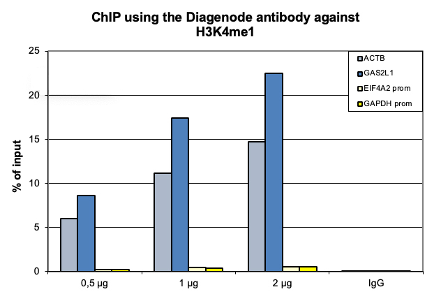
Figure 1. ChIP results obtained with the monoclonal antibody directed against H3K36me3
ChIP was performed with the antibody against H3K36me3 (cat. No. C15210013) on sheared chromatin from 500,000 HeLaS3 cells using the iDeal ChIP-seq kit (cat. No. C01010051). A titration of the antibody consisting of 0.5, 1, and 2 µg per ChIP experiment was analysed. IgG (1 µg/IP) was used as negative IP control. Quantitative PCR was performed with primers specific for the CCT5 coding region, used as positive control, and for a region upstream the ACTB promoter, used as negative control. The graph shows the recovery, expressed as a % of input (the relative amount of immunoprecipitated DNA compared to input DNA after qPCR analysis).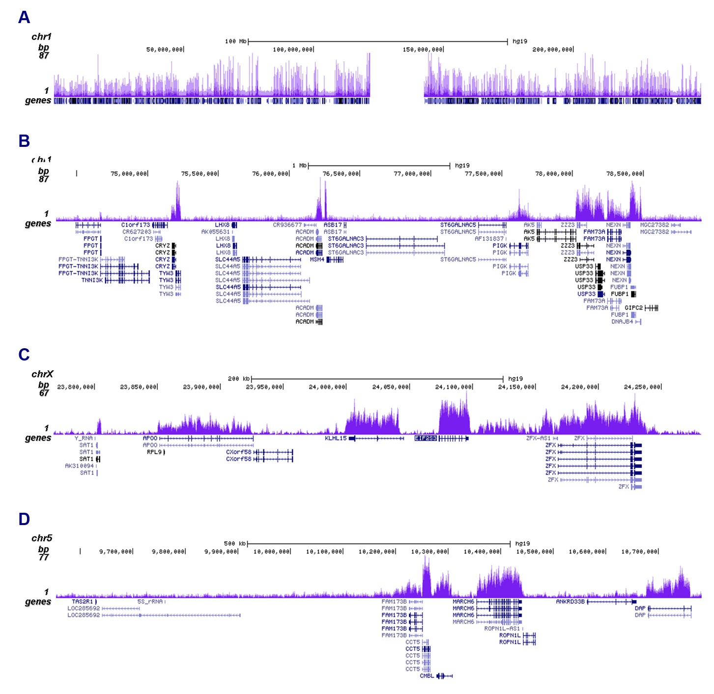
Figure 2. ChIP-seq results obtained with the monoclonal antibody directed against H3K36me3
ChIP was performed on sheared chromatin from 500,000 HeLaS3 cells using 1 µg of the antibody against H3K36me3 (cat. No. C15210013) as described above. The IP’d DNA was subsequently analysed on an Illumina NovaSeq. Library preparation, cluster generation and sequencing were performed according to the manufacturer’s instructions. The 51 bp tags were aligned to the human genome using the BWA algorithm. Figure 2 shows the H3K36me3 signal along the complete sequence and 3 Mb region of human chromosome 1 (figure 2A and B), in a genomic region on chromosome X surrounding the EIF2S3 gene (figure 2C) and in a genomic region on chromosome 5 surrounding the CCT5 positive control (figure 2D).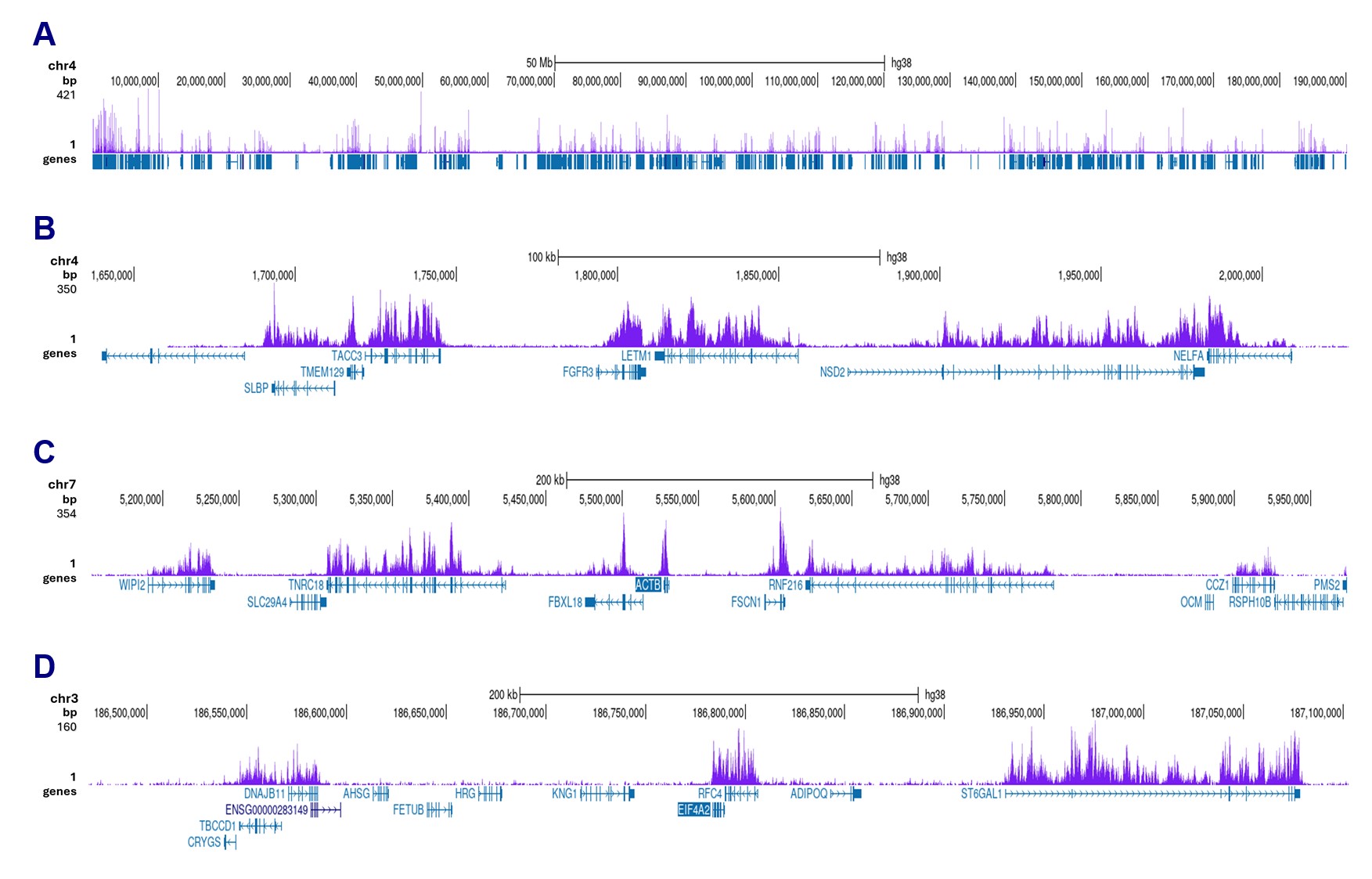
Figure 3. CUT&Tag results obtained with the monoclonal antibody directed against H3K36me3
CUT&Tag was performed on 50,000 K562 cells using 1 µg of the antibody against H3K36me3 (cat. No. C15210013) and the Universal CUT&Tag kit (cat. No. C01070024). The libraries were subsequently analysed on an Illumina NovaSeqX sequencer (2x150 bp paired-end reads) according to the manufacturer’s instructions. The tags were aligned to the human genome (hg38) using the BWA algorithm. Figure 2 shows the peak distribution along the complete sequence and a 300 kb region of chromosome 4 (figure 3A and B) and in 2 genomic regions on chromosome 7 and 3 (figure 3C and D, respectively).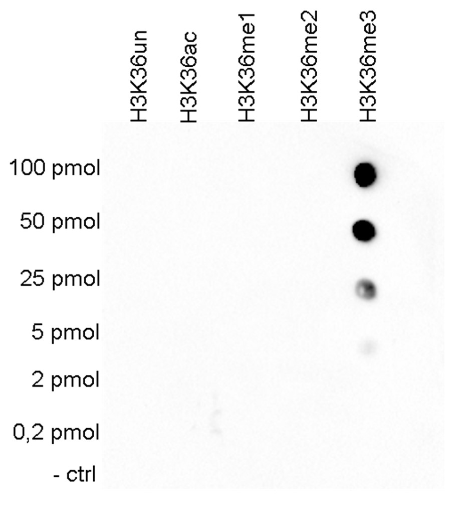
Figure 4. Cross reactivity tests using the monoclonal antibody directed against H3K36me3
To test the cross reactivity of the antibody against H3K36me3 (cat. No. C15210013), a Dot Blot analysis was performed with peptides containing other histone modifications and the unmodified H3K36. One hundred to 0.2 pmol of the respective peptides were spotted on a membrane. The antibody was used at a dilution of 1:2,000. Figure 4 shows a high specificity of the antibody for the modification of interest.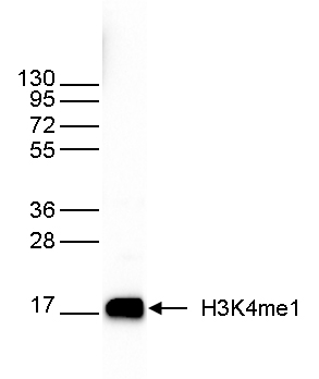
Figure 5. Western blot analysis using the monoclonal antibody directed against H3K36me3
Western blot was performed on recombinant histone H3 (1 µg, lane 1) and on histone extracts from HeLa cells (40 µg, lane 2) using the antibody against H3K36me3 (cat. No. C15210013). The antibody was diluted 1:500 in TBS-Tween containing 5% skimmed milk. The position of the protein of interest is shown on the right, the marker (in kDa) is shown on the left. - 出版物

How to properly cite our product/service in your work
We strongly recommend using this: H3K36me3 monoclonal antibody (Hologic Diagenode Cat# C15210013 Lot# 005). Click here to copy to clipboard.
Using our products or services in your publication? Let us know!
Pig Matrix: a matched multi-omics 3D regulatory genomics database for evolutionary and comparative analyses in pigs
Chao Guo et al.
The pig (Sus scrofa) is an important model for evolutionary, comparative, and translational research, however, current functional genomic resources in pigs remain largely limited to one-dimensional genomic annotation and are therefore insufficient for systematically resolving regulatory region-gene relationships, pa...