RNA polymerase II (pol II) is a key enzyme in the regulation and control of gene transcription. It is able to unwind the DNA double helix, synthesize RNA, and proofread the result. Pol II is a complex enzyme, consisting of 12 subunits, of which the B1 subunit (UniProt/Swiss-Prot entry P24928) is the largest. Together with the second largest subunit, B1 forms the catalytic core of the RNA polymerase II transcription machinery.
Pol II S5p monoclonal antibody

Alternative names: POLR2A, RPB1, POLR2, RPOL2
Monoclonal antibody raised in mouse against the YSPTSPS repeat in the B1 subunit of RNA polymerase II, phosphorylated at Ser5 of the repeat sequence.
| Lot | 001-14 |
|---|---|
| Concentration | 1.0 µg/µl |
| Species reactivity | Human |
| Type | Monoclonal ChIP grade, ChIP-seq grade |
| Purity | Affinity purified monoclonal antibody in PBS containing 0.05% azide. |
| Host | Mouse |
| Storage Conditions | Store at -20°C; for long storage, store at -80°C. Avoid multiple freeze-thaw cycles. |
| Precautions | This product is for research use only. Not for use in diagnostic or therapeutic procedures. |
| Applications | Suggested dilution | References |
|---|---|---|
| ChIP/ChIP-seq * | 1-2 μg/ChIP | Fig 1, 2 |
| ELISA | 1:3,000 | Fig 3 |
| Western Blotting | 1:1,000 | Fig 4, 5 |
| Immunofluorescence | 1:500 | Fig 6 |
* Please note that the optimal antibody amount per IP should be determined by the end-user. We recommend testing 1-5 μg per IP.
- Validation Data
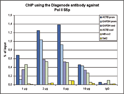
Figure 1. ChIP results obtained with the Diagenode monoclonal antibody directed against Pol II S5p
ChIP assays were performed using human HeLa cells, the Diagenode monoclonal antibody against Pol II S5p (Cat. No. C15200007) and optimized PCR primer pairs for qPCR. ChIP was performed with the “iDeal ChIP-seq” kit (Cat. No. C01010051), using sheared chromatin from 1 million cells. A titration consisting of 1, 2, 5 and 10 μg of antibody per ChIP experiment was analyzed. IgG (2 μg/IP) was used as a negative IP control. Quantitative PCR was performed with primers specific for the promoter and the coding region of the constitutively expressed GAPDH and ACTB genes, used as positive controls, and for exon 2 of the inactive myoglobin (MB) gene and the Sat2 satellite repeat, used as negative controls. Figure 1 shows the recovery, expressed as a % of input (the relative amount of immunoprecipitated DNA compared to input DNA after qPCR analysis).A.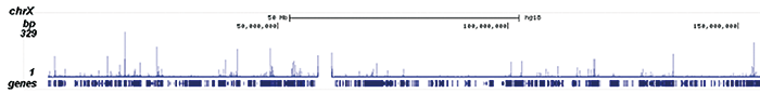
B.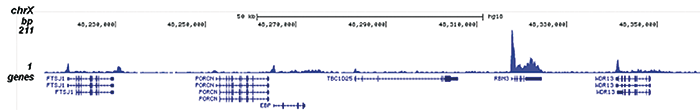
C.
D.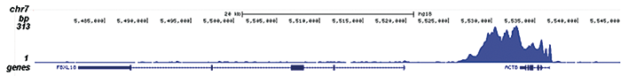
Figure 2. ChIP-seq results obtained with the Diagenode monoclonal antibody directed against Pol II S5p
ChIP was performed on sheared chromatin from 1 million HeLaS3 cells using 1 μg of the Diagenode antibody against Pol II S5p (Cat. No. C15200007) as described above. The IP’d DNA was subsequently analysed on an Illumina Genome Analyzer. Library preparation, cluster generation and sequencing were performed according to the manufacturer’s instructions. The 36 bp tags were aligned to the human genome using the ELAND algorithm. Figure 2 shows the peak distribution along the complete sequence and a 150 kb region of the X-chromosome (figure 2A and B, respectively), and in a two genomic regions surrounding the GAPDH and ACTB positive control genes (figure 2C and D).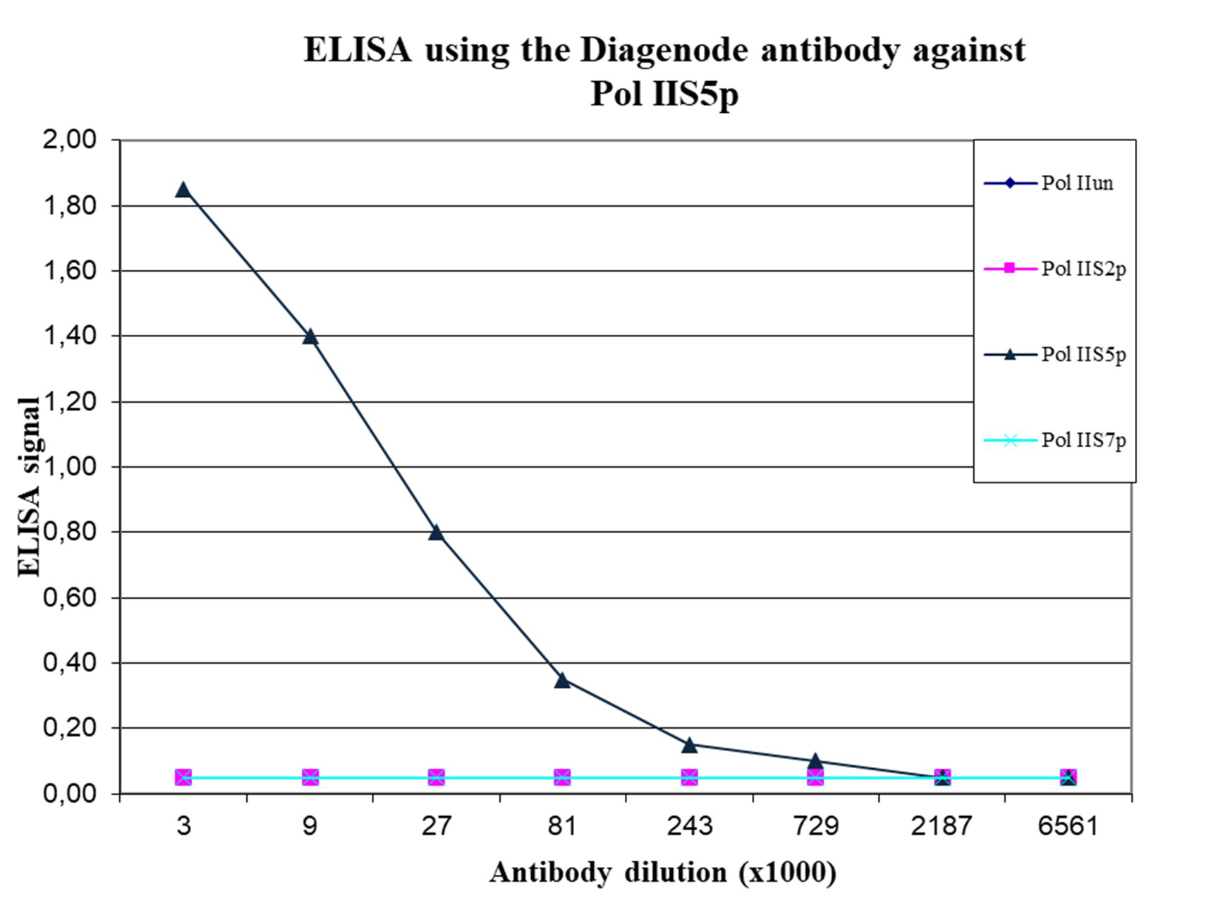
Figure 3. Cross reactivity of the Diagenode monoclonal antibody directed against Pol IIS5p
To test the specificity an ELISA was performed using a serial dilution of the Diagenode monoclonal antibody against Pol IIS5p (Cat. No. C15200007). The wells were coated with peptides containing the unmodified C-terminal repeat sequence as well as different phosphorylated peptides. Figure 3 shows the specificity of the antibody for the S5 phosphorylation.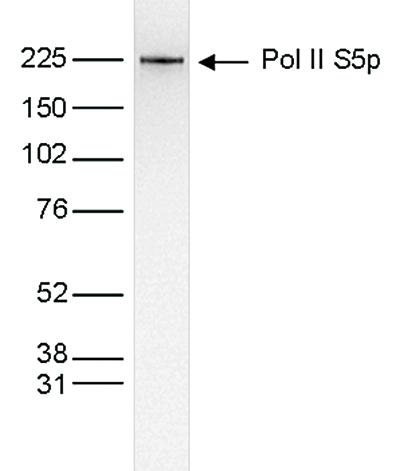
Figure 4. Western blot analysis using the Diagenode monoclonal antibody directed against Pol II S5p
Nuclear extracts (25 μg) from HeLa cells were analysed by Western blot using the Diagenode monoclonal antibody against Pol II S5p (Cat. No. C15200007) diluted 1:1,000 in TBS-Tween containing 5% skimmed milk. The position of the protein of interest is indicated on the right; the marker (in kDa) is shown on the left.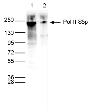
Figure 5. Western blot analysis using the Diagenode monoclonal antibody directed against Pol II S5p
Whole cell extracts (40 μg) from HeLa cells transfected with Pol II siRNA (lane 2) and from an untransfected control (lane 1) were analysed by Western blot using the Diagenode antibody against Pol II S5p (Cat. No. C15200007) diluted 1:1,000 in TBS-Tween containing 5% skimmed milk. The position of the protein of interest is indicated on the right; the marker (in kDa) is shown on the left.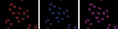
Figure 6. Immunofluorescence using the Diagenode monoclonal antibody directed against Pol II S5p
HeLa cells were stained with the Diagenode antibody against Pol II S5p (Cat. No. C15200007) and with DAPI. Cells were fixed with methanol and blocked with PBS/TX-100 containing 5% normal goat serum and 1% BSA. The cells were immunofluorescently labelled with the Pol II S5p antibody (left) diluted 1:500 in blocking solution followed by an anti-mouse antibody conjugated to Alexa594. The middle panel shows staining of the nuclei with DAPI. A merge of the two stainings is shown on the right. - 出版物

How to properly cite our product/service in your work
We strongly recommend using this: Pol II S5p monoclonal antibody (Hologic Diagenode Cat# C15200007 Lot# 001-14). Click here to copy to clipboard.
Using our products or services in your publication? Let us know!
CDK7 Inhibition is Effective in all the Subtypes of Breast Cancer: Determinants of Response and Synergy with EGFR Inhibition.
McDermott MSJ, Sharko AC, Munie J, Kassler S, Melendez T, Lim CU, Broude EV
CDK7, a transcriptional cyclin-dependent kinase, is emerging as a novel cancer target. Triple-negative breast cancers (TNBC) but not estrogen receptor-positive (ER+) breast cancers have been reported to be uniquely sensitive to the CDK7 inhibitor THZ1 due to the inhibition of a cluster of TNBC-specific genes. Howeve...Transcription Elongation Can Affect Genome 3D Structure.
Heinz S, Texari L, Hayes MGB, Urbanowski M, Chang MW, Givarkes N, Rialdi A, White KM, Albrecht RA, Pache L, Marazzi I, García-Sastre A, Shaw ML, Benner C
How transcription affects genome 3D organization is not well understood. We found that during influenza A (IAV) infection, rampant transcription rapidly reorganizes host cell chromatin interactions. These changes occur at the ends of highly transcribed genes, where global inhibition of transcription termination by I...Meg3 Non-coding RNA Expression Controls Imprinting by Preventing Transcriptional Upregulation in cis.
Sanli I, Lalevée S, Cammisa M, Perrin A, Rage F, Llères D, Riccio A, Bertrand E, Feil R
Although many long non-coding RNAs (lncRNAs) are imprinted, their roles often remain unknown. The Dlk1-Dio3 domain expresses the lncRNA Meg3 and multiple microRNAs and small nucleolar RNAs (snoRNAs) on the maternal chromosome and constitutes an epigenetic model for development. The domain's Dlk1 (Delta-like-1) gene ...CDK8/19 Mediator kinases potentiate induction of transcription by NFκB
Chen M. et al.
The nuclear factor-κB (NFκB) family of transcription factors has been implicated in inflammatory disorders, viral infections, and cancer. Most of the drugs that inhibit NFκB show significant side effects, possibly due to sustained NFκB suppression. Drugs affecting induced, but not basal, NF&k...Functional incompatibility between the generic NF-κB motif and a subtype-specific Sp1III element drives the formation of HIV-1 subtype C viral promoter
Verma A et al.
Of the various genetic subtypes of HIV-1, HIV-2 and SIV, only in subtype C of HIV-1, a genetically variant NF-κB binding site is found at the core of the viral promoter in association with a subtype-specific Sp1III motif. How the subtype-associated variations in the core transcription factor binding sites (TFB...