Notice (8): Undefined variable: solution_of_interest [APP/View/Products/view.ctp, line 755]Code Context<!-- BEGIN: REQUEST_FORM MODAL -->
<div id="request_formModal" class="reveal-modal medium" data-reveal aria-labelledby="modalTitle" aria-hidden="true" role="dialog">
<?= $this->element('Forms/simple_form', array('solution_of_interest' => $solution_of_interest, 'header' => $header, 'message' => $message, 'campaign_id' => $campaign_id)) ?>
$viewFile = '/home/website-server/www/app/View/Products/view.ctp'
$dataForView = array(
'language' => 'jp',
'meta_keywords' => '',
'meta_description' => 'H3K79me3 polyclonal antibody - Classic (sample size)',
'meta_title' => 'H3K79me3 polyclonal antibody - Classic (sample size)',
'product' => array(
'Product' => array(
'id' => '2230',
'antibody_id' => '77',
'name' => 'H3K79me3 polyclonal antibody (sample size)',
'description' => '<p><span>Polyclonal antibody raised in rabbit against histone H3 containing the trimethylated lysine 79 (H3K79me3), using a KLH-conjugated synthetic peptide.</span></p>',
'label1' => 'Validation Data',
'info1' => '',
'label2' => '',
'info2' => '',
'label3' => '',
'info3' => '',
'format' => '10 µg',
'catalog_number' => 'C15410068-10',
'old_catalog_number' => 'pAb-068-050',
'sf_code' => 'C15410068-D001-000582',
'type' => 'FRE',
'search_order' => '03-Antibody',
'price_EUR' => '105',
'price_USD' => '115',
'price_GBP' => '100',
'price_JPY' => '16450',
'price_CNY' => '',
'price_AUD' => '288',
'country' => 'ALL',
'except_countries' => 'None',
'quote' => false,
'in_stock' => false,
'featured' => false,
'no_promo' => false,
'online' => true,
'master' => false,
'last_datasheet_update' => '0000-00-00',
'slug' => 'h3k79me3-polyclonal-antibody-classic-sample-size-10-ug',
'meta_title' => 'H3K79me3 polyclonal antibody - Classic (sample size)',
'meta_keywords' => '',
'meta_description' => 'H3K79me3 polyclonal antibody - Classic (sample size)',
'modified' => '2024-01-16 14:53:16',
'created' => '2015-06-29 14:08:20',
'locale' => 'jpn'
),
'Antibody' => array(
'host' => '*****',
'id' => '77',
'name' => 'H3K79me3 polyclonal antibody',
'description' => 'Histones are the main constituents of the protein part of chromosomes of eukaryotic cells. They are rich in the amino acids arginine and lysine and have been greatly conserved during evolution. Histones pack the DNA into tight masses of chromatin. Two core histones of each class H2A, H2B, H3 and H4 assemble and are wrapped by 146 base pairs of DNA to form one octameric nucleosome. Histone tails undergo numerous post-translational modifications, which either directly or indirectly alter chromatin structure to facilitate transcriptional activation or repression or other nuclear processes. In addition to the genetic code, combinations of the different histone modifications reveal the so-called “histone code”. Histone methylation and demethylation is dynamically regulated by respectively histone methyl transferases and histone demethylases. Trimethylation of histone H3 on K79 was shown to be more present at active promotors than at silent promotors.',
'clonality' => '',
'isotype' => '',
'lot' => 'A2014P',
'concentration' => '0.53 µg/µl',
'reactivity' => 'Human, mouse, yeast: positive. Other species: not tested.',
'type' => 'Polyclonal',
'purity' => 'Affinity purified polyclonal antibody',
'classification' => '',
'application_table' => '<table>
<thead>
<tr>
<th>Applications</th>
<th>Suggested dilution</th>
<th>References</th>
</tr>
</thead>
<tbody>
<tr>
<td>ChIP/ChIP-seq <sup>*</sup></td>
<td>1-2 μg/ChIP</td>
<td>Fig 1, 2</td>
</tr>
<tr>
<td>ELISA</td>
<td>1:500 – 1/1,000</td>
<td>Fig 3</td>
</tr>
<tr>
<td>Dot Blotting</td>
<td>1:20,000</td>
<td>Fig 4</td>
</tr>
<tr>
<td>Western Blotting</td>
<td>1:1,00</td>
<td>Fig 5</td>
</tr>
</tbody>
</table>
<p></p>
<p><small><sup>*</sup> Please note that the optimal antibody amount per IP should be determined by the end-user. We recommend testing 0.5-5 μg per IP.</small></p>',
'storage_conditions' => 'Store at -20°C; for long storage, store at -80°C. Avoid multiple freeze-thaw cycles.',
'storage_buffer' => 'PBS containing 0.05% azide and 0.05% ProClin 300.',
'precautions' => 'This product is for research use only. Not for use in diagnostic or therapeutic procedures.',
'uniprot_acc' => '',
'slug' => '',
'meta_keywords' => '',
'meta_description' => '',
'modified' => '2024-04-24 11:11:17',
'created' => '0000-00-00 00:00:00',
'select_label' => '77 - H3K79me3 polyclonal antibody (A2014P - 0.53 µg/µl - Human, mouse, yeast: positive. Other species: not tested. - Affinity purified polyclonal antibody - Rabbit)'
),
'Slave' => array(),
'Group' => array(
'Group' => array(
[maximum depth reached]
),
'Master' => array(
[maximum depth reached]
),
'Product' => array(
[maximum depth reached]
)
),
'Related' => array(
(int) 0 => array(
[maximum depth reached]
)
),
'Application' => array(
(int) 0 => array(
[maximum depth reached]
),
(int) 1 => array(
[maximum depth reached]
),
(int) 2 => array(
[maximum depth reached]
),
(int) 3 => array(
[maximum depth reached]
),
(int) 4 => array(
[maximum depth reached]
)
),
'Category' => array(
(int) 0 => array(
[maximum depth reached]
),
(int) 1 => array(
[maximum depth reached]
),
(int) 2 => array(
[maximum depth reached]
),
(int) 3 => array(
[maximum depth reached]
),
(int) 4 => array(
[maximum depth reached]
)
),
'Document' => array(
(int) 0 => array(
[maximum depth reached]
),
(int) 1 => array(
[maximum depth reached]
),
(int) 2 => array(
[maximum depth reached]
)
),
'Feature' => array(),
'Image' => array(
(int) 0 => array(
[maximum depth reached]
)
),
'Promotion' => array(),
'Protocol' => array(),
'Publication' => array(
(int) 0 => array(
[maximum depth reached]
),
(int) 1 => array(
[maximum depth reached]
),
(int) 2 => array(
[maximum depth reached]
),
(int) 3 => array(
[maximum depth reached]
),
(int) 4 => array(
[maximum depth reached]
),
(int) 5 => array(
[maximum depth reached]
),
(int) 6 => array(
[maximum depth reached]
),
(int) 7 => array(
[maximum depth reached]
),
(int) 8 => array(
[maximum depth reached]
),
(int) 9 => array(
[maximum depth reached]
)
),
'Testimonial' => array(),
'Area' => array(),
'SafetySheet' => array(
(int) 0 => array(
[maximum depth reached]
),
(int) 1 => array(
[maximum depth reached]
),
(int) 2 => array(
[maximum depth reached]
),
(int) 3 => array(
[maximum depth reached]
),
(int) 4 => array(
[maximum depth reached]
),
(int) 5 => array(
[maximum depth reached]
),
(int) 6 => array(
[maximum depth reached]
),
(int) 7 => array(
[maximum depth reached]
)
)
),
'meta_canonical' => 'https://www.diagenode.com/jp/p/h3k79me3-polyclonal-antibody-classic-50-ug-42-ul'
)
$language = 'jp'
$meta_keywords = ''
$meta_description = 'H3K79me3 polyclonal antibody - Classic (sample size)'
$meta_title = 'H3K79me3 polyclonal antibody - Classic (sample size)'
$product = array(
'Product' => array(
'id' => '2230',
'antibody_id' => '77',
'name' => 'H3K79me3 polyclonal antibody (sample size)',
'description' => '<p><span>Polyclonal antibody raised in rabbit against histone H3 containing the trimethylated lysine 79 (H3K79me3), using a KLH-conjugated synthetic peptide.</span></p>',
'label1' => 'Validation Data',
'info1' => '<div class="row">
<div class="small-6 columns"><img style="border: 1px solid black;" src="https://www.diagenode.com/img/product/antibodies/C15410068-chip.jpg" alt="H3K79me3 Antibody ChIP Grade" /></div>
<div class="small-6 columns">
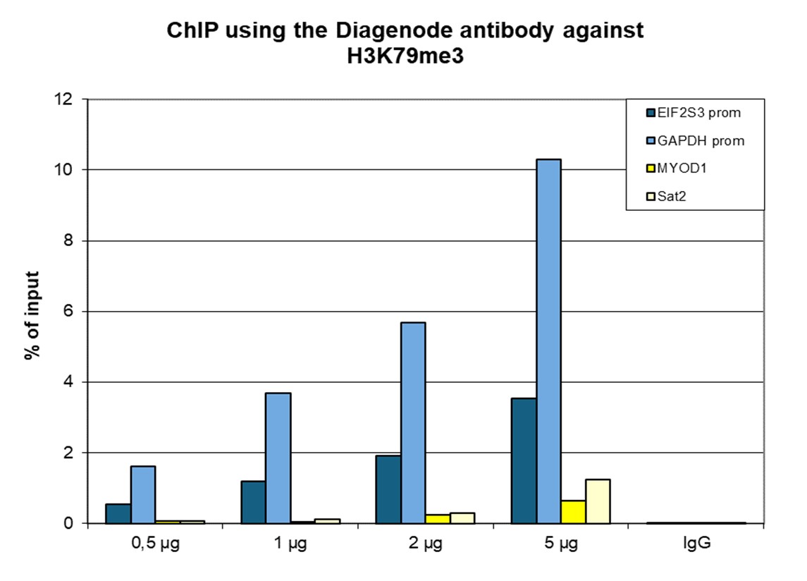
<p><small><strong> Figure 1. ChIP results obtained with the Diagenode antibody directed against H3K79me3</strong><br /> ChIP was performed with the Diagenode antibody against H3K79me3 (cat. No. C15410068) on sheared chromatin from 1 million HeLaS3 cells using the “Auto Histone ChIP-seq” kit and the SX-8G IP-Star automated system. A titration of the antibody consisting of 1, 2, 5 and 10 µg per ChIP experiment was analysed. IgG (5 µg/IP) was used as negative IP control. Quantitative PCR was performed with primers for the GAPDH promoter and for exon 2 of the inactive myoglobin gene. Figure 1 shows the recovery, expressed as a % of input (the relative amount of immunoprecipitated DNA compared to input DNA after qPCR analysis). These results are in accordance with the observation that H3K79me3 shows a preference for active promoters.</small></p>
</div>
</div>
<p><br /><br /></p>
<div class="row">
<div class="small-12 columns">
<p>A.<img src="https://www.diagenode.com/img/product/antibodies/C15410068-chip-seq-A.jpg" alt="H3K79me3 Antibody ChIP-seq Grade" /></p>
<p>B.<img src="https://www.diagenode.com/img/product/antibodies/C15410068-chip-seq-B.jpg" alt="H3K79me3 Antibody for ChIP-seq" /></p>
<p>C.<img src="https://www.diagenode.com/img/product/antibodies/C15410068-chip-seq-C.jpg" alt="H3K79me3 Antibody for ChIP-seq assay" /></p>
<p>D.<img src="https://www.diagenode.com/img/product/antibodies/C15410068-chip-seq-D.jpg" alt="H3K79me3 Antibody validated in ChIP-seq" /></p>
</div>
</div>
<div class="extra-spaced"></div>
<div class="row">
<div class="small-12 columns">
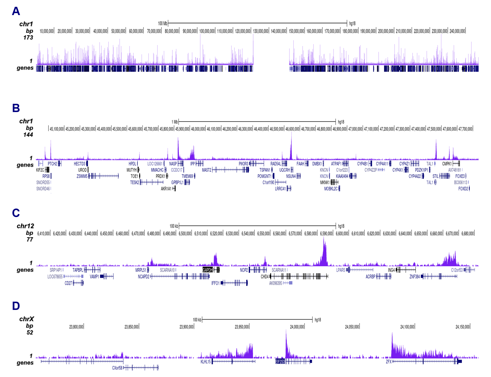
<p><small><strong> Figure 2. ChIP-seq results obtained with the Diagenode antibody directed against H3K79me3</strong><br /> ChIP was performed as described above using 1 µg of the Diagenode antibody against H3K79me3 (cat. No. C15410068). The IP'd DNA was analysed on an Illumina Genome Analyzer. Library preparation, cluster generation and sequencing were performed according to the manufacturer's instructions. The 36 bp tags were aligned to the human genome using the ELAND algorithm. Figure 2 shows the peak distribution along the complete sequence and a 600 kb region of the X-chromosome (figure 2A and B), in a 2 Mb region from chromosome 11 (figure 2C), and in a 100 kb region surrounding the GAPDH positive control gene (figure 2D).</small></p>
</div>
</div>
<p><br /><br /></p>
<div class="row">
<div class="small-6 columns"><img style="border: 1px solid black;" src="https://www.diagenode.com/img/product/antibodies/C15410068-ELISA.jpg" alt="H3K79me3 Antibody ELISA validation" /></div>
<div class="small-6 columns">
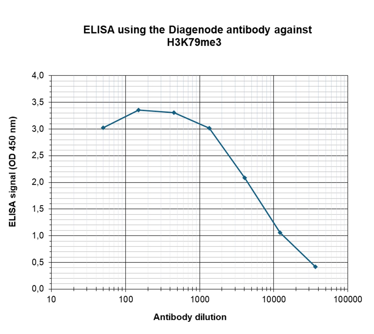
<p><small><strong> Figure 3. Determination of the antibody titer</strong><br /> To determine the titer of the antibody, an ELISA was performed using a serial dilution of Diagenode antibody directed against H3K79me3 (cat. No. C15410068) in antigen coated wells. The antigen used was a peptide containing the histone modification of interest. By plotting the absorbance against the antibody dilution (Figure 3), the titer of the antibody was estimated to be 1:8,500.</small></p>
</div>
</div>
<p><br /><br /></p>
<div class="row">
<div class="small-4 columns"><img src="https://www.diagenode.com/img/product/antibodies/C15410068-dot-blot.jpg" alt="H3K79me3 Antibody validated in Dot Blot" /></div>
<div class="small-8 columns">
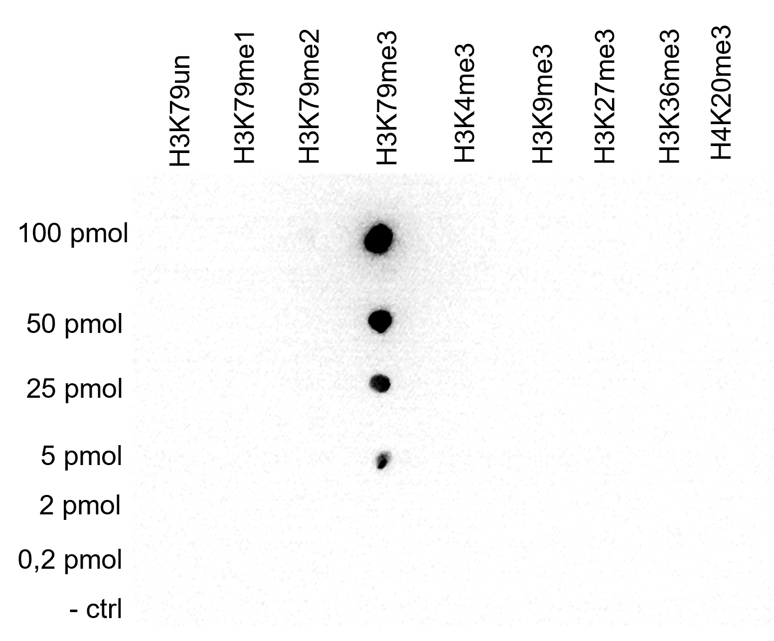
<p><small><strong> Figure 4. Cross reactivity tests using the Diagenode antibody directed against H3K79me3</strong><br /> A Dot Blot analysis was performed to test the cross reactivity of the Diagenode antibody against H3K79me3 (cat. No.<span>C15410068</span>) with peptides containing other histone modifications and the unmodified H3K79. One hundred to 0.2 pmol of the respective peptides were spotted on a membrane. The antibody was used at a dilution of 1:100,000. Figure 4 shows a high specificity of the antibody for the modification of interest. </small></p>
</div>
</div>
<p><br /><br /></p>
<div class="row">
<div class="small-4 columns"><img src="https://www.diagenode.com/img/product/antibodies/C15410068-wb.jpg" alt="H3K79me3 Antibody validated for Western Blot" /></div>
<div class="small-8 columns">
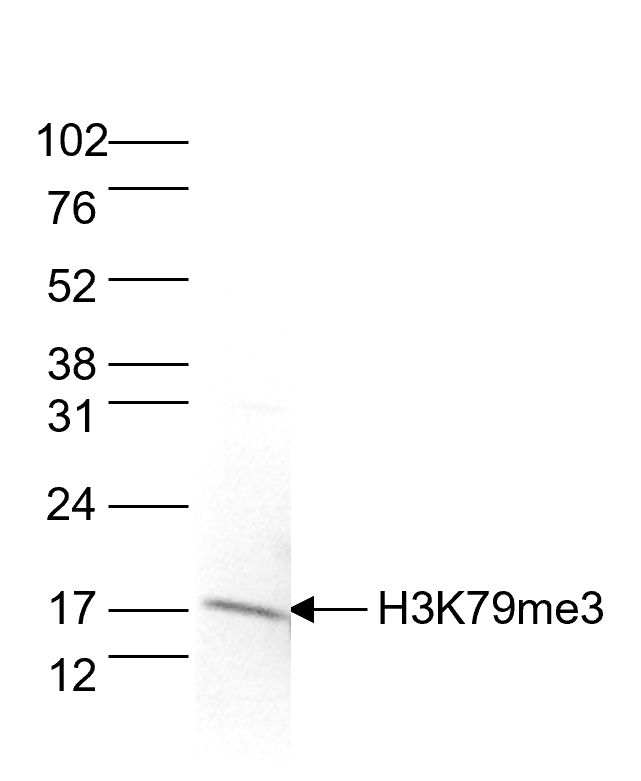
<p><small><strong> Figure 5. Western blot analysis using the Diagenode antibody directed against H3K79me3</strong><br /> Histone extracts of HeLa cells (15 µg) were analysed by Western blot using the Diagenode antibody against H3K79me3 (cat. No.<span>C15410068</span>), diluted 1:1,000 in TBS-Tween containing 5% skimmed milk. The molecular weight marker is shown on the right; the location of the protein of interest is indicated on the left.</small></p>
</div>
</div>',
'label2' => 'Target Description',
'info2' => '<p>Histones are the main constituents of the protein part of chromosomes of eukaryotic cells. They are rich in the amino acids arginine and lysine and have been greatly conserved during evolution. Histones pack the DNA into tight masses of chromatin. Two core histones of each class H2A, H2B, H3 and H4 assemble and are wrapped by 146 base pairs of DNA to form one octameric nucleosome. Histone tails undergo numerous post-translational modifications, which either directly or indirectly alter chromatin structure to facilitate transcriptional activation or repression or other nuclear processes. In addition to the genetic code, combinations of the different histone modifications reveal the so-called “histone code”. Histone methylation and demethylation is dynamically regulated by respectively histone methyl transferases and histone demethylases. Trimethylation of histone H3 on K79 was shown to be more present at active promotors than at silent promotors.</p>',
'label3' => '',
'info3' => '',
'format' => '10 µg',
'catalog_number' => 'C15410068-10',
'old_catalog_number' => 'pAb-068-050',
'sf_code' => 'C15410068-D001-000582',
'type' => 'FRE',
'search_order' => '03-Antibody',
'price_EUR' => '105',
'price_USD' => '115',
'price_GBP' => '100',
'price_JPY' => '16450',
'price_CNY' => '',
'price_AUD' => '288',
'country' => 'ALL',
'except_countries' => 'None',
'quote' => false,
'in_stock' => false,
'featured' => false,
'no_promo' => false,
'online' => true,
'master' => false,
'last_datasheet_update' => '0000-00-00',
'slug' => 'h3k79me3-polyclonal-antibody-classic-sample-size-10-ug',
'meta_title' => 'H3K79me3 polyclonal antibody - Classic (sample size)',
'meta_keywords' => '',
'meta_description' => 'H3K79me3 polyclonal antibody - Classic (sample size)',
'modified' => '2024-01-16 14:53:16',
'created' => '2015-06-29 14:08:20',
'locale' => 'jpn'
),
'Antibody' => array(
'host' => '*****',
'id' => '77',
'name' => 'H3K79me3 polyclonal antibody',
'description' => 'Histones are the main constituents of the protein part of chromosomes of eukaryotic cells. They are rich in the amino acids arginine and lysine and have been greatly conserved during evolution. Histones pack the DNA into tight masses of chromatin. Two core histones of each class H2A, H2B, H3 and H4 assemble and are wrapped by 146 base pairs of DNA to form one octameric nucleosome. Histone tails undergo numerous post-translational modifications, which either directly or indirectly alter chromatin structure to facilitate transcriptional activation or repression or other nuclear processes. In addition to the genetic code, combinations of the different histone modifications reveal the so-called “histone code”. Histone methylation and demethylation is dynamically regulated by respectively histone methyl transferases and histone demethylases. Trimethylation of histone H3 on K79 was shown to be more present at active promotors than at silent promotors.',
'clonality' => '',
'isotype' => '',
'lot' => 'A2014P',
'concentration' => '0.53 µg/µl',
'reactivity' => 'Human, mouse, yeast: positive. Other species: not tested.',
'type' => 'Polyclonal',
'purity' => 'Affinity purified polyclonal antibody',
'classification' => '',
'application_table' => '<table>
<thead>
<tr>
<th>Applications</th>
<th>Suggested dilution</th>
<th>References</th>
</tr>
</thead>
<tbody>
<tr>
<td>ChIP/ChIP-seq <sup>*</sup></td>
<td>1-2 μg/ChIP</td>
<td>Fig 1, 2</td>
</tr>
<tr>
<td>ELISA</td>
<td>1:500 – 1/1,000</td>
<td>Fig 3</td>
</tr>
<tr>
<td>Dot Blotting</td>
<td>1:20,000</td>
<td>Fig 4</td>
</tr>
<tr>
<td>Western Blotting</td>
<td>1:1,00</td>
<td>Fig 5</td>
</tr>
</tbody>
</table>
<p></p>
<p><small><sup>*</sup> Please note that the optimal antibody amount per IP should be determined by the end-user. We recommend testing 0.5-5 μg per IP.</small></p>',
'storage_conditions' => 'Store at -20°C; for long storage, store at -80°C. Avoid multiple freeze-thaw cycles.',
'storage_buffer' => 'PBS containing 0.05% azide and 0.05% ProClin 300.',
'precautions' => 'This product is for research use only. Not for use in diagnostic or therapeutic procedures.',
'uniprot_acc' => '',
'slug' => '',
'meta_keywords' => '',
'meta_description' => '',
'modified' => '2024-04-24 11:11:17',
'created' => '0000-00-00 00:00:00',
'select_label' => '77 - H3K79me3 polyclonal antibody (A2014P - 0.53 µg/µl - Human, mouse, yeast: positive. Other species: not tested. - Affinity purified polyclonal antibody - Rabbit)'
),
'Slave' => array(),
'Group' => array(
'Group' => array(
'id' => '49',
'name' => 'C15410068',
'product_id' => '2229',
'modified' => '2016-02-18 20:51:25',
'created' => '2016-02-18 20:51:25'
),
'Master' => array(
'id' => '2229',
'antibody_id' => '77',
'name' => 'H3K79me3 Antibody',
'description' => '<p><span>Polyclonal antibody raised in rabbit against histone <strong>H3 containing the trimethylated lysine 79</strong> (<strong>H3K79me3</strong>), using a KLH-conjugated synthetic peptide.</span></p>',
'label1' => 'Validation Data',
'info1' => '<div class="row">
<div class="small-6 columns"><img style="border: 1px solid black;" src="https://www.diagenode.com/img/product/antibodies/C15410068-chip.jpg" alt="H3K79me3 Antibody ChIP Grade" /></div>
<div class="small-6 columns">
<p><small><strong> Figure 1. ChIP results obtained with the Diagenode antibody directed against H3K79me3</strong><br /> ChIP was performed with the Diagenode antibody against H3K79me3 (cat. No. C15410068) on sheared chromatin from 1 million HeLaS3 cells using the “Auto Histone ChIP-seq” kit and the SX-8G IP-Star automated system. A titration of the antibody consisting of 1, 2, 5 and 10 µg per ChIP experiment was analysed. IgG (5 µg/IP) was used as negative IP control. Quantitative PCR was performed with primers for the GAPDH promoter and for exon 2 of the inactive myoglobin gene. Figure 1 shows the recovery, expressed as a % of input (the relative amount of immunoprecipitated DNA compared to input DNA after qPCR analysis). These results are in accordance with the observation that H3K79me3 shows a preference for active promoters.</small></p>
</div>
</div>
<p><br /><br /></p>
<div class="row">
<div class="small-12 columns">
<p>A.<img src="https://www.diagenode.com/img/product/antibodies/C15410068-chip-seq-A.jpg" alt="H3K79me3 Antibody ChIP-seq Grade" /></p>
<p>B.<img src="https://www.diagenode.com/img/product/antibodies/C15410068-chip-seq-B.jpg" alt="H3K79me3 Antibody for ChIP-seq" /></p>
<p>C.<img src="https://www.diagenode.com/img/product/antibodies/C15410068-chip-seq-C.jpg" alt="H3K79me3 Antibody for ChIP-seq assay" /></p>
<p>D.<img src="https://www.diagenode.com/img/product/antibodies/C15410068-chip-seq-D.jpg" alt="H3K79me3 Antibody validated in ChIP-seq" /></p>
</div>
</div>
<div class="extra-spaced"></div>
<div class="row">
<div class="small-12 columns">
<p><small><strong> Figure 2. ChIP-seq results obtained with the Diagenode antibody directed against H3K79me3</strong><br /> ChIP was performed as described above using 1 µg of the Diagenode antibody against H3K79me3 (cat. No. C15410068). The IP'd DNA was analysed on an Illumina Genome Analyzer. Library preparation, cluster generation and sequencing were performed according to the manufacturer's instructions. The 36 bp tags were aligned to the human genome using the ELAND algorithm. Figure 2 shows the peak distribution along the complete sequence and a 600 kb region of the X-chromosome (figure 2A and B), in a 2 Mb region from chromosome 11 (figure 2C), and in a 100 kb region surrounding the GAPDH positive control gene (figure 2D).</small></p>
</div>
</div>
<p><br /><br /></p>
<div class="row">
<div class="small-6 columns"><img style="border: 1px solid black;" src="https://www.diagenode.com/img/product/antibodies/C15410068-ELISA.jpg" alt="H3K79me3 Antibody ELISA validation" /></div>
<div class="small-6 columns">
<p><small><strong> Figure 3. Determination of the antibody titer</strong><br /> To determine the titer of the antibody, an ELISA was performed using a serial dilution of Diagenode antibody directed against H3K79me3 (cat. No. C15410068) in antigen coated wells. The antigen used was a peptide containing the histone modification of interest. By plotting the absorbance against the antibody dilution (Figure 3), the titer of the antibody was estimated to be 1:8,500.</small></p>
</div>
</div>
<p><br /><br /></p>
<div class="row">
<div class="small-4 columns"><img src="https://www.diagenode.com/img/product/antibodies/C15410068-dot-blot.jpg" alt="H3K79me3 Antibody validated in Dot Blot" /></div>
<div class="small-8 columns">
<p><small><strong> Figure 4. Cross reactivity tests using the Diagenode antibody directed against H3K79me3</strong><br /> A Dot Blot analysis was performed to test the cross reactivity of the Diagenode antibody against H3K79me3 (cat. No.<span>C15410068</span>) with peptides containing other histone modifications and the unmodified H3K79. One hundred to 0.2 pmol of the respective peptides were spotted on a membrane. The antibody was used at a dilution of 1:100,000. Figure 4 shows a high specificity of the antibody for the modification of interest. </small></p>
</div>
</div>
<p><br /><br /></p>
<div class="row">
<div class="small-4 columns"><img src="https://www.diagenode.com/img/product/antibodies/C15410068-wb.jpg" alt="H3K79me3 Antibody validated for Western Blot" /></div>
<div class="small-8 columns">
<p><small><strong> Figure 5. Western blot analysis using the Diagenode antibody directed against H3K79me3</strong><br /> Histone extracts of HeLa cells (15 µg) were analysed by Western blot using the Diagenode antibody against H3K79me3 (cat. No.<span>C15410068</span>), diluted 1:1,000 in TBS-Tween containing 5% skimmed milk. The molecular weight marker is shown on the right; the location of the protein of interest is indicated on the left.</small></p>
</div>
</div>',
'label2' => 'Target Description',
'info2' => '<p>Histones are the main constituents of the protein part of chromosomes of eukaryotic cells. They are rich in the amino acids arginine and lysine and have been greatly conserved during evolution. Histones pack the DNA into tight masses of chromatin. Two core histones of each class H2A, H2B, H3 and H4 assemble and are wrapped by 146 base pairs of DNA to form one octameric nucleosome. Histone tails undergo numerous post-translational modifications, which either directly or indirectly alter chromatin structure to facilitate transcriptional activation or repression or other nuclear processes. In addition to the genetic code, combinations of the different histone modifications reveal the so-called “histone code”. Histone methylation and demethylation is dynamically regulated by respectively histone methyl transferases and histone demethylases. Trimethylation of histone H3 on K79 was shown to be more present at active promotors than at silent promotors.</p>',
'label3' => '',
'info3' => '',
'format' => '50 µg',
'catalog_number' => 'C15410068',
'old_catalog_number' => 'pAb-068-050',
'sf_code' => 'C15410068-D001-000581',
'type' => 'FRE',
'search_order' => '03-Antibody',
'price_EUR' => '380',
'price_USD' => '380',
'price_GBP' => '340',
'price_JPY' => '59525',
'price_CNY' => '',
'price_AUD' => '950',
'country' => 'ALL',
'except_countries' => 'None',
'quote' => false,
'in_stock' => false,
'featured' => false,
'no_promo' => false,
'online' => true,
'master' => true,
'last_datasheet_update' => '0000-00-00',
'slug' => 'h3k79me3-polyclonal-antibody-classic-50-ug-42-ul',
'meta_title' => 'H3K79me3 Antibody - ChIP-seq Grade (C15410068) | Diagenode',
'meta_keywords' => '',
'meta_description' => 'H3K79me3 (Histone H3 trimethylated at lysine 79) Polyclonal Antibody validated in ChIP-seq, ChIP-qPCR, ELISA, WB and DB. Batch-specific data available on the website. Sample size available.',
'modified' => '2024-04-24 11:19:39',
'created' => '2015-06-29 14:08:20'
),
'Product' => array(
(int) 0 => array(
[maximum depth reached]
)
)
),
'Related' => array(
(int) 0 => array(
'id' => '2229',
'antibody_id' => '77',
'name' => 'H3K79me3 Antibody',
'description' => '<p><span>Polyclonal antibody raised in rabbit against histone <strong>H3 containing the trimethylated lysine 79</strong> (<strong>H3K79me3</strong>), using a KLH-conjugated synthetic peptide.</span></p>',
'label1' => 'Validation Data',
'info1' => '<div class="row">
<div class="small-6 columns"><img style="border: 1px solid black;" src="https://www.diagenode.com/img/product/antibodies/C15410068-chip.jpg" alt="H3K79me3 Antibody ChIP Grade" /></div>
<div class="small-6 columns">
<p><small><strong> Figure 1. ChIP results obtained with the Diagenode antibody directed against H3K79me3</strong><br /> ChIP was performed with the Diagenode antibody against H3K79me3 (cat. No. C15410068) on sheared chromatin from 1 million HeLaS3 cells using the “Auto Histone ChIP-seq” kit and the SX-8G IP-Star automated system. A titration of the antibody consisting of 1, 2, 5 and 10 µg per ChIP experiment was analysed. IgG (5 µg/IP) was used as negative IP control. Quantitative PCR was performed with primers for the GAPDH promoter and for exon 2 of the inactive myoglobin gene. Figure 1 shows the recovery, expressed as a % of input (the relative amount of immunoprecipitated DNA compared to input DNA after qPCR analysis). These results are in accordance with the observation that H3K79me3 shows a preference for active promoters.</small></p>
</div>
</div>
<p><br /><br /></p>
<div class="row">
<div class="small-12 columns">
<p>A.<img src="https://www.diagenode.com/img/product/antibodies/C15410068-chip-seq-A.jpg" alt="H3K79me3 Antibody ChIP-seq Grade" /></p>
<p>B.<img src="https://www.diagenode.com/img/product/antibodies/C15410068-chip-seq-B.jpg" alt="H3K79me3 Antibody for ChIP-seq" /></p>
<p>C.<img src="https://www.diagenode.com/img/product/antibodies/C15410068-chip-seq-C.jpg" alt="H3K79me3 Antibody for ChIP-seq assay" /></p>
<p>D.<img src="https://www.diagenode.com/img/product/antibodies/C15410068-chip-seq-D.jpg" alt="H3K79me3 Antibody validated in ChIP-seq" /></p>
</div>
</div>
<div class="extra-spaced"></div>
<div class="row">
<div class="small-12 columns">
<p><small><strong> Figure 2. ChIP-seq results obtained with the Diagenode antibody directed against H3K79me3</strong><br /> ChIP was performed as described above using 1 µg of the Diagenode antibody against H3K79me3 (cat. No. C15410068). The IP'd DNA was analysed on an Illumina Genome Analyzer. Library preparation, cluster generation and sequencing were performed according to the manufacturer's instructions. The 36 bp tags were aligned to the human genome using the ELAND algorithm. Figure 2 shows the peak distribution along the complete sequence and a 600 kb region of the X-chromosome (figure 2A and B), in a 2 Mb region from chromosome 11 (figure 2C), and in a 100 kb region surrounding the GAPDH positive control gene (figure 2D).</small></p>
</div>
</div>
<p><br /><br /></p>
<div class="row">
<div class="small-6 columns"><img style="border: 1px solid black;" src="https://www.diagenode.com/img/product/antibodies/C15410068-ELISA.jpg" alt="H3K79me3 Antibody ELISA validation" /></div>
<div class="small-6 columns">
<p><small><strong> Figure 3. Determination of the antibody titer</strong><br /> To determine the titer of the antibody, an ELISA was performed using a serial dilution of Diagenode antibody directed against H3K79me3 (cat. No. C15410068) in antigen coated wells. The antigen used was a peptide containing the histone modification of interest. By plotting the absorbance against the antibody dilution (Figure 3), the titer of the antibody was estimated to be 1:8,500.</small></p>
</div>
</div>
<p><br /><br /></p>
<div class="row">
<div class="small-4 columns"><img src="https://www.diagenode.com/img/product/antibodies/C15410068-dot-blot.jpg" alt="H3K79me3 Antibody validated in Dot Blot" /></div>
<div class="small-8 columns">
<p><small><strong> Figure 4. Cross reactivity tests using the Diagenode antibody directed against H3K79me3</strong><br /> A Dot Blot analysis was performed to test the cross reactivity of the Diagenode antibody against H3K79me3 (cat. No.<span>C15410068</span>) with peptides containing other histone modifications and the unmodified H3K79. One hundred to 0.2 pmol of the respective peptides were spotted on a membrane. The antibody was used at a dilution of 1:100,000. Figure 4 shows a high specificity of the antibody for the modification of interest. </small></p>
</div>
</div>
<p><br /><br /></p>
<div class="row">
<div class="small-4 columns"><img src="https://www.diagenode.com/img/product/antibodies/C15410068-wb.jpg" alt="H3K79me3 Antibody validated for Western Blot" /></div>
<div class="small-8 columns">
<p><small><strong> Figure 5. Western blot analysis using the Diagenode antibody directed against H3K79me3</strong><br /> Histone extracts of HeLa cells (15 µg) were analysed by Western blot using the Diagenode antibody against H3K79me3 (cat. No.<span>C15410068</span>), diluted 1:1,000 in TBS-Tween containing 5% skimmed milk. The molecular weight marker is shown on the right; the location of the protein of interest is indicated on the left.</small></p>
</div>
</div>',
'label2' => 'Target Description',
'info2' => '<p>Histones are the main constituents of the protein part of chromosomes of eukaryotic cells. They are rich in the amino acids arginine and lysine and have been greatly conserved during evolution. Histones pack the DNA into tight masses of chromatin. Two core histones of each class H2A, H2B, H3 and H4 assemble and are wrapped by 146 base pairs of DNA to form one octameric nucleosome. Histone tails undergo numerous post-translational modifications, which either directly or indirectly alter chromatin structure to facilitate transcriptional activation or repression or other nuclear processes. In addition to the genetic code, combinations of the different histone modifications reveal the so-called “histone code”. Histone methylation and demethylation is dynamically regulated by respectively histone methyl transferases and histone demethylases. Trimethylation of histone H3 on K79 was shown to be more present at active promotors than at silent promotors.</p>',
'label3' => '',
'info3' => '',
'format' => '50 µg',
'catalog_number' => 'C15410068',
'old_catalog_number' => 'pAb-068-050',
'sf_code' => 'C15410068-D001-000581',
'type' => 'FRE',
'search_order' => '03-Antibody',
'price_EUR' => '380',
'price_USD' => '380',
'price_GBP' => '340',
'price_JPY' => '59525',
'price_CNY' => '',
'price_AUD' => '950',
'country' => 'ALL',
'except_countries' => 'None',
'quote' => false,
'in_stock' => false,
'featured' => false,
'no_promo' => false,
'online' => true,
'master' => true,
'last_datasheet_update' => '0000-00-00',
'slug' => 'h3k79me3-polyclonal-antibody-classic-50-ug-42-ul',
'meta_title' => 'H3K79me3 Antibody - ChIP-seq Grade (C15410068) | Diagenode',
'meta_keywords' => '',
'meta_description' => 'H3K79me3 (Histone H3 trimethylated at lysine 79) Polyclonal Antibody validated in ChIP-seq, ChIP-qPCR, ELISA, WB and DB. Batch-specific data available on the website. Sample size available.',
'modified' => '2024-04-24 11:19:39',
'created' => '2015-06-29 14:08:20',
'ProductsRelated' => array(
[maximum depth reached]
),
'Image' => array(
[maximum depth reached]
)
)
),
'Application' => array(
(int) 0 => array(
'id' => '20',
'position' => '10',
'parent_id' => '40',
'name' => 'ELISA',
'description' => '<div class="row">
<div class="small-12 medium-12 large-12 columns">Enzyme-linked immunosorbent assay.</div>
</div>',
'in_footer' => false,
'in_menu' => false,
'online' => true,
'tabular' => true,
'slug' => 'elisa-antibodies',
'meta_keywords' => ' ELISA Antibodies,Monoclonal antibody, Polyclonal antibody',
'meta_description' => 'Diagenode offers Monoclonal & Polyclonal antibodies for ELISA applications',
'meta_title' => 'ELISA Antibodies - Monoclonal & Polyclonal antibody | Diagenode',
'modified' => '2016-01-13 12:21:41',
'created' => '2014-07-08 08:13:28',
'ProductsApplication' => array(
[maximum depth reached]
)
),
(int) 1 => array(
'id' => '28',
'position' => '10',
'parent_id' => '40',
'name' => 'DB',
'description' => '<p>Dot blotting</p>',
'in_footer' => false,
'in_menu' => false,
'online' => true,
'tabular' => true,
'slug' => 'dot-blotting',
'meta_keywords' => 'Dot blotting,Monoclonal & Polyclonal antibody,',
'meta_description' => 'Diagenode offers Monoclonal & Polyclonal antibodies for Dot blotting applications',
'meta_title' => 'Dot blotting Antibodies - Monoclonal & Polyclonal antibody | Diagenode',
'modified' => '2016-01-13 14:40:49',
'created' => '2015-07-08 13:45:05',
'ProductsApplication' => array(
[maximum depth reached]
)
),
(int) 2 => array(
'id' => '19',
'position' => '10',
'parent_id' => '40',
'name' => 'WB',
'description' => '<p><strong>Western blot</strong> : The quality of antibodies used in this technique is crucial for correct and specific protein identification. Diagenode offers huge selection of highly sensitive and specific western blot-validated antibodies.</p>
<p>Learn more about: <a href="https://www.diagenode.com/applications/western-blot">Loading control, MW marker visualization</a><em>. <br /></em></p>
<p><em></em>Check our selection of antibodies validated in Western blot.</p>',
'in_footer' => false,
'in_menu' => false,
'online' => true,
'tabular' => true,
'slug' => 'western-blot-antibodies',
'meta_keywords' => ' Western Blot Antibodies ,western blot protocol,Western Blotting Products,Polyclonal antibodies ,monoclonal antibodies ',
'meta_description' => 'Diagenode offers a wide range of antibodies and technical support for western blot applications',
'meta_title' => ' Western Blot - Monoclonal antibody - Polyclonal antibody | Diagenode',
'modified' => '2016-04-26 12:44:51',
'created' => '2015-01-07 09:20:00',
'ProductsApplication' => array(
[maximum depth reached]
)
),
(int) 3 => array(
'id' => '42',
'position' => '10',
'parent_id' => '40',
'name' => 'ChIP-seq (ab)',
'description' => '',
'in_footer' => false,
'in_menu' => false,
'online' => true,
'tabular' => true,
'slug' => 'chip-seq-antibodies',
'meta_keywords' => 'Chromatin Immunoprecipitation Sequencing,ChIP-Seq,ChIP-seq grade antibodies,DNA purification,qPCR,Shearing of chromatin',
'meta_description' => 'Diagenode offers a wide range of antibodies and technical support for ChIP Sequencing applications',
'meta_title' => 'ChIP Sequencing Antibodies (ChIP-Seq) | Diagenode',
'modified' => '2016-01-20 11:06:19',
'created' => '2015-10-20 11:44:45',
'ProductsApplication' => array(
[maximum depth reached]
)
),
(int) 4 => array(
'id' => '43',
'position' => '10',
'parent_id' => '40',
'name' => 'ChIP-qPCR (ab)',
'description' => '',
'in_footer' => false,
'in_menu' => false,
'online' => true,
'tabular' => true,
'slug' => 'chip-qpcr-antibodies',
'meta_keywords' => 'Chromatin Immunoprecipitation Sequencing,ChIP-Seq,ChIP-seq grade antibodies,DNA purification,qPCR,Shearing of chromatin',
'meta_description' => 'Diagenode offers a wide range of antibodies and technical support for ChIP-qPCR applications',
'meta_title' => 'ChIP Quantitative PCR Antibodies (ChIP-qPCR) | Diagenode',
'modified' => '2016-01-20 11:30:24',
'created' => '2015-10-20 11:45:36',
'ProductsApplication' => array(
[maximum depth reached]
)
)
),
'Category' => array(
(int) 0 => array(
'id' => '17',
'position' => '10',
'parent_id' => '4',
'name' => 'ChIP-seq grade antibodies',
'description' => '<p><b>Unparalleled ChIP-Seq results with the most rigorously validated antibodies</b></p>
<p><span style="font-weight: 400;">Diagenode provides leading solutions for epigenetic research. Because ChIP-seq is a widely-used technique, we validate our antibodies in ChIP and ChIP-seq experiments (in addition to conventional methods like Western blot, Dot blot, ELISA, and immunofluorescence) to provide the highest quality antibody. We standardize our validation and production to guarantee high product quality without technical bias. Diagenode guarantees ChIP-seq grade antibody performance under our suggested conditions.</span></p>
<div class="row">
<div class="small-12 medium-9 large-9 columns">
<p><strong>ChIP-seq profile</strong> of active (H3K4me3 and H3K36me3) and inactive (H3K27me3) marks using Diagenode antibodies.</p>
<img src="https://www.diagenode.com/img/categories/antibodies/chip-seq-grade-antibodies.png" /></div>
<div class="small-12 medium-3 large-3 columns">
<p><small> ChIP was performed on sheared chromatin from 100,000 K562 cells using iDeal ChIP-seq kit for Histones (cat. No. C01010051) with 1 µg of the Diagenode antibodies against H3K27me3 (cat. No. C15410195) and H3K4me3 (cat. No. C15410003), and 0.5 µg of the antibody against H3K36me3 (cat. No. C15410192). The IP'd DNA was subsequently analysed on an Illumina Genome Analyzer. Library preparation, cluster generation and sequencing were performed according to the manufacturer's instructions. The 36 bp tags were aligned to the human genome using the ELAND algorithm. The figure shows the signal distribution along the complete sequence of human chromosome 3, a zoomin to a 10 Mb region and a further zoomin to a 1.5 Mb region. </small></p>
</div>
</div>
<p>Diagenode’s highly validated antibodies:</p>
<ul>
<li>Highly sensitive and specific</li>
<li>Cost-effective (requires less antibody per reaction)</li>
<li>Batch-specific data is available on the website</li>
<li>Expert technical support</li>
<li>Sample sizes available</li>
<li>100% satisfaction guarantee</li>
</ul>',
'no_promo' => false,
'in_menu' => true,
'online' => true,
'tabular' => false,
'hide' => true,
'all_format' => false,
'is_antibody' => true,
'slug' => 'chip-seq-grade-antibodies',
'cookies_tag_id' => null,
'meta_keywords' => 'ChIP-seq grade antibodies,polyclonal antibody,WB, ELISA, ChIP-seq (ab), ChIP-qPCR (ab)',
'meta_description' => 'Diagenode Offers Wide Range of Validated ChIP-Seq Grade Antibodies for Unparalleled ChIP-Seq Results',
'meta_title' => 'Chromatin Immunoprecipitation ChIP-Seq Grade Antibodies | Diagenode',
'modified' => '2019-07-03 10:57:22',
'created' => '2015-02-16 02:24:01',
'ProductsCategory' => array(
[maximum depth reached]
),
'CookiesTag' => array([maximum depth reached])
),
(int) 1 => array(
'id' => '111',
'position' => '40',
'parent_id' => '4',
'name' => 'Histone antibodies',
'description' => '<p>Histones are the main protein components of chromatin involved in the compaction of DNA into nucleosomes, the basic units of chromatin. A <strong>nucleosome</strong> consists of one pair of each of the core histones (<strong>H2A</strong>, <strong>H2B</strong>, <strong>H3</strong> and <strong>H4</strong>) forming an octameric structure wrapped by 146 base pairs of DNA. The different nucleosomes are linked by the linker histone<strong> H1, </strong>allowing for further condensation of chromatin.</p>
<p>The core histones have a globular structure with large unstructured N-terminal tails protruding from the nucleosome. They can undergo to multiple post-translational modifications (PTM), mainly at the N-terminal tails. These <strong>post-translational modifications </strong>include methylation, acetylation, phosphorylation, ubiquitinylation, citrullination, sumoylation, deamination and crotonylation. The most well characterized PTMs are <strong>methylation,</strong> <strong>acetylation and phosphorylation</strong>. Histone methylation occurs mainly on lysine (K) residues, which can be mono-, di- or tri-methylated, and on arginines (R), which can be mono-methylated and symmetrically or asymmetrically di-methylated. Histone acetylation occurs on lysines and histone phosphorylation mainly on serines (S), threonines (T) and tyrosines (Y).</p>
<p>The PTMs of the different residues are involved in numerous processes such as DNA repair, DNA replication and chromosome condensation. They influence the chromatin organization and can be positively or negatively associated with gene expression. Trimethylation of H3K4, H3K36 and H3K79, and lysine acetylation generally result in an open chromatin configuration (figure below) and are therefore associated with <strong>euchromatin</strong> and gene activation. Trimethylation of H3K9, K3K27 and H4K20, on the other hand, is enriched in <strong>heterochromatin </strong>and associated with gene silencing. The combination of different histone modifications is called the "<strong>histone code</strong>”, analogous to the genetic code.</p>
<p><img src="https://www.diagenode.com/img/categories/antibodies/histone-marks-illustration.png" /></p>
<p>Diagenode is proud to offer a large range of antibodies against histones and histone modifications. Our antibodies are highly specific and have been validated in many applications, including <strong>ChIP</strong> and <strong>ChIP-seq</strong>.</p>
<p>Diagenode’s collection includes antibodies recognizing:</p>
<ul>
<li><strong>Histone H1 variants</strong></li>
<li><strong>Histone H2A, H2A variants and histone H2A</strong> <strong>modifications</strong> (serine phosphorylation, lysine acetylation, lysine ubiquitinylation)</li>
<li><strong>Histone H2B and H2B</strong> <strong>modifications </strong>(serine phosphorylation, lysine acetylation)</li>
<li><strong>Histone H3 and H3 modifications </strong>(lysine methylation (mono-, di- and tri-methylated), lysine acetylation, serine phosphorylation, threonine phosphorylation, arginine methylation (mono-methylated, symmetrically and asymmetrically di-methylated))</li>
<li><strong>Histone H4 and H4 modifications (</strong>lysine methylation (mono-, di- and tri-methylated), lysine acetylation, arginine methylation (mono-methylated and symmetrically di-methylated), serine phosphorylation )</li>
</ul>
<p><span style="font-weight: 400;"><strong>HDAC's HAT's, HMT's and other</strong> <strong>enzymes</strong> which modify histones can be found in the category <a href="../categories/chromatin-modifying-proteins-histone-transferase">Histone modifying enzymes</a><br /></span></p>
<p><span style="font-weight: 400;"> Diagenode’s highly validated antibodies:</span></p>
<ul>
<li><span style="font-weight: 400;"> Highly sensitive and specific</span></li>
<li><span style="font-weight: 400;"> Cost-effective (requires less antibody per reaction)</span></li>
<li><span style="font-weight: 400;"> Batch-specific data is available on the website</span></li>
<li><span style="font-weight: 400;"> Expert technical support</span></li>
<li><span style="font-weight: 400;"> Sample sizes available</span></li>
<li><span style="font-weight: 400;"> 100% satisfaction guarantee</span></li>
</ul>',
'no_promo' => false,
'in_menu' => false,
'online' => true,
'tabular' => false,
'hide' => true,
'all_format' => false,
'is_antibody' => true,
'slug' => 'histone-antibodies',
'cookies_tag_id' => null,
'meta_keywords' => 'Histone, antibody, histone h1, histone h2, histone h3, histone h4',
'meta_description' => 'Polyclonal and Monoclonal Antibodies against Histones and their modifications validated for many applications, including Chromatin Immunoprecipitation (ChIP) and ChIP-Sequencing (ChIP-seq)',
'meta_title' => 'Histone and Modified Histone Antibodies | Diagenode',
'modified' => '2020-09-17 13:34:56',
'created' => '2016-04-01 16:01:32',
'ProductsCategory' => array(
[maximum depth reached]
),
'CookiesTag' => array([maximum depth reached])
),
(int) 2 => array(
'id' => '102',
'position' => '1',
'parent_id' => '4',
'name' => 'Sample size antibodies',
'description' => '<h1><strong>Validated epigenetics antibodies</strong> – care for a sample?<br /> </h1>
<p>Diagenode has partnered with leading epigenetics experts and numerous epigenetics consortiums to bring to you a validated and comprehensive collection of epigenetic antibodies. As an expert in epigenetics, we are committed to offering highly-specific antibodies validated for ChIP/ChIP-seq and many other applications. All batch-specific validation data is available on our website.<br /><a href="../categories/antibodies">Read about our expertise in antibody production</a>.</p>
<ul>
<li><strong>Focused</strong> - Diagenode's selection of antibodies is exclusively dedicated for epigenetic research. <a title="See the full collection." href="../categories/all-antibodies">See the full collection.</a></li>
<li><strong>Strict quality standards</strong> with rigorous QC and validation</li>
<li><strong>Classified</strong> based on level of validation for flexibility of application</li>
</ul>
<p>Existing sample sizes are listed below. We will soon expand our collection. Are you looking for a sample size of another antibody? Just <a href="mailto:agnieszka.zelisko@diagenode.com?Subject=Sample%20Size%20Request" target="_top">Contact us</a>.</p>',
'no_promo' => false,
'in_menu' => true,
'online' => true,
'tabular' => false,
'hide' => true,
'all_format' => true,
'is_antibody' => true,
'slug' => 'sample-size-antibodies',
'cookies_tag_id' => null,
'meta_keywords' => '5-hmC monoclonal antibody,CRISPR/Cas9 polyclonal antibody ,H3K36me3 polyclonal antibody,diagenode',
'meta_description' => 'Diagenode offers sample volume on selected antibodies for researchers to test, validate and provide confidence and flexibility in choosing from our wide range of antibodies ',
'meta_title' => 'Sample-size Antibodies | Diagenode',
'modified' => '2019-07-03 10:57:05',
'created' => '2015-10-27 12:13:34',
'ProductsCategory' => array(
[maximum depth reached]
),
'CookiesTag' => array([maximum depth reached])
),
(int) 3 => array(
'id' => '103',
'position' => '0',
'parent_id' => '4',
'name' => 'All antibodies',
'description' => '<p><span style="font-weight: 400;">All Diagenode’s antibodies are listed below. Please, use our Quick search field to find the antibody of interest by target name, application, purity.</span></p>
<p><span style="font-weight: 400;">Diagenode’s highly validated antibodies:</span></p>
<ul>
<li>Highly sensitive and specific</li>
<li>Cost-effective (requires less antibody per reaction)</li>
<li>Batch-specific data is available on the website</li>
<li>Expert technical support</li>
<li>Sample sizes available</li>
<li>100% satisfaction guarantee</li>
</ul>',
'no_promo' => false,
'in_menu' => true,
'online' => true,
'tabular' => false,
'hide' => true,
'all_format' => false,
'is_antibody' => true,
'slug' => 'all-antibodies',
'cookies_tag_id' => null,
'meta_keywords' => 'Antibodies,Premium Antibodies,Classic,Pioneer',
'meta_description' => 'Diagenode Offers Strict quality standards with Rigorous QC and validated Antibodies. Classified based on level of validation for flexibility of Application. Comprehensive selection of histone and non-histone Antibodies',
'meta_title' => 'Diagenode's selection of Antibodies is exclusively dedicated for Epigenetic Research | Diagenode',
'modified' => '2019-07-03 10:55:44',
'created' => '2015-11-02 14:49:22',
'ProductsCategory' => array(
[maximum depth reached]
),
'CookiesTag' => array([maximum depth reached])
),
(int) 4 => array(
'id' => '127',
'position' => '10',
'parent_id' => '4',
'name' => 'ChIP-grade antibodies',
'description' => '<div class="row">
<div class="small-10 columns"><center></center>
<p><br />Chromatin immunoprecipitation (<b>ChIP</b>) is a technique to study the associations of proteins with the specific genomic regions in intact cells. One of the most important steps of this protocol is the immunoprecipitation of targeted protein using the antibody specifically recognizing it. The quality of antibodies used in ChIP is essential for the success of the experiment. Diagenode offers extensively validated ChIP-grade antibodies, confirmed for their specificity, and high level of performance in ChIP. Each batch is validated, and batch-specific data are available on the website.</p>
<p></p>
</div>
<div class="small-2 columns"><img src="https://www.diagenode.com/emailing/images/epi-success-guaranteed-icon.png" alt="Epigenetic success guaranteed" /></div>
</div>
<p><strong>ChIP results</strong> obtained with the antibody directed against H3K4me3 (Cat. No. <a href="../p/h3k4me3-polyclonal-antibody-premium-50-ug-50-ul">C15410003</a>). </p>
<div class="row">
<div class="small-12 medium-6 large-6 columns"><img src="https://www.diagenode.com/img/product/antibodies/C15410003-fig1-ChIP.jpg" alt="" width="400" height="315" /> </div>
<div class="small-12 medium-6 large-6 columns">
<p></p>
<p></p>
<p></p>
</div>
</div>
<p></p>
<p>Our aim at Diagenode is to offer the largest collection of highly specific <strong>ChIP-grade antibodies</strong>. We add new antibodies monthly. Find your ChIP-grade antibody in the list below and check more information about tested applications, extensive validation data, and product information.</p>',
'no_promo' => false,
'in_menu' => true,
'online' => true,
'tabular' => false,
'hide' => true,
'all_format' => false,
'is_antibody' => true,
'slug' => 'chip-grade-antibodies',
'cookies_tag_id' => null,
'meta_keywords' => 'ChIP-grade antibodies, polyclonal antibody, monoclonal antibody, Diagenode',
'meta_description' => 'Diagenode Offers Extensively Validated ChIP-Grade Antibodies, Confirmed for their Specificity, and high level of Performance in Chromatin Immunoprecipitation ChIP',
'meta_title' => 'Chromatin immunoprecipitation ChIP-grade antibodies | Diagenode',
'modified' => '2021-07-01 10:22:38',
'created' => '2017-02-14 11:16:04',
'ProductsCategory' => array(
[maximum depth reached]
),
'CookiesTag' => array([maximum depth reached])
)
),
'Document' => array(
(int) 0 => array(
'id' => '539',
'name' => 'Datasheet H3K79me3 C15410068',
'description' => '<p>Datasheet description</p>',
'image_id' => null,
'type' => 'Datasheet',
'url' => 'files/products/antibodies/Datasheet_H3K79me3_C15410068.pdf',
'slug' => 'datasheet-h3k79me3-C15410068',
'meta_keywords' => '',
'meta_description' => '',
'modified' => '2015-11-23 17:20:31',
'created' => '2015-07-07 11:47:44',
'ProductsDocument' => array(
[maximum depth reached]
)
),
(int) 1 => array(
'id' => '11',
'name' => 'Antibodies you can trust',
'description' => '<p style="text-align: justify;"><span>Epigenetic research tools have evolved over time from endpoint PCR to qPCR to the analyses of large sets of genome-wide sequencing data. ChIP sequencing (ChIP-seq) has now become the gold standard method for chromatin studies, given the accuracy and coverage scale of the approach over other methods. Successful ChIP-seq, however, requires a higher level of experimental accuracy and consistency in all steps of ChIP than ever before. Particularly crucial is the quality of ChIP antibodies. </span></p>',
'image_id' => null,
'type' => 'Poster',
'url' => 'files/posters/Antibodies_you_can_trust_Poster.pdf',
'slug' => 'antibodies-you-can-trust-poster',
'meta_keywords' => '',
'meta_description' => '',
'modified' => '2015-10-01 20:18:31',
'created' => '2015-07-03 16:05:15',
'ProductsDocument' => array(
[maximum depth reached]
)
),
(int) 2 => array(
'id' => '38',
'name' => 'Epigenetic Antibodies Brochure',
'description' => '<p>More than in any other immuoprecipitation assays, quality antibodies are critical tools in many epigenetics experiments. Since 10 years, Diagenode has developed the most stringent quality production available on the market for antibodies exclusively focused on epigenetic uses. All our antibodies have been qualified to work in epigenetic applications.</p>',
'image_id' => null,
'type' => 'Brochure',
'url' => 'files/brochures/Epigenetic_Antibodies_Brochure.pdf',
'slug' => 'epigenetic-antibodies-brochure',
'meta_keywords' => '',
'meta_description' => '',
'modified' => '2016-06-15 11:24:06',
'created' => '2015-07-03 16:05:27',
'ProductsDocument' => array(
[maximum depth reached]
)
)
),
'Feature' => array(),
'Image' => array(
(int) 0 => array(
'id' => '1783',
'name' => 'product/antibodies/chipseq-grade-ab-icon.png',
'alt' => 'ChIP-seq Grade',
'modified' => '2020-11-27 07:04:40',
'created' => '2018-03-15 15:54:09',
'ProductsImage' => array(
[maximum depth reached]
)
)
),
'Promotion' => array(),
'Protocol' => array(),
'Publication' => array(
(int) 0 => array(
'id' => '4448',
'name' => 'Distinct silencer states generate epigenetic states of heterochromatin.',
'authors' => 'Saxton Daniel S and Rine Jasper',
'description' => '<p>Heterochromatic loci can exhibit different transcriptional states in genetically identical cells. A popular model posits that the inheritance of modified histones is sufficient for inheritance of the silenced state. However, silencing inheritance requires silencers and therefore cannot be driven by the inheritance of modified histones alone. To address these observations, we determined the chromatin architectures produced by strong and weak silencers in Saccharomyces. Strong silencers recruited Sir proteins and silenced the locus in all cells. Strikingly, weakening these silencers reduced Sir protein recruitment and stably silenced the locus in some cells; however, this silenced state could probabilistically convert to an expressed state that lacked Sir protein recruitment. Additionally, changes in the constellation of silencer-bound proteins or the concentration of a structural Sir protein modulated the probability that a locus exhibited the silenced or expressed state. These findings argued that distinct silencer states generate epigenetic states and regulate their dynamics.</p>',
'date' => '2022-08-01',
'pmid' => 'https://www.ncbi.nlm.nih.gov/pubmed/36041432',
'doi' => '10.1016/j.molcel.2022.08.002',
'modified' => '2022-10-14 16:43:29',
'created' => '2022-09-28 09:53:13',
'ProductsPublication' => array(
[maximum depth reached]
)
),
(int) 1 => array(
'id' => '4294',
'name' => 'DOT1L O-GlcNAcylation promotes its protein stability andMLL-fusion leukemia cell proliferation.',
'authors' => 'Song Tanjing et al.',
'description' => '<p>Histone lysine methylation functions at the interface of the extracellular environment and intracellular gene expression. DOT1L is a versatile histone H3K79 methyltransferase with a prominent role in MLL-fusion leukemia, yet little is known about how DOT1L responds to extracellular stimuli. Here, we report that DOT1L protein stability is regulated by the extracellular glucose level through the hexosamine biosynthetic pathway (HBP). Mechanistically, DOT1L is O-GlcNAcylated at evolutionarily conserved S1511 in its C terminus. We identify UBE3C as a DOT1L E3 ubiquitin ligase promoting DOT1L degradation whose interaction with DOT1L is susceptible to O-GlcNAcylation. Consequently, HBP enhances H3K79 methylation and expression of critical DOT1L target genes such as HOXA9/MEIS1, promoting cell proliferation in MLL-fusion leukemia. Inhibiting HBP or O-GlcNAc transferase (OGT) increases cellular sensitivity to DOT1L inhibitor. Overall, our work uncovers O-GlcNAcylation and UBE3C as critical determinants of DOT1L protein abundance, revealing a mechanism by which glucose metabolism affects malignancy progression through histone methylation.</p>',
'date' => '2021-09-01',
'pmid' => 'https://www.ncbi.nlm.nih.gov/pubmed/34551297',
'doi' => '10.1016/j.celrep.2021.109739',
'modified' => '2022-05-24 09:20:37',
'created' => '2022-05-19 10:41:50',
'ProductsPublication' => array(
[maximum depth reached]
)
),
(int) 2 => array(
'id' => '3657',
'name' => 'The histone methyltransferase DOT1L is required for proper DNA damage response, DNA repair, and modulates chemotherapy responsiveness.',
'authors' => 'Kari V, Raul SK, Henck JM, Kitz J, Kramer F, Kosinsky RL, Übelmesser N, Mansour WY, Eggert J, Spitzner M, Najafova Z, Bastians H, Grade M, Gaedcke J, Wegwitz F, Johnsen SA',
'description' => '<p>BACKGROUND: Disruptor of telomeric silencing 1-like (DOT1L) is a non-SET domain containing methyltransferase known to catalyze mono-, di-, and tri-methylation of histone 3 on lysine 79 (H3K79me). DOT1L-mediated H3K79me has been implicated in chromatin-associated functions including gene transcription, heterochromatin formation, and DNA repair. Recent studies have uncovered a role for DOT1L in the initiation and progression of leukemia and other solid tumors. The development and availability of small molecule inhibitors of DOT1L may provide new and unique therapeutic options for certain types or subgroups of cancer. METHODS: In this study, we examined the role of DOT1L in DNA double-strand break (DSB) response and repair by depleting DOT1L using siRNA or inhibiting its methyltransferase activity using small molecule inhibitors in colorectal cancer cells. Cells were treated with different agents to induce DNA damage in DOT1L-depleted or -inhibited cells and analyzed for DNA repair efficiency and survival. Further, rectal cancer patient samples were analyzed for H3K79me3 levels in order to determine whether it may serve as a potential marker for personalized therapy. RESULTS: Our results indicate that DOT1L is required for a proper DNA damage response following DNA double-strand breaks by regulating the phosphorylation of the variant histone H2AX (γH2AX) and repair via homologous recombination (HR). Importantly, we show that small molecule inhibitors of DOT1L combined with chemotherapeutic agents that are used to treat colorectal cancers show additive effects. Furthermore, examination of H3K79me3 levels in rectal cancer patients demonstrates that lower levels correlate with a poorer prognosis. CONCLUSIONS: In this study, we conclude that DOT1L plays an important role in an early DNA damage response and repair of DNA double-strand breaks via the HR pathway. Moreover, DOT1L inhibition leads to increased sensitivity to chemotherapeutic agents and PARP inhibition, which further highlights its potential clinical utility. Our results further suggest that H3K79me3 can be useful as a predictive and or prognostic marker for rectal cancer patients.</p>',
'date' => '2019-01-07',
'pmid' => 'http://www.pubmed.gov/30616689',
'doi' => '10.1186/s13148-018-0601-1',
'modified' => '2019-06-07 09:01:28',
'created' => '2019-06-06 12:11:18',
'ProductsPublication' => array(
[maximum depth reached]
)
),
(int) 3 => array(
'id' => '3226',
'name' => 'Tri-methylation of H3K79 is decreased in TGF-β1-induced epithelial-to-mesenchymal transition in lung cancer',
'authors' => 'Evanno E. et al.',
'description' => '<div class="">
<h4>BACKGROUND:</h4>
<p><abstracttext label="BACKGROUND" nlmcategory="BACKGROUND">The epithelial-to-mesenchymal transition (EMT) enables epithelial cancer cells to acquire mesenchymal features and contributes to metastasis and resistance to treatment. This process involves epigenetic reprogramming for gene expression. We explored global histone modifications during TGF-β1-induced EMT in two non-small cell lung cancer (NSCLC) cell lines and tested different epigenetic treatment to modulate or partially reverse EMT.</abstracttext></p>
<h4>RESULTS:</h4>
<p><abstracttext label="RESULTS" nlmcategory="RESULTS">Loss of classical epithelial markers and gain of mesenchymal markers were verified in A549 and H358 cell lines during TGF-β1-induced EMT. In addition, we noticed increased expression of the axonal guidance protein semaphorin 3C (SEMA3C) and PD-L1 (programmed death-ligand 1) involved in the inhibition of the immune system, suggesting that both SEMA3C and PD-L1 could be the new markers of TGF-β1-induced EMT. H3K79me3 and H2BK120me1 were decreased in A549 and H358 cell lines after a 48-h TGF-β1 treatment, as well as H2BK120ac in A549 cells. However, decreased H3K79me3 was not associated with expression of the histone methyltransferase DOT1L. Furthermore, H3K79me3 was decreased in tumors compared in normal tissues and not associated with cell proliferation. Associations of histone deacetylase inhibitor (SAHA) with DOT1L inhibitors (EPZ5676 or SGC0946) or BET bromodomain inhibitor (PFI-1) were efficient to partially reverse TGF-β1 effects by decreasing expression of PD-L1, SEMA3C, and its receptor neuropilin-2 (NRP2) and by increasing epithelial markers such as E-cadherin.</abstracttext></p>
<h4>CONCLUSION:</h4>
<p><abstracttext label="CONCLUSION" nlmcategory="CONCLUSIONS">Histone methylation was modified during EMT, and combination of epigenetic compounds with conventional or targeted chemotherapy might contribute to reduce metastasis and to enhance clinical responses.</abstracttext></p>
</div>',
'date' => '2017-08-08',
'pmid' => 'https://www.ncbi.nlm.nih.gov/pubmed/28804523',
'doi' => '',
'modified' => '2017-08-22 14:03:21',
'created' => '2017-08-22 14:03:21',
'ProductsPublication' => array(
[maximum depth reached]
)
),
(int) 4 => array(
'id' => '2877',
'name' => 'DOT1L Activity Promotes Proliferation and Protects Cortical Neural Stem Cells from Activation of ATF4-DDIT3-Mediated ER Stress In Vitro',
'authors' => 'Roidl D, Hellbach N, Bovio PP, Villarreal A, Heidrich S, Nestel S, Grüning BA, Boenisch U, Vogel T',
'description' => '<p>Growing evidence suggests that the lysine methyltransferase DOT1L/KMT4 has important roles in proliferation, survival, and differentiation of stem cells in development and in disease. We investigated the function of DOT1L in neural stem cells (NSCs) of the cerebral cortex. The pharmacological inhibition and shRNA-mediated knockdown of DOT1L impaired proliferation and survival of NSCs. DOT1L inhibition specifically induced genes that are activated during the unfolded protein response (UPR) in the endoplasmic reticulum (ER). Chromatin-immunoprecipitation analyses revealed that two genes encoding for central molecules involved in the ER stress response, Atf4 and Ddit3 (Chop), are marked with H3K79 methylation. Interference with DOT1L activity resulted in transcriptional activation of both genes accompanied by decreased levels of H3K79 dimethylation. Although downstream effectors of the UPR, such as Ppp1r15a/Gadd34, Atf3, and Tnfrsf10b/Dr5 were also transcriptionally activated, this most likely occurred in response to increased ATF4 expression rather than as a direct consequence of altered H3K79 methylation. While stem cells are particularly vulnerable to stress, the UPR and ER stress have not been extensively studied in these cells yet. Since activation of the ER stress program is also implicated in directing stem cells into differentiation or to maintain a proliferative status, the UPR must be tightly regulated. Our and published data suggest that histone modifications, including H3K4me3, H3K14ac, and H3K79me2, are implicated in the control of transcriptional activation of ER stress genes. In this context, the loss of H3K79me2 at the Atf4- and Ddit3-promoters appears to mark a point-of-no-return that activates the death program in NSCs.</p>',
'date' => '2016-01-01',
'pmid' => 'http://www.ncbi.nlm.nih.gov/pubmed/26299268',
'doi' => '10.1002/stem.2187',
'modified' => '2016-03-30 12:03:02',
'created' => '2016-03-30 12:03:02',
'ProductsPublication' => array(
[maximum depth reached]
)
),
(int) 5 => array(
'id' => '2849',
'name' => 'MLL-Rearranged Acute Lymphoblastic Leukemias Activate BCL-2 through H3K79 Methylation and Are Sensitive to the BCL-2-Specific Antagonist ABT-199',
'authors' => 'Benito JM et al.',
'description' => '<p>Targeted therapies designed to exploit specific molecular pathways in aggressive cancers are an exciting area of current research. <em>Mixed Lineage Leukemia</em> (<em>MLL</em>) mutations such as the t(4;11) translocation cause aggressive leukemias that are refractory to conventional treatment. The t(4;11) translocation produces an MLL/AF4 fusion protein that activates key target genes through both epigenetic and transcriptional elongation mechanisms. In this study, we show that t(4;11) patient cells express high levels of BCL-2 and are highly sensitive to treatment with the BCL-2-specific BH3 mimetic ABT-199. We demonstrate that MLL/AF4 specifically upregulates the <em>BCL-2</em> gene but not other BCL-2 family members via DOT1L-mediated H3K79me2/3. We use this information to show that a t(4;11) cell line is sensitive to a combination of ABT-199 and DOT1L inhibitors. In addition, ABT-199 synergizes with standard induction-type therapy in a xenotransplant model, advocating for the introduction of ABT-199 into therapeutic regimens for MLL-rearranged leukemias.</p>',
'date' => '2015-12-29',
'pmid' => 'http://www.cell.com/cell-reports/abstract/S2211-1247%2815%2901415-1',
'doi' => ' http://dx.doi.org/10.1016/j.celrep.2015.12.003',
'modified' => '2016-03-11 17:31:23',
'created' => '2016-03-11 17:11:09',
'ProductsPublication' => array(
[maximum depth reached]
)
),
(int) 6 => array(
'id' => '2760',
'name' => 'Degree of recruitment of DOT1L to MLL-AF9 defines level of H3K79 Di- and tri-methylation on target genes and transformation potential.',
'authors' => 'Kuntimaddi A, Achille NJ, Thorpe J, Lokken AA, Singh R, Hemenway CS, Adli M, Zeleznik-Le NJ, Bushweller JH',
'description' => 'The MLL gene is a common target of chromosomal translocations found in human leukemia. MLL-fusion leukemia has a consistently poor outcome. One of the most common translocation partners is AF9 (MLLT3). MLL-AF9 recruits DOT1L, a histone 3 lysine 79 methyltransferase (H3K79me1/me2/me3), leading to aberrant gene transcription. We show that DOT1L has three AF9 binding sites and present the nuclear magnetic resonance (NMR) solution structure of a DOT1L-AF9 complex. We generate structure-guided point mutations and find that they have graded effects on recruitment of DOT1L to MLL-AF9. Chromatin immunoprecipitation sequencing (ChIP-seq) analyses of H3K79me2 and H3K79me3 show that graded reduction of the DOT1L interaction with MLL-AF9 results in differential loss of H3K79me2 and me3 at MLL-AF9 target genes. Furthermore, the degree of DOT1L recruitment is linked to the level of MLL-AF9 hematopoietic transformation.',
'date' => '2015-05-05',
'pmid' => 'https://www.ncbi.nlm.nih.gov/pubmed/25921540',
'doi' => '',
'modified' => '2015-07-24 15:39:05',
'created' => '2015-07-24 15:39:05',
'ProductsPublication' => array(
[maximum depth reached]
)
),
(int) 7 => array(
'id' => '455',
'name' => 'Anticheckpoint pathways at telomeres in yeast',
'authors' => 'Ribeyre Cyril, Shore David',
'description' => 'Telomeres hide (or ‘cap’) chromosome ends from DNA-damage surveillance mechanisms that arrest the cell cycle and promote repair, but the checkpoint status of telomeres is not well understood. Here we characterize the response in Saccharomyces cerevisiae to DNA double-strand breaks (DSBs) flanked by varying amounts of telomeric repeat sequences (TG1–3). We show that even short arrays of TG1–3 repeats do not induce G2/M arrest. Both Rif1 1 and Rif2 are required for capping at short, rapidly elongating ends, yet are largely dispensable for protection of longer telomeric arrays. Rif1 1 and Rif2 act through parallel pathways to block accumulation of both RPA and Rad24, activators of checkpoint kinase Mec1 1 (ATR). Finally, we show that Rif function is correlated with an ‘anticheckpoint’ effect, in which checkpoint recovery at an adjacent unprotected end is stimulated, and we provide insight into the molecular mechanism of this phenomenon.',
'date' => '2012-02-12',
'pmid' => 'http://www.ncbi.nlm.nih.gov/pubmed/22343724',
'doi' => '',
'modified' => '2015-07-24 15:38:57',
'created' => '2015-07-24 15:38:57',
'ProductsPublication' => array(
[maximum depth reached]
)
),
(int) 8 => array(
'id' => '915',
'name' => 'Promoter-exon relationship of H3 lysine 9, 27, 36 and 79 methylation on pluripotency-associated genes.',
'authors' => 'Barrand S, Andersen IS, Collas P',
'description' => 'Evidence links pluripotency to a gene regulatory network organized by the transcription factors Oct4, Nanog and Sox2. Expression of these genes is controlled by epigenetic modifications on regulatory regions. However, little is known on profiles of trimethylated H3 lysine residues on coding regions of these genes in pluripotent and differentiated cells, and on the interdependence between promoter and exon occupancy of modified H3. Here, we determine how H3K9, H3K27, H3K36 and H3K79 methylation profiles on exons of OCT4, NANOG and SOX2 correlate with expression and promoter occupancy. Expression of OCT4, SOX2 and NANOG in embryonal carcinoma cells is associated with a looser chromatin configuration than mesenchymal progenitors or fibroblasts, determined by H3 occupancy. Promoter H3K27 trimethylation extends into the first exon of repressed OCT4, NANOG and SOX2, while H3K9me3 occupies the first exon of these genes irrespective of expression. Both H3K36me3 and H3K79me3 are enriched on exons of expressed genes, yet with a distinct pattern: H3K36me3 increases towards the 3' end of genes, while H3K79me3 is preferentially enriched on first exons. Down-regulation of the H3K36 methyltransferase SetD2 by siRNA causes global and gene-specific H3K36 demethylation and global H3K27 hypermethylation; however it does not affect promoter levels of H3K27me3, suggesting for the genes examined independence of occupancy of H3K27me3 on promoters and H3K36me3 on exons. mRNA levels are however affected, raising the hypothesis of a role of SetD2 on transcription elongation and/or termination.',
'date' => '2010-10-29',
'pmid' => 'https://www.ncbi.nlm.nih.gov/pubmed/20920475',
'doi' => '',
'modified' => '2015-07-24 15:38:58',
'created' => '2015-07-24 15:38:58',
'ProductsPublication' => array(
[maximum depth reached]
)
),
(int) 9 => array(
'id' => '916',
'name' => 'Chromatin states of core pluripotency-associated genes in pluripotent, multipotent and differentiated cells.',
'authors' => 'Barrand S, Collas P',
'description' => 'Oct4, Nanog and Sox2 constitute a core of transcription factors controlling pluripotency. Differentiation and reprogramming studies have unraveled a few epigenetic modifications associated in relation to the expression state of OCT4, NANOG and SOX2. There is, however, no comprehensive map of chromatin states on these genes in human primary cells at different stages of differentiation. We report here a profile of DNA methylation and of 10 histone modifications on regulatory regions of OCT4, NANOG and SOX2 in embryonal carcinoma cells, mesenchymal stem cells and fibroblasts. Bisulfite sequencing reveals correlation between promoter CpG methylation and repression of OCT4, but not NANOG or SOX2, suggesting distinct repression mechanisms. Whereas none of these genes, even when inactive, harbor repressive trimethylated H3K9, CpG hypomethylated NANOG and SOX2, but not CpG methylated OCT4, are enriched in repressive H3K27me3. H3K79me1 and H3K79me3 tend to parallel each other and are linked to repression. Moreover, we highlight an inverse relationship between H3K27me3 occupancy on promoters and H3K36me3 occupancy on coding regions of OCT4, NANOG and SOX2, suggesting a cross-talk between K27 and K36 methylation. Establishment of distinct repression mechanisms for pluripotency-associated genes may constitute a safeguard system to prevent promiscuous reactivation during development or differentiation.',
'date' => '2010-01-01',
'pmid' => 'https://www.ncbi.nlm.nih.gov/pubmed/19944068',
'doi' => '',
'modified' => '2015-07-24 15:38:58',
'created' => '2015-07-24 15:38:58',
'ProductsPublication' => array(
[maximum depth reached]
)
)
),
'Testimonial' => array(),
'Area' => array(),
'SafetySheet' => array(
(int) 0 => array(
'id' => '3499',
'name' => 'SDS C15410068 H3K79me3 Antibody GB en',
'language' => 'en',
'url' => 'files/SDS/H3K79me3/SDS-C15410068-H3K79me3_Antibody-GB-en-GHS_2_0.pdf',
'countries' => 'GB',
'modified' => '2024-01-16 14:50:36',
'created' => '2024-01-16 14:50:36',
'ProductsSafetySheet' => array(
[maximum depth reached]
)
),
(int) 1 => array(
'id' => '3505',
'name' => 'SDS C15410068 H3K79me3 Antibody US en',
'language' => 'en',
'url' => 'files/SDS/H3K79me3/SDS-C15410068-H3K79me3_Antibody-US-en-GHS_2_0.pdf',
'countries' => 'US',
'modified' => '2024-01-16 14:52:28',
'created' => '2024-01-16 14:52:28',
'ProductsSafetySheet' => array(
[maximum depth reached]
)
),
(int) 2 => array(
'id' => '3501',
'name' => 'SDS C15410068 H3K79me3 Antibody ES es',
'language' => 'es',
'url' => 'files/SDS/H3K79me3/SDS-C15410068-H3K79me3_Antibody-ES-es-GHS_2_0.pdf',
'countries' => 'ES',
'modified' => '2024-01-16 14:51:15',
'created' => '2024-01-16 14:51:15',
'ProductsSafetySheet' => array(
[maximum depth reached]
)
),
(int) 3 => array(
'id' => '3504',
'name' => 'SDS C15410068 H3K79me3 Antibody BE fr',
'language' => 'fr',
'url' => 'files/SDS/H3K79me3/SDS-C15410068-H3K79me3_Antibody-BE-fr-GHS_2_0.pdf',
'countries' => 'BE',
'modified' => '2024-01-16 14:52:09',
'created' => '2024-01-16 14:52:09',
'ProductsSafetySheet' => array(
[maximum depth reached]
)
),
(int) 4 => array(
'id' => '3500',
'name' => 'SDS C15410068 H3K79me3 Antibody FR fr',
'language' => 'fr',
'url' => 'files/SDS/H3K79me3/SDS-C15410068-H3K79me3_Antibody-FR-fr-GHS_2_0.pdf',
'countries' => 'FR',
'modified' => '2024-01-16 14:50:55',
'created' => '2024-01-16 14:50:55',
'ProductsSafetySheet' => array(
[maximum depth reached]
)
),
(int) 5 => array(
'id' => '3503',
'name' => 'SDS C15410068 H3K79me3 Antibody BE nl',
'language' => 'nl',
'url' => 'files/SDS/H3K79me3/SDS-C15410068-H3K79me3_Antibody-BE-nl-GHS_2_0.pdf',
'countries' => 'BE',
'modified' => '2024-01-16 14:51:52',
'created' => '2024-01-16 14:51:52',
'ProductsSafetySheet' => array(
[maximum depth reached]
)
),
(int) 6 => array(
'id' => '3498',
'name' => 'SDS C15410068 H3K79me3 Antibody JP ja',
'language' => 'ja',
'url' => 'files/SDS/H3K79me3/SDS-C15410068-H3K79me3_Antibody-JP-ja-GHS_2_0.pdf',
'countries' => 'JP',
'modified' => '2024-01-16 14:50:16',
'created' => '2024-01-16 14:50:16',
'ProductsSafetySheet' => array(
[maximum depth reached]
)
),
(int) 7 => array(
'id' => '3502',
'name' => 'SDS C15410068 H3K79me3 Antibody DE de',
'language' => 'de',
'url' => 'files/SDS/H3K79me3/SDS-C15410068-H3K79me3_Antibody-DE-de-GHS_2_0.pdf',
'countries' => 'DE',
'modified' => '2024-01-16 14:51:34',
'created' => '2024-01-16 14:51:34',
'ProductsSafetySheet' => array(
[maximum depth reached]
)
)
)
)
$meta_canonical = 'https://www.diagenode.com/jp/p/h3k79me3-polyclonal-antibody-classic-50-ug-42-ul'
$country = 'US'
$countries_allowed = array(
(int) 0 => 'CA',
(int) 1 => 'US',
(int) 2 => 'IE',
(int) 3 => 'GB',
(int) 4 => 'DK',
(int) 5 => 'NO',
(int) 6 => 'SE',
(int) 7 => 'FI',
(int) 8 => 'NL',
(int) 9 => 'BE',
(int) 10 => 'LU',
(int) 11 => 'FR',
(int) 12 => 'DE',
(int) 13 => 'CH',
(int) 14 => 'AT',
(int) 15 => 'ES',
(int) 16 => 'IT',
(int) 17 => 'PT'
)
$outsource = true
$other_formats = array(
(int) 0 => array(
'id' => '2229',
'antibody_id' => '77',
'name' => 'H3K79me3 Antibody',
'description' => '<p><span>Polyclonal antibody raised in rabbit against histone <strong>H3 containing the trimethylated lysine 79</strong> (<strong>H3K79me3</strong>), using a KLH-conjugated synthetic peptide.</span></p>',
'label1' => 'Validation Data',
'info1' => '<div class="row">
<div class="small-6 columns"><img style="border: 1px solid black;" src="https://www.diagenode.com/img/product/antibodies/C15410068-chip.jpg" alt="H3K79me3 Antibody ChIP Grade" /></div>
<div class="small-6 columns">
<p><small><strong> Figure 1. ChIP results obtained with the Diagenode antibody directed against H3K79me3</strong><br /> ChIP was performed with the Diagenode antibody against H3K79me3 (cat. No. C15410068) on sheared chromatin from 1 million HeLaS3 cells using the “Auto Histone ChIP-seq” kit and the SX-8G IP-Star automated system. A titration of the antibody consisting of 1, 2, 5 and 10 µg per ChIP experiment was analysed. IgG (5 µg/IP) was used as negative IP control. Quantitative PCR was performed with primers for the GAPDH promoter and for exon 2 of the inactive myoglobin gene. Figure 1 shows the recovery, expressed as a % of input (the relative amount of immunoprecipitated DNA compared to input DNA after qPCR analysis). These results are in accordance with the observation that H3K79me3 shows a preference for active promoters.</small></p>
</div>
</div>
<p><br /><br /></p>
<div class="row">
<div class="small-12 columns">
<p>A.<img src="https://www.diagenode.com/img/product/antibodies/C15410068-chip-seq-A.jpg" alt="H3K79me3 Antibody ChIP-seq Grade" /></p>
<p>B.<img src="https://www.diagenode.com/img/product/antibodies/C15410068-chip-seq-B.jpg" alt="H3K79me3 Antibody for ChIP-seq" /></p>
<p>C.<img src="https://www.diagenode.com/img/product/antibodies/C15410068-chip-seq-C.jpg" alt="H3K79me3 Antibody for ChIP-seq assay" /></p>
<p>D.<img src="https://www.diagenode.com/img/product/antibodies/C15410068-chip-seq-D.jpg" alt="H3K79me3 Antibody validated in ChIP-seq" /></p>
</div>
</div>
<div class="extra-spaced"></div>
<div class="row">
<div class="small-12 columns">
<p><small><strong> Figure 2. ChIP-seq results obtained with the Diagenode antibody directed against H3K79me3</strong><br /> ChIP was performed as described above using 1 µg of the Diagenode antibody against H3K79me3 (cat. No. C15410068). The IP'd DNA was analysed on an Illumina Genome Analyzer. Library preparation, cluster generation and sequencing were performed according to the manufacturer's instructions. The 36 bp tags were aligned to the human genome using the ELAND algorithm. Figure 2 shows the peak distribution along the complete sequence and a 600 kb region of the X-chromosome (figure 2A and B), in a 2 Mb region from chromosome 11 (figure 2C), and in a 100 kb region surrounding the GAPDH positive control gene (figure 2D).</small></p>
</div>
</div>
<p><br /><br /></p>
<div class="row">
<div class="small-6 columns"><img style="border: 1px solid black;" src="https://www.diagenode.com/img/product/antibodies/C15410068-ELISA.jpg" alt="H3K79me3 Antibody ELISA validation" /></div>
<div class="small-6 columns">
<p><small><strong> Figure 3. Determination of the antibody titer</strong><br /> To determine the titer of the antibody, an ELISA was performed using a serial dilution of Diagenode antibody directed against H3K79me3 (cat. No. C15410068) in antigen coated wells. The antigen used was a peptide containing the histone modification of interest. By plotting the absorbance against the antibody dilution (Figure 3), the titer of the antibody was estimated to be 1:8,500.</small></p>
</div>
</div>
<p><br /><br /></p>
<div class="row">
<div class="small-4 columns"><img src="https://www.diagenode.com/img/product/antibodies/C15410068-dot-blot.jpg" alt="H3K79me3 Antibody validated in Dot Blot" /></div>
<div class="small-8 columns">
<p><small><strong> Figure 4. Cross reactivity tests using the Diagenode antibody directed against H3K79me3</strong><br /> A Dot Blot analysis was performed to test the cross reactivity of the Diagenode antibody against H3K79me3 (cat. No.<span>C15410068</span>) with peptides containing other histone modifications and the unmodified H3K79. One hundred to 0.2 pmol of the respective peptides were spotted on a membrane. The antibody was used at a dilution of 1:100,000. Figure 4 shows a high specificity of the antibody for the modification of interest. </small></p>
</div>
</div>
<p><br /><br /></p>
<div class="row">
<div class="small-4 columns"><img src="https://www.diagenode.com/img/product/antibodies/C15410068-wb.jpg" alt="H3K79me3 Antibody validated for Western Blot" /></div>
<div class="small-8 columns">
<p><small><strong> Figure 5. Western blot analysis using the Diagenode antibody directed against H3K79me3</strong><br /> Histone extracts of HeLa cells (15 µg) were analysed by Western blot using the Diagenode antibody against H3K79me3 (cat. No.<span>C15410068</span>), diluted 1:1,000 in TBS-Tween containing 5% skimmed milk. The molecular weight marker is shown on the right; the location of the protein of interest is indicated on the left.</small></p>
</div>
</div>',
'label2' => 'Target Description',
'info2' => '<p>Histones are the main constituents of the protein part of chromosomes of eukaryotic cells. They are rich in the amino acids arginine and lysine and have been greatly conserved during evolution. Histones pack the DNA into tight masses of chromatin. Two core histones of each class H2A, H2B, H3 and H4 assemble and are wrapped by 146 base pairs of DNA to form one octameric nucleosome. Histone tails undergo numerous post-translational modifications, which either directly or indirectly alter chromatin structure to facilitate transcriptional activation or repression or other nuclear processes. In addition to the genetic code, combinations of the different histone modifications reveal the so-called “histone code”. Histone methylation and demethylation is dynamically regulated by respectively histone methyl transferases and histone demethylases. Trimethylation of histone H3 on K79 was shown to be more present at active promotors than at silent promotors.</p>',
'label3' => '',
'info3' => '',
'format' => '50 µg',
'catalog_number' => 'C15410068',
'old_catalog_number' => 'pAb-068-050',
'sf_code' => 'C15410068-D001-000581',
'type' => 'FRE',
'search_order' => '03-Antibody',
'price_EUR' => '380',
'price_USD' => '380',
'price_GBP' => '340',
'price_JPY' => '59525',
'price_CNY' => '',
'price_AUD' => '950',
'country' => 'ALL',
'except_countries' => 'None',
'quote' => false,
'in_stock' => false,
'featured' => false,
'no_promo' => false,
'online' => true,
'master' => true,
'last_datasheet_update' => '0000-00-00',
'slug' => 'h3k79me3-polyclonal-antibody-classic-50-ug-42-ul',
'meta_title' => 'H3K79me3 Antibody - ChIP-seq Grade (C15410068) | Diagenode',
'meta_keywords' => '',
'meta_description' => 'H3K79me3 (Histone H3 trimethylated at lysine 79) Polyclonal Antibody validated in ChIP-seq, ChIP-qPCR, ELISA, WB and DB. Batch-specific data available on the website. Sample size available.',
'modified' => '2024-04-24 11:19:39',
'created' => '2015-06-29 14:08:20'
)
)
$pro = array(
'id' => '2230',
'antibody_id' => '77',
'name' => 'H3K79me3 Antibody (sample size)',
'description' => '',
'label1' => 'Validation Data',
'info1' => '',
'label2' => '',
'info2' => '',
'label3' => '',
'info3' => '',
'format' => '10 µg',
'catalog_number' => 'C15410068-10',
'old_catalog_number' => 'pAb-068-050',
'sf_code' => 'C15410068-D001-000582',
'type' => 'FRE',
'search_order' => '03-Antibody',
'price_EUR' => '105',
'price_USD' => '115',
'price_GBP' => '100',
'price_JPY' => '16450',
'price_CNY' => '',
'price_AUD' => '288',
'country' => 'ALL',
'except_countries' => 'None',
'quote' => false,
'in_stock' => false,
'featured' => false,
'no_promo' => false,
'online' => true,
'master' => false,
'last_datasheet_update' => '0000-00-00',
'slug' => 'h3k79me3-polyclonal-antibody-classic-sample-size-10-ug',
'meta_title' => 'H3K79me3 Antibody - ChIP-seq Grade (C15410068) | Diagenode',
'meta_keywords' => '',
'meta_description' => 'H3K79me3 (Histone H3 trimethylated at lysine 79) Polyclonal Antibody validated in DB, WB, ELISA, ChIP-seq and ChIP-qPCR . Batch-specific data available on the website. Sample size available',
'modified' => '2024-01-16 14:53:16',
'created' => '2015-06-29 14:08:20',
'ProductsGroup' => array(
'id' => '56',
'product_id' => '2230',
'group_id' => '49'
)
)
$edit = ''
$testimonials = ''
$featured_testimonials = ''
$related_products = '<li>
<div class="row">
<div class="small-12 columns">
<a href="/jp/p/h3k79me3-polyclonal-antibody-classic-50-ug-42-ul"><img src="/img/product/antibodies/chipseq-grade-ab-icon.png" alt="ChIP-seq Grade" class="th"/></a> </div>
<div class="small-12 columns">
<div class="small-6 columns" style="padding-left:0px;padding-right:0px;margin-top:-6px;margin-left:-1px">
<span class="success label" style="">C15410068</span>
</div>
<div class="small-6 columns text-right" style="padding-left:0px;padding-right:0px;margin-top:-6px">
<!--a href="#" style="color:#B21329"><i class="fa fa-info-circle"></i></a-->
<!-- BEGIN: ADD TO CART MODAL --><div id="cartModal-2229" class="reveal-modal small" data-reveal aria-labelledby="modalTitle" aria-hidden="true" role="dialog">
<form action="/jp/carts/add/2229" id="CartAdd/2229Form" method="post" accept-charset="utf-8"><div style="display:none;"><input type="hidden" name="_method" value="POST"/></div><input type="hidden" name="data[Cart][product_id]" value="2229" id="CartProductId"/>
<div class="row">
<div class="small-12 medium-12 large-12 columns">
<p><strong><input name="data[Cart][quantity]" placeholder="1" value="1" min="1" style="width:60px;display:inline" type="number" id="CartQuantity" required="required"/></strong>H3K79me3 Antibody個カートに追加。</p>
<div class="row">
<div class="small-6 medium-6 large-6 columns">
<button class="alert small button expand" onclick="$(this).addToCart('H3K79me3 Antibody',
'C15410068',
'380',
$('#CartQuantity').val());" name="checkout" id="checkout" value="checkout" type="submit">お会計</button> </div>
<div class="small-6 medium-6 large-6 columns">
<button class="alert small button expand" onclick="$(this).addToCart('H3K79me3 Antibody',
'C15410068',
'380',
$('#CartQuantity').val());" name="keepshop" id="keepshop" type="submit">お買い物を続ける</button> </div>
</div>
</div>
</div>
</form><a class="close-reveal-modal" aria-label="Close">×</a></div><!-- END: ADD TO CART MODAL --><a href="#" id="h3k79me3-polyclonal-antibody-classic-50-ug-42-ul" data-reveal-id="cartModal-2229" class="" style="color:#B21329"><i class="fa fa-cart-plus"></i></a>
</div>
</div>
<div class="small-12 columns" >
<h6 style="height:60px">H3K79me3 polyclonal antibody </h6>
</div>
</div>
</li>
'
$related = array(
'id' => '2229',
'antibody_id' => '77',
'name' => 'H3K79me3 Antibody',
'description' => '<p><span>Polyclonal antibody raised in rabbit against histone <strong>H3 containing the trimethylated lysine 79</strong> (<strong>H3K79me3</strong>), using a KLH-conjugated synthetic peptide.</span></p>',
'label1' => 'Validation Data',
'info1' => '<div class="row">
<div class="small-6 columns"><img style="border: 1px solid black;" src="https://www.diagenode.com/img/product/antibodies/C15410068-chip.jpg" alt="H3K79me3 Antibody ChIP Grade" /></div>
<div class="small-6 columns">
<p><small><strong> Figure 1. ChIP results obtained with the Diagenode antibody directed against H3K79me3</strong><br /> ChIP was performed with the Diagenode antibody against H3K79me3 (cat. No. C15410068) on sheared chromatin from 1 million HeLaS3 cells using the “Auto Histone ChIP-seq” kit and the SX-8G IP-Star automated system. A titration of the antibody consisting of 1, 2, 5 and 10 µg per ChIP experiment was analysed. IgG (5 µg/IP) was used as negative IP control. Quantitative PCR was performed with primers for the GAPDH promoter and for exon 2 of the inactive myoglobin gene. Figure 1 shows the recovery, expressed as a % of input (the relative amount of immunoprecipitated DNA compared to input DNA after qPCR analysis). These results are in accordance with the observation that H3K79me3 shows a preference for active promoters.</small></p>
</div>
</div>
<p><br /><br /></p>
<div class="row">
<div class="small-12 columns">
<p>A.<img src="https://www.diagenode.com/img/product/antibodies/C15410068-chip-seq-A.jpg" alt="H3K79me3 Antibody ChIP-seq Grade" /></p>
<p>B.<img src="https://www.diagenode.com/img/product/antibodies/C15410068-chip-seq-B.jpg" alt="H3K79me3 Antibody for ChIP-seq" /></p>
<p>C.<img src="https://www.diagenode.com/img/product/antibodies/C15410068-chip-seq-C.jpg" alt="H3K79me3 Antibody for ChIP-seq assay" /></p>
<p>D.<img src="https://www.diagenode.com/img/product/antibodies/C15410068-chip-seq-D.jpg" alt="H3K79me3 Antibody validated in ChIP-seq" /></p>
</div>
</div>
<div class="extra-spaced"></div>
<div class="row">
<div class="small-12 columns">
<p><small><strong> Figure 2. ChIP-seq results obtained with the Diagenode antibody directed against H3K79me3</strong><br /> ChIP was performed as described above using 1 µg of the Diagenode antibody against H3K79me3 (cat. No. C15410068). The IP'd DNA was analysed on an Illumina Genome Analyzer. Library preparation, cluster generation and sequencing were performed according to the manufacturer's instructions. The 36 bp tags were aligned to the human genome using the ELAND algorithm. Figure 2 shows the peak distribution along the complete sequence and a 600 kb region of the X-chromosome (figure 2A and B), in a 2 Mb region from chromosome 11 (figure 2C), and in a 100 kb region surrounding the GAPDH positive control gene (figure 2D).</small></p>
</div>
</div>
<p><br /><br /></p>
<div class="row">
<div class="small-6 columns"><img style="border: 1px solid black;" src="https://www.diagenode.com/img/product/antibodies/C15410068-ELISA.jpg" alt="H3K79me3 Antibody ELISA validation" /></div>
<div class="small-6 columns">
<p><small><strong> Figure 3. Determination of the antibody titer</strong><br /> To determine the titer of the antibody, an ELISA was performed using a serial dilution of Diagenode antibody directed against H3K79me3 (cat. No. C15410068) in antigen coated wells. The antigen used was a peptide containing the histone modification of interest. By plotting the absorbance against the antibody dilution (Figure 3), the titer of the antibody was estimated to be 1:8,500.</small></p>
</div>
</div>
<p><br /><br /></p>
<div class="row">
<div class="small-4 columns"><img src="https://www.diagenode.com/img/product/antibodies/C15410068-dot-blot.jpg" alt="H3K79me3 Antibody validated in Dot Blot" /></div>
<div class="small-8 columns">
<p><small><strong> Figure 4. Cross reactivity tests using the Diagenode antibody directed against H3K79me3</strong><br /> A Dot Blot analysis was performed to test the cross reactivity of the Diagenode antibody against H3K79me3 (cat. No.<span>C15410068</span>) with peptides containing other histone modifications and the unmodified H3K79. One hundred to 0.2 pmol of the respective peptides were spotted on a membrane. The antibody was used at a dilution of 1:100,000. Figure 4 shows a high specificity of the antibody for the modification of interest. </small></p>
</div>
</div>
<p><br /><br /></p>
<div class="row">
<div class="small-4 columns"><img src="https://www.diagenode.com/img/product/antibodies/C15410068-wb.jpg" alt="H3K79me3 Antibody validated for Western Blot" /></div>
<div class="small-8 columns">
<p><small><strong> Figure 5. Western blot analysis using the Diagenode antibody directed against H3K79me3</strong><br /> Histone extracts of HeLa cells (15 µg) were analysed by Western blot using the Diagenode antibody against H3K79me3 (cat. No.<span>C15410068</span>), diluted 1:1,000 in TBS-Tween containing 5% skimmed milk. The molecular weight marker is shown on the right; the location of the protein of interest is indicated on the left.</small></p>
</div>
</div>',
'label2' => 'Target Description',
'info2' => '<p>Histones are the main constituents of the protein part of chromosomes of eukaryotic cells. They are rich in the amino acids arginine and lysine and have been greatly conserved during evolution. Histones pack the DNA into tight masses of chromatin. Two core histones of each class H2A, H2B, H3 and H4 assemble and are wrapped by 146 base pairs of DNA to form one octameric nucleosome. Histone tails undergo numerous post-translational modifications, which either directly or indirectly alter chromatin structure to facilitate transcriptional activation or repression or other nuclear processes. In addition to the genetic code, combinations of the different histone modifications reveal the so-called “histone code”. Histone methylation and demethylation is dynamically regulated by respectively histone methyl transferases and histone demethylases. Trimethylation of histone H3 on K79 was shown to be more present at active promotors than at silent promotors.</p>',
'label3' => '',
'info3' => '',
'format' => '50 µg',
'catalog_number' => 'C15410068',
'old_catalog_number' => 'pAb-068-050',
'sf_code' => 'C15410068-D001-000581',
'type' => 'FRE',
'search_order' => '03-Antibody',
'price_EUR' => '380',
'price_USD' => '380',
'price_GBP' => '340',
'price_JPY' => '59525',
'price_CNY' => '',
'price_AUD' => '950',
'country' => 'ALL',
'except_countries' => 'None',
'quote' => false,
'in_stock' => false,
'featured' => false,
'no_promo' => false,
'online' => true,
'master' => true,
'last_datasheet_update' => '0000-00-00',
'slug' => 'h3k79me3-polyclonal-antibody-classic-50-ug-42-ul',
'meta_title' => 'H3K79me3 Antibody - ChIP-seq Grade (C15410068) | Diagenode',
'meta_keywords' => '',
'meta_description' => 'H3K79me3 (Histone H3 trimethylated at lysine 79) Polyclonal Antibody validated in ChIP-seq, ChIP-qPCR, ELISA, WB and DB. Batch-specific data available on the website. Sample size available.',
'modified' => '2024-04-24 11:19:39',
'created' => '2015-06-29 14:08:20',
'ProductsRelated' => array(
'id' => '3200',
'product_id' => '2230',
'related_id' => '2229'
),
'Image' => array(
(int) 0 => array(
'id' => '1783',
'name' => 'product/antibodies/chipseq-grade-ab-icon.png',
'alt' => 'ChIP-seq Grade',
'modified' => '2020-11-27 07:04:40',
'created' => '2018-03-15 15:54:09',
'ProductsImage' => array(
[maximum depth reached]
)
)
)
)
$rrbs_service = array(
(int) 0 => (int) 1894,
(int) 1 => (int) 1895
)
$chipseq_service = array(
(int) 0 => (int) 2683,
(int) 1 => (int) 1835,
(int) 2 => (int) 1836,
(int) 3 => (int) 2684,
(int) 4 => (int) 1838,
(int) 5 => (int) 1839,
(int) 6 => (int) 1856
)
$labelize = object(Closure) {
}
$old_catalog_number = ' <span style="color:#CCC">(pAb-068-050)</span>'
$country_code = 'US'
$other_format = array(
'id' => '2229',
'antibody_id' => '77',
'name' => 'H3K79me3 Antibody',
'description' => '<p><span>Polyclonal antibody raised in rabbit against histone <strong>H3 containing the trimethylated lysine 79</strong> (<strong>H3K79me3</strong>), using a KLH-conjugated synthetic peptide.</span></p>',
'label1' => 'Validation Data',
'info1' => '<div class="row">
<div class="small-6 columns"><img style="border: 1px solid black;" src="https://www.diagenode.com/img/product/antibodies/C15410068-chip.jpg" alt="H3K79me3 Antibody ChIP Grade" /></div>
<div class="small-6 columns">
<p><small><strong> Figure 1. ChIP results obtained with the Diagenode antibody directed against H3K79me3</strong><br /> ChIP was performed with the Diagenode antibody against H3K79me3 (cat. No. C15410068) on sheared chromatin from 1 million HeLaS3 cells using the “Auto Histone ChIP-seq” kit and the SX-8G IP-Star automated system. A titration of the antibody consisting of 1, 2, 5 and 10 µg per ChIP experiment was analysed. IgG (5 µg/IP) was used as negative IP control. Quantitative PCR was performed with primers for the GAPDH promoter and for exon 2 of the inactive myoglobin gene. Figure 1 shows the recovery, expressed as a % of input (the relative amount of immunoprecipitated DNA compared to input DNA after qPCR analysis). These results are in accordance with the observation that H3K79me3 shows a preference for active promoters.</small></p>
</div>
</div>
<p><br /><br /></p>
<div class="row">
<div class="small-12 columns">
<p>A.<img src="https://www.diagenode.com/img/product/antibodies/C15410068-chip-seq-A.jpg" alt="H3K79me3 Antibody ChIP-seq Grade" /></p>
<p>B.<img src="https://www.diagenode.com/img/product/antibodies/C15410068-chip-seq-B.jpg" alt="H3K79me3 Antibody for ChIP-seq" /></p>
<p>C.<img src="https://www.diagenode.com/img/product/antibodies/C15410068-chip-seq-C.jpg" alt="H3K79me3 Antibody for ChIP-seq assay" /></p>
<p>D.<img src="https://www.diagenode.com/img/product/antibodies/C15410068-chip-seq-D.jpg" alt="H3K79me3 Antibody validated in ChIP-seq" /></p>
</div>
</div>
<div class="extra-spaced"></div>
<div class="row">
<div class="small-12 columns">
<p><small><strong> Figure 2. ChIP-seq results obtained with the Diagenode antibody directed against H3K79me3</strong><br /> ChIP was performed as described above using 1 µg of the Diagenode antibody against H3K79me3 (cat. No. C15410068). The IP'd DNA was analysed on an Illumina Genome Analyzer. Library preparation, cluster generation and sequencing were performed according to the manufacturer's instructions. The 36 bp tags were aligned to the human genome using the ELAND algorithm. Figure 2 shows the peak distribution along the complete sequence and a 600 kb region of the X-chromosome (figure 2A and B), in a 2 Mb region from chromosome 11 (figure 2C), and in a 100 kb region surrounding the GAPDH positive control gene (figure 2D).</small></p>
</div>
</div>
<p><br /><br /></p>
<div class="row">
<div class="small-6 columns"><img style="border: 1px solid black;" src="https://www.diagenode.com/img/product/antibodies/C15410068-ELISA.jpg" alt="H3K79me3 Antibody ELISA validation" /></div>
<div class="small-6 columns">
<p><small><strong> Figure 3. Determination of the antibody titer</strong><br /> To determine the titer of the antibody, an ELISA was performed using a serial dilution of Diagenode antibody directed against H3K79me3 (cat. No. C15410068) in antigen coated wells. The antigen used was a peptide containing the histone modification of interest. By plotting the absorbance against the antibody dilution (Figure 3), the titer of the antibody was estimated to be 1:8,500.</small></p>
</div>
</div>
<p><br /><br /></p>
<div class="row">
<div class="small-4 columns"><img src="https://www.diagenode.com/img/product/antibodies/C15410068-dot-blot.jpg" alt="H3K79me3 Antibody validated in Dot Blot" /></div>
<div class="small-8 columns">
<p><small><strong> Figure 4. Cross reactivity tests using the Diagenode antibody directed against H3K79me3</strong><br /> A Dot Blot analysis was performed to test the cross reactivity of the Diagenode antibody against H3K79me3 (cat. No.<span>C15410068</span>) with peptides containing other histone modifications and the unmodified H3K79. One hundred to 0.2 pmol of the respective peptides were spotted on a membrane. The antibody was used at a dilution of 1:100,000. Figure 4 shows a high specificity of the antibody for the modification of interest. </small></p>
</div>
</div>
<p><br /><br /></p>
<div class="row">
<div class="small-4 columns"><img src="https://www.diagenode.com/img/product/antibodies/C15410068-wb.jpg" alt="H3K79me3 Antibody validated for Western Blot" /></div>
<div class="small-8 columns">
<p><small><strong> Figure 5. Western blot analysis using the Diagenode antibody directed against H3K79me3</strong><br /> Histone extracts of HeLa cells (15 µg) were analysed by Western blot using the Diagenode antibody against H3K79me3 (cat. No.<span>C15410068</span>), diluted 1:1,000 in TBS-Tween containing 5% skimmed milk. The molecular weight marker is shown on the right; the location of the protein of interest is indicated on the left.</small></p>
</div>
</div>',
'label2' => 'Target Description',
'info2' => '<p>Histones are the main constituents of the protein part of chromosomes of eukaryotic cells. They are rich in the amino acids arginine and lysine and have been greatly conserved during evolution. Histones pack the DNA into tight masses of chromatin. Two core histones of each class H2A, H2B, H3 and H4 assemble and are wrapped by 146 base pairs of DNA to form one octameric nucleosome. Histone tails undergo numerous post-translational modifications, which either directly or indirectly alter chromatin structure to facilitate transcriptional activation or repression or other nuclear processes. In addition to the genetic code, combinations of the different histone modifications reveal the so-called “histone code”. Histone methylation and demethylation is dynamically regulated by respectively histone methyl transferases and histone demethylases. Trimethylation of histone H3 on K79 was shown to be more present at active promotors than at silent promotors.</p>',
'label3' => '',
'info3' => '',
'format' => '50 µg',
'catalog_number' => 'C15410068',
'old_catalog_number' => 'pAb-068-050',
'sf_code' => 'C15410068-D001-000581',
'type' => 'FRE',
'search_order' => '03-Antibody',
'price_EUR' => '380',
'price_USD' => '380',
'price_GBP' => '340',
'price_JPY' => '59525',
'price_CNY' => '',
'price_AUD' => '950',
'country' => 'ALL',
'except_countries' => 'None',
'quote' => false,
'in_stock' => false,
'featured' => false,
'no_promo' => false,
'online' => true,
'master' => true,
'last_datasheet_update' => '0000-00-00',
'slug' => 'h3k79me3-polyclonal-antibody-classic-50-ug-42-ul',
'meta_title' => 'H3K79me3 Antibody - ChIP-seq Grade (C15410068) | Diagenode',
'meta_keywords' => '',
'meta_description' => 'H3K79me3 (Histone H3 trimethylated at lysine 79) Polyclonal Antibody validated in ChIP-seq, ChIP-qPCR, ELISA, WB and DB. Batch-specific data available on the website. Sample size available.',
'modified' => '2024-04-24 11:19:39',
'created' => '2015-06-29 14:08:20'
)
$img = 'banners/banner-cut_tag-chipmentation-500.jpg'
$label = '<img src="/img/banners/banner-customizer-back.png" alt=""/>'
$application = array(
'id' => '43',
'position' => '10',
'parent_id' => '40',
'name' => 'ChIP-qPCR (ab)',
'description' => '',
'in_footer' => false,
'in_menu' => false,
'online' => true,
'tabular' => true,
'slug' => 'chip-qpcr-antibodies',
'meta_keywords' => 'Chromatin Immunoprecipitation Sequencing,ChIP-Seq,ChIP-seq grade antibodies,DNA purification,qPCR,Shearing of chromatin',
'meta_description' => 'Diagenode offers a wide range of antibodies and technical support for ChIP-qPCR applications',
'meta_title' => 'ChIP Quantitative PCR Antibodies (ChIP-qPCR) | Diagenode',
'modified' => '2016-01-20 11:30:24',
'created' => '2015-10-20 11:45:36',
'ProductsApplication' => array(
'id' => '4072',
'product_id' => '2230',
'application_id' => '43'
)
)
$slugs = array(
(int) 0 => 'chip-qpcr-antibodies'
)
$applications = array(
'id' => '43',
'position' => '10',
'parent_id' => '40',
'name' => 'ChIP-qPCR (ab)',
'description' => '',
'in_footer' => false,
'in_menu' => false,
'online' => true,
'tabular' => true,
'slug' => 'chip-qpcr-antibodies',
'meta_keywords' => 'Chromatin Immunoprecipitation Sequencing,ChIP-Seq,ChIP-seq grade antibodies,DNA purification,qPCR,Shearing of chromatin',
'meta_description' => 'Diagenode offers a wide range of antibodies and technical support for ChIP-qPCR applications',
'meta_title' => 'ChIP Quantitative PCR Antibodies (ChIP-qPCR) | Diagenode',
'modified' => '2016-01-20 11:30:24',
'created' => '2015-10-20 11:45:36',
'locale' => 'jpn'
)
$description = ''
$name = 'ChIP-qPCR (ab)'
$document = array(
'id' => '38',
'name' => 'Epigenetic Antibodies Brochure',
'description' => '<p>More than in any other immuoprecipitation assays, quality antibodies are critical tools in many epigenetics experiments. Since 10 years, Diagenode has developed the most stringent quality production available on the market for antibodies exclusively focused on epigenetic uses. All our antibodies have been qualified to work in epigenetic applications.</p>',
'image_id' => null,
'type' => 'Brochure',
'url' => 'files/brochures/Epigenetic_Antibodies_Brochure.pdf',
'slug' => 'epigenetic-antibodies-brochure',
'meta_keywords' => '',
'meta_description' => '',
'modified' => '2016-06-15 11:24:06',
'created' => '2015-07-03 16:05:27',
'ProductsDocument' => array(
'id' => '1370',
'product_id' => '2230',
'document_id' => '38'
)
)
$sds = array(
'id' => '3502',
'name' => 'SDS C15410068 H3K79me3 Antibody DE de',
'language' => 'de',
'url' => 'files/SDS/H3K79me3/SDS-C15410068-H3K79me3_Antibody-DE-de-GHS_2_0.pdf',
'countries' => 'DE',
'modified' => '2024-01-16 14:51:34',
'created' => '2024-01-16 14:51:34',
'ProductsSafetySheet' => array(
'id' => '5704',
'product_id' => '2230',
'safety_sheet_id' => '3502'
)
)
$publication = array(
'id' => '916',
'name' => 'Chromatin states of core pluripotency-associated genes in pluripotent, multipotent and differentiated cells.',
'authors' => 'Barrand S, Collas P',
'description' => 'Oct4, Nanog and Sox2 constitute a core of transcription factors controlling pluripotency. Differentiation and reprogramming studies have unraveled a few epigenetic modifications associated in relation to the expression state of OCT4, NANOG and SOX2. There is, however, no comprehensive map of chromatin states on these genes in human primary cells at different stages of differentiation. We report here a profile of DNA methylation and of 10 histone modifications on regulatory regions of OCT4, NANOG and SOX2 in embryonal carcinoma cells, mesenchymal stem cells and fibroblasts. Bisulfite sequencing reveals correlation between promoter CpG methylation and repression of OCT4, but not NANOG or SOX2, suggesting distinct repression mechanisms. Whereas none of these genes, even when inactive, harbor repressive trimethylated H3K9, CpG hypomethylated NANOG and SOX2, but not CpG methylated OCT4, are enriched in repressive H3K27me3. H3K79me1 and H3K79me3 tend to parallel each other and are linked to repression. Moreover, we highlight an inverse relationship between H3K27me3 occupancy on promoters and H3K36me3 occupancy on coding regions of OCT4, NANOG and SOX2, suggesting a cross-talk between K27 and K36 methylation. Establishment of distinct repression mechanisms for pluripotency-associated genes may constitute a safeguard system to prevent promiscuous reactivation during development or differentiation.',
'date' => '2010-01-01',
'pmid' => 'https://www.ncbi.nlm.nih.gov/pubmed/19944068',
'doi' => '',
'modified' => '2015-07-24 15:38:58',
'created' => '2015-07-24 15:38:58',
'ProductsPublication' => array(
'id' => '313',
'product_id' => '2230',
'publication_id' => '916'
)
)
$externalLink = ' <a href="https://www.ncbi.nlm.nih.gov/pubmed/19944068" target="_blank"><i class="fa fa-external-link"></i></a>'
include - APP/View/Products/view.ctp, line 755
View::_evaluate() - CORE/Cake/View/View.php, line 971
View::_render() - CORE/Cake/View/View.php, line 933
View::render() - CORE/Cake/View/View.php, line 473
Controller::render() - CORE/Cake/Controller/Controller.php, line 963
ProductsController::slug() - APP/Controller/ProductsController.php, line 1052
ReflectionMethod::invokeArgs() - [internal], line ??
Controller::invokeAction() - CORE/Cake/Controller/Controller.php, line 491
Dispatcher::_invoke() - CORE/Cake/Routing/Dispatcher.php, line 193
Dispatcher::dispatch() - CORE/Cake/Routing/Dispatcher.php, line 167
[main] - APP/webroot/index.php, line 118
Notice (8): Undefined variable: header [APP/View/Products/view.ctp, line 755]Code Context<!-- BEGIN: REQUEST_FORM MODAL -->
<div id="request_formModal" class="reveal-modal medium" data-reveal aria-labelledby="modalTitle" aria-hidden="true" role="dialog">
<?= $this->element('Forms/simple_form', array('solution_of_interest' => $solution_of_interest, 'header' => $header, 'message' => $message, 'campaign_id' => $campaign_id)) ?>
$viewFile = '/home/website-server/www/app/View/Products/view.ctp'
$dataForView = array(
'language' => 'jp',
'meta_keywords' => '',
'meta_description' => 'H3K79me3 polyclonal antibody - Classic (sample size)',
'meta_title' => 'H3K79me3 polyclonal antibody - Classic (sample size)',
'product' => array(
'Product' => array(
'id' => '2230',
'antibody_id' => '77',
'name' => 'H3K79me3 polyclonal antibody (sample size)',
'description' => '<p><span>Polyclonal antibody raised in rabbit against histone H3 containing the trimethylated lysine 79 (H3K79me3), using a KLH-conjugated synthetic peptide.</span></p>',
'label1' => 'Validation Data',
'info1' => '',
'label2' => '',
'info2' => '',
'label3' => '',
'info3' => '',
'format' => '10 µg',
'catalog_number' => 'C15410068-10',
'old_catalog_number' => 'pAb-068-050',
'sf_code' => 'C15410068-D001-000582',
'type' => 'FRE',
'search_order' => '03-Antibody',
'price_EUR' => '105',
'price_USD' => '115',
'price_GBP' => '100',
'price_JPY' => '16450',
'price_CNY' => '',
'price_AUD' => '288',
'country' => 'ALL',
'except_countries' => 'None',
'quote' => false,
'in_stock' => false,
'featured' => false,
'no_promo' => false,
'online' => true,
'master' => false,
'last_datasheet_update' => '0000-00-00',
'slug' => 'h3k79me3-polyclonal-antibody-classic-sample-size-10-ug',
'meta_title' => 'H3K79me3 polyclonal antibody - Classic (sample size)',
'meta_keywords' => '',
'meta_description' => 'H3K79me3 polyclonal antibody - Classic (sample size)',
'modified' => '2024-01-16 14:53:16',
'created' => '2015-06-29 14:08:20',
'locale' => 'jpn'
),
'Antibody' => array(
'host' => '*****',
'id' => '77',
'name' => 'H3K79me3 polyclonal antibody',
'description' => 'Histones are the main constituents of the protein part of chromosomes of eukaryotic cells. They are rich in the amino acids arginine and lysine and have been greatly conserved during evolution. Histones pack the DNA into tight masses of chromatin. Two core histones of each class H2A, H2B, H3 and H4 assemble and are wrapped by 146 base pairs of DNA to form one octameric nucleosome. Histone tails undergo numerous post-translational modifications, which either directly or indirectly alter chromatin structure to facilitate transcriptional activation or repression or other nuclear processes. In addition to the genetic code, combinations of the different histone modifications reveal the so-called “histone code”. Histone methylation and demethylation is dynamically regulated by respectively histone methyl transferases and histone demethylases. Trimethylation of histone H3 on K79 was shown to be more present at active promotors than at silent promotors.',
'clonality' => '',
'isotype' => '',
'lot' => 'A2014P',
'concentration' => '0.53 µg/µl',
'reactivity' => 'Human, mouse, yeast: positive. Other species: not tested.',
'type' => 'Polyclonal',
'purity' => 'Affinity purified polyclonal antibody',
'classification' => '',
'application_table' => '<table>
<thead>
<tr>
<th>Applications</th>
<th>Suggested dilution</th>
<th>References</th>
</tr>
</thead>
<tbody>
<tr>
<td>ChIP/ChIP-seq <sup>*</sup></td>
<td>1-2 μg/ChIP</td>
<td>Fig 1, 2</td>
</tr>
<tr>
<td>ELISA</td>
<td>1:500 – 1/1,000</td>
<td>Fig 3</td>
</tr>
<tr>
<td>Dot Blotting</td>
<td>1:20,000</td>
<td>Fig 4</td>
</tr>
<tr>
<td>Western Blotting</td>
<td>1:1,00</td>
<td>Fig 5</td>
</tr>
</tbody>
</table>
<p></p>
<p><small><sup>*</sup> Please note that the optimal antibody amount per IP should be determined by the end-user. We recommend testing 0.5-5 μg per IP.</small></p>',
'storage_conditions' => 'Store at -20°C; for long storage, store at -80°C. Avoid multiple freeze-thaw cycles.',
'storage_buffer' => 'PBS containing 0.05% azide and 0.05% ProClin 300.',
'precautions' => 'This product is for research use only. Not for use in diagnostic or therapeutic procedures.',
'uniprot_acc' => '',
'slug' => '',
'meta_keywords' => '',
'meta_description' => '',
'modified' => '2024-04-24 11:11:17',
'created' => '0000-00-00 00:00:00',
'select_label' => '77 - H3K79me3 polyclonal antibody (A2014P - 0.53 µg/µl - Human, mouse, yeast: positive. Other species: not tested. - Affinity purified polyclonal antibody - Rabbit)'
),
'Slave' => array(),
'Group' => array(
'Group' => array(
[maximum depth reached]
),
'Master' => array(
[maximum depth reached]
),
'Product' => array(
[maximum depth reached]
)
),
'Related' => array(
(int) 0 => array(
[maximum depth reached]
)
),
'Application' => array(
(int) 0 => array(
[maximum depth reached]
),
(int) 1 => array(
[maximum depth reached]
),
(int) 2 => array(
[maximum depth reached]
),
(int) 3 => array(
[maximum depth reached]
),
(int) 4 => array(
[maximum depth reached]
)
),
'Category' => array(
(int) 0 => array(
[maximum depth reached]
),
(int) 1 => array(
[maximum depth reached]
),
(int) 2 => array(
[maximum depth reached]
),
(int) 3 => array(
[maximum depth reached]
),
(int) 4 => array(
[maximum depth reached]
)
),
'Document' => array(
(int) 0 => array(
[maximum depth reached]
),
(int) 1 => array(
[maximum depth reached]
),
(int) 2 => array(
[maximum depth reached]
)
),
'Feature' => array(),
'Image' => array(
(int) 0 => array(
[maximum depth reached]
)
),
'Promotion' => array(),
'Protocol' => array(),
'Publication' => array(
(int) 0 => array(
[maximum depth reached]
),
(int) 1 => array(
[maximum depth reached]
),
(int) 2 => array(
[maximum depth reached]
),
(int) 3 => array(
[maximum depth reached]
),
(int) 4 => array(
[maximum depth reached]
),
(int) 5 => array(
[maximum depth reached]
),
(int) 6 => array(
[maximum depth reached]
),
(int) 7 => array(
[maximum depth reached]
),
(int) 8 => array(
[maximum depth reached]
),
(int) 9 => array(
[maximum depth reached]
)
),
'Testimonial' => array(),
'Area' => array(),
'SafetySheet' => array(
(int) 0 => array(
[maximum depth reached]
),
(int) 1 => array(
[maximum depth reached]
),
(int) 2 => array(
[maximum depth reached]
),
(int) 3 => array(
[maximum depth reached]
),
(int) 4 => array(
[maximum depth reached]
),
(int) 5 => array(
[maximum depth reached]
),
(int) 6 => array(
[maximum depth reached]
),
(int) 7 => array(
[maximum depth reached]
)
)
),
'meta_canonical' => 'https://www.diagenode.com/jp/p/h3k79me3-polyclonal-antibody-classic-50-ug-42-ul'
)
$language = 'jp'
$meta_keywords = ''
$meta_description = 'H3K79me3 polyclonal antibody - Classic (sample size)'
$meta_title = 'H3K79me3 polyclonal antibody - Classic (sample size)'
$product = array(
'Product' => array(
'id' => '2230',
'antibody_id' => '77',
'name' => 'H3K79me3 polyclonal antibody (sample size)',
'description' => '<p><span>Polyclonal antibody raised in rabbit against histone H3 containing the trimethylated lysine 79 (H3K79me3), using a KLH-conjugated synthetic peptide.</span></p>',
'label1' => 'Validation Data',
'info1' => '<div class="row">
<div class="small-6 columns"><img style="border: 1px solid black;" src="https://www.diagenode.com/img/product/antibodies/C15410068-chip.jpg" alt="H3K79me3 Antibody ChIP Grade" /></div>
<div class="small-6 columns">
<p><small><strong> Figure 1. ChIP results obtained with the Diagenode antibody directed against H3K79me3</strong><br /> ChIP was performed with the Diagenode antibody against H3K79me3 (cat. No. C15410068) on sheared chromatin from 1 million HeLaS3 cells using the “Auto Histone ChIP-seq” kit and the SX-8G IP-Star automated system. A titration of the antibody consisting of 1, 2, 5 and 10 µg per ChIP experiment was analysed. IgG (5 µg/IP) was used as negative IP control. Quantitative PCR was performed with primers for the GAPDH promoter and for exon 2 of the inactive myoglobin gene. Figure 1 shows the recovery, expressed as a % of input (the relative amount of immunoprecipitated DNA compared to input DNA after qPCR analysis). These results are in accordance with the observation that H3K79me3 shows a preference for active promoters.</small></p>
</div>
</div>
<p><br /><br /></p>
<div class="row">
<div class="small-12 columns">
<p>A.<img src="https://www.diagenode.com/img/product/antibodies/C15410068-chip-seq-A.jpg" alt="H3K79me3 Antibody ChIP-seq Grade" /></p>
<p>B.<img src="https://www.diagenode.com/img/product/antibodies/C15410068-chip-seq-B.jpg" alt="H3K79me3 Antibody for ChIP-seq" /></p>
<p>C.<img src="https://www.diagenode.com/img/product/antibodies/C15410068-chip-seq-C.jpg" alt="H3K79me3 Antibody for ChIP-seq assay" /></p>
<p>D.<img src="https://www.diagenode.com/img/product/antibodies/C15410068-chip-seq-D.jpg" alt="H3K79me3 Antibody validated in ChIP-seq" /></p>
</div>
</div>
<div class="extra-spaced"></div>
<div class="row">
<div class="small-12 columns">
<p><small><strong> Figure 2. ChIP-seq results obtained with the Diagenode antibody directed against H3K79me3</strong><br /> ChIP was performed as described above using 1 µg of the Diagenode antibody against H3K79me3 (cat. No. C15410068). The IP'd DNA was analysed on an Illumina Genome Analyzer. Library preparation, cluster generation and sequencing were performed according to the manufacturer's instructions. The 36 bp tags were aligned to the human genome using the ELAND algorithm. Figure 2 shows the peak distribution along the complete sequence and a 600 kb region of the X-chromosome (figure 2A and B), in a 2 Mb region from chromosome 11 (figure 2C), and in a 100 kb region surrounding the GAPDH positive control gene (figure 2D).</small></p>
</div>
</div>
<p><br /><br /></p>
<div class="row">
<div class="small-6 columns"><img style="border: 1px solid black;" src="https://www.diagenode.com/img/product/antibodies/C15410068-ELISA.jpg" alt="H3K79me3 Antibody ELISA validation" /></div>
<div class="small-6 columns">
<p><small><strong> Figure 3. Determination of the antibody titer</strong><br /> To determine the titer of the antibody, an ELISA was performed using a serial dilution of Diagenode antibody directed against H3K79me3 (cat. No. C15410068) in antigen coated wells. The antigen used was a peptide containing the histone modification of interest. By plotting the absorbance against the antibody dilution (Figure 3), the titer of the antibody was estimated to be 1:8,500.</small></p>
</div>
</div>
<p><br /><br /></p>
<div class="row">
<div class="small-4 columns"><img src="https://www.diagenode.com/img/product/antibodies/C15410068-dot-blot.jpg" alt="H3K79me3 Antibody validated in Dot Blot" /></div>
<div class="small-8 columns">
<p><small><strong> Figure 4. Cross reactivity tests using the Diagenode antibody directed against H3K79me3</strong><br /> A Dot Blot analysis was performed to test the cross reactivity of the Diagenode antibody against H3K79me3 (cat. No.<span>C15410068</span>) with peptides containing other histone modifications and the unmodified H3K79. One hundred to 0.2 pmol of the respective peptides were spotted on a membrane. The antibody was used at a dilution of 1:100,000. Figure 4 shows a high specificity of the antibody for the modification of interest. </small></p>
</div>
</div>
<p><br /><br /></p>
<div class="row">
<div class="small-4 columns"><img src="https://www.diagenode.com/img/product/antibodies/C15410068-wb.jpg" alt="H3K79me3 Antibody validated for Western Blot" /></div>
<div class="small-8 columns">
<p><small><strong> Figure 5. Western blot analysis using the Diagenode antibody directed against H3K79me3</strong><br /> Histone extracts of HeLa cells (15 µg) were analysed by Western blot using the Diagenode antibody against H3K79me3 (cat. No.<span>C15410068</span>), diluted 1:1,000 in TBS-Tween containing 5% skimmed milk. The molecular weight marker is shown on the right; the location of the protein of interest is indicated on the left.</small></p>
</div>
</div>',
'label2' => 'Target Description',
'info2' => '<p>Histones are the main constituents of the protein part of chromosomes of eukaryotic cells. They are rich in the amino acids arginine and lysine and have been greatly conserved during evolution. Histones pack the DNA into tight masses of chromatin. Two core histones of each class H2A, H2B, H3 and H4 assemble and are wrapped by 146 base pairs of DNA to form one octameric nucleosome. Histone tails undergo numerous post-translational modifications, which either directly or indirectly alter chromatin structure to facilitate transcriptional activation or repression or other nuclear processes. In addition to the genetic code, combinations of the different histone modifications reveal the so-called “histone code”. Histone methylation and demethylation is dynamically regulated by respectively histone methyl transferases and histone demethylases. Trimethylation of histone H3 on K79 was shown to be more present at active promotors than at silent promotors.</p>',
'label3' => '',
'info3' => '',
'format' => '10 µg',
'catalog_number' => 'C15410068-10',
'old_catalog_number' => 'pAb-068-050',
'sf_code' => 'C15410068-D001-000582',
'type' => 'FRE',
'search_order' => '03-Antibody',
'price_EUR' => '105',
'price_USD' => '115',
'price_GBP' => '100',
'price_JPY' => '16450',
'price_CNY' => '',
'price_AUD' => '288',
'country' => 'ALL',
'except_countries' => 'None',
'quote' => false,
'in_stock' => false,
'featured' => false,
'no_promo' => false,
'online' => true,
'master' => false,
'last_datasheet_update' => '0000-00-00',
'slug' => 'h3k79me3-polyclonal-antibody-classic-sample-size-10-ug',
'meta_title' => 'H3K79me3 polyclonal antibody - Classic (sample size)',
'meta_keywords' => '',
'meta_description' => 'H3K79me3 polyclonal antibody - Classic (sample size)',
'modified' => '2024-01-16 14:53:16',
'created' => '2015-06-29 14:08:20',
'locale' => 'jpn'
),
'Antibody' => array(
'host' => '*****',
'id' => '77',
'name' => 'H3K79me3 polyclonal antibody',
'description' => 'Histones are the main constituents of the protein part of chromosomes of eukaryotic cells. They are rich in the amino acids arginine and lysine and have been greatly conserved during evolution. Histones pack the DNA into tight masses of chromatin. Two core histones of each class H2A, H2B, H3 and H4 assemble and are wrapped by 146 base pairs of DNA to form one octameric nucleosome. Histone tails undergo numerous post-translational modifications, which either directly or indirectly alter chromatin structure to facilitate transcriptional activation or repression or other nuclear processes. In addition to the genetic code, combinations of the different histone modifications reveal the so-called “histone code”. Histone methylation and demethylation is dynamically regulated by respectively histone methyl transferases and histone demethylases. Trimethylation of histone H3 on K79 was shown to be more present at active promotors than at silent promotors.',
'clonality' => '',
'isotype' => '',
'lot' => 'A2014P',
'concentration' => '0.53 µg/µl',
'reactivity' => 'Human, mouse, yeast: positive. Other species: not tested.',
'type' => 'Polyclonal',
'purity' => 'Affinity purified polyclonal antibody',
'classification' => '',
'application_table' => '<table>
<thead>
<tr>
<th>Applications</th>
<th>Suggested dilution</th>
<th>References</th>
</tr>
</thead>
<tbody>
<tr>
<td>ChIP/ChIP-seq <sup>*</sup></td>
<td>1-2 μg/ChIP</td>
<td>Fig 1, 2</td>
</tr>
<tr>
<td>ELISA</td>
<td>1:500 – 1/1,000</td>
<td>Fig 3</td>
</tr>
<tr>
<td>Dot Blotting</td>
<td>1:20,000</td>
<td>Fig 4</td>
</tr>
<tr>
<td>Western Blotting</td>
<td>1:1,00</td>
<td>Fig 5</td>
</tr>
</tbody>
</table>
<p></p>
<p><small><sup>*</sup> Please note that the optimal antibody amount per IP should be determined by the end-user. We recommend testing 0.5-5 μg per IP.</small></p>',
'storage_conditions' => 'Store at -20°C; for long storage, store at -80°C. Avoid multiple freeze-thaw cycles.',
'storage_buffer' => 'PBS containing 0.05% azide and 0.05% ProClin 300.',
'precautions' => 'This product is for research use only. Not for use in diagnostic or therapeutic procedures.',
'uniprot_acc' => '',
'slug' => '',
'meta_keywords' => '',
'meta_description' => '',
'modified' => '2024-04-24 11:11:17',
'created' => '0000-00-00 00:00:00',
'select_label' => '77 - H3K79me3 polyclonal antibody (A2014P - 0.53 µg/µl - Human, mouse, yeast: positive. Other species: not tested. - Affinity purified polyclonal antibody - Rabbit)'
),
'Slave' => array(),
'Group' => array(
'Group' => array(
'id' => '49',
'name' => 'C15410068',
'product_id' => '2229',
'modified' => '2016-02-18 20:51:25',
'created' => '2016-02-18 20:51:25'
),
'Master' => array(
'id' => '2229',
'antibody_id' => '77',
'name' => 'H3K79me3 Antibody',
'description' => '<p><span>Polyclonal antibody raised in rabbit against histone <strong>H3 containing the trimethylated lysine 79</strong> (<strong>H3K79me3</strong>), using a KLH-conjugated synthetic peptide.</span></p>',
'label1' => 'Validation Data',
'info1' => '<div class="row">
<div class="small-6 columns"><img style="border: 1px solid black;" src="https://www.diagenode.com/img/product/antibodies/C15410068-chip.jpg" alt="H3K79me3 Antibody ChIP Grade" /></div>
<div class="small-6 columns">
<p><small><strong> Figure 1. ChIP results obtained with the Diagenode antibody directed against H3K79me3</strong><br /> ChIP was performed with the Diagenode antibody against H3K79me3 (cat. No. C15410068) on sheared chromatin from 1 million HeLaS3 cells using the “Auto Histone ChIP-seq” kit and the SX-8G IP-Star automated system. A titration of the antibody consisting of 1, 2, 5 and 10 µg per ChIP experiment was analysed. IgG (5 µg/IP) was used as negative IP control. Quantitative PCR was performed with primers for the GAPDH promoter and for exon 2 of the inactive myoglobin gene. Figure 1 shows the recovery, expressed as a % of input (the relative amount of immunoprecipitated DNA compared to input DNA after qPCR analysis). These results are in accordance with the observation that H3K79me3 shows a preference for active promoters.</small></p>
</div>
</div>
<p><br /><br /></p>
<div class="row">
<div class="small-12 columns">
<p>A.<img src="https://www.diagenode.com/img/product/antibodies/C15410068-chip-seq-A.jpg" alt="H3K79me3 Antibody ChIP-seq Grade" /></p>
<p>B.<img src="https://www.diagenode.com/img/product/antibodies/C15410068-chip-seq-B.jpg" alt="H3K79me3 Antibody for ChIP-seq" /></p>
<p>C.<img src="https://www.diagenode.com/img/product/antibodies/C15410068-chip-seq-C.jpg" alt="H3K79me3 Antibody for ChIP-seq assay" /></p>
<p>D.<img src="https://www.diagenode.com/img/product/antibodies/C15410068-chip-seq-D.jpg" alt="H3K79me3 Antibody validated in ChIP-seq" /></p>
</div>
</div>
<div class="extra-spaced"></div>
<div class="row">
<div class="small-12 columns">
<p><small><strong> Figure 2. ChIP-seq results obtained with the Diagenode antibody directed against H3K79me3</strong><br /> ChIP was performed as described above using 1 µg of the Diagenode antibody against H3K79me3 (cat. No. C15410068). The IP'd DNA was analysed on an Illumina Genome Analyzer. Library preparation, cluster generation and sequencing were performed according to the manufacturer's instructions. The 36 bp tags were aligned to the human genome using the ELAND algorithm. Figure 2 shows the peak distribution along the complete sequence and a 600 kb region of the X-chromosome (figure 2A and B), in a 2 Mb region from chromosome 11 (figure 2C), and in a 100 kb region surrounding the GAPDH positive control gene (figure 2D).</small></p>
</div>
</div>
<p><br /><br /></p>
<div class="row">
<div class="small-6 columns"><img style="border: 1px solid black;" src="https://www.diagenode.com/img/product/antibodies/C15410068-ELISA.jpg" alt="H3K79me3 Antibody ELISA validation" /></div>
<div class="small-6 columns">
<p><small><strong> Figure 3. Determination of the antibody titer</strong><br /> To determine the titer of the antibody, an ELISA was performed using a serial dilution of Diagenode antibody directed against H3K79me3 (cat. No. C15410068) in antigen coated wells. The antigen used was a peptide containing the histone modification of interest. By plotting the absorbance against the antibody dilution (Figure 3), the titer of the antibody was estimated to be 1:8,500.</small></p>
</div>
</div>
<p><br /><br /></p>
<div class="row">
<div class="small-4 columns"><img src="https://www.diagenode.com/img/product/antibodies/C15410068-dot-blot.jpg" alt="H3K79me3 Antibody validated in Dot Blot" /></div>
<div class="small-8 columns">
<p><small><strong> Figure 4. Cross reactivity tests using the Diagenode antibody directed against H3K79me3</strong><br /> A Dot Blot analysis was performed to test the cross reactivity of the Diagenode antibody against H3K79me3 (cat. No.<span>C15410068</span>) with peptides containing other histone modifications and the unmodified H3K79. One hundred to 0.2 pmol of the respective peptides were spotted on a membrane. The antibody was used at a dilution of 1:100,000. Figure 4 shows a high specificity of the antibody for the modification of interest. </small></p>
</div>
</div>
<p><br /><br /></p>
<div class="row">
<div class="small-4 columns"><img src="https://www.diagenode.com/img/product/antibodies/C15410068-wb.jpg" alt="H3K79me3 Antibody validated for Western Blot" /></div>
<div class="small-8 columns">
<p><small><strong> Figure 5. Western blot analysis using the Diagenode antibody directed against H3K79me3</strong><br /> Histone extracts of HeLa cells (15 µg) were analysed by Western blot using the Diagenode antibody against H3K79me3 (cat. No.<span>C15410068</span>), diluted 1:1,000 in TBS-Tween containing 5% skimmed milk. The molecular weight marker is shown on the right; the location of the protein of interest is indicated on the left.</small></p>
</div>
</div>',
'label2' => 'Target Description',
'info2' => '<p>Histones are the main constituents of the protein part of chromosomes of eukaryotic cells. They are rich in the amino acids arginine and lysine and have been greatly conserved during evolution. Histones pack the DNA into tight masses of chromatin. Two core histones of each class H2A, H2B, H3 and H4 assemble and are wrapped by 146 base pairs of DNA to form one octameric nucleosome. Histone tails undergo numerous post-translational modifications, which either directly or indirectly alter chromatin structure to facilitate transcriptional activation or repression or other nuclear processes. In addition to the genetic code, combinations of the different histone modifications reveal the so-called “histone code”. Histone methylation and demethylation is dynamically regulated by respectively histone methyl transferases and histone demethylases. Trimethylation of histone H3 on K79 was shown to be more present at active promotors than at silent promotors.</p>',
'label3' => '',
'info3' => '',
'format' => '50 µg',
'catalog_number' => 'C15410068',
'old_catalog_number' => 'pAb-068-050',
'sf_code' => 'C15410068-D001-000581',
'type' => 'FRE',
'search_order' => '03-Antibody',
'price_EUR' => '380',
'price_USD' => '380',
'price_GBP' => '340',
'price_JPY' => '59525',
'price_CNY' => '',
'price_AUD' => '950',
'country' => 'ALL',
'except_countries' => 'None',
'quote' => false,
'in_stock' => false,
'featured' => false,
'no_promo' => false,
'online' => true,
'master' => true,
'last_datasheet_update' => '0000-00-00',
'slug' => 'h3k79me3-polyclonal-antibody-classic-50-ug-42-ul',
'meta_title' => 'H3K79me3 Antibody - ChIP-seq Grade (C15410068) | Diagenode',
'meta_keywords' => '',
'meta_description' => 'H3K79me3 (Histone H3 trimethylated at lysine 79) Polyclonal Antibody validated in ChIP-seq, ChIP-qPCR, ELISA, WB and DB. Batch-specific data available on the website. Sample size available.',
'modified' => '2024-04-24 11:19:39',
'created' => '2015-06-29 14:08:20'
),
'Product' => array(
(int) 0 => array(
[maximum depth reached]
)
)
),
'Related' => array(
(int) 0 => array(
'id' => '2229',
'antibody_id' => '77',
'name' => 'H3K79me3 Antibody',
'description' => '<p><span>Polyclonal antibody raised in rabbit against histone <strong>H3 containing the trimethylated lysine 79</strong> (<strong>H3K79me3</strong>), using a KLH-conjugated synthetic peptide.</span></p>',
'label1' => 'Validation Data',
'info1' => '<div class="row">
<div class="small-6 columns"><img style="border: 1px solid black;" src="https://www.diagenode.com/img/product/antibodies/C15410068-chip.jpg" alt="H3K79me3 Antibody ChIP Grade" /></div>
<div class="small-6 columns">
<p><small><strong> Figure 1. ChIP results obtained with the Diagenode antibody directed against H3K79me3</strong><br /> ChIP was performed with the Diagenode antibody against H3K79me3 (cat. No. C15410068) on sheared chromatin from 1 million HeLaS3 cells using the “Auto Histone ChIP-seq” kit and the SX-8G IP-Star automated system. A titration of the antibody consisting of 1, 2, 5 and 10 µg per ChIP experiment was analysed. IgG (5 µg/IP) was used as negative IP control. Quantitative PCR was performed with primers for the GAPDH promoter and for exon 2 of the inactive myoglobin gene. Figure 1 shows the recovery, expressed as a % of input (the relative amount of immunoprecipitated DNA compared to input DNA after qPCR analysis). These results are in accordance with the observation that H3K79me3 shows a preference for active promoters.</small></p>
</div>
</div>
<p><br /><br /></p>
<div class="row">
<div class="small-12 columns">
<p>A.<img src="https://www.diagenode.com/img/product/antibodies/C15410068-chip-seq-A.jpg" alt="H3K79me3 Antibody ChIP-seq Grade" /></p>
<p>B.<img src="https://www.diagenode.com/img/product/antibodies/C15410068-chip-seq-B.jpg" alt="H3K79me3 Antibody for ChIP-seq" /></p>
<p>C.<img src="https://www.diagenode.com/img/product/antibodies/C15410068-chip-seq-C.jpg" alt="H3K79me3 Antibody for ChIP-seq assay" /></p>
<p>D.<img src="https://www.diagenode.com/img/product/antibodies/C15410068-chip-seq-D.jpg" alt="H3K79me3 Antibody validated in ChIP-seq" /></p>
</div>
</div>
<div class="extra-spaced"></div>
<div class="row">
<div class="small-12 columns">
<p><small><strong> Figure 2. ChIP-seq results obtained with the Diagenode antibody directed against H3K79me3</strong><br /> ChIP was performed as described above using 1 µg of the Diagenode antibody against H3K79me3 (cat. No. C15410068). The IP'd DNA was analysed on an Illumina Genome Analyzer. Library preparation, cluster generation and sequencing were performed according to the manufacturer's instructions. The 36 bp tags were aligned to the human genome using the ELAND algorithm. Figure 2 shows the peak distribution along the complete sequence and a 600 kb region of the X-chromosome (figure 2A and B), in a 2 Mb region from chromosome 11 (figure 2C), and in a 100 kb region surrounding the GAPDH positive control gene (figure 2D).</small></p>
</div>
</div>
<p><br /><br /></p>
<div class="row">
<div class="small-6 columns"><img style="border: 1px solid black;" src="https://www.diagenode.com/img/product/antibodies/C15410068-ELISA.jpg" alt="H3K79me3 Antibody ELISA validation" /></div>
<div class="small-6 columns">
<p><small><strong> Figure 3. Determination of the antibody titer</strong><br /> To determine the titer of the antibody, an ELISA was performed using a serial dilution of Diagenode antibody directed against H3K79me3 (cat. No. C15410068) in antigen coated wells. The antigen used was a peptide containing the histone modification of interest. By plotting the absorbance against the antibody dilution (Figure 3), the titer of the antibody was estimated to be 1:8,500.</small></p>
</div>
</div>
<p><br /><br /></p>
<div class="row">
<div class="small-4 columns"><img src="https://www.diagenode.com/img/product/antibodies/C15410068-dot-blot.jpg" alt="H3K79me3 Antibody validated in Dot Blot" /></div>
<div class="small-8 columns">
<p><small><strong> Figure 4. Cross reactivity tests using the Diagenode antibody directed against H3K79me3</strong><br /> A Dot Blot analysis was performed to test the cross reactivity of the Diagenode antibody against H3K79me3 (cat. No.<span>C15410068</span>) with peptides containing other histone modifications and the unmodified H3K79. One hundred to 0.2 pmol of the respective peptides were spotted on a membrane. The antibody was used at a dilution of 1:100,000. Figure 4 shows a high specificity of the antibody for the modification of interest. </small></p>
</div>
</div>
<p><br /><br /></p>
<div class="row">
<div class="small-4 columns"><img src="https://www.diagenode.com/img/product/antibodies/C15410068-wb.jpg" alt="H3K79me3 Antibody validated for Western Blot" /></div>
<div class="small-8 columns">
<p><small><strong> Figure 5. Western blot analysis using the Diagenode antibody directed against H3K79me3</strong><br /> Histone extracts of HeLa cells (15 µg) were analysed by Western blot using the Diagenode antibody against H3K79me3 (cat. No.<span>C15410068</span>), diluted 1:1,000 in TBS-Tween containing 5% skimmed milk. The molecular weight marker is shown on the right; the location of the protein of interest is indicated on the left.</small></p>
</div>
</div>',
'label2' => 'Target Description',
'info2' => '<p>Histones are the main constituents of the protein part of chromosomes of eukaryotic cells. They are rich in the amino acids arginine and lysine and have been greatly conserved during evolution. Histones pack the DNA into tight masses of chromatin. Two core histones of each class H2A, H2B, H3 and H4 assemble and are wrapped by 146 base pairs of DNA to form one octameric nucleosome. Histone tails undergo numerous post-translational modifications, which either directly or indirectly alter chromatin structure to facilitate transcriptional activation or repression or other nuclear processes. In addition to the genetic code, combinations of the different histone modifications reveal the so-called “histone code”. Histone methylation and demethylation is dynamically regulated by respectively histone methyl transferases and histone demethylases. Trimethylation of histone H3 on K79 was shown to be more present at active promotors than at silent promotors.</p>',
'label3' => '',
'info3' => '',
'format' => '50 µg',
'catalog_number' => 'C15410068',
'old_catalog_number' => 'pAb-068-050',
'sf_code' => 'C15410068-D001-000581',
'type' => 'FRE',
'search_order' => '03-Antibody',
'price_EUR' => '380',
'price_USD' => '380',
'price_GBP' => '340',
'price_JPY' => '59525',
'price_CNY' => '',
'price_AUD' => '950',
'country' => 'ALL',
'except_countries' => 'None',
'quote' => false,
'in_stock' => false,
'featured' => false,
'no_promo' => false,
'online' => true,
'master' => true,
'last_datasheet_update' => '0000-00-00',
'slug' => 'h3k79me3-polyclonal-antibody-classic-50-ug-42-ul',
'meta_title' => 'H3K79me3 Antibody - ChIP-seq Grade (C15410068) | Diagenode',
'meta_keywords' => '',
'meta_description' => 'H3K79me3 (Histone H3 trimethylated at lysine 79) Polyclonal Antibody validated in ChIP-seq, ChIP-qPCR, ELISA, WB and DB. Batch-specific data available on the website. Sample size available.',
'modified' => '2024-04-24 11:19:39',
'created' => '2015-06-29 14:08:20',
'ProductsRelated' => array(
[maximum depth reached]
),
'Image' => array(
[maximum depth reached]
)
)
),
'Application' => array(
(int) 0 => array(
'id' => '20',
'position' => '10',
'parent_id' => '40',
'name' => 'ELISA',
'description' => '<div class="row">
<div class="small-12 medium-12 large-12 columns">Enzyme-linked immunosorbent assay.</div>
</div>',
'in_footer' => false,
'in_menu' => false,
'online' => true,
'tabular' => true,
'slug' => 'elisa-antibodies',
'meta_keywords' => ' ELISA Antibodies,Monoclonal antibody, Polyclonal antibody',
'meta_description' => 'Diagenode offers Monoclonal & Polyclonal antibodies for ELISA applications',
'meta_title' => 'ELISA Antibodies - Monoclonal & Polyclonal antibody | Diagenode',
'modified' => '2016-01-13 12:21:41',
'created' => '2014-07-08 08:13:28',
'ProductsApplication' => array(
[maximum depth reached]
)
),
(int) 1 => array(
'id' => '28',
'position' => '10',
'parent_id' => '40',
'name' => 'DB',
'description' => '<p>Dot blotting</p>',
'in_footer' => false,
'in_menu' => false,
'online' => true,
'tabular' => true,
'slug' => 'dot-blotting',
'meta_keywords' => 'Dot blotting,Monoclonal & Polyclonal antibody,',
'meta_description' => 'Diagenode offers Monoclonal & Polyclonal antibodies for Dot blotting applications',
'meta_title' => 'Dot blotting Antibodies - Monoclonal & Polyclonal antibody | Diagenode',
'modified' => '2016-01-13 14:40:49',
'created' => '2015-07-08 13:45:05',
'ProductsApplication' => array(
[maximum depth reached]
)
),
(int) 2 => array(
'id' => '19',
'position' => '10',
'parent_id' => '40',
'name' => 'WB',
'description' => '<p><strong>Western blot</strong> : The quality of antibodies used in this technique is crucial for correct and specific protein identification. Diagenode offers huge selection of highly sensitive and specific western blot-validated antibodies.</p>
<p>Learn more about: <a href="https://www.diagenode.com/applications/western-blot">Loading control, MW marker visualization</a><em>. <br /></em></p>
<p><em></em>Check our selection of antibodies validated in Western blot.</p>',
'in_footer' => false,
'in_menu' => false,
'online' => true,
'tabular' => true,
'slug' => 'western-blot-antibodies',
'meta_keywords' => ' Western Blot Antibodies ,western blot protocol,Western Blotting Products,Polyclonal antibodies ,monoclonal antibodies ',
'meta_description' => 'Diagenode offers a wide range of antibodies and technical support for western blot applications',
'meta_title' => ' Western Blot - Monoclonal antibody - Polyclonal antibody | Diagenode',
'modified' => '2016-04-26 12:44:51',
'created' => '2015-01-07 09:20:00',
'ProductsApplication' => array(
[maximum depth reached]
)
),
(int) 3 => array(
'id' => '42',
'position' => '10',
'parent_id' => '40',
'name' => 'ChIP-seq (ab)',
'description' => '',
'in_footer' => false,
'in_menu' => false,
'online' => true,
'tabular' => true,
'slug' => 'chip-seq-antibodies',
'meta_keywords' => 'Chromatin Immunoprecipitation Sequencing,ChIP-Seq,ChIP-seq grade antibodies,DNA purification,qPCR,Shearing of chromatin',
'meta_description' => 'Diagenode offers a wide range of antibodies and technical support for ChIP Sequencing applications',
'meta_title' => 'ChIP Sequencing Antibodies (ChIP-Seq) | Diagenode',
'modified' => '2016-01-20 11:06:19',
'created' => '2015-10-20 11:44:45',
'ProductsApplication' => array(
[maximum depth reached]
)
),
(int) 4 => array(
'id' => '43',
'position' => '10',
'parent_id' => '40',
'name' => 'ChIP-qPCR (ab)',
'description' => '',
'in_footer' => false,
'in_menu' => false,
'online' => true,
'tabular' => true,
'slug' => 'chip-qpcr-antibodies',
'meta_keywords' => 'Chromatin Immunoprecipitation Sequencing,ChIP-Seq,ChIP-seq grade antibodies,DNA purification,qPCR,Shearing of chromatin',
'meta_description' => 'Diagenode offers a wide range of antibodies and technical support for ChIP-qPCR applications',
'meta_title' => 'ChIP Quantitative PCR Antibodies (ChIP-qPCR) | Diagenode',
'modified' => '2016-01-20 11:30:24',
'created' => '2015-10-20 11:45:36',
'ProductsApplication' => array(
[maximum depth reached]
)
)
),
'Category' => array(
(int) 0 => array(
'id' => '17',
'position' => '10',
'parent_id' => '4',
'name' => 'ChIP-seq grade antibodies',
'description' => '<p><b>Unparalleled ChIP-Seq results with the most rigorously validated antibodies</b></p>
<p><span style="font-weight: 400;">Diagenode provides leading solutions for epigenetic research. Because ChIP-seq is a widely-used technique, we validate our antibodies in ChIP and ChIP-seq experiments (in addition to conventional methods like Western blot, Dot blot, ELISA, and immunofluorescence) to provide the highest quality antibody. We standardize our validation and production to guarantee high product quality without technical bias. Diagenode guarantees ChIP-seq grade antibody performance under our suggested conditions.</span></p>
<div class="row">
<div class="small-12 medium-9 large-9 columns">
<p><strong>ChIP-seq profile</strong> of active (H3K4me3 and H3K36me3) and inactive (H3K27me3) marks using Diagenode antibodies.</p>
<img src="https://www.diagenode.com/img/categories/antibodies/chip-seq-grade-antibodies.png" /></div>
<div class="small-12 medium-3 large-3 columns">
<p><small> ChIP was performed on sheared chromatin from 100,000 K562 cells using iDeal ChIP-seq kit for Histones (cat. No. C01010051) with 1 µg of the Diagenode antibodies against H3K27me3 (cat. No. C15410195) and H3K4me3 (cat. No. C15410003), and 0.5 µg of the antibody against H3K36me3 (cat. No. C15410192). The IP'd DNA was subsequently analysed on an Illumina Genome Analyzer. Library preparation, cluster generation and sequencing were performed according to the manufacturer's instructions. The 36 bp tags were aligned to the human genome using the ELAND algorithm. The figure shows the signal distribution along the complete sequence of human chromosome 3, a zoomin to a 10 Mb region and a further zoomin to a 1.5 Mb region. </small></p>
</div>
</div>
<p>Diagenode’s highly validated antibodies:</p>
<ul>
<li>Highly sensitive and specific</li>
<li>Cost-effective (requires less antibody per reaction)</li>
<li>Batch-specific data is available on the website</li>
<li>Expert technical support</li>
<li>Sample sizes available</li>
<li>100% satisfaction guarantee</li>
</ul>',
'no_promo' => false,
'in_menu' => true,
'online' => true,
'tabular' => false,
'hide' => true,
'all_format' => false,
'is_antibody' => true,
'slug' => 'chip-seq-grade-antibodies',
'cookies_tag_id' => null,
'meta_keywords' => 'ChIP-seq grade antibodies,polyclonal antibody,WB, ELISA, ChIP-seq (ab), ChIP-qPCR (ab)',
'meta_description' => 'Diagenode Offers Wide Range of Validated ChIP-Seq Grade Antibodies for Unparalleled ChIP-Seq Results',
'meta_title' => 'Chromatin Immunoprecipitation ChIP-Seq Grade Antibodies | Diagenode',
'modified' => '2019-07-03 10:57:22',
'created' => '2015-02-16 02:24:01',
'ProductsCategory' => array(
[maximum depth reached]
),
'CookiesTag' => array([maximum depth reached])
),
(int) 1 => array(
'id' => '111',
'position' => '40',
'parent_id' => '4',
'name' => 'Histone antibodies',
'description' => '<p>Histones are the main protein components of chromatin involved in the compaction of DNA into nucleosomes, the basic units of chromatin. A <strong>nucleosome</strong> consists of one pair of each of the core histones (<strong>H2A</strong>, <strong>H2B</strong>, <strong>H3</strong> and <strong>H4</strong>) forming an octameric structure wrapped by 146 base pairs of DNA. The different nucleosomes are linked by the linker histone<strong> H1, </strong>allowing for further condensation of chromatin.</p>
<p>The core histones have a globular structure with large unstructured N-terminal tails protruding from the nucleosome. They can undergo to multiple post-translational modifications (PTM), mainly at the N-terminal tails. These <strong>post-translational modifications </strong>include methylation, acetylation, phosphorylation, ubiquitinylation, citrullination, sumoylation, deamination and crotonylation. The most well characterized PTMs are <strong>methylation,</strong> <strong>acetylation and phosphorylation</strong>. Histone methylation occurs mainly on lysine (K) residues, which can be mono-, di- or tri-methylated, and on arginines (R), which can be mono-methylated and symmetrically or asymmetrically di-methylated. Histone acetylation occurs on lysines and histone phosphorylation mainly on serines (S), threonines (T) and tyrosines (Y).</p>
<p>The PTMs of the different residues are involved in numerous processes such as DNA repair, DNA replication and chromosome condensation. They influence the chromatin organization and can be positively or negatively associated with gene expression. Trimethylation of H3K4, H3K36 and H3K79, and lysine acetylation generally result in an open chromatin configuration (figure below) and are therefore associated with <strong>euchromatin</strong> and gene activation. Trimethylation of H3K9, K3K27 and H4K20, on the other hand, is enriched in <strong>heterochromatin </strong>and associated with gene silencing. The combination of different histone modifications is called the "<strong>histone code</strong>”, analogous to the genetic code.</p>
<p><img src="https://www.diagenode.com/img/categories/antibodies/histone-marks-illustration.png" /></p>
<p>Diagenode is proud to offer a large range of antibodies against histones and histone modifications. Our antibodies are highly specific and have been validated in many applications, including <strong>ChIP</strong> and <strong>ChIP-seq</strong>.</p>
<p>Diagenode’s collection includes antibodies recognizing:</p>
<ul>
<li><strong>Histone H1 variants</strong></li>
<li><strong>Histone H2A, H2A variants and histone H2A</strong> <strong>modifications</strong> (serine phosphorylation, lysine acetylation, lysine ubiquitinylation)</li>
<li><strong>Histone H2B and H2B</strong> <strong>modifications </strong>(serine phosphorylation, lysine acetylation)</li>
<li><strong>Histone H3 and H3 modifications </strong>(lysine methylation (mono-, di- and tri-methylated), lysine acetylation, serine phosphorylation, threonine phosphorylation, arginine methylation (mono-methylated, symmetrically and asymmetrically di-methylated))</li>
<li><strong>Histone H4 and H4 modifications (</strong>lysine methylation (mono-, di- and tri-methylated), lysine acetylation, arginine methylation (mono-methylated and symmetrically di-methylated), serine phosphorylation )</li>
</ul>
<p><span style="font-weight: 400;"><strong>HDAC's HAT's, HMT's and other</strong> <strong>enzymes</strong> which modify histones can be found in the category <a href="../categories/chromatin-modifying-proteins-histone-transferase">Histone modifying enzymes</a><br /></span></p>
<p><span style="font-weight: 400;"> Diagenode’s highly validated antibodies:</span></p>
<ul>
<li><span style="font-weight: 400;"> Highly sensitive and specific</span></li>
<li><span style="font-weight: 400;"> Cost-effective (requires less antibody per reaction)</span></li>
<li><span style="font-weight: 400;"> Batch-specific data is available on the website</span></li>
<li><span style="font-weight: 400;"> Expert technical support</span></li>
<li><span style="font-weight: 400;"> Sample sizes available</span></li>
<li><span style="font-weight: 400;"> 100% satisfaction guarantee</span></li>
</ul>',
'no_promo' => false,
'in_menu' => false,
'online' => true,
'tabular' => false,
'hide' => true,
'all_format' => false,
'is_antibody' => true,
'slug' => 'histone-antibodies',
'cookies_tag_id' => null,
'meta_keywords' => 'Histone, antibody, histone h1, histone h2, histone h3, histone h4',
'meta_description' => 'Polyclonal and Monoclonal Antibodies against Histones and their modifications validated for many applications, including Chromatin Immunoprecipitation (ChIP) and ChIP-Sequencing (ChIP-seq)',
'meta_title' => 'Histone and Modified Histone Antibodies | Diagenode',
'modified' => '2020-09-17 13:34:56',
'created' => '2016-04-01 16:01:32',
'ProductsCategory' => array(
[maximum depth reached]
),
'CookiesTag' => array([maximum depth reached])
),
(int) 2 => array(
'id' => '102',
'position' => '1',
'parent_id' => '4',
'name' => 'Sample size antibodies',
'description' => '<h1><strong>Validated epigenetics antibodies</strong> – care for a sample?<br /> </h1>
<p>Diagenode has partnered with leading epigenetics experts and numerous epigenetics consortiums to bring to you a validated and comprehensive collection of epigenetic antibodies. As an expert in epigenetics, we are committed to offering highly-specific antibodies validated for ChIP/ChIP-seq and many other applications. All batch-specific validation data is available on our website.<br /><a href="../categories/antibodies">Read about our expertise in antibody production</a>.</p>
<ul>
<li><strong>Focused</strong> - Diagenode's selection of antibodies is exclusively dedicated for epigenetic research. <a title="See the full collection." href="../categories/all-antibodies">See the full collection.</a></li>
<li><strong>Strict quality standards</strong> with rigorous QC and validation</li>
<li><strong>Classified</strong> based on level of validation for flexibility of application</li>
</ul>
<p>Existing sample sizes are listed below. We will soon expand our collection. Are you looking for a sample size of another antibody? Just <a href="mailto:agnieszka.zelisko@diagenode.com?Subject=Sample%20Size%20Request" target="_top">Contact us</a>.</p>',
'no_promo' => false,
'in_menu' => true,
'online' => true,
'tabular' => false,
'hide' => true,
'all_format' => true,
'is_antibody' => true,
'slug' => 'sample-size-antibodies',
'cookies_tag_id' => null,
'meta_keywords' => '5-hmC monoclonal antibody,CRISPR/Cas9 polyclonal antibody ,H3K36me3 polyclonal antibody,diagenode',
'meta_description' => 'Diagenode offers sample volume on selected antibodies for researchers to test, validate and provide confidence and flexibility in choosing from our wide range of antibodies ',
'meta_title' => 'Sample-size Antibodies | Diagenode',
'modified' => '2019-07-03 10:57:05',
'created' => '2015-10-27 12:13:34',
'ProductsCategory' => array(
[maximum depth reached]
),
'CookiesTag' => array([maximum depth reached])
),
(int) 3 => array(
'id' => '103',
'position' => '0',
'parent_id' => '4',
'name' => 'All antibodies',
'description' => '<p><span style="font-weight: 400;">All Diagenode’s antibodies are listed below. Please, use our Quick search field to find the antibody of interest by target name, application, purity.</span></p>
<p><span style="font-weight: 400;">Diagenode’s highly validated antibodies:</span></p>
<ul>
<li>Highly sensitive and specific</li>
<li>Cost-effective (requires less antibody per reaction)</li>
<li>Batch-specific data is available on the website</li>
<li>Expert technical support</li>
<li>Sample sizes available</li>
<li>100% satisfaction guarantee</li>
</ul>',
'no_promo' => false,
'in_menu' => true,
'online' => true,
'tabular' => false,
'hide' => true,
'all_format' => false,
'is_antibody' => true,
'slug' => 'all-antibodies',
'cookies_tag_id' => null,
'meta_keywords' => 'Antibodies,Premium Antibodies,Classic,Pioneer',
'meta_description' => 'Diagenode Offers Strict quality standards with Rigorous QC and validated Antibodies. Classified based on level of validation for flexibility of Application. Comprehensive selection of histone and non-histone Antibodies',
'meta_title' => 'Diagenode's selection of Antibodies is exclusively dedicated for Epigenetic Research | Diagenode',
'modified' => '2019-07-03 10:55:44',
'created' => '2015-11-02 14:49:22',
'ProductsCategory' => array(
[maximum depth reached]
),
'CookiesTag' => array([maximum depth reached])
),
(int) 4 => array(
'id' => '127',
'position' => '10',
'parent_id' => '4',
'name' => 'ChIP-grade antibodies',
'description' => '<div class="row">
<div class="small-10 columns"><center></center>
<p><br />Chromatin immunoprecipitation (<b>ChIP</b>) is a technique to study the associations of proteins with the specific genomic regions in intact cells. One of the most important steps of this protocol is the immunoprecipitation of targeted protein using the antibody specifically recognizing it. The quality of antibodies used in ChIP is essential for the success of the experiment. Diagenode offers extensively validated ChIP-grade antibodies, confirmed for their specificity, and high level of performance in ChIP. Each batch is validated, and batch-specific data are available on the website.</p>
<p></p>
</div>
<div class="small-2 columns"><img src="https://www.diagenode.com/emailing/images/epi-success-guaranteed-icon.png" alt="Epigenetic success guaranteed" /></div>
</div>
<p><strong>ChIP results</strong> obtained with the antibody directed against H3K4me3 (Cat. No. <a href="../p/h3k4me3-polyclonal-antibody-premium-50-ug-50-ul">C15410003</a>). </p>
<div class="row">
<div class="small-12 medium-6 large-6 columns"><img src="https://www.diagenode.com/img/product/antibodies/C15410003-fig1-ChIP.jpg" alt="" width="400" height="315" /> </div>
<div class="small-12 medium-6 large-6 columns">
<p></p>
<p></p>
<p></p>
</div>
</div>
<p></p>
<p>Our aim at Diagenode is to offer the largest collection of highly specific <strong>ChIP-grade antibodies</strong>. We add new antibodies monthly. Find your ChIP-grade antibody in the list below and check more information about tested applications, extensive validation data, and product information.</p>',
'no_promo' => false,
'in_menu' => true,
'online' => true,
'tabular' => false,
'hide' => true,
'all_format' => false,
'is_antibody' => true,
'slug' => 'chip-grade-antibodies',
'cookies_tag_id' => null,
'meta_keywords' => 'ChIP-grade antibodies, polyclonal antibody, monoclonal antibody, Diagenode',
'meta_description' => 'Diagenode Offers Extensively Validated ChIP-Grade Antibodies, Confirmed for their Specificity, and high level of Performance in Chromatin Immunoprecipitation ChIP',
'meta_title' => 'Chromatin immunoprecipitation ChIP-grade antibodies | Diagenode',
'modified' => '2021-07-01 10:22:38',
'created' => '2017-02-14 11:16:04',
'ProductsCategory' => array(
[maximum depth reached]
),
'CookiesTag' => array([maximum depth reached])
)
),
'Document' => array(
(int) 0 => array(
'id' => '539',
'name' => 'Datasheet H3K79me3 C15410068',
'description' => '<p>Datasheet description</p>',
'image_id' => null,
'type' => 'Datasheet',
'url' => 'files/products/antibodies/Datasheet_H3K79me3_C15410068.pdf',
'slug' => 'datasheet-h3k79me3-C15410068',
'meta_keywords' => '',
'meta_description' => '',
'modified' => '2015-11-23 17:20:31',
'created' => '2015-07-07 11:47:44',
'ProductsDocument' => array(
[maximum depth reached]
)
),
(int) 1 => array(
'id' => '11',
'name' => 'Antibodies you can trust',
'description' => '<p style="text-align: justify;"><span>Epigenetic research tools have evolved over time from endpoint PCR to qPCR to the analyses of large sets of genome-wide sequencing data. ChIP sequencing (ChIP-seq) has now become the gold standard method for chromatin studies, given the accuracy and coverage scale of the approach over other methods. Successful ChIP-seq, however, requires a higher level of experimental accuracy and consistency in all steps of ChIP than ever before. Particularly crucial is the quality of ChIP antibodies. </span></p>',
'image_id' => null,
'type' => 'Poster',
'url' => 'files/posters/Antibodies_you_can_trust_Poster.pdf',
'slug' => 'antibodies-you-can-trust-poster',
'meta_keywords' => '',
'meta_description' => '',
'modified' => '2015-10-01 20:18:31',
'created' => '2015-07-03 16:05:15',
'ProductsDocument' => array(
[maximum depth reached]
)
),
(int) 2 => array(
'id' => '38',
'name' => 'Epigenetic Antibodies Brochure',
'description' => '<p>More than in any other immuoprecipitation assays, quality antibodies are critical tools in many epigenetics experiments. Since 10 years, Diagenode has developed the most stringent quality production available on the market for antibodies exclusively focused on epigenetic uses. All our antibodies have been qualified to work in epigenetic applications.</p>',
'image_id' => null,
'type' => 'Brochure',
'url' => 'files/brochures/Epigenetic_Antibodies_Brochure.pdf',
'slug' => 'epigenetic-antibodies-brochure',
'meta_keywords' => '',
'meta_description' => '',
'modified' => '2016-06-15 11:24:06',
'created' => '2015-07-03 16:05:27',
'ProductsDocument' => array(
[maximum depth reached]
)
)
),
'Feature' => array(),
'Image' => array(
(int) 0 => array(
'id' => '1783',
'name' => 'product/antibodies/chipseq-grade-ab-icon.png',
'alt' => 'ChIP-seq Grade',
'modified' => '2020-11-27 07:04:40',
'created' => '2018-03-15 15:54:09',
'ProductsImage' => array(
[maximum depth reached]
)
)
),
'Promotion' => array(),
'Protocol' => array(),
'Publication' => array(
(int) 0 => array(
'id' => '4448',
'name' => 'Distinct silencer states generate epigenetic states of heterochromatin.',
'authors' => 'Saxton Daniel S and Rine Jasper',
'description' => '<p>Heterochromatic loci can exhibit different transcriptional states in genetically identical cells. A popular model posits that the inheritance of modified histones is sufficient for inheritance of the silenced state. However, silencing inheritance requires silencers and therefore cannot be driven by the inheritance of modified histones alone. To address these observations, we determined the chromatin architectures produced by strong and weak silencers in Saccharomyces. Strong silencers recruited Sir proteins and silenced the locus in all cells. Strikingly, weakening these silencers reduced Sir protein recruitment and stably silenced the locus in some cells; however, this silenced state could probabilistically convert to an expressed state that lacked Sir protein recruitment. Additionally, changes in the constellation of silencer-bound proteins or the concentration of a structural Sir protein modulated the probability that a locus exhibited the silenced or expressed state. These findings argued that distinct silencer states generate epigenetic states and regulate their dynamics.</p>',
'date' => '2022-08-01',
'pmid' => 'https://www.ncbi.nlm.nih.gov/pubmed/36041432',
'doi' => '10.1016/j.molcel.2022.08.002',
'modified' => '2022-10-14 16:43:29',
'created' => '2022-09-28 09:53:13',
'ProductsPublication' => array(
[maximum depth reached]
)
),
(int) 1 => array(
'id' => '4294',
'name' => 'DOT1L O-GlcNAcylation promotes its protein stability andMLL-fusion leukemia cell proliferation.',
'authors' => 'Song Tanjing et al.',
'description' => '<p>Histone lysine methylation functions at the interface of the extracellular environment and intracellular gene expression. DOT1L is a versatile histone H3K79 methyltransferase with a prominent role in MLL-fusion leukemia, yet little is known about how DOT1L responds to extracellular stimuli. Here, we report that DOT1L protein stability is regulated by the extracellular glucose level through the hexosamine biosynthetic pathway (HBP). Mechanistically, DOT1L is O-GlcNAcylated at evolutionarily conserved S1511 in its C terminus. We identify UBE3C as a DOT1L E3 ubiquitin ligase promoting DOT1L degradation whose interaction with DOT1L is susceptible to O-GlcNAcylation. Consequently, HBP enhances H3K79 methylation and expression of critical DOT1L target genes such as HOXA9/MEIS1, promoting cell proliferation in MLL-fusion leukemia. Inhibiting HBP or O-GlcNAc transferase (OGT) increases cellular sensitivity to DOT1L inhibitor. Overall, our work uncovers O-GlcNAcylation and UBE3C as critical determinants of DOT1L protein abundance, revealing a mechanism by which glucose metabolism affects malignancy progression through histone methylation.</p>',
'date' => '2021-09-01',
'pmid' => 'https://www.ncbi.nlm.nih.gov/pubmed/34551297',
'doi' => '10.1016/j.celrep.2021.109739',
'modified' => '2022-05-24 09:20:37',
'created' => '2022-05-19 10:41:50',
'ProductsPublication' => array(
[maximum depth reached]
)
),
(int) 2 => array(
'id' => '3657',
'name' => 'The histone methyltransferase DOT1L is required for proper DNA damage response, DNA repair, and modulates chemotherapy responsiveness.',
'authors' => 'Kari V, Raul SK, Henck JM, Kitz J, Kramer F, Kosinsky RL, Übelmesser N, Mansour WY, Eggert J, Spitzner M, Najafova Z, Bastians H, Grade M, Gaedcke J, Wegwitz F, Johnsen SA',
'description' => '<p>BACKGROUND: Disruptor of telomeric silencing 1-like (DOT1L) is a non-SET domain containing methyltransferase known to catalyze mono-, di-, and tri-methylation of histone 3 on lysine 79 (H3K79me). DOT1L-mediated H3K79me has been implicated in chromatin-associated functions including gene transcription, heterochromatin formation, and DNA repair. Recent studies have uncovered a role for DOT1L in the initiation and progression of leukemia and other solid tumors. The development and availability of small molecule inhibitors of DOT1L may provide new and unique therapeutic options for certain types or subgroups of cancer. METHODS: In this study, we examined the role of DOT1L in DNA double-strand break (DSB) response and repair by depleting DOT1L using siRNA or inhibiting its methyltransferase activity using small molecule inhibitors in colorectal cancer cells. Cells were treated with different agents to induce DNA damage in DOT1L-depleted or -inhibited cells and analyzed for DNA repair efficiency and survival. Further, rectal cancer patient samples were analyzed for H3K79me3 levels in order to determine whether it may serve as a potential marker for personalized therapy. RESULTS: Our results indicate that DOT1L is required for a proper DNA damage response following DNA double-strand breaks by regulating the phosphorylation of the variant histone H2AX (γH2AX) and repair via homologous recombination (HR). Importantly, we show that small molecule inhibitors of DOT1L combined with chemotherapeutic agents that are used to treat colorectal cancers show additive effects. Furthermore, examination of H3K79me3 levels in rectal cancer patients demonstrates that lower levels correlate with a poorer prognosis. CONCLUSIONS: In this study, we conclude that DOT1L plays an important role in an early DNA damage response and repair of DNA double-strand breaks via the HR pathway. Moreover, DOT1L inhibition leads to increased sensitivity to chemotherapeutic agents and PARP inhibition, which further highlights its potential clinical utility. Our results further suggest that H3K79me3 can be useful as a predictive and or prognostic marker for rectal cancer patients.</p>',
'date' => '2019-01-07',
'pmid' => 'http://www.pubmed.gov/30616689',
'doi' => '10.1186/s13148-018-0601-1',
'modified' => '2019-06-07 09:01:28',
'created' => '2019-06-06 12:11:18',
'ProductsPublication' => array(
[maximum depth reached]
)
),
(int) 3 => array(
'id' => '3226',
'name' => 'Tri-methylation of H3K79 is decreased in TGF-β1-induced epithelial-to-mesenchymal transition in lung cancer',
'authors' => 'Evanno E. et al.',
'description' => '<div class="">
<h4>BACKGROUND:</h4>
<p><abstracttext label="BACKGROUND" nlmcategory="BACKGROUND">The epithelial-to-mesenchymal transition (EMT) enables epithelial cancer cells to acquire mesenchymal features and contributes to metastasis and resistance to treatment. This process involves epigenetic reprogramming for gene expression. We explored global histone modifications during TGF-β1-induced EMT in two non-small cell lung cancer (NSCLC) cell lines and tested different epigenetic treatment to modulate or partially reverse EMT.</abstracttext></p>
<h4>RESULTS:</h4>
<p><abstracttext label="RESULTS" nlmcategory="RESULTS">Loss of classical epithelial markers and gain of mesenchymal markers were verified in A549 and H358 cell lines during TGF-β1-induced EMT. In addition, we noticed increased expression of the axonal guidance protein semaphorin 3C (SEMA3C) and PD-L1 (programmed death-ligand 1) involved in the inhibition of the immune system, suggesting that both SEMA3C and PD-L1 could be the new markers of TGF-β1-induced EMT. H3K79me3 and H2BK120me1 were decreased in A549 and H358 cell lines after a 48-h TGF-β1 treatment, as well as H2BK120ac in A549 cells. However, decreased H3K79me3 was not associated with expression of the histone methyltransferase DOT1L. Furthermore, H3K79me3 was decreased in tumors compared in normal tissues and not associated with cell proliferation. Associations of histone deacetylase inhibitor (SAHA) with DOT1L inhibitors (EPZ5676 or SGC0946) or BET bromodomain inhibitor (PFI-1) were efficient to partially reverse TGF-β1 effects by decreasing expression of PD-L1, SEMA3C, and its receptor neuropilin-2 (NRP2) and by increasing epithelial markers such as E-cadherin.</abstracttext></p>
<h4>CONCLUSION:</h4>
<p><abstracttext label="CONCLUSION" nlmcategory="CONCLUSIONS">Histone methylation was modified during EMT, and combination of epigenetic compounds with conventional or targeted chemotherapy might contribute to reduce metastasis and to enhance clinical responses.</abstracttext></p>
</div>',
'date' => '2017-08-08',
'pmid' => 'https://www.ncbi.nlm.nih.gov/pubmed/28804523',
'doi' => '',
'modified' => '2017-08-22 14:03:21',
'created' => '2017-08-22 14:03:21',
'ProductsPublication' => array(
[maximum depth reached]
)
),
(int) 4 => array(
'id' => '2877',
'name' => 'DOT1L Activity Promotes Proliferation and Protects Cortical Neural Stem Cells from Activation of ATF4-DDIT3-Mediated ER Stress In Vitro',
'authors' => 'Roidl D, Hellbach N, Bovio PP, Villarreal A, Heidrich S, Nestel S, Grüning BA, Boenisch U, Vogel T',
'description' => '<p>Growing evidence suggests that the lysine methyltransferase DOT1L/KMT4 has important roles in proliferation, survival, and differentiation of stem cells in development and in disease. We investigated the function of DOT1L in neural stem cells (NSCs) of the cerebral cortex. The pharmacological inhibition and shRNA-mediated knockdown of DOT1L impaired proliferation and survival of NSCs. DOT1L inhibition specifically induced genes that are activated during the unfolded protein response (UPR) in the endoplasmic reticulum (ER). Chromatin-immunoprecipitation analyses revealed that two genes encoding for central molecules involved in the ER stress response, Atf4 and Ddit3 (Chop), are marked with H3K79 methylation. Interference with DOT1L activity resulted in transcriptional activation of both genes accompanied by decreased levels of H3K79 dimethylation. Although downstream effectors of the UPR, such as Ppp1r15a/Gadd34, Atf3, and Tnfrsf10b/Dr5 were also transcriptionally activated, this most likely occurred in response to increased ATF4 expression rather than as a direct consequence of altered H3K79 methylation. While stem cells are particularly vulnerable to stress, the UPR and ER stress have not been extensively studied in these cells yet. Since activation of the ER stress program is also implicated in directing stem cells into differentiation or to maintain a proliferative status, the UPR must be tightly regulated. Our and published data suggest that histone modifications, including H3K4me3, H3K14ac, and H3K79me2, are implicated in the control of transcriptional activation of ER stress genes. In this context, the loss of H3K79me2 at the Atf4- and Ddit3-promoters appears to mark a point-of-no-return that activates the death program in NSCs.</p>',
'date' => '2016-01-01',
'pmid' => 'http://www.ncbi.nlm.nih.gov/pubmed/26299268',
'doi' => '10.1002/stem.2187',
'modified' => '2016-03-30 12:03:02',
'created' => '2016-03-30 12:03:02',
'ProductsPublication' => array(
[maximum depth reached]
)
),
(int) 5 => array(
'id' => '2849',
'name' => 'MLL-Rearranged Acute Lymphoblastic Leukemias Activate BCL-2 through H3K79 Methylation and Are Sensitive to the BCL-2-Specific Antagonist ABT-199',
'authors' => 'Benito JM et al.',
'description' => '<p>Targeted therapies designed to exploit specific molecular pathways in aggressive cancers are an exciting area of current research. <em>Mixed Lineage Leukemia</em> (<em>MLL</em>) mutations such as the t(4;11) translocation cause aggressive leukemias that are refractory to conventional treatment. The t(4;11) translocation produces an MLL/AF4 fusion protein that activates key target genes through both epigenetic and transcriptional elongation mechanisms. In this study, we show that t(4;11) patient cells express high levels of BCL-2 and are highly sensitive to treatment with the BCL-2-specific BH3 mimetic ABT-199. We demonstrate that MLL/AF4 specifically upregulates the <em>BCL-2</em> gene but not other BCL-2 family members via DOT1L-mediated H3K79me2/3. We use this information to show that a t(4;11) cell line is sensitive to a combination of ABT-199 and DOT1L inhibitors. In addition, ABT-199 synergizes with standard induction-type therapy in a xenotransplant model, advocating for the introduction of ABT-199 into therapeutic regimens for MLL-rearranged leukemias.</p>',
'date' => '2015-12-29',
'pmid' => 'http://www.cell.com/cell-reports/abstract/S2211-1247%2815%2901415-1',
'doi' => ' http://dx.doi.org/10.1016/j.celrep.2015.12.003',
'modified' => '2016-03-11 17:31:23',
'created' => '2016-03-11 17:11:09',
'ProductsPublication' => array(
[maximum depth reached]
)
),
(int) 6 => array(
'id' => '2760',
'name' => 'Degree of recruitment of DOT1L to MLL-AF9 defines level of H3K79 Di- and tri-methylation on target genes and transformation potential.',
'authors' => 'Kuntimaddi A, Achille NJ, Thorpe J, Lokken AA, Singh R, Hemenway CS, Adli M, Zeleznik-Le NJ, Bushweller JH',
'description' => 'The MLL gene is a common target of chromosomal translocations found in human leukemia. MLL-fusion leukemia has a consistently poor outcome. One of the most common translocation partners is AF9 (MLLT3). MLL-AF9 recruits DOT1L, a histone 3 lysine 79 methyltransferase (H3K79me1/me2/me3), leading to aberrant gene transcription. We show that DOT1L has three AF9 binding sites and present the nuclear magnetic resonance (NMR) solution structure of a DOT1L-AF9 complex. We generate structure-guided point mutations and find that they have graded effects on recruitment of DOT1L to MLL-AF9. Chromatin immunoprecipitation sequencing (ChIP-seq) analyses of H3K79me2 and H3K79me3 show that graded reduction of the DOT1L interaction with MLL-AF9 results in differential loss of H3K79me2 and me3 at MLL-AF9 target genes. Furthermore, the degree of DOT1L recruitment is linked to the level of MLL-AF9 hematopoietic transformation.',
'date' => '2015-05-05',
'pmid' => 'https://www.ncbi.nlm.nih.gov/pubmed/25921540',
'doi' => '',
'modified' => '2015-07-24 15:39:05',
'created' => '2015-07-24 15:39:05',
'ProductsPublication' => array(
[maximum depth reached]
)
),
(int) 7 => array(
'id' => '455',
'name' => 'Anticheckpoint pathways at telomeres in yeast',
'authors' => 'Ribeyre Cyril, Shore David',
'description' => 'Telomeres hide (or ‘cap’) chromosome ends from DNA-damage surveillance mechanisms that arrest the cell cycle and promote repair, but the checkpoint status of telomeres is not well understood. Here we characterize the response in Saccharomyces cerevisiae to DNA double-strand breaks (DSBs) flanked by varying amounts of telomeric repeat sequences (TG1–3). We show that even short arrays of TG1–3 repeats do not induce G2/M arrest. Both Rif1 1 and Rif2 are required for capping at short, rapidly elongating ends, yet are largely dispensable for protection of longer telomeric arrays. Rif1 1 and Rif2 act through parallel pathways to block accumulation of both RPA and Rad24, activators of checkpoint kinase Mec1 1 (ATR). Finally, we show that Rif function is correlated with an ‘anticheckpoint’ effect, in which checkpoint recovery at an adjacent unprotected end is stimulated, and we provide insight into the molecular mechanism of this phenomenon.',
'date' => '2012-02-12',
'pmid' => 'http://www.ncbi.nlm.nih.gov/pubmed/22343724',
'doi' => '',
'modified' => '2015-07-24 15:38:57',
'created' => '2015-07-24 15:38:57',
'ProductsPublication' => array(
[maximum depth reached]
)
),
(int) 8 => array(
'id' => '915',
'name' => 'Promoter-exon relationship of H3 lysine 9, 27, 36 and 79 methylation on pluripotency-associated genes.',
'authors' => 'Barrand S, Andersen IS, Collas P',
'description' => 'Evidence links pluripotency to a gene regulatory network organized by the transcription factors Oct4, Nanog and Sox2. Expression of these genes is controlled by epigenetic modifications on regulatory regions. However, little is known on profiles of trimethylated H3 lysine residues on coding regions of these genes in pluripotent and differentiated cells, and on the interdependence between promoter and exon occupancy of modified H3. Here, we determine how H3K9, H3K27, H3K36 and H3K79 methylation profiles on exons of OCT4, NANOG and SOX2 correlate with expression and promoter occupancy. Expression of OCT4, SOX2 and NANOG in embryonal carcinoma cells is associated with a looser chromatin configuration than mesenchymal progenitors or fibroblasts, determined by H3 occupancy. Promoter H3K27 trimethylation extends into the first exon of repressed OCT4, NANOG and SOX2, while H3K9me3 occupies the first exon of these genes irrespective of expression. Both H3K36me3 and H3K79me3 are enriched on exons of expressed genes, yet with a distinct pattern: H3K36me3 increases towards the 3' end of genes, while H3K79me3 is preferentially enriched on first exons. Down-regulation of the H3K36 methyltransferase SetD2 by siRNA causes global and gene-specific H3K36 demethylation and global H3K27 hypermethylation; however it does not affect promoter levels of H3K27me3, suggesting for the genes examined independence of occupancy of H3K27me3 on promoters and H3K36me3 on exons. mRNA levels are however affected, raising the hypothesis of a role of SetD2 on transcription elongation and/or termination.',
'date' => '2010-10-29',
'pmid' => 'https://www.ncbi.nlm.nih.gov/pubmed/20920475',
'doi' => '',
'modified' => '2015-07-24 15:38:58',
'created' => '2015-07-24 15:38:58',
'ProductsPublication' => array(
[maximum depth reached]
)
),
(int) 9 => array(
'id' => '916',
'name' => 'Chromatin states of core pluripotency-associated genes in pluripotent, multipotent and differentiated cells.',
'authors' => 'Barrand S, Collas P',
'description' => 'Oct4, Nanog and Sox2 constitute a core of transcription factors controlling pluripotency. Differentiation and reprogramming studies have unraveled a few epigenetic modifications associated in relation to the expression state of OCT4, NANOG and SOX2. There is, however, no comprehensive map of chromatin states on these genes in human primary cells at different stages of differentiation. We report here a profile of DNA methylation and of 10 histone modifications on regulatory regions of OCT4, NANOG and SOX2 in embryonal carcinoma cells, mesenchymal stem cells and fibroblasts. Bisulfite sequencing reveals correlation between promoter CpG methylation and repression of OCT4, but not NANOG or SOX2, suggesting distinct repression mechanisms. Whereas none of these genes, even when inactive, harbor repressive trimethylated H3K9, CpG hypomethylated NANOG and SOX2, but not CpG methylated OCT4, are enriched in repressive H3K27me3. H3K79me1 and H3K79me3 tend to parallel each other and are linked to repression. Moreover, we highlight an inverse relationship between H3K27me3 occupancy on promoters and H3K36me3 occupancy on coding regions of OCT4, NANOG and SOX2, suggesting a cross-talk between K27 and K36 methylation. Establishment of distinct repression mechanisms for pluripotency-associated genes may constitute a safeguard system to prevent promiscuous reactivation during development or differentiation.',
'date' => '2010-01-01',
'pmid' => 'https://www.ncbi.nlm.nih.gov/pubmed/19944068',
'doi' => '',
'modified' => '2015-07-24 15:38:58',
'created' => '2015-07-24 15:38:58',
'ProductsPublication' => array(
[maximum depth reached]
)
)
),
'Testimonial' => array(),
'Area' => array(),
'SafetySheet' => array(
(int) 0 => array(
'id' => '3499',
'name' => 'SDS C15410068 H3K79me3 Antibody GB en',
'language' => 'en',
'url' => 'files/SDS/H3K79me3/SDS-C15410068-H3K79me3_Antibody-GB-en-GHS_2_0.pdf',
'countries' => 'GB',
'modified' => '2024-01-16 14:50:36',
'created' => '2024-01-16 14:50:36',
'ProductsSafetySheet' => array(
[maximum depth reached]
)
),
(int) 1 => array(
'id' => '3505',
'name' => 'SDS C15410068 H3K79me3 Antibody US en',
'language' => 'en',
'url' => 'files/SDS/H3K79me3/SDS-C15410068-H3K79me3_Antibody-US-en-GHS_2_0.pdf',
'countries' => 'US',
'modified' => '2024-01-16 14:52:28',
'created' => '2024-01-16 14:52:28',
'ProductsSafetySheet' => array(
[maximum depth reached]
)
),
(int) 2 => array(
'id' => '3501',
'name' => 'SDS C15410068 H3K79me3 Antibody ES es',
'language' => 'es',
'url' => 'files/SDS/H3K79me3/SDS-C15410068-H3K79me3_Antibody-ES-es-GHS_2_0.pdf',
'countries' => 'ES',
'modified' => '2024-01-16 14:51:15',
'created' => '2024-01-16 14:51:15',
'ProductsSafetySheet' => array(
[maximum depth reached]
)
),
(int) 3 => array(
'id' => '3504',
'name' => 'SDS C15410068 H3K79me3 Antibody BE fr',
'language' => 'fr',
'url' => 'files/SDS/H3K79me3/SDS-C15410068-H3K79me3_Antibody-BE-fr-GHS_2_0.pdf',
'countries' => 'BE',
'modified' => '2024-01-16 14:52:09',
'created' => '2024-01-16 14:52:09',
'ProductsSafetySheet' => array(
[maximum depth reached]
)
),
(int) 4 => array(
'id' => '3500',
'name' => 'SDS C15410068 H3K79me3 Antibody FR fr',
'language' => 'fr',
'url' => 'files/SDS/H3K79me3/SDS-C15410068-H3K79me3_Antibody-FR-fr-GHS_2_0.pdf',
'countries' => 'FR',
'modified' => '2024-01-16 14:50:55',
'created' => '2024-01-16 14:50:55',
'ProductsSafetySheet' => array(
[maximum depth reached]
)
),
(int) 5 => array(
'id' => '3503',
'name' => 'SDS C15410068 H3K79me3 Antibody BE nl',
'language' => 'nl',
'url' => 'files/SDS/H3K79me3/SDS-C15410068-H3K79me3_Antibody-BE-nl-GHS_2_0.pdf',
'countries' => 'BE',
'modified' => '2024-01-16 14:51:52',
'created' => '2024-01-16 14:51:52',
'ProductsSafetySheet' => array(
[maximum depth reached]
)
),
(int) 6 => array(
'id' => '3498',
'name' => 'SDS C15410068 H3K79me3 Antibody JP ja',
'language' => 'ja',
'url' => 'files/SDS/H3K79me3/SDS-C15410068-H3K79me3_Antibody-JP-ja-GHS_2_0.pdf',
'countries' => 'JP',
'modified' => '2024-01-16 14:50:16',
'created' => '2024-01-16 14:50:16',
'ProductsSafetySheet' => array(
[maximum depth reached]
)
),
(int) 7 => array(
'id' => '3502',
'name' => 'SDS C15410068 H3K79me3 Antibody DE de',
'language' => 'de',
'url' => 'files/SDS/H3K79me3/SDS-C15410068-H3K79me3_Antibody-DE-de-GHS_2_0.pdf',
'countries' => 'DE',
'modified' => '2024-01-16 14:51:34',
'created' => '2024-01-16 14:51:34',
'ProductsSafetySheet' => array(
[maximum depth reached]
)
)
)
)
$meta_canonical = 'https://www.diagenode.com/jp/p/h3k79me3-polyclonal-antibody-classic-50-ug-42-ul'
$country = 'US'
$countries_allowed = array(
(int) 0 => 'CA',
(int) 1 => 'US',
(int) 2 => 'IE',
(int) 3 => 'GB',
(int) 4 => 'DK',
(int) 5 => 'NO',
(int) 6 => 'SE',
(int) 7 => 'FI',
(int) 8 => 'NL',
(int) 9 => 'BE',
(int) 10 => 'LU',
(int) 11 => 'FR',
(int) 12 => 'DE',
(int) 13 => 'CH',
(int) 14 => 'AT',
(int) 15 => 'ES',
(int) 16 => 'IT',
(int) 17 => 'PT'
)
$outsource = true
$other_formats = array(
(int) 0 => array(
'id' => '2229',
'antibody_id' => '77',
'name' => 'H3K79me3 Antibody',
'description' => '<p><span>Polyclonal antibody raised in rabbit against histone <strong>H3 containing the trimethylated lysine 79</strong> (<strong>H3K79me3</strong>), using a KLH-conjugated synthetic peptide.</span></p>',
'label1' => 'Validation Data',
'info1' => '<div class="row">
<div class="small-6 columns"><img style="border: 1px solid black;" src="https://www.diagenode.com/img/product/antibodies/C15410068-chip.jpg" alt="H3K79me3 Antibody ChIP Grade" /></div>
<div class="small-6 columns">
<p><small><strong> Figure 1. ChIP results obtained with the Diagenode antibody directed against H3K79me3</strong><br /> ChIP was performed with the Diagenode antibody against H3K79me3 (cat. No. C15410068) on sheared chromatin from 1 million HeLaS3 cells using the “Auto Histone ChIP-seq” kit and the SX-8G IP-Star automated system. A titration of the antibody consisting of 1, 2, 5 and 10 µg per ChIP experiment was analysed. IgG (5 µg/IP) was used as negative IP control. Quantitative PCR was performed with primers for the GAPDH promoter and for exon 2 of the inactive myoglobin gene. Figure 1 shows the recovery, expressed as a % of input (the relative amount of immunoprecipitated DNA compared to input DNA after qPCR analysis). These results are in accordance with the observation that H3K79me3 shows a preference for active promoters.</small></p>
</div>
</div>
<p><br /><br /></p>
<div class="row">
<div class="small-12 columns">
<p>A.<img src="https://www.diagenode.com/img/product/antibodies/C15410068-chip-seq-A.jpg" alt="H3K79me3 Antibody ChIP-seq Grade" /></p>
<p>B.<img src="https://www.diagenode.com/img/product/antibodies/C15410068-chip-seq-B.jpg" alt="H3K79me3 Antibody for ChIP-seq" /></p>
<p>C.<img src="https://www.diagenode.com/img/product/antibodies/C15410068-chip-seq-C.jpg" alt="H3K79me3 Antibody for ChIP-seq assay" /></p>
<p>D.<img src="https://www.diagenode.com/img/product/antibodies/C15410068-chip-seq-D.jpg" alt="H3K79me3 Antibody validated in ChIP-seq" /></p>
</div>
</div>
<div class="extra-spaced"></div>
<div class="row">
<div class="small-12 columns">
<p><small><strong> Figure 2. ChIP-seq results obtained with the Diagenode antibody directed against H3K79me3</strong><br /> ChIP was performed as described above using 1 µg of the Diagenode antibody against H3K79me3 (cat. No. C15410068). The IP'd DNA was analysed on an Illumina Genome Analyzer. Library preparation, cluster generation and sequencing were performed according to the manufacturer's instructions. The 36 bp tags were aligned to the human genome using the ELAND algorithm. Figure 2 shows the peak distribution along the complete sequence and a 600 kb region of the X-chromosome (figure 2A and B), in a 2 Mb region from chromosome 11 (figure 2C), and in a 100 kb region surrounding the GAPDH positive control gene (figure 2D).</small></p>
</div>
</div>
<p><br /><br /></p>
<div class="row">
<div class="small-6 columns"><img style="border: 1px solid black;" src="https://www.diagenode.com/img/product/antibodies/C15410068-ELISA.jpg" alt="H3K79me3 Antibody ELISA validation" /></div>
<div class="small-6 columns">
<p><small><strong> Figure 3. Determination of the antibody titer</strong><br /> To determine the titer of the antibody, an ELISA was performed using a serial dilution of Diagenode antibody directed against H3K79me3 (cat. No. C15410068) in antigen coated wells. The antigen used was a peptide containing the histone modification of interest. By plotting the absorbance against the antibody dilution (Figure 3), the titer of the antibody was estimated to be 1:8,500.</small></p>
</div>
</div>
<p><br /><br /></p>
<div class="row">
<div class="small-4 columns"><img src="https://www.diagenode.com/img/product/antibodies/C15410068-dot-blot.jpg" alt="H3K79me3 Antibody validated in Dot Blot" /></div>
<div class="small-8 columns">
<p><small><strong> Figure 4. Cross reactivity tests using the Diagenode antibody directed against H3K79me3</strong><br /> A Dot Blot analysis was performed to test the cross reactivity of the Diagenode antibody against H3K79me3 (cat. No.<span>C15410068</span>) with peptides containing other histone modifications and the unmodified H3K79. One hundred to 0.2 pmol of the respective peptides were spotted on a membrane. The antibody was used at a dilution of 1:100,000. Figure 4 shows a high specificity of the antibody for the modification of interest. </small></p>
</div>
</div>
<p><br /><br /></p>
<div class="row">
<div class="small-4 columns"><img src="https://www.diagenode.com/img/product/antibodies/C15410068-wb.jpg" alt="H3K79me3 Antibody validated for Western Blot" /></div>
<div class="small-8 columns">
<p><small><strong> Figure 5. Western blot analysis using the Diagenode antibody directed against H3K79me3</strong><br /> Histone extracts of HeLa cells (15 µg) were analysed by Western blot using the Diagenode antibody against H3K79me3 (cat. No.<span>C15410068</span>), diluted 1:1,000 in TBS-Tween containing 5% skimmed milk. The molecular weight marker is shown on the right; the location of the protein of interest is indicated on the left.</small></p>
</div>
</div>',
'label2' => 'Target Description',
'info2' => '<p>Histones are the main constituents of the protein part of chromosomes of eukaryotic cells. They are rich in the amino acids arginine and lysine and have been greatly conserved during evolution. Histones pack the DNA into tight masses of chromatin. Two core histones of each class H2A, H2B, H3 and H4 assemble and are wrapped by 146 base pairs of DNA to form one octameric nucleosome. Histone tails undergo numerous post-translational modifications, which either directly or indirectly alter chromatin structure to facilitate transcriptional activation or repression or other nuclear processes. In addition to the genetic code, combinations of the different histone modifications reveal the so-called “histone code”. Histone methylation and demethylation is dynamically regulated by respectively histone methyl transferases and histone demethylases. Trimethylation of histone H3 on K79 was shown to be more present at active promotors than at silent promotors.</p>',
'label3' => '',
'info3' => '',
'format' => '50 µg',
'catalog_number' => 'C15410068',
'old_catalog_number' => 'pAb-068-050',
'sf_code' => 'C15410068-D001-000581',
'type' => 'FRE',
'search_order' => '03-Antibody',
'price_EUR' => '380',
'price_USD' => '380',
'price_GBP' => '340',
'price_JPY' => '59525',
'price_CNY' => '',
'price_AUD' => '950',
'country' => 'ALL',
'except_countries' => 'None',
'quote' => false,
'in_stock' => false,
'featured' => false,
'no_promo' => false,
'online' => true,
'master' => true,
'last_datasheet_update' => '0000-00-00',
'slug' => 'h3k79me3-polyclonal-antibody-classic-50-ug-42-ul',
'meta_title' => 'H3K79me3 Antibody - ChIP-seq Grade (C15410068) | Diagenode',
'meta_keywords' => '',
'meta_description' => 'H3K79me3 (Histone H3 trimethylated at lysine 79) Polyclonal Antibody validated in ChIP-seq, ChIP-qPCR, ELISA, WB and DB. Batch-specific data available on the website. Sample size available.',
'modified' => '2024-04-24 11:19:39',
'created' => '2015-06-29 14:08:20'
)
)
$pro = array(
'id' => '2230',
'antibody_id' => '77',
'name' => 'H3K79me3 Antibody (sample size)',
'description' => '',
'label1' => 'Validation Data',
'info1' => '',
'label2' => '',
'info2' => '',
'label3' => '',
'info3' => '',
'format' => '10 µg',
'catalog_number' => 'C15410068-10',
'old_catalog_number' => 'pAb-068-050',
'sf_code' => 'C15410068-D001-000582',
'type' => 'FRE',
'search_order' => '03-Antibody',
'price_EUR' => '105',
'price_USD' => '115',
'price_GBP' => '100',
'price_JPY' => '16450',
'price_CNY' => '',
'price_AUD' => '288',
'country' => 'ALL',
'except_countries' => 'None',
'quote' => false,
'in_stock' => false,
'featured' => false,
'no_promo' => false,
'online' => true,
'master' => false,
'last_datasheet_update' => '0000-00-00',
'slug' => 'h3k79me3-polyclonal-antibody-classic-sample-size-10-ug',
'meta_title' => 'H3K79me3 Antibody - ChIP-seq Grade (C15410068) | Diagenode',
'meta_keywords' => '',
'meta_description' => 'H3K79me3 (Histone H3 trimethylated at lysine 79) Polyclonal Antibody validated in DB, WB, ELISA, ChIP-seq and ChIP-qPCR . Batch-specific data available on the website. Sample size available',
'modified' => '2024-01-16 14:53:16',
'created' => '2015-06-29 14:08:20',
'ProductsGroup' => array(
'id' => '56',
'product_id' => '2230',
'group_id' => '49'
)
)
$edit = ''
$testimonials = ''
$featured_testimonials = ''
$related_products = '<li>
<div class="row">
<div class="small-12 columns">
<a href="/jp/p/h3k79me3-polyclonal-antibody-classic-50-ug-42-ul"><img src="/img/product/antibodies/chipseq-grade-ab-icon.png" alt="ChIP-seq Grade" class="th"/></a> </div>
<div class="small-12 columns">
<div class="small-6 columns" style="padding-left:0px;padding-right:0px;margin-top:-6px;margin-left:-1px">
<span class="success label" style="">C15410068</span>
</div>
<div class="small-6 columns text-right" style="padding-left:0px;padding-right:0px;margin-top:-6px">
<!--a href="#" style="color:#B21329"><i class="fa fa-info-circle"></i></a-->
<!-- BEGIN: ADD TO CART MODAL --><div id="cartModal-2229" class="reveal-modal small" data-reveal aria-labelledby="modalTitle" aria-hidden="true" role="dialog">
<form action="/jp/carts/add/2229" id="CartAdd/2229Form" method="post" accept-charset="utf-8"><div style="display:none;"><input type="hidden" name="_method" value="POST"/></div><input type="hidden" name="data[Cart][product_id]" value="2229" id="CartProductId"/>
<div class="row">
<div class="small-12 medium-12 large-12 columns">
<p><strong><input name="data[Cart][quantity]" placeholder="1" value="1" min="1" style="width:60px;display:inline" type="number" id="CartQuantity" required="required"/></strong>H3K79me3 Antibody個カートに追加。</p>
<div class="row">
<div class="small-6 medium-6 large-6 columns">
<button class="alert small button expand" onclick="$(this).addToCart('H3K79me3 Antibody',
'C15410068',
'380',
$('#CartQuantity').val());" name="checkout" id="checkout" value="checkout" type="submit">お会計</button> </div>
<div class="small-6 medium-6 large-6 columns">
<button class="alert small button expand" onclick="$(this).addToCart('H3K79me3 Antibody',
'C15410068',
'380',
$('#CartQuantity').val());" name="keepshop" id="keepshop" type="submit">お買い物を続ける</button> </div>
</div>
</div>
</div>
</form><a class="close-reveal-modal" aria-label="Close">×</a></div><!-- END: ADD TO CART MODAL --><a href="#" id="h3k79me3-polyclonal-antibody-classic-50-ug-42-ul" data-reveal-id="cartModal-2229" class="" style="color:#B21329"><i class="fa fa-cart-plus"></i></a>
</div>
</div>
<div class="small-12 columns" >
<h6 style="height:60px">H3K79me3 polyclonal antibody </h6>
</div>
</div>
</li>
'
$related = array(
'id' => '2229',
'antibody_id' => '77',
'name' => 'H3K79me3 Antibody',
'description' => '<p><span>Polyclonal antibody raised in rabbit against histone <strong>H3 containing the trimethylated lysine 79</strong> (<strong>H3K79me3</strong>), using a KLH-conjugated synthetic peptide.</span></p>',
'label1' => 'Validation Data',
'info1' => '<div class="row">
<div class="small-6 columns"><img style="border: 1px solid black;" src="https://www.diagenode.com/img/product/antibodies/C15410068-chip.jpg" alt="H3K79me3 Antibody ChIP Grade" /></div>
<div class="small-6 columns">
<p><small><strong> Figure 1. ChIP results obtained with the Diagenode antibody directed against H3K79me3</strong><br /> ChIP was performed with the Diagenode antibody against H3K79me3 (cat. No. C15410068) on sheared chromatin from 1 million HeLaS3 cells using the “Auto Histone ChIP-seq” kit and the SX-8G IP-Star automated system. A titration of the antibody consisting of 1, 2, 5 and 10 µg per ChIP experiment was analysed. IgG (5 µg/IP) was used as negative IP control. Quantitative PCR was performed with primers for the GAPDH promoter and for exon 2 of the inactive myoglobin gene. Figure 1 shows the recovery, expressed as a % of input (the relative amount of immunoprecipitated DNA compared to input DNA after qPCR analysis). These results are in accordance with the observation that H3K79me3 shows a preference for active promoters.</small></p>
</div>
</div>
<p><br /><br /></p>
<div class="row">
<div class="small-12 columns">
<p>A.<img src="https://www.diagenode.com/img/product/antibodies/C15410068-chip-seq-A.jpg" alt="H3K79me3 Antibody ChIP-seq Grade" /></p>
<p>B.<img src="https://www.diagenode.com/img/product/antibodies/C15410068-chip-seq-B.jpg" alt="H3K79me3 Antibody for ChIP-seq" /></p>
<p>C.<img src="https://www.diagenode.com/img/product/antibodies/C15410068-chip-seq-C.jpg" alt="H3K79me3 Antibody for ChIP-seq assay" /></p>
<p>D.<img src="https://www.diagenode.com/img/product/antibodies/C15410068-chip-seq-D.jpg" alt="H3K79me3 Antibody validated in ChIP-seq" /></p>
</div>
</div>
<div class="extra-spaced"></div>
<div class="row">
<div class="small-12 columns">
<p><small><strong> Figure 2. ChIP-seq results obtained with the Diagenode antibody directed against H3K79me3</strong><br /> ChIP was performed as described above using 1 µg of the Diagenode antibody against H3K79me3 (cat. No. C15410068). The IP'd DNA was analysed on an Illumina Genome Analyzer. Library preparation, cluster generation and sequencing were performed according to the manufacturer's instructions. The 36 bp tags were aligned to the human genome using the ELAND algorithm. Figure 2 shows the peak distribution along the complete sequence and a 600 kb region of the X-chromosome (figure 2A and B), in a 2 Mb region from chromosome 11 (figure 2C), and in a 100 kb region surrounding the GAPDH positive control gene (figure 2D).</small></p>
</div>
</div>
<p><br /><br /></p>
<div class="row">
<div class="small-6 columns"><img style="border: 1px solid black;" src="https://www.diagenode.com/img/product/antibodies/C15410068-ELISA.jpg" alt="H3K79me3 Antibody ELISA validation" /></div>
<div class="small-6 columns">
<p><small><strong> Figure 3. Determination of the antibody titer</strong><br /> To determine the titer of the antibody, an ELISA was performed using a serial dilution of Diagenode antibody directed against H3K79me3 (cat. No. C15410068) in antigen coated wells. The antigen used was a peptide containing the histone modification of interest. By plotting the absorbance against the antibody dilution (Figure 3), the titer of the antibody was estimated to be 1:8,500.</small></p>
</div>
</div>
<p><br /><br /></p>
<div class="row">
<div class="small-4 columns"><img src="https://www.diagenode.com/img/product/antibodies/C15410068-dot-blot.jpg" alt="H3K79me3 Antibody validated in Dot Blot" /></div>
<div class="small-8 columns">
<p><small><strong> Figure 4. Cross reactivity tests using the Diagenode antibody directed against H3K79me3</strong><br /> A Dot Blot analysis was performed to test the cross reactivity of the Diagenode antibody against H3K79me3 (cat. No.<span>C15410068</span>) with peptides containing other histone modifications and the unmodified H3K79. One hundred to 0.2 pmol of the respective peptides were spotted on a membrane. The antibody was used at a dilution of 1:100,000. Figure 4 shows a high specificity of the antibody for the modification of interest. </small></p>
</div>
</div>
<p><br /><br /></p>
<div class="row">
<div class="small-4 columns"><img src="https://www.diagenode.com/img/product/antibodies/C15410068-wb.jpg" alt="H3K79me3 Antibody validated for Western Blot" /></div>
<div class="small-8 columns">
<p><small><strong> Figure 5. Western blot analysis using the Diagenode antibody directed against H3K79me3</strong><br /> Histone extracts of HeLa cells (15 µg) were analysed by Western blot using the Diagenode antibody against H3K79me3 (cat. No.<span>C15410068</span>), diluted 1:1,000 in TBS-Tween containing 5% skimmed milk. The molecular weight marker is shown on the right; the location of the protein of interest is indicated on the left.</small></p>
</div>
</div>',
'label2' => 'Target Description',
'info2' => '<p>Histones are the main constituents of the protein part of chromosomes of eukaryotic cells. They are rich in the amino acids arginine and lysine and have been greatly conserved during evolution. Histones pack the DNA into tight masses of chromatin. Two core histones of each class H2A, H2B, H3 and H4 assemble and are wrapped by 146 base pairs of DNA to form one octameric nucleosome. Histone tails undergo numerous post-translational modifications, which either directly or indirectly alter chromatin structure to facilitate transcriptional activation or repression or other nuclear processes. In addition to the genetic code, combinations of the different histone modifications reveal the so-called “histone code”. Histone methylation and demethylation is dynamically regulated by respectively histone methyl transferases and histone demethylases. Trimethylation of histone H3 on K79 was shown to be more present at active promotors than at silent promotors.</p>',
'label3' => '',
'info3' => '',
'format' => '50 µg',
'catalog_number' => 'C15410068',
'old_catalog_number' => 'pAb-068-050',
'sf_code' => 'C15410068-D001-000581',
'type' => 'FRE',
'search_order' => '03-Antibody',
'price_EUR' => '380',
'price_USD' => '380',
'price_GBP' => '340',
'price_JPY' => '59525',
'price_CNY' => '',
'price_AUD' => '950',
'country' => 'ALL',
'except_countries' => 'None',
'quote' => false,
'in_stock' => false,
'featured' => false,
'no_promo' => false,
'online' => true,
'master' => true,
'last_datasheet_update' => '0000-00-00',
'slug' => 'h3k79me3-polyclonal-antibody-classic-50-ug-42-ul',
'meta_title' => 'H3K79me3 Antibody - ChIP-seq Grade (C15410068) | Diagenode',
'meta_keywords' => '',
'meta_description' => 'H3K79me3 (Histone H3 trimethylated at lysine 79) Polyclonal Antibody validated in ChIP-seq, ChIP-qPCR, ELISA, WB and DB. Batch-specific data available on the website. Sample size available.',
'modified' => '2024-04-24 11:19:39',
'created' => '2015-06-29 14:08:20',
'ProductsRelated' => array(
'id' => '3200',
'product_id' => '2230',
'related_id' => '2229'
),
'Image' => array(
(int) 0 => array(
'id' => '1783',
'name' => 'product/antibodies/chipseq-grade-ab-icon.png',
'alt' => 'ChIP-seq Grade',
'modified' => '2020-11-27 07:04:40',
'created' => '2018-03-15 15:54:09',
'ProductsImage' => array(
[maximum depth reached]
)
)
)
)
$rrbs_service = array(
(int) 0 => (int) 1894,
(int) 1 => (int) 1895
)
$chipseq_service = array(
(int) 0 => (int) 2683,
(int) 1 => (int) 1835,
(int) 2 => (int) 1836,
(int) 3 => (int) 2684,
(int) 4 => (int) 1838,
(int) 5 => (int) 1839,
(int) 6 => (int) 1856
)
$labelize = object(Closure) {
}
$old_catalog_number = ' <span style="color:#CCC">(pAb-068-050)</span>'
$country_code = 'US'
$other_format = array(
'id' => '2229',
'antibody_id' => '77',
'name' => 'H3K79me3 Antibody',
'description' => '<p><span>Polyclonal antibody raised in rabbit against histone <strong>H3 containing the trimethylated lysine 79</strong> (<strong>H3K79me3</strong>), using a KLH-conjugated synthetic peptide.</span></p>',
'label1' => 'Validation Data',
'info1' => '<div class="row">
<div class="small-6 columns"><img style="border: 1px solid black;" src="https://www.diagenode.com/img/product/antibodies/C15410068-chip.jpg" alt="H3K79me3 Antibody ChIP Grade" /></div>
<div class="small-6 columns">
<p><small><strong> Figure 1. ChIP results obtained with the Diagenode antibody directed against H3K79me3</strong><br /> ChIP was performed with the Diagenode antibody against H3K79me3 (cat. No. C15410068) on sheared chromatin from 1 million HeLaS3 cells using the “Auto Histone ChIP-seq” kit and the SX-8G IP-Star automated system. A titration of the antibody consisting of 1, 2, 5 and 10 µg per ChIP experiment was analysed. IgG (5 µg/IP) was used as negative IP control. Quantitative PCR was performed with primers for the GAPDH promoter and for exon 2 of the inactive myoglobin gene. Figure 1 shows the recovery, expressed as a % of input (the relative amount of immunoprecipitated DNA compared to input DNA after qPCR analysis). These results are in accordance with the observation that H3K79me3 shows a preference for active promoters.</small></p>
</div>
</div>
<p><br /><br /></p>
<div class="row">
<div class="small-12 columns">
<p>A.<img src="https://www.diagenode.com/img/product/antibodies/C15410068-chip-seq-A.jpg" alt="H3K79me3 Antibody ChIP-seq Grade" /></p>
<p>B.<img src="https://www.diagenode.com/img/product/antibodies/C15410068-chip-seq-B.jpg" alt="H3K79me3 Antibody for ChIP-seq" /></p>
<p>C.<img src="https://www.diagenode.com/img/product/antibodies/C15410068-chip-seq-C.jpg" alt="H3K79me3 Antibody for ChIP-seq assay" /></p>
<p>D.<img src="https://www.diagenode.com/img/product/antibodies/C15410068-chip-seq-D.jpg" alt="H3K79me3 Antibody validated in ChIP-seq" /></p>
</div>
</div>
<div class="extra-spaced"></div>
<div class="row">
<div class="small-12 columns">
<p><small><strong> Figure 2. ChIP-seq results obtained with the Diagenode antibody directed against H3K79me3</strong><br /> ChIP was performed as described above using 1 µg of the Diagenode antibody against H3K79me3 (cat. No. C15410068). The IP'd DNA was analysed on an Illumina Genome Analyzer. Library preparation, cluster generation and sequencing were performed according to the manufacturer's instructions. The 36 bp tags were aligned to the human genome using the ELAND algorithm. Figure 2 shows the peak distribution along the complete sequence and a 600 kb region of the X-chromosome (figure 2A and B), in a 2 Mb region from chromosome 11 (figure 2C), and in a 100 kb region surrounding the GAPDH positive control gene (figure 2D).</small></p>
</div>
</div>
<p><br /><br /></p>
<div class="row">
<div class="small-6 columns"><img style="border: 1px solid black;" src="https://www.diagenode.com/img/product/antibodies/C15410068-ELISA.jpg" alt="H3K79me3 Antibody ELISA validation" /></div>
<div class="small-6 columns">
<p><small><strong> Figure 3. Determination of the antibody titer</strong><br /> To determine the titer of the antibody, an ELISA was performed using a serial dilution of Diagenode antibody directed against H3K79me3 (cat. No. C15410068) in antigen coated wells. The antigen used was a peptide containing the histone modification of interest. By plotting the absorbance against the antibody dilution (Figure 3), the titer of the antibody was estimated to be 1:8,500.</small></p>
</div>
</div>
<p><br /><br /></p>
<div class="row">
<div class="small-4 columns"><img src="https://www.diagenode.com/img/product/antibodies/C15410068-dot-blot.jpg" alt="H3K79me3 Antibody validated in Dot Blot" /></div>
<div class="small-8 columns">
<p><small><strong> Figure 4. Cross reactivity tests using the Diagenode antibody directed against H3K79me3</strong><br /> A Dot Blot analysis was performed to test the cross reactivity of the Diagenode antibody against H3K79me3 (cat. No.<span>C15410068</span>) with peptides containing other histone modifications and the unmodified H3K79. One hundred to 0.2 pmol of the respective peptides were spotted on a membrane. The antibody was used at a dilution of 1:100,000. Figure 4 shows a high specificity of the antibody for the modification of interest. </small></p>
</div>
</div>
<p><br /><br /></p>
<div class="row">
<div class="small-4 columns"><img src="https://www.diagenode.com/img/product/antibodies/C15410068-wb.jpg" alt="H3K79me3 Antibody validated for Western Blot" /></div>
<div class="small-8 columns">
<p><small><strong> Figure 5. Western blot analysis using the Diagenode antibody directed against H3K79me3</strong><br /> Histone extracts of HeLa cells (15 µg) were analysed by Western blot using the Diagenode antibody against H3K79me3 (cat. No.<span>C15410068</span>), diluted 1:1,000 in TBS-Tween containing 5% skimmed milk. The molecular weight marker is shown on the right; the location of the protein of interest is indicated on the left.</small></p>
</div>
</div>',
'label2' => 'Target Description',
'info2' => '<p>Histones are the main constituents of the protein part of chromosomes of eukaryotic cells. They are rich in the amino acids arginine and lysine and have been greatly conserved during evolution. Histones pack the DNA into tight masses of chromatin. Two core histones of each class H2A, H2B, H3 and H4 assemble and are wrapped by 146 base pairs of DNA to form one octameric nucleosome. Histone tails undergo numerous post-translational modifications, which either directly or indirectly alter chromatin structure to facilitate transcriptional activation or repression or other nuclear processes. In addition to the genetic code, combinations of the different histone modifications reveal the so-called “histone code”. Histone methylation and demethylation is dynamically regulated by respectively histone methyl transferases and histone demethylases. Trimethylation of histone H3 on K79 was shown to be more present at active promotors than at silent promotors.</p>',
'label3' => '',
'info3' => '',
'format' => '50 µg',
'catalog_number' => 'C15410068',
'old_catalog_number' => 'pAb-068-050',
'sf_code' => 'C15410068-D001-000581',
'type' => 'FRE',
'search_order' => '03-Antibody',
'price_EUR' => '380',
'price_USD' => '380',
'price_GBP' => '340',
'price_JPY' => '59525',
'price_CNY' => '',
'price_AUD' => '950',
'country' => 'ALL',
'except_countries' => 'None',
'quote' => false,
'in_stock' => false,
'featured' => false,
'no_promo' => false,
'online' => true,
'master' => true,
'last_datasheet_update' => '0000-00-00',
'slug' => 'h3k79me3-polyclonal-antibody-classic-50-ug-42-ul',
'meta_title' => 'H3K79me3 Antibody - ChIP-seq Grade (C15410068) | Diagenode',
'meta_keywords' => '',
'meta_description' => 'H3K79me3 (Histone H3 trimethylated at lysine 79) Polyclonal Antibody validated in ChIP-seq, ChIP-qPCR, ELISA, WB and DB. Batch-specific data available on the website. Sample size available.',
'modified' => '2024-04-24 11:19:39',
'created' => '2015-06-29 14:08:20'
)
$img = 'banners/banner-cut_tag-chipmentation-500.jpg'
$label = '<img src="/img/banners/banner-customizer-back.png" alt=""/>'
$application = array(
'id' => '43',
'position' => '10',
'parent_id' => '40',
'name' => 'ChIP-qPCR (ab)',
'description' => '',
'in_footer' => false,
'in_menu' => false,
'online' => true,
'tabular' => true,
'slug' => 'chip-qpcr-antibodies',
'meta_keywords' => 'Chromatin Immunoprecipitation Sequencing,ChIP-Seq,ChIP-seq grade antibodies,DNA purification,qPCR,Shearing of chromatin',
'meta_description' => 'Diagenode offers a wide range of antibodies and technical support for ChIP-qPCR applications',
'meta_title' => 'ChIP Quantitative PCR Antibodies (ChIP-qPCR) | Diagenode',
'modified' => '2016-01-20 11:30:24',
'created' => '2015-10-20 11:45:36',
'ProductsApplication' => array(
'id' => '4072',
'product_id' => '2230',
'application_id' => '43'
)
)
$slugs = array(
(int) 0 => 'chip-qpcr-antibodies'
)
$applications = array(
'id' => '43',
'position' => '10',
'parent_id' => '40',
'name' => 'ChIP-qPCR (ab)',
'description' => '',
'in_footer' => false,
'in_menu' => false,
'online' => true,
'tabular' => true,
'slug' => 'chip-qpcr-antibodies',
'meta_keywords' => 'Chromatin Immunoprecipitation Sequencing,ChIP-Seq,ChIP-seq grade antibodies,DNA purification,qPCR,Shearing of chromatin',
'meta_description' => 'Diagenode offers a wide range of antibodies and technical support for ChIP-qPCR applications',
'meta_title' => 'ChIP Quantitative PCR Antibodies (ChIP-qPCR) | Diagenode',
'modified' => '2016-01-20 11:30:24',
'created' => '2015-10-20 11:45:36',
'locale' => 'jpn'
)
$description = ''
$name = 'ChIP-qPCR (ab)'
$document = array(
'id' => '38',
'name' => 'Epigenetic Antibodies Brochure',
'description' => '<p>More than in any other immuoprecipitation assays, quality antibodies are critical tools in many epigenetics experiments. Since 10 years, Diagenode has developed the most stringent quality production available on the market for antibodies exclusively focused on epigenetic uses. All our antibodies have been qualified to work in epigenetic applications.</p>',
'image_id' => null,
'type' => 'Brochure',
'url' => 'files/brochures/Epigenetic_Antibodies_Brochure.pdf',
'slug' => 'epigenetic-antibodies-brochure',
'meta_keywords' => '',
'meta_description' => '',
'modified' => '2016-06-15 11:24:06',
'created' => '2015-07-03 16:05:27',
'ProductsDocument' => array(
'id' => '1370',
'product_id' => '2230',
'document_id' => '38'
)
)
$sds = array(
'id' => '3502',
'name' => 'SDS C15410068 H3K79me3 Antibody DE de',
'language' => 'de',
'url' => 'files/SDS/H3K79me3/SDS-C15410068-H3K79me3_Antibody-DE-de-GHS_2_0.pdf',
'countries' => 'DE',
'modified' => '2024-01-16 14:51:34',
'created' => '2024-01-16 14:51:34',
'ProductsSafetySheet' => array(
'id' => '5704',
'product_id' => '2230',
'safety_sheet_id' => '3502'
)
)
$publication = array(
'id' => '916',
'name' => 'Chromatin states of core pluripotency-associated genes in pluripotent, multipotent and differentiated cells.',
'authors' => 'Barrand S, Collas P',
'description' => 'Oct4, Nanog and Sox2 constitute a core of transcription factors controlling pluripotency. Differentiation and reprogramming studies have unraveled a few epigenetic modifications associated in relation to the expression state of OCT4, NANOG and SOX2. There is, however, no comprehensive map of chromatin states on these genes in human primary cells at different stages of differentiation. We report here a profile of DNA methylation and of 10 histone modifications on regulatory regions of OCT4, NANOG and SOX2 in embryonal carcinoma cells, mesenchymal stem cells and fibroblasts. Bisulfite sequencing reveals correlation between promoter CpG methylation and repression of OCT4, but not NANOG or SOX2, suggesting distinct repression mechanisms. Whereas none of these genes, even when inactive, harbor repressive trimethylated H3K9, CpG hypomethylated NANOG and SOX2, but not CpG methylated OCT4, are enriched in repressive H3K27me3. H3K79me1 and H3K79me3 tend to parallel each other and are linked to repression. Moreover, we highlight an inverse relationship between H3K27me3 occupancy on promoters and H3K36me3 occupancy on coding regions of OCT4, NANOG and SOX2, suggesting a cross-talk between K27 and K36 methylation. Establishment of distinct repression mechanisms for pluripotency-associated genes may constitute a safeguard system to prevent promiscuous reactivation during development or differentiation.',
'date' => '2010-01-01',
'pmid' => 'https://www.ncbi.nlm.nih.gov/pubmed/19944068',
'doi' => '',
'modified' => '2015-07-24 15:38:58',
'created' => '2015-07-24 15:38:58',
'ProductsPublication' => array(
'id' => '313',
'product_id' => '2230',
'publication_id' => '916'
)
)
$externalLink = ' <a href="https://www.ncbi.nlm.nih.gov/pubmed/19944068" target="_blank"><i class="fa fa-external-link"></i></a>'
include - APP/View/Products/view.ctp, line 755
View::_evaluate() - CORE/Cake/View/View.php, line 971
View::_render() - CORE/Cake/View/View.php, line 933
View::render() - CORE/Cake/View/View.php, line 473
Controller::render() - CORE/Cake/Controller/Controller.php, line 963
ProductsController::slug() - APP/Controller/ProductsController.php, line 1052
ReflectionMethod::invokeArgs() - [internal], line ??
Controller::invokeAction() - CORE/Cake/Controller/Controller.php, line 491
Dispatcher::_invoke() - CORE/Cake/Routing/Dispatcher.php, line 193
Dispatcher::dispatch() - CORE/Cake/Routing/Dispatcher.php, line 167
[main] - APP/webroot/index.php, line 118
Notice (8): Undefined variable: message [APP/View/Products/view.ctp, line 755]Code Context<!-- BEGIN: REQUEST_FORM MODAL -->
<div id="request_formModal" class="reveal-modal medium" data-reveal aria-labelledby="modalTitle" aria-hidden="true" role="dialog">
<?= $this->element('Forms/simple_form', array('solution_of_interest' => $solution_of_interest, 'header' => $header, 'message' => $message, 'campaign_id' => $campaign_id)) ?>
$viewFile = '/home/website-server/www/app/View/Products/view.ctp'
$dataForView = array(
'language' => 'jp',
'meta_keywords' => '',
'meta_description' => 'H3K79me3 polyclonal antibody - Classic (sample size)',
'meta_title' => 'H3K79me3 polyclonal antibody - Classic (sample size)',
'product' => array(
'Product' => array(
'id' => '2230',
'antibody_id' => '77',
'name' => 'H3K79me3 polyclonal antibody (sample size)',
'description' => '<p><span>Polyclonal antibody raised in rabbit against histone H3 containing the trimethylated lysine 79 (H3K79me3), using a KLH-conjugated synthetic peptide.</span></p>',
'label1' => 'Validation Data',
'info1' => '',
'label2' => '',
'info2' => '',
'label3' => '',
'info3' => '',
'format' => '10 µg',
'catalog_number' => 'C15410068-10',
'old_catalog_number' => 'pAb-068-050',
'sf_code' => 'C15410068-D001-000582',
'type' => 'FRE',
'search_order' => '03-Antibody',
'price_EUR' => '105',
'price_USD' => '115',
'price_GBP' => '100',
'price_JPY' => '16450',
'price_CNY' => '',
'price_AUD' => '288',
'country' => 'ALL',
'except_countries' => 'None',
'quote' => false,
'in_stock' => false,
'featured' => false,
'no_promo' => false,
'online' => true,
'master' => false,
'last_datasheet_update' => '0000-00-00',
'slug' => 'h3k79me3-polyclonal-antibody-classic-sample-size-10-ug',
'meta_title' => 'H3K79me3 polyclonal antibody - Classic (sample size)',
'meta_keywords' => '',
'meta_description' => 'H3K79me3 polyclonal antibody - Classic (sample size)',
'modified' => '2024-01-16 14:53:16',
'created' => '2015-06-29 14:08:20',
'locale' => 'jpn'
),
'Antibody' => array(
'host' => '*****',
'id' => '77',
'name' => 'H3K79me3 polyclonal antibody',
'description' => 'Histones are the main constituents of the protein part of chromosomes of eukaryotic cells. They are rich in the amino acids arginine and lysine and have been greatly conserved during evolution. Histones pack the DNA into tight masses of chromatin. Two core histones of each class H2A, H2B, H3 and H4 assemble and are wrapped by 146 base pairs of DNA to form one octameric nucleosome. Histone tails undergo numerous post-translational modifications, which either directly or indirectly alter chromatin structure to facilitate transcriptional activation or repression or other nuclear processes. In addition to the genetic code, combinations of the different histone modifications reveal the so-called “histone code”. Histone methylation and demethylation is dynamically regulated by respectively histone methyl transferases and histone demethylases. Trimethylation of histone H3 on K79 was shown to be more present at active promotors than at silent promotors.',
'clonality' => '',
'isotype' => '',
'lot' => 'A2014P',
'concentration' => '0.53 µg/µl',
'reactivity' => 'Human, mouse, yeast: positive. Other species: not tested.',
'type' => 'Polyclonal',
'purity' => 'Affinity purified polyclonal antibody',
'classification' => '',
'application_table' => '<table>
<thead>
<tr>
<th>Applications</th>
<th>Suggested dilution</th>
<th>References</th>
</tr>
</thead>
<tbody>
<tr>
<td>ChIP/ChIP-seq <sup>*</sup></td>
<td>1-2 μg/ChIP</td>
<td>Fig 1, 2</td>
</tr>
<tr>
<td>ELISA</td>
<td>1:500 – 1/1,000</td>
<td>Fig 3</td>
</tr>
<tr>
<td>Dot Blotting</td>
<td>1:20,000</td>
<td>Fig 4</td>
</tr>
<tr>
<td>Western Blotting</td>
<td>1:1,00</td>
<td>Fig 5</td>
</tr>
</tbody>
</table>
<p></p>
<p><small><sup>*</sup> Please note that the optimal antibody amount per IP should be determined by the end-user. We recommend testing 0.5-5 μg per IP.</small></p>',
'storage_conditions' => 'Store at -20°C; for long storage, store at -80°C. Avoid multiple freeze-thaw cycles.',
'storage_buffer' => 'PBS containing 0.05% azide and 0.05% ProClin 300.',
'precautions' => 'This product is for research use only. Not for use in diagnostic or therapeutic procedures.',
'uniprot_acc' => '',
'slug' => '',
'meta_keywords' => '',
'meta_description' => '',
'modified' => '2024-04-24 11:11:17',
'created' => '0000-00-00 00:00:00',
'select_label' => '77 - H3K79me3 polyclonal antibody (A2014P - 0.53 µg/µl - Human, mouse, yeast: positive. Other species: not tested. - Affinity purified polyclonal antibody - Rabbit)'
),
'Slave' => array(),
'Group' => array(
'Group' => array(
[maximum depth reached]
),
'Master' => array(
[maximum depth reached]
),
'Product' => array(
[maximum depth reached]
)
),
'Related' => array(
(int) 0 => array(
[maximum depth reached]
)
),
'Application' => array(
(int) 0 => array(
[maximum depth reached]
),
(int) 1 => array(
[maximum depth reached]
),
(int) 2 => array(
[maximum depth reached]
),
(int) 3 => array(
[maximum depth reached]
),
(int) 4 => array(
[maximum depth reached]
)
),
'Category' => array(
(int) 0 => array(
[maximum depth reached]
),
(int) 1 => array(
[maximum depth reached]
),
(int) 2 => array(
[maximum depth reached]
),
(int) 3 => array(
[maximum depth reached]
),
(int) 4 => array(
[maximum depth reached]
)
),
'Document' => array(
(int) 0 => array(
[maximum depth reached]
),
(int) 1 => array(
[maximum depth reached]
),
(int) 2 => array(
[maximum depth reached]
)
),
'Feature' => array(),
'Image' => array(
(int) 0 => array(
[maximum depth reached]
)
),
'Promotion' => array(),
'Protocol' => array(),
'Publication' => array(
(int) 0 => array(
[maximum depth reached]
),
(int) 1 => array(
[maximum depth reached]
),
(int) 2 => array(
[maximum depth reached]
),
(int) 3 => array(
[maximum depth reached]
),
(int) 4 => array(
[maximum depth reached]
),
(int) 5 => array(
[maximum depth reached]
),
(int) 6 => array(
[maximum depth reached]
),
(int) 7 => array(
[maximum depth reached]
),
(int) 8 => array(
[maximum depth reached]
),
(int) 9 => array(
[maximum depth reached]
)
),
'Testimonial' => array(),
'Area' => array(),
'SafetySheet' => array(
(int) 0 => array(
[maximum depth reached]
),
(int) 1 => array(
[maximum depth reached]
),
(int) 2 => array(
[maximum depth reached]
),
(int) 3 => array(
[maximum depth reached]
),
(int) 4 => array(
[maximum depth reached]
),
(int) 5 => array(
[maximum depth reached]
),
(int) 6 => array(
[maximum depth reached]
),
(int) 7 => array(
[maximum depth reached]
)
)
),
'meta_canonical' => 'https://www.diagenode.com/jp/p/h3k79me3-polyclonal-antibody-classic-50-ug-42-ul'
)
$language = 'jp'
$meta_keywords = ''
$meta_description = 'H3K79me3 polyclonal antibody - Classic (sample size)'
$meta_title = 'H3K79me3 polyclonal antibody - Classic (sample size)'
$product = array(
'Product' => array(
'id' => '2230',
'antibody_id' => '77',
'name' => 'H3K79me3 polyclonal antibody (sample size)',
'description' => '<p><span>Polyclonal antibody raised in rabbit against histone H3 containing the trimethylated lysine 79 (H3K79me3), using a KLH-conjugated synthetic peptide.</span></p>',
'label1' => 'Validation Data',
'info1' => '<div class="row">
<div class="small-6 columns"><img style="border: 1px solid black;" src="https://www.diagenode.com/img/product/antibodies/C15410068-chip.jpg" alt="H3K79me3 Antibody ChIP Grade" /></div>
<div class="small-6 columns">
<p><small><strong> Figure 1. ChIP results obtained with the Diagenode antibody directed against H3K79me3</strong><br /> ChIP was performed with the Diagenode antibody against H3K79me3 (cat. No. C15410068) on sheared chromatin from 1 million HeLaS3 cells using the “Auto Histone ChIP-seq” kit and the SX-8G IP-Star automated system. A titration of the antibody consisting of 1, 2, 5 and 10 µg per ChIP experiment was analysed. IgG (5 µg/IP) was used as negative IP control. Quantitative PCR was performed with primers for the GAPDH promoter and for exon 2 of the inactive myoglobin gene. Figure 1 shows the recovery, expressed as a % of input (the relative amount of immunoprecipitated DNA compared to input DNA after qPCR analysis). These results are in accordance with the observation that H3K79me3 shows a preference for active promoters.</small></p>
</div>
</div>
<p><br /><br /></p>
<div class="row">
<div class="small-12 columns">
<p>A.<img src="https://www.diagenode.com/img/product/antibodies/C15410068-chip-seq-A.jpg" alt="H3K79me3 Antibody ChIP-seq Grade" /></p>
<p>B.<img src="https://www.diagenode.com/img/product/antibodies/C15410068-chip-seq-B.jpg" alt="H3K79me3 Antibody for ChIP-seq" /></p>
<p>C.<img src="https://www.diagenode.com/img/product/antibodies/C15410068-chip-seq-C.jpg" alt="H3K79me3 Antibody for ChIP-seq assay" /></p>
<p>D.<img src="https://www.diagenode.com/img/product/antibodies/C15410068-chip-seq-D.jpg" alt="H3K79me3 Antibody validated in ChIP-seq" /></p>
</div>
</div>
<div class="extra-spaced"></div>
<div class="row">
<div class="small-12 columns">
<p><small><strong> Figure 2. ChIP-seq results obtained with the Diagenode antibody directed against H3K79me3</strong><br /> ChIP was performed as described above using 1 µg of the Diagenode antibody against H3K79me3 (cat. No. C15410068). The IP'd DNA was analysed on an Illumina Genome Analyzer. Library preparation, cluster generation and sequencing were performed according to the manufacturer's instructions. The 36 bp tags were aligned to the human genome using the ELAND algorithm. Figure 2 shows the peak distribution along the complete sequence and a 600 kb region of the X-chromosome (figure 2A and B), in a 2 Mb region from chromosome 11 (figure 2C), and in a 100 kb region surrounding the GAPDH positive control gene (figure 2D).</small></p>
</div>
</div>
<p><br /><br /></p>
<div class="row">
<div class="small-6 columns"><img style="border: 1px solid black;" src="https://www.diagenode.com/img/product/antibodies/C15410068-ELISA.jpg" alt="H3K79me3 Antibody ELISA validation" /></div>
<div class="small-6 columns">
<p><small><strong> Figure 3. Determination of the antibody titer</strong><br /> To determine the titer of the antibody, an ELISA was performed using a serial dilution of Diagenode antibody directed against H3K79me3 (cat. No. C15410068) in antigen coated wells. The antigen used was a peptide containing the histone modification of interest. By plotting the absorbance against the antibody dilution (Figure 3), the titer of the antibody was estimated to be 1:8,500.</small></p>
</div>
</div>
<p><br /><br /></p>
<div class="row">
<div class="small-4 columns"><img src="https://www.diagenode.com/img/product/antibodies/C15410068-dot-blot.jpg" alt="H3K79me3 Antibody validated in Dot Blot" /></div>
<div class="small-8 columns">
<p><small><strong> Figure 4. Cross reactivity tests using the Diagenode antibody directed against H3K79me3</strong><br /> A Dot Blot analysis was performed to test the cross reactivity of the Diagenode antibody against H3K79me3 (cat. No.<span>C15410068</span>) with peptides containing other histone modifications and the unmodified H3K79. One hundred to 0.2 pmol of the respective peptides were spotted on a membrane. The antibody was used at a dilution of 1:100,000. Figure 4 shows a high specificity of the antibody for the modification of interest. </small></p>
</div>
</div>
<p><br /><br /></p>
<div class="row">
<div class="small-4 columns"><img src="https://www.diagenode.com/img/product/antibodies/C15410068-wb.jpg" alt="H3K79me3 Antibody validated for Western Blot" /></div>
<div class="small-8 columns">
<p><small><strong> Figure 5. Western blot analysis using the Diagenode antibody directed against H3K79me3</strong><br /> Histone extracts of HeLa cells (15 µg) were analysed by Western blot using the Diagenode antibody against H3K79me3 (cat. No.<span>C15410068</span>), diluted 1:1,000 in TBS-Tween containing 5% skimmed milk. The molecular weight marker is shown on the right; the location of the protein of interest is indicated on the left.</small></p>
</div>
</div>',
'label2' => 'Target Description',
'info2' => '<p>Histones are the main constituents of the protein part of chromosomes of eukaryotic cells. They are rich in the amino acids arginine and lysine and have been greatly conserved during evolution. Histones pack the DNA into tight masses of chromatin. Two core histones of each class H2A, H2B, H3 and H4 assemble and are wrapped by 146 base pairs of DNA to form one octameric nucleosome. Histone tails undergo numerous post-translational modifications, which either directly or indirectly alter chromatin structure to facilitate transcriptional activation or repression or other nuclear processes. In addition to the genetic code, combinations of the different histone modifications reveal the so-called “histone code”. Histone methylation and demethylation is dynamically regulated by respectively histone methyl transferases and histone demethylases. Trimethylation of histone H3 on K79 was shown to be more present at active promotors than at silent promotors.</p>',
'label3' => '',
'info3' => '',
'format' => '10 µg',
'catalog_number' => 'C15410068-10',
'old_catalog_number' => 'pAb-068-050',
'sf_code' => 'C15410068-D001-000582',
'type' => 'FRE',
'search_order' => '03-Antibody',
'price_EUR' => '105',
'price_USD' => '115',
'price_GBP' => '100',
'price_JPY' => '16450',
'price_CNY' => '',
'price_AUD' => '288',
'country' => 'ALL',
'except_countries' => 'None',
'quote' => false,
'in_stock' => false,
'featured' => false,
'no_promo' => false,
'online' => true,
'master' => false,
'last_datasheet_update' => '0000-00-00',
'slug' => 'h3k79me3-polyclonal-antibody-classic-sample-size-10-ug',
'meta_title' => 'H3K79me3 polyclonal antibody - Classic (sample size)',
'meta_keywords' => '',
'meta_description' => 'H3K79me3 polyclonal antibody - Classic (sample size)',
'modified' => '2024-01-16 14:53:16',
'created' => '2015-06-29 14:08:20',
'locale' => 'jpn'
),
'Antibody' => array(
'host' => '*****',
'id' => '77',
'name' => 'H3K79me3 polyclonal antibody',
'description' => 'Histones are the main constituents of the protein part of chromosomes of eukaryotic cells. They are rich in the amino acids arginine and lysine and have been greatly conserved during evolution. Histones pack the DNA into tight masses of chromatin. Two core histones of each class H2A, H2B, H3 and H4 assemble and are wrapped by 146 base pairs of DNA to form one octameric nucleosome. Histone tails undergo numerous post-translational modifications, which either directly or indirectly alter chromatin structure to facilitate transcriptional activation or repression or other nuclear processes. In addition to the genetic code, combinations of the different histone modifications reveal the so-called “histone code”. Histone methylation and demethylation is dynamically regulated by respectively histone methyl transferases and histone demethylases. Trimethylation of histone H3 on K79 was shown to be more present at active promotors than at silent promotors.',
'clonality' => '',
'isotype' => '',
'lot' => 'A2014P',
'concentration' => '0.53 µg/µl',
'reactivity' => 'Human, mouse, yeast: positive. Other species: not tested.',
'type' => 'Polyclonal',
'purity' => 'Affinity purified polyclonal antibody',
'classification' => '',
'application_table' => '<table>
<thead>
<tr>
<th>Applications</th>
<th>Suggested dilution</th>
<th>References</th>
</tr>
</thead>
<tbody>
<tr>
<td>ChIP/ChIP-seq <sup>*</sup></td>
<td>1-2 μg/ChIP</td>
<td>Fig 1, 2</td>
</tr>
<tr>
<td>ELISA</td>
<td>1:500 – 1/1,000</td>
<td>Fig 3</td>
</tr>
<tr>
<td>Dot Blotting</td>
<td>1:20,000</td>
<td>Fig 4</td>
</tr>
<tr>
<td>Western Blotting</td>
<td>1:1,00</td>
<td>Fig 5</td>
</tr>
</tbody>
</table>
<p></p>
<p><small><sup>*</sup> Please note that the optimal antibody amount per IP should be determined by the end-user. We recommend testing 0.5-5 μg per IP.</small></p>',
'storage_conditions' => 'Store at -20°C; for long storage, store at -80°C. Avoid multiple freeze-thaw cycles.',
'storage_buffer' => 'PBS containing 0.05% azide and 0.05% ProClin 300.',
'precautions' => 'This product is for research use only. Not for use in diagnostic or therapeutic procedures.',
'uniprot_acc' => '',
'slug' => '',
'meta_keywords' => '',
'meta_description' => '',
'modified' => '2024-04-24 11:11:17',
'created' => '0000-00-00 00:00:00',
'select_label' => '77 - H3K79me3 polyclonal antibody (A2014P - 0.53 µg/µl - Human, mouse, yeast: positive. Other species: not tested. - Affinity purified polyclonal antibody - Rabbit)'
),
'Slave' => array(),
'Group' => array(
'Group' => array(
'id' => '49',
'name' => 'C15410068',
'product_id' => '2229',
'modified' => '2016-02-18 20:51:25',
'created' => '2016-02-18 20:51:25'
),
'Master' => array(
'id' => '2229',
'antibody_id' => '77',
'name' => 'H3K79me3 Antibody',
'description' => '<p><span>Polyclonal antibody raised in rabbit against histone <strong>H3 containing the trimethylated lysine 79</strong> (<strong>H3K79me3</strong>), using a KLH-conjugated synthetic peptide.</span></p>',
'label1' => 'Validation Data',
'info1' => '<div class="row">
<div class="small-6 columns"><img style="border: 1px solid black;" src="https://www.diagenode.com/img/product/antibodies/C15410068-chip.jpg" alt="H3K79me3 Antibody ChIP Grade" /></div>
<div class="small-6 columns">
<p><small><strong> Figure 1. ChIP results obtained with the Diagenode antibody directed against H3K79me3</strong><br /> ChIP was performed with the Diagenode antibody against H3K79me3 (cat. No. C15410068) on sheared chromatin from 1 million HeLaS3 cells using the “Auto Histone ChIP-seq” kit and the SX-8G IP-Star automated system. A titration of the antibody consisting of 1, 2, 5 and 10 µg per ChIP experiment was analysed. IgG (5 µg/IP) was used as negative IP control. Quantitative PCR was performed with primers for the GAPDH promoter and for exon 2 of the inactive myoglobin gene. Figure 1 shows the recovery, expressed as a % of input (the relative amount of immunoprecipitated DNA compared to input DNA after qPCR analysis). These results are in accordance with the observation that H3K79me3 shows a preference for active promoters.</small></p>
</div>
</div>
<p><br /><br /></p>
<div class="row">
<div class="small-12 columns">
<p>A.<img src="https://www.diagenode.com/img/product/antibodies/C15410068-chip-seq-A.jpg" alt="H3K79me3 Antibody ChIP-seq Grade" /></p>
<p>B.<img src="https://www.diagenode.com/img/product/antibodies/C15410068-chip-seq-B.jpg" alt="H3K79me3 Antibody for ChIP-seq" /></p>
<p>C.<img src="https://www.diagenode.com/img/product/antibodies/C15410068-chip-seq-C.jpg" alt="H3K79me3 Antibody for ChIP-seq assay" /></p>
<p>D.<img src="https://www.diagenode.com/img/product/antibodies/C15410068-chip-seq-D.jpg" alt="H3K79me3 Antibody validated in ChIP-seq" /></p>
</div>
</div>
<div class="extra-spaced"></div>
<div class="row">
<div class="small-12 columns">
<p><small><strong> Figure 2. ChIP-seq results obtained with the Diagenode antibody directed against H3K79me3</strong><br /> ChIP was performed as described above using 1 µg of the Diagenode antibody against H3K79me3 (cat. No. C15410068). The IP'd DNA was analysed on an Illumina Genome Analyzer. Library preparation, cluster generation and sequencing were performed according to the manufacturer's instructions. The 36 bp tags were aligned to the human genome using the ELAND algorithm. Figure 2 shows the peak distribution along the complete sequence and a 600 kb region of the X-chromosome (figure 2A and B), in a 2 Mb region from chromosome 11 (figure 2C), and in a 100 kb region surrounding the GAPDH positive control gene (figure 2D).</small></p>
</div>
</div>
<p><br /><br /></p>
<div class="row">
<div class="small-6 columns"><img style="border: 1px solid black;" src="https://www.diagenode.com/img/product/antibodies/C15410068-ELISA.jpg" alt="H3K79me3 Antibody ELISA validation" /></div>
<div class="small-6 columns">
<p><small><strong> Figure 3. Determination of the antibody titer</strong><br /> To determine the titer of the antibody, an ELISA was performed using a serial dilution of Diagenode antibody directed against H3K79me3 (cat. No. C15410068) in antigen coated wells. The antigen used was a peptide containing the histone modification of interest. By plotting the absorbance against the antibody dilution (Figure 3), the titer of the antibody was estimated to be 1:8,500.</small></p>
</div>
</div>
<p><br /><br /></p>
<div class="row">
<div class="small-4 columns"><img src="https://www.diagenode.com/img/product/antibodies/C15410068-dot-blot.jpg" alt="H3K79me3 Antibody validated in Dot Blot" /></div>
<div class="small-8 columns">
<p><small><strong> Figure 4. Cross reactivity tests using the Diagenode antibody directed against H3K79me3</strong><br /> A Dot Blot analysis was performed to test the cross reactivity of the Diagenode antibody against H3K79me3 (cat. No.<span>C15410068</span>) with peptides containing other histone modifications and the unmodified H3K79. One hundred to 0.2 pmol of the respective peptides were spotted on a membrane. The antibody was used at a dilution of 1:100,000. Figure 4 shows a high specificity of the antibody for the modification of interest. </small></p>
</div>
</div>
<p><br /><br /></p>
<div class="row">
<div class="small-4 columns"><img src="https://www.diagenode.com/img/product/antibodies/C15410068-wb.jpg" alt="H3K79me3 Antibody validated for Western Blot" /></div>
<div class="small-8 columns">
<p><small><strong> Figure 5. Western blot analysis using the Diagenode antibody directed against H3K79me3</strong><br /> Histone extracts of HeLa cells (15 µg) were analysed by Western blot using the Diagenode antibody against H3K79me3 (cat. No.<span>C15410068</span>), diluted 1:1,000 in TBS-Tween containing 5% skimmed milk. The molecular weight marker is shown on the right; the location of the protein of interest is indicated on the left.</small></p>
</div>
</div>',
'label2' => 'Target Description',
'info2' => '<p>Histones are the main constituents of the protein part of chromosomes of eukaryotic cells. They are rich in the amino acids arginine and lysine and have been greatly conserved during evolution. Histones pack the DNA into tight masses of chromatin. Two core histones of each class H2A, H2B, H3 and H4 assemble and are wrapped by 146 base pairs of DNA to form one octameric nucleosome. Histone tails undergo numerous post-translational modifications, which either directly or indirectly alter chromatin structure to facilitate transcriptional activation or repression or other nuclear processes. In addition to the genetic code, combinations of the different histone modifications reveal the so-called “histone code”. Histone methylation and demethylation is dynamically regulated by respectively histone methyl transferases and histone demethylases. Trimethylation of histone H3 on K79 was shown to be more present at active promotors than at silent promotors.</p>',
'label3' => '',
'info3' => '',
'format' => '50 µg',
'catalog_number' => 'C15410068',
'old_catalog_number' => 'pAb-068-050',
'sf_code' => 'C15410068-D001-000581',
'type' => 'FRE',
'search_order' => '03-Antibody',
'price_EUR' => '380',
'price_USD' => '380',
'price_GBP' => '340',
'price_JPY' => '59525',
'price_CNY' => '',
'price_AUD' => '950',
'country' => 'ALL',
'except_countries' => 'None',
'quote' => false,
'in_stock' => false,
'featured' => false,
'no_promo' => false,
'online' => true,
'master' => true,
'last_datasheet_update' => '0000-00-00',
'slug' => 'h3k79me3-polyclonal-antibody-classic-50-ug-42-ul',
'meta_title' => 'H3K79me3 Antibody - ChIP-seq Grade (C15410068) | Diagenode',
'meta_keywords' => '',
'meta_description' => 'H3K79me3 (Histone H3 trimethylated at lysine 79) Polyclonal Antibody validated in ChIP-seq, ChIP-qPCR, ELISA, WB and DB. Batch-specific data available on the website. Sample size available.',
'modified' => '2024-04-24 11:19:39',
'created' => '2015-06-29 14:08:20'
),
'Product' => array(
(int) 0 => array(
[maximum depth reached]
)
)
),
'Related' => array(
(int) 0 => array(
'id' => '2229',
'antibody_id' => '77',
'name' => 'H3K79me3 Antibody',
'description' => '<p><span>Polyclonal antibody raised in rabbit against histone <strong>H3 containing the trimethylated lysine 79</strong> (<strong>H3K79me3</strong>), using a KLH-conjugated synthetic peptide.</span></p>',
'label1' => 'Validation Data',
'info1' => '<div class="row">
<div class="small-6 columns"><img style="border: 1px solid black;" src="https://www.diagenode.com/img/product/antibodies/C15410068-chip.jpg" alt="H3K79me3 Antibody ChIP Grade" /></div>
<div class="small-6 columns">
<p><small><strong> Figure 1. ChIP results obtained with the Diagenode antibody directed against H3K79me3</strong><br /> ChIP was performed with the Diagenode antibody against H3K79me3 (cat. No. C15410068) on sheared chromatin from 1 million HeLaS3 cells using the “Auto Histone ChIP-seq” kit and the SX-8G IP-Star automated system. A titration of the antibody consisting of 1, 2, 5 and 10 µg per ChIP experiment was analysed. IgG (5 µg/IP) was used as negative IP control. Quantitative PCR was performed with primers for the GAPDH promoter and for exon 2 of the inactive myoglobin gene. Figure 1 shows the recovery, expressed as a % of input (the relative amount of immunoprecipitated DNA compared to input DNA after qPCR analysis). These results are in accordance with the observation that H3K79me3 shows a preference for active promoters.</small></p>
</div>
</div>
<p><br /><br /></p>
<div class="row">
<div class="small-12 columns">
<p>A.<img src="https://www.diagenode.com/img/product/antibodies/C15410068-chip-seq-A.jpg" alt="H3K79me3 Antibody ChIP-seq Grade" /></p>
<p>B.<img src="https://www.diagenode.com/img/product/antibodies/C15410068-chip-seq-B.jpg" alt="H3K79me3 Antibody for ChIP-seq" /></p>
<p>C.<img src="https://www.diagenode.com/img/product/antibodies/C15410068-chip-seq-C.jpg" alt="H3K79me3 Antibody for ChIP-seq assay" /></p>
<p>D.<img src="https://www.diagenode.com/img/product/antibodies/C15410068-chip-seq-D.jpg" alt="H3K79me3 Antibody validated in ChIP-seq" /></p>
</div>
</div>
<div class="extra-spaced"></div>
<div class="row">
<div class="small-12 columns">
<p><small><strong> Figure 2. ChIP-seq results obtained with the Diagenode antibody directed against H3K79me3</strong><br /> ChIP was performed as described above using 1 µg of the Diagenode antibody against H3K79me3 (cat. No. C15410068). The IP'd DNA was analysed on an Illumina Genome Analyzer. Library preparation, cluster generation and sequencing were performed according to the manufacturer's instructions. The 36 bp tags were aligned to the human genome using the ELAND algorithm. Figure 2 shows the peak distribution along the complete sequence and a 600 kb region of the X-chromosome (figure 2A and B), in a 2 Mb region from chromosome 11 (figure 2C), and in a 100 kb region surrounding the GAPDH positive control gene (figure 2D).</small></p>
</div>
</div>
<p><br /><br /></p>
<div class="row">
<div class="small-6 columns"><img style="border: 1px solid black;" src="https://www.diagenode.com/img/product/antibodies/C15410068-ELISA.jpg" alt="H3K79me3 Antibody ELISA validation" /></div>
<div class="small-6 columns">
<p><small><strong> Figure 3. Determination of the antibody titer</strong><br /> To determine the titer of the antibody, an ELISA was performed using a serial dilution of Diagenode antibody directed against H3K79me3 (cat. No. C15410068) in antigen coated wells. The antigen used was a peptide containing the histone modification of interest. By plotting the absorbance against the antibody dilution (Figure 3), the titer of the antibody was estimated to be 1:8,500.</small></p>
</div>
</div>
<p><br /><br /></p>
<div class="row">
<div class="small-4 columns"><img src="https://www.diagenode.com/img/product/antibodies/C15410068-dot-blot.jpg" alt="H3K79me3 Antibody validated in Dot Blot" /></div>
<div class="small-8 columns">
<p><small><strong> Figure 4. Cross reactivity tests using the Diagenode antibody directed against H3K79me3</strong><br /> A Dot Blot analysis was performed to test the cross reactivity of the Diagenode antibody against H3K79me3 (cat. No.<span>C15410068</span>) with peptides containing other histone modifications and the unmodified H3K79. One hundred to 0.2 pmol of the respective peptides were spotted on a membrane. The antibody was used at a dilution of 1:100,000. Figure 4 shows a high specificity of the antibody for the modification of interest. </small></p>
</div>
</div>
<p><br /><br /></p>
<div class="row">
<div class="small-4 columns"><img src="https://www.diagenode.com/img/product/antibodies/C15410068-wb.jpg" alt="H3K79me3 Antibody validated for Western Blot" /></div>
<div class="small-8 columns">
<p><small><strong> Figure 5. Western blot analysis using the Diagenode antibody directed against H3K79me3</strong><br /> Histone extracts of HeLa cells (15 µg) were analysed by Western blot using the Diagenode antibody against H3K79me3 (cat. No.<span>C15410068</span>), diluted 1:1,000 in TBS-Tween containing 5% skimmed milk. The molecular weight marker is shown on the right; the location of the protein of interest is indicated on the left.</small></p>
</div>
</div>',
'label2' => 'Target Description',
'info2' => '<p>Histones are the main constituents of the protein part of chromosomes of eukaryotic cells. They are rich in the amino acids arginine and lysine and have been greatly conserved during evolution. Histones pack the DNA into tight masses of chromatin. Two core histones of each class H2A, H2B, H3 and H4 assemble and are wrapped by 146 base pairs of DNA to form one octameric nucleosome. Histone tails undergo numerous post-translational modifications, which either directly or indirectly alter chromatin structure to facilitate transcriptional activation or repression or other nuclear processes. In addition to the genetic code, combinations of the different histone modifications reveal the so-called “histone code”. Histone methylation and demethylation is dynamically regulated by respectively histone methyl transferases and histone demethylases. Trimethylation of histone H3 on K79 was shown to be more present at active promotors than at silent promotors.</p>',
'label3' => '',
'info3' => '',
'format' => '50 µg',
'catalog_number' => 'C15410068',
'old_catalog_number' => 'pAb-068-050',
'sf_code' => 'C15410068-D001-000581',
'type' => 'FRE',
'search_order' => '03-Antibody',
'price_EUR' => '380',
'price_USD' => '380',
'price_GBP' => '340',
'price_JPY' => '59525',
'price_CNY' => '',
'price_AUD' => '950',
'country' => 'ALL',
'except_countries' => 'None',
'quote' => false,
'in_stock' => false,
'featured' => false,
'no_promo' => false,
'online' => true,
'master' => true,
'last_datasheet_update' => '0000-00-00',
'slug' => 'h3k79me3-polyclonal-antibody-classic-50-ug-42-ul',
'meta_title' => 'H3K79me3 Antibody - ChIP-seq Grade (C15410068) | Diagenode',
'meta_keywords' => '',
'meta_description' => 'H3K79me3 (Histone H3 trimethylated at lysine 79) Polyclonal Antibody validated in ChIP-seq, ChIP-qPCR, ELISA, WB and DB. Batch-specific data available on the website. Sample size available.',
'modified' => '2024-04-24 11:19:39',
'created' => '2015-06-29 14:08:20',
'ProductsRelated' => array(
[maximum depth reached]
),
'Image' => array(
[maximum depth reached]
)
)
),
'Application' => array(
(int) 0 => array(
'id' => '20',
'position' => '10',
'parent_id' => '40',
'name' => 'ELISA',
'description' => '<div class="row">
<div class="small-12 medium-12 large-12 columns">Enzyme-linked immunosorbent assay.</div>
</div>',
'in_footer' => false,
'in_menu' => false,
'online' => true,
'tabular' => true,
'slug' => 'elisa-antibodies',
'meta_keywords' => ' ELISA Antibodies,Monoclonal antibody, Polyclonal antibody',
'meta_description' => 'Diagenode offers Monoclonal & Polyclonal antibodies for ELISA applications',
'meta_title' => 'ELISA Antibodies - Monoclonal & Polyclonal antibody | Diagenode',
'modified' => '2016-01-13 12:21:41',
'created' => '2014-07-08 08:13:28',
'ProductsApplication' => array(
[maximum depth reached]
)
),
(int) 1 => array(
'id' => '28',
'position' => '10',
'parent_id' => '40',
'name' => 'DB',
'description' => '<p>Dot blotting</p>',
'in_footer' => false,
'in_menu' => false,
'online' => true,
'tabular' => true,
'slug' => 'dot-blotting',
'meta_keywords' => 'Dot blotting,Monoclonal & Polyclonal antibody,',
'meta_description' => 'Diagenode offers Monoclonal & Polyclonal antibodies for Dot blotting applications',
'meta_title' => 'Dot blotting Antibodies - Monoclonal & Polyclonal antibody | Diagenode',
'modified' => '2016-01-13 14:40:49',
'created' => '2015-07-08 13:45:05',
'ProductsApplication' => array(
[maximum depth reached]
)
),
(int) 2 => array(
'id' => '19',
'position' => '10',
'parent_id' => '40',
'name' => 'WB',
'description' => '<p><strong>Western blot</strong> : The quality of antibodies used in this technique is crucial for correct and specific protein identification. Diagenode offers huge selection of highly sensitive and specific western blot-validated antibodies.</p>
<p>Learn more about: <a href="https://www.diagenode.com/applications/western-blot">Loading control, MW marker visualization</a><em>. <br /></em></p>
<p><em></em>Check our selection of antibodies validated in Western blot.</p>',
'in_footer' => false,
'in_menu' => false,
'online' => true,
'tabular' => true,
'slug' => 'western-blot-antibodies',
'meta_keywords' => ' Western Blot Antibodies ,western blot protocol,Western Blotting Products,Polyclonal antibodies ,monoclonal antibodies ',
'meta_description' => 'Diagenode offers a wide range of antibodies and technical support for western blot applications',
'meta_title' => ' Western Blot - Monoclonal antibody - Polyclonal antibody | Diagenode',
'modified' => '2016-04-26 12:44:51',
'created' => '2015-01-07 09:20:00',
'ProductsApplication' => array(
[maximum depth reached]
)
),
(int) 3 => array(
'id' => '42',
'position' => '10',
'parent_id' => '40',
'name' => 'ChIP-seq (ab)',
'description' => '',
'in_footer' => false,
'in_menu' => false,
'online' => true,
'tabular' => true,
'slug' => 'chip-seq-antibodies',
'meta_keywords' => 'Chromatin Immunoprecipitation Sequencing,ChIP-Seq,ChIP-seq grade antibodies,DNA purification,qPCR,Shearing of chromatin',
'meta_description' => 'Diagenode offers a wide range of antibodies and technical support for ChIP Sequencing applications',
'meta_title' => 'ChIP Sequencing Antibodies (ChIP-Seq) | Diagenode',
'modified' => '2016-01-20 11:06:19',
'created' => '2015-10-20 11:44:45',
'ProductsApplication' => array(
[maximum depth reached]
)
),
(int) 4 => array(
'id' => '43',
'position' => '10',
'parent_id' => '40',
'name' => 'ChIP-qPCR (ab)',
'description' => '',
'in_footer' => false,
'in_menu' => false,
'online' => true,
'tabular' => true,
'slug' => 'chip-qpcr-antibodies',
'meta_keywords' => 'Chromatin Immunoprecipitation Sequencing,ChIP-Seq,ChIP-seq grade antibodies,DNA purification,qPCR,Shearing of chromatin',
'meta_description' => 'Diagenode offers a wide range of antibodies and technical support for ChIP-qPCR applications',
'meta_title' => 'ChIP Quantitative PCR Antibodies (ChIP-qPCR) | Diagenode',
'modified' => '2016-01-20 11:30:24',
'created' => '2015-10-20 11:45:36',
'ProductsApplication' => array(
[maximum depth reached]
)
)
),
'Category' => array(
(int) 0 => array(
'id' => '17',
'position' => '10',
'parent_id' => '4',
'name' => 'ChIP-seq grade antibodies',
'description' => '<p><b>Unparalleled ChIP-Seq results with the most rigorously validated antibodies</b></p>
<p><span style="font-weight: 400;">Diagenode provides leading solutions for epigenetic research. Because ChIP-seq is a widely-used technique, we validate our antibodies in ChIP and ChIP-seq experiments (in addition to conventional methods like Western blot, Dot blot, ELISA, and immunofluorescence) to provide the highest quality antibody. We standardize our validation and production to guarantee high product quality without technical bias. Diagenode guarantees ChIP-seq grade antibody performance under our suggested conditions.</span></p>
<div class="row">
<div class="small-12 medium-9 large-9 columns">
<p><strong>ChIP-seq profile</strong> of active (H3K4me3 and H3K36me3) and inactive (H3K27me3) marks using Diagenode antibodies.</p>
<img src="https://www.diagenode.com/img/categories/antibodies/chip-seq-grade-antibodies.png" /></div>
<div class="small-12 medium-3 large-3 columns">
<p><small> ChIP was performed on sheared chromatin from 100,000 K562 cells using iDeal ChIP-seq kit for Histones (cat. No. C01010051) with 1 µg of the Diagenode antibodies against H3K27me3 (cat. No. C15410195) and H3K4me3 (cat. No. C15410003), and 0.5 µg of the antibody against H3K36me3 (cat. No. C15410192). The IP'd DNA was subsequently analysed on an Illumina Genome Analyzer. Library preparation, cluster generation and sequencing were performed according to the manufacturer's instructions. The 36 bp tags were aligned to the human genome using the ELAND algorithm. The figure shows the signal distribution along the complete sequence of human chromosome 3, a zoomin to a 10 Mb region and a further zoomin to a 1.5 Mb region. </small></p>
</div>
</div>
<p>Diagenode’s highly validated antibodies:</p>
<ul>
<li>Highly sensitive and specific</li>
<li>Cost-effective (requires less antibody per reaction)</li>
<li>Batch-specific data is available on the website</li>
<li>Expert technical support</li>
<li>Sample sizes available</li>
<li>100% satisfaction guarantee</li>
</ul>',
'no_promo' => false,
'in_menu' => true,
'online' => true,
'tabular' => false,
'hide' => true,
'all_format' => false,
'is_antibody' => true,
'slug' => 'chip-seq-grade-antibodies',
'cookies_tag_id' => null,
'meta_keywords' => 'ChIP-seq grade antibodies,polyclonal antibody,WB, ELISA, ChIP-seq (ab), ChIP-qPCR (ab)',
'meta_description' => 'Diagenode Offers Wide Range of Validated ChIP-Seq Grade Antibodies for Unparalleled ChIP-Seq Results',
'meta_title' => 'Chromatin Immunoprecipitation ChIP-Seq Grade Antibodies | Diagenode',
'modified' => '2019-07-03 10:57:22',
'created' => '2015-02-16 02:24:01',
'ProductsCategory' => array(
[maximum depth reached]
),
'CookiesTag' => array([maximum depth reached])
),
(int) 1 => array(
'id' => '111',
'position' => '40',
'parent_id' => '4',
'name' => 'Histone antibodies',
'description' => '<p>Histones are the main protein components of chromatin involved in the compaction of DNA into nucleosomes, the basic units of chromatin. A <strong>nucleosome</strong> consists of one pair of each of the core histones (<strong>H2A</strong>, <strong>H2B</strong>, <strong>H3</strong> and <strong>H4</strong>) forming an octameric structure wrapped by 146 base pairs of DNA. The different nucleosomes are linked by the linker histone<strong> H1, </strong>allowing for further condensation of chromatin.</p>
<p>The core histones have a globular structure with large unstructured N-terminal tails protruding from the nucleosome. They can undergo to multiple post-translational modifications (PTM), mainly at the N-terminal tails. These <strong>post-translational modifications </strong>include methylation, acetylation, phosphorylation, ubiquitinylation, citrullination, sumoylation, deamination and crotonylation. The most well characterized PTMs are <strong>methylation,</strong> <strong>acetylation and phosphorylation</strong>. Histone methylation occurs mainly on lysine (K) residues, which can be mono-, di- or tri-methylated, and on arginines (R), which can be mono-methylated and symmetrically or asymmetrically di-methylated. Histone acetylation occurs on lysines and histone phosphorylation mainly on serines (S), threonines (T) and tyrosines (Y).</p>
<p>The PTMs of the different residues are involved in numerous processes such as DNA repair, DNA replication and chromosome condensation. They influence the chromatin organization and can be positively or negatively associated with gene expression. Trimethylation of H3K4, H3K36 and H3K79, and lysine acetylation generally result in an open chromatin configuration (figure below) and are therefore associated with <strong>euchromatin</strong> and gene activation. Trimethylation of H3K9, K3K27 and H4K20, on the other hand, is enriched in <strong>heterochromatin </strong>and associated with gene silencing. The combination of different histone modifications is called the "<strong>histone code</strong>”, analogous to the genetic code.</p>
<p><img src="https://www.diagenode.com/img/categories/antibodies/histone-marks-illustration.png" /></p>
<p>Diagenode is proud to offer a large range of antibodies against histones and histone modifications. Our antibodies are highly specific and have been validated in many applications, including <strong>ChIP</strong> and <strong>ChIP-seq</strong>.</p>
<p>Diagenode’s collection includes antibodies recognizing:</p>
<ul>
<li><strong>Histone H1 variants</strong></li>
<li><strong>Histone H2A, H2A variants and histone H2A</strong> <strong>modifications</strong> (serine phosphorylation, lysine acetylation, lysine ubiquitinylation)</li>
<li><strong>Histone H2B and H2B</strong> <strong>modifications </strong>(serine phosphorylation, lysine acetylation)</li>
<li><strong>Histone H3 and H3 modifications </strong>(lysine methylation (mono-, di- and tri-methylated), lysine acetylation, serine phosphorylation, threonine phosphorylation, arginine methylation (mono-methylated, symmetrically and asymmetrically di-methylated))</li>
<li><strong>Histone H4 and H4 modifications (</strong>lysine methylation (mono-, di- and tri-methylated), lysine acetylation, arginine methylation (mono-methylated and symmetrically di-methylated), serine phosphorylation )</li>
</ul>
<p><span style="font-weight: 400;"><strong>HDAC's HAT's, HMT's and other</strong> <strong>enzymes</strong> which modify histones can be found in the category <a href="../categories/chromatin-modifying-proteins-histone-transferase">Histone modifying enzymes</a><br /></span></p>
<p><span style="font-weight: 400;"> Diagenode’s highly validated antibodies:</span></p>
<ul>
<li><span style="font-weight: 400;"> Highly sensitive and specific</span></li>
<li><span style="font-weight: 400;"> Cost-effective (requires less antibody per reaction)</span></li>
<li><span style="font-weight: 400;"> Batch-specific data is available on the website</span></li>
<li><span style="font-weight: 400;"> Expert technical support</span></li>
<li><span style="font-weight: 400;"> Sample sizes available</span></li>
<li><span style="font-weight: 400;"> 100% satisfaction guarantee</span></li>
</ul>',
'no_promo' => false,
'in_menu' => false,
'online' => true,
'tabular' => false,
'hide' => true,
'all_format' => false,
'is_antibody' => true,
'slug' => 'histone-antibodies',
'cookies_tag_id' => null,
'meta_keywords' => 'Histone, antibody, histone h1, histone h2, histone h3, histone h4',
'meta_description' => 'Polyclonal and Monoclonal Antibodies against Histones and their modifications validated for many applications, including Chromatin Immunoprecipitation (ChIP) and ChIP-Sequencing (ChIP-seq)',
'meta_title' => 'Histone and Modified Histone Antibodies | Diagenode',
'modified' => '2020-09-17 13:34:56',
'created' => '2016-04-01 16:01:32',
'ProductsCategory' => array(
[maximum depth reached]
),
'CookiesTag' => array([maximum depth reached])
),
(int) 2 => array(
'id' => '102',
'position' => '1',
'parent_id' => '4',
'name' => 'Sample size antibodies',
'description' => '<h1><strong>Validated epigenetics antibodies</strong> – care for a sample?<br /> </h1>
<p>Diagenode has partnered with leading epigenetics experts and numerous epigenetics consortiums to bring to you a validated and comprehensive collection of epigenetic antibodies. As an expert in epigenetics, we are committed to offering highly-specific antibodies validated for ChIP/ChIP-seq and many other applications. All batch-specific validation data is available on our website.<br /><a href="../categories/antibodies">Read about our expertise in antibody production</a>.</p>
<ul>
<li><strong>Focused</strong> - Diagenode's selection of antibodies is exclusively dedicated for epigenetic research. <a title="See the full collection." href="../categories/all-antibodies">See the full collection.</a></li>
<li><strong>Strict quality standards</strong> with rigorous QC and validation</li>
<li><strong>Classified</strong> based on level of validation for flexibility of application</li>
</ul>
<p>Existing sample sizes are listed below. We will soon expand our collection. Are you looking for a sample size of another antibody? Just <a href="mailto:agnieszka.zelisko@diagenode.com?Subject=Sample%20Size%20Request" target="_top">Contact us</a>.</p>',
'no_promo' => false,
'in_menu' => true,
'online' => true,
'tabular' => false,
'hide' => true,
'all_format' => true,
'is_antibody' => true,
'slug' => 'sample-size-antibodies',
'cookies_tag_id' => null,
'meta_keywords' => '5-hmC monoclonal antibody,CRISPR/Cas9 polyclonal antibody ,H3K36me3 polyclonal antibody,diagenode',
'meta_description' => 'Diagenode offers sample volume on selected antibodies for researchers to test, validate and provide confidence and flexibility in choosing from our wide range of antibodies ',
'meta_title' => 'Sample-size Antibodies | Diagenode',
'modified' => '2019-07-03 10:57:05',
'created' => '2015-10-27 12:13:34',
'ProductsCategory' => array(
[maximum depth reached]
),
'CookiesTag' => array([maximum depth reached])
),
(int) 3 => array(
'id' => '103',
'position' => '0',
'parent_id' => '4',
'name' => 'All antibodies',
'description' => '<p><span style="font-weight: 400;">All Diagenode’s antibodies are listed below. Please, use our Quick search field to find the antibody of interest by target name, application, purity.</span></p>
<p><span style="font-weight: 400;">Diagenode’s highly validated antibodies:</span></p>
<ul>
<li>Highly sensitive and specific</li>
<li>Cost-effective (requires less antibody per reaction)</li>
<li>Batch-specific data is available on the website</li>
<li>Expert technical support</li>
<li>Sample sizes available</li>
<li>100% satisfaction guarantee</li>
</ul>',
'no_promo' => false,
'in_menu' => true,
'online' => true,
'tabular' => false,
'hide' => true,
'all_format' => false,
'is_antibody' => true,
'slug' => 'all-antibodies',
'cookies_tag_id' => null,
'meta_keywords' => 'Antibodies,Premium Antibodies,Classic,Pioneer',
'meta_description' => 'Diagenode Offers Strict quality standards with Rigorous QC and validated Antibodies. Classified based on level of validation for flexibility of Application. Comprehensive selection of histone and non-histone Antibodies',
'meta_title' => 'Diagenode's selection of Antibodies is exclusively dedicated for Epigenetic Research | Diagenode',
'modified' => '2019-07-03 10:55:44',
'created' => '2015-11-02 14:49:22',
'ProductsCategory' => array(
[maximum depth reached]
),
'CookiesTag' => array([maximum depth reached])
),
(int) 4 => array(
'id' => '127',
'position' => '10',
'parent_id' => '4',
'name' => 'ChIP-grade antibodies',
'description' => '<div class="row">
<div class="small-10 columns"><center></center>
<p><br />Chromatin immunoprecipitation (<b>ChIP</b>) is a technique to study the associations of proteins with the specific genomic regions in intact cells. One of the most important steps of this protocol is the immunoprecipitation of targeted protein using the antibody specifically recognizing it. The quality of antibodies used in ChIP is essential for the success of the experiment. Diagenode offers extensively validated ChIP-grade antibodies, confirmed for their specificity, and high level of performance in ChIP. Each batch is validated, and batch-specific data are available on the website.</p>
<p></p>
</div>
<div class="small-2 columns"><img src="https://www.diagenode.com/emailing/images/epi-success-guaranteed-icon.png" alt="Epigenetic success guaranteed" /></div>
</div>
<p><strong>ChIP results</strong> obtained with the antibody directed against H3K4me3 (Cat. No. <a href="../p/h3k4me3-polyclonal-antibody-premium-50-ug-50-ul">C15410003</a>). </p>
<div class="row">
<div class="small-12 medium-6 large-6 columns"><img src="https://www.diagenode.com/img/product/antibodies/C15410003-fig1-ChIP.jpg" alt="" width="400" height="315" /> </div>
<div class="small-12 medium-6 large-6 columns">
<p></p>
<p></p>
<p></p>
</div>
</div>
<p></p>
<p>Our aim at Diagenode is to offer the largest collection of highly specific <strong>ChIP-grade antibodies</strong>. We add new antibodies monthly. Find your ChIP-grade antibody in the list below and check more information about tested applications, extensive validation data, and product information.</p>',
'no_promo' => false,
'in_menu' => true,
'online' => true,
'tabular' => false,
'hide' => true,
'all_format' => false,
'is_antibody' => true,
'slug' => 'chip-grade-antibodies',
'cookies_tag_id' => null,
'meta_keywords' => 'ChIP-grade antibodies, polyclonal antibody, monoclonal antibody, Diagenode',
'meta_description' => 'Diagenode Offers Extensively Validated ChIP-Grade Antibodies, Confirmed for their Specificity, and high level of Performance in Chromatin Immunoprecipitation ChIP',
'meta_title' => 'Chromatin immunoprecipitation ChIP-grade antibodies | Diagenode',
'modified' => '2021-07-01 10:22:38',
'created' => '2017-02-14 11:16:04',
'ProductsCategory' => array(
[maximum depth reached]
),
'CookiesTag' => array([maximum depth reached])
)
),
'Document' => array(
(int) 0 => array(
'id' => '539',
'name' => 'Datasheet H3K79me3 C15410068',
'description' => '<p>Datasheet description</p>',
'image_id' => null,
'type' => 'Datasheet',
'url' => 'files/products/antibodies/Datasheet_H3K79me3_C15410068.pdf',
'slug' => 'datasheet-h3k79me3-C15410068',
'meta_keywords' => '',
'meta_description' => '',
'modified' => '2015-11-23 17:20:31',
'created' => '2015-07-07 11:47:44',
'ProductsDocument' => array(
[maximum depth reached]
)
),
(int) 1 => array(
'id' => '11',
'name' => 'Antibodies you can trust',
'description' => '<p style="text-align: justify;"><span>Epigenetic research tools have evolved over time from endpoint PCR to qPCR to the analyses of large sets of genome-wide sequencing data. ChIP sequencing (ChIP-seq) has now become the gold standard method for chromatin studies, given the accuracy and coverage scale of the approach over other methods. Successful ChIP-seq, however, requires a higher level of experimental accuracy and consistency in all steps of ChIP than ever before. Particularly crucial is the quality of ChIP antibodies. </span></p>',
'image_id' => null,
'type' => 'Poster',
'url' => 'files/posters/Antibodies_you_can_trust_Poster.pdf',
'slug' => 'antibodies-you-can-trust-poster',
'meta_keywords' => '',
'meta_description' => '',
'modified' => '2015-10-01 20:18:31',
'created' => '2015-07-03 16:05:15',
'ProductsDocument' => array(
[maximum depth reached]
)
),
(int) 2 => array(
'id' => '38',
'name' => 'Epigenetic Antibodies Brochure',
'description' => '<p>More than in any other immuoprecipitation assays, quality antibodies are critical tools in many epigenetics experiments. Since 10 years, Diagenode has developed the most stringent quality production available on the market for antibodies exclusively focused on epigenetic uses. All our antibodies have been qualified to work in epigenetic applications.</p>',
'image_id' => null,
'type' => 'Brochure',
'url' => 'files/brochures/Epigenetic_Antibodies_Brochure.pdf',
'slug' => 'epigenetic-antibodies-brochure',
'meta_keywords' => '',
'meta_description' => '',
'modified' => '2016-06-15 11:24:06',
'created' => '2015-07-03 16:05:27',
'ProductsDocument' => array(
[maximum depth reached]
)
)
),
'Feature' => array(),
'Image' => array(
(int) 0 => array(
'id' => '1783',
'name' => 'product/antibodies/chipseq-grade-ab-icon.png',
'alt' => 'ChIP-seq Grade',
'modified' => '2020-11-27 07:04:40',
'created' => '2018-03-15 15:54:09',
'ProductsImage' => array(
[maximum depth reached]
)
)
),
'Promotion' => array(),
'Protocol' => array(),
'Publication' => array(
(int) 0 => array(
'id' => '4448',
'name' => 'Distinct silencer states generate epigenetic states of heterochromatin.',
'authors' => 'Saxton Daniel S and Rine Jasper',
'description' => '<p>Heterochromatic loci can exhibit different transcriptional states in genetically identical cells. A popular model posits that the inheritance of modified histones is sufficient for inheritance of the silenced state. However, silencing inheritance requires silencers and therefore cannot be driven by the inheritance of modified histones alone. To address these observations, we determined the chromatin architectures produced by strong and weak silencers in Saccharomyces. Strong silencers recruited Sir proteins and silenced the locus in all cells. Strikingly, weakening these silencers reduced Sir protein recruitment and stably silenced the locus in some cells; however, this silenced state could probabilistically convert to an expressed state that lacked Sir protein recruitment. Additionally, changes in the constellation of silencer-bound proteins or the concentration of a structural Sir protein modulated the probability that a locus exhibited the silenced or expressed state. These findings argued that distinct silencer states generate epigenetic states and regulate their dynamics.</p>',
'date' => '2022-08-01',
'pmid' => 'https://www.ncbi.nlm.nih.gov/pubmed/36041432',
'doi' => '10.1016/j.molcel.2022.08.002',
'modified' => '2022-10-14 16:43:29',
'created' => '2022-09-28 09:53:13',
'ProductsPublication' => array(
[maximum depth reached]
)
),
(int) 1 => array(
'id' => '4294',
'name' => 'DOT1L O-GlcNAcylation promotes its protein stability andMLL-fusion leukemia cell proliferation.',
'authors' => 'Song Tanjing et al.',
'description' => '<p>Histone lysine methylation functions at the interface of the extracellular environment and intracellular gene expression. DOT1L is a versatile histone H3K79 methyltransferase with a prominent role in MLL-fusion leukemia, yet little is known about how DOT1L responds to extracellular stimuli. Here, we report that DOT1L protein stability is regulated by the extracellular glucose level through the hexosamine biosynthetic pathway (HBP). Mechanistically, DOT1L is O-GlcNAcylated at evolutionarily conserved S1511 in its C terminus. We identify UBE3C as a DOT1L E3 ubiquitin ligase promoting DOT1L degradation whose interaction with DOT1L is susceptible to O-GlcNAcylation. Consequently, HBP enhances H3K79 methylation and expression of critical DOT1L target genes such as HOXA9/MEIS1, promoting cell proliferation in MLL-fusion leukemia. Inhibiting HBP or O-GlcNAc transferase (OGT) increases cellular sensitivity to DOT1L inhibitor. Overall, our work uncovers O-GlcNAcylation and UBE3C as critical determinants of DOT1L protein abundance, revealing a mechanism by which glucose metabolism affects malignancy progression through histone methylation.</p>',
'date' => '2021-09-01',
'pmid' => 'https://www.ncbi.nlm.nih.gov/pubmed/34551297',
'doi' => '10.1016/j.celrep.2021.109739',
'modified' => '2022-05-24 09:20:37',
'created' => '2022-05-19 10:41:50',
'ProductsPublication' => array(
[maximum depth reached]
)
),
(int) 2 => array(
'id' => '3657',
'name' => 'The histone methyltransferase DOT1L is required for proper DNA damage response, DNA repair, and modulates chemotherapy responsiveness.',
'authors' => 'Kari V, Raul SK, Henck JM, Kitz J, Kramer F, Kosinsky RL, Übelmesser N, Mansour WY, Eggert J, Spitzner M, Najafova Z, Bastians H, Grade M, Gaedcke J, Wegwitz F, Johnsen SA',
'description' => '<p>BACKGROUND: Disruptor of telomeric silencing 1-like (DOT1L) is a non-SET domain containing methyltransferase known to catalyze mono-, di-, and tri-methylation of histone 3 on lysine 79 (H3K79me). DOT1L-mediated H3K79me has been implicated in chromatin-associated functions including gene transcription, heterochromatin formation, and DNA repair. Recent studies have uncovered a role for DOT1L in the initiation and progression of leukemia and other solid tumors. The development and availability of small molecule inhibitors of DOT1L may provide new and unique therapeutic options for certain types or subgroups of cancer. METHODS: In this study, we examined the role of DOT1L in DNA double-strand break (DSB) response and repair by depleting DOT1L using siRNA or inhibiting its methyltransferase activity using small molecule inhibitors in colorectal cancer cells. Cells were treated with different agents to induce DNA damage in DOT1L-depleted or -inhibited cells and analyzed for DNA repair efficiency and survival. Further, rectal cancer patient samples were analyzed for H3K79me3 levels in order to determine whether it may serve as a potential marker for personalized therapy. RESULTS: Our results indicate that DOT1L is required for a proper DNA damage response following DNA double-strand breaks by regulating the phosphorylation of the variant histone H2AX (γH2AX) and repair via homologous recombination (HR). Importantly, we show that small molecule inhibitors of DOT1L combined with chemotherapeutic agents that are used to treat colorectal cancers show additive effects. Furthermore, examination of H3K79me3 levels in rectal cancer patients demonstrates that lower levels correlate with a poorer prognosis. CONCLUSIONS: In this study, we conclude that DOT1L plays an important role in an early DNA damage response and repair of DNA double-strand breaks via the HR pathway. Moreover, DOT1L inhibition leads to increased sensitivity to chemotherapeutic agents and PARP inhibition, which further highlights its potential clinical utility. Our results further suggest that H3K79me3 can be useful as a predictive and or prognostic marker for rectal cancer patients.</p>',
'date' => '2019-01-07',
'pmid' => 'http://www.pubmed.gov/30616689',
'doi' => '10.1186/s13148-018-0601-1',
'modified' => '2019-06-07 09:01:28',
'created' => '2019-06-06 12:11:18',
'ProductsPublication' => array(
[maximum depth reached]
)
),
(int) 3 => array(
'id' => '3226',
'name' => 'Tri-methylation of H3K79 is decreased in TGF-β1-induced epithelial-to-mesenchymal transition in lung cancer',
'authors' => 'Evanno E. et al.',
'description' => '<div class="">
<h4>BACKGROUND:</h4>
<p><abstracttext label="BACKGROUND" nlmcategory="BACKGROUND">The epithelial-to-mesenchymal transition (EMT) enables epithelial cancer cells to acquire mesenchymal features and contributes to metastasis and resistance to treatment. This process involves epigenetic reprogramming for gene expression. We explored global histone modifications during TGF-β1-induced EMT in two non-small cell lung cancer (NSCLC) cell lines and tested different epigenetic treatment to modulate or partially reverse EMT.</abstracttext></p>
<h4>RESULTS:</h4>
<p><abstracttext label="RESULTS" nlmcategory="RESULTS">Loss of classical epithelial markers and gain of mesenchymal markers were verified in A549 and H358 cell lines during TGF-β1-induced EMT. In addition, we noticed increased expression of the axonal guidance protein semaphorin 3C (SEMA3C) and PD-L1 (programmed death-ligand 1) involved in the inhibition of the immune system, suggesting that both SEMA3C and PD-L1 could be the new markers of TGF-β1-induced EMT. H3K79me3 and H2BK120me1 were decreased in A549 and H358 cell lines after a 48-h TGF-β1 treatment, as well as H2BK120ac in A549 cells. However, decreased H3K79me3 was not associated with expression of the histone methyltransferase DOT1L. Furthermore, H3K79me3 was decreased in tumors compared in normal tissues and not associated with cell proliferation. Associations of histone deacetylase inhibitor (SAHA) with DOT1L inhibitors (EPZ5676 or SGC0946) or BET bromodomain inhibitor (PFI-1) were efficient to partially reverse TGF-β1 effects by decreasing expression of PD-L1, SEMA3C, and its receptor neuropilin-2 (NRP2) and by increasing epithelial markers such as E-cadherin.</abstracttext></p>
<h4>CONCLUSION:</h4>
<p><abstracttext label="CONCLUSION" nlmcategory="CONCLUSIONS">Histone methylation was modified during EMT, and combination of epigenetic compounds with conventional or targeted chemotherapy might contribute to reduce metastasis and to enhance clinical responses.</abstracttext></p>
</div>',
'date' => '2017-08-08',
'pmid' => 'https://www.ncbi.nlm.nih.gov/pubmed/28804523',
'doi' => '',
'modified' => '2017-08-22 14:03:21',
'created' => '2017-08-22 14:03:21',
'ProductsPublication' => array(
[maximum depth reached]
)
),
(int) 4 => array(
'id' => '2877',
'name' => 'DOT1L Activity Promotes Proliferation and Protects Cortical Neural Stem Cells from Activation of ATF4-DDIT3-Mediated ER Stress In Vitro',
'authors' => 'Roidl D, Hellbach N, Bovio PP, Villarreal A, Heidrich S, Nestel S, Grüning BA, Boenisch U, Vogel T',
'description' => '<p>Growing evidence suggests that the lysine methyltransferase DOT1L/KMT4 has important roles in proliferation, survival, and differentiation of stem cells in development and in disease. We investigated the function of DOT1L in neural stem cells (NSCs) of the cerebral cortex. The pharmacological inhibition and shRNA-mediated knockdown of DOT1L impaired proliferation and survival of NSCs. DOT1L inhibition specifically induced genes that are activated during the unfolded protein response (UPR) in the endoplasmic reticulum (ER). Chromatin-immunoprecipitation analyses revealed that two genes encoding for central molecules involved in the ER stress response, Atf4 and Ddit3 (Chop), are marked with H3K79 methylation. Interference with DOT1L activity resulted in transcriptional activation of both genes accompanied by decreased levels of H3K79 dimethylation. Although downstream effectors of the UPR, such as Ppp1r15a/Gadd34, Atf3, and Tnfrsf10b/Dr5 were also transcriptionally activated, this most likely occurred in response to increased ATF4 expression rather than as a direct consequence of altered H3K79 methylation. While stem cells are particularly vulnerable to stress, the UPR and ER stress have not been extensively studied in these cells yet. Since activation of the ER stress program is also implicated in directing stem cells into differentiation or to maintain a proliferative status, the UPR must be tightly regulated. Our and published data suggest that histone modifications, including H3K4me3, H3K14ac, and H3K79me2, are implicated in the control of transcriptional activation of ER stress genes. In this context, the loss of H3K79me2 at the Atf4- and Ddit3-promoters appears to mark a point-of-no-return that activates the death program in NSCs.</p>',
'date' => '2016-01-01',
'pmid' => 'http://www.ncbi.nlm.nih.gov/pubmed/26299268',
'doi' => '10.1002/stem.2187',
'modified' => '2016-03-30 12:03:02',
'created' => '2016-03-30 12:03:02',
'ProductsPublication' => array(
[maximum depth reached]
)
),
(int) 5 => array(
'id' => '2849',
'name' => 'MLL-Rearranged Acute Lymphoblastic Leukemias Activate BCL-2 through H3K79 Methylation and Are Sensitive to the BCL-2-Specific Antagonist ABT-199',
'authors' => 'Benito JM et al.',
'description' => '<p>Targeted therapies designed to exploit specific molecular pathways in aggressive cancers are an exciting area of current research. <em>Mixed Lineage Leukemia</em> (<em>MLL</em>) mutations such as the t(4;11) translocation cause aggressive leukemias that are refractory to conventional treatment. The t(4;11) translocation produces an MLL/AF4 fusion protein that activates key target genes through both epigenetic and transcriptional elongation mechanisms. In this study, we show that t(4;11) patient cells express high levels of BCL-2 and are highly sensitive to treatment with the BCL-2-specific BH3 mimetic ABT-199. We demonstrate that MLL/AF4 specifically upregulates the <em>BCL-2</em> gene but not other BCL-2 family members via DOT1L-mediated H3K79me2/3. We use this information to show that a t(4;11) cell line is sensitive to a combination of ABT-199 and DOT1L inhibitors. In addition, ABT-199 synergizes with standard induction-type therapy in a xenotransplant model, advocating for the introduction of ABT-199 into therapeutic regimens for MLL-rearranged leukemias.</p>',
'date' => '2015-12-29',
'pmid' => 'http://www.cell.com/cell-reports/abstract/S2211-1247%2815%2901415-1',
'doi' => ' http://dx.doi.org/10.1016/j.celrep.2015.12.003',
'modified' => '2016-03-11 17:31:23',
'created' => '2016-03-11 17:11:09',
'ProductsPublication' => array(
[maximum depth reached]
)
),
(int) 6 => array(
'id' => '2760',
'name' => 'Degree of recruitment of DOT1L to MLL-AF9 defines level of H3K79 Di- and tri-methylation on target genes and transformation potential.',
'authors' => 'Kuntimaddi A, Achille NJ, Thorpe J, Lokken AA, Singh R, Hemenway CS, Adli M, Zeleznik-Le NJ, Bushweller JH',
'description' => 'The MLL gene is a common target of chromosomal translocations found in human leukemia. MLL-fusion leukemia has a consistently poor outcome. One of the most common translocation partners is AF9 (MLLT3). MLL-AF9 recruits DOT1L, a histone 3 lysine 79 methyltransferase (H3K79me1/me2/me3), leading to aberrant gene transcription. We show that DOT1L has three AF9 binding sites and present the nuclear magnetic resonance (NMR) solution structure of a DOT1L-AF9 complex. We generate structure-guided point mutations and find that they have graded effects on recruitment of DOT1L to MLL-AF9. Chromatin immunoprecipitation sequencing (ChIP-seq) analyses of H3K79me2 and H3K79me3 show that graded reduction of the DOT1L interaction with MLL-AF9 results in differential loss of H3K79me2 and me3 at MLL-AF9 target genes. Furthermore, the degree of DOT1L recruitment is linked to the level of MLL-AF9 hematopoietic transformation.',
'date' => '2015-05-05',
'pmid' => 'https://www.ncbi.nlm.nih.gov/pubmed/25921540',
'doi' => '',
'modified' => '2015-07-24 15:39:05',
'created' => '2015-07-24 15:39:05',
'ProductsPublication' => array(
[maximum depth reached]
)
),
(int) 7 => array(
'id' => '455',
'name' => 'Anticheckpoint pathways at telomeres in yeast',
'authors' => 'Ribeyre Cyril, Shore David',
'description' => 'Telomeres hide (or ‘cap’) chromosome ends from DNA-damage surveillance mechanisms that arrest the cell cycle and promote repair, but the checkpoint status of telomeres is not well understood. Here we characterize the response in Saccharomyces cerevisiae to DNA double-strand breaks (DSBs) flanked by varying amounts of telomeric repeat sequences (TG1–3). We show that even short arrays of TG1–3 repeats do not induce G2/M arrest. Both Rif1 1 and Rif2 are required for capping at short, rapidly elongating ends, yet are largely dispensable for protection of longer telomeric arrays. Rif1 1 and Rif2 act through parallel pathways to block accumulation of both RPA and Rad24, activators of checkpoint kinase Mec1 1 (ATR). Finally, we show that Rif function is correlated with an ‘anticheckpoint’ effect, in which checkpoint recovery at an adjacent unprotected end is stimulated, and we provide insight into the molecular mechanism of this phenomenon.',
'date' => '2012-02-12',
'pmid' => 'http://www.ncbi.nlm.nih.gov/pubmed/22343724',
'doi' => '',
'modified' => '2015-07-24 15:38:57',
'created' => '2015-07-24 15:38:57',
'ProductsPublication' => array(
[maximum depth reached]
)
),
(int) 8 => array(
'id' => '915',
'name' => 'Promoter-exon relationship of H3 lysine 9, 27, 36 and 79 methylation on pluripotency-associated genes.',
'authors' => 'Barrand S, Andersen IS, Collas P',
'description' => 'Evidence links pluripotency to a gene regulatory network organized by the transcription factors Oct4, Nanog and Sox2. Expression of these genes is controlled by epigenetic modifications on regulatory regions. However, little is known on profiles of trimethylated H3 lysine residues on coding regions of these genes in pluripotent and differentiated cells, and on the interdependence between promoter and exon occupancy of modified H3. Here, we determine how H3K9, H3K27, H3K36 and H3K79 methylation profiles on exons of OCT4, NANOG and SOX2 correlate with expression and promoter occupancy. Expression of OCT4, SOX2 and NANOG in embryonal carcinoma cells is associated with a looser chromatin configuration than mesenchymal progenitors or fibroblasts, determined by H3 occupancy. Promoter H3K27 trimethylation extends into the first exon of repressed OCT4, NANOG and SOX2, while H3K9me3 occupies the first exon of these genes irrespective of expression. Both H3K36me3 and H3K79me3 are enriched on exons of expressed genes, yet with a distinct pattern: H3K36me3 increases towards the 3' end of genes, while H3K79me3 is preferentially enriched on first exons. Down-regulation of the H3K36 methyltransferase SetD2 by siRNA causes global and gene-specific H3K36 demethylation and global H3K27 hypermethylation; however it does not affect promoter levels of H3K27me3, suggesting for the genes examined independence of occupancy of H3K27me3 on promoters and H3K36me3 on exons. mRNA levels are however affected, raising the hypothesis of a role of SetD2 on transcription elongation and/or termination.',
'date' => '2010-10-29',
'pmid' => 'https://www.ncbi.nlm.nih.gov/pubmed/20920475',
'doi' => '',
'modified' => '2015-07-24 15:38:58',
'created' => '2015-07-24 15:38:58',
'ProductsPublication' => array(
[maximum depth reached]
)
),
(int) 9 => array(
'id' => '916',
'name' => 'Chromatin states of core pluripotency-associated genes in pluripotent, multipotent and differentiated cells.',
'authors' => 'Barrand S, Collas P',
'description' => 'Oct4, Nanog and Sox2 constitute a core of transcription factors controlling pluripotency. Differentiation and reprogramming studies have unraveled a few epigenetic modifications associated in relation to the expression state of OCT4, NANOG and SOX2. There is, however, no comprehensive map of chromatin states on these genes in human primary cells at different stages of differentiation. We report here a profile of DNA methylation and of 10 histone modifications on regulatory regions of OCT4, NANOG and SOX2 in embryonal carcinoma cells, mesenchymal stem cells and fibroblasts. Bisulfite sequencing reveals correlation between promoter CpG methylation and repression of OCT4, but not NANOG or SOX2, suggesting distinct repression mechanisms. Whereas none of these genes, even when inactive, harbor repressive trimethylated H3K9, CpG hypomethylated NANOG and SOX2, but not CpG methylated OCT4, are enriched in repressive H3K27me3. H3K79me1 and H3K79me3 tend to parallel each other and are linked to repression. Moreover, we highlight an inverse relationship between H3K27me3 occupancy on promoters and H3K36me3 occupancy on coding regions of OCT4, NANOG and SOX2, suggesting a cross-talk between K27 and K36 methylation. Establishment of distinct repression mechanisms for pluripotency-associated genes may constitute a safeguard system to prevent promiscuous reactivation during development or differentiation.',
'date' => '2010-01-01',
'pmid' => 'https://www.ncbi.nlm.nih.gov/pubmed/19944068',
'doi' => '',
'modified' => '2015-07-24 15:38:58',
'created' => '2015-07-24 15:38:58',
'ProductsPublication' => array(
[maximum depth reached]
)
)
),
'Testimonial' => array(),
'Area' => array(),
'SafetySheet' => array(
(int) 0 => array(
'id' => '3499',
'name' => 'SDS C15410068 H3K79me3 Antibody GB en',
'language' => 'en',
'url' => 'files/SDS/H3K79me3/SDS-C15410068-H3K79me3_Antibody-GB-en-GHS_2_0.pdf',
'countries' => 'GB',
'modified' => '2024-01-16 14:50:36',
'created' => '2024-01-16 14:50:36',
'ProductsSafetySheet' => array(
[maximum depth reached]
)
),
(int) 1 => array(
'id' => '3505',
'name' => 'SDS C15410068 H3K79me3 Antibody US en',
'language' => 'en',
'url' => 'files/SDS/H3K79me3/SDS-C15410068-H3K79me3_Antibody-US-en-GHS_2_0.pdf',
'countries' => 'US',
'modified' => '2024-01-16 14:52:28',
'created' => '2024-01-16 14:52:28',
'ProductsSafetySheet' => array(
[maximum depth reached]
)
),
(int) 2 => array(
'id' => '3501',
'name' => 'SDS C15410068 H3K79me3 Antibody ES es',
'language' => 'es',
'url' => 'files/SDS/H3K79me3/SDS-C15410068-H3K79me3_Antibody-ES-es-GHS_2_0.pdf',
'countries' => 'ES',
'modified' => '2024-01-16 14:51:15',
'created' => '2024-01-16 14:51:15',
'ProductsSafetySheet' => array(
[maximum depth reached]
)
),
(int) 3 => array(
'id' => '3504',
'name' => 'SDS C15410068 H3K79me3 Antibody BE fr',
'language' => 'fr',
'url' => 'files/SDS/H3K79me3/SDS-C15410068-H3K79me3_Antibody-BE-fr-GHS_2_0.pdf',
'countries' => 'BE',
'modified' => '2024-01-16 14:52:09',
'created' => '2024-01-16 14:52:09',
'ProductsSafetySheet' => array(
[maximum depth reached]
)
),
(int) 4 => array(
'id' => '3500',
'name' => 'SDS C15410068 H3K79me3 Antibody FR fr',
'language' => 'fr',
'url' => 'files/SDS/H3K79me3/SDS-C15410068-H3K79me3_Antibody-FR-fr-GHS_2_0.pdf',
'countries' => 'FR',
'modified' => '2024-01-16 14:50:55',
'created' => '2024-01-16 14:50:55',
'ProductsSafetySheet' => array(
[maximum depth reached]
)
),
(int) 5 => array(
'id' => '3503',
'name' => 'SDS C15410068 H3K79me3 Antibody BE nl',
'language' => 'nl',
'url' => 'files/SDS/H3K79me3/SDS-C15410068-H3K79me3_Antibody-BE-nl-GHS_2_0.pdf',
'countries' => 'BE',
'modified' => '2024-01-16 14:51:52',
'created' => '2024-01-16 14:51:52',
'ProductsSafetySheet' => array(
[maximum depth reached]
)
),
(int) 6 => array(
'id' => '3498',
'name' => 'SDS C15410068 H3K79me3 Antibody JP ja',
'language' => 'ja',
'url' => 'files/SDS/H3K79me3/SDS-C15410068-H3K79me3_Antibody-JP-ja-GHS_2_0.pdf',
'countries' => 'JP',
'modified' => '2024-01-16 14:50:16',
'created' => '2024-01-16 14:50:16',
'ProductsSafetySheet' => array(
[maximum depth reached]
)
),
(int) 7 => array(
'id' => '3502',
'name' => 'SDS C15410068 H3K79me3 Antibody DE de',
'language' => 'de',
'url' => 'files/SDS/H3K79me3/SDS-C15410068-H3K79me3_Antibody-DE-de-GHS_2_0.pdf',
'countries' => 'DE',
'modified' => '2024-01-16 14:51:34',
'created' => '2024-01-16 14:51:34',
'ProductsSafetySheet' => array(
[maximum depth reached]
)
)
)
)
$meta_canonical = 'https://www.diagenode.com/jp/p/h3k79me3-polyclonal-antibody-classic-50-ug-42-ul'
$country = 'US'
$countries_allowed = array(
(int) 0 => 'CA',
(int) 1 => 'US',
(int) 2 => 'IE',
(int) 3 => 'GB',
(int) 4 => 'DK',
(int) 5 => 'NO',
(int) 6 => 'SE',
(int) 7 => 'FI',
(int) 8 => 'NL',
(int) 9 => 'BE',
(int) 10 => 'LU',
(int) 11 => 'FR',
(int) 12 => 'DE',
(int) 13 => 'CH',
(int) 14 => 'AT',
(int) 15 => 'ES',
(int) 16 => 'IT',
(int) 17 => 'PT'
)
$outsource = true
$other_formats = array(
(int) 0 => array(
'id' => '2229',
'antibody_id' => '77',
'name' => 'H3K79me3 Antibody',
'description' => '<p><span>Polyclonal antibody raised in rabbit against histone <strong>H3 containing the trimethylated lysine 79</strong> (<strong>H3K79me3</strong>), using a KLH-conjugated synthetic peptide.</span></p>',
'label1' => 'Validation Data',
'info1' => '<div class="row">
<div class="small-6 columns"><img style="border: 1px solid black;" src="https://www.diagenode.com/img/product/antibodies/C15410068-chip.jpg" alt="H3K79me3 Antibody ChIP Grade" /></div>
<div class="small-6 columns">
<p><small><strong> Figure 1. ChIP results obtained with the Diagenode antibody directed against H3K79me3</strong><br /> ChIP was performed with the Diagenode antibody against H3K79me3 (cat. No. C15410068) on sheared chromatin from 1 million HeLaS3 cells using the “Auto Histone ChIP-seq” kit and the SX-8G IP-Star automated system. A titration of the antibody consisting of 1, 2, 5 and 10 µg per ChIP experiment was analysed. IgG (5 µg/IP) was used as negative IP control. Quantitative PCR was performed with primers for the GAPDH promoter and for exon 2 of the inactive myoglobin gene. Figure 1 shows the recovery, expressed as a % of input (the relative amount of immunoprecipitated DNA compared to input DNA after qPCR analysis). These results are in accordance with the observation that H3K79me3 shows a preference for active promoters.</small></p>
</div>
</div>
<p><br /><br /></p>
<div class="row">
<div class="small-12 columns">
<p>A.<img src="https://www.diagenode.com/img/product/antibodies/C15410068-chip-seq-A.jpg" alt="H3K79me3 Antibody ChIP-seq Grade" /></p>
<p>B.<img src="https://www.diagenode.com/img/product/antibodies/C15410068-chip-seq-B.jpg" alt="H3K79me3 Antibody for ChIP-seq" /></p>
<p>C.<img src="https://www.diagenode.com/img/product/antibodies/C15410068-chip-seq-C.jpg" alt="H3K79me3 Antibody for ChIP-seq assay" /></p>
<p>D.<img src="https://www.diagenode.com/img/product/antibodies/C15410068-chip-seq-D.jpg" alt="H3K79me3 Antibody validated in ChIP-seq" /></p>
</div>
</div>
<div class="extra-spaced"></div>
<div class="row">
<div class="small-12 columns">
<p><small><strong> Figure 2. ChIP-seq results obtained with the Diagenode antibody directed against H3K79me3</strong><br /> ChIP was performed as described above using 1 µg of the Diagenode antibody against H3K79me3 (cat. No. C15410068). The IP'd DNA was analysed on an Illumina Genome Analyzer. Library preparation, cluster generation and sequencing were performed according to the manufacturer's instructions. The 36 bp tags were aligned to the human genome using the ELAND algorithm. Figure 2 shows the peak distribution along the complete sequence and a 600 kb region of the X-chromosome (figure 2A and B), in a 2 Mb region from chromosome 11 (figure 2C), and in a 100 kb region surrounding the GAPDH positive control gene (figure 2D).</small></p>
</div>
</div>
<p><br /><br /></p>
<div class="row">
<div class="small-6 columns"><img style="border: 1px solid black;" src="https://www.diagenode.com/img/product/antibodies/C15410068-ELISA.jpg" alt="H3K79me3 Antibody ELISA validation" /></div>
<div class="small-6 columns">
<p><small><strong> Figure 3. Determination of the antibody titer</strong><br /> To determine the titer of the antibody, an ELISA was performed using a serial dilution of Diagenode antibody directed against H3K79me3 (cat. No. C15410068) in antigen coated wells. The antigen used was a peptide containing the histone modification of interest. By plotting the absorbance against the antibody dilution (Figure 3), the titer of the antibody was estimated to be 1:8,500.</small></p>
</div>
</div>
<p><br /><br /></p>
<div class="row">
<div class="small-4 columns"><img src="https://www.diagenode.com/img/product/antibodies/C15410068-dot-blot.jpg" alt="H3K79me3 Antibody validated in Dot Blot" /></div>
<div class="small-8 columns">
<p><small><strong> Figure 4. Cross reactivity tests using the Diagenode antibody directed against H3K79me3</strong><br /> A Dot Blot analysis was performed to test the cross reactivity of the Diagenode antibody against H3K79me3 (cat. No.<span>C15410068</span>) with peptides containing other histone modifications and the unmodified H3K79. One hundred to 0.2 pmol of the respective peptides were spotted on a membrane. The antibody was used at a dilution of 1:100,000. Figure 4 shows a high specificity of the antibody for the modification of interest. </small></p>
</div>
</div>
<p><br /><br /></p>
<div class="row">
<div class="small-4 columns"><img src="https://www.diagenode.com/img/product/antibodies/C15410068-wb.jpg" alt="H3K79me3 Antibody validated for Western Blot" /></div>
<div class="small-8 columns">
<p><small><strong> Figure 5. Western blot analysis using the Diagenode antibody directed against H3K79me3</strong><br /> Histone extracts of HeLa cells (15 µg) were analysed by Western blot using the Diagenode antibody against H3K79me3 (cat. No.<span>C15410068</span>), diluted 1:1,000 in TBS-Tween containing 5% skimmed milk. The molecular weight marker is shown on the right; the location of the protein of interest is indicated on the left.</small></p>
</div>
</div>',
'label2' => 'Target Description',
'info2' => '<p>Histones are the main constituents of the protein part of chromosomes of eukaryotic cells. They are rich in the amino acids arginine and lysine and have been greatly conserved during evolution. Histones pack the DNA into tight masses of chromatin. Two core histones of each class H2A, H2B, H3 and H4 assemble and are wrapped by 146 base pairs of DNA to form one octameric nucleosome. Histone tails undergo numerous post-translational modifications, which either directly or indirectly alter chromatin structure to facilitate transcriptional activation or repression or other nuclear processes. In addition to the genetic code, combinations of the different histone modifications reveal the so-called “histone code”. Histone methylation and demethylation is dynamically regulated by respectively histone methyl transferases and histone demethylases. Trimethylation of histone H3 on K79 was shown to be more present at active promotors than at silent promotors.</p>',
'label3' => '',
'info3' => '',
'format' => '50 µg',
'catalog_number' => 'C15410068',
'old_catalog_number' => 'pAb-068-050',
'sf_code' => 'C15410068-D001-000581',
'type' => 'FRE',
'search_order' => '03-Antibody',
'price_EUR' => '380',
'price_USD' => '380',
'price_GBP' => '340',
'price_JPY' => '59525',
'price_CNY' => '',
'price_AUD' => '950',
'country' => 'ALL',
'except_countries' => 'None',
'quote' => false,
'in_stock' => false,
'featured' => false,
'no_promo' => false,
'online' => true,
'master' => true,
'last_datasheet_update' => '0000-00-00',
'slug' => 'h3k79me3-polyclonal-antibody-classic-50-ug-42-ul',
'meta_title' => 'H3K79me3 Antibody - ChIP-seq Grade (C15410068) | Diagenode',
'meta_keywords' => '',
'meta_description' => 'H3K79me3 (Histone H3 trimethylated at lysine 79) Polyclonal Antibody validated in ChIP-seq, ChIP-qPCR, ELISA, WB and DB. Batch-specific data available on the website. Sample size available.',
'modified' => '2024-04-24 11:19:39',
'created' => '2015-06-29 14:08:20'
)
)
$pro = array(
'id' => '2230',
'antibody_id' => '77',
'name' => 'H3K79me3 Antibody (sample size)',
'description' => '',
'label1' => 'Validation Data',
'info1' => '',
'label2' => '',
'info2' => '',
'label3' => '',
'info3' => '',
'format' => '10 µg',
'catalog_number' => 'C15410068-10',
'old_catalog_number' => 'pAb-068-050',
'sf_code' => 'C15410068-D001-000582',
'type' => 'FRE',
'search_order' => '03-Antibody',
'price_EUR' => '105',
'price_USD' => '115',
'price_GBP' => '100',
'price_JPY' => '16450',
'price_CNY' => '',
'price_AUD' => '288',
'country' => 'ALL',
'except_countries' => 'None',
'quote' => false,
'in_stock' => false,
'featured' => false,
'no_promo' => false,
'online' => true,
'master' => false,
'last_datasheet_update' => '0000-00-00',
'slug' => 'h3k79me3-polyclonal-antibody-classic-sample-size-10-ug',
'meta_title' => 'H3K79me3 Antibody - ChIP-seq Grade (C15410068) | Diagenode',
'meta_keywords' => '',
'meta_description' => 'H3K79me3 (Histone H3 trimethylated at lysine 79) Polyclonal Antibody validated in DB, WB, ELISA, ChIP-seq and ChIP-qPCR . Batch-specific data available on the website. Sample size available',
'modified' => '2024-01-16 14:53:16',
'created' => '2015-06-29 14:08:20',
'ProductsGroup' => array(
'id' => '56',
'product_id' => '2230',
'group_id' => '49'
)
)
$edit = ''
$testimonials = ''
$featured_testimonials = ''
$related_products = '<li>
<div class="row">
<div class="small-12 columns">
<a href="/jp/p/h3k79me3-polyclonal-antibody-classic-50-ug-42-ul"><img src="/img/product/antibodies/chipseq-grade-ab-icon.png" alt="ChIP-seq Grade" class="th"/></a> </div>
<div class="small-12 columns">
<div class="small-6 columns" style="padding-left:0px;padding-right:0px;margin-top:-6px;margin-left:-1px">
<span class="success label" style="">C15410068</span>
</div>
<div class="small-6 columns text-right" style="padding-left:0px;padding-right:0px;margin-top:-6px">
<!--a href="#" style="color:#B21329"><i class="fa fa-info-circle"></i></a-->
<!-- BEGIN: ADD TO CART MODAL --><div id="cartModal-2229" class="reveal-modal small" data-reveal aria-labelledby="modalTitle" aria-hidden="true" role="dialog">
<form action="/jp/carts/add/2229" id="CartAdd/2229Form" method="post" accept-charset="utf-8"><div style="display:none;"><input type="hidden" name="_method" value="POST"/></div><input type="hidden" name="data[Cart][product_id]" value="2229" id="CartProductId"/>
<div class="row">
<div class="small-12 medium-12 large-12 columns">
<p><strong><input name="data[Cart][quantity]" placeholder="1" value="1" min="1" style="width:60px;display:inline" type="number" id="CartQuantity" required="required"/></strong>H3K79me3 Antibody個カートに追加。</p>
<div class="row">
<div class="small-6 medium-6 large-6 columns">
<button class="alert small button expand" onclick="$(this).addToCart('H3K79me3 Antibody',
'C15410068',
'380',
$('#CartQuantity').val());" name="checkout" id="checkout" value="checkout" type="submit">お会計</button> </div>
<div class="small-6 medium-6 large-6 columns">
<button class="alert small button expand" onclick="$(this).addToCart('H3K79me3 Antibody',
'C15410068',
'380',
$('#CartQuantity').val());" name="keepshop" id="keepshop" type="submit">お買い物を続ける</button> </div>
</div>
</div>
</div>
</form><a class="close-reveal-modal" aria-label="Close">×</a></div><!-- END: ADD TO CART MODAL --><a href="#" id="h3k79me3-polyclonal-antibody-classic-50-ug-42-ul" data-reveal-id="cartModal-2229" class="" style="color:#B21329"><i class="fa fa-cart-plus"></i></a>
</div>
</div>
<div class="small-12 columns" >
<h6 style="height:60px">H3K79me3 polyclonal antibody </h6>
</div>
</div>
</li>
'
$related = array(
'id' => '2229',
'antibody_id' => '77',
'name' => 'H3K79me3 Antibody',
'description' => '<p><span>Polyclonal antibody raised in rabbit against histone <strong>H3 containing the trimethylated lysine 79</strong> (<strong>H3K79me3</strong>), using a KLH-conjugated synthetic peptide.</span></p>',
'label1' => 'Validation Data',
'info1' => '<div class="row">
<div class="small-6 columns"><img style="border: 1px solid black;" src="https://www.diagenode.com/img/product/antibodies/C15410068-chip.jpg" alt="H3K79me3 Antibody ChIP Grade" /></div>
<div class="small-6 columns">
<p><small><strong> Figure 1. ChIP results obtained with the Diagenode antibody directed against H3K79me3</strong><br /> ChIP was performed with the Diagenode antibody against H3K79me3 (cat. No. C15410068) on sheared chromatin from 1 million HeLaS3 cells using the “Auto Histone ChIP-seq” kit and the SX-8G IP-Star automated system. A titration of the antibody consisting of 1, 2, 5 and 10 µg per ChIP experiment was analysed. IgG (5 µg/IP) was used as negative IP control. Quantitative PCR was performed with primers for the GAPDH promoter and for exon 2 of the inactive myoglobin gene. Figure 1 shows the recovery, expressed as a % of input (the relative amount of immunoprecipitated DNA compared to input DNA after qPCR analysis). These results are in accordance with the observation that H3K79me3 shows a preference for active promoters.</small></p>
</div>
</div>
<p><br /><br /></p>
<div class="row">
<div class="small-12 columns">
<p>A.<img src="https://www.diagenode.com/img/product/antibodies/C15410068-chip-seq-A.jpg" alt="H3K79me3 Antibody ChIP-seq Grade" /></p>
<p>B.<img src="https://www.diagenode.com/img/product/antibodies/C15410068-chip-seq-B.jpg" alt="H3K79me3 Antibody for ChIP-seq" /></p>
<p>C.<img src="https://www.diagenode.com/img/product/antibodies/C15410068-chip-seq-C.jpg" alt="H3K79me3 Antibody for ChIP-seq assay" /></p>
<p>D.<img src="https://www.diagenode.com/img/product/antibodies/C15410068-chip-seq-D.jpg" alt="H3K79me3 Antibody validated in ChIP-seq" /></p>
</div>
</div>
<div class="extra-spaced"></div>
<div class="row">
<div class="small-12 columns">
<p><small><strong> Figure 2. ChIP-seq results obtained with the Diagenode antibody directed against H3K79me3</strong><br /> ChIP was performed as described above using 1 µg of the Diagenode antibody against H3K79me3 (cat. No. C15410068). The IP'd DNA was analysed on an Illumina Genome Analyzer. Library preparation, cluster generation and sequencing were performed according to the manufacturer's instructions. The 36 bp tags were aligned to the human genome using the ELAND algorithm. Figure 2 shows the peak distribution along the complete sequence and a 600 kb region of the X-chromosome (figure 2A and B), in a 2 Mb region from chromosome 11 (figure 2C), and in a 100 kb region surrounding the GAPDH positive control gene (figure 2D).</small></p>
</div>
</div>
<p><br /><br /></p>
<div class="row">
<div class="small-6 columns"><img style="border: 1px solid black;" src="https://www.diagenode.com/img/product/antibodies/C15410068-ELISA.jpg" alt="H3K79me3 Antibody ELISA validation" /></div>
<div class="small-6 columns">
<p><small><strong> Figure 3. Determination of the antibody titer</strong><br /> To determine the titer of the antibody, an ELISA was performed using a serial dilution of Diagenode antibody directed against H3K79me3 (cat. No. C15410068) in antigen coated wells. The antigen used was a peptide containing the histone modification of interest. By plotting the absorbance against the antibody dilution (Figure 3), the titer of the antibody was estimated to be 1:8,500.</small></p>
</div>
</div>
<p><br /><br /></p>
<div class="row">
<div class="small-4 columns"><img src="https://www.diagenode.com/img/product/antibodies/C15410068-dot-blot.jpg" alt="H3K79me3 Antibody validated in Dot Blot" /></div>
<div class="small-8 columns">
<p><small><strong> Figure 4. Cross reactivity tests using the Diagenode antibody directed against H3K79me3</strong><br /> A Dot Blot analysis was performed to test the cross reactivity of the Diagenode antibody against H3K79me3 (cat. No.<span>C15410068</span>) with peptides containing other histone modifications and the unmodified H3K79. One hundred to 0.2 pmol of the respective peptides were spotted on a membrane. The antibody was used at a dilution of 1:100,000. Figure 4 shows a high specificity of the antibody for the modification of interest. </small></p>
</div>
</div>
<p><br /><br /></p>
<div class="row">
<div class="small-4 columns"><img src="https://www.diagenode.com/img/product/antibodies/C15410068-wb.jpg" alt="H3K79me3 Antibody validated for Western Blot" /></div>
<div class="small-8 columns">
<p><small><strong> Figure 5. Western blot analysis using the Diagenode antibody directed against H3K79me3</strong><br /> Histone extracts of HeLa cells (15 µg) were analysed by Western blot using the Diagenode antibody against H3K79me3 (cat. No.<span>C15410068</span>), diluted 1:1,000 in TBS-Tween containing 5% skimmed milk. The molecular weight marker is shown on the right; the location of the protein of interest is indicated on the left.</small></p>
</div>
</div>',
'label2' => 'Target Description',
'info2' => '<p>Histones are the main constituents of the protein part of chromosomes of eukaryotic cells. They are rich in the amino acids arginine and lysine and have been greatly conserved during evolution. Histones pack the DNA into tight masses of chromatin. Two core histones of each class H2A, H2B, H3 and H4 assemble and are wrapped by 146 base pairs of DNA to form one octameric nucleosome. Histone tails undergo numerous post-translational modifications, which either directly or indirectly alter chromatin structure to facilitate transcriptional activation or repression or other nuclear processes. In addition to the genetic code, combinations of the different histone modifications reveal the so-called “histone code”. Histone methylation and demethylation is dynamically regulated by respectively histone methyl transferases and histone demethylases. Trimethylation of histone H3 on K79 was shown to be more present at active promotors than at silent promotors.</p>',
'label3' => '',
'info3' => '',
'format' => '50 µg',
'catalog_number' => 'C15410068',
'old_catalog_number' => 'pAb-068-050',
'sf_code' => 'C15410068-D001-000581',
'type' => 'FRE',
'search_order' => '03-Antibody',
'price_EUR' => '380',
'price_USD' => '380',
'price_GBP' => '340',
'price_JPY' => '59525',
'price_CNY' => '',
'price_AUD' => '950',
'country' => 'ALL',
'except_countries' => 'None',
'quote' => false,
'in_stock' => false,
'featured' => false,
'no_promo' => false,
'online' => true,
'master' => true,
'last_datasheet_update' => '0000-00-00',
'slug' => 'h3k79me3-polyclonal-antibody-classic-50-ug-42-ul',
'meta_title' => 'H3K79me3 Antibody - ChIP-seq Grade (C15410068) | Diagenode',
'meta_keywords' => '',
'meta_description' => 'H3K79me3 (Histone H3 trimethylated at lysine 79) Polyclonal Antibody validated in ChIP-seq, ChIP-qPCR, ELISA, WB and DB. Batch-specific data available on the website. Sample size available.',
'modified' => '2024-04-24 11:19:39',
'created' => '2015-06-29 14:08:20',
'ProductsRelated' => array(
'id' => '3200',
'product_id' => '2230',
'related_id' => '2229'
),
'Image' => array(
(int) 0 => array(
'id' => '1783',
'name' => 'product/antibodies/chipseq-grade-ab-icon.png',
'alt' => 'ChIP-seq Grade',
'modified' => '2020-11-27 07:04:40',
'created' => '2018-03-15 15:54:09',
'ProductsImage' => array(
[maximum depth reached]
)
)
)
)
$rrbs_service = array(
(int) 0 => (int) 1894,
(int) 1 => (int) 1895
)
$chipseq_service = array(
(int) 0 => (int) 2683,
(int) 1 => (int) 1835,
(int) 2 => (int) 1836,
(int) 3 => (int) 2684,
(int) 4 => (int) 1838,
(int) 5 => (int) 1839,
(int) 6 => (int) 1856
)
$labelize = object(Closure) {
}
$old_catalog_number = ' <span style="color:#CCC">(pAb-068-050)</span>'
$country_code = 'US'
$other_format = array(
'id' => '2229',
'antibody_id' => '77',
'name' => 'H3K79me3 Antibody',
'description' => '<p><span>Polyclonal antibody raised in rabbit against histone <strong>H3 containing the trimethylated lysine 79</strong> (<strong>H3K79me3</strong>), using a KLH-conjugated synthetic peptide.</span></p>',
'label1' => 'Validation Data',
'info1' => '<div class="row">
<div class="small-6 columns"><img style="border: 1px solid black;" src="https://www.diagenode.com/img/product/antibodies/C15410068-chip.jpg" alt="H3K79me3 Antibody ChIP Grade" /></div>
<div class="small-6 columns">
<p><small><strong> Figure 1. ChIP results obtained with the Diagenode antibody directed against H3K79me3</strong><br /> ChIP was performed with the Diagenode antibody against H3K79me3 (cat. No. C15410068) on sheared chromatin from 1 million HeLaS3 cells using the “Auto Histone ChIP-seq” kit and the SX-8G IP-Star automated system. A titration of the antibody consisting of 1, 2, 5 and 10 µg per ChIP experiment was analysed. IgG (5 µg/IP) was used as negative IP control. Quantitative PCR was performed with primers for the GAPDH promoter and for exon 2 of the inactive myoglobin gene. Figure 1 shows the recovery, expressed as a % of input (the relative amount of immunoprecipitated DNA compared to input DNA after qPCR analysis). These results are in accordance with the observation that H3K79me3 shows a preference for active promoters.</small></p>
</div>
</div>
<p><br /><br /></p>
<div class="row">
<div class="small-12 columns">
<p>A.<img src="https://www.diagenode.com/img/product/antibodies/C15410068-chip-seq-A.jpg" alt="H3K79me3 Antibody ChIP-seq Grade" /></p>
<p>B.<img src="https://www.diagenode.com/img/product/antibodies/C15410068-chip-seq-B.jpg" alt="H3K79me3 Antibody for ChIP-seq" /></p>
<p>C.<img src="https://www.diagenode.com/img/product/antibodies/C15410068-chip-seq-C.jpg" alt="H3K79me3 Antibody for ChIP-seq assay" /></p>
<p>D.<img src="https://www.diagenode.com/img/product/antibodies/C15410068-chip-seq-D.jpg" alt="H3K79me3 Antibody validated in ChIP-seq" /></p>
</div>
</div>
<div class="extra-spaced"></div>
<div class="row">
<div class="small-12 columns">
<p><small><strong> Figure 2. ChIP-seq results obtained with the Diagenode antibody directed against H3K79me3</strong><br /> ChIP was performed as described above using 1 µg of the Diagenode antibody against H3K79me3 (cat. No. C15410068). The IP'd DNA was analysed on an Illumina Genome Analyzer. Library preparation, cluster generation and sequencing were performed according to the manufacturer's instructions. The 36 bp tags were aligned to the human genome using the ELAND algorithm. Figure 2 shows the peak distribution along the complete sequence and a 600 kb region of the X-chromosome (figure 2A and B), in a 2 Mb region from chromosome 11 (figure 2C), and in a 100 kb region surrounding the GAPDH positive control gene (figure 2D).</small></p>
</div>
</div>
<p><br /><br /></p>
<div class="row">
<div class="small-6 columns"><img style="border: 1px solid black;" src="https://www.diagenode.com/img/product/antibodies/C15410068-ELISA.jpg" alt="H3K79me3 Antibody ELISA validation" /></div>
<div class="small-6 columns">
<p><small><strong> Figure 3. Determination of the antibody titer</strong><br /> To determine the titer of the antibody, an ELISA was performed using a serial dilution of Diagenode antibody directed against H3K79me3 (cat. No. C15410068) in antigen coated wells. The antigen used was a peptide containing the histone modification of interest. By plotting the absorbance against the antibody dilution (Figure 3), the titer of the antibody was estimated to be 1:8,500.</small></p>
</div>
</div>
<p><br /><br /></p>
<div class="row">
<div class="small-4 columns"><img src="https://www.diagenode.com/img/product/antibodies/C15410068-dot-blot.jpg" alt="H3K79me3 Antibody validated in Dot Blot" /></div>
<div class="small-8 columns">
<p><small><strong> Figure 4. Cross reactivity tests using the Diagenode antibody directed against H3K79me3</strong><br /> A Dot Blot analysis was performed to test the cross reactivity of the Diagenode antibody against H3K79me3 (cat. No.<span>C15410068</span>) with peptides containing other histone modifications and the unmodified H3K79. One hundred to 0.2 pmol of the respective peptides were spotted on a membrane. The antibody was used at a dilution of 1:100,000. Figure 4 shows a high specificity of the antibody for the modification of interest. </small></p>
</div>
</div>
<p><br /><br /></p>
<div class="row">
<div class="small-4 columns"><img src="https://www.diagenode.com/img/product/antibodies/C15410068-wb.jpg" alt="H3K79me3 Antibody validated for Western Blot" /></div>
<div class="small-8 columns">
<p><small><strong> Figure 5. Western blot analysis using the Diagenode antibody directed against H3K79me3</strong><br /> Histone extracts of HeLa cells (15 µg) were analysed by Western blot using the Diagenode antibody against H3K79me3 (cat. No.<span>C15410068</span>), diluted 1:1,000 in TBS-Tween containing 5% skimmed milk. The molecular weight marker is shown on the right; the location of the protein of interest is indicated on the left.</small></p>
</div>
</div>',
'label2' => 'Target Description',
'info2' => '<p>Histones are the main constituents of the protein part of chromosomes of eukaryotic cells. They are rich in the amino acids arginine and lysine and have been greatly conserved during evolution. Histones pack the DNA into tight masses of chromatin. Two core histones of each class H2A, H2B, H3 and H4 assemble and are wrapped by 146 base pairs of DNA to form one octameric nucleosome. Histone tails undergo numerous post-translational modifications, which either directly or indirectly alter chromatin structure to facilitate transcriptional activation or repression or other nuclear processes. In addition to the genetic code, combinations of the different histone modifications reveal the so-called “histone code”. Histone methylation and demethylation is dynamically regulated by respectively histone methyl transferases and histone demethylases. Trimethylation of histone H3 on K79 was shown to be more present at active promotors than at silent promotors.</p>',
'label3' => '',
'info3' => '',
'format' => '50 µg',
'catalog_number' => 'C15410068',
'old_catalog_number' => 'pAb-068-050',
'sf_code' => 'C15410068-D001-000581',
'type' => 'FRE',
'search_order' => '03-Antibody',
'price_EUR' => '380',
'price_USD' => '380',
'price_GBP' => '340',
'price_JPY' => '59525',
'price_CNY' => '',
'price_AUD' => '950',
'country' => 'ALL',
'except_countries' => 'None',
'quote' => false,
'in_stock' => false,
'featured' => false,
'no_promo' => false,
'online' => true,
'master' => true,
'last_datasheet_update' => '0000-00-00',
'slug' => 'h3k79me3-polyclonal-antibody-classic-50-ug-42-ul',
'meta_title' => 'H3K79me3 Antibody - ChIP-seq Grade (C15410068) | Diagenode',
'meta_keywords' => '',
'meta_description' => 'H3K79me3 (Histone H3 trimethylated at lysine 79) Polyclonal Antibody validated in ChIP-seq, ChIP-qPCR, ELISA, WB and DB. Batch-specific data available on the website. Sample size available.',
'modified' => '2024-04-24 11:19:39',
'created' => '2015-06-29 14:08:20'
)
$img = 'banners/banner-cut_tag-chipmentation-500.jpg'
$label = '<img src="/img/banners/banner-customizer-back.png" alt=""/>'
$application = array(
'id' => '43',
'position' => '10',
'parent_id' => '40',
'name' => 'ChIP-qPCR (ab)',
'description' => '',
'in_footer' => false,
'in_menu' => false,
'online' => true,
'tabular' => true,
'slug' => 'chip-qpcr-antibodies',
'meta_keywords' => 'Chromatin Immunoprecipitation Sequencing,ChIP-Seq,ChIP-seq grade antibodies,DNA purification,qPCR,Shearing of chromatin',
'meta_description' => 'Diagenode offers a wide range of antibodies and technical support for ChIP-qPCR applications',
'meta_title' => 'ChIP Quantitative PCR Antibodies (ChIP-qPCR) | Diagenode',
'modified' => '2016-01-20 11:30:24',
'created' => '2015-10-20 11:45:36',
'ProductsApplication' => array(
'id' => '4072',
'product_id' => '2230',
'application_id' => '43'
)
)
$slugs = array(
(int) 0 => 'chip-qpcr-antibodies'
)
$applications = array(
'id' => '43',
'position' => '10',
'parent_id' => '40',
'name' => 'ChIP-qPCR (ab)',
'description' => '',
'in_footer' => false,
'in_menu' => false,
'online' => true,
'tabular' => true,
'slug' => 'chip-qpcr-antibodies',
'meta_keywords' => 'Chromatin Immunoprecipitation Sequencing,ChIP-Seq,ChIP-seq grade antibodies,DNA purification,qPCR,Shearing of chromatin',
'meta_description' => 'Diagenode offers a wide range of antibodies and technical support for ChIP-qPCR applications',
'meta_title' => 'ChIP Quantitative PCR Antibodies (ChIP-qPCR) | Diagenode',
'modified' => '2016-01-20 11:30:24',
'created' => '2015-10-20 11:45:36',
'locale' => 'jpn'
)
$description = ''
$name = 'ChIP-qPCR (ab)'
$document = array(
'id' => '38',
'name' => 'Epigenetic Antibodies Brochure',
'description' => '<p>More than in any other immuoprecipitation assays, quality antibodies are critical tools in many epigenetics experiments. Since 10 years, Diagenode has developed the most stringent quality production available on the market for antibodies exclusively focused on epigenetic uses. All our antibodies have been qualified to work in epigenetic applications.</p>',
'image_id' => null,
'type' => 'Brochure',
'url' => 'files/brochures/Epigenetic_Antibodies_Brochure.pdf',
'slug' => 'epigenetic-antibodies-brochure',
'meta_keywords' => '',
'meta_description' => '',
'modified' => '2016-06-15 11:24:06',
'created' => '2015-07-03 16:05:27',
'ProductsDocument' => array(
'id' => '1370',
'product_id' => '2230',
'document_id' => '38'
)
)
$sds = array(
'id' => '3502',
'name' => 'SDS C15410068 H3K79me3 Antibody DE de',
'language' => 'de',
'url' => 'files/SDS/H3K79me3/SDS-C15410068-H3K79me3_Antibody-DE-de-GHS_2_0.pdf',
'countries' => 'DE',
'modified' => '2024-01-16 14:51:34',
'created' => '2024-01-16 14:51:34',
'ProductsSafetySheet' => array(
'id' => '5704',
'product_id' => '2230',
'safety_sheet_id' => '3502'
)
)
$publication = array(
'id' => '916',
'name' => 'Chromatin states of core pluripotency-associated genes in pluripotent, multipotent and differentiated cells.',
'authors' => 'Barrand S, Collas P',
'description' => 'Oct4, Nanog and Sox2 constitute a core of transcription factors controlling pluripotency. Differentiation and reprogramming studies have unraveled a few epigenetic modifications associated in relation to the expression state of OCT4, NANOG and SOX2. There is, however, no comprehensive map of chromatin states on these genes in human primary cells at different stages of differentiation. We report here a profile of DNA methylation and of 10 histone modifications on regulatory regions of OCT4, NANOG and SOX2 in embryonal carcinoma cells, mesenchymal stem cells and fibroblasts. Bisulfite sequencing reveals correlation between promoter CpG methylation and repression of OCT4, but not NANOG or SOX2, suggesting distinct repression mechanisms. Whereas none of these genes, even when inactive, harbor repressive trimethylated H3K9, CpG hypomethylated NANOG and SOX2, but not CpG methylated OCT4, are enriched in repressive H3K27me3. H3K79me1 and H3K79me3 tend to parallel each other and are linked to repression. Moreover, we highlight an inverse relationship between H3K27me3 occupancy on promoters and H3K36me3 occupancy on coding regions of OCT4, NANOG and SOX2, suggesting a cross-talk between K27 and K36 methylation. Establishment of distinct repression mechanisms for pluripotency-associated genes may constitute a safeguard system to prevent promiscuous reactivation during development or differentiation.',
'date' => '2010-01-01',
'pmid' => 'https://www.ncbi.nlm.nih.gov/pubmed/19944068',
'doi' => '',
'modified' => '2015-07-24 15:38:58',
'created' => '2015-07-24 15:38:58',
'ProductsPublication' => array(
'id' => '313',
'product_id' => '2230',
'publication_id' => '916'
)
)
$externalLink = ' <a href="https://www.ncbi.nlm.nih.gov/pubmed/19944068" target="_blank"><i class="fa fa-external-link"></i></a>'
include - APP/View/Products/view.ctp, line 755
View::_evaluate() - CORE/Cake/View/View.php, line 971
View::_render() - CORE/Cake/View/View.php, line 933
View::render() - CORE/Cake/View/View.php, line 473
Controller::render() - CORE/Cake/Controller/Controller.php, line 963
ProductsController::slug() - APP/Controller/ProductsController.php, line 1052
ReflectionMethod::invokeArgs() - [internal], line ??
Controller::invokeAction() - CORE/Cake/Controller/Controller.php, line 491
Dispatcher::_invoke() - CORE/Cake/Routing/Dispatcher.php, line 193
Dispatcher::dispatch() - CORE/Cake/Routing/Dispatcher.php, line 167
[main] - APP/webroot/index.php, line 118
Notice (8): Undefined variable: campaign_id [APP/View/Products/view.ctp, line 755]Code Context<!-- BEGIN: REQUEST_FORM MODAL -->
<div id="request_formModal" class="reveal-modal medium" data-reveal aria-labelledby="modalTitle" aria-hidden="true" role="dialog">
<?= $this->element('Forms/simple_form', array('solution_of_interest' => $solution_of_interest, 'header' => $header, 'message' => $message, 'campaign_id' => $campaign_id)) ?>
$viewFile = '/home/website-server/www/app/View/Products/view.ctp'
$dataForView = array(
'language' => 'jp',
'meta_keywords' => '',
'meta_description' => 'H3K79me3 polyclonal antibody - Classic (sample size)',
'meta_title' => 'H3K79me3 polyclonal antibody - Classic (sample size)',
'product' => array(
'Product' => array(
'id' => '2230',
'antibody_id' => '77',
'name' => 'H3K79me3 polyclonal antibody (sample size)',
'description' => '<p><span>Polyclonal antibody raised in rabbit against histone H3 containing the trimethylated lysine 79 (H3K79me3), using a KLH-conjugated synthetic peptide.</span></p>',
'label1' => 'Validation Data',
'info1' => '',
'label2' => '',
'info2' => '',
'label3' => '',
'info3' => '',
'format' => '10 µg',
'catalog_number' => 'C15410068-10',
'old_catalog_number' => 'pAb-068-050',
'sf_code' => 'C15410068-D001-000582',
'type' => 'FRE',
'search_order' => '03-Antibody',
'price_EUR' => '105',
'price_USD' => '115',
'price_GBP' => '100',
'price_JPY' => '16450',
'price_CNY' => '',
'price_AUD' => '288',
'country' => 'ALL',
'except_countries' => 'None',
'quote' => false,
'in_stock' => false,
'featured' => false,
'no_promo' => false,
'online' => true,
'master' => false,
'last_datasheet_update' => '0000-00-00',
'slug' => 'h3k79me3-polyclonal-antibody-classic-sample-size-10-ug',
'meta_title' => 'H3K79me3 polyclonal antibody - Classic (sample size)',
'meta_keywords' => '',
'meta_description' => 'H3K79me3 polyclonal antibody - Classic (sample size)',
'modified' => '2024-01-16 14:53:16',
'created' => '2015-06-29 14:08:20',
'locale' => 'jpn'
),
'Antibody' => array(
'host' => '*****',
'id' => '77',
'name' => 'H3K79me3 polyclonal antibody',
'description' => 'Histones are the main constituents of the protein part of chromosomes of eukaryotic cells. They are rich in the amino acids arginine and lysine and have been greatly conserved during evolution. Histones pack the DNA into tight masses of chromatin. Two core histones of each class H2A, H2B, H3 and H4 assemble and are wrapped by 146 base pairs of DNA to form one octameric nucleosome. Histone tails undergo numerous post-translational modifications, which either directly or indirectly alter chromatin structure to facilitate transcriptional activation or repression or other nuclear processes. In addition to the genetic code, combinations of the different histone modifications reveal the so-called “histone code”. Histone methylation and demethylation is dynamically regulated by respectively histone methyl transferases and histone demethylases. Trimethylation of histone H3 on K79 was shown to be more present at active promotors than at silent promotors.',
'clonality' => '',
'isotype' => '',
'lot' => 'A2014P',
'concentration' => '0.53 µg/µl',
'reactivity' => 'Human, mouse, yeast: positive. Other species: not tested.',
'type' => 'Polyclonal',
'purity' => 'Affinity purified polyclonal antibody',
'classification' => '',
'application_table' => '<table>
<thead>
<tr>
<th>Applications</th>
<th>Suggested dilution</th>
<th>References</th>
</tr>
</thead>
<tbody>
<tr>
<td>ChIP/ChIP-seq <sup>*</sup></td>
<td>1-2 μg/ChIP</td>
<td>Fig 1, 2</td>
</tr>
<tr>
<td>ELISA</td>
<td>1:500 – 1/1,000</td>
<td>Fig 3</td>
</tr>
<tr>
<td>Dot Blotting</td>
<td>1:20,000</td>
<td>Fig 4</td>
</tr>
<tr>
<td>Western Blotting</td>
<td>1:1,00</td>
<td>Fig 5</td>
</tr>
</tbody>
</table>
<p></p>
<p><small><sup>*</sup> Please note that the optimal antibody amount per IP should be determined by the end-user. We recommend testing 0.5-5 μg per IP.</small></p>',
'storage_conditions' => 'Store at -20°C; for long storage, store at -80°C. Avoid multiple freeze-thaw cycles.',
'storage_buffer' => 'PBS containing 0.05% azide and 0.05% ProClin 300.',
'precautions' => 'This product is for research use only. Not for use in diagnostic or therapeutic procedures.',
'uniprot_acc' => '',
'slug' => '',
'meta_keywords' => '',
'meta_description' => '',
'modified' => '2024-04-24 11:11:17',
'created' => '0000-00-00 00:00:00',
'select_label' => '77 - H3K79me3 polyclonal antibody (A2014P - 0.53 µg/µl - Human, mouse, yeast: positive. Other species: not tested. - Affinity purified polyclonal antibody - Rabbit)'
),
'Slave' => array(),
'Group' => array(
'Group' => array(
[maximum depth reached]
),
'Master' => array(
[maximum depth reached]
),
'Product' => array(
[maximum depth reached]
)
),
'Related' => array(
(int) 0 => array(
[maximum depth reached]
)
),
'Application' => array(
(int) 0 => array(
[maximum depth reached]
),
(int) 1 => array(
[maximum depth reached]
),
(int) 2 => array(
[maximum depth reached]
),
(int) 3 => array(
[maximum depth reached]
),
(int) 4 => array(
[maximum depth reached]
)
),
'Category' => array(
(int) 0 => array(
[maximum depth reached]
),
(int) 1 => array(
[maximum depth reached]
),
(int) 2 => array(
[maximum depth reached]
),
(int) 3 => array(
[maximum depth reached]
),
(int) 4 => array(
[maximum depth reached]
)
),
'Document' => array(
(int) 0 => array(
[maximum depth reached]
),
(int) 1 => array(
[maximum depth reached]
),
(int) 2 => array(
[maximum depth reached]
)
),
'Feature' => array(),
'Image' => array(
(int) 0 => array(
[maximum depth reached]
)
),
'Promotion' => array(),
'Protocol' => array(),
'Publication' => array(
(int) 0 => array(
[maximum depth reached]
),
(int) 1 => array(
[maximum depth reached]
),
(int) 2 => array(
[maximum depth reached]
),
(int) 3 => array(
[maximum depth reached]
),
(int) 4 => array(
[maximum depth reached]
),
(int) 5 => array(
[maximum depth reached]
),
(int) 6 => array(
[maximum depth reached]
),
(int) 7 => array(
[maximum depth reached]
),
(int) 8 => array(
[maximum depth reached]
),
(int) 9 => array(
[maximum depth reached]
)
),
'Testimonial' => array(),
'Area' => array(),
'SafetySheet' => array(
(int) 0 => array(
[maximum depth reached]
),
(int) 1 => array(
[maximum depth reached]
),
(int) 2 => array(
[maximum depth reached]
),
(int) 3 => array(
[maximum depth reached]
),
(int) 4 => array(
[maximum depth reached]
),
(int) 5 => array(
[maximum depth reached]
),
(int) 6 => array(
[maximum depth reached]
),
(int) 7 => array(
[maximum depth reached]
)
)
),
'meta_canonical' => 'https://www.diagenode.com/jp/p/h3k79me3-polyclonal-antibody-classic-50-ug-42-ul'
)
$language = 'jp'
$meta_keywords = ''
$meta_description = 'H3K79me3 polyclonal antibody - Classic (sample size)'
$meta_title = 'H3K79me3 polyclonal antibody - Classic (sample size)'
$product = array(
'Product' => array(
'id' => '2230',
'antibody_id' => '77',
'name' => 'H3K79me3 polyclonal antibody (sample size)',
'description' => '<p><span>Polyclonal antibody raised in rabbit against histone H3 containing the trimethylated lysine 79 (H3K79me3), using a KLH-conjugated synthetic peptide.</span></p>',
'label1' => 'Validation Data',
'info1' => '<div class="row">
<div class="small-6 columns"><img style="border: 1px solid black;" src="https://www.diagenode.com/img/product/antibodies/C15410068-chip.jpg" alt="H3K79me3 Antibody ChIP Grade" /></div>
<div class="small-6 columns">
<p><small><strong> Figure 1. ChIP results obtained with the Diagenode antibody directed against H3K79me3</strong><br /> ChIP was performed with the Diagenode antibody against H3K79me3 (cat. No. C15410068) on sheared chromatin from 1 million HeLaS3 cells using the “Auto Histone ChIP-seq” kit and the SX-8G IP-Star automated system. A titration of the antibody consisting of 1, 2, 5 and 10 µg per ChIP experiment was analysed. IgG (5 µg/IP) was used as negative IP control. Quantitative PCR was performed with primers for the GAPDH promoter and for exon 2 of the inactive myoglobin gene. Figure 1 shows the recovery, expressed as a % of input (the relative amount of immunoprecipitated DNA compared to input DNA after qPCR analysis). These results are in accordance with the observation that H3K79me3 shows a preference for active promoters.</small></p>
</div>
</div>
<p><br /><br /></p>
<div class="row">
<div class="small-12 columns">
<p>A.<img src="https://www.diagenode.com/img/product/antibodies/C15410068-chip-seq-A.jpg" alt="H3K79me3 Antibody ChIP-seq Grade" /></p>
<p>B.<img src="https://www.diagenode.com/img/product/antibodies/C15410068-chip-seq-B.jpg" alt="H3K79me3 Antibody for ChIP-seq" /></p>
<p>C.<img src="https://www.diagenode.com/img/product/antibodies/C15410068-chip-seq-C.jpg" alt="H3K79me3 Antibody for ChIP-seq assay" /></p>
<p>D.<img src="https://www.diagenode.com/img/product/antibodies/C15410068-chip-seq-D.jpg" alt="H3K79me3 Antibody validated in ChIP-seq" /></p>
</div>
</div>
<div class="extra-spaced"></div>
<div class="row">
<div class="small-12 columns">
<p><small><strong> Figure 2. ChIP-seq results obtained with the Diagenode antibody directed against H3K79me3</strong><br /> ChIP was performed as described above using 1 µg of the Diagenode antibody against H3K79me3 (cat. No. C15410068). The IP'd DNA was analysed on an Illumina Genome Analyzer. Library preparation, cluster generation and sequencing were performed according to the manufacturer's instructions. The 36 bp tags were aligned to the human genome using the ELAND algorithm. Figure 2 shows the peak distribution along the complete sequence and a 600 kb region of the X-chromosome (figure 2A and B), in a 2 Mb region from chromosome 11 (figure 2C), and in a 100 kb region surrounding the GAPDH positive control gene (figure 2D).</small></p>
</div>
</div>
<p><br /><br /></p>
<div class="row">
<div class="small-6 columns"><img style="border: 1px solid black;" src="https://www.diagenode.com/img/product/antibodies/C15410068-ELISA.jpg" alt="H3K79me3 Antibody ELISA validation" /></div>
<div class="small-6 columns">
<p><small><strong> Figure 3. Determination of the antibody titer</strong><br /> To determine the titer of the antibody, an ELISA was performed using a serial dilution of Diagenode antibody directed against H3K79me3 (cat. No. C15410068) in antigen coated wells. The antigen used was a peptide containing the histone modification of interest. By plotting the absorbance against the antibody dilution (Figure 3), the titer of the antibody was estimated to be 1:8,500.</small></p>
</div>
</div>
<p><br /><br /></p>
<div class="row">
<div class="small-4 columns"><img src="https://www.diagenode.com/img/product/antibodies/C15410068-dot-blot.jpg" alt="H3K79me3 Antibody validated in Dot Blot" /></div>
<div class="small-8 columns">
<p><small><strong> Figure 4. Cross reactivity tests using the Diagenode antibody directed against H3K79me3</strong><br /> A Dot Blot analysis was performed to test the cross reactivity of the Diagenode antibody against H3K79me3 (cat. No.<span>C15410068</span>) with peptides containing other histone modifications and the unmodified H3K79. One hundred to 0.2 pmol of the respective peptides were spotted on a membrane. The antibody was used at a dilution of 1:100,000. Figure 4 shows a high specificity of the antibody for the modification of interest. </small></p>
</div>
</div>
<p><br /><br /></p>
<div class="row">
<div class="small-4 columns"><img src="https://www.diagenode.com/img/product/antibodies/C15410068-wb.jpg" alt="H3K79me3 Antibody validated for Western Blot" /></div>
<div class="small-8 columns">
<p><small><strong> Figure 5. Western blot analysis using the Diagenode antibody directed against H3K79me3</strong><br /> Histone extracts of HeLa cells (15 µg) were analysed by Western blot using the Diagenode antibody against H3K79me3 (cat. No.<span>C15410068</span>), diluted 1:1,000 in TBS-Tween containing 5% skimmed milk. The molecular weight marker is shown on the right; the location of the protein of interest is indicated on the left.</small></p>
</div>
</div>',
'label2' => 'Target Description',
'info2' => '<p>Histones are the main constituents of the protein part of chromosomes of eukaryotic cells. They are rich in the amino acids arginine and lysine and have been greatly conserved during evolution. Histones pack the DNA into tight masses of chromatin. Two core histones of each class H2A, H2B, H3 and H4 assemble and are wrapped by 146 base pairs of DNA to form one octameric nucleosome. Histone tails undergo numerous post-translational modifications, which either directly or indirectly alter chromatin structure to facilitate transcriptional activation or repression or other nuclear processes. In addition to the genetic code, combinations of the different histone modifications reveal the so-called “histone code”. Histone methylation and demethylation is dynamically regulated by respectively histone methyl transferases and histone demethylases. Trimethylation of histone H3 on K79 was shown to be more present at active promotors than at silent promotors.</p>',
'label3' => '',
'info3' => '',
'format' => '10 µg',
'catalog_number' => 'C15410068-10',
'old_catalog_number' => 'pAb-068-050',
'sf_code' => 'C15410068-D001-000582',
'type' => 'FRE',
'search_order' => '03-Antibody',
'price_EUR' => '105',
'price_USD' => '115',
'price_GBP' => '100',
'price_JPY' => '16450',
'price_CNY' => '',
'price_AUD' => '288',
'country' => 'ALL',
'except_countries' => 'None',
'quote' => false,
'in_stock' => false,
'featured' => false,
'no_promo' => false,
'online' => true,
'master' => false,
'last_datasheet_update' => '0000-00-00',
'slug' => 'h3k79me3-polyclonal-antibody-classic-sample-size-10-ug',
'meta_title' => 'H3K79me3 polyclonal antibody - Classic (sample size)',
'meta_keywords' => '',
'meta_description' => 'H3K79me3 polyclonal antibody - Classic (sample size)',
'modified' => '2024-01-16 14:53:16',
'created' => '2015-06-29 14:08:20',
'locale' => 'jpn'
),
'Antibody' => array(
'host' => '*****',
'id' => '77',
'name' => 'H3K79me3 polyclonal antibody',
'description' => 'Histones are the main constituents of the protein part of chromosomes of eukaryotic cells. They are rich in the amino acids arginine and lysine and have been greatly conserved during evolution. Histones pack the DNA into tight masses of chromatin. Two core histones of each class H2A, H2B, H3 and H4 assemble and are wrapped by 146 base pairs of DNA to form one octameric nucleosome. Histone tails undergo numerous post-translational modifications, which either directly or indirectly alter chromatin structure to facilitate transcriptional activation or repression or other nuclear processes. In addition to the genetic code, combinations of the different histone modifications reveal the so-called “histone code”. Histone methylation and demethylation is dynamically regulated by respectively histone methyl transferases and histone demethylases. Trimethylation of histone H3 on K79 was shown to be more present at active promotors than at silent promotors.',
'clonality' => '',
'isotype' => '',
'lot' => 'A2014P',
'concentration' => '0.53 µg/µl',
'reactivity' => 'Human, mouse, yeast: positive. Other species: not tested.',
'type' => 'Polyclonal',
'purity' => 'Affinity purified polyclonal antibody',
'classification' => '',
'application_table' => '<table>
<thead>
<tr>
<th>Applications</th>
<th>Suggested dilution</th>
<th>References</th>
</tr>
</thead>
<tbody>
<tr>
<td>ChIP/ChIP-seq <sup>*</sup></td>
<td>1-2 μg/ChIP</td>
<td>Fig 1, 2</td>
</tr>
<tr>
<td>ELISA</td>
<td>1:500 – 1/1,000</td>
<td>Fig 3</td>
</tr>
<tr>
<td>Dot Blotting</td>
<td>1:20,000</td>
<td>Fig 4</td>
</tr>
<tr>
<td>Western Blotting</td>
<td>1:1,00</td>
<td>Fig 5</td>
</tr>
</tbody>
</table>
<p></p>
<p><small><sup>*</sup> Please note that the optimal antibody amount per IP should be determined by the end-user. We recommend testing 0.5-5 μg per IP.</small></p>',
'storage_conditions' => 'Store at -20°C; for long storage, store at -80°C. Avoid multiple freeze-thaw cycles.',
'storage_buffer' => 'PBS containing 0.05% azide and 0.05% ProClin 300.',
'precautions' => 'This product is for research use only. Not for use in diagnostic or therapeutic procedures.',
'uniprot_acc' => '',
'slug' => '',
'meta_keywords' => '',
'meta_description' => '',
'modified' => '2024-04-24 11:11:17',
'created' => '0000-00-00 00:00:00',
'select_label' => '77 - H3K79me3 polyclonal antibody (A2014P - 0.53 µg/µl - Human, mouse, yeast: positive. Other species: not tested. - Affinity purified polyclonal antibody - Rabbit)'
),
'Slave' => array(),
'Group' => array(
'Group' => array(
'id' => '49',
'name' => 'C15410068',
'product_id' => '2229',
'modified' => '2016-02-18 20:51:25',
'created' => '2016-02-18 20:51:25'
),
'Master' => array(
'id' => '2229',
'antibody_id' => '77',
'name' => 'H3K79me3 Antibody',
'description' => '<p><span>Polyclonal antibody raised in rabbit against histone <strong>H3 containing the trimethylated lysine 79</strong> (<strong>H3K79me3</strong>), using a KLH-conjugated synthetic peptide.</span></p>',
'label1' => 'Validation Data',
'info1' => '<div class="row">
<div class="small-6 columns"><img style="border: 1px solid black;" src="https://www.diagenode.com/img/product/antibodies/C15410068-chip.jpg" alt="H3K79me3 Antibody ChIP Grade" /></div>
<div class="small-6 columns">
<p><small><strong> Figure 1. ChIP results obtained with the Diagenode antibody directed against H3K79me3</strong><br /> ChIP was performed with the Diagenode antibody against H3K79me3 (cat. No. C15410068) on sheared chromatin from 1 million HeLaS3 cells using the “Auto Histone ChIP-seq” kit and the SX-8G IP-Star automated system. A titration of the antibody consisting of 1, 2, 5 and 10 µg per ChIP experiment was analysed. IgG (5 µg/IP) was used as negative IP control. Quantitative PCR was performed with primers for the GAPDH promoter and for exon 2 of the inactive myoglobin gene. Figure 1 shows the recovery, expressed as a % of input (the relative amount of immunoprecipitated DNA compared to input DNA after qPCR analysis). These results are in accordance with the observation that H3K79me3 shows a preference for active promoters.</small></p>
</div>
</div>
<p><br /><br /></p>
<div class="row">
<div class="small-12 columns">
<p>A.<img src="https://www.diagenode.com/img/product/antibodies/C15410068-chip-seq-A.jpg" alt="H3K79me3 Antibody ChIP-seq Grade" /></p>
<p>B.<img src="https://www.diagenode.com/img/product/antibodies/C15410068-chip-seq-B.jpg" alt="H3K79me3 Antibody for ChIP-seq" /></p>
<p>C.<img src="https://www.diagenode.com/img/product/antibodies/C15410068-chip-seq-C.jpg" alt="H3K79me3 Antibody for ChIP-seq assay" /></p>
<p>D.<img src="https://www.diagenode.com/img/product/antibodies/C15410068-chip-seq-D.jpg" alt="H3K79me3 Antibody validated in ChIP-seq" /></p>
</div>
</div>
<div class="extra-spaced"></div>
<div class="row">
<div class="small-12 columns">
<p><small><strong> Figure 2. ChIP-seq results obtained with the Diagenode antibody directed against H3K79me3</strong><br /> ChIP was performed as described above using 1 µg of the Diagenode antibody against H3K79me3 (cat. No. C15410068). The IP'd DNA was analysed on an Illumina Genome Analyzer. Library preparation, cluster generation and sequencing were performed according to the manufacturer's instructions. The 36 bp tags were aligned to the human genome using the ELAND algorithm. Figure 2 shows the peak distribution along the complete sequence and a 600 kb region of the X-chromosome (figure 2A and B), in a 2 Mb region from chromosome 11 (figure 2C), and in a 100 kb region surrounding the GAPDH positive control gene (figure 2D).</small></p>
</div>
</div>
<p><br /><br /></p>
<div class="row">
<div class="small-6 columns"><img style="border: 1px solid black;" src="https://www.diagenode.com/img/product/antibodies/C15410068-ELISA.jpg" alt="H3K79me3 Antibody ELISA validation" /></div>
<div class="small-6 columns">
<p><small><strong> Figure 3. Determination of the antibody titer</strong><br /> To determine the titer of the antibody, an ELISA was performed using a serial dilution of Diagenode antibody directed against H3K79me3 (cat. No. C15410068) in antigen coated wells. The antigen used was a peptide containing the histone modification of interest. By plotting the absorbance against the antibody dilution (Figure 3), the titer of the antibody was estimated to be 1:8,500.</small></p>
</div>
</div>
<p><br /><br /></p>
<div class="row">
<div class="small-4 columns"><img src="https://www.diagenode.com/img/product/antibodies/C15410068-dot-blot.jpg" alt="H3K79me3 Antibody validated in Dot Blot" /></div>
<div class="small-8 columns">
<p><small><strong> Figure 4. Cross reactivity tests using the Diagenode antibody directed against H3K79me3</strong><br /> A Dot Blot analysis was performed to test the cross reactivity of the Diagenode antibody against H3K79me3 (cat. No.<span>C15410068</span>) with peptides containing other histone modifications and the unmodified H3K79. One hundred to 0.2 pmol of the respective peptides were spotted on a membrane. The antibody was used at a dilution of 1:100,000. Figure 4 shows a high specificity of the antibody for the modification of interest. </small></p>
</div>
</div>
<p><br /><br /></p>
<div class="row">
<div class="small-4 columns"><img src="https://www.diagenode.com/img/product/antibodies/C15410068-wb.jpg" alt="H3K79me3 Antibody validated for Western Blot" /></div>
<div class="small-8 columns">
<p><small><strong> Figure 5. Western blot analysis using the Diagenode antibody directed against H3K79me3</strong><br /> Histone extracts of HeLa cells (15 µg) were analysed by Western blot using the Diagenode antibody against H3K79me3 (cat. No.<span>C15410068</span>), diluted 1:1,000 in TBS-Tween containing 5% skimmed milk. The molecular weight marker is shown on the right; the location of the protein of interest is indicated on the left.</small></p>
</div>
</div>',
'label2' => 'Target Description',
'info2' => '<p>Histones are the main constituents of the protein part of chromosomes of eukaryotic cells. They are rich in the amino acids arginine and lysine and have been greatly conserved during evolution. Histones pack the DNA into tight masses of chromatin. Two core histones of each class H2A, H2B, H3 and H4 assemble and are wrapped by 146 base pairs of DNA to form one octameric nucleosome. Histone tails undergo numerous post-translational modifications, which either directly or indirectly alter chromatin structure to facilitate transcriptional activation or repression or other nuclear processes. In addition to the genetic code, combinations of the different histone modifications reveal the so-called “histone code”. Histone methylation and demethylation is dynamically regulated by respectively histone methyl transferases and histone demethylases. Trimethylation of histone H3 on K79 was shown to be more present at active promotors than at silent promotors.</p>',
'label3' => '',
'info3' => '',
'format' => '50 µg',
'catalog_number' => 'C15410068',
'old_catalog_number' => 'pAb-068-050',
'sf_code' => 'C15410068-D001-000581',
'type' => 'FRE',
'search_order' => '03-Antibody',
'price_EUR' => '380',
'price_USD' => '380',
'price_GBP' => '340',
'price_JPY' => '59525',
'price_CNY' => '',
'price_AUD' => '950',
'country' => 'ALL',
'except_countries' => 'None',
'quote' => false,
'in_stock' => false,
'featured' => false,
'no_promo' => false,
'online' => true,
'master' => true,
'last_datasheet_update' => '0000-00-00',
'slug' => 'h3k79me3-polyclonal-antibody-classic-50-ug-42-ul',
'meta_title' => 'H3K79me3 Antibody - ChIP-seq Grade (C15410068) | Diagenode',
'meta_keywords' => '',
'meta_description' => 'H3K79me3 (Histone H3 trimethylated at lysine 79) Polyclonal Antibody validated in ChIP-seq, ChIP-qPCR, ELISA, WB and DB. Batch-specific data available on the website. Sample size available.',
'modified' => '2024-04-24 11:19:39',
'created' => '2015-06-29 14:08:20'
),
'Product' => array(
(int) 0 => array(
[maximum depth reached]
)
)
),
'Related' => array(
(int) 0 => array(
'id' => '2229',
'antibody_id' => '77',
'name' => 'H3K79me3 Antibody',
'description' => '<p><span>Polyclonal antibody raised in rabbit against histone <strong>H3 containing the trimethylated lysine 79</strong> (<strong>H3K79me3</strong>), using a KLH-conjugated synthetic peptide.</span></p>',
'label1' => 'Validation Data',
'info1' => '<div class="row">
<div class="small-6 columns"><img style="border: 1px solid black;" src="https://www.diagenode.com/img/product/antibodies/C15410068-chip.jpg" alt="H3K79me3 Antibody ChIP Grade" /></div>
<div class="small-6 columns">
<p><small><strong> Figure 1. ChIP results obtained with the Diagenode antibody directed against H3K79me3</strong><br /> ChIP was performed with the Diagenode antibody against H3K79me3 (cat. No. C15410068) on sheared chromatin from 1 million HeLaS3 cells using the “Auto Histone ChIP-seq” kit and the SX-8G IP-Star automated system. A titration of the antibody consisting of 1, 2, 5 and 10 µg per ChIP experiment was analysed. IgG (5 µg/IP) was used as negative IP control. Quantitative PCR was performed with primers for the GAPDH promoter and for exon 2 of the inactive myoglobin gene. Figure 1 shows the recovery, expressed as a % of input (the relative amount of immunoprecipitated DNA compared to input DNA after qPCR analysis). These results are in accordance with the observation that H3K79me3 shows a preference for active promoters.</small></p>
</div>
</div>
<p><br /><br /></p>
<div class="row">
<div class="small-12 columns">
<p>A.<img src="https://www.diagenode.com/img/product/antibodies/C15410068-chip-seq-A.jpg" alt="H3K79me3 Antibody ChIP-seq Grade" /></p>
<p>B.<img src="https://www.diagenode.com/img/product/antibodies/C15410068-chip-seq-B.jpg" alt="H3K79me3 Antibody for ChIP-seq" /></p>
<p>C.<img src="https://www.diagenode.com/img/product/antibodies/C15410068-chip-seq-C.jpg" alt="H3K79me3 Antibody for ChIP-seq assay" /></p>
<p>D.<img src="https://www.diagenode.com/img/product/antibodies/C15410068-chip-seq-D.jpg" alt="H3K79me3 Antibody validated in ChIP-seq" /></p>
</div>
</div>
<div class="extra-spaced"></div>
<div class="row">
<div class="small-12 columns">
<p><small><strong> Figure 2. ChIP-seq results obtained with the Diagenode antibody directed against H3K79me3</strong><br /> ChIP was performed as described above using 1 µg of the Diagenode antibody against H3K79me3 (cat. No. C15410068). The IP'd DNA was analysed on an Illumina Genome Analyzer. Library preparation, cluster generation and sequencing were performed according to the manufacturer's instructions. The 36 bp tags were aligned to the human genome using the ELAND algorithm. Figure 2 shows the peak distribution along the complete sequence and a 600 kb region of the X-chromosome (figure 2A and B), in a 2 Mb region from chromosome 11 (figure 2C), and in a 100 kb region surrounding the GAPDH positive control gene (figure 2D).</small></p>
</div>
</div>
<p><br /><br /></p>
<div class="row">
<div class="small-6 columns"><img style="border: 1px solid black;" src="https://www.diagenode.com/img/product/antibodies/C15410068-ELISA.jpg" alt="H3K79me3 Antibody ELISA validation" /></div>
<div class="small-6 columns">
<p><small><strong> Figure 3. Determination of the antibody titer</strong><br /> To determine the titer of the antibody, an ELISA was performed using a serial dilution of Diagenode antibody directed against H3K79me3 (cat. No. C15410068) in antigen coated wells. The antigen used was a peptide containing the histone modification of interest. By plotting the absorbance against the antibody dilution (Figure 3), the titer of the antibody was estimated to be 1:8,500.</small></p>
</div>
</div>
<p><br /><br /></p>
<div class="row">
<div class="small-4 columns"><img src="https://www.diagenode.com/img/product/antibodies/C15410068-dot-blot.jpg" alt="H3K79me3 Antibody validated in Dot Blot" /></div>
<div class="small-8 columns">
<p><small><strong> Figure 4. Cross reactivity tests using the Diagenode antibody directed against H3K79me3</strong><br /> A Dot Blot analysis was performed to test the cross reactivity of the Diagenode antibody against H3K79me3 (cat. No.<span>C15410068</span>) with peptides containing other histone modifications and the unmodified H3K79. One hundred to 0.2 pmol of the respective peptides were spotted on a membrane. The antibody was used at a dilution of 1:100,000. Figure 4 shows a high specificity of the antibody for the modification of interest. </small></p>
</div>
</div>
<p><br /><br /></p>
<div class="row">
<div class="small-4 columns"><img src="https://www.diagenode.com/img/product/antibodies/C15410068-wb.jpg" alt="H3K79me3 Antibody validated for Western Blot" /></div>
<div class="small-8 columns">
<p><small><strong> Figure 5. Western blot analysis using the Diagenode antibody directed against H3K79me3</strong><br /> Histone extracts of HeLa cells (15 µg) were analysed by Western blot using the Diagenode antibody against H3K79me3 (cat. No.<span>C15410068</span>), diluted 1:1,000 in TBS-Tween containing 5% skimmed milk. The molecular weight marker is shown on the right; the location of the protein of interest is indicated on the left.</small></p>
</div>
</div>',
'label2' => 'Target Description',
'info2' => '<p>Histones are the main constituents of the protein part of chromosomes of eukaryotic cells. They are rich in the amino acids arginine and lysine and have been greatly conserved during evolution. Histones pack the DNA into tight masses of chromatin. Two core histones of each class H2A, H2B, H3 and H4 assemble and are wrapped by 146 base pairs of DNA to form one octameric nucleosome. Histone tails undergo numerous post-translational modifications, which either directly or indirectly alter chromatin structure to facilitate transcriptional activation or repression or other nuclear processes. In addition to the genetic code, combinations of the different histone modifications reveal the so-called “histone code”. Histone methylation and demethylation is dynamically regulated by respectively histone methyl transferases and histone demethylases. Trimethylation of histone H3 on K79 was shown to be more present at active promotors than at silent promotors.</p>',
'label3' => '',
'info3' => '',
'format' => '50 µg',
'catalog_number' => 'C15410068',
'old_catalog_number' => 'pAb-068-050',
'sf_code' => 'C15410068-D001-000581',
'type' => 'FRE',
'search_order' => '03-Antibody',
'price_EUR' => '380',
'price_USD' => '380',
'price_GBP' => '340',
'price_JPY' => '59525',
'price_CNY' => '',
'price_AUD' => '950',
'country' => 'ALL',
'except_countries' => 'None',
'quote' => false,
'in_stock' => false,
'featured' => false,
'no_promo' => false,
'online' => true,
'master' => true,
'last_datasheet_update' => '0000-00-00',
'slug' => 'h3k79me3-polyclonal-antibody-classic-50-ug-42-ul',
'meta_title' => 'H3K79me3 Antibody - ChIP-seq Grade (C15410068) | Diagenode',
'meta_keywords' => '',
'meta_description' => 'H3K79me3 (Histone H3 trimethylated at lysine 79) Polyclonal Antibody validated in ChIP-seq, ChIP-qPCR, ELISA, WB and DB. Batch-specific data available on the website. Sample size available.',
'modified' => '2024-04-24 11:19:39',
'created' => '2015-06-29 14:08:20',
'ProductsRelated' => array(
[maximum depth reached]
),
'Image' => array(
[maximum depth reached]
)
)
),
'Application' => array(
(int) 0 => array(
'id' => '20',
'position' => '10',
'parent_id' => '40',
'name' => 'ELISA',
'description' => '<div class="row">
<div class="small-12 medium-12 large-12 columns">Enzyme-linked immunosorbent assay.</div>
</div>',
'in_footer' => false,
'in_menu' => false,
'online' => true,
'tabular' => true,
'slug' => 'elisa-antibodies',
'meta_keywords' => ' ELISA Antibodies,Monoclonal antibody, Polyclonal antibody',
'meta_description' => 'Diagenode offers Monoclonal & Polyclonal antibodies for ELISA applications',
'meta_title' => 'ELISA Antibodies - Monoclonal & Polyclonal antibody | Diagenode',
'modified' => '2016-01-13 12:21:41',
'created' => '2014-07-08 08:13:28',
'ProductsApplication' => array(
[maximum depth reached]
)
),
(int) 1 => array(
'id' => '28',
'position' => '10',
'parent_id' => '40',
'name' => 'DB',
'description' => '<p>Dot blotting</p>',
'in_footer' => false,
'in_menu' => false,
'online' => true,
'tabular' => true,
'slug' => 'dot-blotting',
'meta_keywords' => 'Dot blotting,Monoclonal & Polyclonal antibody,',
'meta_description' => 'Diagenode offers Monoclonal & Polyclonal antibodies for Dot blotting applications',
'meta_title' => 'Dot blotting Antibodies - Monoclonal & Polyclonal antibody | Diagenode',
'modified' => '2016-01-13 14:40:49',
'created' => '2015-07-08 13:45:05',
'ProductsApplication' => array(
[maximum depth reached]
)
),
(int) 2 => array(
'id' => '19',
'position' => '10',
'parent_id' => '40',
'name' => 'WB',
'description' => '<p><strong>Western blot</strong> : The quality of antibodies used in this technique is crucial for correct and specific protein identification. Diagenode offers huge selection of highly sensitive and specific western blot-validated antibodies.</p>
<p>Learn more about: <a href="https://www.diagenode.com/applications/western-blot">Loading control, MW marker visualization</a><em>. <br /></em></p>
<p><em></em>Check our selection of antibodies validated in Western blot.</p>',
'in_footer' => false,
'in_menu' => false,
'online' => true,
'tabular' => true,
'slug' => 'western-blot-antibodies',
'meta_keywords' => ' Western Blot Antibodies ,western blot protocol,Western Blotting Products,Polyclonal antibodies ,monoclonal antibodies ',
'meta_description' => 'Diagenode offers a wide range of antibodies and technical support for western blot applications',
'meta_title' => ' Western Blot - Monoclonal antibody - Polyclonal antibody | Diagenode',
'modified' => '2016-04-26 12:44:51',
'created' => '2015-01-07 09:20:00',
'ProductsApplication' => array(
[maximum depth reached]
)
),
(int) 3 => array(
'id' => '42',
'position' => '10',
'parent_id' => '40',
'name' => 'ChIP-seq (ab)',
'description' => '',
'in_footer' => false,
'in_menu' => false,
'online' => true,
'tabular' => true,
'slug' => 'chip-seq-antibodies',
'meta_keywords' => 'Chromatin Immunoprecipitation Sequencing,ChIP-Seq,ChIP-seq grade antibodies,DNA purification,qPCR,Shearing of chromatin',
'meta_description' => 'Diagenode offers a wide range of antibodies and technical support for ChIP Sequencing applications',
'meta_title' => 'ChIP Sequencing Antibodies (ChIP-Seq) | Diagenode',
'modified' => '2016-01-20 11:06:19',
'created' => '2015-10-20 11:44:45',
'ProductsApplication' => array(
[maximum depth reached]
)
),
(int) 4 => array(
'id' => '43',
'position' => '10',
'parent_id' => '40',
'name' => 'ChIP-qPCR (ab)',
'description' => '',
'in_footer' => false,
'in_menu' => false,
'online' => true,
'tabular' => true,
'slug' => 'chip-qpcr-antibodies',
'meta_keywords' => 'Chromatin Immunoprecipitation Sequencing,ChIP-Seq,ChIP-seq grade antibodies,DNA purification,qPCR,Shearing of chromatin',
'meta_description' => 'Diagenode offers a wide range of antibodies and technical support for ChIP-qPCR applications',
'meta_title' => 'ChIP Quantitative PCR Antibodies (ChIP-qPCR) | Diagenode',
'modified' => '2016-01-20 11:30:24',
'created' => '2015-10-20 11:45:36',
'ProductsApplication' => array(
[maximum depth reached]
)
)
),
'Category' => array(
(int) 0 => array(
'id' => '17',
'position' => '10',
'parent_id' => '4',
'name' => 'ChIP-seq grade antibodies',
'description' => '<p><b>Unparalleled ChIP-Seq results with the most rigorously validated antibodies</b></p>
<p><span style="font-weight: 400;">Diagenode provides leading solutions for epigenetic research. Because ChIP-seq is a widely-used technique, we validate our antibodies in ChIP and ChIP-seq experiments (in addition to conventional methods like Western blot, Dot blot, ELISA, and immunofluorescence) to provide the highest quality antibody. We standardize our validation and production to guarantee high product quality without technical bias. Diagenode guarantees ChIP-seq grade antibody performance under our suggested conditions.</span></p>
<div class="row">
<div class="small-12 medium-9 large-9 columns">
<p><strong>ChIP-seq profile</strong> of active (H3K4me3 and H3K36me3) and inactive (H3K27me3) marks using Diagenode antibodies.</p>
<img src="https://www.diagenode.com/img/categories/antibodies/chip-seq-grade-antibodies.png" /></div>
<div class="small-12 medium-3 large-3 columns">
<p><small> ChIP was performed on sheared chromatin from 100,000 K562 cells using iDeal ChIP-seq kit for Histones (cat. No. C01010051) with 1 µg of the Diagenode antibodies against H3K27me3 (cat. No. C15410195) and H3K4me3 (cat. No. C15410003), and 0.5 µg of the antibody against H3K36me3 (cat. No. C15410192). The IP'd DNA was subsequently analysed on an Illumina Genome Analyzer. Library preparation, cluster generation and sequencing were performed according to the manufacturer's instructions. The 36 bp tags were aligned to the human genome using the ELAND algorithm. The figure shows the signal distribution along the complete sequence of human chromosome 3, a zoomin to a 10 Mb region and a further zoomin to a 1.5 Mb region. </small></p>
</div>
</div>
<p>Diagenode’s highly validated antibodies:</p>
<ul>
<li>Highly sensitive and specific</li>
<li>Cost-effective (requires less antibody per reaction)</li>
<li>Batch-specific data is available on the website</li>
<li>Expert technical support</li>
<li>Sample sizes available</li>
<li>100% satisfaction guarantee</li>
</ul>',
'no_promo' => false,
'in_menu' => true,
'online' => true,
'tabular' => false,
'hide' => true,
'all_format' => false,
'is_antibody' => true,
'slug' => 'chip-seq-grade-antibodies',
'cookies_tag_id' => null,
'meta_keywords' => 'ChIP-seq grade antibodies,polyclonal antibody,WB, ELISA, ChIP-seq (ab), ChIP-qPCR (ab)',
'meta_description' => 'Diagenode Offers Wide Range of Validated ChIP-Seq Grade Antibodies for Unparalleled ChIP-Seq Results',
'meta_title' => 'Chromatin Immunoprecipitation ChIP-Seq Grade Antibodies | Diagenode',
'modified' => '2019-07-03 10:57:22',
'created' => '2015-02-16 02:24:01',
'ProductsCategory' => array(
[maximum depth reached]
),
'CookiesTag' => array([maximum depth reached])
),
(int) 1 => array(
'id' => '111',
'position' => '40',
'parent_id' => '4',
'name' => 'Histone antibodies',
'description' => '<p>Histones are the main protein components of chromatin involved in the compaction of DNA into nucleosomes, the basic units of chromatin. A <strong>nucleosome</strong> consists of one pair of each of the core histones (<strong>H2A</strong>, <strong>H2B</strong>, <strong>H3</strong> and <strong>H4</strong>) forming an octameric structure wrapped by 146 base pairs of DNA. The different nucleosomes are linked by the linker histone<strong> H1, </strong>allowing for further condensation of chromatin.</p>
<p>The core histones have a globular structure with large unstructured N-terminal tails protruding from the nucleosome. They can undergo to multiple post-translational modifications (PTM), mainly at the N-terminal tails. These <strong>post-translational modifications </strong>include methylation, acetylation, phosphorylation, ubiquitinylation, citrullination, sumoylation, deamination and crotonylation. The most well characterized PTMs are <strong>methylation,</strong> <strong>acetylation and phosphorylation</strong>. Histone methylation occurs mainly on lysine (K) residues, which can be mono-, di- or tri-methylated, and on arginines (R), which can be mono-methylated and symmetrically or asymmetrically di-methylated. Histone acetylation occurs on lysines and histone phosphorylation mainly on serines (S), threonines (T) and tyrosines (Y).</p>
<p>The PTMs of the different residues are involved in numerous processes such as DNA repair, DNA replication and chromosome condensation. They influence the chromatin organization and can be positively or negatively associated with gene expression. Trimethylation of H3K4, H3K36 and H3K79, and lysine acetylation generally result in an open chromatin configuration (figure below) and are therefore associated with <strong>euchromatin</strong> and gene activation. Trimethylation of H3K9, K3K27 and H4K20, on the other hand, is enriched in <strong>heterochromatin </strong>and associated with gene silencing. The combination of different histone modifications is called the "<strong>histone code</strong>”, analogous to the genetic code.</p>
<p><img src="https://www.diagenode.com/img/categories/antibodies/histone-marks-illustration.png" /></p>
<p>Diagenode is proud to offer a large range of antibodies against histones and histone modifications. Our antibodies are highly specific and have been validated in many applications, including <strong>ChIP</strong> and <strong>ChIP-seq</strong>.</p>
<p>Diagenode’s collection includes antibodies recognizing:</p>
<ul>
<li><strong>Histone H1 variants</strong></li>
<li><strong>Histone H2A, H2A variants and histone H2A</strong> <strong>modifications</strong> (serine phosphorylation, lysine acetylation, lysine ubiquitinylation)</li>
<li><strong>Histone H2B and H2B</strong> <strong>modifications </strong>(serine phosphorylation, lysine acetylation)</li>
<li><strong>Histone H3 and H3 modifications </strong>(lysine methylation (mono-, di- and tri-methylated), lysine acetylation, serine phosphorylation, threonine phosphorylation, arginine methylation (mono-methylated, symmetrically and asymmetrically di-methylated))</li>
<li><strong>Histone H4 and H4 modifications (</strong>lysine methylation (mono-, di- and tri-methylated), lysine acetylation, arginine methylation (mono-methylated and symmetrically di-methylated), serine phosphorylation )</li>
</ul>
<p><span style="font-weight: 400;"><strong>HDAC's HAT's, HMT's and other</strong> <strong>enzymes</strong> which modify histones can be found in the category <a href="../categories/chromatin-modifying-proteins-histone-transferase">Histone modifying enzymes</a><br /></span></p>
<p><span style="font-weight: 400;"> Diagenode’s highly validated antibodies:</span></p>
<ul>
<li><span style="font-weight: 400;"> Highly sensitive and specific</span></li>
<li><span style="font-weight: 400;"> Cost-effective (requires less antibody per reaction)</span></li>
<li><span style="font-weight: 400;"> Batch-specific data is available on the website</span></li>
<li><span style="font-weight: 400;"> Expert technical support</span></li>
<li><span style="font-weight: 400;"> Sample sizes available</span></li>
<li><span style="font-weight: 400;"> 100% satisfaction guarantee</span></li>
</ul>',
'no_promo' => false,
'in_menu' => false,
'online' => true,
'tabular' => false,
'hide' => true,
'all_format' => false,
'is_antibody' => true,
'slug' => 'histone-antibodies',
'cookies_tag_id' => null,
'meta_keywords' => 'Histone, antibody, histone h1, histone h2, histone h3, histone h4',
'meta_description' => 'Polyclonal and Monoclonal Antibodies against Histones and their modifications validated for many applications, including Chromatin Immunoprecipitation (ChIP) and ChIP-Sequencing (ChIP-seq)',
'meta_title' => 'Histone and Modified Histone Antibodies | Diagenode',
'modified' => '2020-09-17 13:34:56',
'created' => '2016-04-01 16:01:32',
'ProductsCategory' => array(
[maximum depth reached]
),
'CookiesTag' => array([maximum depth reached])
),
(int) 2 => array(
'id' => '102',
'position' => '1',
'parent_id' => '4',
'name' => 'Sample size antibodies',
'description' => '<h1><strong>Validated epigenetics antibodies</strong> – care for a sample?<br /> </h1>
<p>Diagenode has partnered with leading epigenetics experts and numerous epigenetics consortiums to bring to you a validated and comprehensive collection of epigenetic antibodies. As an expert in epigenetics, we are committed to offering highly-specific antibodies validated for ChIP/ChIP-seq and many other applications. All batch-specific validation data is available on our website.<br /><a href="../categories/antibodies">Read about our expertise in antibody production</a>.</p>
<ul>
<li><strong>Focused</strong> - Diagenode's selection of antibodies is exclusively dedicated for epigenetic research. <a title="See the full collection." href="../categories/all-antibodies">See the full collection.</a></li>
<li><strong>Strict quality standards</strong> with rigorous QC and validation</li>
<li><strong>Classified</strong> based on level of validation for flexibility of application</li>
</ul>
<p>Existing sample sizes are listed below. We will soon expand our collection. Are you looking for a sample size of another antibody? Just <a href="mailto:agnieszka.zelisko@diagenode.com?Subject=Sample%20Size%20Request" target="_top">Contact us</a>.</p>',
'no_promo' => false,
'in_menu' => true,
'online' => true,
'tabular' => false,
'hide' => true,
'all_format' => true,
'is_antibody' => true,
'slug' => 'sample-size-antibodies',
'cookies_tag_id' => null,
'meta_keywords' => '5-hmC monoclonal antibody,CRISPR/Cas9 polyclonal antibody ,H3K36me3 polyclonal antibody,diagenode',
'meta_description' => 'Diagenode offers sample volume on selected antibodies for researchers to test, validate and provide confidence and flexibility in choosing from our wide range of antibodies ',
'meta_title' => 'Sample-size Antibodies | Diagenode',
'modified' => '2019-07-03 10:57:05',
'created' => '2015-10-27 12:13:34',
'ProductsCategory' => array(
[maximum depth reached]
),
'CookiesTag' => array([maximum depth reached])
),
(int) 3 => array(
'id' => '103',
'position' => '0',
'parent_id' => '4',
'name' => 'All antibodies',
'description' => '<p><span style="font-weight: 400;">All Diagenode’s antibodies are listed below. Please, use our Quick search field to find the antibody of interest by target name, application, purity.</span></p>
<p><span style="font-weight: 400;">Diagenode’s highly validated antibodies:</span></p>
<ul>
<li>Highly sensitive and specific</li>
<li>Cost-effective (requires less antibody per reaction)</li>
<li>Batch-specific data is available on the website</li>
<li>Expert technical support</li>
<li>Sample sizes available</li>
<li>100% satisfaction guarantee</li>
</ul>',
'no_promo' => false,
'in_menu' => true,
'online' => true,
'tabular' => false,
'hide' => true,
'all_format' => false,
'is_antibody' => true,
'slug' => 'all-antibodies',
'cookies_tag_id' => null,
'meta_keywords' => 'Antibodies,Premium Antibodies,Classic,Pioneer',
'meta_description' => 'Diagenode Offers Strict quality standards with Rigorous QC and validated Antibodies. Classified based on level of validation for flexibility of Application. Comprehensive selection of histone and non-histone Antibodies',
'meta_title' => 'Diagenode's selection of Antibodies is exclusively dedicated for Epigenetic Research | Diagenode',
'modified' => '2019-07-03 10:55:44',
'created' => '2015-11-02 14:49:22',
'ProductsCategory' => array(
[maximum depth reached]
),
'CookiesTag' => array([maximum depth reached])
),
(int) 4 => array(
'id' => '127',
'position' => '10',
'parent_id' => '4',
'name' => 'ChIP-grade antibodies',
'description' => '<div class="row">
<div class="small-10 columns"><center></center>
<p><br />Chromatin immunoprecipitation (<b>ChIP</b>) is a technique to study the associations of proteins with the specific genomic regions in intact cells. One of the most important steps of this protocol is the immunoprecipitation of targeted protein using the antibody specifically recognizing it. The quality of antibodies used in ChIP is essential for the success of the experiment. Diagenode offers extensively validated ChIP-grade antibodies, confirmed for their specificity, and high level of performance in ChIP. Each batch is validated, and batch-specific data are available on the website.</p>
<p></p>
</div>
<div class="small-2 columns"><img src="https://www.diagenode.com/emailing/images/epi-success-guaranteed-icon.png" alt="Epigenetic success guaranteed" /></div>
</div>
<p><strong>ChIP results</strong> obtained with the antibody directed against H3K4me3 (Cat. No. <a href="../p/h3k4me3-polyclonal-antibody-premium-50-ug-50-ul">C15410003</a>). </p>
<div class="row">
<div class="small-12 medium-6 large-6 columns"><img src="https://www.diagenode.com/img/product/antibodies/C15410003-fig1-ChIP.jpg" alt="" width="400" height="315" /> </div>
<div class="small-12 medium-6 large-6 columns">
<p></p>
<p></p>
<p></p>
</div>
</div>
<p></p>
<p>Our aim at Diagenode is to offer the largest collection of highly specific <strong>ChIP-grade antibodies</strong>. We add new antibodies monthly. Find your ChIP-grade antibody in the list below and check more information about tested applications, extensive validation data, and product information.</p>',
'no_promo' => false,
'in_menu' => true,
'online' => true,
'tabular' => false,
'hide' => true,
'all_format' => false,
'is_antibody' => true,
'slug' => 'chip-grade-antibodies',
'cookies_tag_id' => null,
'meta_keywords' => 'ChIP-grade antibodies, polyclonal antibody, monoclonal antibody, Diagenode',
'meta_description' => 'Diagenode Offers Extensively Validated ChIP-Grade Antibodies, Confirmed for their Specificity, and high level of Performance in Chromatin Immunoprecipitation ChIP',
'meta_title' => 'Chromatin immunoprecipitation ChIP-grade antibodies | Diagenode',
'modified' => '2021-07-01 10:22:38',
'created' => '2017-02-14 11:16:04',
'ProductsCategory' => array(
[maximum depth reached]
),
'CookiesTag' => array([maximum depth reached])
)
),
'Document' => array(
(int) 0 => array(
'id' => '539',
'name' => 'Datasheet H3K79me3 C15410068',
'description' => '<p>Datasheet description</p>',
'image_id' => null,
'type' => 'Datasheet',
'url' => 'files/products/antibodies/Datasheet_H3K79me3_C15410068.pdf',
'slug' => 'datasheet-h3k79me3-C15410068',
'meta_keywords' => '',
'meta_description' => '',
'modified' => '2015-11-23 17:20:31',
'created' => '2015-07-07 11:47:44',
'ProductsDocument' => array(
[maximum depth reached]
)
),
(int) 1 => array(
'id' => '11',
'name' => 'Antibodies you can trust',
'description' => '<p style="text-align: justify;"><span>Epigenetic research tools have evolved over time from endpoint PCR to qPCR to the analyses of large sets of genome-wide sequencing data. ChIP sequencing (ChIP-seq) has now become the gold standard method for chromatin studies, given the accuracy and coverage scale of the approach over other methods. Successful ChIP-seq, however, requires a higher level of experimental accuracy and consistency in all steps of ChIP than ever before. Particularly crucial is the quality of ChIP antibodies. </span></p>',
'image_id' => null,
'type' => 'Poster',
'url' => 'files/posters/Antibodies_you_can_trust_Poster.pdf',
'slug' => 'antibodies-you-can-trust-poster',
'meta_keywords' => '',
'meta_description' => '',
'modified' => '2015-10-01 20:18:31',
'created' => '2015-07-03 16:05:15',
'ProductsDocument' => array(
[maximum depth reached]
)
),
(int) 2 => array(
'id' => '38',
'name' => 'Epigenetic Antibodies Brochure',
'description' => '<p>More than in any other immuoprecipitation assays, quality antibodies are critical tools in many epigenetics experiments. Since 10 years, Diagenode has developed the most stringent quality production available on the market for antibodies exclusively focused on epigenetic uses. All our antibodies have been qualified to work in epigenetic applications.</p>',
'image_id' => null,
'type' => 'Brochure',
'url' => 'files/brochures/Epigenetic_Antibodies_Brochure.pdf',
'slug' => 'epigenetic-antibodies-brochure',
'meta_keywords' => '',
'meta_description' => '',
'modified' => '2016-06-15 11:24:06',
'created' => '2015-07-03 16:05:27',
'ProductsDocument' => array(
[maximum depth reached]
)
)
),
'Feature' => array(),
'Image' => array(
(int) 0 => array(
'id' => '1783',
'name' => 'product/antibodies/chipseq-grade-ab-icon.png',
'alt' => 'ChIP-seq Grade',
'modified' => '2020-11-27 07:04:40',
'created' => '2018-03-15 15:54:09',
'ProductsImage' => array(
[maximum depth reached]
)
)
),
'Promotion' => array(),
'Protocol' => array(),
'Publication' => array(
(int) 0 => array(
'id' => '4448',
'name' => 'Distinct silencer states generate epigenetic states of heterochromatin.',
'authors' => 'Saxton Daniel S and Rine Jasper',
'description' => '<p>Heterochromatic loci can exhibit different transcriptional states in genetically identical cells. A popular model posits that the inheritance of modified histones is sufficient for inheritance of the silenced state. However, silencing inheritance requires silencers and therefore cannot be driven by the inheritance of modified histones alone. To address these observations, we determined the chromatin architectures produced by strong and weak silencers in Saccharomyces. Strong silencers recruited Sir proteins and silenced the locus in all cells. Strikingly, weakening these silencers reduced Sir protein recruitment and stably silenced the locus in some cells; however, this silenced state could probabilistically convert to an expressed state that lacked Sir protein recruitment. Additionally, changes in the constellation of silencer-bound proteins or the concentration of a structural Sir protein modulated the probability that a locus exhibited the silenced or expressed state. These findings argued that distinct silencer states generate epigenetic states and regulate their dynamics.</p>',
'date' => '2022-08-01',
'pmid' => 'https://www.ncbi.nlm.nih.gov/pubmed/36041432',
'doi' => '10.1016/j.molcel.2022.08.002',
'modified' => '2022-10-14 16:43:29',
'created' => '2022-09-28 09:53:13',
'ProductsPublication' => array(
[maximum depth reached]
)
),
(int) 1 => array(
'id' => '4294',
'name' => 'DOT1L O-GlcNAcylation promotes its protein stability andMLL-fusion leukemia cell proliferation.',
'authors' => 'Song Tanjing et al.',
'description' => '<p>Histone lysine methylation functions at the interface of the extracellular environment and intracellular gene expression. DOT1L is a versatile histone H3K79 methyltransferase with a prominent role in MLL-fusion leukemia, yet little is known about how DOT1L responds to extracellular stimuli. Here, we report that DOT1L protein stability is regulated by the extracellular glucose level through the hexosamine biosynthetic pathway (HBP). Mechanistically, DOT1L is O-GlcNAcylated at evolutionarily conserved S1511 in its C terminus. We identify UBE3C as a DOT1L E3 ubiquitin ligase promoting DOT1L degradation whose interaction with DOT1L is susceptible to O-GlcNAcylation. Consequently, HBP enhances H3K79 methylation and expression of critical DOT1L target genes such as HOXA9/MEIS1, promoting cell proliferation in MLL-fusion leukemia. Inhibiting HBP or O-GlcNAc transferase (OGT) increases cellular sensitivity to DOT1L inhibitor. Overall, our work uncovers O-GlcNAcylation and UBE3C as critical determinants of DOT1L protein abundance, revealing a mechanism by which glucose metabolism affects malignancy progression through histone methylation.</p>',
'date' => '2021-09-01',
'pmid' => 'https://www.ncbi.nlm.nih.gov/pubmed/34551297',
'doi' => '10.1016/j.celrep.2021.109739',
'modified' => '2022-05-24 09:20:37',
'created' => '2022-05-19 10:41:50',
'ProductsPublication' => array(
[maximum depth reached]
)
),
(int) 2 => array(
'id' => '3657',
'name' => 'The histone methyltransferase DOT1L is required for proper DNA damage response, DNA repair, and modulates chemotherapy responsiveness.',
'authors' => 'Kari V, Raul SK, Henck JM, Kitz J, Kramer F, Kosinsky RL, Übelmesser N, Mansour WY, Eggert J, Spitzner M, Najafova Z, Bastians H, Grade M, Gaedcke J, Wegwitz F, Johnsen SA',
'description' => '<p>BACKGROUND: Disruptor of telomeric silencing 1-like (DOT1L) is a non-SET domain containing methyltransferase known to catalyze mono-, di-, and tri-methylation of histone 3 on lysine 79 (H3K79me). DOT1L-mediated H3K79me has been implicated in chromatin-associated functions including gene transcription, heterochromatin formation, and DNA repair. Recent studies have uncovered a role for DOT1L in the initiation and progression of leukemia and other solid tumors. The development and availability of small molecule inhibitors of DOT1L may provide new and unique therapeutic options for certain types or subgroups of cancer. METHODS: In this study, we examined the role of DOT1L in DNA double-strand break (DSB) response and repair by depleting DOT1L using siRNA or inhibiting its methyltransferase activity using small molecule inhibitors in colorectal cancer cells. Cells were treated with different agents to induce DNA damage in DOT1L-depleted or -inhibited cells and analyzed for DNA repair efficiency and survival. Further, rectal cancer patient samples were analyzed for H3K79me3 levels in order to determine whether it may serve as a potential marker for personalized therapy. RESULTS: Our results indicate that DOT1L is required for a proper DNA damage response following DNA double-strand breaks by regulating the phosphorylation of the variant histone H2AX (γH2AX) and repair via homologous recombination (HR). Importantly, we show that small molecule inhibitors of DOT1L combined with chemotherapeutic agents that are used to treat colorectal cancers show additive effects. Furthermore, examination of H3K79me3 levels in rectal cancer patients demonstrates that lower levels correlate with a poorer prognosis. CONCLUSIONS: In this study, we conclude that DOT1L plays an important role in an early DNA damage response and repair of DNA double-strand breaks via the HR pathway. Moreover, DOT1L inhibition leads to increased sensitivity to chemotherapeutic agents and PARP inhibition, which further highlights its potential clinical utility. Our results further suggest that H3K79me3 can be useful as a predictive and or prognostic marker for rectal cancer patients.</p>',
'date' => '2019-01-07',
'pmid' => 'http://www.pubmed.gov/30616689',
'doi' => '10.1186/s13148-018-0601-1',
'modified' => '2019-06-07 09:01:28',
'created' => '2019-06-06 12:11:18',
'ProductsPublication' => array(
[maximum depth reached]
)
),
(int) 3 => array(
'id' => '3226',
'name' => 'Tri-methylation of H3K79 is decreased in TGF-β1-induced epithelial-to-mesenchymal transition in lung cancer',
'authors' => 'Evanno E. et al.',
'description' => '<div class="">
<h4>BACKGROUND:</h4>
<p><abstracttext label="BACKGROUND" nlmcategory="BACKGROUND">The epithelial-to-mesenchymal transition (EMT) enables epithelial cancer cells to acquire mesenchymal features and contributes to metastasis and resistance to treatment. This process involves epigenetic reprogramming for gene expression. We explored global histone modifications during TGF-β1-induced EMT in two non-small cell lung cancer (NSCLC) cell lines and tested different epigenetic treatment to modulate or partially reverse EMT.</abstracttext></p>
<h4>RESULTS:</h4>
<p><abstracttext label="RESULTS" nlmcategory="RESULTS">Loss of classical epithelial markers and gain of mesenchymal markers were verified in A549 and H358 cell lines during TGF-β1-induced EMT. In addition, we noticed increased expression of the axonal guidance protein semaphorin 3C (SEMA3C) and PD-L1 (programmed death-ligand 1) involved in the inhibition of the immune system, suggesting that both SEMA3C and PD-L1 could be the new markers of TGF-β1-induced EMT. H3K79me3 and H2BK120me1 were decreased in A549 and H358 cell lines after a 48-h TGF-β1 treatment, as well as H2BK120ac in A549 cells. However, decreased H3K79me3 was not associated with expression of the histone methyltransferase DOT1L. Furthermore, H3K79me3 was decreased in tumors compared in normal tissues and not associated with cell proliferation. Associations of histone deacetylase inhibitor (SAHA) with DOT1L inhibitors (EPZ5676 or SGC0946) or BET bromodomain inhibitor (PFI-1) were efficient to partially reverse TGF-β1 effects by decreasing expression of PD-L1, SEMA3C, and its receptor neuropilin-2 (NRP2) and by increasing epithelial markers such as E-cadherin.</abstracttext></p>
<h4>CONCLUSION:</h4>
<p><abstracttext label="CONCLUSION" nlmcategory="CONCLUSIONS">Histone methylation was modified during EMT, and combination of epigenetic compounds with conventional or targeted chemotherapy might contribute to reduce metastasis and to enhance clinical responses.</abstracttext></p>
</div>',
'date' => '2017-08-08',
'pmid' => 'https://www.ncbi.nlm.nih.gov/pubmed/28804523',
'doi' => '',
'modified' => '2017-08-22 14:03:21',
'created' => '2017-08-22 14:03:21',
'ProductsPublication' => array(
[maximum depth reached]
)
),
(int) 4 => array(
'id' => '2877',
'name' => 'DOT1L Activity Promotes Proliferation and Protects Cortical Neural Stem Cells from Activation of ATF4-DDIT3-Mediated ER Stress In Vitro',
'authors' => 'Roidl D, Hellbach N, Bovio PP, Villarreal A, Heidrich S, Nestel S, Grüning BA, Boenisch U, Vogel T',
'description' => '<p>Growing evidence suggests that the lysine methyltransferase DOT1L/KMT4 has important roles in proliferation, survival, and differentiation of stem cells in development and in disease. We investigated the function of DOT1L in neural stem cells (NSCs) of the cerebral cortex. The pharmacological inhibition and shRNA-mediated knockdown of DOT1L impaired proliferation and survival of NSCs. DOT1L inhibition specifically induced genes that are activated during the unfolded protein response (UPR) in the endoplasmic reticulum (ER). Chromatin-immunoprecipitation analyses revealed that two genes encoding for central molecules involved in the ER stress response, Atf4 and Ddit3 (Chop), are marked with H3K79 methylation. Interference with DOT1L activity resulted in transcriptional activation of both genes accompanied by decreased levels of H3K79 dimethylation. Although downstream effectors of the UPR, such as Ppp1r15a/Gadd34, Atf3, and Tnfrsf10b/Dr5 were also transcriptionally activated, this most likely occurred in response to increased ATF4 expression rather than as a direct consequence of altered H3K79 methylation. While stem cells are particularly vulnerable to stress, the UPR and ER stress have not been extensively studied in these cells yet. Since activation of the ER stress program is also implicated in directing stem cells into differentiation or to maintain a proliferative status, the UPR must be tightly regulated. Our and published data suggest that histone modifications, including H3K4me3, H3K14ac, and H3K79me2, are implicated in the control of transcriptional activation of ER stress genes. In this context, the loss of H3K79me2 at the Atf4- and Ddit3-promoters appears to mark a point-of-no-return that activates the death program in NSCs.</p>',
'date' => '2016-01-01',
'pmid' => 'http://www.ncbi.nlm.nih.gov/pubmed/26299268',
'doi' => '10.1002/stem.2187',
'modified' => '2016-03-30 12:03:02',
'created' => '2016-03-30 12:03:02',
'ProductsPublication' => array(
[maximum depth reached]
)
),
(int) 5 => array(
'id' => '2849',
'name' => 'MLL-Rearranged Acute Lymphoblastic Leukemias Activate BCL-2 through H3K79 Methylation and Are Sensitive to the BCL-2-Specific Antagonist ABT-199',
'authors' => 'Benito JM et al.',
'description' => '<p>Targeted therapies designed to exploit specific molecular pathways in aggressive cancers are an exciting area of current research. <em>Mixed Lineage Leukemia</em> (<em>MLL</em>) mutations such as the t(4;11) translocation cause aggressive leukemias that are refractory to conventional treatment. The t(4;11) translocation produces an MLL/AF4 fusion protein that activates key target genes through both epigenetic and transcriptional elongation mechanisms. In this study, we show that t(4;11) patient cells express high levels of BCL-2 and are highly sensitive to treatment with the BCL-2-specific BH3 mimetic ABT-199. We demonstrate that MLL/AF4 specifically upregulates the <em>BCL-2</em> gene but not other BCL-2 family members via DOT1L-mediated H3K79me2/3. We use this information to show that a t(4;11) cell line is sensitive to a combination of ABT-199 and DOT1L inhibitors. In addition, ABT-199 synergizes with standard induction-type therapy in a xenotransplant model, advocating for the introduction of ABT-199 into therapeutic regimens for MLL-rearranged leukemias.</p>',
'date' => '2015-12-29',
'pmid' => 'http://www.cell.com/cell-reports/abstract/S2211-1247%2815%2901415-1',
'doi' => ' http://dx.doi.org/10.1016/j.celrep.2015.12.003',
'modified' => '2016-03-11 17:31:23',
'created' => '2016-03-11 17:11:09',
'ProductsPublication' => array(
[maximum depth reached]
)
),
(int) 6 => array(
'id' => '2760',
'name' => 'Degree of recruitment of DOT1L to MLL-AF9 defines level of H3K79 Di- and tri-methylation on target genes and transformation potential.',
'authors' => 'Kuntimaddi A, Achille NJ, Thorpe J, Lokken AA, Singh R, Hemenway CS, Adli M, Zeleznik-Le NJ, Bushweller JH',
'description' => 'The MLL gene is a common target of chromosomal translocations found in human leukemia. MLL-fusion leukemia has a consistently poor outcome. One of the most common translocation partners is AF9 (MLLT3). MLL-AF9 recruits DOT1L, a histone 3 lysine 79 methyltransferase (H3K79me1/me2/me3), leading to aberrant gene transcription. We show that DOT1L has three AF9 binding sites and present the nuclear magnetic resonance (NMR) solution structure of a DOT1L-AF9 complex. We generate structure-guided point mutations and find that they have graded effects on recruitment of DOT1L to MLL-AF9. Chromatin immunoprecipitation sequencing (ChIP-seq) analyses of H3K79me2 and H3K79me3 show that graded reduction of the DOT1L interaction with MLL-AF9 results in differential loss of H3K79me2 and me3 at MLL-AF9 target genes. Furthermore, the degree of DOT1L recruitment is linked to the level of MLL-AF9 hematopoietic transformation.',
'date' => '2015-05-05',
'pmid' => 'https://www.ncbi.nlm.nih.gov/pubmed/25921540',
'doi' => '',
'modified' => '2015-07-24 15:39:05',
'created' => '2015-07-24 15:39:05',
'ProductsPublication' => array(
[maximum depth reached]
)
),
(int) 7 => array(
'id' => '455',
'name' => 'Anticheckpoint pathways at telomeres in yeast',
'authors' => 'Ribeyre Cyril, Shore David',
'description' => 'Telomeres hide (or ‘cap’) chromosome ends from DNA-damage surveillance mechanisms that arrest the cell cycle and promote repair, but the checkpoint status of telomeres is not well understood. Here we characterize the response in Saccharomyces cerevisiae to DNA double-strand breaks (DSBs) flanked by varying amounts of telomeric repeat sequences (TG1–3). We show that even short arrays of TG1–3 repeats do not induce G2/M arrest. Both Rif1 1 and Rif2 are required for capping at short, rapidly elongating ends, yet are largely dispensable for protection of longer telomeric arrays. Rif1 1 and Rif2 act through parallel pathways to block accumulation of both RPA and Rad24, activators of checkpoint kinase Mec1 1 (ATR). Finally, we show that Rif function is correlated with an ‘anticheckpoint’ effect, in which checkpoint recovery at an adjacent unprotected end is stimulated, and we provide insight into the molecular mechanism of this phenomenon.',
'date' => '2012-02-12',
'pmid' => 'http://www.ncbi.nlm.nih.gov/pubmed/22343724',
'doi' => '',
'modified' => '2015-07-24 15:38:57',
'created' => '2015-07-24 15:38:57',
'ProductsPublication' => array(
[maximum depth reached]
)
),
(int) 8 => array(
'id' => '915',
'name' => 'Promoter-exon relationship of H3 lysine 9, 27, 36 and 79 methylation on pluripotency-associated genes.',
'authors' => 'Barrand S, Andersen IS, Collas P',
'description' => 'Evidence links pluripotency to a gene regulatory network organized by the transcription factors Oct4, Nanog and Sox2. Expression of these genes is controlled by epigenetic modifications on regulatory regions. However, little is known on profiles of trimethylated H3 lysine residues on coding regions of these genes in pluripotent and differentiated cells, and on the interdependence between promoter and exon occupancy of modified H3. Here, we determine how H3K9, H3K27, H3K36 and H3K79 methylation profiles on exons of OCT4, NANOG and SOX2 correlate with expression and promoter occupancy. Expression of OCT4, SOX2 and NANOG in embryonal carcinoma cells is associated with a looser chromatin configuration than mesenchymal progenitors or fibroblasts, determined by H3 occupancy. Promoter H3K27 trimethylation extends into the first exon of repressed OCT4, NANOG and SOX2, while H3K9me3 occupies the first exon of these genes irrespective of expression. Both H3K36me3 and H3K79me3 are enriched on exons of expressed genes, yet with a distinct pattern: H3K36me3 increases towards the 3' end of genes, while H3K79me3 is preferentially enriched on first exons. Down-regulation of the H3K36 methyltransferase SetD2 by siRNA causes global and gene-specific H3K36 demethylation and global H3K27 hypermethylation; however it does not affect promoter levels of H3K27me3, suggesting for the genes examined independence of occupancy of H3K27me3 on promoters and H3K36me3 on exons. mRNA levels are however affected, raising the hypothesis of a role of SetD2 on transcription elongation and/or termination.',
'date' => '2010-10-29',
'pmid' => 'https://www.ncbi.nlm.nih.gov/pubmed/20920475',
'doi' => '',
'modified' => '2015-07-24 15:38:58',
'created' => '2015-07-24 15:38:58',
'ProductsPublication' => array(
[maximum depth reached]
)
),
(int) 9 => array(
'id' => '916',
'name' => 'Chromatin states of core pluripotency-associated genes in pluripotent, multipotent and differentiated cells.',
'authors' => 'Barrand S, Collas P',
'description' => 'Oct4, Nanog and Sox2 constitute a core of transcription factors controlling pluripotency. Differentiation and reprogramming studies have unraveled a few epigenetic modifications associated in relation to the expression state of OCT4, NANOG and SOX2. There is, however, no comprehensive map of chromatin states on these genes in human primary cells at different stages of differentiation. We report here a profile of DNA methylation and of 10 histone modifications on regulatory regions of OCT4, NANOG and SOX2 in embryonal carcinoma cells, mesenchymal stem cells and fibroblasts. Bisulfite sequencing reveals correlation between promoter CpG methylation and repression of OCT4, but not NANOG or SOX2, suggesting distinct repression mechanisms. Whereas none of these genes, even when inactive, harbor repressive trimethylated H3K9, CpG hypomethylated NANOG and SOX2, but not CpG methylated OCT4, are enriched in repressive H3K27me3. H3K79me1 and H3K79me3 tend to parallel each other and are linked to repression. Moreover, we highlight an inverse relationship between H3K27me3 occupancy on promoters and H3K36me3 occupancy on coding regions of OCT4, NANOG and SOX2, suggesting a cross-talk between K27 and K36 methylation. Establishment of distinct repression mechanisms for pluripotency-associated genes may constitute a safeguard system to prevent promiscuous reactivation during development or differentiation.',
'date' => '2010-01-01',
'pmid' => 'https://www.ncbi.nlm.nih.gov/pubmed/19944068',
'doi' => '',
'modified' => '2015-07-24 15:38:58',
'created' => '2015-07-24 15:38:58',
'ProductsPublication' => array(
[maximum depth reached]
)
)
),
'Testimonial' => array(),
'Area' => array(),
'SafetySheet' => array(
(int) 0 => array(
'id' => '3499',
'name' => 'SDS C15410068 H3K79me3 Antibody GB en',
'language' => 'en',
'url' => 'files/SDS/H3K79me3/SDS-C15410068-H3K79me3_Antibody-GB-en-GHS_2_0.pdf',
'countries' => 'GB',
'modified' => '2024-01-16 14:50:36',
'created' => '2024-01-16 14:50:36',
'ProductsSafetySheet' => array(
[maximum depth reached]
)
),
(int) 1 => array(
'id' => '3505',
'name' => 'SDS C15410068 H3K79me3 Antibody US en',
'language' => 'en',
'url' => 'files/SDS/H3K79me3/SDS-C15410068-H3K79me3_Antibody-US-en-GHS_2_0.pdf',
'countries' => 'US',
'modified' => '2024-01-16 14:52:28',
'created' => '2024-01-16 14:52:28',
'ProductsSafetySheet' => array(
[maximum depth reached]
)
),
(int) 2 => array(
'id' => '3501',
'name' => 'SDS C15410068 H3K79me3 Antibody ES es',
'language' => 'es',
'url' => 'files/SDS/H3K79me3/SDS-C15410068-H3K79me3_Antibody-ES-es-GHS_2_0.pdf',
'countries' => 'ES',
'modified' => '2024-01-16 14:51:15',
'created' => '2024-01-16 14:51:15',
'ProductsSafetySheet' => array(
[maximum depth reached]
)
),
(int) 3 => array(
'id' => '3504',
'name' => 'SDS C15410068 H3K79me3 Antibody BE fr',
'language' => 'fr',
'url' => 'files/SDS/H3K79me3/SDS-C15410068-H3K79me3_Antibody-BE-fr-GHS_2_0.pdf',
'countries' => 'BE',
'modified' => '2024-01-16 14:52:09',
'created' => '2024-01-16 14:52:09',
'ProductsSafetySheet' => array(
[maximum depth reached]
)
),
(int) 4 => array(
'id' => '3500',
'name' => 'SDS C15410068 H3K79me3 Antibody FR fr',
'language' => 'fr',
'url' => 'files/SDS/H3K79me3/SDS-C15410068-H3K79me3_Antibody-FR-fr-GHS_2_0.pdf',
'countries' => 'FR',
'modified' => '2024-01-16 14:50:55',
'created' => '2024-01-16 14:50:55',
'ProductsSafetySheet' => array(
[maximum depth reached]
)
),
(int) 5 => array(
'id' => '3503',
'name' => 'SDS C15410068 H3K79me3 Antibody BE nl',
'language' => 'nl',
'url' => 'files/SDS/H3K79me3/SDS-C15410068-H3K79me3_Antibody-BE-nl-GHS_2_0.pdf',
'countries' => 'BE',
'modified' => '2024-01-16 14:51:52',
'created' => '2024-01-16 14:51:52',
'ProductsSafetySheet' => array(
[maximum depth reached]
)
),
(int) 6 => array(
'id' => '3498',
'name' => 'SDS C15410068 H3K79me3 Antibody JP ja',
'language' => 'ja',
'url' => 'files/SDS/H3K79me3/SDS-C15410068-H3K79me3_Antibody-JP-ja-GHS_2_0.pdf',
'countries' => 'JP',
'modified' => '2024-01-16 14:50:16',
'created' => '2024-01-16 14:50:16',
'ProductsSafetySheet' => array(
[maximum depth reached]
)
),
(int) 7 => array(
'id' => '3502',
'name' => 'SDS C15410068 H3K79me3 Antibody DE de',
'language' => 'de',
'url' => 'files/SDS/H3K79me3/SDS-C15410068-H3K79me3_Antibody-DE-de-GHS_2_0.pdf',
'countries' => 'DE',
'modified' => '2024-01-16 14:51:34',
'created' => '2024-01-16 14:51:34',
'ProductsSafetySheet' => array(
[maximum depth reached]
)
)
)
)
$meta_canonical = 'https://www.diagenode.com/jp/p/h3k79me3-polyclonal-antibody-classic-50-ug-42-ul'
$country = 'US'
$countries_allowed = array(
(int) 0 => 'CA',
(int) 1 => 'US',
(int) 2 => 'IE',
(int) 3 => 'GB',
(int) 4 => 'DK',
(int) 5 => 'NO',
(int) 6 => 'SE',
(int) 7 => 'FI',
(int) 8 => 'NL',
(int) 9 => 'BE',
(int) 10 => 'LU',
(int) 11 => 'FR',
(int) 12 => 'DE',
(int) 13 => 'CH',
(int) 14 => 'AT',
(int) 15 => 'ES',
(int) 16 => 'IT',
(int) 17 => 'PT'
)
$outsource = true
$other_formats = array(
(int) 0 => array(
'id' => '2229',
'antibody_id' => '77',
'name' => 'H3K79me3 Antibody',
'description' => '<p><span>Polyclonal antibody raised in rabbit against histone <strong>H3 containing the trimethylated lysine 79</strong> (<strong>H3K79me3</strong>), using a KLH-conjugated synthetic peptide.</span></p>',
'label1' => 'Validation Data',
'info1' => '<div class="row">
<div class="small-6 columns"><img style="border: 1px solid black;" src="https://www.diagenode.com/img/product/antibodies/C15410068-chip.jpg" alt="H3K79me3 Antibody ChIP Grade" /></div>
<div class="small-6 columns">
<p><small><strong> Figure 1. ChIP results obtained with the Diagenode antibody directed against H3K79me3</strong><br /> ChIP was performed with the Diagenode antibody against H3K79me3 (cat. No. C15410068) on sheared chromatin from 1 million HeLaS3 cells using the “Auto Histone ChIP-seq” kit and the SX-8G IP-Star automated system. A titration of the antibody consisting of 1, 2, 5 and 10 µg per ChIP experiment was analysed. IgG (5 µg/IP) was used as negative IP control. Quantitative PCR was performed with primers for the GAPDH promoter and for exon 2 of the inactive myoglobin gene. Figure 1 shows the recovery, expressed as a % of input (the relative amount of immunoprecipitated DNA compared to input DNA after qPCR analysis). These results are in accordance with the observation that H3K79me3 shows a preference for active promoters.</small></p>
</div>
</div>
<p><br /><br /></p>
<div class="row">
<div class="small-12 columns">
<p>A.<img src="https://www.diagenode.com/img/product/antibodies/C15410068-chip-seq-A.jpg" alt="H3K79me3 Antibody ChIP-seq Grade" /></p>
<p>B.<img src="https://www.diagenode.com/img/product/antibodies/C15410068-chip-seq-B.jpg" alt="H3K79me3 Antibody for ChIP-seq" /></p>
<p>C.<img src="https://www.diagenode.com/img/product/antibodies/C15410068-chip-seq-C.jpg" alt="H3K79me3 Antibody for ChIP-seq assay" /></p>
<p>D.<img src="https://www.diagenode.com/img/product/antibodies/C15410068-chip-seq-D.jpg" alt="H3K79me3 Antibody validated in ChIP-seq" /></p>
</div>
</div>
<div class="extra-spaced"></div>
<div class="row">
<div class="small-12 columns">
<p><small><strong> Figure 2. ChIP-seq results obtained with the Diagenode antibody directed against H3K79me3</strong><br /> ChIP was performed as described above using 1 µg of the Diagenode antibody against H3K79me3 (cat. No. C15410068). The IP'd DNA was analysed on an Illumina Genome Analyzer. Library preparation, cluster generation and sequencing were performed according to the manufacturer's instructions. The 36 bp tags were aligned to the human genome using the ELAND algorithm. Figure 2 shows the peak distribution along the complete sequence and a 600 kb region of the X-chromosome (figure 2A and B), in a 2 Mb region from chromosome 11 (figure 2C), and in a 100 kb region surrounding the GAPDH positive control gene (figure 2D).</small></p>
</div>
</div>
<p><br /><br /></p>
<div class="row">
<div class="small-6 columns"><img style="border: 1px solid black;" src="https://www.diagenode.com/img/product/antibodies/C15410068-ELISA.jpg" alt="H3K79me3 Antibody ELISA validation" /></div>
<div class="small-6 columns">
<p><small><strong> Figure 3. Determination of the antibody titer</strong><br /> To determine the titer of the antibody, an ELISA was performed using a serial dilution of Diagenode antibody directed against H3K79me3 (cat. No. C15410068) in antigen coated wells. The antigen used was a peptide containing the histone modification of interest. By plotting the absorbance against the antibody dilution (Figure 3), the titer of the antibody was estimated to be 1:8,500.</small></p>
</div>
</div>
<p><br /><br /></p>
<div class="row">
<div class="small-4 columns"><img src="https://www.diagenode.com/img/product/antibodies/C15410068-dot-blot.jpg" alt="H3K79me3 Antibody validated in Dot Blot" /></div>
<div class="small-8 columns">
<p><small><strong> Figure 4. Cross reactivity tests using the Diagenode antibody directed against H3K79me3</strong><br /> A Dot Blot analysis was performed to test the cross reactivity of the Diagenode antibody against H3K79me3 (cat. No.<span>C15410068</span>) with peptides containing other histone modifications and the unmodified H3K79. One hundred to 0.2 pmol of the respective peptides were spotted on a membrane. The antibody was used at a dilution of 1:100,000. Figure 4 shows a high specificity of the antibody for the modification of interest. </small></p>
</div>
</div>
<p><br /><br /></p>
<div class="row">
<div class="small-4 columns"><img src="https://www.diagenode.com/img/product/antibodies/C15410068-wb.jpg" alt="H3K79me3 Antibody validated for Western Blot" /></div>
<div class="small-8 columns">
<p><small><strong> Figure 5. Western blot analysis using the Diagenode antibody directed against H3K79me3</strong><br /> Histone extracts of HeLa cells (15 µg) were analysed by Western blot using the Diagenode antibody against H3K79me3 (cat. No.<span>C15410068</span>), diluted 1:1,000 in TBS-Tween containing 5% skimmed milk. The molecular weight marker is shown on the right; the location of the protein of interest is indicated on the left.</small></p>
</div>
</div>',
'label2' => 'Target Description',
'info2' => '<p>Histones are the main constituents of the protein part of chromosomes of eukaryotic cells. They are rich in the amino acids arginine and lysine and have been greatly conserved during evolution. Histones pack the DNA into tight masses of chromatin. Two core histones of each class H2A, H2B, H3 and H4 assemble and are wrapped by 146 base pairs of DNA to form one octameric nucleosome. Histone tails undergo numerous post-translational modifications, which either directly or indirectly alter chromatin structure to facilitate transcriptional activation or repression or other nuclear processes. In addition to the genetic code, combinations of the different histone modifications reveal the so-called “histone code”. Histone methylation and demethylation is dynamically regulated by respectively histone methyl transferases and histone demethylases. Trimethylation of histone H3 on K79 was shown to be more present at active promotors than at silent promotors.</p>',
'label3' => '',
'info3' => '',
'format' => '50 µg',
'catalog_number' => 'C15410068',
'old_catalog_number' => 'pAb-068-050',
'sf_code' => 'C15410068-D001-000581',
'type' => 'FRE',
'search_order' => '03-Antibody',
'price_EUR' => '380',
'price_USD' => '380',
'price_GBP' => '340',
'price_JPY' => '59525',
'price_CNY' => '',
'price_AUD' => '950',
'country' => 'ALL',
'except_countries' => 'None',
'quote' => false,
'in_stock' => false,
'featured' => false,
'no_promo' => false,
'online' => true,
'master' => true,
'last_datasheet_update' => '0000-00-00',
'slug' => 'h3k79me3-polyclonal-antibody-classic-50-ug-42-ul',
'meta_title' => 'H3K79me3 Antibody - ChIP-seq Grade (C15410068) | Diagenode',
'meta_keywords' => '',
'meta_description' => 'H3K79me3 (Histone H3 trimethylated at lysine 79) Polyclonal Antibody validated in ChIP-seq, ChIP-qPCR, ELISA, WB and DB. Batch-specific data available on the website. Sample size available.',
'modified' => '2024-04-24 11:19:39',
'created' => '2015-06-29 14:08:20'
)
)
$pro = array(
'id' => '2230',
'antibody_id' => '77',
'name' => 'H3K79me3 Antibody (sample size)',
'description' => '',
'label1' => 'Validation Data',
'info1' => '',
'label2' => '',
'info2' => '',
'label3' => '',
'info3' => '',
'format' => '10 µg',
'catalog_number' => 'C15410068-10',
'old_catalog_number' => 'pAb-068-050',
'sf_code' => 'C15410068-D001-000582',
'type' => 'FRE',
'search_order' => '03-Antibody',
'price_EUR' => '105',
'price_USD' => '115',
'price_GBP' => '100',
'price_JPY' => '16450',
'price_CNY' => '',
'price_AUD' => '288',
'country' => 'ALL',
'except_countries' => 'None',
'quote' => false,
'in_stock' => false,
'featured' => false,
'no_promo' => false,
'online' => true,
'master' => false,
'last_datasheet_update' => '0000-00-00',
'slug' => 'h3k79me3-polyclonal-antibody-classic-sample-size-10-ug',
'meta_title' => 'H3K79me3 Antibody - ChIP-seq Grade (C15410068) | Diagenode',
'meta_keywords' => '',
'meta_description' => 'H3K79me3 (Histone H3 trimethylated at lysine 79) Polyclonal Antibody validated in DB, WB, ELISA, ChIP-seq and ChIP-qPCR . Batch-specific data available on the website. Sample size available',
'modified' => '2024-01-16 14:53:16',
'created' => '2015-06-29 14:08:20',
'ProductsGroup' => array(
'id' => '56',
'product_id' => '2230',
'group_id' => '49'
)
)
$edit = ''
$testimonials = ''
$featured_testimonials = ''
$related_products = '<li>
<div class="row">
<div class="small-12 columns">
<a href="/jp/p/h3k79me3-polyclonal-antibody-classic-50-ug-42-ul"><img src="/img/product/antibodies/chipseq-grade-ab-icon.png" alt="ChIP-seq Grade" class="th"/></a> </div>
<div class="small-12 columns">
<div class="small-6 columns" style="padding-left:0px;padding-right:0px;margin-top:-6px;margin-left:-1px">
<span class="success label" style="">C15410068</span>
</div>
<div class="small-6 columns text-right" style="padding-left:0px;padding-right:0px;margin-top:-6px">
<!--a href="#" style="color:#B21329"><i class="fa fa-info-circle"></i></a-->
<!-- BEGIN: ADD TO CART MODAL --><div id="cartModal-2229" class="reveal-modal small" data-reveal aria-labelledby="modalTitle" aria-hidden="true" role="dialog">
<form action="/jp/carts/add/2229" id="CartAdd/2229Form" method="post" accept-charset="utf-8"><div style="display:none;"><input type="hidden" name="_method" value="POST"/></div><input type="hidden" name="data[Cart][product_id]" value="2229" id="CartProductId"/>
<div class="row">
<div class="small-12 medium-12 large-12 columns">
<p><strong><input name="data[Cart][quantity]" placeholder="1" value="1" min="1" style="width:60px;display:inline" type="number" id="CartQuantity" required="required"/></strong>H3K79me3 Antibody個カートに追加。</p>
<div class="row">
<div class="small-6 medium-6 large-6 columns">
<button class="alert small button expand" onclick="$(this).addToCart('H3K79me3 Antibody',
'C15410068',
'380',
$('#CartQuantity').val());" name="checkout" id="checkout" value="checkout" type="submit">お会計</button> </div>
<div class="small-6 medium-6 large-6 columns">
<button class="alert small button expand" onclick="$(this).addToCart('H3K79me3 Antibody',
'C15410068',
'380',
$('#CartQuantity').val());" name="keepshop" id="keepshop" type="submit">お買い物を続ける</button> </div>
</div>
</div>
</div>
</form><a class="close-reveal-modal" aria-label="Close">×</a></div><!-- END: ADD TO CART MODAL --><a href="#" id="h3k79me3-polyclonal-antibody-classic-50-ug-42-ul" data-reveal-id="cartModal-2229" class="" style="color:#B21329"><i class="fa fa-cart-plus"></i></a>
</div>
</div>
<div class="small-12 columns" >
<h6 style="height:60px">H3K79me3 polyclonal antibody </h6>
</div>
</div>
</li>
'
$related = array(
'id' => '2229',
'antibody_id' => '77',
'name' => 'H3K79me3 Antibody',
'description' => '<p><span>Polyclonal antibody raised in rabbit against histone <strong>H3 containing the trimethylated lysine 79</strong> (<strong>H3K79me3</strong>), using a KLH-conjugated synthetic peptide.</span></p>',
'label1' => 'Validation Data',
'info1' => '<div class="row">
<div class="small-6 columns"><img style="border: 1px solid black;" src="https://www.diagenode.com/img/product/antibodies/C15410068-chip.jpg" alt="H3K79me3 Antibody ChIP Grade" /></div>
<div class="small-6 columns">
<p><small><strong> Figure 1. ChIP results obtained with the Diagenode antibody directed against H3K79me3</strong><br /> ChIP was performed with the Diagenode antibody against H3K79me3 (cat. No. C15410068) on sheared chromatin from 1 million HeLaS3 cells using the “Auto Histone ChIP-seq” kit and the SX-8G IP-Star automated system. A titration of the antibody consisting of 1, 2, 5 and 10 µg per ChIP experiment was analysed. IgG (5 µg/IP) was used as negative IP control. Quantitative PCR was performed with primers for the GAPDH promoter and for exon 2 of the inactive myoglobin gene. Figure 1 shows the recovery, expressed as a % of input (the relative amount of immunoprecipitated DNA compared to input DNA after qPCR analysis). These results are in accordance with the observation that H3K79me3 shows a preference for active promoters.</small></p>
</div>
</div>
<p><br /><br /></p>
<div class="row">
<div class="small-12 columns">
<p>A.<img src="https://www.diagenode.com/img/product/antibodies/C15410068-chip-seq-A.jpg" alt="H3K79me3 Antibody ChIP-seq Grade" /></p>
<p>B.<img src="https://www.diagenode.com/img/product/antibodies/C15410068-chip-seq-B.jpg" alt="H3K79me3 Antibody for ChIP-seq" /></p>
<p>C.<img src="https://www.diagenode.com/img/product/antibodies/C15410068-chip-seq-C.jpg" alt="H3K79me3 Antibody for ChIP-seq assay" /></p>
<p>D.<img src="https://www.diagenode.com/img/product/antibodies/C15410068-chip-seq-D.jpg" alt="H3K79me3 Antibody validated in ChIP-seq" /></p>
</div>
</div>
<div class="extra-spaced"></div>
<div class="row">
<div class="small-12 columns">
<p><small><strong> Figure 2. ChIP-seq results obtained with the Diagenode antibody directed against H3K79me3</strong><br /> ChIP was performed as described above using 1 µg of the Diagenode antibody against H3K79me3 (cat. No. C15410068). The IP'd DNA was analysed on an Illumina Genome Analyzer. Library preparation, cluster generation and sequencing were performed according to the manufacturer's instructions. The 36 bp tags were aligned to the human genome using the ELAND algorithm. Figure 2 shows the peak distribution along the complete sequence and a 600 kb region of the X-chromosome (figure 2A and B), in a 2 Mb region from chromosome 11 (figure 2C), and in a 100 kb region surrounding the GAPDH positive control gene (figure 2D).</small></p>
</div>
</div>
<p><br /><br /></p>
<div class="row">
<div class="small-6 columns"><img style="border: 1px solid black;" src="https://www.diagenode.com/img/product/antibodies/C15410068-ELISA.jpg" alt="H3K79me3 Antibody ELISA validation" /></div>
<div class="small-6 columns">
<p><small><strong> Figure 3. Determination of the antibody titer</strong><br /> To determine the titer of the antibody, an ELISA was performed using a serial dilution of Diagenode antibody directed against H3K79me3 (cat. No. C15410068) in antigen coated wells. The antigen used was a peptide containing the histone modification of interest. By plotting the absorbance against the antibody dilution (Figure 3), the titer of the antibody was estimated to be 1:8,500.</small></p>
</div>
</div>
<p><br /><br /></p>
<div class="row">
<div class="small-4 columns"><img src="https://www.diagenode.com/img/product/antibodies/C15410068-dot-blot.jpg" alt="H3K79me3 Antibody validated in Dot Blot" /></div>
<div class="small-8 columns">
<p><small><strong> Figure 4. Cross reactivity tests using the Diagenode antibody directed against H3K79me3</strong><br /> A Dot Blot analysis was performed to test the cross reactivity of the Diagenode antibody against H3K79me3 (cat. No.<span>C15410068</span>) with peptides containing other histone modifications and the unmodified H3K79. One hundred to 0.2 pmol of the respective peptides were spotted on a membrane. The antibody was used at a dilution of 1:100,000. Figure 4 shows a high specificity of the antibody for the modification of interest. </small></p>
</div>
</div>
<p><br /><br /></p>
<div class="row">
<div class="small-4 columns"><img src="https://www.diagenode.com/img/product/antibodies/C15410068-wb.jpg" alt="H3K79me3 Antibody validated for Western Blot" /></div>
<div class="small-8 columns">
<p><small><strong> Figure 5. Western blot analysis using the Diagenode antibody directed against H3K79me3</strong><br /> Histone extracts of HeLa cells (15 µg) were analysed by Western blot using the Diagenode antibody against H3K79me3 (cat. No.<span>C15410068</span>), diluted 1:1,000 in TBS-Tween containing 5% skimmed milk. The molecular weight marker is shown on the right; the location of the protein of interest is indicated on the left.</small></p>
</div>
</div>',
'label2' => 'Target Description',
'info2' => '<p>Histones are the main constituents of the protein part of chromosomes of eukaryotic cells. They are rich in the amino acids arginine and lysine and have been greatly conserved during evolution. Histones pack the DNA into tight masses of chromatin. Two core histones of each class H2A, H2B, H3 and H4 assemble and are wrapped by 146 base pairs of DNA to form one octameric nucleosome. Histone tails undergo numerous post-translational modifications, which either directly or indirectly alter chromatin structure to facilitate transcriptional activation or repression or other nuclear processes. In addition to the genetic code, combinations of the different histone modifications reveal the so-called “histone code”. Histone methylation and demethylation is dynamically regulated by respectively histone methyl transferases and histone demethylases. Trimethylation of histone H3 on K79 was shown to be more present at active promotors than at silent promotors.</p>',
'label3' => '',
'info3' => '',
'format' => '50 µg',
'catalog_number' => 'C15410068',
'old_catalog_number' => 'pAb-068-050',
'sf_code' => 'C15410068-D001-000581',
'type' => 'FRE',
'search_order' => '03-Antibody',
'price_EUR' => '380',
'price_USD' => '380',
'price_GBP' => '340',
'price_JPY' => '59525',
'price_CNY' => '',
'price_AUD' => '950',
'country' => 'ALL',
'except_countries' => 'None',
'quote' => false,
'in_stock' => false,
'featured' => false,
'no_promo' => false,
'online' => true,
'master' => true,
'last_datasheet_update' => '0000-00-00',
'slug' => 'h3k79me3-polyclonal-antibody-classic-50-ug-42-ul',
'meta_title' => 'H3K79me3 Antibody - ChIP-seq Grade (C15410068) | Diagenode',
'meta_keywords' => '',
'meta_description' => 'H3K79me3 (Histone H3 trimethylated at lysine 79) Polyclonal Antibody validated in ChIP-seq, ChIP-qPCR, ELISA, WB and DB. Batch-specific data available on the website. Sample size available.',
'modified' => '2024-04-24 11:19:39',
'created' => '2015-06-29 14:08:20',
'ProductsRelated' => array(
'id' => '3200',
'product_id' => '2230',
'related_id' => '2229'
),
'Image' => array(
(int) 0 => array(
'id' => '1783',
'name' => 'product/antibodies/chipseq-grade-ab-icon.png',
'alt' => 'ChIP-seq Grade',
'modified' => '2020-11-27 07:04:40',
'created' => '2018-03-15 15:54:09',
'ProductsImage' => array(
[maximum depth reached]
)
)
)
)
$rrbs_service = array(
(int) 0 => (int) 1894,
(int) 1 => (int) 1895
)
$chipseq_service = array(
(int) 0 => (int) 2683,
(int) 1 => (int) 1835,
(int) 2 => (int) 1836,
(int) 3 => (int) 2684,
(int) 4 => (int) 1838,
(int) 5 => (int) 1839,
(int) 6 => (int) 1856
)
$labelize = object(Closure) {
}
$old_catalog_number = ' <span style="color:#CCC">(pAb-068-050)</span>'
$country_code = 'US'
$other_format = array(
'id' => '2229',
'antibody_id' => '77',
'name' => 'H3K79me3 Antibody',
'description' => '<p><span>Polyclonal antibody raised in rabbit against histone <strong>H3 containing the trimethylated lysine 79</strong> (<strong>H3K79me3</strong>), using a KLH-conjugated synthetic peptide.</span></p>',
'label1' => 'Validation Data',
'info1' => '<div class="row">
<div class="small-6 columns"><img style="border: 1px solid black;" src="https://www.diagenode.com/img/product/antibodies/C15410068-chip.jpg" alt="H3K79me3 Antibody ChIP Grade" /></div>
<div class="small-6 columns">
<p><small><strong> Figure 1. ChIP results obtained with the Diagenode antibody directed against H3K79me3</strong><br /> ChIP was performed with the Diagenode antibody against H3K79me3 (cat. No. C15410068) on sheared chromatin from 1 million HeLaS3 cells using the “Auto Histone ChIP-seq” kit and the SX-8G IP-Star automated system. A titration of the antibody consisting of 1, 2, 5 and 10 µg per ChIP experiment was analysed. IgG (5 µg/IP) was used as negative IP control. Quantitative PCR was performed with primers for the GAPDH promoter and for exon 2 of the inactive myoglobin gene. Figure 1 shows the recovery, expressed as a % of input (the relative amount of immunoprecipitated DNA compared to input DNA after qPCR analysis). These results are in accordance with the observation that H3K79me3 shows a preference for active promoters.</small></p>
</div>
</div>
<p><br /><br /></p>
<div class="row">
<div class="small-12 columns">
<p>A.<img src="https://www.diagenode.com/img/product/antibodies/C15410068-chip-seq-A.jpg" alt="H3K79me3 Antibody ChIP-seq Grade" /></p>
<p>B.<img src="https://www.diagenode.com/img/product/antibodies/C15410068-chip-seq-B.jpg" alt="H3K79me3 Antibody for ChIP-seq" /></p>
<p>C.<img src="https://www.diagenode.com/img/product/antibodies/C15410068-chip-seq-C.jpg" alt="H3K79me3 Antibody for ChIP-seq assay" /></p>
<p>D.<img src="https://www.diagenode.com/img/product/antibodies/C15410068-chip-seq-D.jpg" alt="H3K79me3 Antibody validated in ChIP-seq" /></p>
</div>
</div>
<div class="extra-spaced"></div>
<div class="row">
<div class="small-12 columns">
<p><small><strong> Figure 2. ChIP-seq results obtained with the Diagenode antibody directed against H3K79me3</strong><br /> ChIP was performed as described above using 1 µg of the Diagenode antibody against H3K79me3 (cat. No. C15410068). The IP'd DNA was analysed on an Illumina Genome Analyzer. Library preparation, cluster generation and sequencing were performed according to the manufacturer's instructions. The 36 bp tags were aligned to the human genome using the ELAND algorithm. Figure 2 shows the peak distribution along the complete sequence and a 600 kb region of the X-chromosome (figure 2A and B), in a 2 Mb region from chromosome 11 (figure 2C), and in a 100 kb region surrounding the GAPDH positive control gene (figure 2D).</small></p>
</div>
</div>
<p><br /><br /></p>
<div class="row">
<div class="small-6 columns"><img style="border: 1px solid black;" src="https://www.diagenode.com/img/product/antibodies/C15410068-ELISA.jpg" alt="H3K79me3 Antibody ELISA validation" /></div>
<div class="small-6 columns">
<p><small><strong> Figure 3. Determination of the antibody titer</strong><br /> To determine the titer of the antibody, an ELISA was performed using a serial dilution of Diagenode antibody directed against H3K79me3 (cat. No. C15410068) in antigen coated wells. The antigen used was a peptide containing the histone modification of interest. By plotting the absorbance against the antibody dilution (Figure 3), the titer of the antibody was estimated to be 1:8,500.</small></p>
</div>
</div>
<p><br /><br /></p>
<div class="row">
<div class="small-4 columns"><img src="https://www.diagenode.com/img/product/antibodies/C15410068-dot-blot.jpg" alt="H3K79me3 Antibody validated in Dot Blot" /></div>
<div class="small-8 columns">
<p><small><strong> Figure 4. Cross reactivity tests using the Diagenode antibody directed against H3K79me3</strong><br /> A Dot Blot analysis was performed to test the cross reactivity of the Diagenode antibody against H3K79me3 (cat. No.<span>C15410068</span>) with peptides containing other histone modifications and the unmodified H3K79. One hundred to 0.2 pmol of the respective peptides were spotted on a membrane. The antibody was used at a dilution of 1:100,000. Figure 4 shows a high specificity of the antibody for the modification of interest. </small></p>
</div>
</div>
<p><br /><br /></p>
<div class="row">
<div class="small-4 columns"><img src="https://www.diagenode.com/img/product/antibodies/C15410068-wb.jpg" alt="H3K79me3 Antibody validated for Western Blot" /></div>
<div class="small-8 columns">
<p><small><strong> Figure 5. Western blot analysis using the Diagenode antibody directed against H3K79me3</strong><br /> Histone extracts of HeLa cells (15 µg) were analysed by Western blot using the Diagenode antibody against H3K79me3 (cat. No.<span>C15410068</span>), diluted 1:1,000 in TBS-Tween containing 5% skimmed milk. The molecular weight marker is shown on the right; the location of the protein of interest is indicated on the left.</small></p>
</div>
</div>',
'label2' => 'Target Description',
'info2' => '<p>Histones are the main constituents of the protein part of chromosomes of eukaryotic cells. They are rich in the amino acids arginine and lysine and have been greatly conserved during evolution. Histones pack the DNA into tight masses of chromatin. Two core histones of each class H2A, H2B, H3 and H4 assemble and are wrapped by 146 base pairs of DNA to form one octameric nucleosome. Histone tails undergo numerous post-translational modifications, which either directly or indirectly alter chromatin structure to facilitate transcriptional activation or repression or other nuclear processes. In addition to the genetic code, combinations of the different histone modifications reveal the so-called “histone code”. Histone methylation and demethylation is dynamically regulated by respectively histone methyl transferases and histone demethylases. Trimethylation of histone H3 on K79 was shown to be more present at active promotors than at silent promotors.</p>',
'label3' => '',
'info3' => '',
'format' => '50 µg',
'catalog_number' => 'C15410068',
'old_catalog_number' => 'pAb-068-050',
'sf_code' => 'C15410068-D001-000581',
'type' => 'FRE',
'search_order' => '03-Antibody',
'price_EUR' => '380',
'price_USD' => '380',
'price_GBP' => '340',
'price_JPY' => '59525',
'price_CNY' => '',
'price_AUD' => '950',
'country' => 'ALL',
'except_countries' => 'None',
'quote' => false,
'in_stock' => false,
'featured' => false,
'no_promo' => false,
'online' => true,
'master' => true,
'last_datasheet_update' => '0000-00-00',
'slug' => 'h3k79me3-polyclonal-antibody-classic-50-ug-42-ul',
'meta_title' => 'H3K79me3 Antibody - ChIP-seq Grade (C15410068) | Diagenode',
'meta_keywords' => '',
'meta_description' => 'H3K79me3 (Histone H3 trimethylated at lysine 79) Polyclonal Antibody validated in ChIP-seq, ChIP-qPCR, ELISA, WB and DB. Batch-specific data available on the website. Sample size available.',
'modified' => '2024-04-24 11:19:39',
'created' => '2015-06-29 14:08:20'
)
$img = 'banners/banner-cut_tag-chipmentation-500.jpg'
$label = '<img src="/img/banners/banner-customizer-back.png" alt=""/>'
$application = array(
'id' => '43',
'position' => '10',
'parent_id' => '40',
'name' => 'ChIP-qPCR (ab)',
'description' => '',
'in_footer' => false,
'in_menu' => false,
'online' => true,
'tabular' => true,
'slug' => 'chip-qpcr-antibodies',
'meta_keywords' => 'Chromatin Immunoprecipitation Sequencing,ChIP-Seq,ChIP-seq grade antibodies,DNA purification,qPCR,Shearing of chromatin',
'meta_description' => 'Diagenode offers a wide range of antibodies and technical support for ChIP-qPCR applications',
'meta_title' => 'ChIP Quantitative PCR Antibodies (ChIP-qPCR) | Diagenode',
'modified' => '2016-01-20 11:30:24',
'created' => '2015-10-20 11:45:36',
'ProductsApplication' => array(
'id' => '4072',
'product_id' => '2230',
'application_id' => '43'
)
)
$slugs = array(
(int) 0 => 'chip-qpcr-antibodies'
)
$applications = array(
'id' => '43',
'position' => '10',
'parent_id' => '40',
'name' => 'ChIP-qPCR (ab)',
'description' => '',
'in_footer' => false,
'in_menu' => false,
'online' => true,
'tabular' => true,
'slug' => 'chip-qpcr-antibodies',
'meta_keywords' => 'Chromatin Immunoprecipitation Sequencing,ChIP-Seq,ChIP-seq grade antibodies,DNA purification,qPCR,Shearing of chromatin',
'meta_description' => 'Diagenode offers a wide range of antibodies and technical support for ChIP-qPCR applications',
'meta_title' => 'ChIP Quantitative PCR Antibodies (ChIP-qPCR) | Diagenode',
'modified' => '2016-01-20 11:30:24',
'created' => '2015-10-20 11:45:36',
'locale' => 'jpn'
)
$description = ''
$name = 'ChIP-qPCR (ab)'
$document = array(
'id' => '38',
'name' => 'Epigenetic Antibodies Brochure',
'description' => '<p>More than in any other immuoprecipitation assays, quality antibodies are critical tools in many epigenetics experiments. Since 10 years, Diagenode has developed the most stringent quality production available on the market for antibodies exclusively focused on epigenetic uses. All our antibodies have been qualified to work in epigenetic applications.</p>',
'image_id' => null,
'type' => 'Brochure',
'url' => 'files/brochures/Epigenetic_Antibodies_Brochure.pdf',
'slug' => 'epigenetic-antibodies-brochure',
'meta_keywords' => '',
'meta_description' => '',
'modified' => '2016-06-15 11:24:06',
'created' => '2015-07-03 16:05:27',
'ProductsDocument' => array(
'id' => '1370',
'product_id' => '2230',
'document_id' => '38'
)
)
$sds = array(
'id' => '3502',
'name' => 'SDS C15410068 H3K79me3 Antibody DE de',
'language' => 'de',
'url' => 'files/SDS/H3K79me3/SDS-C15410068-H3K79me3_Antibody-DE-de-GHS_2_0.pdf',
'countries' => 'DE',
'modified' => '2024-01-16 14:51:34',
'created' => '2024-01-16 14:51:34',
'ProductsSafetySheet' => array(
'id' => '5704',
'product_id' => '2230',
'safety_sheet_id' => '3502'
)
)
$publication = array(
'id' => '916',
'name' => 'Chromatin states of core pluripotency-associated genes in pluripotent, multipotent and differentiated cells.',
'authors' => 'Barrand S, Collas P',
'description' => 'Oct4, Nanog and Sox2 constitute a core of transcription factors controlling pluripotency. Differentiation and reprogramming studies have unraveled a few epigenetic modifications associated in relation to the expression state of OCT4, NANOG and SOX2. There is, however, no comprehensive map of chromatin states on these genes in human primary cells at different stages of differentiation. We report here a profile of DNA methylation and of 10 histone modifications on regulatory regions of OCT4, NANOG and SOX2 in embryonal carcinoma cells, mesenchymal stem cells and fibroblasts. Bisulfite sequencing reveals correlation between promoter CpG methylation and repression of OCT4, but not NANOG or SOX2, suggesting distinct repression mechanisms. Whereas none of these genes, even when inactive, harbor repressive trimethylated H3K9, CpG hypomethylated NANOG and SOX2, but not CpG methylated OCT4, are enriched in repressive H3K27me3. H3K79me1 and H3K79me3 tend to parallel each other and are linked to repression. Moreover, we highlight an inverse relationship between H3K27me3 occupancy on promoters and H3K36me3 occupancy on coding regions of OCT4, NANOG and SOX2, suggesting a cross-talk between K27 and K36 methylation. Establishment of distinct repression mechanisms for pluripotency-associated genes may constitute a safeguard system to prevent promiscuous reactivation during development or differentiation.',
'date' => '2010-01-01',
'pmid' => 'https://www.ncbi.nlm.nih.gov/pubmed/19944068',
'doi' => '',
'modified' => '2015-07-24 15:38:58',
'created' => '2015-07-24 15:38:58',
'ProductsPublication' => array(
'id' => '313',
'product_id' => '2230',
'publication_id' => '916'
)
)
$externalLink = ' <a href="https://www.ncbi.nlm.nih.gov/pubmed/19944068" target="_blank"><i class="fa fa-external-link"></i></a>'
include - APP/View/Products/view.ctp, line 755
View::_evaluate() - CORE/Cake/View/View.php, line 971
View::_render() - CORE/Cake/View/View.php, line 933
View::render() - CORE/Cake/View/View.php, line 473
Controller::render() - CORE/Cake/Controller/Controller.php, line 963
ProductsController::slug() - APP/Controller/ProductsController.php, line 1052
ReflectionMethod::invokeArgs() - [internal], line ??
Controller::invokeAction() - CORE/Cake/Controller/Controller.php, line 491
Dispatcher::_invoke() - CORE/Cake/Routing/Dispatcher.php, line 193
Dispatcher::dispatch() - CORE/Cake/Routing/Dispatcher.php, line 167
[main] - APP/webroot/index.php, line 118
×