| ELISA Enzyme-linked immunosorbent assay. Read more |
| DB Dot blotting Read more |
| WB Western blot : The quality of antibodies used in this technique is crucial for correct and specific protein identification. Diagenode offers huge selection of highly sensitive and specific western blot-validated antibodies. Learn more about: Load... Read more |
| IF Immunofluorescence: Diagenode offers huge selection of highly sensitive antibodies validated in IF. Immunofluorescence using the Diagenode monoclonal antibody directed against CRISPR/Cas9 HeLa cells transfected with a Cas9 expression vector (... Read more |
| ChIP-seq (ab) Read more |
| ChIP-qPCR (ab) Read more |
| CUT&Tag CUT&Tagアッセイを成功させるための重要な要素の1つは使用される抗体の品質です。 特異性高い抗体は、目的のタンパク質のみをターゲットとした確実な結果を可能にします。 CUT&Tagで検証済みの抗体のセレクションはこちらからご覧ください。 Read more: Products for CUT&Tag assay Performance of Diagenode's antibodies in CUT&Tag Read more |
H3K36me2 polyclonal antibody
Catalog Number
Format
Price
Other format

Unavailable in Japan
Polyclonal antibody raised in rabbit against histone H3 containing the dimethylated lysine 36 (H3K36me2), using a KLH-conjugated synthetic peptide.
| Lot | A239-001 |
|---|---|
| Concentration | Not determined |
| Species reactivity | Human, mouse, yeast: positive. Other species: not tested. |
| Type | Polyclonal |
| Purity | Whole antiserum from rabbit. |
| Host | Rabbit |
| Storage Conditions | Store at -20°C; for long storage, store at -80°C. Avoid multiple freeze-thaw cycles. |
| Storage Buffer | Whole antiserum from rabbit containing 0.05% azide |
| Precautions | This product is for research use only. Not for use in diagnostic or therapeutic procedures. |
| Applications | Suggested dilution | References |
|---|---|---|
| ChIP/ChIP-seq * | 0.5-1 µl/ChIP | Fig 1, 2 |
| CUT&TAG | 1 µg | Fig 3 |
| ELISA | 1:1,000 | Fig 4 |
| Dot Blotting | 1:100,000 | Fig 5 |
| Western Blotting | 1:1,000 | Fig 6 |
| Immunofluorescence | 1:500 | Fig 7 |
* Please note that the optimal antibody amount per IP should be determined by the end-user. We recommend testing 0.5-10 µl per IP.
- Validation Data
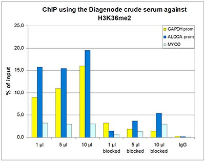
Figure 1. ChIP results obtained with the Diagenode antibody directed against H3K36me2
ChIP assays were performed using human HeLa cells, the Diagenode antibody against H3K36me2 (Cat. No. C15310127) and optimized PCR primer sets for qPCR. ChIP was performed with the “LowCell# ChIP” kit (Cat. No. C01010070), using sheared chromatin from 10,000 cells. A titration of the antibody consisting of 1, 5, and 10 µl per ChIP experiment was analysed. Additionally, the same titration was analysed after incubation of the antibody with 5 nmol blocking peptide (Cat. No. C16000127 ) for 1 hour at room temperature. IgG (5 µg/IP) was used as negative IP control. QPCR was performed with primers for the promoter of the active genes GAPDH and ALDOA and for the coding region of the myogenic differentiation gene (MYOD). Figure 1 shows the recovery, expressed as a % of input (the relative amount of immunoprecipitated DNA compared to input DNA after qPCR analysis).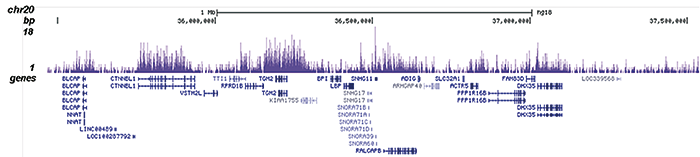
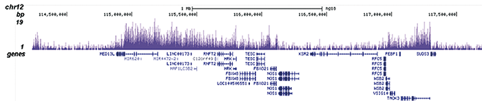
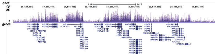
Figure 2. ChIP-seq results obtained with the Diagenode antibody directed against H3K36me2
ChIP was performed with 0.5 µl of the Diagenode antibody against H3K36me2 (Cat. No. C15310127) on sheared chromatin from 1 million HeLa cells using the “iDeal ChIP-seq” kit (Cat. No. C01010051). The IP’d DNA was subsequently analysed on an Illumina Genome Analyzer. Library preparation, cluster generation and sequencing were performed according to the manufacturer’s instructions. The 36 bp tags were aligned to the human genome using the ELAND algorithm. Figure 2 shows the signal distribution along 3 genomic regions of chromosome 20, 12 and X, respectively.A.
B.
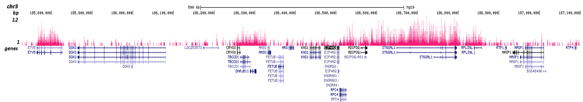
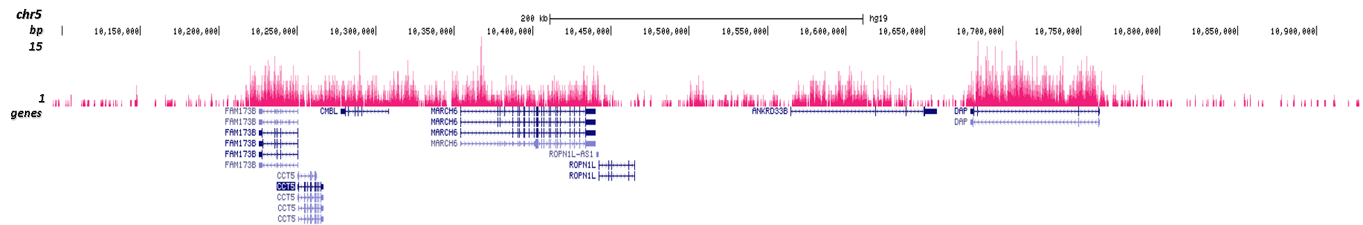
Figure 3. Cut&Tag results obtained with the Diagenode antibody directed against H3K36me2
CUT&TAG (Kaya-Okur, H.S., Nat Commun 10, 1930, 2019) was performed on 50,000 K562 cells using 1 µg of the Diagenode antibody against H3K36me2 (cat. No. C15310127) and the Diagenode pA-Tn5 transposase (C01070001). The libraries were subsequently analysed on an Illumina NextSeq 500 sequencer (2x75 paired-end reads) according to the manufacturer's instructions. The tags were aligned to the human genome (hg19) using the BWA algorithm. Figure 3 shows the peak distribution in 2 genomic regions surrounding the MARCH6 gene on chromosome 5 and the EIF4A2 gene on chromosome 3 (figure 3A and B, respectively).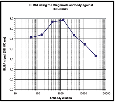
Figure 4. Determination of the titer
To determine the titer, an ELISA was performed using a serial dilution of the Diagenode antibody directed against H3K36me2 (Cat. No. C15310127). The antigen used was a peptide containing the histone modification of interest. By plotting the absorbance against the antibody dilution (Figure 4), the titer of the antibody was estimated to be 1:31,000.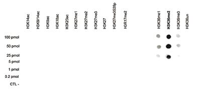
Figure 5. Cross reactivity test using the Diagenode antibody directed against H3K36me2
A dot blot analysis was performed to test the cross reactivity of the Diagenode antibody against H3K36me2 (Cat. No. C15310127) with peptides containing other modifications and unmodified sequences of histone H3. One hundred to 0.2 pmol of the peptide containing the respective histone modification were spotted on a membrane. The antibody was used at a dilution of 1:100,000. Figure 5 shows a high specificity of the antibody for the modification of interest.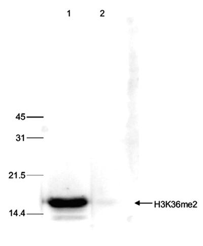
Figure 6. Western blot analysis using the Diagenode antibody directed against H3K36me2
Histone extracts of HeLa cells (15 µg) were analysed by Western blot using the Diagenode antibody against H3K36me2 (Cat. No. C15310127) diluted 1:1,000 in TBS-Tween containing 5% skimmed milk. The position of the protein of interest is indicated on the right; the marker (in kDa) is shown on the left. The result of the Western analysis with the antibody is shown in lane 1; lane 2 shows the same analysis after incubation of the antibody with 5 nmol blocking peptide (Cat. No. C16000127 ) for 1 hour at room temperature.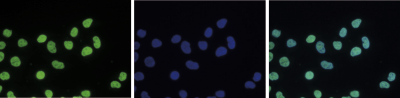
Figure 7. Immunofluorescence using the Diagenode antibody directed against H3K36me2
HeLa cells were stained with the Diagenode antibody against H3K36me2 (Cat. No. C15310127) and with DAPI. Cells were fixed with 4% formaldehyde for 10’ and blocked with PBS/TX-100 containing 5% normal goat serum and 1% BSA. The cells were immunofluorescently labelled with the H3K36me2 antibody (left) diluted 1:500 in blocking solution followed by an anti-rabbit antibody conjugated to Alexa488. The middle panel shows staining of the nuclei with DAPI. A merge of the two stainings is shown on the right. - 出版物

How to properly cite our product/service in your work
We strongly recommend using this: H3K36me2 polyclonal antibody (Hologic Diagenode Cat# C15310127 Lot# A239-001). Click here to copy to clipboard.
Using our products or services in your publication? Let us know!
Motif distribution and DNA methylation underlie distinct Cdx2 binding during development and homeostasis
Alireza Lorzadeh et al.
Transcription factors guide tissue development by binding to developmental stage-specific targets and establishing an appropriate enhancer landscape. In turn, DNA and chromatin modifications direct the genomic binding of transcription factors. However, how transcription factors navigate chromatin features to selecti...Histone H3 lysine 36 methyltransferase mobilizes NER factors to regulate tolerance against alkylation damage in fission yeast.
Lim KK, Nguyen TTT, Li AY, Yeo YP, Chen ES
The Set2 methyltransferase and its target, histone H3 lysine 36 (H3K36), affect chromatin architecture during the transcription and repair of DNA double-stranded breaks. Set2 also confers resistance against the alkylating agent, methyl methanesulfonate (MMS), through an unknown mechanism. Here, we show that Schizosa...Miz1 Controls Schwann Cell Proliferation via H3K36me2 Demethylase Kdm8 to Prevent Peripheral Nerve Demyelination
Fuhrmann D. et al.
Schwann cell differentiation and myelination depends on chromatin remodeling, histone acetylation, and methylation, which all affect Schwann cell proliferation. We previously reported that the deletion of the POZ (POxvirus and Zinc finger) domain of the transcription factor Miz1 (Myc-interacting zinc finger protein;...