

| WB Western blot : The quality of antibodies used in this technique is crucial for correct and specific protein identification. Diagenode offers huge selection of highly sensitive and specific western blot-validated antibodies. Learn more about: Load... Read more |
| IF Immunofluorescence: Diagenode offers huge selection of highly sensitive antibodies validated in IF. Immunofluorescence using the Diagenode monoclonal antibody directed against CRISPR/Cas9 HeLa cells transfected with a Cas9 expression vector (... Read more |
| ChIP-qPCR (ab) Read more |
| siRNA Knockdown Epigenetic antibodies you can trust! Antibody quality is essential for assay success. Diagenode offers antibodies that are actually validated and have been widely used and published by the scientific community. Now we are adding a new level o... Read more |
| CUT&Tag CUT&Tagアッセイを成功させるための重要な要素の1つは使用される抗体の品質です。 特異性高い抗体は、目的のタンパク質のみをターゲットとした確実な結果を可能にします。 CUT&Tagで検証済みの抗体のセレクションはこちらからご覧ください。 Read more: Products for CUT&Tag assay Performance of Diagenode's antibodies in CUT&Tag Read more |
HDAC1 monoclonal antibody
Catalog Number
Format
Price

Alternative names: HD1, RPD3, RPD3L1, GON-10
Monoclonal antibody raised in mouse against human HDAC1 (Histone deacetylase 1), using a KLH-conjugated synthetic peptide containing a sequence from the C-terminal region of the protein.
| Lot | 001 |
|---|---|
| Concentration | 2.0 µg/µl |
| Species reactivity | Human: positive. Other species: not tested. |
| Type | Monoclonal, ChIP-grade, CUT&Tag-grade |
| Purity | Protein A purified |
| Host | Mouse |
| Storage Conditions | Store at -20°C; for long storage, store at -80°C. Avoid multiple freeze-thaw cycles. |
| Storage Buffer | PBS containing 0.05% azide |
| Precautions | This product is for research use only. Not for use in diagnostic or therapeutic procedures. |
| Applications | Suggested dilution | References |
|---|---|---|
| ChIP * | 2 μg/ChIP | Fig 1 |
| CUT&Tag | 1 µg | Fig 2 |
| Western Blotting | 1:2,000 | Fig 3, 4 |
| Immunofluorescence | 1:500 | Fig 5 |
* Please note that the optimal antibody amount per ChIP should be determined by the end-user. We recommend testing 1-5 μg per IP.
- Validation Data
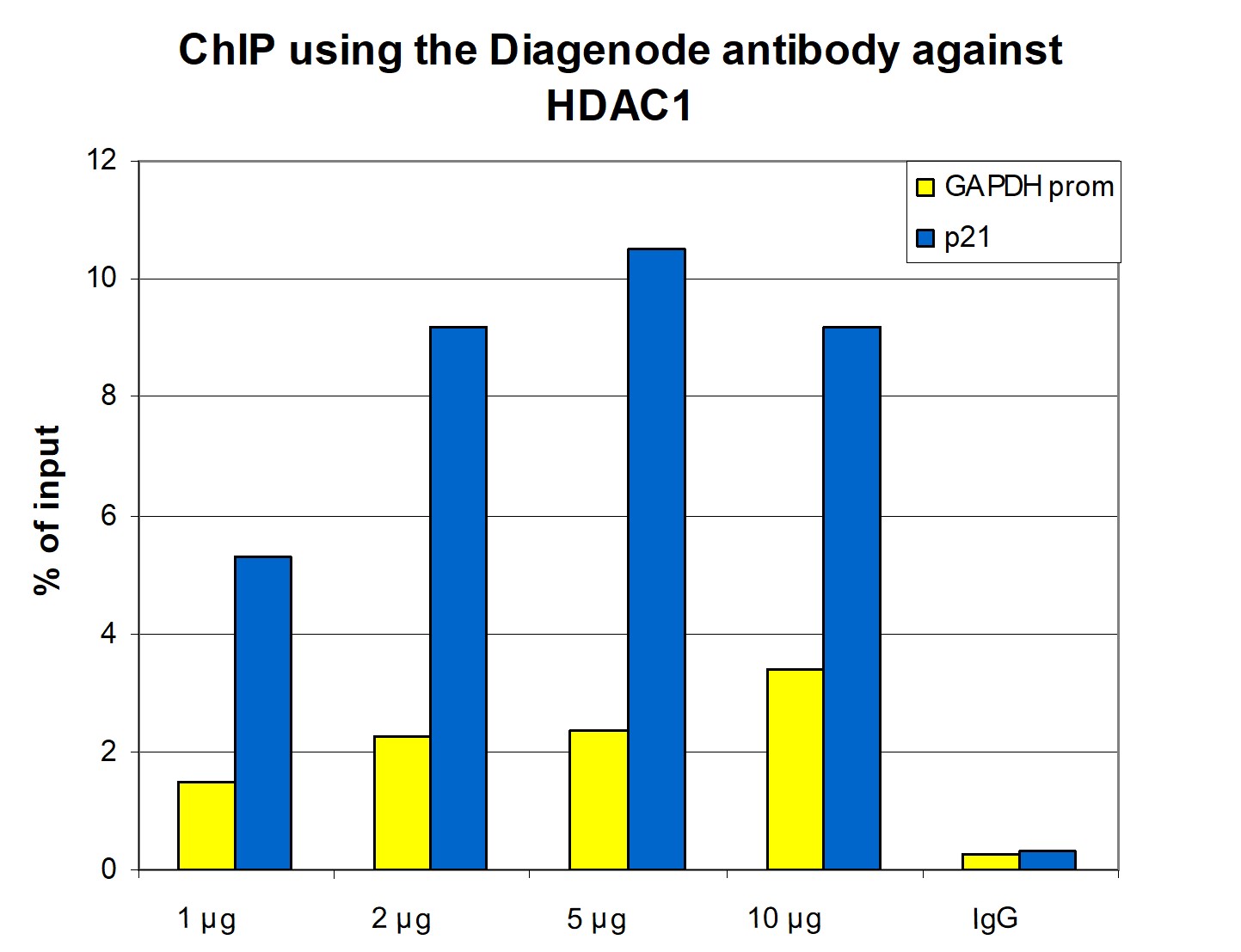
Figure 1. ChIP results obtained with the monoclonal antibody directed against HDAC1
ChIP assays were performed using human HeLa cells, the monoclonal antibody against HDAC1 (cat. No. C15200144) and optimized PCR primer sets for qPCR. A titration of the antibody consisting of 1, 2, 5, and 10 µg per ChIP experiment was analysed. IgG (5 µg/IP) was used as negative IP control. QPCR was performed with primers for the GAPDH promoter and for the coding region of p21, a known target gene of HDAC1. Figure 1 shows the recovery, expressed as a % of input (the relative amount of immunoprecipitated DNA compared to input DNA after qPCR analysis).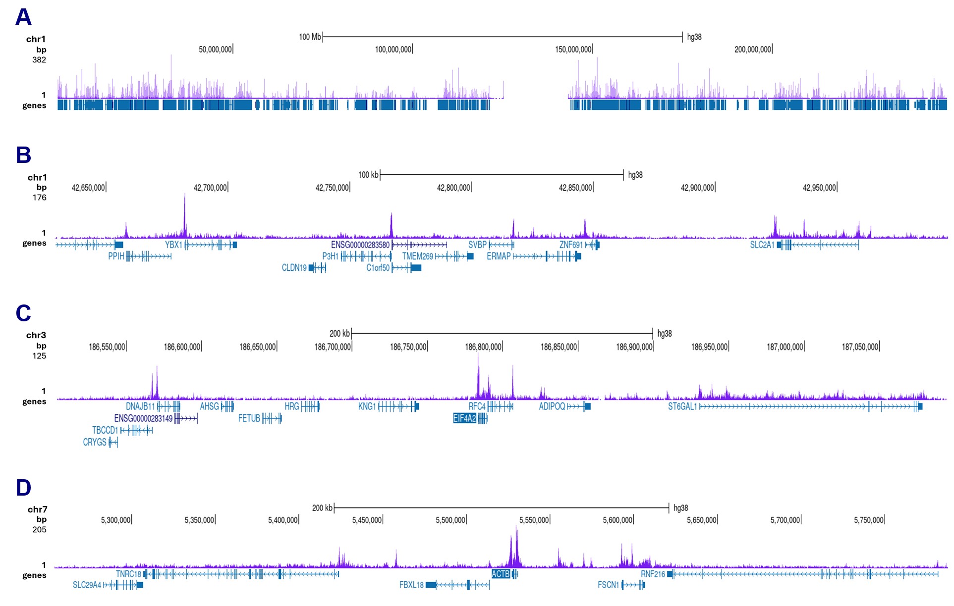
Figure 2. CUT&Tag results obtained with the monoclonal antibody directed against HDAC1
CUT&Tag was performed on 50,000 K562 cells using 1 µg of the antibody against HDAC1 (cat. No. C15200144) and the Universal CUT&Tag kit (cat. No. C01070024). The libraries were subsequently analysed on an Illumina NovaSeqX sequencer (2x150 bp paired-end reads) according to the manufacturer’s instructions. The tags were aligned to the human genome (hg38) using the BWA algorithm. Figure 2 shows the peak distribution along the complete sequence and a 300 kb region of chromosome 1 (figure 2A and B) and in 2 genomic regions on chromosome 3 and 7 (figure 2C and D, respectively).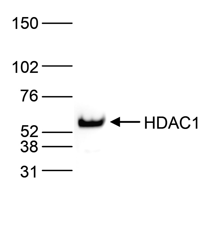
Figure 3. Western blot analysis using the monoclonal antibody directed against HDAC1
Nuclear extracts from HeLa cells (40 µg) were analysed by Western blot using the monoclonal antibody against HDAC1 (cat. No. C15200144) diluted 1:2,000 in TBS-Tween containing 5% skimmed milk. The position of the protein of interest is indicated on the right (expected size: 55 kDa); the marker (in kDa) is shown on the left.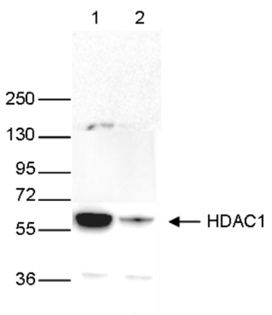
Figure 4. Western blot analysis using the monoclonal antibody directed against HDAC1
Whole cell extracts (40 µg) from HeLa cells transfected with HDAC1 siRNA (lane 2) and from an untransfected control (lane 1) were analysed by Western blot using the antibody against HDAC1 (cat. No. C15200144) diluted 1:1,000 in TBS-Tween containing 5% skimmed milk. The position of the protein of interest is indicated on the right (expected size: 55 kDa); the marker (in kDa) is shown on the left.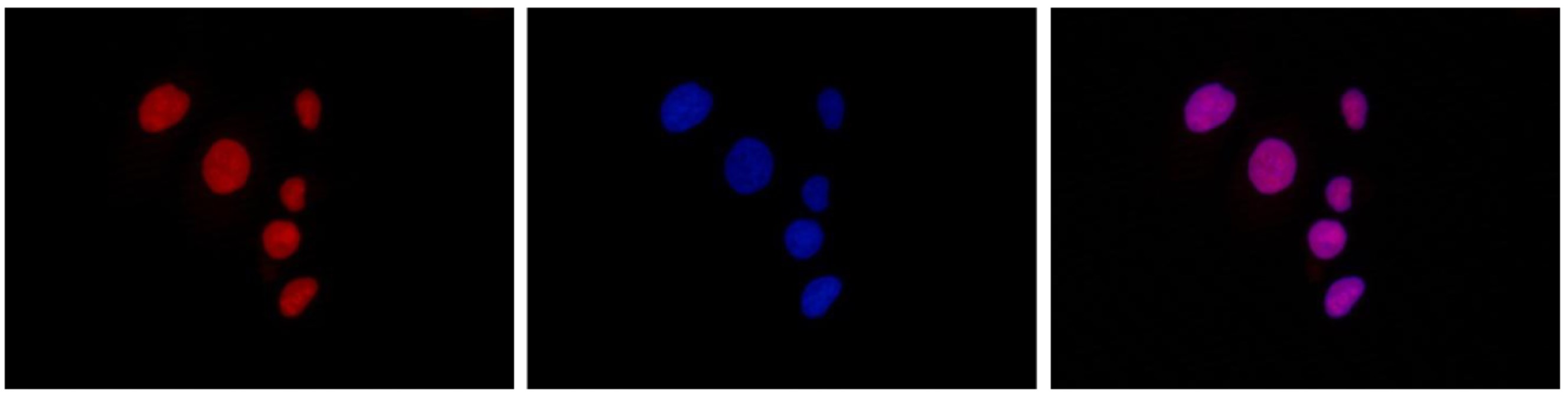
Figure 5. Immunofluorescence using the monoclonal antibody directed against HDAC1
HeLa cells were stained with the antibody against HDAC1 (cat. No. C15200144) and with DAPI. Cells were fixed with 4% formaldehyde for 10’ and blocked with PBS/TX-100 containing 5% normal goat serum and 1% BSA. The cells were immunofluorescently labelled with the HDAC1 antibody (left) diluted 1:500 in blocking solution followed by an anti-mouse antibody conjugated to Alexa594. The middle panel shows staining of the nuclei with DAPI. A merge of the two stains is shown on the right. - 出版物

How to properly cite our product/service in your work
We strongly recommend using this: HDAC1 monoclonal antibody (Hologic Diagenode Cat# C15200144 Lot# 001). Click here to copy to clipboard.
Using our products or services in your publication? Let us know!