
tHistones are the main constituents of the protein part of chromosomes of eukaryotic cells. They are rich in the amino acids arginine and lysine and have been greatly conserved during evolution. Histones pack the DNA into tight masses of chromatin. Two core histones of each class H2A, H2B, H3 and H4 assemble and are wrapped by 146 base pairs of DNA to form one octameric nucleosome. Histone tails undergo numerous post-translational modifications, which either directly or indirectly alter chromatin structure to facilitate transcriptional activation or repression or other nuclear processes. In addition to the genetic code, combinations of the different histone modifications reveal the so-called “histone code”. Histone methylation and demethylation is dynamically regulated by respectively histone methyl transferases and histone demethylases.
H3K4me1 monoclonal antibody

Monoclonal antibody raised in mouse against histone H3, monomethylated at lysine 4 (H3K4me1), using a KLH-conjugated synthetic peptide.
| Lot | 001-12 |
|---|---|
| Concentration | 1.0 µg/µl |
| Species reactivity | Human |
| Type | Monoclonal |
| Purity | Protein A purified |
| Host | Mouse |
| Precautions | This product is for research use only. Not for use in diagnostic or therapeutic procedures. |
| Applications | Suggested dilution | References |
|---|---|---|
| ChIP * | 1 μg/ChIP | Fig 1 |
| ELISA | 1:3,000 | Fig 2 |
| Western Blotting | 1:1,000 | Fig 3 |
| Immunofluorescence | 1:500 | Fig 4 |
* Please note that the optimal antibody amount per IP should be determined by the end-user. We recommend testing 1-5 μg per IP.
- Validation Data
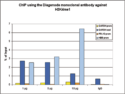
Figure 1. ChIP results obtained with the Diagenode monoclonal antibody directed against H3K4me1
ChIP assays were performed using HeLa cells, the monoclonal antibody against H3K4me1 (Cat. No. MAb-150- 050) and optimized PCR primer sets for qPCR. Chromatin was sheared with the Diagenode Bioruptor using the “Shearing ChIP” kit (Cat. No. kch-redmod-100). ChIP was performed with the “OneDay ChIP” kit (Cat. No. kch-oneDIP-060), using sheared chromatin from 1.6 million cells. A titration of the antibody consisting of 1, 5 and 10 μg per ChIP experiment was analysed. IgG (5 μg/IP) was used as negative IP control. QPCR was performed with primers for the promoter and the coding region of the GAPDH gene, and for the RPL10 and HBB promoters. Figure 1 shows the recovery, expressed as a % of input (the relative amount of immunoprecipitated DNA compared to input DNA after qPCR analysis).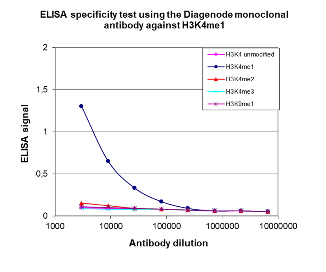
Figure 2. Cross reactivity of the Diagenode monoclonal antibody directed against H3K4me1
To test the specificity an ELISA was performed using a serial dilution of the Diagenode monoclonal antibody against H3K4me1 (cat. No. C15200150). The wells were coated with peptides containing the unmodified H3K4 as well as the mono-, di- and trimethylated H3K4 and the monomethylated H3K9. Figure 2 shows a high specificity of the antibody for the modification of interest.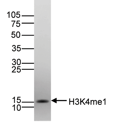
Figure 3. Western blot analysis using the Diagenode monoclonal directed antibody against H3K4me1
Histone extracts (15 μg) from HeLa cells were analysed by Western blot using the Diagenode monoclonal antibody against H3K4me1 (Cat. No. MAb-150- 050) diluted 1:1,000 in TBS-Tween containing 5% skimmed milk. The position of the protein of interest is indicated on the right; the marker (in kDa) is shown on the left.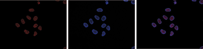
Figure 4. Immunofluorescence using the Diagenode monoclonal antibody directed against H3K4me1
HeLa cells were stained with the Diagenode antibody against H3K4me1 (Cat. No. MAb-150-050) and with DAPI. Cells were fixed with 4% formaldehyde for 10’ and blocked with PBS/TX-100 containing 5% normal goat serum and 1% BSA. The cells were immunofluorescently labelled with the H3K4me1 antibody (left) diluted 1:500 in blocking solution followed by an anti-mouse antibody conjugated to Alexa594. The middle panel shows staining of the nuclei with DAPI. A merge of the two stainings is shown on the right. - 出版物

How to properly cite our product/service in your work
We strongly recommend using this: H3K4me1 monoclonal antibody (Hologic Diagenode Cat# C15200150 Lot# 001-12). Click here to copy to clipboard.
Using our products or services in your publication? Let us know!
The glucocorticoid receptor recruits the COMPASS complex to regulateinflammatory transcription at macrophage enhancers.
Greulich, Franziska et al.
Glucocorticoids (GCs) are effective anti-inflammatory drugs; yet, their mechanisms of action are poorly understood. GCs bind to the glucocorticoid receptor (GR), a ligand-gated transcription factor controlling gene expression in numerous cell types. Here, we characterize GR's protein interactome and find the SETD1A ...The genetic association of RUNX3 with ankylosing spondylitis can be explained by allele-specific effects on IRF4 recruitment that alter gene expression
Matteo Vecellio, Amity R Roberts, Carla J Cohen, Adrian Cortes, Julian C Knight, Paul Bowness, B Paul Wordsworth
The authors sought to identify the functional basis for the genetic association of single nucleotide polymorphisms (SNP), upstream of the RUNX3 promoter, with ankylosing spondylitis (AS). They performed conditional analysis of genetic association data and used ENCODE data on chromatin remodelling and transcription f...