Notice (8): Undefined variable: solution_of_interest [APP/View/Products/view.ctp, line 755]Code Context<!-- BEGIN: REQUEST_FORM MODAL -->
<div id="request_formModal" class="reveal-modal medium" data-reveal aria-labelledby="modalTitle" aria-hidden="true" role="dialog">
<?= $this->element('Forms/simple_form', array('solution_of_interest' => $solution_of_interest, 'header' => $header, 'message' => $message, 'campaign_id' => $campaign_id)) ?>
$viewFile = '/home/website-server/www/app/View/Products/view.ctp'
$dataForView = array(
'language' => 'en',
'meta_keywords' => '',
'meta_description' => 'Histone variant H2A.Z Polyclonal Antibody validated in ChIP-seq, ChIP-qPCR, IF, WB and ELISA. Batch-specific data available on the website. Sample size available',
'meta_title' => 'H2A.Z Antibody - ChIP-seq Grade (C15410201) | Diagenode',
'product' => array(
'Product' => array(
'id' => '2275',
'antibody_id' => '191',
'name' => 'H2A.Z Antibody (sample size)',
'description' => '<p>Polyclonal antibody raised in rabbit against histone variant <strong>H2A.Z</strong>, using a KLH-conjugated synthetic peptide containing a sequence from the C-terminal part of the protein.</p>',
'label1' => 'Validation Data',
'info1' => '<div class="row">
<div class="small-4 columns">
<p><img src="https://www.diagenode.com/img/product/antibodies/C15410201-Fig1A-ChIP.png" alt="H2A.Z Antibody ChIP Grade" /><br /><img src="https://www.diagenode.com/img/product/antibodies/C15410201-Fig1B-ChIP.png" alt="H2A.Z Antibody for ChIP" /></p>
</div>
<div class="small-8 columns">
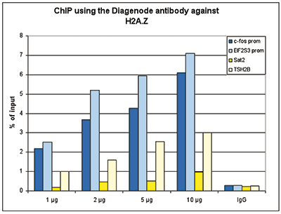
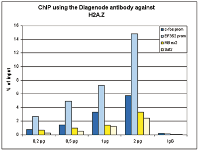
<p><small><strong> Figure 1. ChIP results obtained with the Diagenode antibody directed against H2A.Z</strong><br /> Figure 1A ChIP assays were performed using human HeLa cells, the Diagenode antibody against H2A.Z (cat. No. C15410201) and optimized PCR primer pairs for qPCR. ChIP was performed with the ““Auto Histone ChIP-seq” kit (cat. No. AB-Auto02-A100) on the IP-Star automated system, using sheared chromatin from 1,000,000 cells. A titration consisting of 1, 2, 5 and 10 μg of antibody per ChIP experiment was analyzed. IgG (2 μg/IP) was used as a negative IP control. Quantitative PCR was performed with primers specific for the promoter of the active genes c-fos and EIF2S3, used as positive controls, and for the inactive TSH2B gene and the Sat2 satellite repeat, used as negative controls. Figure 1B ChIP assays were performed using human K562 cells, the Diagenode antibody against H2A.Z (cat. No. C15410201) and optimized PCR primer sets for qPCR. ChIP was performed with the “iDeal ChIP- seq” kit (cat. No. AB-001-0024) on sheared chromatin from 100,000 cells. A titration of the antibody consisting of 0.2, 0.5, 1 and 2 μg per ChIP experiment was analysed. IgG (1 μg/IP) was used as negative IP control. Quantitative PCR was performed with primers specific for the promoter of the active genes c-fos and EIF2S3, used as positive controls, and for the coding region of the inactive MB gene and the Sat2 satellite repeat, used as negative controls. Figure 1 shows the recovery, expressed as a % of input (the relative amount of immunoprecipitated DNA compared to input DNA after qPCR analysis). </small></p>
</div>
</div>
<div class="row">
<div class="small-6 columns">
<p>A.<img src="https://www.diagenode.com/img/product/antibodies/C15410201-Fig2A-ChIP-seq.png" alt="H2A.Z Antibody ChIP-seq Grade" /></p>
<p>B.<img src="https://www.diagenode.com/img/product/antibodies/C15410201-Fig2B-ChIP-seq.png" alt="H2A.Z Antibody for ChIP-seq" /></p>
<p>C.<img src="https://www.diagenode.com/img/product/antibodies/C15410201-Fig2C-ChIP-seq.png" alt="H2A.Z Antibody for ChIP-seq assay" /></p>
<p>D.<img src="https://www.diagenode.com/img/product/antibodies/C15410201-Fig2D-ChIP-seq.png" alt="H2A.Z Antibody validated in ChIP-seq" /></p>
</div>
<div class="small-6 columns">
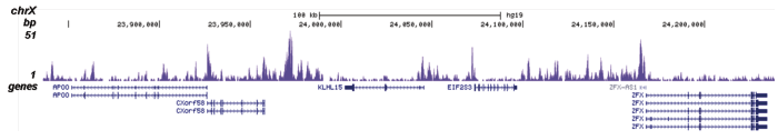
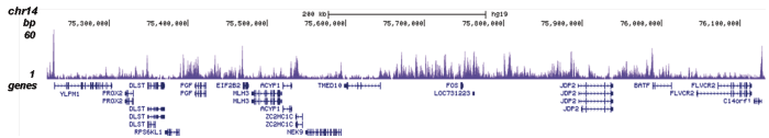
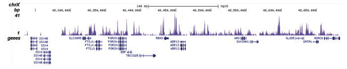
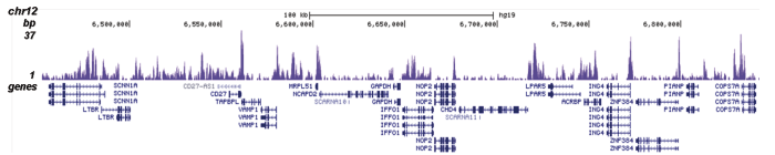
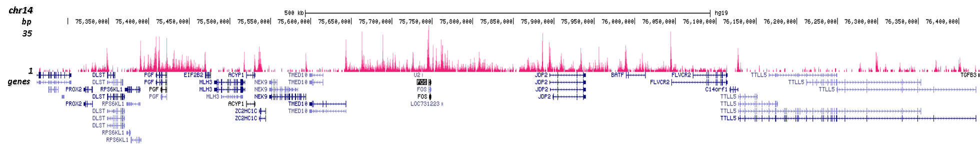
<p><small><strong> Figure 2. ChIP-seq results obtained with the Diagenode antibody directed against H2A.Z</strong><br /> ChIP was performed on sheared chromatin from 100,000 K562 cells using 0.5 μg of the Diagenode antibody against H2A.Z (cat. No. C15410201) with the “iDeal ChIP-seq” kit (cat. No. AB-001-0024) as described above. The IP’d DNA was subsequently analysed with an Illumina Genome Analyzer. Library preparation, cluster generation and sequencing were performed according to the manufacturer’s instructions. The 36 bp tags were aligned to the human genome (hg19) using the ELAND algorithm. Figure 2 shows the peak distribution in four genomic regions including the regions surrounding the EIF2S3 and c-fos positive control genes on chromosome X and 14, respectively (figure 2A and B). </small></p>
</div>
</div>
<div class="row">
<div class="small-6 columns">
<p>A.<img src="https://www.diagenode.com/img/product/antibodies/C15410201-cuttagA.png" /></p>
<p>B.<img src="https://www.diagenode.com/img/product/antibodies/C15410201-cuttagB.png" /></p>
</div>
<div class="small-6 columns">
<p><small><strong> Figure 3. Cut&Tag results obtained with the Diagenode antibody directed against H2A.Z</strong><br /> CUT&TAG (Kaya-Okur, H.S., Nat Commun 10, 1930, 2019) was performed on 50,000 K562 cells using 1 µg of the Diagenode antibody against H2A.Z (cat. No. C15410201) and the Diagenode pA-Tn5 transposase (C01070001). The libraries were subsequently analysed on an Illumina NextSeq 500 sequencer (2x75 paired-end reads) according to the manufacturer’s instructions. The tags were aligned to the human genome (hg19) using the BWA algorithm. Figure 3 shows the peak distribution along the complete sequence of chromosome 1 and in a 1 Mb region surrounding the FOS gene on chromosome 14 (figure 3A and B, respectively).</small></p>
</div>
</div>
<div class="row">
<div class="small-4 columns">
<p><img src="https://www.diagenode.com/img/product/antibodies/C15410201-Fig3-ELISA.png" alt="H2A.Z Antibody ELISA Validation" /></p>
</div>
<div class="small-8 columns">
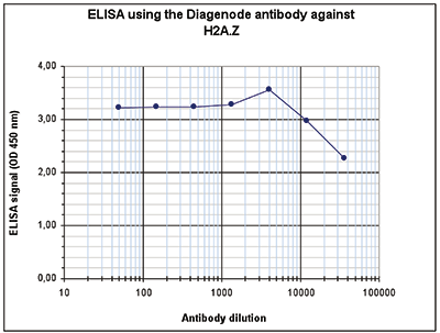
<p><small><strong> Figure 4. Determination of the antibody titer</strong><br /> To determine the titer of the antibody, an ELISA was performed using a serial dilution of the Diagenode antibody against H2A.Z (cat. No. C15410201). The antigen used was a peptide containing the histone modification of interest. By plotting the absorbance against the antibody dilution (Figure 4), the titer of the antibody was estimated to be 1:87,500. </small></p>
</div>
</div>
<div class="row">
<div class="small-4 columns">
<p><img src="https://www.diagenode.com/img/product/antibodies/C15410201-Fig4-WB.png" alt="H2A.Z Antibody validated in Western Blot" /></p>
</div>
<div class="small-8 columns">
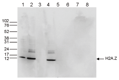
<p><small><strong> Figure 5. Western blot analysis using the Diagenode antibody directed against H2A.Z</strong><br /> Western blot was performed on whole cell (25 μg, lane 1) and histone extracts (15 μg, lane 2) from HeLa cells, and on 1 μg of recombinant histone H2A, H2B, H3 and H4 (lane 5, 6, 7 and 8, respectively) using the Diagenode antibody against H2A.Z (cat. No. C15410201). The antibody was diluted 1:1,000 in TBS-Tween containing 5% skimmed milk. Alternatively, Western blot was performed on histone extracts after incubation of the antibody with 1 μg of the peptide used for immunisation of the rabbit (1 hour at room temperature) (lane 3) or with a peptide containing a sequence from the central part of the H2A.Z protein (lane 4). The position of the protein of interest is indicated on the right, the marker (in kDa) is shown on the left. </small></p>
</div>
</div>
<div class="row">
<div class="small-6 columns">
<p><img src="https://www.diagenode.com/img/product/antibodies/C15410201-Fig5A-IF.png" alt="H2A.Z Antibody validated in Immunofluorescence" /> <br /> <br /> <img src="https://www.diagenode.com/img/product/antibodies/C15410201-Fig5B-IF.png" alt="H2A.Z Antibody validated for Immunofluorescence" /> <br /> <br /> <img src="https://www.diagenode.com/img/product/antibodies/C15410201-Fig5C-IF.png" alt="H2A.Z Antibody validated in Immunofluorescence " /></p>
</div>
<div class="small-6 columns">
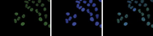
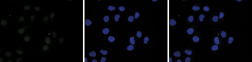
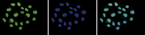
<p><small><strong> Figure 6. Immunofluorescence using the Diagenode antibody directed against H2A.Z</strong><br /> HeLa cells were stained with the Diagenode antibody against H2A.Z (cat. No. C15410201) and with DAPI. Cells were fixed with 4% formaldehyde for 10’ and blocked with PBS/TX-100 containing 5% normal goat serum and 1% BSA. Figure 5A: cells were immunofluorescently labeled with the H2A.Z antibody (left) diluted 1:500 in blocking solution followed by an anti-rabbit antibody conjugated to Alexa488. The middle panel shows staining of the nuclei with DAPI. A merge of the two stainings is shown on the right. Figure 6B and C: staining of the cells with the H2A.Z antibody after incubation of the antibody with 10 ng/μl of the peptide used for immunisation of the rabbit (figure 6B) and with a peptide containing a sequence from the central part of the H2A.Z protein (figure 6C). </small></p>
</div>
</div>',
'label2' => 'Target Description',
'info2' => '<p>Histones are the main constituents of the protein part of chromosomes of eukaryotic cells. They are rich in the amino acids arginine and lysine and have been greatly conserved during evolution. Histones pack the DNA into tight masses of chromatin. Two core histones of each class H2A, H2B, H3 and H4 assemble and are wrapped by 146 base pairs of DNA to form one octameric nucleosome. Histone tails undergo numerous post-translational modifications, which either directly or indirectly alter chromatin structure to facilitate transcriptional activation or repression or other nuclear processes. In addition to the genetic code, combinations of the different histone modifications reveal the so-called “histone code”. Histone methylation and demethylation is dynamically regulated by respectively histone methyl transferases and histone demethylases. Histone variant H2A.Z is associated with the active genes.</p>',
'label3' => '',
'info3' => '',
'format' => '10 µg',
'catalog_number' => 'C15410201-10',
'old_catalog_number' => '',
'sf_code' => 'C15410201-D001-000582',
'type' => 'FRE',
'search_order' => '03-Antibody',
'price_EUR' => '125',
'price_USD' => '115',
'price_GBP' => '115',
'price_JPY' => '19580',
'price_CNY' => '',
'price_AUD' => '288',
'country' => 'ALL',
'except_countries' => 'None',
'quote' => false,
'in_stock' => false,
'featured' => false,
'no_promo' => false,
'online' => true,
'master' => false,
'last_datasheet_update' => '0000-00-00',
'slug' => 'h2a-z-polyclonal-antibody-premium-sample-size-10-ug',
'meta_title' => 'H2A.Z Antibody - ChIP-seq Grade (C15410201) | Diagenode',
'meta_keywords' => '',
'meta_description' => 'Histone variant H2A.Z Polyclonal Antibody validated in ChIP-seq, ChIP-qPCR, IF, WB and ELISA. Batch-specific data available on the website. Sample size available',
'modified' => '2021-12-22 15:44:50',
'created' => '2015-06-29 14:08:20',
'locale' => 'eng'
),
'Antibody' => array(
'host' => '*****',
'id' => '191',
'name' => 'H2A.Z polyclonal antibody',
'description' => 'Histones are the main constituents of the protein part of chromosomes of eukaryotic cells. They are rich in the amino acids arginine and lysine and have been greatly conserved during evolution. Histones pack the DNA into tight masses of chromatin. Two core histones of each class H2A, H2B, H3 and H4 assemble and are wrapped by 146 base pairs of DNA to form one octameric nucleosome. Histone tails undergo numerous post-translational modifications, which either directly or indirectly alter chromatin structure to facilitate transcriptional activation or repression or other nuclear processes. In addition to the genetic code, combinations of the different histone modifications reveal the so-called “histone code”. Histone methylation and demethylation is dynamically regulated by respectively histone methyl transferases and histone demethylases. Histone variant H2A.Z is associated with the active genes.',
'clonality' => '',
'isotype' => '',
'lot' => 'A2039P',
'concentration' => '1.55 µg/µl',
'reactivity' => 'Human',
'type' => 'Polyclonal',
'purity' => 'Affinity purified',
'classification' => 'Premium',
'application_table' => '<table>
<thead>
<tr>
<th>Applications</th>
<th>Suggested dilution</th>
<th>References</th>
</tr>
</thead>
<tbody>
<tr>
<td>ChIP/ChIP-seq <sup>*</sup></td>
<td>0.5-1 μg/IP</td>
<td>Fig 1, 2</td>
</tr>
<tr>
<td>CUT&TAG</td>
<td>1 μg</td>
<td>Fig 3</td>
</tr>
<tr>
<td>ELISA</td>
<td>1:5,000</td>
<td>Fig 4</td>
</tr>
<tr>
<td>Western Blotting</td>
<td>1:1,000</td>
<td>Fig 5</td>
</tr>
<tr>
<td>Immunofluorescence</td>
<td>1:500</td>
<td>Fig 6</td>
</tr>
</tbody>
</table>
<p><small><sup>*</sup> Please note that the optimal antibody amount per ChIP should be determined by the end-user. We recommend testing 0.5-5 μg per IP.</small></p>',
'storage_conditions' => '',
'storage_buffer' => '',
'precautions' => 'This product is for research use only. Not for use in diagnostic or therapeutic procedures.',
'uniprot_acc' => '',
'slug' => '',
'meta_keywords' => '',
'meta_description' => '',
'modified' => '2021-12-22 15:37:17',
'created' => '0000-00-00 00:00:00',
'select_label' => '191 - H2A.Z polyclonal antibody (A2039P - 1.55 µg/µl - Human - Affinity purified - Rabbit)'
),
'Slave' => array(),
'Group' => array(
'Group' => array(
[maximum depth reached]
),
'Master' => array(
[maximum depth reached]
),
'Product' => array(
[maximum depth reached]
)
),
'Related' => array(
(int) 0 => array(
[maximum depth reached]
),
(int) 1 => array(
[maximum depth reached]
)
),
'Application' => array(
(int) 0 => array(
[maximum depth reached]
),
(int) 1 => array(
[maximum depth reached]
),
(int) 2 => array(
[maximum depth reached]
),
(int) 3 => array(
[maximum depth reached]
),
(int) 4 => array(
[maximum depth reached]
),
(int) 5 => array(
[maximum depth reached]
)
),
'Category' => array(
(int) 0 => array(
[maximum depth reached]
),
(int) 1 => array(
[maximum depth reached]
),
(int) 2 => array(
[maximum depth reached]
),
(int) 3 => array(
[maximum depth reached]
),
(int) 4 => array(
[maximum depth reached]
)
),
'Document' => array(
(int) 0 => array(
[maximum depth reached]
),
(int) 1 => array(
[maximum depth reached]
),
(int) 2 => array(
[maximum depth reached]
)
),
'Feature' => array(),
'Image' => array(
(int) 0 => array(
[maximum depth reached]
)
),
'Promotion' => array(),
'Protocol' => array(),
'Publication' => array(),
'Testimonial' => array(),
'Area' => array(),
'SafetySheet' => array(
(int) 0 => array(
[maximum depth reached]
),
(int) 1 => array(
[maximum depth reached]
),
(int) 2 => array(
[maximum depth reached]
),
(int) 3 => array(
[maximum depth reached]
),
(int) 4 => array(
[maximum depth reached]
),
(int) 5 => array(
[maximum depth reached]
),
(int) 6 => array(
[maximum depth reached]
),
(int) 7 => array(
[maximum depth reached]
)
)
),
'meta_canonical' => 'https://www.diagenode.com/en/p/h2a-z-polyclonal-antibody-premium-50-mg-33-ml'
)
$language = 'en'
$meta_keywords = ''
$meta_description = 'Histone variant H2A.Z Polyclonal Antibody validated in ChIP-seq, ChIP-qPCR, IF, WB and ELISA. Batch-specific data available on the website. Sample size available'
$meta_title = 'H2A.Z Antibody - ChIP-seq Grade (C15410201) | Diagenode'
$product = array(
'Product' => array(
'id' => '2275',
'antibody_id' => '191',
'name' => 'H2A.Z Antibody (sample size)',
'description' => '<p>Polyclonal antibody raised in rabbit against histone variant <strong>H2A.Z</strong>, using a KLH-conjugated synthetic peptide containing a sequence from the C-terminal part of the protein.</p>',
'label1' => 'Validation Data',
'info1' => '<div class="row">
<div class="small-4 columns">
<p><img src="https://www.diagenode.com/img/product/antibodies/C15410201-Fig1A-ChIP.png" alt="H2A.Z Antibody ChIP Grade" /><br /><img src="https://www.diagenode.com/img/product/antibodies/C15410201-Fig1B-ChIP.png" alt="H2A.Z Antibody for ChIP" /></p>
</div>
<div class="small-8 columns">
<p><small><strong> Figure 1. ChIP results obtained with the Diagenode antibody directed against H2A.Z</strong><br /> Figure 1A ChIP assays were performed using human HeLa cells, the Diagenode antibody against H2A.Z (cat. No. C15410201) and optimized PCR primer pairs for qPCR. ChIP was performed with the ““Auto Histone ChIP-seq” kit (cat. No. AB-Auto02-A100) on the IP-Star automated system, using sheared chromatin from 1,000,000 cells. A titration consisting of 1, 2, 5 and 10 μg of antibody per ChIP experiment was analyzed. IgG (2 μg/IP) was used as a negative IP control. Quantitative PCR was performed with primers specific for the promoter of the active genes c-fos and EIF2S3, used as positive controls, and for the inactive TSH2B gene and the Sat2 satellite repeat, used as negative controls. Figure 1B ChIP assays were performed using human K562 cells, the Diagenode antibody against H2A.Z (cat. No. C15410201) and optimized PCR primer sets for qPCR. ChIP was performed with the “iDeal ChIP- seq” kit (cat. No. AB-001-0024) on sheared chromatin from 100,000 cells. A titration of the antibody consisting of 0.2, 0.5, 1 and 2 μg per ChIP experiment was analysed. IgG (1 μg/IP) was used as negative IP control. Quantitative PCR was performed with primers specific for the promoter of the active genes c-fos and EIF2S3, used as positive controls, and for the coding region of the inactive MB gene and the Sat2 satellite repeat, used as negative controls. Figure 1 shows the recovery, expressed as a % of input (the relative amount of immunoprecipitated DNA compared to input DNA after qPCR analysis). </small></p>
</div>
</div>
<div class="row">
<div class="small-6 columns">
<p>A.<img src="https://www.diagenode.com/img/product/antibodies/C15410201-Fig2A-ChIP-seq.png" alt="H2A.Z Antibody ChIP-seq Grade" /></p>
<p>B.<img src="https://www.diagenode.com/img/product/antibodies/C15410201-Fig2B-ChIP-seq.png" alt="H2A.Z Antibody for ChIP-seq" /></p>
<p>C.<img src="https://www.diagenode.com/img/product/antibodies/C15410201-Fig2C-ChIP-seq.png" alt="H2A.Z Antibody for ChIP-seq assay" /></p>
<p>D.<img src="https://www.diagenode.com/img/product/antibodies/C15410201-Fig2D-ChIP-seq.png" alt="H2A.Z Antibody validated in ChIP-seq" /></p>
</div>
<div class="small-6 columns">
<p><small><strong> Figure 2. ChIP-seq results obtained with the Diagenode antibody directed against H2A.Z</strong><br /> ChIP was performed on sheared chromatin from 100,000 K562 cells using 0.5 μg of the Diagenode antibody against H2A.Z (cat. No. C15410201) with the “iDeal ChIP-seq” kit (cat. No. AB-001-0024) as described above. The IP’d DNA was subsequently analysed with an Illumina Genome Analyzer. Library preparation, cluster generation and sequencing were performed according to the manufacturer’s instructions. The 36 bp tags were aligned to the human genome (hg19) using the ELAND algorithm. Figure 2 shows the peak distribution in four genomic regions including the regions surrounding the EIF2S3 and c-fos positive control genes on chromosome X and 14, respectively (figure 2A and B). </small></p>
</div>
</div>
<div class="row">
<div class="small-6 columns">
<p>A.<img src="https://www.diagenode.com/img/product/antibodies/C15410201-cuttagA.png" /></p>
<p>B.<img src="https://www.diagenode.com/img/product/antibodies/C15410201-cuttagB.png" /></p>
</div>
<div class="small-6 columns">
<p><small><strong> Figure 3. Cut&Tag results obtained with the Diagenode antibody directed against H2A.Z</strong><br /> CUT&TAG (Kaya-Okur, H.S., Nat Commun 10, 1930, 2019) was performed on 50,000 K562 cells using 1 µg of the Diagenode antibody against H2A.Z (cat. No. C15410201) and the Diagenode pA-Tn5 transposase (C01070001). The libraries were subsequently analysed on an Illumina NextSeq 500 sequencer (2x75 paired-end reads) according to the manufacturer’s instructions. The tags were aligned to the human genome (hg19) using the BWA algorithm. Figure 3 shows the peak distribution along the complete sequence of chromosome 1 and in a 1 Mb region surrounding the FOS gene on chromosome 14 (figure 3A and B, respectively).</small></p>
</div>
</div>
<div class="row">
<div class="small-4 columns">
<p><img src="https://www.diagenode.com/img/product/antibodies/C15410201-Fig3-ELISA.png" alt="H2A.Z Antibody ELISA Validation" /></p>
</div>
<div class="small-8 columns">
<p><small><strong> Figure 4. Determination of the antibody titer</strong><br /> To determine the titer of the antibody, an ELISA was performed using a serial dilution of the Diagenode antibody against H2A.Z (cat. No. C15410201). The antigen used was a peptide containing the histone modification of interest. By plotting the absorbance against the antibody dilution (Figure 4), the titer of the antibody was estimated to be 1:87,500. </small></p>
</div>
</div>
<div class="row">
<div class="small-4 columns">
<p><img src="https://www.diagenode.com/img/product/antibodies/C15410201-Fig4-WB.png" alt="H2A.Z Antibody validated in Western Blot" /></p>
</div>
<div class="small-8 columns">
<p><small><strong> Figure 5. Western blot analysis using the Diagenode antibody directed against H2A.Z</strong><br /> Western blot was performed on whole cell (25 μg, lane 1) and histone extracts (15 μg, lane 2) from HeLa cells, and on 1 μg of recombinant histone H2A, H2B, H3 and H4 (lane 5, 6, 7 and 8, respectively) using the Diagenode antibody against H2A.Z (cat. No. C15410201). The antibody was diluted 1:1,000 in TBS-Tween containing 5% skimmed milk. Alternatively, Western blot was performed on histone extracts after incubation of the antibody with 1 μg of the peptide used for immunisation of the rabbit (1 hour at room temperature) (lane 3) or with a peptide containing a sequence from the central part of the H2A.Z protein (lane 4). The position of the protein of interest is indicated on the right, the marker (in kDa) is shown on the left. </small></p>
</div>
</div>
<div class="row">
<div class="small-6 columns">
<p><img src="https://www.diagenode.com/img/product/antibodies/C15410201-Fig5A-IF.png" alt="H2A.Z Antibody validated in Immunofluorescence" /> <br /> <br /> <img src="https://www.diagenode.com/img/product/antibodies/C15410201-Fig5B-IF.png" alt="H2A.Z Antibody validated for Immunofluorescence" /> <br /> <br /> <img src="https://www.diagenode.com/img/product/antibodies/C15410201-Fig5C-IF.png" alt="H2A.Z Antibody validated in Immunofluorescence " /></p>
</div>
<div class="small-6 columns">
<p><small><strong> Figure 6. Immunofluorescence using the Diagenode antibody directed against H2A.Z</strong><br /> HeLa cells were stained with the Diagenode antibody against H2A.Z (cat. No. C15410201) and with DAPI. Cells were fixed with 4% formaldehyde for 10’ and blocked with PBS/TX-100 containing 5% normal goat serum and 1% BSA. Figure 5A: cells were immunofluorescently labeled with the H2A.Z antibody (left) diluted 1:500 in blocking solution followed by an anti-rabbit antibody conjugated to Alexa488. The middle panel shows staining of the nuclei with DAPI. A merge of the two stainings is shown on the right. Figure 6B and C: staining of the cells with the H2A.Z antibody after incubation of the antibody with 10 ng/μl of the peptide used for immunisation of the rabbit (figure 6B) and with a peptide containing a sequence from the central part of the H2A.Z protein (figure 6C). </small></p>
</div>
</div>',
'label2' => 'Target Description',
'info2' => '<p>Histones are the main constituents of the protein part of chromosomes of eukaryotic cells. They are rich in the amino acids arginine and lysine and have been greatly conserved during evolution. Histones pack the DNA into tight masses of chromatin. Two core histones of each class H2A, H2B, H3 and H4 assemble and are wrapped by 146 base pairs of DNA to form one octameric nucleosome. Histone tails undergo numerous post-translational modifications, which either directly or indirectly alter chromatin structure to facilitate transcriptional activation or repression or other nuclear processes. In addition to the genetic code, combinations of the different histone modifications reveal the so-called “histone code”. Histone methylation and demethylation is dynamically regulated by respectively histone methyl transferases and histone demethylases. Histone variant H2A.Z is associated with the active genes.</p>',
'label3' => '',
'info3' => '',
'format' => '10 µg',
'catalog_number' => 'C15410201-10',
'old_catalog_number' => '',
'sf_code' => 'C15410201-D001-000582',
'type' => 'FRE',
'search_order' => '03-Antibody',
'price_EUR' => '125',
'price_USD' => '115',
'price_GBP' => '115',
'price_JPY' => '19580',
'price_CNY' => '',
'price_AUD' => '288',
'country' => 'ALL',
'except_countries' => 'None',
'quote' => false,
'in_stock' => false,
'featured' => false,
'no_promo' => false,
'online' => true,
'master' => false,
'last_datasheet_update' => '0000-00-00',
'slug' => 'h2a-z-polyclonal-antibody-premium-sample-size-10-ug',
'meta_title' => 'H2A.Z Antibody - ChIP-seq Grade (C15410201) | Diagenode',
'meta_keywords' => '',
'meta_description' => 'Histone variant H2A.Z Polyclonal Antibody validated in ChIP-seq, ChIP-qPCR, IF, WB and ELISA. Batch-specific data available on the website. Sample size available',
'modified' => '2021-12-22 15:44:50',
'created' => '2015-06-29 14:08:20',
'locale' => 'eng'
),
'Antibody' => array(
'host' => '*****',
'id' => '191',
'name' => 'H2A.Z polyclonal antibody',
'description' => 'Histones are the main constituents of the protein part of chromosomes of eukaryotic cells. They are rich in the amino acids arginine and lysine and have been greatly conserved during evolution. Histones pack the DNA into tight masses of chromatin. Two core histones of each class H2A, H2B, H3 and H4 assemble and are wrapped by 146 base pairs of DNA to form one octameric nucleosome. Histone tails undergo numerous post-translational modifications, which either directly or indirectly alter chromatin structure to facilitate transcriptional activation or repression or other nuclear processes. In addition to the genetic code, combinations of the different histone modifications reveal the so-called “histone code”. Histone methylation and demethylation is dynamically regulated by respectively histone methyl transferases and histone demethylases. Histone variant H2A.Z is associated with the active genes.',
'clonality' => '',
'isotype' => '',
'lot' => 'A2039P',
'concentration' => '1.55 µg/µl',
'reactivity' => 'Human',
'type' => 'Polyclonal',
'purity' => 'Affinity purified',
'classification' => 'Premium',
'application_table' => '<table>
<thead>
<tr>
<th>Applications</th>
<th>Suggested dilution</th>
<th>References</th>
</tr>
</thead>
<tbody>
<tr>
<td>ChIP/ChIP-seq <sup>*</sup></td>
<td>0.5-1 μg/IP</td>
<td>Fig 1, 2</td>
</tr>
<tr>
<td>CUT&TAG</td>
<td>1 μg</td>
<td>Fig 3</td>
</tr>
<tr>
<td>ELISA</td>
<td>1:5,000</td>
<td>Fig 4</td>
</tr>
<tr>
<td>Western Blotting</td>
<td>1:1,000</td>
<td>Fig 5</td>
</tr>
<tr>
<td>Immunofluorescence</td>
<td>1:500</td>
<td>Fig 6</td>
</tr>
</tbody>
</table>
<p><small><sup>*</sup> Please note that the optimal antibody amount per ChIP should be determined by the end-user. We recommend testing 0.5-5 μg per IP.</small></p>',
'storage_conditions' => '',
'storage_buffer' => '',
'precautions' => 'This product is for research use only. Not for use in diagnostic or therapeutic procedures.',
'uniprot_acc' => '',
'slug' => '',
'meta_keywords' => '',
'meta_description' => '',
'modified' => '2021-12-22 15:37:17',
'created' => '0000-00-00 00:00:00',
'select_label' => '191 - H2A.Z polyclonal antibody (A2039P - 1.55 µg/µl - Human - Affinity purified - Rabbit)'
),
'Slave' => array(),
'Group' => array(
'Group' => array(
'id' => '32',
'name' => 'C15410201',
'product_id' => '2274',
'modified' => '2016-02-18 18:07:33',
'created' => '2016-02-18 18:07:33'
),
'Master' => array(
'id' => '2274',
'antibody_id' => '191',
'name' => 'H2A.Z Antibody',
'description' => '<div style="left: 94.4882px; top: 479.039px; font-size: 15px; font-family: sans-serif; transform: scaleX(1.02952); text-align: left; padding-left: 30px;">Polyclonal antibody raised in rabbit against histone variant <strong>H2A.Z</strong>, using a KLH-conjugated synthetic peptide containing a sequence from the C-terminal part of the protein.</div>',
'label1' => 'Validation Data',
'info1' => '<div class="row">
<div class="small-4 columns">
<p><img src="https://www.diagenode.com/img/product/antibodies/C15410201-Fig1A-ChIP.png" alt="H2A.Z Antibody ChIP Grade" /><br /><img src="https://www.diagenode.com/img/product/antibodies/C15410201-Fig1B-ChIP.png" alt="H2A.Z Antibody for ChIP" /></p>
</div>
<div class="small-8 columns">
<p><small><strong> Figure 1. ChIP results obtained with the Diagenode antibody directed against H2A.Z</strong><br /> Figure 1A ChIP assays were performed using human HeLa cells, the Diagenode antibody against H2A.Z (cat. No. C15410201) and optimized PCR primer pairs for qPCR. ChIP was performed with the ““Auto Histone ChIP-seq” kit (cat. No. AB-Auto02-A100) on the IP-Star automated system, using sheared chromatin from 1,000,000 cells. A titration consisting of 1, 2, 5 and 10 μg of antibody per ChIP experiment was analyzed. IgG (2 μg/IP) was used as a negative IP control. Quantitative PCR was performed with primers specific for the promoter of the active genes c-fos and EIF2S3, used as positive controls, and for the inactive TSH2B gene and the Sat2 satellite repeat, used as negative controls. Figure 1B ChIP assays were performed using human K562 cells, the Diagenode antibody against H2A.Z (cat. No. C15410201) and optimized PCR primer sets for qPCR. ChIP was performed with the “iDeal ChIP- seq” kit (cat. No. AB-001-0024) on sheared chromatin from 100,000 cells. A titration of the antibody consisting of 0.2, 0.5, 1 and 2 μg per ChIP experiment was analysed. IgG (1 μg/IP) was used as negative IP control. Quantitative PCR was performed with primers specific for the promoter of the active genes c-fos and EIF2S3, used as positive controls, and for the coding region of the inactive MB gene and the Sat2 satellite repeat, used as negative controls. Figure 1 shows the recovery, expressed as a % of input (the relative amount of immunoprecipitated DNA compared to input DNA after qPCR analysis). </small></p>
</div>
</div>
<div class="row">
<div class="small-6 columns">
<p>A.<img src="https://www.diagenode.com/img/product/antibodies/C15410201-Fig2A-ChIP-seq.png" alt="H2A.Z Antibody ChIP-seq Grade" /></p>
<p>B.<img src="https://www.diagenode.com/img/product/antibodies/C15410201-Fig2B-ChIP-seq.png" alt="H2A.Z Antibody for ChIP-seq" /></p>
<p>C.<img src="https://www.diagenode.com/img/product/antibodies/C15410201-Fig2C-ChIP-seq.png" alt="H2A.Z Antibody for ChIP-seq assay" /></p>
<p>D.<img src="https://www.diagenode.com/img/product/antibodies/C15410201-Fig2D-ChIP-seq.png" alt="H2A.Z Antibody validated in ChIP-seq" /></p>
</div>
<div class="small-6 columns">
<p><small><strong> Figure 2. ChIP-seq results obtained with the Diagenode antibody directed against H2A.Z</strong><br /> ChIP was performed on sheared chromatin from 100,000 K562 cells using 0.5 μg of the Diagenode antibody against H2A.Z (cat. No. C15410201) with the “iDeal ChIP-seq” kit (cat. No. AB-001-0024) as described above. The IP’d DNA was subsequently analysed with an Illumina Genome Analyzer. Library preparation, cluster generation and sequencing were performed according to the manufacturer’s instructions. The 36 bp tags were aligned to the human genome (hg19) using the ELAND algorithm. Figure 2 shows the peak distribution in four genomic regions including the regions surrounding the EIF2S3 and c-fos positive control genes on chromosome X and 14, respectively (figure 2A and B). </small></p>
</div>
</div>
<div class="row">
<div class="small-6 columns">
<p>A.<img src="https://www.diagenode.com/img/product/antibodies/C15410201-cuttagA.png" /></p>
<p>B.<img src="https://www.diagenode.com/img/product/antibodies/C15410201-cuttagB.png" /></p>
</div>
<div class="small-6 columns">
<p><small><strong> Figure 3. Cut&Tag results obtained with the Diagenode antibody directed against H2A.Z</strong><br /> CUT&TAG (Kaya-Okur, H.S., Nat Commun 10, 1930, 2019) was performed on 50,000 K562 cells using 1 µg of the Diagenode antibody against H2A.Z (cat. No. C15410201) and the Diagenode pA-Tn5 transposase (C01070001). The libraries were subsequently analysed on an Illumina NextSeq 500 sequencer (2x75 paired-end reads) according to the manufacturer’s instructions. The tags were aligned to the human genome (hg19) using the BWA algorithm. Figure 3 shows the peak distribution along the complete sequence of chromosome 1 and in a 1 Mb region surrounding the FOS gene on chromosome 14 (figure 3A and B, respectively).</small></p>
</div>
</div>
<div class="row">
<div class="small-4 columns">
<p><img src="https://www.diagenode.com/img/product/antibodies/C15410201-Fig3-ELISA.png" alt="H2A.Z Antibody ELISA Validation" /></p>
</div>
<div class="small-8 columns">
<p><small><strong> Figure 4. Determination of the antibody titer</strong><br /> To determine the titer of the antibody, an ELISA was performed using a serial dilution of the Diagenode antibody against H2A.Z (cat. No. C15410201). The antigen used was a peptide containing the histone modification of interest. By plotting the absorbance against the antibody dilution (Figure 4), the titer of the antibody was estimated to be 1:87,500. </small></p>
</div>
</div>
<div class="row">
<div class="small-4 columns">
<p><img src="https://www.diagenode.com/img/product/antibodies/C15410201-Fig4-WB.png" alt="H2A.Z Antibody validated in Western Blot" /></p>
</div>
<div class="small-8 columns">
<p><small><strong> Figure 5. Western blot analysis using the Diagenode antibody directed against H2A.Z</strong><br /> Western blot was performed on whole cell (25 μg, lane 1) and histone extracts (15 μg, lane 2) from HeLa cells, and on 1 μg of recombinant histone H2A, H2B, H3 and H4 (lane 5, 6, 7 and 8, respectively) using the Diagenode antibody against H2A.Z (cat. No. C15410201). The antibody was diluted 1:1,000 in TBS-Tween containing 5% skimmed milk. Alternatively, Western blot was performed on histone extracts after incubation of the antibody with 1 μg of the peptide used for immunisation of the rabbit (1 hour at room temperature) (lane 3) or with a peptide containing a sequence from the central part of the H2A.Z protein (lane 4). The position of the protein of interest is indicated on the right, the marker (in kDa) is shown on the left. </small></p>
</div>
</div>
<div class="row">
<div class="small-6 columns">
<p><img src="https://www.diagenode.com/img/product/antibodies/C15410201-Fig5A-IF.png" alt="H2A.Z Antibody validated in Immunofluorescence" /> <br /> <br /> <img src="https://www.diagenode.com/img/product/antibodies/C15410201-Fig5B-IF.png" alt="H2A.Z Antibody validated for Immunofluorescence" /> <br /> <br /> <img src="https://www.diagenode.com/img/product/antibodies/C15410201-Fig5C-IF.png" alt="H2A.Z Antibody validated in Immunofluorescence " /></p>
</div>
<div class="small-6 columns">
<p><small><strong> Figure 6. Immunofluorescence using the Diagenode antibody directed against H2A.Z</strong><br /> HeLa cells were stained with the Diagenode antibody against H2A.Z (cat. No. C15410201) and with DAPI. Cells were fixed with 4% formaldehyde for 10’ and blocked with PBS/TX-100 containing 5% normal goat serum and 1% BSA. Figure 5A: cells were immunofluorescently labeled with the H2A.Z antibody (left) diluted 1:500 in blocking solution followed by an anti-rabbit antibody conjugated to Alexa488. The middle panel shows staining of the nuclei with DAPI. A merge of the two stainings is shown on the right. Figure 6B and C: staining of the cells with the H2A.Z antibody after incubation of the antibody with 10 ng/μl of the peptide used for immunisation of the rabbit (figure 6B) and with a peptide containing a sequence from the central part of the H2A.Z protein (figure 6C). </small></p>
</div>
</div>',
'label2' => 'Target Description',
'info2' => '<p>Histones are the main constituents of the protein part of chromosomes of eukaryotic cells. They are rich in the amino acids arginine and lysine and have been greatly conserved during evolution. Histones pack the DNA into tight masses of chromatin. Two core histones of each class H2A, H2B, H3 and H4 assemble and are wrapped by 146 base pairs of DNA to form one octameric nucleosome. Histone tails undergo numerous post-translational modifications, which either directly or indirectly alter chromatin structure to facilitate transcriptional activation or repression or other nuclear processes. In addition to the genetic code, combinations of the different histone modifications reveal the so-called “histone code”. Histone methylation and demethylation is dynamically regulated by respectively histone methyl transferases and histone demethylases. Histone variant H2A.Z is associated with the active genes.</p>',
'label3' => '',
'info3' => '',
'format' => '50 μg',
'catalog_number' => 'C15410201',
'old_catalog_number' => '',
'sf_code' => 'C15410201-D001-000581',
'type' => 'FRE',
'search_order' => '03-Antibody',
'price_EUR' => '480',
'price_USD' => '470',
'price_GBP' => '430',
'price_JPY' => '75190',
'price_CNY' => '',
'price_AUD' => '1175',
'country' => 'ALL',
'except_countries' => 'None',
'quote' => false,
'in_stock' => false,
'featured' => false,
'no_promo' => false,
'online' => true,
'master' => true,
'last_datasheet_update' => '0000-00-00',
'slug' => 'h2a-z-polyclonal-antibody-premium-50-mg-33-ml',
'meta_title' => 'H2A.Z Antibody - ChIP-seq Grade (C15410201) | Diagenode',
'meta_keywords' => '',
'meta_description' => 'Histone variant H2A.Z Polyclonal Antibody validated in ChIP-seq, ChIP-qPCR, WB, IF and ELISA. Batch-specific data available on the website. Sample size available.',
'modified' => '2021-12-22 15:43:09',
'created' => '2015-06-29 14:08:20'
),
'Product' => array(
(int) 0 => array(
[maximum depth reached]
)
)
),
'Related' => array(
(int) 0 => array(
'id' => '1787',
'antibody_id' => null,
'name' => 'Bioruptor<sup>®</sup> Pico sonication device',
'description' => '<p><a href="https://go.diagenode.com/bioruptor-upgrade"><img src="https://www.diagenode.com/img/banners/banner-br-trade.png" /></a></p>
<p>The Bioruptor® Pico (2013-2019) represented a breakthrough for shearing micro-volumes of 5 μl to larger volumes of up to 2 ml. <span>The new generation keeps the features you like the most and bring even more innovation. Check it now:</span></p>
<center><span></span></center><center><a href="https://www.diagenode.com/p/bioruptorpico2"> <img alt="New Bioruptor Pico" src="https://www.diagenode.com/img/product/shearing_technologies/new-pico-product-banner.jpg" /></a></center>
<p></p>
<p><span>Watch our short video about the Bioruptor Pico and how it can help you accomplish perfect shearing for any application including chromatin shearing, DNA shearing for NGS, unmatched DNA extraction from FFPE samples, RNA shearing, protein extraction, and much more.</span></p>
<p>
<script>// <![CDATA[
(function(){var qs,js,q,s,d=document,gi=d.getElementById,ce=d.createElement,gt=d.getElementsByTagName,id='typef_orm',b='https://s3-eu-west-1.amazonaws.com/share.typeform.com/';if(!gi.call(d,id)){js=ce.call(d,'script');js.id=id;js.src=b+'share.js';q=gt.call(d,'script')[0];q.parentNode.insertBefore(js,q)}id=id+'_';if(!gi.call(d,id)){qs=ce.call(d,'link');qs.rel='stylesheet';qs.id=id;qs.href=b+'share-button.css';s=gt.call(d,'head')[0];s.appendChild(qs,s)}})()
// ]]></script>
</p>
<center><iframe width="560" height="315" src="https://www.youtube.com/embed/ckLc4owudIM" frameborder="0" allowfullscreen="allowfullscreen"></iframe></center><center>
<p></p>
</center><center><a href="https://www.diagenode.com/en/pages/osha"><img src="https://www.diagenode.com/img/banners/banner-osha-580.jpg" width="635" height="243" /></a></center>
<div id="ConnectiveDocSignExtentionInstalled" data-extension-version="1.0.4"></div>
<div id="ConnectiveDocSignExtentionInstalled" data-extension-version="1.0.4"></div>
<div id="ConnectiveDocSignExtentionInstalled" data-extension-version="1.0.4"></div>
<div id="ConnectiveDocSignExtentionInstalled" data-extension-version="1.0.4"></div>
<div id="ConnectiveDocSignExtentionInstalled" data-extension-version="1.0.4"></div>
<div id="ConnectiveDocSignExtentionInstalled" data-extension-version="1.0.4"></div>
<div id="ConnectiveDocSignExtentionInstalled" data-extension-version="1.0.4"></div>
<div id="ConnectiveDocSignExtentionInstalled" data-extension-version="1.0.4"></div>
<div id="ConnectiveDocSignExtentionInstalled" data-extension-version="1.0.4"></div>
<div id="ConnectiveDocSignExtentionInstalled" data-extension-version="1.0.4"></div>
<div id="ConnectiveDocSignExtentionInstalled" data-extension-version="1.0.4"></div>
<div id="ConnectiveDocSignExtentionInstalled" data-extension-version="1.0.4"></div>
<div id="ConnectiveDocSignExtentionInstalled" data-extension-version="1.0.4"></div>
<div id="ConnectiveDocSignExtentionInstalled" data-extension-version="1.0.4"></div>
<div id="ConnectiveDocSignExtentionInstalled" data-extension-version="1.0.4"></div>
<div id="ConnectiveDocSignExtentionInstalled" data-extension-version="1.0.4"></div>
<div id="ConnectiveDocSignExtentionInstalled" data-extension-version="1.0.4"></div>
<div id="ConnectiveDocSignExtentionInstalled" data-extension-version="1.0.4"></div>
<div id="ConnectiveDocSignExtentionInstalled" data-extension-version="1.0.4"></div>',
'label1' => 'User manual ',
'info1' => '<p><a href="https://www.diagenode.com/files/products/shearing_technology/bioruptor/Bioruptor_pico_cooler_manual.pdf">Download</a></p>
<div id="ConnectiveDocSignExtentionInstalled" data-extension-version="1.0.4"></div>
<div id="ConnectiveDocSignExtentionInstalled" data-extension-version="1.0.4"></div>
<div id="ConnectiveDocSignExtentionInstalled" data-extension-version="1.0.4"></div>
<div id="ConnectiveDocSignExtentionInstalled" data-extension-version="1.0.4"></div>
<div id="ConnectiveDocSignExtentionInstalled" data-extension-version="1.0.4"></div>
<div id="ConnectiveDocSignExtentionInstalled" data-extension-version="1.0.4"></div>
<div id="ConnectiveDocSignExtentionInstalled" data-extension-version="1.0.4"></div>
<script src="chrome-extension://hhojmcideegachlhfgfdhailpfhgknjm/web_accessible_resources/index.js"></script>
<script src="chrome-extension://hhojmcideegachlhfgfdhailpfhgknjm/web_accessible_resources/index.js"></script>
<script src="chrome-extension://hhojmcideegachlhfgfdhailpfhgknjm/web_accessible_resources/index.js"></script>
<script src="chrome-extension://hhojmcideegachlhfgfdhailpfhgknjm/web_accessible_resources/index.js"></script>
<div id="ConnectiveDocSignExtentionInstalled" data-extension-version="1.0.4"></div>
<div id="ConnectiveDocSignExtentionInstalled" data-extension-version="1.0.4"></div>
<div id="ConnectiveDocSignExtentionInstalled" data-extension-version="1.0.4"></div>
<div id="ConnectiveDocSignExtentionInstalled" data-extension-version="1.0.4"></div>
<div id="ConnectiveDocSignExtentionInstalled" data-extension-version="1.0.4"></div>
<div id="ConnectiveDocSignExtentionInstalled" data-extension-version="1.0.4"></div>
<div id="ConnectiveDocSignExtentionInstalled" data-extension-version="1.0.4"></div>
<div id="ConnectiveDocSignExtentionInstalled" data-extension-version="1.0.4"></div>
<div id="ConnectiveDocSignExtentionInstalled" data-extension-version="1.0.4"></div>
<div id="ConnectiveDocSignExtentionInstalled" data-extension-version="1.0.4"></div>
<div id="ConnectiveDocSignExtentionInstalled" data-extension-version="1.0.4"></div>
<div id="ConnectiveDocSignExtentionInstalled" data-extension-version="1.0.4"></div>',
'label2' => 'Recommended settings for DNA shearing with Bioruptor® Pico',
'info2' => '<p>Follow our guidelines and find the good parameters for your expected DNA size: <a href="https://pybrevet.typeform.com/to/o8cQfM">DNA shearing with the Bioruptor® Pico</a></p>
<p></p>
<p>
<script>// <![CDATA[
(function(){var qs,js,q,s,d=document,gi=d.getElementById,ce=d.createElement,gt=d.getElementsByTagName,id='typef_orm',b='https://s3-eu-west-1.amazonaws.com/share.typeform.com/';if(!gi.call(d,id)){js=ce.call(d,'script');js.id=id;js.src=b+'share.js';q=gt.call(d,'script')[0];q.parentNode.insertBefore(js,q)}id=id+'_';if(!gi.call(d,id)){qs=ce.call(d,'link');qs.rel='stylesheet';qs.id=id;qs.href=b+'share-button.css';s=gt.call(d,'head')[0];s.appendChild(qs,s)}})()
// ]]></script>
</p>
<div id="ConnectiveDocSignExtentionInstalled" data-extension-version="1.0.4"></div>
<div id="ConnectiveDocSignExtentionInstalled" data-extension-version="1.0.4"></div>
<div id="ConnectiveDocSignExtentionInstalled" data-extension-version="1.0.4"></div>
<div id="ConnectiveDocSignExtentionInstalled" data-extension-version="1.0.4"></div>
<div id="ConnectiveDocSignExtentionInstalled" data-extension-version="1.0.4"></div>
<div id="ConnectiveDocSignExtentionInstalled" data-extension-version="1.0.4"></div>
<div id="ConnectiveDocSignExtentionInstalled" data-extension-version="1.0.4"></div>
<script src="chrome-extension://hhojmcideegachlhfgfdhailpfhgknjm/web_accessible_resources/index.js"></script>
<script src="chrome-extension://hhojmcideegachlhfgfdhailpfhgknjm/web_accessible_resources/index.js"></script>
<script src="chrome-extension://hhojmcideegachlhfgfdhailpfhgknjm/web_accessible_resources/index.js"></script>
<script src="chrome-extension://hhojmcideegachlhfgfdhailpfhgknjm/web_accessible_resources/index.js"></script>
<div id="ConnectiveDocSignExtentionInstalled" data-extension-version="1.0.4"></div>
<div id="ConnectiveDocSignExtentionInstalled" data-extension-version="1.0.4"></div>
<div id="ConnectiveDocSignExtentionInstalled" data-extension-version="1.0.4"></div>
<div id="ConnectiveDocSignExtentionInstalled" data-extension-version="1.0.4"></div>
<div id="ConnectiveDocSignExtentionInstalled" data-extension-version="1.0.4"></div>
<div id="ConnectiveDocSignExtentionInstalled" data-extension-version="1.0.4"></div>
<div id="ConnectiveDocSignExtentionInstalled" data-extension-version="1.0.4"></div>
<div id="ConnectiveDocSignExtentionInstalled" data-extension-version="1.0.4"></div>
<div id="ConnectiveDocSignExtentionInstalled" data-extension-version="1.0.4"></div>
<div id="ConnectiveDocSignExtentionInstalled" data-extension-version="1.0.4"></div>
<div id="ConnectiveDocSignExtentionInstalled" data-extension-version="1.0.4"></div>
<div id="ConnectiveDocSignExtentionInstalled" data-extension-version="1.0.4"></div>',
'label3' => 'Available chromatin shearing kits',
'info3' => '<p>It is important to establish optimal conditions to shear crosslinked chromatin to get the correct fragment sizes needed for ChIP. Usually this process requires both optimizing sonication conditions as well as optimizing SDS concentration, which is laborious. With the Chromatin Shearing Optimization Kits, optimization is fast and easy - we provide optimization reagents with varying concentrations of SDS. Moreover, our Chromatin Shearing Optimization Kits can be used for the optimization of chromatin preparation with our kits for ChIP.</p>
<table style="width: 925px;">
<tbody>
<tr valign="middle">
<td style="width: 213px;"></td>
<td style="text-align: center; width: 208px;"><strong><a href="../p/chromatin-shearing-optimization-kit-low-sds-100-million-cells">Chromatin Shearing Kit Low SDS (for Histones)</a></strong></td>
<td style="text-align: center; width: 180px;"><strong><a href="../p/chromatin-shearing-optimization-kit-low-sds-for-tfs-25-rxns">Chromatin Shearing Kit Low SDS (for TF)</a></strong></td>
<td style="text-align: center; width: 154px;"><strong><a href="../p/chromatin-shearing-optimization-kit-high-sds-100-million-cells">Chromatin Shearing Kit High SDS</a></strong></td>
<td style="text-align: center; width: 155px;"><strong><a href="../p/chromatin-shearing-plant-chip-seq-kit">Chromatin Shearing Kit (for Plant)</a></strong></td>
</tr>
<tr style="background-color: #fff;" valign="middle">
<td style="width: 213px;">
<p style="text-align: left;"><strong>SDS concentration</strong></p>
</td>
<td style="text-align: center; width: 208px;">
<p style="text-align: center;">< 0.1%</p>
</td>
<td style="text-align: center; width: 180px;">
<p style="text-align: center;">0.2%</p>
</td>
<td style="text-align: center; width: 154px;">
<p style="text-align: center;">1%</p>
</td>
<td style="text-align: center; width: 155px;">
<p style="text-align: center;">0.5%</p>
</td>
</tr>
<tr style="background-color: #fff;" valign="middle">
<td style="width: 213px;">
<p style="text-align: left;"><strong>Nuclei isolation</strong></p>
</td>
<td style="text-align: center; width: 208px;">
<p style="text-align: center;">Yes</p>
</td>
<td style="text-align: center; width: 180px;">
<p style="text-align: center;">Yes</p>
</td>
<td style="text-align: center; width: 154px;">
<p style="text-align: center;">No</p>
</td>
<td style="text-align: center; width: 155px;">
<p style="text-align: center;">Yes</p>
</td>
</tr>
<tr style="background-color: #fff;" valign="middle">
<td style="width: 213px;">
<p style="text-align: left;"><strong>Allows for shearing of... cells/tissue</strong></p>
</td>
<td style="text-align: center; width: 208px;">
<p style="text-align: center;">100 million cells</p>
</td>
<td style="text-align: center; width: 180px;">
<p style="text-align: center;">100 million cells</p>
</td>
<td style="text-align: center; width: 154px;">
<p style="text-align: center;">100 million cells</p>
</td>
<td style="text-align: center; width: 155px;">
<p style="text-align: center;">up to 25 g of tissue</p>
</td>
</tr>
<tr style="background-color: #fff;" valign="middle">
<td style="width: 213px;">
<p style="text-align: left;"><strong>Corresponding to shearing buffers from</strong></p>
</td>
<td style="text-align: center; width: 208px;">
<p style="text-align: center;"><a href="../p/ideal-chip-seq-kit-x24-24-rxns">iDeal ChIP-seq kit for Histones</a></p>
<p style="text-align: center;"><a href="https://www.diagenode.com/en/p/manual-chipmentation-kit-for-histones-24-rxns">ChIPmentation Kit for Histones</a></p>
</td>
<td style="text-align: center; width: 180px;">
<p style="text-align: center;"><a href="../p/ideal-chip-seq-kit-for-transcription-factors-x24-24-rxns">iDeal ChIP-seq Kit for Transcription Factors</a></p>
<p style="text-align: center;"><a href="../p/ideal-chip-qpcr-kit">iDeal ChIP qPCR kit</a></p>
</td>
<td style="text-align: center; width: 154px;">
<p style="text-align: center;"><a href="../p/true-microchip-kit-x16-16-rxns">True MicroChIP kit</a></p>
</td>
<td style="text-align: center; width: 155px;">
<p style="text-align: center;"><a href="../p/universal-plant-chip-seq-kit-x24-24-rxns">Universal Plant <br />ChIP-seq kit</a></p>
</td>
</tr>
</tbody>
</table>
<div id="ConnectiveDocSignExtentionInstalled" data-extension-version="1.0.4"></div>
<div id="ConnectiveDocSignExtentionInstalled" data-extension-version="1.0.4"></div>
<div id="ConnectiveDocSignExtentionInstalled" data-extension-version="1.0.4"></div>
<div id="ConnectiveDocSignExtentionInstalled" data-extension-version="1.0.4"></div>
<div id="ConnectiveDocSignExtentionInstalled" data-extension-version="1.0.4"></div>
<div id="ConnectiveDocSignExtentionInstalled" data-extension-version="1.0.4"></div>
<div id="ConnectiveDocSignExtentionInstalled" data-extension-version="1.0.4"></div>
<script src="chrome-extension://hhojmcideegachlhfgfdhailpfhgknjm/web_accessible_resources/index.js"></script>
<script src="chrome-extension://hhojmcideegachlhfgfdhailpfhgknjm/web_accessible_resources/index.js"></script>
<script src="chrome-extension://hhojmcideegachlhfgfdhailpfhgknjm/web_accessible_resources/index.js"></script>
<script src="chrome-extension://hhojmcideegachlhfgfdhailpfhgknjm/web_accessible_resources/index.js"></script>
<div id="ConnectiveDocSignExtentionInstalled" data-extension-version="1.0.4"></div>
<div id="ConnectiveDocSignExtentionInstalled" data-extension-version="1.0.4"></div>
<div id="ConnectiveDocSignExtentionInstalled" data-extension-version="1.0.4"></div>
<div id="ConnectiveDocSignExtentionInstalled" data-extension-version="1.0.4"></div>
<div id="ConnectiveDocSignExtentionInstalled" data-extension-version="1.0.4"></div>
<div id="ConnectiveDocSignExtentionInstalled" data-extension-version="1.0.4"></div>
<div id="ConnectiveDocSignExtentionInstalled" data-extension-version="1.0.4"></div>
<div id="ConnectiveDocSignExtentionInstalled" data-extension-version="1.0.4"></div>
<div id="ConnectiveDocSignExtentionInstalled" data-extension-version="1.0.4"></div>
<div id="ConnectiveDocSignExtentionInstalled" data-extension-version="1.0.4"></div>
<div id="ConnectiveDocSignExtentionInstalled" data-extension-version="1.0.4"></div>
<div id="ConnectiveDocSignExtentionInstalled" data-extension-version="1.0.4"></div>',
'format' => '1 unit',
'catalog_number' => 'B01060010',
'old_catalog_number' => '',
'sf_code' => 'B01060010-',
'type' => 'ACC',
'search_order' => '00-Machine',
'price_EUR' => '24000',
'price_USD' => '27500',
'price_GBP' => '21000',
'price_JPY' => '3759600',
'price_CNY' => 'Discontinued',
'price_AUD' => '68750',
'country' => 'ALL',
'except_countries' => 'None',
'quote' => false,
'in_stock' => true,
'featured' => false,
'no_promo' => false,
'online' => true,
'master' => true,
'last_datasheet_update' => '0000-00-00',
'slug' => 'bioruptor-pico-sonication-device',
'meta_title' => 'Bioruptor® Pico sonication device for RNA,Chromatin and DNA shearing for Next-Generation-Sequencing | Diagenode',
'meta_keywords' => 'Bioruptor, sonication, Next-Generation-Sequencing,DNA shearing,Protein extraction',
'meta_description' => 'An all-in-one shearing system Ideal for DNA shearing for Next-Generation-Sequencing,Chromatin shearing,RNA shearing,Protein extraction from tissues and cells and FFPE DNA extraction',
'modified' => '2023-12-20 14:21:02',
'created' => '2015-06-29 14:08:20',
'ProductsRelated' => array(
[maximum depth reached]
),
'Image' => array(
[maximum depth reached]
)
),
(int) 1 => array(
'id' => '2274',
'antibody_id' => '191',
'name' => 'H2A.Z Antibody',
'description' => '<div style="left: 94.4882px; top: 479.039px; font-size: 15px; font-family: sans-serif; transform: scaleX(1.02952); text-align: left; padding-left: 30px;">Polyclonal antibody raised in rabbit against histone variant <strong>H2A.Z</strong>, using a KLH-conjugated synthetic peptide containing a sequence from the C-terminal part of the protein.</div>',
'label1' => 'Validation Data',
'info1' => '<div class="row">
<div class="small-4 columns">
<p><img src="https://www.diagenode.com/img/product/antibodies/C15410201-Fig1A-ChIP.png" alt="H2A.Z Antibody ChIP Grade" /><br /><img src="https://www.diagenode.com/img/product/antibodies/C15410201-Fig1B-ChIP.png" alt="H2A.Z Antibody for ChIP" /></p>
</div>
<div class="small-8 columns">
<p><small><strong> Figure 1. ChIP results obtained with the Diagenode antibody directed against H2A.Z</strong><br /> Figure 1A ChIP assays were performed using human HeLa cells, the Diagenode antibody against H2A.Z (cat. No. C15410201) and optimized PCR primer pairs for qPCR. ChIP was performed with the ““Auto Histone ChIP-seq” kit (cat. No. AB-Auto02-A100) on the IP-Star automated system, using sheared chromatin from 1,000,000 cells. A titration consisting of 1, 2, 5 and 10 μg of antibody per ChIP experiment was analyzed. IgG (2 μg/IP) was used as a negative IP control. Quantitative PCR was performed with primers specific for the promoter of the active genes c-fos and EIF2S3, used as positive controls, and for the inactive TSH2B gene and the Sat2 satellite repeat, used as negative controls. Figure 1B ChIP assays were performed using human K562 cells, the Diagenode antibody against H2A.Z (cat. No. C15410201) and optimized PCR primer sets for qPCR. ChIP was performed with the “iDeal ChIP- seq” kit (cat. No. AB-001-0024) on sheared chromatin from 100,000 cells. A titration of the antibody consisting of 0.2, 0.5, 1 and 2 μg per ChIP experiment was analysed. IgG (1 μg/IP) was used as negative IP control. Quantitative PCR was performed with primers specific for the promoter of the active genes c-fos and EIF2S3, used as positive controls, and for the coding region of the inactive MB gene and the Sat2 satellite repeat, used as negative controls. Figure 1 shows the recovery, expressed as a % of input (the relative amount of immunoprecipitated DNA compared to input DNA after qPCR analysis). </small></p>
</div>
</div>
<div class="row">
<div class="small-6 columns">
<p>A.<img src="https://www.diagenode.com/img/product/antibodies/C15410201-Fig2A-ChIP-seq.png" alt="H2A.Z Antibody ChIP-seq Grade" /></p>
<p>B.<img src="https://www.diagenode.com/img/product/antibodies/C15410201-Fig2B-ChIP-seq.png" alt="H2A.Z Antibody for ChIP-seq" /></p>
<p>C.<img src="https://www.diagenode.com/img/product/antibodies/C15410201-Fig2C-ChIP-seq.png" alt="H2A.Z Antibody for ChIP-seq assay" /></p>
<p>D.<img src="https://www.diagenode.com/img/product/antibodies/C15410201-Fig2D-ChIP-seq.png" alt="H2A.Z Antibody validated in ChIP-seq" /></p>
</div>
<div class="small-6 columns">
<p><small><strong> Figure 2. ChIP-seq results obtained with the Diagenode antibody directed against H2A.Z</strong><br /> ChIP was performed on sheared chromatin from 100,000 K562 cells using 0.5 μg of the Diagenode antibody against H2A.Z (cat. No. C15410201) with the “iDeal ChIP-seq” kit (cat. No. AB-001-0024) as described above. The IP’d DNA was subsequently analysed with an Illumina Genome Analyzer. Library preparation, cluster generation and sequencing were performed according to the manufacturer’s instructions. The 36 bp tags were aligned to the human genome (hg19) using the ELAND algorithm. Figure 2 shows the peak distribution in four genomic regions including the regions surrounding the EIF2S3 and c-fos positive control genes on chromosome X and 14, respectively (figure 2A and B). </small></p>
</div>
</div>
<div class="row">
<div class="small-6 columns">
<p>A.<img src="https://www.diagenode.com/img/product/antibodies/C15410201-cuttagA.png" /></p>
<p>B.<img src="https://www.diagenode.com/img/product/antibodies/C15410201-cuttagB.png" /></p>
</div>
<div class="small-6 columns">
<p><small><strong> Figure 3. Cut&Tag results obtained with the Diagenode antibody directed against H2A.Z</strong><br /> CUT&TAG (Kaya-Okur, H.S., Nat Commun 10, 1930, 2019) was performed on 50,000 K562 cells using 1 µg of the Diagenode antibody against H2A.Z (cat. No. C15410201) and the Diagenode pA-Tn5 transposase (C01070001). The libraries were subsequently analysed on an Illumina NextSeq 500 sequencer (2x75 paired-end reads) according to the manufacturer’s instructions. The tags were aligned to the human genome (hg19) using the BWA algorithm. Figure 3 shows the peak distribution along the complete sequence of chromosome 1 and in a 1 Mb region surrounding the FOS gene on chromosome 14 (figure 3A and B, respectively).</small></p>
</div>
</div>
<div class="row">
<div class="small-4 columns">
<p><img src="https://www.diagenode.com/img/product/antibodies/C15410201-Fig3-ELISA.png" alt="H2A.Z Antibody ELISA Validation" /></p>
</div>
<div class="small-8 columns">
<p><small><strong> Figure 4. Determination of the antibody titer</strong><br /> To determine the titer of the antibody, an ELISA was performed using a serial dilution of the Diagenode antibody against H2A.Z (cat. No. C15410201). The antigen used was a peptide containing the histone modification of interest. By plotting the absorbance against the antibody dilution (Figure 4), the titer of the antibody was estimated to be 1:87,500. </small></p>
</div>
</div>
<div class="row">
<div class="small-4 columns">
<p><img src="https://www.diagenode.com/img/product/antibodies/C15410201-Fig4-WB.png" alt="H2A.Z Antibody validated in Western Blot" /></p>
</div>
<div class="small-8 columns">
<p><small><strong> Figure 5. Western blot analysis using the Diagenode antibody directed against H2A.Z</strong><br /> Western blot was performed on whole cell (25 μg, lane 1) and histone extracts (15 μg, lane 2) from HeLa cells, and on 1 μg of recombinant histone H2A, H2B, H3 and H4 (lane 5, 6, 7 and 8, respectively) using the Diagenode antibody against H2A.Z (cat. No. C15410201). The antibody was diluted 1:1,000 in TBS-Tween containing 5% skimmed milk. Alternatively, Western blot was performed on histone extracts after incubation of the antibody with 1 μg of the peptide used for immunisation of the rabbit (1 hour at room temperature) (lane 3) or with a peptide containing a sequence from the central part of the H2A.Z protein (lane 4). The position of the protein of interest is indicated on the right, the marker (in kDa) is shown on the left. </small></p>
</div>
</div>
<div class="row">
<div class="small-6 columns">
<p><img src="https://www.diagenode.com/img/product/antibodies/C15410201-Fig5A-IF.png" alt="H2A.Z Antibody validated in Immunofluorescence" /> <br /> <br /> <img src="https://www.diagenode.com/img/product/antibodies/C15410201-Fig5B-IF.png" alt="H2A.Z Antibody validated for Immunofluorescence" /> <br /> <br /> <img src="https://www.diagenode.com/img/product/antibodies/C15410201-Fig5C-IF.png" alt="H2A.Z Antibody validated in Immunofluorescence " /></p>
</div>
<div class="small-6 columns">
<p><small><strong> Figure 6. Immunofluorescence using the Diagenode antibody directed against H2A.Z</strong><br /> HeLa cells were stained with the Diagenode antibody against H2A.Z (cat. No. C15410201) and with DAPI. Cells were fixed with 4% formaldehyde for 10’ and blocked with PBS/TX-100 containing 5% normal goat serum and 1% BSA. Figure 5A: cells were immunofluorescently labeled with the H2A.Z antibody (left) diluted 1:500 in blocking solution followed by an anti-rabbit antibody conjugated to Alexa488. The middle panel shows staining of the nuclei with DAPI. A merge of the two stainings is shown on the right. Figure 6B and C: staining of the cells with the H2A.Z antibody after incubation of the antibody with 10 ng/μl of the peptide used for immunisation of the rabbit (figure 6B) and with a peptide containing a sequence from the central part of the H2A.Z protein (figure 6C). </small></p>
</div>
</div>',
'label2' => 'Target Description',
'info2' => '<p>Histones are the main constituents of the protein part of chromosomes of eukaryotic cells. They are rich in the amino acids arginine and lysine and have been greatly conserved during evolution. Histones pack the DNA into tight masses of chromatin. Two core histones of each class H2A, H2B, H3 and H4 assemble and are wrapped by 146 base pairs of DNA to form one octameric nucleosome. Histone tails undergo numerous post-translational modifications, which either directly or indirectly alter chromatin structure to facilitate transcriptional activation or repression or other nuclear processes. In addition to the genetic code, combinations of the different histone modifications reveal the so-called “histone code”. Histone methylation and demethylation is dynamically regulated by respectively histone methyl transferases and histone demethylases. Histone variant H2A.Z is associated with the active genes.</p>',
'label3' => '',
'info3' => '',
'format' => '50 μg',
'catalog_number' => 'C15410201',
'old_catalog_number' => '',
'sf_code' => 'C15410201-D001-000581',
'type' => 'FRE',
'search_order' => '03-Antibody',
'price_EUR' => '480',
'price_USD' => '470',
'price_GBP' => '430',
'price_JPY' => '75190',
'price_CNY' => '',
'price_AUD' => '1175',
'country' => 'ALL',
'except_countries' => 'None',
'quote' => false,
'in_stock' => false,
'featured' => false,
'no_promo' => false,
'online' => true,
'master' => true,
'last_datasheet_update' => '0000-00-00',
'slug' => 'h2a-z-polyclonal-antibody-premium-50-mg-33-ml',
'meta_title' => 'H2A.Z Antibody - ChIP-seq Grade (C15410201) | Diagenode',
'meta_keywords' => '',
'meta_description' => 'Histone variant H2A.Z Polyclonal Antibody validated in ChIP-seq, ChIP-qPCR, WB, IF and ELISA. Batch-specific data available on the website. Sample size available.',
'modified' => '2021-12-22 15:43:09',
'created' => '2015-06-29 14:08:20',
'ProductsRelated' => array(
[maximum depth reached]
),
'Image' => array(
[maximum depth reached]
)
)
),
'Application' => array(
(int) 0 => array(
'id' => '20',
'position' => '10',
'parent_id' => '40',
'name' => 'ELISA',
'description' => '<div class="row">
<div class="small-12 medium-12 large-12 columns">Enzyme-linked immunosorbent assay.</div>
</div>',
'in_footer' => false,
'in_menu' => false,
'online' => true,
'tabular' => true,
'slug' => 'elisa-antibodies',
'meta_keywords' => ' ELISA Antibodies,Monoclonal antibody, Polyclonal antibody',
'meta_description' => 'Diagenode offers Monoclonal & Polyclonal antibodies for ELISA applications',
'meta_title' => 'ELISA Antibodies - Monoclonal & Polyclonal antibody | Diagenode',
'modified' => '2016-01-13 12:21:41',
'created' => '2014-07-08 08:13:28',
'ProductsApplication' => array(
[maximum depth reached]
)
),
(int) 1 => array(
'id' => '19',
'position' => '10',
'parent_id' => '40',
'name' => 'WB',
'description' => '<p><strong>Western blot</strong> : The quality of antibodies used in this technique is crucial for correct and specific protein identification. Diagenode offers huge selection of highly sensitive and specific western blot-validated antibodies.</p>
<p>Learn more about: <a href="https://www.diagenode.com/applications/western-blot">Loading control, MW marker visualization</a><em>. <br /></em></p>
<p><em></em>Check our selection of antibodies validated in Western blot.</p>',
'in_footer' => false,
'in_menu' => false,
'online' => true,
'tabular' => true,
'slug' => 'western-blot-antibodies',
'meta_keywords' => ' Western Blot Antibodies ,western blot protocol,Western Blotting Products,Polyclonal antibodies ,monoclonal antibodies ',
'meta_description' => 'Diagenode offers a wide range of antibodies and technical support for western blot applications',
'meta_title' => ' Western Blot - Monoclonal antibody - Polyclonal antibody | Diagenode',
'modified' => '2016-04-26 12:44:51',
'created' => '2015-01-07 09:20:00',
'ProductsApplication' => array(
[maximum depth reached]
)
),
(int) 2 => array(
'id' => '29',
'position' => '10',
'parent_id' => '40',
'name' => 'IF',
'description' => '<p><strong>Immunofluorescence</strong>:</p>
<p>Diagenode offers huge selection of highly sensitive antibodies validated in IF.</p>
<p><img src="https://www.diagenode.com/img/product/antibodies/C15200229-IF.jpg" alt="" height="245" width="256" /></p>
<p><sup><strong>Immunofluorescence using the Diagenode monoclonal antibody directed against CRISPR/Cas9</strong></sup></p>
<p><sup>HeLa cells transfected with a Cas9 expression vector (left) or untransfected cells (right) were fixed in methanol at -20°C, permeabilized with acetone at -20°C and blocked with PBS containing 2% BSA. The cells were stained with the Cas9 C-terminal antibody (Cat. No. C15200229) diluted 1:400, followed by incubation with an anti-mouse secondary antibody coupled to AF488. The bottom images show counter-staining of the nuclei with Hoechst 33342.</sup></p>
<h5><sup>Check our selection of antibodies validated in IF.</sup></h5>',
'in_footer' => false,
'in_menu' => false,
'online' => true,
'tabular' => true,
'slug' => 'immunofluorescence',
'meta_keywords' => 'Immunofluorescence,Monoclonal antibody,Polyclonal antibody',
'meta_description' => 'Diagenode offers a wide range of antibodies and technical support for Immunofluorescence applications',
'meta_title' => 'Immunofluorescence - Monoclonal antibody - Polyclonal antibody | Diagenode',
'modified' => '2016-04-27 16:23:10',
'created' => '2015-07-08 13:46:02',
'ProductsApplication' => array(
[maximum depth reached]
)
),
(int) 3 => array(
'id' => '42',
'position' => '10',
'parent_id' => '40',
'name' => 'ChIP-seq (ab)',
'description' => '',
'in_footer' => false,
'in_menu' => false,
'online' => true,
'tabular' => true,
'slug' => 'chip-seq-antibodies',
'meta_keywords' => 'Chromatin Immunoprecipitation Sequencing,ChIP-Seq,ChIP-seq grade antibodies,DNA purification,qPCR,Shearing of chromatin',
'meta_description' => 'Diagenode offers a wide range of antibodies and technical support for ChIP Sequencing applications',
'meta_title' => 'ChIP Sequencing Antibodies (ChIP-Seq) | Diagenode',
'modified' => '2016-01-20 11:06:19',
'created' => '2015-10-20 11:44:45',
'ProductsApplication' => array(
[maximum depth reached]
)
),
(int) 4 => array(
'id' => '43',
'position' => '10',
'parent_id' => '40',
'name' => 'ChIP-qPCR (ab)',
'description' => '',
'in_footer' => false,
'in_menu' => false,
'online' => true,
'tabular' => true,
'slug' => 'chip-qpcr-antibodies',
'meta_keywords' => 'Chromatin Immunoprecipitation Sequencing,ChIP-Seq,ChIP-seq grade antibodies,DNA purification,qPCR,Shearing of chromatin',
'meta_description' => 'Diagenode offers a wide range of antibodies and technical support for ChIP-qPCR applications',
'meta_title' => 'ChIP Quantitative PCR Antibodies (ChIP-qPCR) | Diagenode',
'modified' => '2016-01-20 11:30:24',
'created' => '2015-10-20 11:45:36',
'ProductsApplication' => array(
[maximum depth reached]
)
),
(int) 5 => array(
'id' => '55',
'position' => '10',
'parent_id' => '40',
'name' => 'CUT&Tag',
'description' => '<p>CUT&Tagアッセイを成功させるための重要な要素の1つは使用される抗体の品質です。 特異性高い抗体は、目的のタンパク質のみをターゲットとした確実な結果を可能にします。 CUT&Tagで検証済みの抗体のセレクションはこちらからご覧ください。</p>
<p>Read more:</p>
<p><a href="https://www.diagenode.com/en/categories/cutandtag">Products for CUT&Tag assay</a></p>
<p><a href="https://www.diagenode.com/en/pages/cut-and-tag">Performance of Diagenode's antibodies in CUT&Tag</a></p>
<script src="chrome-extension://hhojmcideegachlhfgfdhailpfhgknjm/web_accessible_resources/index.js"></script>',
'in_footer' => false,
'in_menu' => false,
'online' => true,
'tabular' => true,
'slug' => 'cut-and-tag',
'meta_keywords' => 'CUT&Tag',
'meta_description' => 'CUT&Tag',
'meta_title' => 'CUT&Tag',
'modified' => '2021-04-27 05:17:46',
'created' => '2020-08-20 10:13:47',
'ProductsApplication' => array(
[maximum depth reached]
)
)
),
'Category' => array(
(int) 0 => array(
'id' => '111',
'position' => '40',
'parent_id' => '4',
'name' => 'Histone antibodies',
'description' => '<p>Histones are the main protein components of chromatin involved in the compaction of DNA into nucleosomes, the basic units of chromatin. A <strong>nucleosome</strong> consists of one pair of each of the core histones (<strong>H2A</strong>, <strong>H2B</strong>, <strong>H3</strong> and <strong>H4</strong>) forming an octameric structure wrapped by 146 base pairs of DNA. The different nucleosomes are linked by the linker histone<strong> H1, </strong>allowing for further condensation of chromatin.</p>
<p>The core histones have a globular structure with large unstructured N-terminal tails protruding from the nucleosome. They can undergo to multiple post-translational modifications (PTM), mainly at the N-terminal tails. These <strong>post-translational modifications </strong>include methylation, acetylation, phosphorylation, ubiquitinylation, citrullination, sumoylation, deamination and crotonylation. The most well characterized PTMs are <strong>methylation,</strong> <strong>acetylation and phosphorylation</strong>. Histone methylation occurs mainly on lysine (K) residues, which can be mono-, di- or tri-methylated, and on arginines (R), which can be mono-methylated and symmetrically or asymmetrically di-methylated. Histone acetylation occurs on lysines and histone phosphorylation mainly on serines (S), threonines (T) and tyrosines (Y).</p>
<p>The PTMs of the different residues are involved in numerous processes such as DNA repair, DNA replication and chromosome condensation. They influence the chromatin organization and can be positively or negatively associated with gene expression. Trimethylation of H3K4, H3K36 and H3K79, and lysine acetylation generally result in an open chromatin configuration (figure below) and are therefore associated with <strong>euchromatin</strong> and gene activation. Trimethylation of H3K9, K3K27 and H4K20, on the other hand, is enriched in <strong>heterochromatin </strong>and associated with gene silencing. The combination of different histone modifications is called the "<strong>histone code</strong>”, analogous to the genetic code.</p>
<p><img src="https://www.diagenode.com/img/categories/antibodies/histone-marks-illustration.png" /></p>
<p>Diagenode is proud to offer a large range of antibodies against histones and histone modifications. Our antibodies are highly specific and have been validated in many applications, including <strong>ChIP</strong> and <strong>ChIP-seq</strong>.</p>
<p>Diagenode’s collection includes antibodies recognizing:</p>
<ul>
<li><strong>Histone H1 variants</strong></li>
<li><strong>Histone H2A, H2A variants and histone H2A</strong> <strong>modifications</strong> (serine phosphorylation, lysine acetylation, lysine ubiquitinylation)</li>
<li><strong>Histone H2B and H2B</strong> <strong>modifications </strong>(serine phosphorylation, lysine acetylation)</li>
<li><strong>Histone H3 and H3 modifications </strong>(lysine methylation (mono-, di- and tri-methylated), lysine acetylation, serine phosphorylation, threonine phosphorylation, arginine methylation (mono-methylated, symmetrically and asymmetrically di-methylated))</li>
<li><strong>Histone H4 and H4 modifications (</strong>lysine methylation (mono-, di- and tri-methylated), lysine acetylation, arginine methylation (mono-methylated and symmetrically di-methylated), serine phosphorylation )</li>
</ul>
<p><span style="font-weight: 400;"><strong>HDAC's HAT's, HMT's and other</strong> <strong>enzymes</strong> which modify histones can be found in the category <a href="../categories/chromatin-modifying-proteins-histone-transferase">Histone modifying enzymes</a><br /></span></p>
<p><span style="font-weight: 400;"> Diagenode’s highly validated antibodies:</span></p>
<ul>
<li><span style="font-weight: 400;"> Highly sensitive and specific</span></li>
<li><span style="font-weight: 400;"> Cost-effective (requires less antibody per reaction)</span></li>
<li><span style="font-weight: 400;"> Batch-specific data is available on the website</span></li>
<li><span style="font-weight: 400;"> Expert technical support</span></li>
<li><span style="font-weight: 400;"> Sample sizes available</span></li>
<li><span style="font-weight: 400;"> 100% satisfaction guarantee</span></li>
</ul>',
'no_promo' => false,
'in_menu' => false,
'online' => true,
'tabular' => false,
'hide' => true,
'all_format' => false,
'is_antibody' => true,
'slug' => 'histone-antibodies',
'cookies_tag_id' => null,
'meta_keywords' => 'Histone, antibody, histone h1, histone h2, histone h3, histone h4',
'meta_description' => 'Polyclonal and Monoclonal Antibodies against Histones and their modifications validated for many applications, including Chromatin Immunoprecipitation (ChIP) and ChIP-Sequencing (ChIP-seq)',
'meta_title' => 'Histone and Modified Histone Antibodies | Diagenode',
'modified' => '2020-09-17 13:34:56',
'created' => '2016-04-01 16:01:32',
'ProductsCategory' => array(
[maximum depth reached]
),
'CookiesTag' => array([maximum depth reached])
),
(int) 1 => array(
'id' => '17',
'position' => '10',
'parent_id' => '4',
'name' => 'ChIP-seq grade antibodies',
'description' => '<p><b>Unparalleled ChIP-Seq results with the most rigorously validated antibodies</b></p>
<p><span style="font-weight: 400;">Diagenode provides leading solutions for epigenetic research. Because ChIP-seq is a widely-used technique, we validate our antibodies in ChIP and ChIP-seq experiments (in addition to conventional methods like Western blot, Dot blot, ELISA, and immunofluorescence) to provide the highest quality antibody. We standardize our validation and production to guarantee high product quality without technical bias. Diagenode guarantees ChIP-seq grade antibody performance under our suggested conditions.</span></p>
<div class="row">
<div class="small-12 medium-9 large-9 columns">
<p><strong>ChIP-seq profile</strong> of active (H3K4me3 and H3K36me3) and inactive (H3K27me3) marks using Diagenode antibodies.</p>
<img src="https://www.diagenode.com/img/categories/antibodies/chip-seq-grade-antibodies.png" /></div>
<div class="small-12 medium-3 large-3 columns">
<p><small> ChIP was performed on sheared chromatin from 100,000 K562 cells using iDeal ChIP-seq kit for Histones (cat. No. C01010051) with 1 µg of the Diagenode antibodies against H3K27me3 (cat. No. C15410195) and H3K4me3 (cat. No. C15410003), and 0.5 µg of the antibody against H3K36me3 (cat. No. C15410192). The IP'd DNA was subsequently analysed on an Illumina Genome Analyzer. Library preparation, cluster generation and sequencing were performed according to the manufacturer's instructions. The 36 bp tags were aligned to the human genome using the ELAND algorithm. The figure shows the signal distribution along the complete sequence of human chromosome 3, a zoomin to a 10 Mb region and a further zoomin to a 1.5 Mb region. </small></p>
</div>
</div>
<p>Diagenode’s highly validated antibodies:</p>
<ul>
<li>Highly sensitive and specific</li>
<li>Cost-effective (requires less antibody per reaction)</li>
<li>Batch-specific data is available on the website</li>
<li>Expert technical support</li>
<li>Sample sizes available</li>
<li>100% satisfaction guarantee</li>
</ul>',
'no_promo' => false,
'in_menu' => true,
'online' => true,
'tabular' => false,
'hide' => true,
'all_format' => false,
'is_antibody' => true,
'slug' => 'chip-seq-grade-antibodies',
'cookies_tag_id' => null,
'meta_keywords' => 'ChIP-seq grade antibodies,polyclonal antibody,WB, ELISA, ChIP-seq (ab), ChIP-qPCR (ab)',
'meta_description' => 'Diagenode Offers Wide Range of Validated ChIP-Seq Grade Antibodies for Unparalleled ChIP-Seq Results',
'meta_title' => 'Chromatin Immunoprecipitation ChIP-Seq Grade Antibodies | Diagenode',
'modified' => '2019-07-03 10:57:22',
'created' => '2015-02-16 02:24:01',
'ProductsCategory' => array(
[maximum depth reached]
),
'CookiesTag' => array([maximum depth reached])
),
(int) 2 => array(
'id' => '102',
'position' => '1',
'parent_id' => '4',
'name' => 'Sample size antibodies',
'description' => '<h1><strong>Validated epigenetics antibodies</strong> – care for a sample?<br /> </h1>
<p>Diagenode has partnered with leading epigenetics experts and numerous epigenetics consortiums to bring to you a validated and comprehensive collection of epigenetic antibodies. As an expert in epigenetics, we are committed to offering highly-specific antibodies validated for ChIP/ChIP-seq and many other applications. All batch-specific validation data is available on our website.<br /><a href="../categories/antibodies">Read about our expertise in antibody production</a>.</p>
<ul>
<li><strong>Focused</strong> - Diagenode's selection of antibodies is exclusively dedicated for epigenetic research. <a title="See the full collection." href="../categories/all-antibodies">See the full collection.</a></li>
<li><strong>Strict quality standards</strong> with rigorous QC and validation</li>
<li><strong>Classified</strong> based on level of validation for flexibility of application</li>
</ul>
<p>Existing sample sizes are listed below. We will soon expand our collection. Are you looking for a sample size of another antibody? Just <a href="mailto:agnieszka.zelisko@diagenode.com?Subject=Sample%20Size%20Request" target="_top">Contact us</a>.</p>',
'no_promo' => false,
'in_menu' => true,
'online' => true,
'tabular' => false,
'hide' => true,
'all_format' => true,
'is_antibody' => true,
'slug' => 'sample-size-antibodies',
'cookies_tag_id' => null,
'meta_keywords' => '5-hmC monoclonal antibody,CRISPR/Cas9 polyclonal antibody ,H3K36me3 polyclonal antibody,diagenode',
'meta_description' => 'Diagenode offers sample volume on selected antibodies for researchers to test, validate and provide confidence and flexibility in choosing from our wide range of antibodies ',
'meta_title' => 'Sample-size Antibodies | Diagenode',
'modified' => '2019-07-03 10:57:05',
'created' => '2015-10-27 12:13:34',
'ProductsCategory' => array(
[maximum depth reached]
),
'CookiesTag' => array([maximum depth reached])
),
(int) 3 => array(
'id' => '103',
'position' => '0',
'parent_id' => '4',
'name' => 'All antibodies',
'description' => '<p><span style="font-weight: 400;">All Diagenode’s antibodies are listed below. Please, use our Quick search field to find the antibody of interest by target name, application, purity.</span></p>
<p><span style="font-weight: 400;">Diagenode’s highly validated antibodies:</span></p>
<ul>
<li>Highly sensitive and specific</li>
<li>Cost-effective (requires less antibody per reaction)</li>
<li>Batch-specific data is available on the website</li>
<li>Expert technical support</li>
<li>Sample sizes available</li>
<li>100% satisfaction guarantee</li>
</ul>',
'no_promo' => false,
'in_menu' => true,
'online' => true,
'tabular' => false,
'hide' => true,
'all_format' => false,
'is_antibody' => true,
'slug' => 'all-antibodies',
'cookies_tag_id' => null,
'meta_keywords' => 'Antibodies,Premium Antibodies,Classic,Pioneer',
'meta_description' => 'Diagenode Offers Strict quality standards with Rigorous QC and validated Antibodies. Classified based on level of validation for flexibility of Application. Comprehensive selection of histone and non-histone Antibodies',
'meta_title' => 'Diagenode's selection of Antibodies is exclusively dedicated for Epigenetic Research | Diagenode',
'modified' => '2019-07-03 10:55:44',
'created' => '2015-11-02 14:49:22',
'ProductsCategory' => array(
[maximum depth reached]
),
'CookiesTag' => array([maximum depth reached])
),
(int) 4 => array(
'id' => '127',
'position' => '10',
'parent_id' => '4',
'name' => 'ChIP-grade antibodies',
'description' => '<div class="row">
<div class="small-10 columns"><center></center>
<p><br />Chromatin immunoprecipitation (<b>ChIP</b>) is a technique to study the associations of proteins with the specific genomic regions in intact cells. One of the most important steps of this protocol is the immunoprecipitation of targeted protein using the antibody specifically recognizing it. The quality of antibodies used in ChIP is essential for the success of the experiment. Diagenode offers extensively validated ChIP-grade antibodies, confirmed for their specificity, and high level of performance in ChIP. Each batch is validated, and batch-specific data are available on the website.</p>
<p></p>
</div>
<div class="small-2 columns"><img src="https://www.diagenode.com/emailing/images/epi-success-guaranteed-icon.png" alt="Epigenetic success guaranteed" /></div>
</div>
<p><strong>ChIP results</strong> obtained with the antibody directed against H3K4me3 (Cat. No. <a href="../p/h3k4me3-polyclonal-antibody-premium-50-ug-50-ul">C15410003</a>). </p>
<div class="row">
<div class="small-12 medium-6 large-6 columns"><img src="https://www.diagenode.com/img/product/antibodies/C15410003-fig1-ChIP.jpg" alt="" width="400" height="315" /> </div>
<div class="small-12 medium-6 large-6 columns">
<p></p>
<p></p>
<p></p>
</div>
</div>
<p></p>
<p>Our aim at Diagenode is to offer the largest collection of highly specific <strong>ChIP-grade antibodies</strong>. We add new antibodies monthly. Find your ChIP-grade antibody in the list below and check more information about tested applications, extensive validation data, and product information.</p>',
'no_promo' => false,
'in_menu' => true,
'online' => true,
'tabular' => false,
'hide' => true,
'all_format' => false,
'is_antibody' => true,
'slug' => 'chip-grade-antibodies',
'cookies_tag_id' => null,
'meta_keywords' => 'ChIP-grade antibodies, polyclonal antibody, monoclonal antibody, Diagenode',
'meta_description' => 'Diagenode Offers Extensively Validated ChIP-Grade Antibodies, Confirmed for their Specificity, and high level of Performance in Chromatin Immunoprecipitation ChIP',
'meta_title' => 'Chromatin immunoprecipitation ChIP-grade antibodies | Diagenode',
'modified' => '2021-07-01 10:22:38',
'created' => '2017-02-14 11:16:04',
'ProductsCategory' => array(
[maximum depth reached]
),
'CookiesTag' => array([maximum depth reached])
)
),
'Document' => array(
(int) 0 => array(
'id' => '282',
'name' => 'Datasheet H2AZ C15410201',
'description' => 'Datasheet description',
'image_id' => null,
'type' => 'Datasheet',
'url' => 'files/products/antibodies/Datasheet_H2AZ_C15410201.pdf',
'slug' => 'datasheet-h2az-c15410201',
'meta_keywords' => null,
'meta_description' => null,
'modified' => '2015-07-07 11:47:43',
'created' => '2015-07-07 11:47:43',
'ProductsDocument' => array(
[maximum depth reached]
)
),
(int) 1 => array(
'id' => '11',
'name' => 'Antibodies you can trust',
'description' => '<p style="text-align: justify;"><span>Epigenetic research tools have evolved over time from endpoint PCR to qPCR to the analyses of large sets of genome-wide sequencing data. ChIP sequencing (ChIP-seq) has now become the gold standard method for chromatin studies, given the accuracy and coverage scale of the approach over other methods. Successful ChIP-seq, however, requires a higher level of experimental accuracy and consistency in all steps of ChIP than ever before. Particularly crucial is the quality of ChIP antibodies. </span></p>',
'image_id' => null,
'type' => 'Poster',
'url' => 'files/posters/Antibodies_you_can_trust_Poster.pdf',
'slug' => 'antibodies-you-can-trust-poster',
'meta_keywords' => '',
'meta_description' => '',
'modified' => '2015-10-01 20:18:31',
'created' => '2015-07-03 16:05:15',
'ProductsDocument' => array(
[maximum depth reached]
)
),
(int) 2 => array(
'id' => '38',
'name' => 'Epigenetic Antibodies Brochure',
'description' => '<p>More than in any other immuoprecipitation assays, quality antibodies are critical tools in many epigenetics experiments. Since 10 years, Diagenode has developed the most stringent quality production available on the market for antibodies exclusively focused on epigenetic uses. All our antibodies have been qualified to work in epigenetic applications.</p>',
'image_id' => null,
'type' => 'Brochure',
'url' => 'files/brochures/Epigenetic_Antibodies_Brochure.pdf',
'slug' => 'epigenetic-antibodies-brochure',
'meta_keywords' => '',
'meta_description' => '',
'modified' => '2016-06-15 11:24:06',
'created' => '2015-07-03 16:05:27',
'ProductsDocument' => array(
[maximum depth reached]
)
)
),
'Feature' => array(),
'Image' => array(
(int) 0 => array(
'id' => '1815',
'name' => 'product/antibodies/ab-cuttag-icon.png',
'alt' => 'cut and tag antibody icon',
'modified' => '2021-02-11 12:45:34',
'created' => '2021-02-11 12:45:34',
'ProductsImage' => array(
[maximum depth reached]
)
)
),
'Promotion' => array(),
'Protocol' => array(),
'Publication' => array(),
'Testimonial' => array(),
'Area' => array(),
'SafetySheet' => array(
(int) 0 => array(
'id' => '414',
'name' => 'H2A.Z antibody SDS GB en',
'language' => 'en',
'url' => 'files/SDS/H2A_Z/SDS-C15410201-H2A_Z_Antibody-GB-en-GHS_2_0.pdf',
'countries' => 'GB',
'modified' => '2020-06-30 18:05:15',
'created' => '2020-06-30 18:05:15',
'ProductsSafetySheet' => array(
[maximum depth reached]
)
),
(int) 1 => array(
'id' => '416',
'name' => 'H2A.Z antibody SDS US en',
'language' => 'en',
'url' => 'files/SDS/H2A_Z/SDS-C15410201-H2A_Z_Antibody-US-en-GHS_2_0.pdf',
'countries' => 'US',
'modified' => '2020-06-30 18:06:17',
'created' => '2020-06-30 18:06:17',
'ProductsSafetySheet' => array(
[maximum depth reached]
)
),
(int) 2 => array(
'id' => '410',
'name' => 'H2A.Z antibody SDS BE nl',
'language' => 'nl',
'url' => 'files/SDS/H2A_Z/SDS-C15410201-H2A_Z_Antibody-BE-nl-GHS_2_0.pdf',
'countries' => 'BE',
'modified' => '2020-06-30 18:03:16',
'created' => '2020-06-30 18:03:16',
'ProductsSafetySheet' => array(
[maximum depth reached]
)
),
(int) 3 => array(
'id' => '409',
'name' => 'H2A.Z antibody SDS BE fr',
'language' => 'fr',
'url' => 'files/SDS/H2A_Z/SDS-C15410201-H2A_Z_Antibody-BE-fr-GHS_2_0.pdf',
'countries' => 'BE',
'modified' => '2020-06-30 18:02:37',
'created' => '2020-06-30 18:02:37',
'ProductsSafetySheet' => array(
[maximum depth reached]
)
),
(int) 4 => array(
'id' => '413',
'name' => 'H2A.Z antibody SDS FR fr',
'language' => 'fr',
'url' => 'files/SDS/H2A_Z/SDS-C15410201-H2A_Z_Antibody-FR-fr-GHS_2_0.pdf',
'countries' => 'FR',
'modified' => '2020-06-30 18:04:43',
'created' => '2020-06-30 18:04:43',
'ProductsSafetySheet' => array(
[maximum depth reached]
)
),
(int) 5 => array(
'id' => '412',
'name' => 'H2A.Z antibody SDS ES es',
'language' => 'es',
'url' => 'files/SDS/H2A_Z/SDS-C15410201-H2A_Z_Antibody-ES-es-GHS_2_0.pdf',
'countries' => 'ES',
'modified' => '2020-06-30 18:04:17',
'created' => '2020-06-30 18:04:17',
'ProductsSafetySheet' => array(
[maximum depth reached]
)
),
(int) 6 => array(
'id' => '411',
'name' => 'H2A.Z antibody SDS DE de',
'language' => 'de',
'url' => 'files/SDS/H2A_Z/SDS-C15410201-H2A_Z_Antibody-DE-de-GHS_2_0.pdf',
'countries' => 'DE',
'modified' => '2020-06-30 18:03:50',
'created' => '2020-06-30 18:03:50',
'ProductsSafetySheet' => array(
[maximum depth reached]
)
),
(int) 7 => array(
'id' => '415',
'name' => 'H2A.Z antibody SDS JP ja',
'language' => 'ja',
'url' => 'files/SDS/H2A_Z/SDS-C15410201-H2A_Z_Antibody-JP-ja-GHS_3_0.pdf',
'countries' => 'JP',
'modified' => '2020-06-30 18:05:50',
'created' => '2020-06-30 18:05:50',
'ProductsSafetySheet' => array(
[maximum depth reached]
)
)
)
)
$meta_canonical = 'https://www.diagenode.com/en/p/h2a-z-polyclonal-antibody-premium-50-mg-33-ml'
$country = 'US'
$countries_allowed = array(
(int) 0 => 'CA',
(int) 1 => 'US',
(int) 2 => 'IE',
(int) 3 => 'GB',
(int) 4 => 'DK',
(int) 5 => 'NO',
(int) 6 => 'SE',
(int) 7 => 'FI',
(int) 8 => 'NL',
(int) 9 => 'BE',
(int) 10 => 'LU',
(int) 11 => 'FR',
(int) 12 => 'DE',
(int) 13 => 'CH',
(int) 14 => 'AT',
(int) 15 => 'ES',
(int) 16 => 'IT',
(int) 17 => 'PT'
)
$outsource = false
$other_formats = array(
(int) 0 => array(
'id' => '2274',
'antibody_id' => '191',
'name' => 'H2A.Z Antibody',
'description' => '<div style="left: 94.4882px; top: 479.039px; font-size: 15px; font-family: sans-serif; transform: scaleX(1.02952); text-align: left; padding-left: 30px;">Polyclonal antibody raised in rabbit against histone variant <strong>H2A.Z</strong>, using a KLH-conjugated synthetic peptide containing a sequence from the C-terminal part of the protein.</div>',
'label1' => 'Validation Data',
'info1' => '<div class="row">
<div class="small-4 columns">
<p><img src="https://www.diagenode.com/img/product/antibodies/C15410201-Fig1A-ChIP.png" alt="H2A.Z Antibody ChIP Grade" /><br /><img src="https://www.diagenode.com/img/product/antibodies/C15410201-Fig1B-ChIP.png" alt="H2A.Z Antibody for ChIP" /></p>
</div>
<div class="small-8 columns">
<p><small><strong> Figure 1. ChIP results obtained with the Diagenode antibody directed against H2A.Z</strong><br /> Figure 1A ChIP assays were performed using human HeLa cells, the Diagenode antibody against H2A.Z (cat. No. C15410201) and optimized PCR primer pairs for qPCR. ChIP was performed with the ““Auto Histone ChIP-seq” kit (cat. No. AB-Auto02-A100) on the IP-Star automated system, using sheared chromatin from 1,000,000 cells. A titration consisting of 1, 2, 5 and 10 μg of antibody per ChIP experiment was analyzed. IgG (2 μg/IP) was used as a negative IP control. Quantitative PCR was performed with primers specific for the promoter of the active genes c-fos and EIF2S3, used as positive controls, and for the inactive TSH2B gene and the Sat2 satellite repeat, used as negative controls. Figure 1B ChIP assays were performed using human K562 cells, the Diagenode antibody against H2A.Z (cat. No. C15410201) and optimized PCR primer sets for qPCR. ChIP was performed with the “iDeal ChIP- seq” kit (cat. No. AB-001-0024) on sheared chromatin from 100,000 cells. A titration of the antibody consisting of 0.2, 0.5, 1 and 2 μg per ChIP experiment was analysed. IgG (1 μg/IP) was used as negative IP control. Quantitative PCR was performed with primers specific for the promoter of the active genes c-fos and EIF2S3, used as positive controls, and for the coding region of the inactive MB gene and the Sat2 satellite repeat, used as negative controls. Figure 1 shows the recovery, expressed as a % of input (the relative amount of immunoprecipitated DNA compared to input DNA after qPCR analysis). </small></p>
</div>
</div>
<div class="row">
<div class="small-6 columns">
<p>A.<img src="https://www.diagenode.com/img/product/antibodies/C15410201-Fig2A-ChIP-seq.png" alt="H2A.Z Antibody ChIP-seq Grade" /></p>
<p>B.<img src="https://www.diagenode.com/img/product/antibodies/C15410201-Fig2B-ChIP-seq.png" alt="H2A.Z Antibody for ChIP-seq" /></p>
<p>C.<img src="https://www.diagenode.com/img/product/antibodies/C15410201-Fig2C-ChIP-seq.png" alt="H2A.Z Antibody for ChIP-seq assay" /></p>
<p>D.<img src="https://www.diagenode.com/img/product/antibodies/C15410201-Fig2D-ChIP-seq.png" alt="H2A.Z Antibody validated in ChIP-seq" /></p>
</div>
<div class="small-6 columns">
<p><small><strong> Figure 2. ChIP-seq results obtained with the Diagenode antibody directed against H2A.Z</strong><br /> ChIP was performed on sheared chromatin from 100,000 K562 cells using 0.5 μg of the Diagenode antibody against H2A.Z (cat. No. C15410201) with the “iDeal ChIP-seq” kit (cat. No. AB-001-0024) as described above. The IP’d DNA was subsequently analysed with an Illumina Genome Analyzer. Library preparation, cluster generation and sequencing were performed according to the manufacturer’s instructions. The 36 bp tags were aligned to the human genome (hg19) using the ELAND algorithm. Figure 2 shows the peak distribution in four genomic regions including the regions surrounding the EIF2S3 and c-fos positive control genes on chromosome X and 14, respectively (figure 2A and B). </small></p>
</div>
</div>
<div class="row">
<div class="small-6 columns">
<p>A.<img src="https://www.diagenode.com/img/product/antibodies/C15410201-cuttagA.png" /></p>
<p>B.<img src="https://www.diagenode.com/img/product/antibodies/C15410201-cuttagB.png" /></p>
</div>
<div class="small-6 columns">
<p><small><strong> Figure 3. Cut&Tag results obtained with the Diagenode antibody directed against H2A.Z</strong><br /> CUT&TAG (Kaya-Okur, H.S., Nat Commun 10, 1930, 2019) was performed on 50,000 K562 cells using 1 µg of the Diagenode antibody against H2A.Z (cat. No. C15410201) and the Diagenode pA-Tn5 transposase (C01070001). The libraries were subsequently analysed on an Illumina NextSeq 500 sequencer (2x75 paired-end reads) according to the manufacturer’s instructions. The tags were aligned to the human genome (hg19) using the BWA algorithm. Figure 3 shows the peak distribution along the complete sequence of chromosome 1 and in a 1 Mb region surrounding the FOS gene on chromosome 14 (figure 3A and B, respectively).</small></p>
</div>
</div>
<div class="row">
<div class="small-4 columns">
<p><img src="https://www.diagenode.com/img/product/antibodies/C15410201-Fig3-ELISA.png" alt="H2A.Z Antibody ELISA Validation" /></p>
</div>
<div class="small-8 columns">
<p><small><strong> Figure 4. Determination of the antibody titer</strong><br /> To determine the titer of the antibody, an ELISA was performed using a serial dilution of the Diagenode antibody against H2A.Z (cat. No. C15410201). The antigen used was a peptide containing the histone modification of interest. By plotting the absorbance against the antibody dilution (Figure 4), the titer of the antibody was estimated to be 1:87,500. </small></p>
</div>
</div>
<div class="row">
<div class="small-4 columns">
<p><img src="https://www.diagenode.com/img/product/antibodies/C15410201-Fig4-WB.png" alt="H2A.Z Antibody validated in Western Blot" /></p>
</div>
<div class="small-8 columns">
<p><small><strong> Figure 5. Western blot analysis using the Diagenode antibody directed against H2A.Z</strong><br /> Western blot was performed on whole cell (25 μg, lane 1) and histone extracts (15 μg, lane 2) from HeLa cells, and on 1 μg of recombinant histone H2A, H2B, H3 and H4 (lane 5, 6, 7 and 8, respectively) using the Diagenode antibody against H2A.Z (cat. No. C15410201). The antibody was diluted 1:1,000 in TBS-Tween containing 5% skimmed milk. Alternatively, Western blot was performed on histone extracts after incubation of the antibody with 1 μg of the peptide used for immunisation of the rabbit (1 hour at room temperature) (lane 3) or with a peptide containing a sequence from the central part of the H2A.Z protein (lane 4). The position of the protein of interest is indicated on the right, the marker (in kDa) is shown on the left. </small></p>
</div>
</div>
<div class="row">
<div class="small-6 columns">
<p><img src="https://www.diagenode.com/img/product/antibodies/C15410201-Fig5A-IF.png" alt="H2A.Z Antibody validated in Immunofluorescence" /> <br /> <br /> <img src="https://www.diagenode.com/img/product/antibodies/C15410201-Fig5B-IF.png" alt="H2A.Z Antibody validated for Immunofluorescence" /> <br /> <br /> <img src="https://www.diagenode.com/img/product/antibodies/C15410201-Fig5C-IF.png" alt="H2A.Z Antibody validated in Immunofluorescence " /></p>
</div>
<div class="small-6 columns">
<p><small><strong> Figure 6. Immunofluorescence using the Diagenode antibody directed against H2A.Z</strong><br /> HeLa cells were stained with the Diagenode antibody against H2A.Z (cat. No. C15410201) and with DAPI. Cells were fixed with 4% formaldehyde for 10’ and blocked with PBS/TX-100 containing 5% normal goat serum and 1% BSA. Figure 5A: cells were immunofluorescently labeled with the H2A.Z antibody (left) diluted 1:500 in blocking solution followed by an anti-rabbit antibody conjugated to Alexa488. The middle panel shows staining of the nuclei with DAPI. A merge of the two stainings is shown on the right. Figure 6B and C: staining of the cells with the H2A.Z antibody after incubation of the antibody with 10 ng/μl of the peptide used for immunisation of the rabbit (figure 6B) and with a peptide containing a sequence from the central part of the H2A.Z protein (figure 6C). </small></p>
</div>
</div>',
'label2' => 'Target Description',
'info2' => '<p>Histones are the main constituents of the protein part of chromosomes of eukaryotic cells. They are rich in the amino acids arginine and lysine and have been greatly conserved during evolution. Histones pack the DNA into tight masses of chromatin. Two core histones of each class H2A, H2B, H3 and H4 assemble and are wrapped by 146 base pairs of DNA to form one octameric nucleosome. Histone tails undergo numerous post-translational modifications, which either directly or indirectly alter chromatin structure to facilitate transcriptional activation or repression or other nuclear processes. In addition to the genetic code, combinations of the different histone modifications reveal the so-called “histone code”. Histone methylation and demethylation is dynamically regulated by respectively histone methyl transferases and histone demethylases. Histone variant H2A.Z is associated with the active genes.</p>',
'label3' => '',
'info3' => '',
'format' => '50 μg',
'catalog_number' => 'C15410201',
'old_catalog_number' => '',
'sf_code' => 'C15410201-D001-000581',
'type' => 'FRE',
'search_order' => '03-Antibody',
'price_EUR' => '480',
'price_USD' => '470',
'price_GBP' => '430',
'price_JPY' => '75190',
'price_CNY' => '',
'price_AUD' => '1175',
'country' => 'ALL',
'except_countries' => 'None',
'quote' => false,
'in_stock' => false,
'featured' => false,
'no_promo' => false,
'online' => true,
'master' => true,
'last_datasheet_update' => '0000-00-00',
'slug' => 'h2a-z-polyclonal-antibody-premium-50-mg-33-ml',
'meta_title' => 'H2A.Z Antibody - ChIP-seq Grade (C15410201) | Diagenode',
'meta_keywords' => '',
'meta_description' => 'Histone variant H2A.Z Polyclonal Antibody validated in ChIP-seq, ChIP-qPCR, WB, IF and ELISA. Batch-specific data available on the website. Sample size available.',
'modified' => '2021-12-22 15:43:09',
'created' => '2015-06-29 14:08:20'
)
)
$pro = array(
'id' => '2275',
'antibody_id' => '191',
'name' => 'H2A.Z Antibody (sample size)',
'description' => '<p>Polyclonal antibody raised in rabbit against histone variant <strong>H2A.Z</strong>, using a KLH-conjugated synthetic peptide containing a sequence from the C-terminal part of the protein.</p>',
'label1' => 'Validation Data',
'info1' => '<div class="row">
<div class="small-4 columns">
<p><img src="https://www.diagenode.com/img/product/antibodies/C15410201-Fig1A-ChIP.png" alt="H2A.Z Antibody ChIP Grade" /><br /><img src="https://www.diagenode.com/img/product/antibodies/C15410201-Fig1B-ChIP.png" alt="H2A.Z Antibody for ChIP" /></p>
</div>
<div class="small-8 columns">
<p><small><strong> Figure 1. ChIP results obtained with the Diagenode antibody directed against H2A.Z</strong><br /> Figure 1A ChIP assays were performed using human HeLa cells, the Diagenode antibody against H2A.Z (cat. No. C15410201) and optimized PCR primer pairs for qPCR. ChIP was performed with the ““Auto Histone ChIP-seq” kit (cat. No. AB-Auto02-A100) on the IP-Star automated system, using sheared chromatin from 1,000,000 cells. A titration consisting of 1, 2, 5 and 10 μg of antibody per ChIP experiment was analyzed. IgG (2 μg/IP) was used as a negative IP control. Quantitative PCR was performed with primers specific for the promoter of the active genes c-fos and EIF2S3, used as positive controls, and for the inactive TSH2B gene and the Sat2 satellite repeat, used as negative controls. Figure 1B ChIP assays were performed using human K562 cells, the Diagenode antibody against H2A.Z (cat. No. C15410201) and optimized PCR primer sets for qPCR. ChIP was performed with the “iDeal ChIP- seq” kit (cat. No. AB-001-0024) on sheared chromatin from 100,000 cells. A titration of the antibody consisting of 0.2, 0.5, 1 and 2 μg per ChIP experiment was analysed. IgG (1 μg/IP) was used as negative IP control. Quantitative PCR was performed with primers specific for the promoter of the active genes c-fos and EIF2S3, used as positive controls, and for the coding region of the inactive MB gene and the Sat2 satellite repeat, used as negative controls. Figure 1 shows the recovery, expressed as a % of input (the relative amount of immunoprecipitated DNA compared to input DNA after qPCR analysis). </small></p>
</div>
</div>
<div class="row">
<div class="small-6 columns">
<p>A.<img src="https://www.diagenode.com/img/product/antibodies/C15410201-Fig2A-ChIP-seq.png" alt="H2A.Z Antibody ChIP-seq Grade" /></p>
<p>B.<img src="https://www.diagenode.com/img/product/antibodies/C15410201-Fig2B-ChIP-seq.png" alt="H2A.Z Antibody for ChIP-seq" /></p>
<p>C.<img src="https://www.diagenode.com/img/product/antibodies/C15410201-Fig2C-ChIP-seq.png" alt="H2A.Z Antibody for ChIP-seq assay" /></p>
<p>D.<img src="https://www.diagenode.com/img/product/antibodies/C15410201-Fig2D-ChIP-seq.png" alt="H2A.Z Antibody validated in ChIP-seq" /></p>
</div>
<div class="small-6 columns">
<p><small><strong> Figure 2. ChIP-seq results obtained with the Diagenode antibody directed against H2A.Z</strong><br /> ChIP was performed on sheared chromatin from 100,000 K562 cells using 0.5 μg of the Diagenode antibody against H2A.Z (cat. No. C15410201) with the “iDeal ChIP-seq” kit (cat. No. AB-001-0024) as described above. The IP’d DNA was subsequently analysed with an Illumina Genome Analyzer. Library preparation, cluster generation and sequencing were performed according to the manufacturer’s instructions. The 36 bp tags were aligned to the human genome (hg19) using the ELAND algorithm. Figure 2 shows the peak distribution in four genomic regions including the regions surrounding the EIF2S3 and c-fos positive control genes on chromosome X and 14, respectively (figure 2A and B). </small></p>
</div>
</div>
<div class="row">
<div class="small-6 columns">
<p>A.<img src="https://www.diagenode.com/img/product/antibodies/C15410201-cuttagA.png" /></p>
<p>B.<img src="https://www.diagenode.com/img/product/antibodies/C15410201-cuttagB.png" /></p>
</div>
<div class="small-6 columns">
<p><small><strong> Figure 3. Cut&Tag results obtained with the Diagenode antibody directed against H2A.Z</strong><br /> CUT&TAG (Kaya-Okur, H.S., Nat Commun 10, 1930, 2019) was performed on 50,000 K562 cells using 1 µg of the Diagenode antibody against H2A.Z (cat. No. C15410201) and the Diagenode pA-Tn5 transposase (C01070001). The libraries were subsequently analysed on an Illumina NextSeq 500 sequencer (2x75 paired-end reads) according to the manufacturer’s instructions. The tags were aligned to the human genome (hg19) using the BWA algorithm. Figure 3 shows the peak distribution along the complete sequence of chromosome 1 and in a 1 Mb region surrounding the FOS gene on chromosome 14 (figure 3A and B, respectively).</small></p>
</div>
</div>
<div class="row">
<div class="small-4 columns">
<p><img src="https://www.diagenode.com/img/product/antibodies/C15410201-Fig3-ELISA.png" alt="H2A.Z Antibody ELISA Validation" /></p>
</div>
<div class="small-8 columns">
<p><small><strong> Figure 4. Determination of the antibody titer</strong><br /> To determine the titer of the antibody, an ELISA was performed using a serial dilution of the Diagenode antibody against H2A.Z (cat. No. C15410201). The antigen used was a peptide containing the histone modification of interest. By plotting the absorbance against the antibody dilution (Figure 4), the titer of the antibody was estimated to be 1:87,500. </small></p>
</div>
</div>
<div class="row">
<div class="small-4 columns">
<p><img src="https://www.diagenode.com/img/product/antibodies/C15410201-Fig4-WB.png" alt="H2A.Z Antibody validated in Western Blot" /></p>
</div>
<div class="small-8 columns">
<p><small><strong> Figure 5. Western blot analysis using the Diagenode antibody directed against H2A.Z</strong><br /> Western blot was performed on whole cell (25 μg, lane 1) and histone extracts (15 μg, lane 2) from HeLa cells, and on 1 μg of recombinant histone H2A, H2B, H3 and H4 (lane 5, 6, 7 and 8, respectively) using the Diagenode antibody against H2A.Z (cat. No. C15410201). The antibody was diluted 1:1,000 in TBS-Tween containing 5% skimmed milk. Alternatively, Western blot was performed on histone extracts after incubation of the antibody with 1 μg of the peptide used for immunisation of the rabbit (1 hour at room temperature) (lane 3) or with a peptide containing a sequence from the central part of the H2A.Z protein (lane 4). The position of the protein of interest is indicated on the right, the marker (in kDa) is shown on the left. </small></p>
</div>
</div>
<div class="row">
<div class="small-6 columns">
<p><img src="https://www.diagenode.com/img/product/antibodies/C15410201-Fig5A-IF.png" alt="H2A.Z Antibody validated in Immunofluorescence" /> <br /> <br /> <img src="https://www.diagenode.com/img/product/antibodies/C15410201-Fig5B-IF.png" alt="H2A.Z Antibody validated for Immunofluorescence" /> <br /> <br /> <img src="https://www.diagenode.com/img/product/antibodies/C15410201-Fig5C-IF.png" alt="H2A.Z Antibody validated in Immunofluorescence " /></p>
</div>
<div class="small-6 columns">
<p><small><strong> Figure 6. Immunofluorescence using the Diagenode antibody directed against H2A.Z</strong><br /> HeLa cells were stained with the Diagenode antibody against H2A.Z (cat. No. C15410201) and with DAPI. Cells were fixed with 4% formaldehyde for 10’ and blocked with PBS/TX-100 containing 5% normal goat serum and 1% BSA. Figure 5A: cells were immunofluorescently labeled with the H2A.Z antibody (left) diluted 1:500 in blocking solution followed by an anti-rabbit antibody conjugated to Alexa488. The middle panel shows staining of the nuclei with DAPI. A merge of the two stainings is shown on the right. Figure 6B and C: staining of the cells with the H2A.Z antibody after incubation of the antibody with 10 ng/μl of the peptide used for immunisation of the rabbit (figure 6B) and with a peptide containing a sequence from the central part of the H2A.Z protein (figure 6C). </small></p>
</div>
</div>',
'label2' => 'Target Description',
'info2' => '<p>Histones are the main constituents of the protein part of chromosomes of eukaryotic cells. They are rich in the amino acids arginine and lysine and have been greatly conserved during evolution. Histones pack the DNA into tight masses of chromatin. Two core histones of each class H2A, H2B, H3 and H4 assemble and are wrapped by 146 base pairs of DNA to form one octameric nucleosome. Histone tails undergo numerous post-translational modifications, which either directly or indirectly alter chromatin structure to facilitate transcriptional activation or repression or other nuclear processes. In addition to the genetic code, combinations of the different histone modifications reveal the so-called “histone code”. Histone methylation and demethylation is dynamically regulated by respectively histone methyl transferases and histone demethylases. Histone variant H2A.Z is associated with the active genes.</p>',
'label3' => '',
'info3' => '',
'format' => '10 µg',
'catalog_number' => 'C15410201-10',
'old_catalog_number' => '',
'sf_code' => 'C15410201-D001-000582',
'type' => 'FRE',
'search_order' => '03-Antibody',
'price_EUR' => '125',
'price_USD' => '115',
'price_GBP' => '115',
'price_JPY' => '19580',
'price_CNY' => '',
'price_AUD' => '288',
'country' => 'ALL',
'except_countries' => 'None',
'quote' => false,
'in_stock' => false,
'featured' => false,
'no_promo' => false,
'online' => true,
'master' => false,
'last_datasheet_update' => '0000-00-00',
'slug' => 'h2a-z-polyclonal-antibody-premium-sample-size-10-ug',
'meta_title' => 'H2A.Z Antibody - ChIP-seq Grade (C15410201) | Diagenode',
'meta_keywords' => '',
'meta_description' => 'Histone variant H2A.Z Polyclonal Antibody validated in ChIP-seq, ChIP-qPCR, IF, WB and ELISA. Batch-specific data available on the website. Sample size available',
'modified' => '2021-12-22 15:44:50',
'created' => '2015-06-29 14:08:20',
'ProductsGroup' => array(
'id' => '39',
'product_id' => '2275',
'group_id' => '32'
)
)
$edit = ''
$testimonials = ''
$featured_testimonials = ''
$related_products = '<li>
<div class="row">
<div class="small-12 columns">
<a href="/en/p/bioruptor-pico-sonication-device"><img src="/img/product/shearing_technologies/bioruptor_pico.jpg" alt="Bioruptor pico next gen sequencing " class="th"/></a> </div>
<div class="small-12 columns">
<div class="small-6 columns" style="padding-left:0px;padding-right:0px;margin-top:-6px;margin-left:-1px">
<span class="success label" style="">B01060010</span>
</div>
<div class="small-6 columns text-right" style="padding-left:0px;padding-right:0px;margin-top:-6px">
<!--a href="#" style="color:#B21329"><i class="fa fa-info-circle"></i></a-->
</div>
</div>
<div class="small-12 columns" >
<h6 style="height:60px">Bioruptor® Pico sonication device</h6>
</div>
</div>
</li>
<li>
<div class="row">
<div class="small-12 columns">
<a href="/en/p/h2a-z-polyclonal-antibody-premium-50-mg-33-ml"><img src="/img/product/antibodies/ab-cuttag-icon.png" alt="cut and tag antibody icon" class="th"/></a> </div>
<div class="small-12 columns">
<div class="small-6 columns" style="padding-left:0px;padding-right:0px;margin-top:-6px;margin-left:-1px">
<span class="success label" style="">C15410201</span>
</div>
<div class="small-6 columns text-right" style="padding-left:0px;padding-right:0px;margin-top:-6px">
<!--a href="#" style="color:#B21329"><i class="fa fa-info-circle"></i></a-->
<!-- BEGIN: ADD TO CART MODAL --><div id="cartModal-2274" class="reveal-modal small" data-reveal aria-labelledby="modalTitle" aria-hidden="true" role="dialog">
<form action="/en/carts/add/2274" id="CartAdd/2274Form" method="post" accept-charset="utf-8"><div style="display:none;"><input type="hidden" name="_method" value="POST"/></div><input type="hidden" name="data[Cart][product_id]" value="2274" id="CartProductId"/>
<div class="row">
<div class="small-12 medium-12 large-12 columns">
<p>Add <input name="data[Cart][quantity]" placeholder="1" value="1" min="1" style="width:60px;display:inline" type="number" id="CartQuantity" required="required"/> <strong> H2A.Z Antibody</strong> to my shopping cart.</p>
<div class="row">
<div class="small-6 medium-6 large-6 columns">
<button class="alert small button expand" onclick="$(this).addToCart('H2A.Z Antibody',
'C15410201',
'470',
$('#CartQuantity').val());" name="checkout" id="checkout" value="checkout" type="submit">Checkout</button> </div>
<div class="small-6 medium-6 large-6 columns">
<button class="alert small button expand" onclick="$(this).addToCart('H2A.Z Antibody',
'C15410201',
'470',
$('#CartQuantity').val());" name="keepshop" id="keepshop" type="submit">Keep shopping</button> </div>
</div>
</div>
</div>
</form><a class="close-reveal-modal" aria-label="Close">×</a></div><!-- END: ADD TO CART MODAL --><a href="#" id="h2a-z-polyclonal-antibody-premium-50-mg-33-ml" data-reveal-id="cartModal-2274" class="" style="color:#B21329"><i class="fa fa-cart-plus"></i></a>
</div>
</div>
<div class="small-12 columns" >
<h6 style="height:60px">H2A.Z Antibody</h6>
</div>
</div>
</li>
'
$related = array(
'id' => '2274',
'antibody_id' => '191',
'name' => 'H2A.Z Antibody',
'description' => '<div style="left: 94.4882px; top: 479.039px; font-size: 15px; font-family: sans-serif; transform: scaleX(1.02952); text-align: left; padding-left: 30px;">Polyclonal antibody raised in rabbit against histone variant <strong>H2A.Z</strong>, using a KLH-conjugated synthetic peptide containing a sequence from the C-terminal part of the protein.</div>',
'label1' => 'Validation Data',
'info1' => '<div class="row">
<div class="small-4 columns">
<p><img src="https://www.diagenode.com/img/product/antibodies/C15410201-Fig1A-ChIP.png" alt="H2A.Z Antibody ChIP Grade" /><br /><img src="https://www.diagenode.com/img/product/antibodies/C15410201-Fig1B-ChIP.png" alt="H2A.Z Antibody for ChIP" /></p>
</div>
<div class="small-8 columns">
<p><small><strong> Figure 1. ChIP results obtained with the Diagenode antibody directed against H2A.Z</strong><br /> Figure 1A ChIP assays were performed using human HeLa cells, the Diagenode antibody against H2A.Z (cat. No. C15410201) and optimized PCR primer pairs for qPCR. ChIP was performed with the ““Auto Histone ChIP-seq” kit (cat. No. AB-Auto02-A100) on the IP-Star automated system, using sheared chromatin from 1,000,000 cells. A titration consisting of 1, 2, 5 and 10 μg of antibody per ChIP experiment was analyzed. IgG (2 μg/IP) was used as a negative IP control. Quantitative PCR was performed with primers specific for the promoter of the active genes c-fos and EIF2S3, used as positive controls, and for the inactive TSH2B gene and the Sat2 satellite repeat, used as negative controls. Figure 1B ChIP assays were performed using human K562 cells, the Diagenode antibody against H2A.Z (cat. No. C15410201) and optimized PCR primer sets for qPCR. ChIP was performed with the “iDeal ChIP- seq” kit (cat. No. AB-001-0024) on sheared chromatin from 100,000 cells. A titration of the antibody consisting of 0.2, 0.5, 1 and 2 μg per ChIP experiment was analysed. IgG (1 μg/IP) was used as negative IP control. Quantitative PCR was performed with primers specific for the promoter of the active genes c-fos and EIF2S3, used as positive controls, and for the coding region of the inactive MB gene and the Sat2 satellite repeat, used as negative controls. Figure 1 shows the recovery, expressed as a % of input (the relative amount of immunoprecipitated DNA compared to input DNA after qPCR analysis). </small></p>
</div>
</div>
<div class="row">
<div class="small-6 columns">
<p>A.<img src="https://www.diagenode.com/img/product/antibodies/C15410201-Fig2A-ChIP-seq.png" alt="H2A.Z Antibody ChIP-seq Grade" /></p>
<p>B.<img src="https://www.diagenode.com/img/product/antibodies/C15410201-Fig2B-ChIP-seq.png" alt="H2A.Z Antibody for ChIP-seq" /></p>
<p>C.<img src="https://www.diagenode.com/img/product/antibodies/C15410201-Fig2C-ChIP-seq.png" alt="H2A.Z Antibody for ChIP-seq assay" /></p>
<p>D.<img src="https://www.diagenode.com/img/product/antibodies/C15410201-Fig2D-ChIP-seq.png" alt="H2A.Z Antibody validated in ChIP-seq" /></p>
</div>
<div class="small-6 columns">
<p><small><strong> Figure 2. ChIP-seq results obtained with the Diagenode antibody directed against H2A.Z</strong><br /> ChIP was performed on sheared chromatin from 100,000 K562 cells using 0.5 μg of the Diagenode antibody against H2A.Z (cat. No. C15410201) with the “iDeal ChIP-seq” kit (cat. No. AB-001-0024) as described above. The IP’d DNA was subsequently analysed with an Illumina Genome Analyzer. Library preparation, cluster generation and sequencing were performed according to the manufacturer’s instructions. The 36 bp tags were aligned to the human genome (hg19) using the ELAND algorithm. Figure 2 shows the peak distribution in four genomic regions including the regions surrounding the EIF2S3 and c-fos positive control genes on chromosome X and 14, respectively (figure 2A and B). </small></p>
</div>
</div>
<div class="row">
<div class="small-6 columns">
<p>A.<img src="https://www.diagenode.com/img/product/antibodies/C15410201-cuttagA.png" /></p>
<p>B.<img src="https://www.diagenode.com/img/product/antibodies/C15410201-cuttagB.png" /></p>
</div>
<div class="small-6 columns">
<p><small><strong> Figure 3. Cut&Tag results obtained with the Diagenode antibody directed against H2A.Z</strong><br /> CUT&TAG (Kaya-Okur, H.S., Nat Commun 10, 1930, 2019) was performed on 50,000 K562 cells using 1 µg of the Diagenode antibody against H2A.Z (cat. No. C15410201) and the Diagenode pA-Tn5 transposase (C01070001). The libraries were subsequently analysed on an Illumina NextSeq 500 sequencer (2x75 paired-end reads) according to the manufacturer’s instructions. The tags were aligned to the human genome (hg19) using the BWA algorithm. Figure 3 shows the peak distribution along the complete sequence of chromosome 1 and in a 1 Mb region surrounding the FOS gene on chromosome 14 (figure 3A and B, respectively).</small></p>
</div>
</div>
<div class="row">
<div class="small-4 columns">
<p><img src="https://www.diagenode.com/img/product/antibodies/C15410201-Fig3-ELISA.png" alt="H2A.Z Antibody ELISA Validation" /></p>
</div>
<div class="small-8 columns">
<p><small><strong> Figure 4. Determination of the antibody titer</strong><br /> To determine the titer of the antibody, an ELISA was performed using a serial dilution of the Diagenode antibody against H2A.Z (cat. No. C15410201). The antigen used was a peptide containing the histone modification of interest. By plotting the absorbance against the antibody dilution (Figure 4), the titer of the antibody was estimated to be 1:87,500. </small></p>
</div>
</div>
<div class="row">
<div class="small-4 columns">
<p><img src="https://www.diagenode.com/img/product/antibodies/C15410201-Fig4-WB.png" alt="H2A.Z Antibody validated in Western Blot" /></p>
</div>
<div class="small-8 columns">
<p><small><strong> Figure 5. Western blot analysis using the Diagenode antibody directed against H2A.Z</strong><br /> Western blot was performed on whole cell (25 μg, lane 1) and histone extracts (15 μg, lane 2) from HeLa cells, and on 1 μg of recombinant histone H2A, H2B, H3 and H4 (lane 5, 6, 7 and 8, respectively) using the Diagenode antibody against H2A.Z (cat. No. C15410201). The antibody was diluted 1:1,000 in TBS-Tween containing 5% skimmed milk. Alternatively, Western blot was performed on histone extracts after incubation of the antibody with 1 μg of the peptide used for immunisation of the rabbit (1 hour at room temperature) (lane 3) or with a peptide containing a sequence from the central part of the H2A.Z protein (lane 4). The position of the protein of interest is indicated on the right, the marker (in kDa) is shown on the left. </small></p>
</div>
</div>
<div class="row">
<div class="small-6 columns">
<p><img src="https://www.diagenode.com/img/product/antibodies/C15410201-Fig5A-IF.png" alt="H2A.Z Antibody validated in Immunofluorescence" /> <br /> <br /> <img src="https://www.diagenode.com/img/product/antibodies/C15410201-Fig5B-IF.png" alt="H2A.Z Antibody validated for Immunofluorescence" /> <br /> <br /> <img src="https://www.diagenode.com/img/product/antibodies/C15410201-Fig5C-IF.png" alt="H2A.Z Antibody validated in Immunofluorescence " /></p>
</div>
<div class="small-6 columns">
<p><small><strong> Figure 6. Immunofluorescence using the Diagenode antibody directed against H2A.Z</strong><br /> HeLa cells were stained with the Diagenode antibody against H2A.Z (cat. No. C15410201) and with DAPI. Cells were fixed with 4% formaldehyde for 10’ and blocked with PBS/TX-100 containing 5% normal goat serum and 1% BSA. Figure 5A: cells were immunofluorescently labeled with the H2A.Z antibody (left) diluted 1:500 in blocking solution followed by an anti-rabbit antibody conjugated to Alexa488. The middle panel shows staining of the nuclei with DAPI. A merge of the two stainings is shown on the right. Figure 6B and C: staining of the cells with the H2A.Z antibody after incubation of the antibody with 10 ng/μl of the peptide used for immunisation of the rabbit (figure 6B) and with a peptide containing a sequence from the central part of the H2A.Z protein (figure 6C). </small></p>
</div>
</div>',
'label2' => 'Target Description',
'info2' => '<p>Histones are the main constituents of the protein part of chromosomes of eukaryotic cells. They are rich in the amino acids arginine and lysine and have been greatly conserved during evolution. Histones pack the DNA into tight masses of chromatin. Two core histones of each class H2A, H2B, H3 and H4 assemble and are wrapped by 146 base pairs of DNA to form one octameric nucleosome. Histone tails undergo numerous post-translational modifications, which either directly or indirectly alter chromatin structure to facilitate transcriptional activation or repression or other nuclear processes. In addition to the genetic code, combinations of the different histone modifications reveal the so-called “histone code”. Histone methylation and demethylation is dynamically regulated by respectively histone methyl transferases and histone demethylases. Histone variant H2A.Z is associated with the active genes.</p>',
'label3' => '',
'info3' => '',
'format' => '50 μg',
'catalog_number' => 'C15410201',
'old_catalog_number' => '',
'sf_code' => 'C15410201-D001-000581',
'type' => 'FRE',
'search_order' => '03-Antibody',
'price_EUR' => '480',
'price_USD' => '470',
'price_GBP' => '430',
'price_JPY' => '75190',
'price_CNY' => '',
'price_AUD' => '1175',
'country' => 'ALL',
'except_countries' => 'None',
'quote' => false,
'in_stock' => false,
'featured' => false,
'no_promo' => false,
'online' => true,
'master' => true,
'last_datasheet_update' => '0000-00-00',
'slug' => 'h2a-z-polyclonal-antibody-premium-50-mg-33-ml',
'meta_title' => 'H2A.Z Antibody - ChIP-seq Grade (C15410201) | Diagenode',
'meta_keywords' => '',
'meta_description' => 'Histone variant H2A.Z Polyclonal Antibody validated in ChIP-seq, ChIP-qPCR, WB, IF and ELISA. Batch-specific data available on the website. Sample size available.',
'modified' => '2021-12-22 15:43:09',
'created' => '2015-06-29 14:08:20',
'ProductsRelated' => array(
'id' => '3163',
'product_id' => '2275',
'related_id' => '2274'
),
'Image' => array(
(int) 0 => array(
'id' => '1815',
'name' => 'product/antibodies/ab-cuttag-icon.png',
'alt' => 'cut and tag antibody icon',
'modified' => '2021-02-11 12:45:34',
'created' => '2021-02-11 12:45:34',
'ProductsImage' => array(
[maximum depth reached]
)
)
)
)
$rrbs_service = array(
(int) 0 => (int) 1894,
(int) 1 => (int) 1895
)
$chipseq_service = array(
(int) 0 => (int) 2683,
(int) 1 => (int) 1835,
(int) 2 => (int) 1836,
(int) 3 => (int) 2684,
(int) 4 => (int) 1838,
(int) 5 => (int) 1839,
(int) 6 => (int) 1856
)
$labelize = object(Closure) {
}
$old_catalog_number = ''
$country_code = 'US'
$other_format = array(
'id' => '2274',
'antibody_id' => '191',
'name' => 'H2A.Z Antibody',
'description' => '<div style="left: 94.4882px; top: 479.039px; font-size: 15px; font-family: sans-serif; transform: scaleX(1.02952); text-align: left; padding-left: 30px;">Polyclonal antibody raised in rabbit against histone variant <strong>H2A.Z</strong>, using a KLH-conjugated synthetic peptide containing a sequence from the C-terminal part of the protein.</div>',
'label1' => 'Validation Data',
'info1' => '<div class="row">
<div class="small-4 columns">
<p><img src="https://www.diagenode.com/img/product/antibodies/C15410201-Fig1A-ChIP.png" alt="H2A.Z Antibody ChIP Grade" /><br /><img src="https://www.diagenode.com/img/product/antibodies/C15410201-Fig1B-ChIP.png" alt="H2A.Z Antibody for ChIP" /></p>
</div>
<div class="small-8 columns">
<p><small><strong> Figure 1. ChIP results obtained with the Diagenode antibody directed against H2A.Z</strong><br /> Figure 1A ChIP assays were performed using human HeLa cells, the Diagenode antibody against H2A.Z (cat. No. C15410201) and optimized PCR primer pairs for qPCR. ChIP was performed with the ““Auto Histone ChIP-seq” kit (cat. No. AB-Auto02-A100) on the IP-Star automated system, using sheared chromatin from 1,000,000 cells. A titration consisting of 1, 2, 5 and 10 μg of antibody per ChIP experiment was analyzed. IgG (2 μg/IP) was used as a negative IP control. Quantitative PCR was performed with primers specific for the promoter of the active genes c-fos and EIF2S3, used as positive controls, and for the inactive TSH2B gene and the Sat2 satellite repeat, used as negative controls. Figure 1B ChIP assays were performed using human K562 cells, the Diagenode antibody against H2A.Z (cat. No. C15410201) and optimized PCR primer sets for qPCR. ChIP was performed with the “iDeal ChIP- seq” kit (cat. No. AB-001-0024) on sheared chromatin from 100,000 cells. A titration of the antibody consisting of 0.2, 0.5, 1 and 2 μg per ChIP experiment was analysed. IgG (1 μg/IP) was used as negative IP control. Quantitative PCR was performed with primers specific for the promoter of the active genes c-fos and EIF2S3, used as positive controls, and for the coding region of the inactive MB gene and the Sat2 satellite repeat, used as negative controls. Figure 1 shows the recovery, expressed as a % of input (the relative amount of immunoprecipitated DNA compared to input DNA after qPCR analysis). </small></p>
</div>
</div>
<div class="row">
<div class="small-6 columns">
<p>A.<img src="https://www.diagenode.com/img/product/antibodies/C15410201-Fig2A-ChIP-seq.png" alt="H2A.Z Antibody ChIP-seq Grade" /></p>
<p>B.<img src="https://www.diagenode.com/img/product/antibodies/C15410201-Fig2B-ChIP-seq.png" alt="H2A.Z Antibody for ChIP-seq" /></p>
<p>C.<img src="https://www.diagenode.com/img/product/antibodies/C15410201-Fig2C-ChIP-seq.png" alt="H2A.Z Antibody for ChIP-seq assay" /></p>
<p>D.<img src="https://www.diagenode.com/img/product/antibodies/C15410201-Fig2D-ChIP-seq.png" alt="H2A.Z Antibody validated in ChIP-seq" /></p>
</div>
<div class="small-6 columns">
<p><small><strong> Figure 2. ChIP-seq results obtained with the Diagenode antibody directed against H2A.Z</strong><br /> ChIP was performed on sheared chromatin from 100,000 K562 cells using 0.5 μg of the Diagenode antibody against H2A.Z (cat. No. C15410201) with the “iDeal ChIP-seq” kit (cat. No. AB-001-0024) as described above. The IP’d DNA was subsequently analysed with an Illumina Genome Analyzer. Library preparation, cluster generation and sequencing were performed according to the manufacturer’s instructions. The 36 bp tags were aligned to the human genome (hg19) using the ELAND algorithm. Figure 2 shows the peak distribution in four genomic regions including the regions surrounding the EIF2S3 and c-fos positive control genes on chromosome X and 14, respectively (figure 2A and B). </small></p>
</div>
</div>
<div class="row">
<div class="small-6 columns">
<p>A.<img src="https://www.diagenode.com/img/product/antibodies/C15410201-cuttagA.png" /></p>
<p>B.<img src="https://www.diagenode.com/img/product/antibodies/C15410201-cuttagB.png" /></p>
</div>
<div class="small-6 columns">
<p><small><strong> Figure 3. Cut&Tag results obtained with the Diagenode antibody directed against H2A.Z</strong><br /> CUT&TAG (Kaya-Okur, H.S., Nat Commun 10, 1930, 2019) was performed on 50,000 K562 cells using 1 µg of the Diagenode antibody against H2A.Z (cat. No. C15410201) and the Diagenode pA-Tn5 transposase (C01070001). The libraries were subsequently analysed on an Illumina NextSeq 500 sequencer (2x75 paired-end reads) according to the manufacturer’s instructions. The tags were aligned to the human genome (hg19) using the BWA algorithm. Figure 3 shows the peak distribution along the complete sequence of chromosome 1 and in a 1 Mb region surrounding the FOS gene on chromosome 14 (figure 3A and B, respectively).</small></p>
</div>
</div>
<div class="row">
<div class="small-4 columns">
<p><img src="https://www.diagenode.com/img/product/antibodies/C15410201-Fig3-ELISA.png" alt="H2A.Z Antibody ELISA Validation" /></p>
</div>
<div class="small-8 columns">
<p><small><strong> Figure 4. Determination of the antibody titer</strong><br /> To determine the titer of the antibody, an ELISA was performed using a serial dilution of the Diagenode antibody against H2A.Z (cat. No. C15410201). The antigen used was a peptide containing the histone modification of interest. By plotting the absorbance against the antibody dilution (Figure 4), the titer of the antibody was estimated to be 1:87,500. </small></p>
</div>
</div>
<div class="row">
<div class="small-4 columns">
<p><img src="https://www.diagenode.com/img/product/antibodies/C15410201-Fig4-WB.png" alt="H2A.Z Antibody validated in Western Blot" /></p>
</div>
<div class="small-8 columns">
<p><small><strong> Figure 5. Western blot analysis using the Diagenode antibody directed against H2A.Z</strong><br /> Western blot was performed on whole cell (25 μg, lane 1) and histone extracts (15 μg, lane 2) from HeLa cells, and on 1 μg of recombinant histone H2A, H2B, H3 and H4 (lane 5, 6, 7 and 8, respectively) using the Diagenode antibody against H2A.Z (cat. No. C15410201). The antibody was diluted 1:1,000 in TBS-Tween containing 5% skimmed milk. Alternatively, Western blot was performed on histone extracts after incubation of the antibody with 1 μg of the peptide used for immunisation of the rabbit (1 hour at room temperature) (lane 3) or with a peptide containing a sequence from the central part of the H2A.Z protein (lane 4). The position of the protein of interest is indicated on the right, the marker (in kDa) is shown on the left. </small></p>
</div>
</div>
<div class="row">
<div class="small-6 columns">
<p><img src="https://www.diagenode.com/img/product/antibodies/C15410201-Fig5A-IF.png" alt="H2A.Z Antibody validated in Immunofluorescence" /> <br /> <br /> <img src="https://www.diagenode.com/img/product/antibodies/C15410201-Fig5B-IF.png" alt="H2A.Z Antibody validated for Immunofluorescence" /> <br /> <br /> <img src="https://www.diagenode.com/img/product/antibodies/C15410201-Fig5C-IF.png" alt="H2A.Z Antibody validated in Immunofluorescence " /></p>
</div>
<div class="small-6 columns">
<p><small><strong> Figure 6. Immunofluorescence using the Diagenode antibody directed against H2A.Z</strong><br /> HeLa cells were stained with the Diagenode antibody against H2A.Z (cat. No. C15410201) and with DAPI. Cells were fixed with 4% formaldehyde for 10’ and blocked with PBS/TX-100 containing 5% normal goat serum and 1% BSA. Figure 5A: cells were immunofluorescently labeled with the H2A.Z antibody (left) diluted 1:500 in blocking solution followed by an anti-rabbit antibody conjugated to Alexa488. The middle panel shows staining of the nuclei with DAPI. A merge of the two stainings is shown on the right. Figure 6B and C: staining of the cells with the H2A.Z antibody after incubation of the antibody with 10 ng/μl of the peptide used for immunisation of the rabbit (figure 6B) and with a peptide containing a sequence from the central part of the H2A.Z protein (figure 6C). </small></p>
</div>
</div>',
'label2' => 'Target Description',
'info2' => '<p>Histones are the main constituents of the protein part of chromosomes of eukaryotic cells. They are rich in the amino acids arginine and lysine and have been greatly conserved during evolution. Histones pack the DNA into tight masses of chromatin. Two core histones of each class H2A, H2B, H3 and H4 assemble and are wrapped by 146 base pairs of DNA to form one octameric nucleosome. Histone tails undergo numerous post-translational modifications, which either directly or indirectly alter chromatin structure to facilitate transcriptional activation or repression or other nuclear processes. In addition to the genetic code, combinations of the different histone modifications reveal the so-called “histone code”. Histone methylation and demethylation is dynamically regulated by respectively histone methyl transferases and histone demethylases. Histone variant H2A.Z is associated with the active genes.</p>',
'label3' => '',
'info3' => '',
'format' => '50 μg',
'catalog_number' => 'C15410201',
'old_catalog_number' => '',
'sf_code' => 'C15410201-D001-000581',
'type' => 'FRE',
'search_order' => '03-Antibody',
'price_EUR' => '480',
'price_USD' => '470',
'price_GBP' => '430',
'price_JPY' => '75190',
'price_CNY' => '',
'price_AUD' => '1175',
'country' => 'ALL',
'except_countries' => 'None',
'quote' => false,
'in_stock' => false,
'featured' => false,
'no_promo' => false,
'online' => true,
'master' => true,
'last_datasheet_update' => '0000-00-00',
'slug' => 'h2a-z-polyclonal-antibody-premium-50-mg-33-ml',
'meta_title' => 'H2A.Z Antibody - ChIP-seq Grade (C15410201) | Diagenode',
'meta_keywords' => '',
'meta_description' => 'Histone variant H2A.Z Polyclonal Antibody validated in ChIP-seq, ChIP-qPCR, WB, IF and ELISA. Batch-specific data available on the website. Sample size available.',
'modified' => '2021-12-22 15:43:09',
'created' => '2015-06-29 14:08:20'
)
$img = 'banners/banner-cut_tag-chipmentation-500.jpg'
$label = '<img src="/img/banners/banner-customizer-back.png" alt=""/>'
$application = array(
'id' => '55',
'position' => '10',
'parent_id' => '40',
'name' => 'CUT&Tag',
'description' => '<p>CUT&Tagアッセイを成功させるための重要な要素の1つは使用される抗体の品質です。 特異性高い抗体は、目的のタンパク質のみをターゲットとした確実な結果を可能にします。 CUT&Tagで検証済みの抗体のセレクションはこちらからご覧ください。</p>
<p>Read more:</p>
<p><a href="https://www.diagenode.com/en/categories/cutandtag">Products for CUT&Tag assay</a></p>
<p><a href="https://www.diagenode.com/en/pages/cut-and-tag">Performance of Diagenode's antibodies in CUT&Tag</a></p>
<script src="chrome-extension://hhojmcideegachlhfgfdhailpfhgknjm/web_accessible_resources/index.js"></script>',
'in_footer' => false,
'in_menu' => false,
'online' => true,
'tabular' => true,
'slug' => 'cut-and-tag',
'meta_keywords' => 'CUT&Tag',
'meta_description' => 'CUT&Tag',
'meta_title' => 'CUT&Tag',
'modified' => '2021-04-27 05:17:46',
'created' => '2020-08-20 10:13:47',
'ProductsApplication' => array(
'id' => '5490',
'product_id' => '2275',
'application_id' => '55'
)
)
$slugs = array(
(int) 0 => 'cut-and-tag'
)
$applications = array(
'id' => '55',
'position' => '10',
'parent_id' => '40',
'name' => 'CUT&Tag',
'description' => '<p>The quality of antibody used in CUT&Tag is one of the crucial factors for assay success. The antibodies with confirmed high specificity will target only the protein of interest, enabling real results. Check out our selection of antibodies validated in CUT&Tag.</p>
<p>Read more:</p>
<p><a href="https://www.diagenode.com/en/categories/cutandtag">Products for CUT&Tag assay</a></p>
<p><a href="https://www.diagenode.com/en/pages/cut-and-tag">Performance of Diagenode's antibodies in CUT&Tag</a></p>',
'in_footer' => false,
'in_menu' => false,
'online' => true,
'tabular' => true,
'slug' => 'cut-and-tag',
'meta_keywords' => 'CUT&Tag',
'meta_description' => 'CUT&Tag',
'meta_title' => 'CUT&Tag',
'modified' => '2021-04-27 05:17:46',
'created' => '2020-08-20 10:13:47',
'locale' => 'eng'
)
$description = '<p>The quality of antibody used in CUT&Tag is one of the crucial factors for assay success. The antibodies with confirmed high specificity will target only the protein of interest, enabling real results. Check out our selection of antibodies validated in CUT&Tag.</p>
<p>Read more:</p>
<p><a href="https://www.diagenode.com/en/categories/cutandtag">Products for CUT&Tag assay</a></p>
<p><a href="https://www.diagenode.com/en/pages/cut-and-tag">Performance of Diagenode's antibodies in CUT&Tag</a></p>'
$name = 'CUT&Tag'
$document = array(
'id' => '38',
'name' => 'Epigenetic Antibodies Brochure',
'description' => '<p>More than in any other immuoprecipitation assays, quality antibodies are critical tools in many epigenetics experiments. Since 10 years, Diagenode has developed the most stringent quality production available on the market for antibodies exclusively focused on epigenetic uses. All our antibodies have been qualified to work in epigenetic applications.</p>',
'image_id' => null,
'type' => 'Brochure',
'url' => 'files/brochures/Epigenetic_Antibodies_Brochure.pdf',
'slug' => 'epigenetic-antibodies-brochure',
'meta_keywords' => '',
'meta_description' => '',
'modified' => '2016-06-15 11:24:06',
'created' => '2015-07-03 16:05:27',
'ProductsDocument' => array(
'id' => '1303',
'product_id' => '2275',
'document_id' => '38'
)
)
$sds = array(
'id' => '415',
'name' => 'H2A.Z antibody SDS JP ja',
'language' => 'ja',
'url' => 'files/SDS/H2A_Z/SDS-C15410201-H2A_Z_Antibody-JP-ja-GHS_3_0.pdf',
'countries' => 'JP',
'modified' => '2020-06-30 18:05:50',
'created' => '2020-06-30 18:05:50',
'ProductsSafetySheet' => array(
'id' => '813',
'product_id' => '2275',
'safety_sheet_id' => '415'
)
)include - APP/View/Products/view.ctp, line 755
View::_evaluate() - CORE/Cake/View/View.php, line 971
View::_render() - CORE/Cake/View/View.php, line 933
View::render() - CORE/Cake/View/View.php, line 473
Controller::render() - CORE/Cake/Controller/Controller.php, line 963
ProductsController::slug() - APP/Controller/ProductsController.php, line 1052
ReflectionMethod::invokeArgs() - [internal], line ??
Controller::invokeAction() - CORE/Cake/Controller/Controller.php, line 491
Dispatcher::_invoke() - CORE/Cake/Routing/Dispatcher.php, line 193
Dispatcher::dispatch() - CORE/Cake/Routing/Dispatcher.php, line 167
[main] - APP/webroot/index.php, line 118
Notice (8): Undefined variable: header [APP/View/Products/view.ctp, line 755]Code Context<!-- BEGIN: REQUEST_FORM MODAL -->
<div id="request_formModal" class="reveal-modal medium" data-reveal aria-labelledby="modalTitle" aria-hidden="true" role="dialog">
<?= $this->element('Forms/simple_form', array('solution_of_interest' => $solution_of_interest, 'header' => $header, 'message' => $message, 'campaign_id' => $campaign_id)) ?>
$viewFile = '/home/website-server/www/app/View/Products/view.ctp'
$dataForView = array(
'language' => 'en',
'meta_keywords' => '',
'meta_description' => 'Histone variant H2A.Z Polyclonal Antibody validated in ChIP-seq, ChIP-qPCR, IF, WB and ELISA. Batch-specific data available on the website. Sample size available',
'meta_title' => 'H2A.Z Antibody - ChIP-seq Grade (C15410201) | Diagenode',
'product' => array(
'Product' => array(
'id' => '2275',
'antibody_id' => '191',
'name' => 'H2A.Z Antibody (sample size)',
'description' => '<p>Polyclonal antibody raised in rabbit against histone variant <strong>H2A.Z</strong>, using a KLH-conjugated synthetic peptide containing a sequence from the C-terminal part of the protein.</p>',
'label1' => 'Validation Data',
'info1' => '<div class="row">
<div class="small-4 columns">
<p><img src="https://www.diagenode.com/img/product/antibodies/C15410201-Fig1A-ChIP.png" alt="H2A.Z Antibody ChIP Grade" /><br /><img src="https://www.diagenode.com/img/product/antibodies/C15410201-Fig1B-ChIP.png" alt="H2A.Z Antibody for ChIP" /></p>
</div>
<div class="small-8 columns">
<p><small><strong> Figure 1. ChIP results obtained with the Diagenode antibody directed against H2A.Z</strong><br /> Figure 1A ChIP assays were performed using human HeLa cells, the Diagenode antibody against H2A.Z (cat. No. C15410201) and optimized PCR primer pairs for qPCR. ChIP was performed with the ““Auto Histone ChIP-seq” kit (cat. No. AB-Auto02-A100) on the IP-Star automated system, using sheared chromatin from 1,000,000 cells. A titration consisting of 1, 2, 5 and 10 μg of antibody per ChIP experiment was analyzed. IgG (2 μg/IP) was used as a negative IP control. Quantitative PCR was performed with primers specific for the promoter of the active genes c-fos and EIF2S3, used as positive controls, and for the inactive TSH2B gene and the Sat2 satellite repeat, used as negative controls. Figure 1B ChIP assays were performed using human K562 cells, the Diagenode antibody against H2A.Z (cat. No. C15410201) and optimized PCR primer sets for qPCR. ChIP was performed with the “iDeal ChIP- seq” kit (cat. No. AB-001-0024) on sheared chromatin from 100,000 cells. A titration of the antibody consisting of 0.2, 0.5, 1 and 2 μg per ChIP experiment was analysed. IgG (1 μg/IP) was used as negative IP control. Quantitative PCR was performed with primers specific for the promoter of the active genes c-fos and EIF2S3, used as positive controls, and for the coding region of the inactive MB gene and the Sat2 satellite repeat, used as negative controls. Figure 1 shows the recovery, expressed as a % of input (the relative amount of immunoprecipitated DNA compared to input DNA after qPCR analysis). </small></p>
</div>
</div>
<div class="row">
<div class="small-6 columns">
<p>A.<img src="https://www.diagenode.com/img/product/antibodies/C15410201-Fig2A-ChIP-seq.png" alt="H2A.Z Antibody ChIP-seq Grade" /></p>
<p>B.<img src="https://www.diagenode.com/img/product/antibodies/C15410201-Fig2B-ChIP-seq.png" alt="H2A.Z Antibody for ChIP-seq" /></p>
<p>C.<img src="https://www.diagenode.com/img/product/antibodies/C15410201-Fig2C-ChIP-seq.png" alt="H2A.Z Antibody for ChIP-seq assay" /></p>
<p>D.<img src="https://www.diagenode.com/img/product/antibodies/C15410201-Fig2D-ChIP-seq.png" alt="H2A.Z Antibody validated in ChIP-seq" /></p>
</div>
<div class="small-6 columns">
<p><small><strong> Figure 2. ChIP-seq results obtained with the Diagenode antibody directed against H2A.Z</strong><br /> ChIP was performed on sheared chromatin from 100,000 K562 cells using 0.5 μg of the Diagenode antibody against H2A.Z (cat. No. C15410201) with the “iDeal ChIP-seq” kit (cat. No. AB-001-0024) as described above. The IP’d DNA was subsequently analysed with an Illumina Genome Analyzer. Library preparation, cluster generation and sequencing were performed according to the manufacturer’s instructions. The 36 bp tags were aligned to the human genome (hg19) using the ELAND algorithm. Figure 2 shows the peak distribution in four genomic regions including the regions surrounding the EIF2S3 and c-fos positive control genes on chromosome X and 14, respectively (figure 2A and B). </small></p>
</div>
</div>
<div class="row">
<div class="small-6 columns">
<p>A.<img src="https://www.diagenode.com/img/product/antibodies/C15410201-cuttagA.png" /></p>
<p>B.<img src="https://www.diagenode.com/img/product/antibodies/C15410201-cuttagB.png" /></p>
</div>
<div class="small-6 columns">
<p><small><strong> Figure 3. Cut&Tag results obtained with the Diagenode antibody directed against H2A.Z</strong><br /> CUT&TAG (Kaya-Okur, H.S., Nat Commun 10, 1930, 2019) was performed on 50,000 K562 cells using 1 µg of the Diagenode antibody against H2A.Z (cat. No. C15410201) and the Diagenode pA-Tn5 transposase (C01070001). The libraries were subsequently analysed on an Illumina NextSeq 500 sequencer (2x75 paired-end reads) according to the manufacturer’s instructions. The tags were aligned to the human genome (hg19) using the BWA algorithm. Figure 3 shows the peak distribution along the complete sequence of chromosome 1 and in a 1 Mb region surrounding the FOS gene on chromosome 14 (figure 3A and B, respectively).</small></p>
</div>
</div>
<div class="row">
<div class="small-4 columns">
<p><img src="https://www.diagenode.com/img/product/antibodies/C15410201-Fig3-ELISA.png" alt="H2A.Z Antibody ELISA Validation" /></p>
</div>
<div class="small-8 columns">
<p><small><strong> Figure 4. Determination of the antibody titer</strong><br /> To determine the titer of the antibody, an ELISA was performed using a serial dilution of the Diagenode antibody against H2A.Z (cat. No. C15410201). The antigen used was a peptide containing the histone modification of interest. By plotting the absorbance against the antibody dilution (Figure 4), the titer of the antibody was estimated to be 1:87,500. </small></p>
</div>
</div>
<div class="row">
<div class="small-4 columns">
<p><img src="https://www.diagenode.com/img/product/antibodies/C15410201-Fig4-WB.png" alt="H2A.Z Antibody validated in Western Blot" /></p>
</div>
<div class="small-8 columns">
<p><small><strong> Figure 5. Western blot analysis using the Diagenode antibody directed against H2A.Z</strong><br /> Western blot was performed on whole cell (25 μg, lane 1) and histone extracts (15 μg, lane 2) from HeLa cells, and on 1 μg of recombinant histone H2A, H2B, H3 and H4 (lane 5, 6, 7 and 8, respectively) using the Diagenode antibody against H2A.Z (cat. No. C15410201). The antibody was diluted 1:1,000 in TBS-Tween containing 5% skimmed milk. Alternatively, Western blot was performed on histone extracts after incubation of the antibody with 1 μg of the peptide used for immunisation of the rabbit (1 hour at room temperature) (lane 3) or with a peptide containing a sequence from the central part of the H2A.Z protein (lane 4). The position of the protein of interest is indicated on the right, the marker (in kDa) is shown on the left. </small></p>
</div>
</div>
<div class="row">
<div class="small-6 columns">
<p><img src="https://www.diagenode.com/img/product/antibodies/C15410201-Fig5A-IF.png" alt="H2A.Z Antibody validated in Immunofluorescence" /> <br /> <br /> <img src="https://www.diagenode.com/img/product/antibodies/C15410201-Fig5B-IF.png" alt="H2A.Z Antibody validated for Immunofluorescence" /> <br /> <br /> <img src="https://www.diagenode.com/img/product/antibodies/C15410201-Fig5C-IF.png" alt="H2A.Z Antibody validated in Immunofluorescence " /></p>
</div>
<div class="small-6 columns">
<p><small><strong> Figure 6. Immunofluorescence using the Diagenode antibody directed against H2A.Z</strong><br /> HeLa cells were stained with the Diagenode antibody against H2A.Z (cat. No. C15410201) and with DAPI. Cells were fixed with 4% formaldehyde for 10’ and blocked with PBS/TX-100 containing 5% normal goat serum and 1% BSA. Figure 5A: cells were immunofluorescently labeled with the H2A.Z antibody (left) diluted 1:500 in blocking solution followed by an anti-rabbit antibody conjugated to Alexa488. The middle panel shows staining of the nuclei with DAPI. A merge of the two stainings is shown on the right. Figure 6B and C: staining of the cells with the H2A.Z antibody after incubation of the antibody with 10 ng/μl of the peptide used for immunisation of the rabbit (figure 6B) and with a peptide containing a sequence from the central part of the H2A.Z protein (figure 6C). </small></p>
</div>
</div>',
'label2' => 'Target Description',
'info2' => '<p>Histones are the main constituents of the protein part of chromosomes of eukaryotic cells. They are rich in the amino acids arginine and lysine and have been greatly conserved during evolution. Histones pack the DNA into tight masses of chromatin. Two core histones of each class H2A, H2B, H3 and H4 assemble and are wrapped by 146 base pairs of DNA to form one octameric nucleosome. Histone tails undergo numerous post-translational modifications, which either directly or indirectly alter chromatin structure to facilitate transcriptional activation or repression or other nuclear processes. In addition to the genetic code, combinations of the different histone modifications reveal the so-called “histone code”. Histone methylation and demethylation is dynamically regulated by respectively histone methyl transferases and histone demethylases. Histone variant H2A.Z is associated with the active genes.</p>',
'label3' => '',
'info3' => '',
'format' => '10 µg',
'catalog_number' => 'C15410201-10',
'old_catalog_number' => '',
'sf_code' => 'C15410201-D001-000582',
'type' => 'FRE',
'search_order' => '03-Antibody',
'price_EUR' => '125',
'price_USD' => '115',
'price_GBP' => '115',
'price_JPY' => '19580',
'price_CNY' => '',
'price_AUD' => '288',
'country' => 'ALL',
'except_countries' => 'None',
'quote' => false,
'in_stock' => false,
'featured' => false,
'no_promo' => false,
'online' => true,
'master' => false,
'last_datasheet_update' => '0000-00-00',
'slug' => 'h2a-z-polyclonal-antibody-premium-sample-size-10-ug',
'meta_title' => 'H2A.Z Antibody - ChIP-seq Grade (C15410201) | Diagenode',
'meta_keywords' => '',
'meta_description' => 'Histone variant H2A.Z Polyclonal Antibody validated in ChIP-seq, ChIP-qPCR, IF, WB and ELISA. Batch-specific data available on the website. Sample size available',
'modified' => '2021-12-22 15:44:50',
'created' => '2015-06-29 14:08:20',
'locale' => 'eng'
),
'Antibody' => array(
'host' => '*****',
'id' => '191',
'name' => 'H2A.Z polyclonal antibody',
'description' => 'Histones are the main constituents of the protein part of chromosomes of eukaryotic cells. They are rich in the amino acids arginine and lysine and have been greatly conserved during evolution. Histones pack the DNA into tight masses of chromatin. Two core histones of each class H2A, H2B, H3 and H4 assemble and are wrapped by 146 base pairs of DNA to form one octameric nucleosome. Histone tails undergo numerous post-translational modifications, which either directly or indirectly alter chromatin structure to facilitate transcriptional activation or repression or other nuclear processes. In addition to the genetic code, combinations of the different histone modifications reveal the so-called “histone code”. Histone methylation and demethylation is dynamically regulated by respectively histone methyl transferases and histone demethylases. Histone variant H2A.Z is associated with the active genes.',
'clonality' => '',
'isotype' => '',
'lot' => 'A2039P',
'concentration' => '1.55 µg/µl',
'reactivity' => 'Human',
'type' => 'Polyclonal',
'purity' => 'Affinity purified',
'classification' => 'Premium',
'application_table' => '<table>
<thead>
<tr>
<th>Applications</th>
<th>Suggested dilution</th>
<th>References</th>
</tr>
</thead>
<tbody>
<tr>
<td>ChIP/ChIP-seq <sup>*</sup></td>
<td>0.5-1 μg/IP</td>
<td>Fig 1, 2</td>
</tr>
<tr>
<td>CUT&TAG</td>
<td>1 μg</td>
<td>Fig 3</td>
</tr>
<tr>
<td>ELISA</td>
<td>1:5,000</td>
<td>Fig 4</td>
</tr>
<tr>
<td>Western Blotting</td>
<td>1:1,000</td>
<td>Fig 5</td>
</tr>
<tr>
<td>Immunofluorescence</td>
<td>1:500</td>
<td>Fig 6</td>
</tr>
</tbody>
</table>
<p><small><sup>*</sup> Please note that the optimal antibody amount per ChIP should be determined by the end-user. We recommend testing 0.5-5 μg per IP.</small></p>',
'storage_conditions' => '',
'storage_buffer' => '',
'precautions' => 'This product is for research use only. Not for use in diagnostic or therapeutic procedures.',
'uniprot_acc' => '',
'slug' => '',
'meta_keywords' => '',
'meta_description' => '',
'modified' => '2021-12-22 15:37:17',
'created' => '0000-00-00 00:00:00',
'select_label' => '191 - H2A.Z polyclonal antibody (A2039P - 1.55 µg/µl - Human - Affinity purified - Rabbit)'
),
'Slave' => array(),
'Group' => array(
'Group' => array(
[maximum depth reached]
),
'Master' => array(
[maximum depth reached]
),
'Product' => array(
[maximum depth reached]
)
),
'Related' => array(
(int) 0 => array(
[maximum depth reached]
),
(int) 1 => array(
[maximum depth reached]
)
),
'Application' => array(
(int) 0 => array(
[maximum depth reached]
),
(int) 1 => array(
[maximum depth reached]
),
(int) 2 => array(
[maximum depth reached]
),
(int) 3 => array(
[maximum depth reached]
),
(int) 4 => array(
[maximum depth reached]
),
(int) 5 => array(
[maximum depth reached]
)
),
'Category' => array(
(int) 0 => array(
[maximum depth reached]
),
(int) 1 => array(
[maximum depth reached]
),
(int) 2 => array(
[maximum depth reached]
),
(int) 3 => array(
[maximum depth reached]
),
(int) 4 => array(
[maximum depth reached]
)
),
'Document' => array(
(int) 0 => array(
[maximum depth reached]
),
(int) 1 => array(
[maximum depth reached]
),
(int) 2 => array(
[maximum depth reached]
)
),
'Feature' => array(),
'Image' => array(
(int) 0 => array(
[maximum depth reached]
)
),
'Promotion' => array(),
'Protocol' => array(),
'Publication' => array(),
'Testimonial' => array(),
'Area' => array(),
'SafetySheet' => array(
(int) 0 => array(
[maximum depth reached]
),
(int) 1 => array(
[maximum depth reached]
),
(int) 2 => array(
[maximum depth reached]
),
(int) 3 => array(
[maximum depth reached]
),
(int) 4 => array(
[maximum depth reached]
),
(int) 5 => array(
[maximum depth reached]
),
(int) 6 => array(
[maximum depth reached]
),
(int) 7 => array(
[maximum depth reached]
)
)
),
'meta_canonical' => 'https://www.diagenode.com/en/p/h2a-z-polyclonal-antibody-premium-50-mg-33-ml'
)
$language = 'en'
$meta_keywords = ''
$meta_description = 'Histone variant H2A.Z Polyclonal Antibody validated in ChIP-seq, ChIP-qPCR, IF, WB and ELISA. Batch-specific data available on the website. Sample size available'
$meta_title = 'H2A.Z Antibody - ChIP-seq Grade (C15410201) | Diagenode'
$product = array(
'Product' => array(
'id' => '2275',
'antibody_id' => '191',
'name' => 'H2A.Z Antibody (sample size)',
'description' => '<p>Polyclonal antibody raised in rabbit against histone variant <strong>H2A.Z</strong>, using a KLH-conjugated synthetic peptide containing a sequence from the C-terminal part of the protein.</p>',
'label1' => 'Validation Data',
'info1' => '<div class="row">
<div class="small-4 columns">
<p><img src="https://www.diagenode.com/img/product/antibodies/C15410201-Fig1A-ChIP.png" alt="H2A.Z Antibody ChIP Grade" /><br /><img src="https://www.diagenode.com/img/product/antibodies/C15410201-Fig1B-ChIP.png" alt="H2A.Z Antibody for ChIP" /></p>
</div>
<div class="small-8 columns">
<p><small><strong> Figure 1. ChIP results obtained with the Diagenode antibody directed against H2A.Z</strong><br /> Figure 1A ChIP assays were performed using human HeLa cells, the Diagenode antibody against H2A.Z (cat. No. C15410201) and optimized PCR primer pairs for qPCR. ChIP was performed with the ““Auto Histone ChIP-seq” kit (cat. No. AB-Auto02-A100) on the IP-Star automated system, using sheared chromatin from 1,000,000 cells. A titration consisting of 1, 2, 5 and 10 μg of antibody per ChIP experiment was analyzed. IgG (2 μg/IP) was used as a negative IP control. Quantitative PCR was performed with primers specific for the promoter of the active genes c-fos and EIF2S3, used as positive controls, and for the inactive TSH2B gene and the Sat2 satellite repeat, used as negative controls. Figure 1B ChIP assays were performed using human K562 cells, the Diagenode antibody against H2A.Z (cat. No. C15410201) and optimized PCR primer sets for qPCR. ChIP was performed with the “iDeal ChIP- seq” kit (cat. No. AB-001-0024) on sheared chromatin from 100,000 cells. A titration of the antibody consisting of 0.2, 0.5, 1 and 2 μg per ChIP experiment was analysed. IgG (1 μg/IP) was used as negative IP control. Quantitative PCR was performed with primers specific for the promoter of the active genes c-fos and EIF2S3, used as positive controls, and for the coding region of the inactive MB gene and the Sat2 satellite repeat, used as negative controls. Figure 1 shows the recovery, expressed as a % of input (the relative amount of immunoprecipitated DNA compared to input DNA after qPCR analysis). </small></p>
</div>
</div>
<div class="row">
<div class="small-6 columns">
<p>A.<img src="https://www.diagenode.com/img/product/antibodies/C15410201-Fig2A-ChIP-seq.png" alt="H2A.Z Antibody ChIP-seq Grade" /></p>
<p>B.<img src="https://www.diagenode.com/img/product/antibodies/C15410201-Fig2B-ChIP-seq.png" alt="H2A.Z Antibody for ChIP-seq" /></p>
<p>C.<img src="https://www.diagenode.com/img/product/antibodies/C15410201-Fig2C-ChIP-seq.png" alt="H2A.Z Antibody for ChIP-seq assay" /></p>
<p>D.<img src="https://www.diagenode.com/img/product/antibodies/C15410201-Fig2D-ChIP-seq.png" alt="H2A.Z Antibody validated in ChIP-seq" /></p>
</div>
<div class="small-6 columns">
<p><small><strong> Figure 2. ChIP-seq results obtained with the Diagenode antibody directed against H2A.Z</strong><br /> ChIP was performed on sheared chromatin from 100,000 K562 cells using 0.5 μg of the Diagenode antibody against H2A.Z (cat. No. C15410201) with the “iDeal ChIP-seq” kit (cat. No. AB-001-0024) as described above. The IP’d DNA was subsequently analysed with an Illumina Genome Analyzer. Library preparation, cluster generation and sequencing were performed according to the manufacturer’s instructions. The 36 bp tags were aligned to the human genome (hg19) using the ELAND algorithm. Figure 2 shows the peak distribution in four genomic regions including the regions surrounding the EIF2S3 and c-fos positive control genes on chromosome X and 14, respectively (figure 2A and B). </small></p>
</div>
</div>
<div class="row">
<div class="small-6 columns">
<p>A.<img src="https://www.diagenode.com/img/product/antibodies/C15410201-cuttagA.png" /></p>
<p>B.<img src="https://www.diagenode.com/img/product/antibodies/C15410201-cuttagB.png" /></p>
</div>
<div class="small-6 columns">
<p><small><strong> Figure 3. Cut&Tag results obtained with the Diagenode antibody directed against H2A.Z</strong><br /> CUT&TAG (Kaya-Okur, H.S., Nat Commun 10, 1930, 2019) was performed on 50,000 K562 cells using 1 µg of the Diagenode antibody against H2A.Z (cat. No. C15410201) and the Diagenode pA-Tn5 transposase (C01070001). The libraries were subsequently analysed on an Illumina NextSeq 500 sequencer (2x75 paired-end reads) according to the manufacturer’s instructions. The tags were aligned to the human genome (hg19) using the BWA algorithm. Figure 3 shows the peak distribution along the complete sequence of chromosome 1 and in a 1 Mb region surrounding the FOS gene on chromosome 14 (figure 3A and B, respectively).</small></p>
</div>
</div>
<div class="row">
<div class="small-4 columns">
<p><img src="https://www.diagenode.com/img/product/antibodies/C15410201-Fig3-ELISA.png" alt="H2A.Z Antibody ELISA Validation" /></p>
</div>
<div class="small-8 columns">
<p><small><strong> Figure 4. Determination of the antibody titer</strong><br /> To determine the titer of the antibody, an ELISA was performed using a serial dilution of the Diagenode antibody against H2A.Z (cat. No. C15410201). The antigen used was a peptide containing the histone modification of interest. By plotting the absorbance against the antibody dilution (Figure 4), the titer of the antibody was estimated to be 1:87,500. </small></p>
</div>
</div>
<div class="row">
<div class="small-4 columns">
<p><img src="https://www.diagenode.com/img/product/antibodies/C15410201-Fig4-WB.png" alt="H2A.Z Antibody validated in Western Blot" /></p>
</div>
<div class="small-8 columns">
<p><small><strong> Figure 5. Western blot analysis using the Diagenode antibody directed against H2A.Z</strong><br /> Western blot was performed on whole cell (25 μg, lane 1) and histone extracts (15 μg, lane 2) from HeLa cells, and on 1 μg of recombinant histone H2A, H2B, H3 and H4 (lane 5, 6, 7 and 8, respectively) using the Diagenode antibody against H2A.Z (cat. No. C15410201). The antibody was diluted 1:1,000 in TBS-Tween containing 5% skimmed milk. Alternatively, Western blot was performed on histone extracts after incubation of the antibody with 1 μg of the peptide used for immunisation of the rabbit (1 hour at room temperature) (lane 3) or with a peptide containing a sequence from the central part of the H2A.Z protein (lane 4). The position of the protein of interest is indicated on the right, the marker (in kDa) is shown on the left. </small></p>
</div>
</div>
<div class="row">
<div class="small-6 columns">
<p><img src="https://www.diagenode.com/img/product/antibodies/C15410201-Fig5A-IF.png" alt="H2A.Z Antibody validated in Immunofluorescence" /> <br /> <br /> <img src="https://www.diagenode.com/img/product/antibodies/C15410201-Fig5B-IF.png" alt="H2A.Z Antibody validated for Immunofluorescence" /> <br /> <br /> <img src="https://www.diagenode.com/img/product/antibodies/C15410201-Fig5C-IF.png" alt="H2A.Z Antibody validated in Immunofluorescence " /></p>
</div>
<div class="small-6 columns">
<p><small><strong> Figure 6. Immunofluorescence using the Diagenode antibody directed against H2A.Z</strong><br /> HeLa cells were stained with the Diagenode antibody against H2A.Z (cat. No. C15410201) and with DAPI. Cells were fixed with 4% formaldehyde for 10’ and blocked with PBS/TX-100 containing 5% normal goat serum and 1% BSA. Figure 5A: cells were immunofluorescently labeled with the H2A.Z antibody (left) diluted 1:500 in blocking solution followed by an anti-rabbit antibody conjugated to Alexa488. The middle panel shows staining of the nuclei with DAPI. A merge of the two stainings is shown on the right. Figure 6B and C: staining of the cells with the H2A.Z antibody after incubation of the antibody with 10 ng/μl of the peptide used for immunisation of the rabbit (figure 6B) and with a peptide containing a sequence from the central part of the H2A.Z protein (figure 6C). </small></p>
</div>
</div>',
'label2' => 'Target Description',
'info2' => '<p>Histones are the main constituents of the protein part of chromosomes of eukaryotic cells. They are rich in the amino acids arginine and lysine and have been greatly conserved during evolution. Histones pack the DNA into tight masses of chromatin. Two core histones of each class H2A, H2B, H3 and H4 assemble and are wrapped by 146 base pairs of DNA to form one octameric nucleosome. Histone tails undergo numerous post-translational modifications, which either directly or indirectly alter chromatin structure to facilitate transcriptional activation or repression or other nuclear processes. In addition to the genetic code, combinations of the different histone modifications reveal the so-called “histone code”. Histone methylation and demethylation is dynamically regulated by respectively histone methyl transferases and histone demethylases. Histone variant H2A.Z is associated with the active genes.</p>',
'label3' => '',
'info3' => '',
'format' => '10 µg',
'catalog_number' => 'C15410201-10',
'old_catalog_number' => '',
'sf_code' => 'C15410201-D001-000582',
'type' => 'FRE',
'search_order' => '03-Antibody',
'price_EUR' => '125',
'price_USD' => '115',
'price_GBP' => '115',
'price_JPY' => '19580',
'price_CNY' => '',
'price_AUD' => '288',
'country' => 'ALL',
'except_countries' => 'None',
'quote' => false,
'in_stock' => false,
'featured' => false,
'no_promo' => false,
'online' => true,
'master' => false,
'last_datasheet_update' => '0000-00-00',
'slug' => 'h2a-z-polyclonal-antibody-premium-sample-size-10-ug',
'meta_title' => 'H2A.Z Antibody - ChIP-seq Grade (C15410201) | Diagenode',
'meta_keywords' => '',
'meta_description' => 'Histone variant H2A.Z Polyclonal Antibody validated in ChIP-seq, ChIP-qPCR, IF, WB and ELISA. Batch-specific data available on the website. Sample size available',
'modified' => '2021-12-22 15:44:50',
'created' => '2015-06-29 14:08:20',
'locale' => 'eng'
),
'Antibody' => array(
'host' => '*****',
'id' => '191',
'name' => 'H2A.Z polyclonal antibody',
'description' => 'Histones are the main constituents of the protein part of chromosomes of eukaryotic cells. They are rich in the amino acids arginine and lysine and have been greatly conserved during evolution. Histones pack the DNA into tight masses of chromatin. Two core histones of each class H2A, H2B, H3 and H4 assemble and are wrapped by 146 base pairs of DNA to form one octameric nucleosome. Histone tails undergo numerous post-translational modifications, which either directly or indirectly alter chromatin structure to facilitate transcriptional activation or repression or other nuclear processes. In addition to the genetic code, combinations of the different histone modifications reveal the so-called “histone code”. Histone methylation and demethylation is dynamically regulated by respectively histone methyl transferases and histone demethylases. Histone variant H2A.Z is associated with the active genes.',
'clonality' => '',
'isotype' => '',
'lot' => 'A2039P',
'concentration' => '1.55 µg/µl',
'reactivity' => 'Human',
'type' => 'Polyclonal',
'purity' => 'Affinity purified',
'classification' => 'Premium',
'application_table' => '<table>
<thead>
<tr>
<th>Applications</th>
<th>Suggested dilution</th>
<th>References</th>
</tr>
</thead>
<tbody>
<tr>
<td>ChIP/ChIP-seq <sup>*</sup></td>
<td>0.5-1 μg/IP</td>
<td>Fig 1, 2</td>
</tr>
<tr>
<td>CUT&TAG</td>
<td>1 μg</td>
<td>Fig 3</td>
</tr>
<tr>
<td>ELISA</td>
<td>1:5,000</td>
<td>Fig 4</td>
</tr>
<tr>
<td>Western Blotting</td>
<td>1:1,000</td>
<td>Fig 5</td>
</tr>
<tr>
<td>Immunofluorescence</td>
<td>1:500</td>
<td>Fig 6</td>
</tr>
</tbody>
</table>
<p><small><sup>*</sup> Please note that the optimal antibody amount per ChIP should be determined by the end-user. We recommend testing 0.5-5 μg per IP.</small></p>',
'storage_conditions' => '',
'storage_buffer' => '',
'precautions' => 'This product is for research use only. Not for use in diagnostic or therapeutic procedures.',
'uniprot_acc' => '',
'slug' => '',
'meta_keywords' => '',
'meta_description' => '',
'modified' => '2021-12-22 15:37:17',
'created' => '0000-00-00 00:00:00',
'select_label' => '191 - H2A.Z polyclonal antibody (A2039P - 1.55 µg/µl - Human - Affinity purified - Rabbit)'
),
'Slave' => array(),
'Group' => array(
'Group' => array(
'id' => '32',
'name' => 'C15410201',
'product_id' => '2274',
'modified' => '2016-02-18 18:07:33',
'created' => '2016-02-18 18:07:33'
),
'Master' => array(
'id' => '2274',
'antibody_id' => '191',
'name' => 'H2A.Z Antibody',
'description' => '<div style="left: 94.4882px; top: 479.039px; font-size: 15px; font-family: sans-serif; transform: scaleX(1.02952); text-align: left; padding-left: 30px;">Polyclonal antibody raised in rabbit against histone variant <strong>H2A.Z</strong>, using a KLH-conjugated synthetic peptide containing a sequence from the C-terminal part of the protein.</div>',
'label1' => 'Validation Data',
'info1' => '<div class="row">
<div class="small-4 columns">
<p><img src="https://www.diagenode.com/img/product/antibodies/C15410201-Fig1A-ChIP.png" alt="H2A.Z Antibody ChIP Grade" /><br /><img src="https://www.diagenode.com/img/product/antibodies/C15410201-Fig1B-ChIP.png" alt="H2A.Z Antibody for ChIP" /></p>
</div>
<div class="small-8 columns">
<p><small><strong> Figure 1. ChIP results obtained with the Diagenode antibody directed against H2A.Z</strong><br /> Figure 1A ChIP assays were performed using human HeLa cells, the Diagenode antibody against H2A.Z (cat. No. C15410201) and optimized PCR primer pairs for qPCR. ChIP was performed with the ““Auto Histone ChIP-seq” kit (cat. No. AB-Auto02-A100) on the IP-Star automated system, using sheared chromatin from 1,000,000 cells. A titration consisting of 1, 2, 5 and 10 μg of antibody per ChIP experiment was analyzed. IgG (2 μg/IP) was used as a negative IP control. Quantitative PCR was performed with primers specific for the promoter of the active genes c-fos and EIF2S3, used as positive controls, and for the inactive TSH2B gene and the Sat2 satellite repeat, used as negative controls. Figure 1B ChIP assays were performed using human K562 cells, the Diagenode antibody against H2A.Z (cat. No. C15410201) and optimized PCR primer sets for qPCR. ChIP was performed with the “iDeal ChIP- seq” kit (cat. No. AB-001-0024) on sheared chromatin from 100,000 cells. A titration of the antibody consisting of 0.2, 0.5, 1 and 2 μg per ChIP experiment was analysed. IgG (1 μg/IP) was used as negative IP control. Quantitative PCR was performed with primers specific for the promoter of the active genes c-fos and EIF2S3, used as positive controls, and for the coding region of the inactive MB gene and the Sat2 satellite repeat, used as negative controls. Figure 1 shows the recovery, expressed as a % of input (the relative amount of immunoprecipitated DNA compared to input DNA after qPCR analysis). </small></p>
</div>
</div>
<div class="row">
<div class="small-6 columns">
<p>A.<img src="https://www.diagenode.com/img/product/antibodies/C15410201-Fig2A-ChIP-seq.png" alt="H2A.Z Antibody ChIP-seq Grade" /></p>
<p>B.<img src="https://www.diagenode.com/img/product/antibodies/C15410201-Fig2B-ChIP-seq.png" alt="H2A.Z Antibody for ChIP-seq" /></p>
<p>C.<img src="https://www.diagenode.com/img/product/antibodies/C15410201-Fig2C-ChIP-seq.png" alt="H2A.Z Antibody for ChIP-seq assay" /></p>
<p>D.<img src="https://www.diagenode.com/img/product/antibodies/C15410201-Fig2D-ChIP-seq.png" alt="H2A.Z Antibody validated in ChIP-seq" /></p>
</div>
<div class="small-6 columns">
<p><small><strong> Figure 2. ChIP-seq results obtained with the Diagenode antibody directed against H2A.Z</strong><br /> ChIP was performed on sheared chromatin from 100,000 K562 cells using 0.5 μg of the Diagenode antibody against H2A.Z (cat. No. C15410201) with the “iDeal ChIP-seq” kit (cat. No. AB-001-0024) as described above. The IP’d DNA was subsequently analysed with an Illumina Genome Analyzer. Library preparation, cluster generation and sequencing were performed according to the manufacturer’s instructions. The 36 bp tags were aligned to the human genome (hg19) using the ELAND algorithm. Figure 2 shows the peak distribution in four genomic regions including the regions surrounding the EIF2S3 and c-fos positive control genes on chromosome X and 14, respectively (figure 2A and B). </small></p>
</div>
</div>
<div class="row">
<div class="small-6 columns">
<p>A.<img src="https://www.diagenode.com/img/product/antibodies/C15410201-cuttagA.png" /></p>
<p>B.<img src="https://www.diagenode.com/img/product/antibodies/C15410201-cuttagB.png" /></p>
</div>
<div class="small-6 columns">
<p><small><strong> Figure 3. Cut&Tag results obtained with the Diagenode antibody directed against H2A.Z</strong><br /> CUT&TAG (Kaya-Okur, H.S., Nat Commun 10, 1930, 2019) was performed on 50,000 K562 cells using 1 µg of the Diagenode antibody against H2A.Z (cat. No. C15410201) and the Diagenode pA-Tn5 transposase (C01070001). The libraries were subsequently analysed on an Illumina NextSeq 500 sequencer (2x75 paired-end reads) according to the manufacturer’s instructions. The tags were aligned to the human genome (hg19) using the BWA algorithm. Figure 3 shows the peak distribution along the complete sequence of chromosome 1 and in a 1 Mb region surrounding the FOS gene on chromosome 14 (figure 3A and B, respectively).</small></p>
</div>
</div>
<div class="row">
<div class="small-4 columns">
<p><img src="https://www.diagenode.com/img/product/antibodies/C15410201-Fig3-ELISA.png" alt="H2A.Z Antibody ELISA Validation" /></p>
</div>
<div class="small-8 columns">
<p><small><strong> Figure 4. Determination of the antibody titer</strong><br /> To determine the titer of the antibody, an ELISA was performed using a serial dilution of the Diagenode antibody against H2A.Z (cat. No. C15410201). The antigen used was a peptide containing the histone modification of interest. By plotting the absorbance against the antibody dilution (Figure 4), the titer of the antibody was estimated to be 1:87,500. </small></p>
</div>
</div>
<div class="row">
<div class="small-4 columns">
<p><img src="https://www.diagenode.com/img/product/antibodies/C15410201-Fig4-WB.png" alt="H2A.Z Antibody validated in Western Blot" /></p>
</div>
<div class="small-8 columns">
<p><small><strong> Figure 5. Western blot analysis using the Diagenode antibody directed against H2A.Z</strong><br /> Western blot was performed on whole cell (25 μg, lane 1) and histone extracts (15 μg, lane 2) from HeLa cells, and on 1 μg of recombinant histone H2A, H2B, H3 and H4 (lane 5, 6, 7 and 8, respectively) using the Diagenode antibody against H2A.Z (cat. No. C15410201). The antibody was diluted 1:1,000 in TBS-Tween containing 5% skimmed milk. Alternatively, Western blot was performed on histone extracts after incubation of the antibody with 1 μg of the peptide used for immunisation of the rabbit (1 hour at room temperature) (lane 3) or with a peptide containing a sequence from the central part of the H2A.Z protein (lane 4). The position of the protein of interest is indicated on the right, the marker (in kDa) is shown on the left. </small></p>
</div>
</div>
<div class="row">
<div class="small-6 columns">
<p><img src="https://www.diagenode.com/img/product/antibodies/C15410201-Fig5A-IF.png" alt="H2A.Z Antibody validated in Immunofluorescence" /> <br /> <br /> <img src="https://www.diagenode.com/img/product/antibodies/C15410201-Fig5B-IF.png" alt="H2A.Z Antibody validated for Immunofluorescence" /> <br /> <br /> <img src="https://www.diagenode.com/img/product/antibodies/C15410201-Fig5C-IF.png" alt="H2A.Z Antibody validated in Immunofluorescence " /></p>
</div>
<div class="small-6 columns">
<p><small><strong> Figure 6. Immunofluorescence using the Diagenode antibody directed against H2A.Z</strong><br /> HeLa cells were stained with the Diagenode antibody against H2A.Z (cat. No. C15410201) and with DAPI. Cells were fixed with 4% formaldehyde for 10’ and blocked with PBS/TX-100 containing 5% normal goat serum and 1% BSA. Figure 5A: cells were immunofluorescently labeled with the H2A.Z antibody (left) diluted 1:500 in blocking solution followed by an anti-rabbit antibody conjugated to Alexa488. The middle panel shows staining of the nuclei with DAPI. A merge of the two stainings is shown on the right. Figure 6B and C: staining of the cells with the H2A.Z antibody after incubation of the antibody with 10 ng/μl of the peptide used for immunisation of the rabbit (figure 6B) and with a peptide containing a sequence from the central part of the H2A.Z protein (figure 6C). </small></p>
</div>
</div>',
'label2' => 'Target Description',
'info2' => '<p>Histones are the main constituents of the protein part of chromosomes of eukaryotic cells. They are rich in the amino acids arginine and lysine and have been greatly conserved during evolution. Histones pack the DNA into tight masses of chromatin. Two core histones of each class H2A, H2B, H3 and H4 assemble and are wrapped by 146 base pairs of DNA to form one octameric nucleosome. Histone tails undergo numerous post-translational modifications, which either directly or indirectly alter chromatin structure to facilitate transcriptional activation or repression or other nuclear processes. In addition to the genetic code, combinations of the different histone modifications reveal the so-called “histone code”. Histone methylation and demethylation is dynamically regulated by respectively histone methyl transferases and histone demethylases. Histone variant H2A.Z is associated with the active genes.</p>',
'label3' => '',
'info3' => '',
'format' => '50 μg',
'catalog_number' => 'C15410201',
'old_catalog_number' => '',
'sf_code' => 'C15410201-D001-000581',
'type' => 'FRE',
'search_order' => '03-Antibody',
'price_EUR' => '480',
'price_USD' => '470',
'price_GBP' => '430',
'price_JPY' => '75190',
'price_CNY' => '',
'price_AUD' => '1175',
'country' => 'ALL',
'except_countries' => 'None',
'quote' => false,
'in_stock' => false,
'featured' => false,
'no_promo' => false,
'online' => true,
'master' => true,
'last_datasheet_update' => '0000-00-00',
'slug' => 'h2a-z-polyclonal-antibody-premium-50-mg-33-ml',
'meta_title' => 'H2A.Z Antibody - ChIP-seq Grade (C15410201) | Diagenode',
'meta_keywords' => '',
'meta_description' => 'Histone variant H2A.Z Polyclonal Antibody validated in ChIP-seq, ChIP-qPCR, WB, IF and ELISA. Batch-specific data available on the website. Sample size available.',
'modified' => '2021-12-22 15:43:09',
'created' => '2015-06-29 14:08:20'
),
'Product' => array(
(int) 0 => array(
[maximum depth reached]
)
)
),
'Related' => array(
(int) 0 => array(
'id' => '1787',
'antibody_id' => null,
'name' => 'Bioruptor<sup>®</sup> Pico sonication device',
'description' => '<p><a href="https://go.diagenode.com/bioruptor-upgrade"><img src="https://www.diagenode.com/img/banners/banner-br-trade.png" /></a></p>
<p>The Bioruptor® Pico (2013-2019) represented a breakthrough for shearing micro-volumes of 5 μl to larger volumes of up to 2 ml. <span>The new generation keeps the features you like the most and bring even more innovation. Check it now:</span></p>
<center><span></span></center><center><a href="https://www.diagenode.com/p/bioruptorpico2"> <img alt="New Bioruptor Pico" src="https://www.diagenode.com/img/product/shearing_technologies/new-pico-product-banner.jpg" /></a></center>
<p></p>
<p><span>Watch our short video about the Bioruptor Pico and how it can help you accomplish perfect shearing for any application including chromatin shearing, DNA shearing for NGS, unmatched DNA extraction from FFPE samples, RNA shearing, protein extraction, and much more.</span></p>
<p>
<script>// <![CDATA[
(function(){var qs,js,q,s,d=document,gi=d.getElementById,ce=d.createElement,gt=d.getElementsByTagName,id='typef_orm',b='https://s3-eu-west-1.amazonaws.com/share.typeform.com/';if(!gi.call(d,id)){js=ce.call(d,'script');js.id=id;js.src=b+'share.js';q=gt.call(d,'script')[0];q.parentNode.insertBefore(js,q)}id=id+'_';if(!gi.call(d,id)){qs=ce.call(d,'link');qs.rel='stylesheet';qs.id=id;qs.href=b+'share-button.css';s=gt.call(d,'head')[0];s.appendChild(qs,s)}})()
// ]]></script>
</p>
<center><iframe width="560" height="315" src="https://www.youtube.com/embed/ckLc4owudIM" frameborder="0" allowfullscreen="allowfullscreen"></iframe></center><center>
<p></p>
</center><center><a href="https://www.diagenode.com/en/pages/osha"><img src="https://www.diagenode.com/img/banners/banner-osha-580.jpg" width="635" height="243" /></a></center>
<div id="ConnectiveDocSignExtentionInstalled" data-extension-version="1.0.4"></div>
<div id="ConnectiveDocSignExtentionInstalled" data-extension-version="1.0.4"></div>
<div id="ConnectiveDocSignExtentionInstalled" data-extension-version="1.0.4"></div>
<div id="ConnectiveDocSignExtentionInstalled" data-extension-version="1.0.4"></div>
<div id="ConnectiveDocSignExtentionInstalled" data-extension-version="1.0.4"></div>
<div id="ConnectiveDocSignExtentionInstalled" data-extension-version="1.0.4"></div>
<div id="ConnectiveDocSignExtentionInstalled" data-extension-version="1.0.4"></div>
<div id="ConnectiveDocSignExtentionInstalled" data-extension-version="1.0.4"></div>
<div id="ConnectiveDocSignExtentionInstalled" data-extension-version="1.0.4"></div>
<div id="ConnectiveDocSignExtentionInstalled" data-extension-version="1.0.4"></div>
<div id="ConnectiveDocSignExtentionInstalled" data-extension-version="1.0.4"></div>
<div id="ConnectiveDocSignExtentionInstalled" data-extension-version="1.0.4"></div>
<div id="ConnectiveDocSignExtentionInstalled" data-extension-version="1.0.4"></div>
<div id="ConnectiveDocSignExtentionInstalled" data-extension-version="1.0.4"></div>
<div id="ConnectiveDocSignExtentionInstalled" data-extension-version="1.0.4"></div>
<div id="ConnectiveDocSignExtentionInstalled" data-extension-version="1.0.4"></div>
<div id="ConnectiveDocSignExtentionInstalled" data-extension-version="1.0.4"></div>
<div id="ConnectiveDocSignExtentionInstalled" data-extension-version="1.0.4"></div>
<div id="ConnectiveDocSignExtentionInstalled" data-extension-version="1.0.4"></div>',
'label1' => 'User manual ',
'info1' => '<p><a href="https://www.diagenode.com/files/products/shearing_technology/bioruptor/Bioruptor_pico_cooler_manual.pdf">Download</a></p>
<div id="ConnectiveDocSignExtentionInstalled" data-extension-version="1.0.4"></div>
<div id="ConnectiveDocSignExtentionInstalled" data-extension-version="1.0.4"></div>
<div id="ConnectiveDocSignExtentionInstalled" data-extension-version="1.0.4"></div>
<div id="ConnectiveDocSignExtentionInstalled" data-extension-version="1.0.4"></div>
<div id="ConnectiveDocSignExtentionInstalled" data-extension-version="1.0.4"></div>
<div id="ConnectiveDocSignExtentionInstalled" data-extension-version="1.0.4"></div>
<div id="ConnectiveDocSignExtentionInstalled" data-extension-version="1.0.4"></div>
<script src="chrome-extension://hhojmcideegachlhfgfdhailpfhgknjm/web_accessible_resources/index.js"></script>
<script src="chrome-extension://hhojmcideegachlhfgfdhailpfhgknjm/web_accessible_resources/index.js"></script>
<script src="chrome-extension://hhojmcideegachlhfgfdhailpfhgknjm/web_accessible_resources/index.js"></script>
<script src="chrome-extension://hhojmcideegachlhfgfdhailpfhgknjm/web_accessible_resources/index.js"></script>
<div id="ConnectiveDocSignExtentionInstalled" data-extension-version="1.0.4"></div>
<div id="ConnectiveDocSignExtentionInstalled" data-extension-version="1.0.4"></div>
<div id="ConnectiveDocSignExtentionInstalled" data-extension-version="1.0.4"></div>
<div id="ConnectiveDocSignExtentionInstalled" data-extension-version="1.0.4"></div>
<div id="ConnectiveDocSignExtentionInstalled" data-extension-version="1.0.4"></div>
<div id="ConnectiveDocSignExtentionInstalled" data-extension-version="1.0.4"></div>
<div id="ConnectiveDocSignExtentionInstalled" data-extension-version="1.0.4"></div>
<div id="ConnectiveDocSignExtentionInstalled" data-extension-version="1.0.4"></div>
<div id="ConnectiveDocSignExtentionInstalled" data-extension-version="1.0.4"></div>
<div id="ConnectiveDocSignExtentionInstalled" data-extension-version="1.0.4"></div>
<div id="ConnectiveDocSignExtentionInstalled" data-extension-version="1.0.4"></div>
<div id="ConnectiveDocSignExtentionInstalled" data-extension-version="1.0.4"></div>',
'label2' => 'Recommended settings for DNA shearing with Bioruptor® Pico',
'info2' => '<p>Follow our guidelines and find the good parameters for your expected DNA size: <a href="https://pybrevet.typeform.com/to/o8cQfM">DNA shearing with the Bioruptor® Pico</a></p>
<p></p>
<p>
<script>// <![CDATA[
(function(){var qs,js,q,s,d=document,gi=d.getElementById,ce=d.createElement,gt=d.getElementsByTagName,id='typef_orm',b='https://s3-eu-west-1.amazonaws.com/share.typeform.com/';if(!gi.call(d,id)){js=ce.call(d,'script');js.id=id;js.src=b+'share.js';q=gt.call(d,'script')[0];q.parentNode.insertBefore(js,q)}id=id+'_';if(!gi.call(d,id)){qs=ce.call(d,'link');qs.rel='stylesheet';qs.id=id;qs.href=b+'share-button.css';s=gt.call(d,'head')[0];s.appendChild(qs,s)}})()
// ]]></script>
</p>
<div id="ConnectiveDocSignExtentionInstalled" data-extension-version="1.0.4"></div>
<div id="ConnectiveDocSignExtentionInstalled" data-extension-version="1.0.4"></div>
<div id="ConnectiveDocSignExtentionInstalled" data-extension-version="1.0.4"></div>
<div id="ConnectiveDocSignExtentionInstalled" data-extension-version="1.0.4"></div>
<div id="ConnectiveDocSignExtentionInstalled" data-extension-version="1.0.4"></div>
<div id="ConnectiveDocSignExtentionInstalled" data-extension-version="1.0.4"></div>
<div id="ConnectiveDocSignExtentionInstalled" data-extension-version="1.0.4"></div>
<script src="chrome-extension://hhojmcideegachlhfgfdhailpfhgknjm/web_accessible_resources/index.js"></script>
<script src="chrome-extension://hhojmcideegachlhfgfdhailpfhgknjm/web_accessible_resources/index.js"></script>
<script src="chrome-extension://hhojmcideegachlhfgfdhailpfhgknjm/web_accessible_resources/index.js"></script>
<script src="chrome-extension://hhojmcideegachlhfgfdhailpfhgknjm/web_accessible_resources/index.js"></script>
<div id="ConnectiveDocSignExtentionInstalled" data-extension-version="1.0.4"></div>
<div id="ConnectiveDocSignExtentionInstalled" data-extension-version="1.0.4"></div>
<div id="ConnectiveDocSignExtentionInstalled" data-extension-version="1.0.4"></div>
<div id="ConnectiveDocSignExtentionInstalled" data-extension-version="1.0.4"></div>
<div id="ConnectiveDocSignExtentionInstalled" data-extension-version="1.0.4"></div>
<div id="ConnectiveDocSignExtentionInstalled" data-extension-version="1.0.4"></div>
<div id="ConnectiveDocSignExtentionInstalled" data-extension-version="1.0.4"></div>
<div id="ConnectiveDocSignExtentionInstalled" data-extension-version="1.0.4"></div>
<div id="ConnectiveDocSignExtentionInstalled" data-extension-version="1.0.4"></div>
<div id="ConnectiveDocSignExtentionInstalled" data-extension-version="1.0.4"></div>
<div id="ConnectiveDocSignExtentionInstalled" data-extension-version="1.0.4"></div>
<div id="ConnectiveDocSignExtentionInstalled" data-extension-version="1.0.4"></div>',
'label3' => 'Available chromatin shearing kits',
'info3' => '<p>It is important to establish optimal conditions to shear crosslinked chromatin to get the correct fragment sizes needed for ChIP. Usually this process requires both optimizing sonication conditions as well as optimizing SDS concentration, which is laborious. With the Chromatin Shearing Optimization Kits, optimization is fast and easy - we provide optimization reagents with varying concentrations of SDS. Moreover, our Chromatin Shearing Optimization Kits can be used for the optimization of chromatin preparation with our kits for ChIP.</p>
<table style="width: 925px;">
<tbody>
<tr valign="middle">
<td style="width: 213px;"></td>
<td style="text-align: center; width: 208px;"><strong><a href="../p/chromatin-shearing-optimization-kit-low-sds-100-million-cells">Chromatin Shearing Kit Low SDS (for Histones)</a></strong></td>
<td style="text-align: center; width: 180px;"><strong><a href="../p/chromatin-shearing-optimization-kit-low-sds-for-tfs-25-rxns">Chromatin Shearing Kit Low SDS (for TF)</a></strong></td>
<td style="text-align: center; width: 154px;"><strong><a href="../p/chromatin-shearing-optimization-kit-high-sds-100-million-cells">Chromatin Shearing Kit High SDS</a></strong></td>
<td style="text-align: center; width: 155px;"><strong><a href="../p/chromatin-shearing-plant-chip-seq-kit">Chromatin Shearing Kit (for Plant)</a></strong></td>
</tr>
<tr style="background-color: #fff;" valign="middle">
<td style="width: 213px;">
<p style="text-align: left;"><strong>SDS concentration</strong></p>
</td>
<td style="text-align: center; width: 208px;">
<p style="text-align: center;">< 0.1%</p>
</td>
<td style="text-align: center; width: 180px;">
<p style="text-align: center;">0.2%</p>
</td>
<td style="text-align: center; width: 154px;">
<p style="text-align: center;">1%</p>
</td>
<td style="text-align: center; width: 155px;">
<p style="text-align: center;">0.5%</p>
</td>
</tr>
<tr style="background-color: #fff;" valign="middle">
<td style="width: 213px;">
<p style="text-align: left;"><strong>Nuclei isolation</strong></p>
</td>
<td style="text-align: center; width: 208px;">
<p style="text-align: center;">Yes</p>
</td>
<td style="text-align: center; width: 180px;">
<p style="text-align: center;">Yes</p>
</td>
<td style="text-align: center; width: 154px;">
<p style="text-align: center;">No</p>
</td>
<td style="text-align: center; width: 155px;">
<p style="text-align: center;">Yes</p>
</td>
</tr>
<tr style="background-color: #fff;" valign="middle">
<td style="width: 213px;">
<p style="text-align: left;"><strong>Allows for shearing of... cells/tissue</strong></p>
</td>
<td style="text-align: center; width: 208px;">
<p style="text-align: center;">100 million cells</p>
</td>
<td style="text-align: center; width: 180px;">
<p style="text-align: center;">100 million cells</p>
</td>
<td style="text-align: center; width: 154px;">
<p style="text-align: center;">100 million cells</p>
</td>
<td style="text-align: center; width: 155px;">
<p style="text-align: center;">up to 25 g of tissue</p>
</td>
</tr>
<tr style="background-color: #fff;" valign="middle">
<td style="width: 213px;">
<p style="text-align: left;"><strong>Corresponding to shearing buffers from</strong></p>
</td>
<td style="text-align: center; width: 208px;">
<p style="text-align: center;"><a href="../p/ideal-chip-seq-kit-x24-24-rxns">iDeal ChIP-seq kit for Histones</a></p>
<p style="text-align: center;"><a href="https://www.diagenode.com/en/p/manual-chipmentation-kit-for-histones-24-rxns">ChIPmentation Kit for Histones</a></p>
</td>
<td style="text-align: center; width: 180px;">
<p style="text-align: center;"><a href="../p/ideal-chip-seq-kit-for-transcription-factors-x24-24-rxns">iDeal ChIP-seq Kit for Transcription Factors</a></p>
<p style="text-align: center;"><a href="../p/ideal-chip-qpcr-kit">iDeal ChIP qPCR kit</a></p>
</td>
<td style="text-align: center; width: 154px;">
<p style="text-align: center;"><a href="../p/true-microchip-kit-x16-16-rxns">True MicroChIP kit</a></p>
</td>
<td style="text-align: center; width: 155px;">
<p style="text-align: center;"><a href="../p/universal-plant-chip-seq-kit-x24-24-rxns">Universal Plant <br />ChIP-seq kit</a></p>
</td>
</tr>
</tbody>
</table>
<div id="ConnectiveDocSignExtentionInstalled" data-extension-version="1.0.4"></div>
<div id="ConnectiveDocSignExtentionInstalled" data-extension-version="1.0.4"></div>
<div id="ConnectiveDocSignExtentionInstalled" data-extension-version="1.0.4"></div>
<div id="ConnectiveDocSignExtentionInstalled" data-extension-version="1.0.4"></div>
<div id="ConnectiveDocSignExtentionInstalled" data-extension-version="1.0.4"></div>
<div id="ConnectiveDocSignExtentionInstalled" data-extension-version="1.0.4"></div>
<div id="ConnectiveDocSignExtentionInstalled" data-extension-version="1.0.4"></div>
<script src="chrome-extension://hhojmcideegachlhfgfdhailpfhgknjm/web_accessible_resources/index.js"></script>
<script src="chrome-extension://hhojmcideegachlhfgfdhailpfhgknjm/web_accessible_resources/index.js"></script>
<script src="chrome-extension://hhojmcideegachlhfgfdhailpfhgknjm/web_accessible_resources/index.js"></script>
<script src="chrome-extension://hhojmcideegachlhfgfdhailpfhgknjm/web_accessible_resources/index.js"></script>
<div id="ConnectiveDocSignExtentionInstalled" data-extension-version="1.0.4"></div>
<div id="ConnectiveDocSignExtentionInstalled" data-extension-version="1.0.4"></div>
<div id="ConnectiveDocSignExtentionInstalled" data-extension-version="1.0.4"></div>
<div id="ConnectiveDocSignExtentionInstalled" data-extension-version="1.0.4"></div>
<div id="ConnectiveDocSignExtentionInstalled" data-extension-version="1.0.4"></div>
<div id="ConnectiveDocSignExtentionInstalled" data-extension-version="1.0.4"></div>
<div id="ConnectiveDocSignExtentionInstalled" data-extension-version="1.0.4"></div>
<div id="ConnectiveDocSignExtentionInstalled" data-extension-version="1.0.4"></div>
<div id="ConnectiveDocSignExtentionInstalled" data-extension-version="1.0.4"></div>
<div id="ConnectiveDocSignExtentionInstalled" data-extension-version="1.0.4"></div>
<div id="ConnectiveDocSignExtentionInstalled" data-extension-version="1.0.4"></div>
<div id="ConnectiveDocSignExtentionInstalled" data-extension-version="1.0.4"></div>',
'format' => '1 unit',
'catalog_number' => 'B01060010',
'old_catalog_number' => '',
'sf_code' => 'B01060010-',
'type' => 'ACC',
'search_order' => '00-Machine',
'price_EUR' => '24000',
'price_USD' => '27500',
'price_GBP' => '21000',
'price_JPY' => '3759600',
'price_CNY' => 'Discontinued',
'price_AUD' => '68750',
'country' => 'ALL',
'except_countries' => 'None',
'quote' => false,
'in_stock' => true,
'featured' => false,
'no_promo' => false,
'online' => true,
'master' => true,
'last_datasheet_update' => '0000-00-00',
'slug' => 'bioruptor-pico-sonication-device',
'meta_title' => 'Bioruptor® Pico sonication device for RNA,Chromatin and DNA shearing for Next-Generation-Sequencing | Diagenode',
'meta_keywords' => 'Bioruptor, sonication, Next-Generation-Sequencing,DNA shearing,Protein extraction',
'meta_description' => 'An all-in-one shearing system Ideal for DNA shearing for Next-Generation-Sequencing,Chromatin shearing,RNA shearing,Protein extraction from tissues and cells and FFPE DNA extraction',
'modified' => '2023-12-20 14:21:02',
'created' => '2015-06-29 14:08:20',
'ProductsRelated' => array(
[maximum depth reached]
),
'Image' => array(
[maximum depth reached]
)
),
(int) 1 => array(
'id' => '2274',
'antibody_id' => '191',
'name' => 'H2A.Z Antibody',
'description' => '<div style="left: 94.4882px; top: 479.039px; font-size: 15px; font-family: sans-serif; transform: scaleX(1.02952); text-align: left; padding-left: 30px;">Polyclonal antibody raised in rabbit against histone variant <strong>H2A.Z</strong>, using a KLH-conjugated synthetic peptide containing a sequence from the C-terminal part of the protein.</div>',
'label1' => 'Validation Data',
'info1' => '<div class="row">
<div class="small-4 columns">
<p><img src="https://www.diagenode.com/img/product/antibodies/C15410201-Fig1A-ChIP.png" alt="H2A.Z Antibody ChIP Grade" /><br /><img src="https://www.diagenode.com/img/product/antibodies/C15410201-Fig1B-ChIP.png" alt="H2A.Z Antibody for ChIP" /></p>
</div>
<div class="small-8 columns">
<p><small><strong> Figure 1. ChIP results obtained with the Diagenode antibody directed against H2A.Z</strong><br /> Figure 1A ChIP assays were performed using human HeLa cells, the Diagenode antibody against H2A.Z (cat. No. C15410201) and optimized PCR primer pairs for qPCR. ChIP was performed with the ““Auto Histone ChIP-seq” kit (cat. No. AB-Auto02-A100) on the IP-Star automated system, using sheared chromatin from 1,000,000 cells. A titration consisting of 1, 2, 5 and 10 μg of antibody per ChIP experiment was analyzed. IgG (2 μg/IP) was used as a negative IP control. Quantitative PCR was performed with primers specific for the promoter of the active genes c-fos and EIF2S3, used as positive controls, and for the inactive TSH2B gene and the Sat2 satellite repeat, used as negative controls. Figure 1B ChIP assays were performed using human K562 cells, the Diagenode antibody against H2A.Z (cat. No. C15410201) and optimized PCR primer sets for qPCR. ChIP was performed with the “iDeal ChIP- seq” kit (cat. No. AB-001-0024) on sheared chromatin from 100,000 cells. A titration of the antibody consisting of 0.2, 0.5, 1 and 2 μg per ChIP experiment was analysed. IgG (1 μg/IP) was used as negative IP control. Quantitative PCR was performed with primers specific for the promoter of the active genes c-fos and EIF2S3, used as positive controls, and for the coding region of the inactive MB gene and the Sat2 satellite repeat, used as negative controls. Figure 1 shows the recovery, expressed as a % of input (the relative amount of immunoprecipitated DNA compared to input DNA after qPCR analysis). </small></p>
</div>
</div>
<div class="row">
<div class="small-6 columns">
<p>A.<img src="https://www.diagenode.com/img/product/antibodies/C15410201-Fig2A-ChIP-seq.png" alt="H2A.Z Antibody ChIP-seq Grade" /></p>
<p>B.<img src="https://www.diagenode.com/img/product/antibodies/C15410201-Fig2B-ChIP-seq.png" alt="H2A.Z Antibody for ChIP-seq" /></p>
<p>C.<img src="https://www.diagenode.com/img/product/antibodies/C15410201-Fig2C-ChIP-seq.png" alt="H2A.Z Antibody for ChIP-seq assay" /></p>
<p>D.<img src="https://www.diagenode.com/img/product/antibodies/C15410201-Fig2D-ChIP-seq.png" alt="H2A.Z Antibody validated in ChIP-seq" /></p>
</div>
<div class="small-6 columns">
<p><small><strong> Figure 2. ChIP-seq results obtained with the Diagenode antibody directed against H2A.Z</strong><br /> ChIP was performed on sheared chromatin from 100,000 K562 cells using 0.5 μg of the Diagenode antibody against H2A.Z (cat. No. C15410201) with the “iDeal ChIP-seq” kit (cat. No. AB-001-0024) as described above. The IP’d DNA was subsequently analysed with an Illumina Genome Analyzer. Library preparation, cluster generation and sequencing were performed according to the manufacturer’s instructions. The 36 bp tags were aligned to the human genome (hg19) using the ELAND algorithm. Figure 2 shows the peak distribution in four genomic regions including the regions surrounding the EIF2S3 and c-fos positive control genes on chromosome X and 14, respectively (figure 2A and B). </small></p>
</div>
</div>
<div class="row">
<div class="small-6 columns">
<p>A.<img src="https://www.diagenode.com/img/product/antibodies/C15410201-cuttagA.png" /></p>
<p>B.<img src="https://www.diagenode.com/img/product/antibodies/C15410201-cuttagB.png" /></p>
</div>
<div class="small-6 columns">
<p><small><strong> Figure 3. Cut&Tag results obtained with the Diagenode antibody directed against H2A.Z</strong><br /> CUT&TAG (Kaya-Okur, H.S., Nat Commun 10, 1930, 2019) was performed on 50,000 K562 cells using 1 µg of the Diagenode antibody against H2A.Z (cat. No. C15410201) and the Diagenode pA-Tn5 transposase (C01070001). The libraries were subsequently analysed on an Illumina NextSeq 500 sequencer (2x75 paired-end reads) according to the manufacturer’s instructions. The tags were aligned to the human genome (hg19) using the BWA algorithm. Figure 3 shows the peak distribution along the complete sequence of chromosome 1 and in a 1 Mb region surrounding the FOS gene on chromosome 14 (figure 3A and B, respectively).</small></p>
</div>
</div>
<div class="row">
<div class="small-4 columns">
<p><img src="https://www.diagenode.com/img/product/antibodies/C15410201-Fig3-ELISA.png" alt="H2A.Z Antibody ELISA Validation" /></p>
</div>
<div class="small-8 columns">
<p><small><strong> Figure 4. Determination of the antibody titer</strong><br /> To determine the titer of the antibody, an ELISA was performed using a serial dilution of the Diagenode antibody against H2A.Z (cat. No. C15410201). The antigen used was a peptide containing the histone modification of interest. By plotting the absorbance against the antibody dilution (Figure 4), the titer of the antibody was estimated to be 1:87,500. </small></p>
</div>
</div>
<div class="row">
<div class="small-4 columns">
<p><img src="https://www.diagenode.com/img/product/antibodies/C15410201-Fig4-WB.png" alt="H2A.Z Antibody validated in Western Blot" /></p>
</div>
<div class="small-8 columns">
<p><small><strong> Figure 5. Western blot analysis using the Diagenode antibody directed against H2A.Z</strong><br /> Western blot was performed on whole cell (25 μg, lane 1) and histone extracts (15 μg, lane 2) from HeLa cells, and on 1 μg of recombinant histone H2A, H2B, H3 and H4 (lane 5, 6, 7 and 8, respectively) using the Diagenode antibody against H2A.Z (cat. No. C15410201). The antibody was diluted 1:1,000 in TBS-Tween containing 5% skimmed milk. Alternatively, Western blot was performed on histone extracts after incubation of the antibody with 1 μg of the peptide used for immunisation of the rabbit (1 hour at room temperature) (lane 3) or with a peptide containing a sequence from the central part of the H2A.Z protein (lane 4). The position of the protein of interest is indicated on the right, the marker (in kDa) is shown on the left. </small></p>
</div>
</div>
<div class="row">
<div class="small-6 columns">
<p><img src="https://www.diagenode.com/img/product/antibodies/C15410201-Fig5A-IF.png" alt="H2A.Z Antibody validated in Immunofluorescence" /> <br /> <br /> <img src="https://www.diagenode.com/img/product/antibodies/C15410201-Fig5B-IF.png" alt="H2A.Z Antibody validated for Immunofluorescence" /> <br /> <br /> <img src="https://www.diagenode.com/img/product/antibodies/C15410201-Fig5C-IF.png" alt="H2A.Z Antibody validated in Immunofluorescence " /></p>
</div>
<div class="small-6 columns">
<p><small><strong> Figure 6. Immunofluorescence using the Diagenode antibody directed against H2A.Z</strong><br /> HeLa cells were stained with the Diagenode antibody against H2A.Z (cat. No. C15410201) and with DAPI. Cells were fixed with 4% formaldehyde for 10’ and blocked with PBS/TX-100 containing 5% normal goat serum and 1% BSA. Figure 5A: cells were immunofluorescently labeled with the H2A.Z antibody (left) diluted 1:500 in blocking solution followed by an anti-rabbit antibody conjugated to Alexa488. The middle panel shows staining of the nuclei with DAPI. A merge of the two stainings is shown on the right. Figure 6B and C: staining of the cells with the H2A.Z antibody after incubation of the antibody with 10 ng/μl of the peptide used for immunisation of the rabbit (figure 6B) and with a peptide containing a sequence from the central part of the H2A.Z protein (figure 6C). </small></p>
</div>
</div>',
'label2' => 'Target Description',
'info2' => '<p>Histones are the main constituents of the protein part of chromosomes of eukaryotic cells. They are rich in the amino acids arginine and lysine and have been greatly conserved during evolution. Histones pack the DNA into tight masses of chromatin. Two core histones of each class H2A, H2B, H3 and H4 assemble and are wrapped by 146 base pairs of DNA to form one octameric nucleosome. Histone tails undergo numerous post-translational modifications, which either directly or indirectly alter chromatin structure to facilitate transcriptional activation or repression or other nuclear processes. In addition to the genetic code, combinations of the different histone modifications reveal the so-called “histone code”. Histone methylation and demethylation is dynamically regulated by respectively histone methyl transferases and histone demethylases. Histone variant H2A.Z is associated with the active genes.</p>',
'label3' => '',
'info3' => '',
'format' => '50 μg',
'catalog_number' => 'C15410201',
'old_catalog_number' => '',
'sf_code' => 'C15410201-D001-000581',
'type' => 'FRE',
'search_order' => '03-Antibody',
'price_EUR' => '480',
'price_USD' => '470',
'price_GBP' => '430',
'price_JPY' => '75190',
'price_CNY' => '',
'price_AUD' => '1175',
'country' => 'ALL',
'except_countries' => 'None',
'quote' => false,
'in_stock' => false,
'featured' => false,
'no_promo' => false,
'online' => true,
'master' => true,
'last_datasheet_update' => '0000-00-00',
'slug' => 'h2a-z-polyclonal-antibody-premium-50-mg-33-ml',
'meta_title' => 'H2A.Z Antibody - ChIP-seq Grade (C15410201) | Diagenode',
'meta_keywords' => '',
'meta_description' => 'Histone variant H2A.Z Polyclonal Antibody validated in ChIP-seq, ChIP-qPCR, WB, IF and ELISA. Batch-specific data available on the website. Sample size available.',
'modified' => '2021-12-22 15:43:09',
'created' => '2015-06-29 14:08:20',
'ProductsRelated' => array(
[maximum depth reached]
),
'Image' => array(
[maximum depth reached]
)
)
),
'Application' => array(
(int) 0 => array(
'id' => '20',
'position' => '10',
'parent_id' => '40',
'name' => 'ELISA',
'description' => '<div class="row">
<div class="small-12 medium-12 large-12 columns">Enzyme-linked immunosorbent assay.</div>
</div>',
'in_footer' => false,
'in_menu' => false,
'online' => true,
'tabular' => true,
'slug' => 'elisa-antibodies',
'meta_keywords' => ' ELISA Antibodies,Monoclonal antibody, Polyclonal antibody',
'meta_description' => 'Diagenode offers Monoclonal & Polyclonal antibodies for ELISA applications',
'meta_title' => 'ELISA Antibodies - Monoclonal & Polyclonal antibody | Diagenode',
'modified' => '2016-01-13 12:21:41',
'created' => '2014-07-08 08:13:28',
'ProductsApplication' => array(
[maximum depth reached]
)
),
(int) 1 => array(
'id' => '19',
'position' => '10',
'parent_id' => '40',
'name' => 'WB',
'description' => '<p><strong>Western blot</strong> : The quality of antibodies used in this technique is crucial for correct and specific protein identification. Diagenode offers huge selection of highly sensitive and specific western blot-validated antibodies.</p>
<p>Learn more about: <a href="https://www.diagenode.com/applications/western-blot">Loading control, MW marker visualization</a><em>. <br /></em></p>
<p><em></em>Check our selection of antibodies validated in Western blot.</p>',
'in_footer' => false,
'in_menu' => false,
'online' => true,
'tabular' => true,
'slug' => 'western-blot-antibodies',
'meta_keywords' => ' Western Blot Antibodies ,western blot protocol,Western Blotting Products,Polyclonal antibodies ,monoclonal antibodies ',
'meta_description' => 'Diagenode offers a wide range of antibodies and technical support for western blot applications',
'meta_title' => ' Western Blot - Monoclonal antibody - Polyclonal antibody | Diagenode',
'modified' => '2016-04-26 12:44:51',
'created' => '2015-01-07 09:20:00',
'ProductsApplication' => array(
[maximum depth reached]
)
),
(int) 2 => array(
'id' => '29',
'position' => '10',
'parent_id' => '40',
'name' => 'IF',
'description' => '<p><strong>Immunofluorescence</strong>:</p>
<p>Diagenode offers huge selection of highly sensitive antibodies validated in IF.</p>
<p><img src="https://www.diagenode.com/img/product/antibodies/C15200229-IF.jpg" alt="" height="245" width="256" /></p>
<p><sup><strong>Immunofluorescence using the Diagenode monoclonal antibody directed against CRISPR/Cas9</strong></sup></p>
<p><sup>HeLa cells transfected with a Cas9 expression vector (left) or untransfected cells (right) were fixed in methanol at -20°C, permeabilized with acetone at -20°C and blocked with PBS containing 2% BSA. The cells were stained with the Cas9 C-terminal antibody (Cat. No. C15200229) diluted 1:400, followed by incubation with an anti-mouse secondary antibody coupled to AF488. The bottom images show counter-staining of the nuclei with Hoechst 33342.</sup></p>
<h5><sup>Check our selection of antibodies validated in IF.</sup></h5>',
'in_footer' => false,
'in_menu' => false,
'online' => true,
'tabular' => true,
'slug' => 'immunofluorescence',
'meta_keywords' => 'Immunofluorescence,Monoclonal antibody,Polyclonal antibody',
'meta_description' => 'Diagenode offers a wide range of antibodies and technical support for Immunofluorescence applications',
'meta_title' => 'Immunofluorescence - Monoclonal antibody - Polyclonal antibody | Diagenode',
'modified' => '2016-04-27 16:23:10',
'created' => '2015-07-08 13:46:02',
'ProductsApplication' => array(
[maximum depth reached]
)
),
(int) 3 => array(
'id' => '42',
'position' => '10',
'parent_id' => '40',
'name' => 'ChIP-seq (ab)',
'description' => '',
'in_footer' => false,
'in_menu' => false,
'online' => true,
'tabular' => true,
'slug' => 'chip-seq-antibodies',
'meta_keywords' => 'Chromatin Immunoprecipitation Sequencing,ChIP-Seq,ChIP-seq grade antibodies,DNA purification,qPCR,Shearing of chromatin',
'meta_description' => 'Diagenode offers a wide range of antibodies and technical support for ChIP Sequencing applications',
'meta_title' => 'ChIP Sequencing Antibodies (ChIP-Seq) | Diagenode',
'modified' => '2016-01-20 11:06:19',
'created' => '2015-10-20 11:44:45',
'ProductsApplication' => array(
[maximum depth reached]
)
),
(int) 4 => array(
'id' => '43',
'position' => '10',
'parent_id' => '40',
'name' => 'ChIP-qPCR (ab)',
'description' => '',
'in_footer' => false,
'in_menu' => false,
'online' => true,
'tabular' => true,
'slug' => 'chip-qpcr-antibodies',
'meta_keywords' => 'Chromatin Immunoprecipitation Sequencing,ChIP-Seq,ChIP-seq grade antibodies,DNA purification,qPCR,Shearing of chromatin',
'meta_description' => 'Diagenode offers a wide range of antibodies and technical support for ChIP-qPCR applications',
'meta_title' => 'ChIP Quantitative PCR Antibodies (ChIP-qPCR) | Diagenode',
'modified' => '2016-01-20 11:30:24',
'created' => '2015-10-20 11:45:36',
'ProductsApplication' => array(
[maximum depth reached]
)
),
(int) 5 => array(
'id' => '55',
'position' => '10',
'parent_id' => '40',
'name' => 'CUT&Tag',
'description' => '<p>CUT&Tagアッセイを成功させるための重要な要素の1つは使用される抗体の品質です。 特異性高い抗体は、目的のタンパク質のみをターゲットとした確実な結果を可能にします。 CUT&Tagで検証済みの抗体のセレクションはこちらからご覧ください。</p>
<p>Read more:</p>
<p><a href="https://www.diagenode.com/en/categories/cutandtag">Products for CUT&Tag assay</a></p>
<p><a href="https://www.diagenode.com/en/pages/cut-and-tag">Performance of Diagenode's antibodies in CUT&Tag</a></p>
<script src="chrome-extension://hhojmcideegachlhfgfdhailpfhgknjm/web_accessible_resources/index.js"></script>',
'in_footer' => false,
'in_menu' => false,
'online' => true,
'tabular' => true,
'slug' => 'cut-and-tag',
'meta_keywords' => 'CUT&Tag',
'meta_description' => 'CUT&Tag',
'meta_title' => 'CUT&Tag',
'modified' => '2021-04-27 05:17:46',
'created' => '2020-08-20 10:13:47',
'ProductsApplication' => array(
[maximum depth reached]
)
)
),
'Category' => array(
(int) 0 => array(
'id' => '111',
'position' => '40',
'parent_id' => '4',
'name' => 'Histone antibodies',
'description' => '<p>Histones are the main protein components of chromatin involved in the compaction of DNA into nucleosomes, the basic units of chromatin. A <strong>nucleosome</strong> consists of one pair of each of the core histones (<strong>H2A</strong>, <strong>H2B</strong>, <strong>H3</strong> and <strong>H4</strong>) forming an octameric structure wrapped by 146 base pairs of DNA. The different nucleosomes are linked by the linker histone<strong> H1, </strong>allowing for further condensation of chromatin.</p>
<p>The core histones have a globular structure with large unstructured N-terminal tails protruding from the nucleosome. They can undergo to multiple post-translational modifications (PTM), mainly at the N-terminal tails. These <strong>post-translational modifications </strong>include methylation, acetylation, phosphorylation, ubiquitinylation, citrullination, sumoylation, deamination and crotonylation. The most well characterized PTMs are <strong>methylation,</strong> <strong>acetylation and phosphorylation</strong>. Histone methylation occurs mainly on lysine (K) residues, which can be mono-, di- or tri-methylated, and on arginines (R), which can be mono-methylated and symmetrically or asymmetrically di-methylated. Histone acetylation occurs on lysines and histone phosphorylation mainly on serines (S), threonines (T) and tyrosines (Y).</p>
<p>The PTMs of the different residues are involved in numerous processes such as DNA repair, DNA replication and chromosome condensation. They influence the chromatin organization and can be positively or negatively associated with gene expression. Trimethylation of H3K4, H3K36 and H3K79, and lysine acetylation generally result in an open chromatin configuration (figure below) and are therefore associated with <strong>euchromatin</strong> and gene activation. Trimethylation of H3K9, K3K27 and H4K20, on the other hand, is enriched in <strong>heterochromatin </strong>and associated with gene silencing. The combination of different histone modifications is called the "<strong>histone code</strong>”, analogous to the genetic code.</p>
<p><img src="https://www.diagenode.com/img/categories/antibodies/histone-marks-illustration.png" /></p>
<p>Diagenode is proud to offer a large range of antibodies against histones and histone modifications. Our antibodies are highly specific and have been validated in many applications, including <strong>ChIP</strong> and <strong>ChIP-seq</strong>.</p>
<p>Diagenode’s collection includes antibodies recognizing:</p>
<ul>
<li><strong>Histone H1 variants</strong></li>
<li><strong>Histone H2A, H2A variants and histone H2A</strong> <strong>modifications</strong> (serine phosphorylation, lysine acetylation, lysine ubiquitinylation)</li>
<li><strong>Histone H2B and H2B</strong> <strong>modifications </strong>(serine phosphorylation, lysine acetylation)</li>
<li><strong>Histone H3 and H3 modifications </strong>(lysine methylation (mono-, di- and tri-methylated), lysine acetylation, serine phosphorylation, threonine phosphorylation, arginine methylation (mono-methylated, symmetrically and asymmetrically di-methylated))</li>
<li><strong>Histone H4 and H4 modifications (</strong>lysine methylation (mono-, di- and tri-methylated), lysine acetylation, arginine methylation (mono-methylated and symmetrically di-methylated), serine phosphorylation )</li>
</ul>
<p><span style="font-weight: 400;"><strong>HDAC's HAT's, HMT's and other</strong> <strong>enzymes</strong> which modify histones can be found in the category <a href="../categories/chromatin-modifying-proteins-histone-transferase">Histone modifying enzymes</a><br /></span></p>
<p><span style="font-weight: 400;"> Diagenode’s highly validated antibodies:</span></p>
<ul>
<li><span style="font-weight: 400;"> Highly sensitive and specific</span></li>
<li><span style="font-weight: 400;"> Cost-effective (requires less antibody per reaction)</span></li>
<li><span style="font-weight: 400;"> Batch-specific data is available on the website</span></li>
<li><span style="font-weight: 400;"> Expert technical support</span></li>
<li><span style="font-weight: 400;"> Sample sizes available</span></li>
<li><span style="font-weight: 400;"> 100% satisfaction guarantee</span></li>
</ul>',
'no_promo' => false,
'in_menu' => false,
'online' => true,
'tabular' => false,
'hide' => true,
'all_format' => false,
'is_antibody' => true,
'slug' => 'histone-antibodies',
'cookies_tag_id' => null,
'meta_keywords' => 'Histone, antibody, histone h1, histone h2, histone h3, histone h4',
'meta_description' => 'Polyclonal and Monoclonal Antibodies against Histones and their modifications validated for many applications, including Chromatin Immunoprecipitation (ChIP) and ChIP-Sequencing (ChIP-seq)',
'meta_title' => 'Histone and Modified Histone Antibodies | Diagenode',
'modified' => '2020-09-17 13:34:56',
'created' => '2016-04-01 16:01:32',
'ProductsCategory' => array(
[maximum depth reached]
),
'CookiesTag' => array([maximum depth reached])
),
(int) 1 => array(
'id' => '17',
'position' => '10',
'parent_id' => '4',
'name' => 'ChIP-seq grade antibodies',
'description' => '<p><b>Unparalleled ChIP-Seq results with the most rigorously validated antibodies</b></p>
<p><span style="font-weight: 400;">Diagenode provides leading solutions for epigenetic research. Because ChIP-seq is a widely-used technique, we validate our antibodies in ChIP and ChIP-seq experiments (in addition to conventional methods like Western blot, Dot blot, ELISA, and immunofluorescence) to provide the highest quality antibody. We standardize our validation and production to guarantee high product quality without technical bias. Diagenode guarantees ChIP-seq grade antibody performance under our suggested conditions.</span></p>
<div class="row">
<div class="small-12 medium-9 large-9 columns">
<p><strong>ChIP-seq profile</strong> of active (H3K4me3 and H3K36me3) and inactive (H3K27me3) marks using Diagenode antibodies.</p>
<img src="https://www.diagenode.com/img/categories/antibodies/chip-seq-grade-antibodies.png" /></div>
<div class="small-12 medium-3 large-3 columns">
<p><small> ChIP was performed on sheared chromatin from 100,000 K562 cells using iDeal ChIP-seq kit for Histones (cat. No. C01010051) with 1 µg of the Diagenode antibodies against H3K27me3 (cat. No. C15410195) and H3K4me3 (cat. No. C15410003), and 0.5 µg of the antibody against H3K36me3 (cat. No. C15410192). The IP'd DNA was subsequently analysed on an Illumina Genome Analyzer. Library preparation, cluster generation and sequencing were performed according to the manufacturer's instructions. The 36 bp tags were aligned to the human genome using the ELAND algorithm. The figure shows the signal distribution along the complete sequence of human chromosome 3, a zoomin to a 10 Mb region and a further zoomin to a 1.5 Mb region. </small></p>
</div>
</div>
<p>Diagenode’s highly validated antibodies:</p>
<ul>
<li>Highly sensitive and specific</li>
<li>Cost-effective (requires less antibody per reaction)</li>
<li>Batch-specific data is available on the website</li>
<li>Expert technical support</li>
<li>Sample sizes available</li>
<li>100% satisfaction guarantee</li>
</ul>',
'no_promo' => false,
'in_menu' => true,
'online' => true,
'tabular' => false,
'hide' => true,
'all_format' => false,
'is_antibody' => true,
'slug' => 'chip-seq-grade-antibodies',
'cookies_tag_id' => null,
'meta_keywords' => 'ChIP-seq grade antibodies,polyclonal antibody,WB, ELISA, ChIP-seq (ab), ChIP-qPCR (ab)',
'meta_description' => 'Diagenode Offers Wide Range of Validated ChIP-Seq Grade Antibodies for Unparalleled ChIP-Seq Results',
'meta_title' => 'Chromatin Immunoprecipitation ChIP-Seq Grade Antibodies | Diagenode',
'modified' => '2019-07-03 10:57:22',
'created' => '2015-02-16 02:24:01',
'ProductsCategory' => array(
[maximum depth reached]
),
'CookiesTag' => array([maximum depth reached])
),
(int) 2 => array(
'id' => '102',
'position' => '1',
'parent_id' => '4',
'name' => 'Sample size antibodies',
'description' => '<h1><strong>Validated epigenetics antibodies</strong> – care for a sample?<br /> </h1>
<p>Diagenode has partnered with leading epigenetics experts and numerous epigenetics consortiums to bring to you a validated and comprehensive collection of epigenetic antibodies. As an expert in epigenetics, we are committed to offering highly-specific antibodies validated for ChIP/ChIP-seq and many other applications. All batch-specific validation data is available on our website.<br /><a href="../categories/antibodies">Read about our expertise in antibody production</a>.</p>
<ul>
<li><strong>Focused</strong> - Diagenode's selection of antibodies is exclusively dedicated for epigenetic research. <a title="See the full collection." href="../categories/all-antibodies">See the full collection.</a></li>
<li><strong>Strict quality standards</strong> with rigorous QC and validation</li>
<li><strong>Classified</strong> based on level of validation for flexibility of application</li>
</ul>
<p>Existing sample sizes are listed below. We will soon expand our collection. Are you looking for a sample size of another antibody? Just <a href="mailto:agnieszka.zelisko@diagenode.com?Subject=Sample%20Size%20Request" target="_top">Contact us</a>.</p>',
'no_promo' => false,
'in_menu' => true,
'online' => true,
'tabular' => false,
'hide' => true,
'all_format' => true,
'is_antibody' => true,
'slug' => 'sample-size-antibodies',
'cookies_tag_id' => null,
'meta_keywords' => '5-hmC monoclonal antibody,CRISPR/Cas9 polyclonal antibody ,H3K36me3 polyclonal antibody,diagenode',
'meta_description' => 'Diagenode offers sample volume on selected antibodies for researchers to test, validate and provide confidence and flexibility in choosing from our wide range of antibodies ',
'meta_title' => 'Sample-size Antibodies | Diagenode',
'modified' => '2019-07-03 10:57:05',
'created' => '2015-10-27 12:13:34',
'ProductsCategory' => array(
[maximum depth reached]
),
'CookiesTag' => array([maximum depth reached])
),
(int) 3 => array(
'id' => '103',
'position' => '0',
'parent_id' => '4',
'name' => 'All antibodies',
'description' => '<p><span style="font-weight: 400;">All Diagenode’s antibodies are listed below. Please, use our Quick search field to find the antibody of interest by target name, application, purity.</span></p>
<p><span style="font-weight: 400;">Diagenode’s highly validated antibodies:</span></p>
<ul>
<li>Highly sensitive and specific</li>
<li>Cost-effective (requires less antibody per reaction)</li>
<li>Batch-specific data is available on the website</li>
<li>Expert technical support</li>
<li>Sample sizes available</li>
<li>100% satisfaction guarantee</li>
</ul>',
'no_promo' => false,
'in_menu' => true,
'online' => true,
'tabular' => false,
'hide' => true,
'all_format' => false,
'is_antibody' => true,
'slug' => 'all-antibodies',
'cookies_tag_id' => null,
'meta_keywords' => 'Antibodies,Premium Antibodies,Classic,Pioneer',
'meta_description' => 'Diagenode Offers Strict quality standards with Rigorous QC and validated Antibodies. Classified based on level of validation for flexibility of Application. Comprehensive selection of histone and non-histone Antibodies',
'meta_title' => 'Diagenode's selection of Antibodies is exclusively dedicated for Epigenetic Research | Diagenode',
'modified' => '2019-07-03 10:55:44',
'created' => '2015-11-02 14:49:22',
'ProductsCategory' => array(
[maximum depth reached]
),
'CookiesTag' => array([maximum depth reached])
),
(int) 4 => array(
'id' => '127',
'position' => '10',
'parent_id' => '4',
'name' => 'ChIP-grade antibodies',
'description' => '<div class="row">
<div class="small-10 columns"><center></center>
<p><br />Chromatin immunoprecipitation (<b>ChIP</b>) is a technique to study the associations of proteins with the specific genomic regions in intact cells. One of the most important steps of this protocol is the immunoprecipitation of targeted protein using the antibody specifically recognizing it. The quality of antibodies used in ChIP is essential for the success of the experiment. Diagenode offers extensively validated ChIP-grade antibodies, confirmed for their specificity, and high level of performance in ChIP. Each batch is validated, and batch-specific data are available on the website.</p>
<p></p>
</div>
<div class="small-2 columns"><img src="https://www.diagenode.com/emailing/images/epi-success-guaranteed-icon.png" alt="Epigenetic success guaranteed" /></div>
</div>
<p><strong>ChIP results</strong> obtained with the antibody directed against H3K4me3 (Cat. No. <a href="../p/h3k4me3-polyclonal-antibody-premium-50-ug-50-ul">C15410003</a>). </p>
<div class="row">
<div class="small-12 medium-6 large-6 columns"><img src="https://www.diagenode.com/img/product/antibodies/C15410003-fig1-ChIP.jpg" alt="" width="400" height="315" /> </div>
<div class="small-12 medium-6 large-6 columns">
<p></p>
<p></p>
<p></p>
</div>
</div>
<p></p>
<p>Our aim at Diagenode is to offer the largest collection of highly specific <strong>ChIP-grade antibodies</strong>. We add new antibodies monthly. Find your ChIP-grade antibody in the list below and check more information about tested applications, extensive validation data, and product information.</p>',
'no_promo' => false,
'in_menu' => true,
'online' => true,
'tabular' => false,
'hide' => true,
'all_format' => false,
'is_antibody' => true,
'slug' => 'chip-grade-antibodies',
'cookies_tag_id' => null,
'meta_keywords' => 'ChIP-grade antibodies, polyclonal antibody, monoclonal antibody, Diagenode',
'meta_description' => 'Diagenode Offers Extensively Validated ChIP-Grade Antibodies, Confirmed for their Specificity, and high level of Performance in Chromatin Immunoprecipitation ChIP',
'meta_title' => 'Chromatin immunoprecipitation ChIP-grade antibodies | Diagenode',
'modified' => '2021-07-01 10:22:38',
'created' => '2017-02-14 11:16:04',
'ProductsCategory' => array(
[maximum depth reached]
),
'CookiesTag' => array([maximum depth reached])
)
),
'Document' => array(
(int) 0 => array(
'id' => '282',
'name' => 'Datasheet H2AZ C15410201',
'description' => 'Datasheet description',
'image_id' => null,
'type' => 'Datasheet',
'url' => 'files/products/antibodies/Datasheet_H2AZ_C15410201.pdf',
'slug' => 'datasheet-h2az-c15410201',
'meta_keywords' => null,
'meta_description' => null,
'modified' => '2015-07-07 11:47:43',
'created' => '2015-07-07 11:47:43',
'ProductsDocument' => array(
[maximum depth reached]
)
),
(int) 1 => array(
'id' => '11',
'name' => 'Antibodies you can trust',
'description' => '<p style="text-align: justify;"><span>Epigenetic research tools have evolved over time from endpoint PCR to qPCR to the analyses of large sets of genome-wide sequencing data. ChIP sequencing (ChIP-seq) has now become the gold standard method for chromatin studies, given the accuracy and coverage scale of the approach over other methods. Successful ChIP-seq, however, requires a higher level of experimental accuracy and consistency in all steps of ChIP than ever before. Particularly crucial is the quality of ChIP antibodies. </span></p>',
'image_id' => null,
'type' => 'Poster',
'url' => 'files/posters/Antibodies_you_can_trust_Poster.pdf',
'slug' => 'antibodies-you-can-trust-poster',
'meta_keywords' => '',
'meta_description' => '',
'modified' => '2015-10-01 20:18:31',
'created' => '2015-07-03 16:05:15',
'ProductsDocument' => array(
[maximum depth reached]
)
),
(int) 2 => array(
'id' => '38',
'name' => 'Epigenetic Antibodies Brochure',
'description' => '<p>More than in any other immuoprecipitation assays, quality antibodies are critical tools in many epigenetics experiments. Since 10 years, Diagenode has developed the most stringent quality production available on the market for antibodies exclusively focused on epigenetic uses. All our antibodies have been qualified to work in epigenetic applications.</p>',
'image_id' => null,
'type' => 'Brochure',
'url' => 'files/brochures/Epigenetic_Antibodies_Brochure.pdf',
'slug' => 'epigenetic-antibodies-brochure',
'meta_keywords' => '',
'meta_description' => '',
'modified' => '2016-06-15 11:24:06',
'created' => '2015-07-03 16:05:27',
'ProductsDocument' => array(
[maximum depth reached]
)
)
),
'Feature' => array(),
'Image' => array(
(int) 0 => array(
'id' => '1815',
'name' => 'product/antibodies/ab-cuttag-icon.png',
'alt' => 'cut and tag antibody icon',
'modified' => '2021-02-11 12:45:34',
'created' => '2021-02-11 12:45:34',
'ProductsImage' => array(
[maximum depth reached]
)
)
),
'Promotion' => array(),
'Protocol' => array(),
'Publication' => array(),
'Testimonial' => array(),
'Area' => array(),
'SafetySheet' => array(
(int) 0 => array(
'id' => '414',
'name' => 'H2A.Z antibody SDS GB en',
'language' => 'en',
'url' => 'files/SDS/H2A_Z/SDS-C15410201-H2A_Z_Antibody-GB-en-GHS_2_0.pdf',
'countries' => 'GB',
'modified' => '2020-06-30 18:05:15',
'created' => '2020-06-30 18:05:15',
'ProductsSafetySheet' => array(
[maximum depth reached]
)
),
(int) 1 => array(
'id' => '416',
'name' => 'H2A.Z antibody SDS US en',
'language' => 'en',
'url' => 'files/SDS/H2A_Z/SDS-C15410201-H2A_Z_Antibody-US-en-GHS_2_0.pdf',
'countries' => 'US',
'modified' => '2020-06-30 18:06:17',
'created' => '2020-06-30 18:06:17',
'ProductsSafetySheet' => array(
[maximum depth reached]
)
),
(int) 2 => array(
'id' => '410',
'name' => 'H2A.Z antibody SDS BE nl',
'language' => 'nl',
'url' => 'files/SDS/H2A_Z/SDS-C15410201-H2A_Z_Antibody-BE-nl-GHS_2_0.pdf',
'countries' => 'BE',
'modified' => '2020-06-30 18:03:16',
'created' => '2020-06-30 18:03:16',
'ProductsSafetySheet' => array(
[maximum depth reached]
)
),
(int) 3 => array(
'id' => '409',
'name' => 'H2A.Z antibody SDS BE fr',
'language' => 'fr',
'url' => 'files/SDS/H2A_Z/SDS-C15410201-H2A_Z_Antibody-BE-fr-GHS_2_0.pdf',
'countries' => 'BE',
'modified' => '2020-06-30 18:02:37',
'created' => '2020-06-30 18:02:37',
'ProductsSafetySheet' => array(
[maximum depth reached]
)
),
(int) 4 => array(
'id' => '413',
'name' => 'H2A.Z antibody SDS FR fr',
'language' => 'fr',
'url' => 'files/SDS/H2A_Z/SDS-C15410201-H2A_Z_Antibody-FR-fr-GHS_2_0.pdf',
'countries' => 'FR',
'modified' => '2020-06-30 18:04:43',
'created' => '2020-06-30 18:04:43',
'ProductsSafetySheet' => array(
[maximum depth reached]
)
),
(int) 5 => array(
'id' => '412',
'name' => 'H2A.Z antibody SDS ES es',
'language' => 'es',
'url' => 'files/SDS/H2A_Z/SDS-C15410201-H2A_Z_Antibody-ES-es-GHS_2_0.pdf',
'countries' => 'ES',
'modified' => '2020-06-30 18:04:17',
'created' => '2020-06-30 18:04:17',
'ProductsSafetySheet' => array(
[maximum depth reached]
)
),
(int) 6 => array(
'id' => '411',
'name' => 'H2A.Z antibody SDS DE de',
'language' => 'de',
'url' => 'files/SDS/H2A_Z/SDS-C15410201-H2A_Z_Antibody-DE-de-GHS_2_0.pdf',
'countries' => 'DE',
'modified' => '2020-06-30 18:03:50',
'created' => '2020-06-30 18:03:50',
'ProductsSafetySheet' => array(
[maximum depth reached]
)
),
(int) 7 => array(
'id' => '415',
'name' => 'H2A.Z antibody SDS JP ja',
'language' => 'ja',
'url' => 'files/SDS/H2A_Z/SDS-C15410201-H2A_Z_Antibody-JP-ja-GHS_3_0.pdf',
'countries' => 'JP',
'modified' => '2020-06-30 18:05:50',
'created' => '2020-06-30 18:05:50',
'ProductsSafetySheet' => array(
[maximum depth reached]
)
)
)
)
$meta_canonical = 'https://www.diagenode.com/en/p/h2a-z-polyclonal-antibody-premium-50-mg-33-ml'
$country = 'US'
$countries_allowed = array(
(int) 0 => 'CA',
(int) 1 => 'US',
(int) 2 => 'IE',
(int) 3 => 'GB',
(int) 4 => 'DK',
(int) 5 => 'NO',
(int) 6 => 'SE',
(int) 7 => 'FI',
(int) 8 => 'NL',
(int) 9 => 'BE',
(int) 10 => 'LU',
(int) 11 => 'FR',
(int) 12 => 'DE',
(int) 13 => 'CH',
(int) 14 => 'AT',
(int) 15 => 'ES',
(int) 16 => 'IT',
(int) 17 => 'PT'
)
$outsource = false
$other_formats = array(
(int) 0 => array(
'id' => '2274',
'antibody_id' => '191',
'name' => 'H2A.Z Antibody',
'description' => '<div style="left: 94.4882px; top: 479.039px; font-size: 15px; font-family: sans-serif; transform: scaleX(1.02952); text-align: left; padding-left: 30px;">Polyclonal antibody raised in rabbit against histone variant <strong>H2A.Z</strong>, using a KLH-conjugated synthetic peptide containing a sequence from the C-terminal part of the protein.</div>',
'label1' => 'Validation Data',
'info1' => '<div class="row">
<div class="small-4 columns">
<p><img src="https://www.diagenode.com/img/product/antibodies/C15410201-Fig1A-ChIP.png" alt="H2A.Z Antibody ChIP Grade" /><br /><img src="https://www.diagenode.com/img/product/antibodies/C15410201-Fig1B-ChIP.png" alt="H2A.Z Antibody for ChIP" /></p>
</div>
<div class="small-8 columns">
<p><small><strong> Figure 1. ChIP results obtained with the Diagenode antibody directed against H2A.Z</strong><br /> Figure 1A ChIP assays were performed using human HeLa cells, the Diagenode antibody against H2A.Z (cat. No. C15410201) and optimized PCR primer pairs for qPCR. ChIP was performed with the ““Auto Histone ChIP-seq” kit (cat. No. AB-Auto02-A100) on the IP-Star automated system, using sheared chromatin from 1,000,000 cells. A titration consisting of 1, 2, 5 and 10 μg of antibody per ChIP experiment was analyzed. IgG (2 μg/IP) was used as a negative IP control. Quantitative PCR was performed with primers specific for the promoter of the active genes c-fos and EIF2S3, used as positive controls, and for the inactive TSH2B gene and the Sat2 satellite repeat, used as negative controls. Figure 1B ChIP assays were performed using human K562 cells, the Diagenode antibody against H2A.Z (cat. No. C15410201) and optimized PCR primer sets for qPCR. ChIP was performed with the “iDeal ChIP- seq” kit (cat. No. AB-001-0024) on sheared chromatin from 100,000 cells. A titration of the antibody consisting of 0.2, 0.5, 1 and 2 μg per ChIP experiment was analysed. IgG (1 μg/IP) was used as negative IP control. Quantitative PCR was performed with primers specific for the promoter of the active genes c-fos and EIF2S3, used as positive controls, and for the coding region of the inactive MB gene and the Sat2 satellite repeat, used as negative controls. Figure 1 shows the recovery, expressed as a % of input (the relative amount of immunoprecipitated DNA compared to input DNA after qPCR analysis). </small></p>
</div>
</div>
<div class="row">
<div class="small-6 columns">
<p>A.<img src="https://www.diagenode.com/img/product/antibodies/C15410201-Fig2A-ChIP-seq.png" alt="H2A.Z Antibody ChIP-seq Grade" /></p>
<p>B.<img src="https://www.diagenode.com/img/product/antibodies/C15410201-Fig2B-ChIP-seq.png" alt="H2A.Z Antibody for ChIP-seq" /></p>
<p>C.<img src="https://www.diagenode.com/img/product/antibodies/C15410201-Fig2C-ChIP-seq.png" alt="H2A.Z Antibody for ChIP-seq assay" /></p>
<p>D.<img src="https://www.diagenode.com/img/product/antibodies/C15410201-Fig2D-ChIP-seq.png" alt="H2A.Z Antibody validated in ChIP-seq" /></p>
</div>
<div class="small-6 columns">
<p><small><strong> Figure 2. ChIP-seq results obtained with the Diagenode antibody directed against H2A.Z</strong><br /> ChIP was performed on sheared chromatin from 100,000 K562 cells using 0.5 μg of the Diagenode antibody against H2A.Z (cat. No. C15410201) with the “iDeal ChIP-seq” kit (cat. No. AB-001-0024) as described above. The IP’d DNA was subsequently analysed with an Illumina Genome Analyzer. Library preparation, cluster generation and sequencing were performed according to the manufacturer’s instructions. The 36 bp tags were aligned to the human genome (hg19) using the ELAND algorithm. Figure 2 shows the peak distribution in four genomic regions including the regions surrounding the EIF2S3 and c-fos positive control genes on chromosome X and 14, respectively (figure 2A and B). </small></p>
</div>
</div>
<div class="row">
<div class="small-6 columns">
<p>A.<img src="https://www.diagenode.com/img/product/antibodies/C15410201-cuttagA.png" /></p>
<p>B.<img src="https://www.diagenode.com/img/product/antibodies/C15410201-cuttagB.png" /></p>
</div>
<div class="small-6 columns">
<p><small><strong> Figure 3. Cut&Tag results obtained with the Diagenode antibody directed against H2A.Z</strong><br /> CUT&TAG (Kaya-Okur, H.S., Nat Commun 10, 1930, 2019) was performed on 50,000 K562 cells using 1 µg of the Diagenode antibody against H2A.Z (cat. No. C15410201) and the Diagenode pA-Tn5 transposase (C01070001). The libraries were subsequently analysed on an Illumina NextSeq 500 sequencer (2x75 paired-end reads) according to the manufacturer’s instructions. The tags were aligned to the human genome (hg19) using the BWA algorithm. Figure 3 shows the peak distribution along the complete sequence of chromosome 1 and in a 1 Mb region surrounding the FOS gene on chromosome 14 (figure 3A and B, respectively).</small></p>
</div>
</div>
<div class="row">
<div class="small-4 columns">
<p><img src="https://www.diagenode.com/img/product/antibodies/C15410201-Fig3-ELISA.png" alt="H2A.Z Antibody ELISA Validation" /></p>
</div>
<div class="small-8 columns">
<p><small><strong> Figure 4. Determination of the antibody titer</strong><br /> To determine the titer of the antibody, an ELISA was performed using a serial dilution of the Diagenode antibody against H2A.Z (cat. No. C15410201). The antigen used was a peptide containing the histone modification of interest. By plotting the absorbance against the antibody dilution (Figure 4), the titer of the antibody was estimated to be 1:87,500. </small></p>
</div>
</div>
<div class="row">
<div class="small-4 columns">
<p><img src="https://www.diagenode.com/img/product/antibodies/C15410201-Fig4-WB.png" alt="H2A.Z Antibody validated in Western Blot" /></p>
</div>
<div class="small-8 columns">
<p><small><strong> Figure 5. Western blot analysis using the Diagenode antibody directed against H2A.Z</strong><br /> Western blot was performed on whole cell (25 μg, lane 1) and histone extracts (15 μg, lane 2) from HeLa cells, and on 1 μg of recombinant histone H2A, H2B, H3 and H4 (lane 5, 6, 7 and 8, respectively) using the Diagenode antibody against H2A.Z (cat. No. C15410201). The antibody was diluted 1:1,000 in TBS-Tween containing 5% skimmed milk. Alternatively, Western blot was performed on histone extracts after incubation of the antibody with 1 μg of the peptide used for immunisation of the rabbit (1 hour at room temperature) (lane 3) or with a peptide containing a sequence from the central part of the H2A.Z protein (lane 4). The position of the protein of interest is indicated on the right, the marker (in kDa) is shown on the left. </small></p>
</div>
</div>
<div class="row">
<div class="small-6 columns">
<p><img src="https://www.diagenode.com/img/product/antibodies/C15410201-Fig5A-IF.png" alt="H2A.Z Antibody validated in Immunofluorescence" /> <br /> <br /> <img src="https://www.diagenode.com/img/product/antibodies/C15410201-Fig5B-IF.png" alt="H2A.Z Antibody validated for Immunofluorescence" /> <br /> <br /> <img src="https://www.diagenode.com/img/product/antibodies/C15410201-Fig5C-IF.png" alt="H2A.Z Antibody validated in Immunofluorescence " /></p>
</div>
<div class="small-6 columns">
<p><small><strong> Figure 6. Immunofluorescence using the Diagenode antibody directed against H2A.Z</strong><br /> HeLa cells were stained with the Diagenode antibody against H2A.Z (cat. No. C15410201) and with DAPI. Cells were fixed with 4% formaldehyde for 10’ and blocked with PBS/TX-100 containing 5% normal goat serum and 1% BSA. Figure 5A: cells were immunofluorescently labeled with the H2A.Z antibody (left) diluted 1:500 in blocking solution followed by an anti-rabbit antibody conjugated to Alexa488. The middle panel shows staining of the nuclei with DAPI. A merge of the two stainings is shown on the right. Figure 6B and C: staining of the cells with the H2A.Z antibody after incubation of the antibody with 10 ng/μl of the peptide used for immunisation of the rabbit (figure 6B) and with a peptide containing a sequence from the central part of the H2A.Z protein (figure 6C). </small></p>
</div>
</div>',
'label2' => 'Target Description',
'info2' => '<p>Histones are the main constituents of the protein part of chromosomes of eukaryotic cells. They are rich in the amino acids arginine and lysine and have been greatly conserved during evolution. Histones pack the DNA into tight masses of chromatin. Two core histones of each class H2A, H2B, H3 and H4 assemble and are wrapped by 146 base pairs of DNA to form one octameric nucleosome. Histone tails undergo numerous post-translational modifications, which either directly or indirectly alter chromatin structure to facilitate transcriptional activation or repression or other nuclear processes. In addition to the genetic code, combinations of the different histone modifications reveal the so-called “histone code”. Histone methylation and demethylation is dynamically regulated by respectively histone methyl transferases and histone demethylases. Histone variant H2A.Z is associated with the active genes.</p>',
'label3' => '',
'info3' => '',
'format' => '50 μg',
'catalog_number' => 'C15410201',
'old_catalog_number' => '',
'sf_code' => 'C15410201-D001-000581',
'type' => 'FRE',
'search_order' => '03-Antibody',
'price_EUR' => '480',
'price_USD' => '470',
'price_GBP' => '430',
'price_JPY' => '75190',
'price_CNY' => '',
'price_AUD' => '1175',
'country' => 'ALL',
'except_countries' => 'None',
'quote' => false,
'in_stock' => false,
'featured' => false,
'no_promo' => false,
'online' => true,
'master' => true,
'last_datasheet_update' => '0000-00-00',
'slug' => 'h2a-z-polyclonal-antibody-premium-50-mg-33-ml',
'meta_title' => 'H2A.Z Antibody - ChIP-seq Grade (C15410201) | Diagenode',
'meta_keywords' => '',
'meta_description' => 'Histone variant H2A.Z Polyclonal Antibody validated in ChIP-seq, ChIP-qPCR, WB, IF and ELISA. Batch-specific data available on the website. Sample size available.',
'modified' => '2021-12-22 15:43:09',
'created' => '2015-06-29 14:08:20'
)
)
$pro = array(
'id' => '2275',
'antibody_id' => '191',
'name' => 'H2A.Z Antibody (sample size)',
'description' => '<p>Polyclonal antibody raised in rabbit against histone variant <strong>H2A.Z</strong>, using a KLH-conjugated synthetic peptide containing a sequence from the C-terminal part of the protein.</p>',
'label1' => 'Validation Data',
'info1' => '<div class="row">
<div class="small-4 columns">
<p><img src="https://www.diagenode.com/img/product/antibodies/C15410201-Fig1A-ChIP.png" alt="H2A.Z Antibody ChIP Grade" /><br /><img src="https://www.diagenode.com/img/product/antibodies/C15410201-Fig1B-ChIP.png" alt="H2A.Z Antibody for ChIP" /></p>
</div>
<div class="small-8 columns">
<p><small><strong> Figure 1. ChIP results obtained with the Diagenode antibody directed against H2A.Z</strong><br /> Figure 1A ChIP assays were performed using human HeLa cells, the Diagenode antibody against H2A.Z (cat. No. C15410201) and optimized PCR primer pairs for qPCR. ChIP was performed with the ““Auto Histone ChIP-seq” kit (cat. No. AB-Auto02-A100) on the IP-Star automated system, using sheared chromatin from 1,000,000 cells. A titration consisting of 1, 2, 5 and 10 μg of antibody per ChIP experiment was analyzed. IgG (2 μg/IP) was used as a negative IP control. Quantitative PCR was performed with primers specific for the promoter of the active genes c-fos and EIF2S3, used as positive controls, and for the inactive TSH2B gene and the Sat2 satellite repeat, used as negative controls. Figure 1B ChIP assays were performed using human K562 cells, the Diagenode antibody against H2A.Z (cat. No. C15410201) and optimized PCR primer sets for qPCR. ChIP was performed with the “iDeal ChIP- seq” kit (cat. No. AB-001-0024) on sheared chromatin from 100,000 cells. A titration of the antibody consisting of 0.2, 0.5, 1 and 2 μg per ChIP experiment was analysed. IgG (1 μg/IP) was used as negative IP control. Quantitative PCR was performed with primers specific for the promoter of the active genes c-fos and EIF2S3, used as positive controls, and for the coding region of the inactive MB gene and the Sat2 satellite repeat, used as negative controls. Figure 1 shows the recovery, expressed as a % of input (the relative amount of immunoprecipitated DNA compared to input DNA after qPCR analysis). </small></p>
</div>
</div>
<div class="row">
<div class="small-6 columns">
<p>A.<img src="https://www.diagenode.com/img/product/antibodies/C15410201-Fig2A-ChIP-seq.png" alt="H2A.Z Antibody ChIP-seq Grade" /></p>
<p>B.<img src="https://www.diagenode.com/img/product/antibodies/C15410201-Fig2B-ChIP-seq.png" alt="H2A.Z Antibody for ChIP-seq" /></p>
<p>C.<img src="https://www.diagenode.com/img/product/antibodies/C15410201-Fig2C-ChIP-seq.png" alt="H2A.Z Antibody for ChIP-seq assay" /></p>
<p>D.<img src="https://www.diagenode.com/img/product/antibodies/C15410201-Fig2D-ChIP-seq.png" alt="H2A.Z Antibody validated in ChIP-seq" /></p>
</div>
<div class="small-6 columns">
<p><small><strong> Figure 2. ChIP-seq results obtained with the Diagenode antibody directed against H2A.Z</strong><br /> ChIP was performed on sheared chromatin from 100,000 K562 cells using 0.5 μg of the Diagenode antibody against H2A.Z (cat. No. C15410201) with the “iDeal ChIP-seq” kit (cat. No. AB-001-0024) as described above. The IP’d DNA was subsequently analysed with an Illumina Genome Analyzer. Library preparation, cluster generation and sequencing were performed according to the manufacturer’s instructions. The 36 bp tags were aligned to the human genome (hg19) using the ELAND algorithm. Figure 2 shows the peak distribution in four genomic regions including the regions surrounding the EIF2S3 and c-fos positive control genes on chromosome X and 14, respectively (figure 2A and B). </small></p>
</div>
</div>
<div class="row">
<div class="small-6 columns">
<p>A.<img src="https://www.diagenode.com/img/product/antibodies/C15410201-cuttagA.png" /></p>
<p>B.<img src="https://www.diagenode.com/img/product/antibodies/C15410201-cuttagB.png" /></p>
</div>
<div class="small-6 columns">
<p><small><strong> Figure 3. Cut&Tag results obtained with the Diagenode antibody directed against H2A.Z</strong><br /> CUT&TAG (Kaya-Okur, H.S., Nat Commun 10, 1930, 2019) was performed on 50,000 K562 cells using 1 µg of the Diagenode antibody against H2A.Z (cat. No. C15410201) and the Diagenode pA-Tn5 transposase (C01070001). The libraries were subsequently analysed on an Illumina NextSeq 500 sequencer (2x75 paired-end reads) according to the manufacturer’s instructions. The tags were aligned to the human genome (hg19) using the BWA algorithm. Figure 3 shows the peak distribution along the complete sequence of chromosome 1 and in a 1 Mb region surrounding the FOS gene on chromosome 14 (figure 3A and B, respectively).</small></p>
</div>
</div>
<div class="row">
<div class="small-4 columns">
<p><img src="https://www.diagenode.com/img/product/antibodies/C15410201-Fig3-ELISA.png" alt="H2A.Z Antibody ELISA Validation" /></p>
</div>
<div class="small-8 columns">
<p><small><strong> Figure 4. Determination of the antibody titer</strong><br /> To determine the titer of the antibody, an ELISA was performed using a serial dilution of the Diagenode antibody against H2A.Z (cat. No. C15410201). The antigen used was a peptide containing the histone modification of interest. By plotting the absorbance against the antibody dilution (Figure 4), the titer of the antibody was estimated to be 1:87,500. </small></p>
</div>
</div>
<div class="row">
<div class="small-4 columns">
<p><img src="https://www.diagenode.com/img/product/antibodies/C15410201-Fig4-WB.png" alt="H2A.Z Antibody validated in Western Blot" /></p>
</div>
<div class="small-8 columns">
<p><small><strong> Figure 5. Western blot analysis using the Diagenode antibody directed against H2A.Z</strong><br /> Western blot was performed on whole cell (25 μg, lane 1) and histone extracts (15 μg, lane 2) from HeLa cells, and on 1 μg of recombinant histone H2A, H2B, H3 and H4 (lane 5, 6, 7 and 8, respectively) using the Diagenode antibody against H2A.Z (cat. No. C15410201). The antibody was diluted 1:1,000 in TBS-Tween containing 5% skimmed milk. Alternatively, Western blot was performed on histone extracts after incubation of the antibody with 1 μg of the peptide used for immunisation of the rabbit (1 hour at room temperature) (lane 3) or with a peptide containing a sequence from the central part of the H2A.Z protein (lane 4). The position of the protein of interest is indicated on the right, the marker (in kDa) is shown on the left. </small></p>
</div>
</div>
<div class="row">
<div class="small-6 columns">
<p><img src="https://www.diagenode.com/img/product/antibodies/C15410201-Fig5A-IF.png" alt="H2A.Z Antibody validated in Immunofluorescence" /> <br /> <br /> <img src="https://www.diagenode.com/img/product/antibodies/C15410201-Fig5B-IF.png" alt="H2A.Z Antibody validated for Immunofluorescence" /> <br /> <br /> <img src="https://www.diagenode.com/img/product/antibodies/C15410201-Fig5C-IF.png" alt="H2A.Z Antibody validated in Immunofluorescence " /></p>
</div>
<div class="small-6 columns">
<p><small><strong> Figure 6. Immunofluorescence using the Diagenode antibody directed against H2A.Z</strong><br /> HeLa cells were stained with the Diagenode antibody against H2A.Z (cat. No. C15410201) and with DAPI. Cells were fixed with 4% formaldehyde for 10’ and blocked with PBS/TX-100 containing 5% normal goat serum and 1% BSA. Figure 5A: cells were immunofluorescently labeled with the H2A.Z antibody (left) diluted 1:500 in blocking solution followed by an anti-rabbit antibody conjugated to Alexa488. The middle panel shows staining of the nuclei with DAPI. A merge of the two stainings is shown on the right. Figure 6B and C: staining of the cells with the H2A.Z antibody after incubation of the antibody with 10 ng/μl of the peptide used for immunisation of the rabbit (figure 6B) and with a peptide containing a sequence from the central part of the H2A.Z protein (figure 6C). </small></p>
</div>
</div>',
'label2' => 'Target Description',
'info2' => '<p>Histones are the main constituents of the protein part of chromosomes of eukaryotic cells. They are rich in the amino acids arginine and lysine and have been greatly conserved during evolution. Histones pack the DNA into tight masses of chromatin. Two core histones of each class H2A, H2B, H3 and H4 assemble and are wrapped by 146 base pairs of DNA to form one octameric nucleosome. Histone tails undergo numerous post-translational modifications, which either directly or indirectly alter chromatin structure to facilitate transcriptional activation or repression or other nuclear processes. In addition to the genetic code, combinations of the different histone modifications reveal the so-called “histone code”. Histone methylation and demethylation is dynamically regulated by respectively histone methyl transferases and histone demethylases. Histone variant H2A.Z is associated with the active genes.</p>',
'label3' => '',
'info3' => '',
'format' => '10 µg',
'catalog_number' => 'C15410201-10',
'old_catalog_number' => '',
'sf_code' => 'C15410201-D001-000582',
'type' => 'FRE',
'search_order' => '03-Antibody',
'price_EUR' => '125',
'price_USD' => '115',
'price_GBP' => '115',
'price_JPY' => '19580',
'price_CNY' => '',
'price_AUD' => '288',
'country' => 'ALL',
'except_countries' => 'None',
'quote' => false,
'in_stock' => false,
'featured' => false,
'no_promo' => false,
'online' => true,
'master' => false,
'last_datasheet_update' => '0000-00-00',
'slug' => 'h2a-z-polyclonal-antibody-premium-sample-size-10-ug',
'meta_title' => 'H2A.Z Antibody - ChIP-seq Grade (C15410201) | Diagenode',
'meta_keywords' => '',
'meta_description' => 'Histone variant H2A.Z Polyclonal Antibody validated in ChIP-seq, ChIP-qPCR, IF, WB and ELISA. Batch-specific data available on the website. Sample size available',
'modified' => '2021-12-22 15:44:50',
'created' => '2015-06-29 14:08:20',
'ProductsGroup' => array(
'id' => '39',
'product_id' => '2275',
'group_id' => '32'
)
)
$edit = ''
$testimonials = ''
$featured_testimonials = ''
$related_products = '<li>
<div class="row">
<div class="small-12 columns">
<a href="/en/p/bioruptor-pico-sonication-device"><img src="/img/product/shearing_technologies/bioruptor_pico.jpg" alt="Bioruptor pico next gen sequencing " class="th"/></a> </div>
<div class="small-12 columns">
<div class="small-6 columns" style="padding-left:0px;padding-right:0px;margin-top:-6px;margin-left:-1px">
<span class="success label" style="">B01060010</span>
</div>
<div class="small-6 columns text-right" style="padding-left:0px;padding-right:0px;margin-top:-6px">
<!--a href="#" style="color:#B21329"><i class="fa fa-info-circle"></i></a-->
</div>
</div>
<div class="small-12 columns" >
<h6 style="height:60px">Bioruptor® Pico sonication device</h6>
</div>
</div>
</li>
<li>
<div class="row">
<div class="small-12 columns">
<a href="/en/p/h2a-z-polyclonal-antibody-premium-50-mg-33-ml"><img src="/img/product/antibodies/ab-cuttag-icon.png" alt="cut and tag antibody icon" class="th"/></a> </div>
<div class="small-12 columns">
<div class="small-6 columns" style="padding-left:0px;padding-right:0px;margin-top:-6px;margin-left:-1px">
<span class="success label" style="">C15410201</span>
</div>
<div class="small-6 columns text-right" style="padding-left:0px;padding-right:0px;margin-top:-6px">
<!--a href="#" style="color:#B21329"><i class="fa fa-info-circle"></i></a-->
<!-- BEGIN: ADD TO CART MODAL --><div id="cartModal-2274" class="reveal-modal small" data-reveal aria-labelledby="modalTitle" aria-hidden="true" role="dialog">
<form action="/en/carts/add/2274" id="CartAdd/2274Form" method="post" accept-charset="utf-8"><div style="display:none;"><input type="hidden" name="_method" value="POST"/></div><input type="hidden" name="data[Cart][product_id]" value="2274" id="CartProductId"/>
<div class="row">
<div class="small-12 medium-12 large-12 columns">
<p>Add <input name="data[Cart][quantity]" placeholder="1" value="1" min="1" style="width:60px;display:inline" type="number" id="CartQuantity" required="required"/> <strong> H2A.Z Antibody</strong> to my shopping cart.</p>
<div class="row">
<div class="small-6 medium-6 large-6 columns">
<button class="alert small button expand" onclick="$(this).addToCart('H2A.Z Antibody',
'C15410201',
'470',
$('#CartQuantity').val());" name="checkout" id="checkout" value="checkout" type="submit">Checkout</button> </div>
<div class="small-6 medium-6 large-6 columns">
<button class="alert small button expand" onclick="$(this).addToCart('H2A.Z Antibody',
'C15410201',
'470',
$('#CartQuantity').val());" name="keepshop" id="keepshop" type="submit">Keep shopping</button> </div>
</div>
</div>
</div>
</form><a class="close-reveal-modal" aria-label="Close">×</a></div><!-- END: ADD TO CART MODAL --><a href="#" id="h2a-z-polyclonal-antibody-premium-50-mg-33-ml" data-reveal-id="cartModal-2274" class="" style="color:#B21329"><i class="fa fa-cart-plus"></i></a>
</div>
</div>
<div class="small-12 columns" >
<h6 style="height:60px">H2A.Z Antibody</h6>
</div>
</div>
</li>
'
$related = array(
'id' => '2274',
'antibody_id' => '191',
'name' => 'H2A.Z Antibody',
'description' => '<div style="left: 94.4882px; top: 479.039px; font-size: 15px; font-family: sans-serif; transform: scaleX(1.02952); text-align: left; padding-left: 30px;">Polyclonal antibody raised in rabbit against histone variant <strong>H2A.Z</strong>, using a KLH-conjugated synthetic peptide containing a sequence from the C-terminal part of the protein.</div>',
'label1' => 'Validation Data',
'info1' => '<div class="row">
<div class="small-4 columns">
<p><img src="https://www.diagenode.com/img/product/antibodies/C15410201-Fig1A-ChIP.png" alt="H2A.Z Antibody ChIP Grade" /><br /><img src="https://www.diagenode.com/img/product/antibodies/C15410201-Fig1B-ChIP.png" alt="H2A.Z Antibody for ChIP" /></p>
</div>
<div class="small-8 columns">
<p><small><strong> Figure 1. ChIP results obtained with the Diagenode antibody directed against H2A.Z</strong><br /> Figure 1A ChIP assays were performed using human HeLa cells, the Diagenode antibody against H2A.Z (cat. No. C15410201) and optimized PCR primer pairs for qPCR. ChIP was performed with the ““Auto Histone ChIP-seq” kit (cat. No. AB-Auto02-A100) on the IP-Star automated system, using sheared chromatin from 1,000,000 cells. A titration consisting of 1, 2, 5 and 10 μg of antibody per ChIP experiment was analyzed. IgG (2 μg/IP) was used as a negative IP control. Quantitative PCR was performed with primers specific for the promoter of the active genes c-fos and EIF2S3, used as positive controls, and for the inactive TSH2B gene and the Sat2 satellite repeat, used as negative controls. Figure 1B ChIP assays were performed using human K562 cells, the Diagenode antibody against H2A.Z (cat. No. C15410201) and optimized PCR primer sets for qPCR. ChIP was performed with the “iDeal ChIP- seq” kit (cat. No. AB-001-0024) on sheared chromatin from 100,000 cells. A titration of the antibody consisting of 0.2, 0.5, 1 and 2 μg per ChIP experiment was analysed. IgG (1 μg/IP) was used as negative IP control. Quantitative PCR was performed with primers specific for the promoter of the active genes c-fos and EIF2S3, used as positive controls, and for the coding region of the inactive MB gene and the Sat2 satellite repeat, used as negative controls. Figure 1 shows the recovery, expressed as a % of input (the relative amount of immunoprecipitated DNA compared to input DNA after qPCR analysis). </small></p>
</div>
</div>
<div class="row">
<div class="small-6 columns">
<p>A.<img src="https://www.diagenode.com/img/product/antibodies/C15410201-Fig2A-ChIP-seq.png" alt="H2A.Z Antibody ChIP-seq Grade" /></p>
<p>B.<img src="https://www.diagenode.com/img/product/antibodies/C15410201-Fig2B-ChIP-seq.png" alt="H2A.Z Antibody for ChIP-seq" /></p>
<p>C.<img src="https://www.diagenode.com/img/product/antibodies/C15410201-Fig2C-ChIP-seq.png" alt="H2A.Z Antibody for ChIP-seq assay" /></p>
<p>D.<img src="https://www.diagenode.com/img/product/antibodies/C15410201-Fig2D-ChIP-seq.png" alt="H2A.Z Antibody validated in ChIP-seq" /></p>
</div>
<div class="small-6 columns">
<p><small><strong> Figure 2. ChIP-seq results obtained with the Diagenode antibody directed against H2A.Z</strong><br /> ChIP was performed on sheared chromatin from 100,000 K562 cells using 0.5 μg of the Diagenode antibody against H2A.Z (cat. No. C15410201) with the “iDeal ChIP-seq” kit (cat. No. AB-001-0024) as described above. The IP’d DNA was subsequently analysed with an Illumina Genome Analyzer. Library preparation, cluster generation and sequencing were performed according to the manufacturer’s instructions. The 36 bp tags were aligned to the human genome (hg19) using the ELAND algorithm. Figure 2 shows the peak distribution in four genomic regions including the regions surrounding the EIF2S3 and c-fos positive control genes on chromosome X and 14, respectively (figure 2A and B). </small></p>
</div>
</div>
<div class="row">
<div class="small-6 columns">
<p>A.<img src="https://www.diagenode.com/img/product/antibodies/C15410201-cuttagA.png" /></p>
<p>B.<img src="https://www.diagenode.com/img/product/antibodies/C15410201-cuttagB.png" /></p>
</div>
<div class="small-6 columns">
<p><small><strong> Figure 3. Cut&Tag results obtained with the Diagenode antibody directed against H2A.Z</strong><br /> CUT&TAG (Kaya-Okur, H.S., Nat Commun 10, 1930, 2019) was performed on 50,000 K562 cells using 1 µg of the Diagenode antibody against H2A.Z (cat. No. C15410201) and the Diagenode pA-Tn5 transposase (C01070001). The libraries were subsequently analysed on an Illumina NextSeq 500 sequencer (2x75 paired-end reads) according to the manufacturer’s instructions. The tags were aligned to the human genome (hg19) using the BWA algorithm. Figure 3 shows the peak distribution along the complete sequence of chromosome 1 and in a 1 Mb region surrounding the FOS gene on chromosome 14 (figure 3A and B, respectively).</small></p>
</div>
</div>
<div class="row">
<div class="small-4 columns">
<p><img src="https://www.diagenode.com/img/product/antibodies/C15410201-Fig3-ELISA.png" alt="H2A.Z Antibody ELISA Validation" /></p>
</div>
<div class="small-8 columns">
<p><small><strong> Figure 4. Determination of the antibody titer</strong><br /> To determine the titer of the antibody, an ELISA was performed using a serial dilution of the Diagenode antibody against H2A.Z (cat. No. C15410201). The antigen used was a peptide containing the histone modification of interest. By plotting the absorbance against the antibody dilution (Figure 4), the titer of the antibody was estimated to be 1:87,500. </small></p>
</div>
</div>
<div class="row">
<div class="small-4 columns">
<p><img src="https://www.diagenode.com/img/product/antibodies/C15410201-Fig4-WB.png" alt="H2A.Z Antibody validated in Western Blot" /></p>
</div>
<div class="small-8 columns">
<p><small><strong> Figure 5. Western blot analysis using the Diagenode antibody directed against H2A.Z</strong><br /> Western blot was performed on whole cell (25 μg, lane 1) and histone extracts (15 μg, lane 2) from HeLa cells, and on 1 μg of recombinant histone H2A, H2B, H3 and H4 (lane 5, 6, 7 and 8, respectively) using the Diagenode antibody against H2A.Z (cat. No. C15410201). The antibody was diluted 1:1,000 in TBS-Tween containing 5% skimmed milk. Alternatively, Western blot was performed on histone extracts after incubation of the antibody with 1 μg of the peptide used for immunisation of the rabbit (1 hour at room temperature) (lane 3) or with a peptide containing a sequence from the central part of the H2A.Z protein (lane 4). The position of the protein of interest is indicated on the right, the marker (in kDa) is shown on the left. </small></p>
</div>
</div>
<div class="row">
<div class="small-6 columns">
<p><img src="https://www.diagenode.com/img/product/antibodies/C15410201-Fig5A-IF.png" alt="H2A.Z Antibody validated in Immunofluorescence" /> <br /> <br /> <img src="https://www.diagenode.com/img/product/antibodies/C15410201-Fig5B-IF.png" alt="H2A.Z Antibody validated for Immunofluorescence" /> <br /> <br /> <img src="https://www.diagenode.com/img/product/antibodies/C15410201-Fig5C-IF.png" alt="H2A.Z Antibody validated in Immunofluorescence " /></p>
</div>
<div class="small-6 columns">
<p><small><strong> Figure 6. Immunofluorescence using the Diagenode antibody directed against H2A.Z</strong><br /> HeLa cells were stained with the Diagenode antibody against H2A.Z (cat. No. C15410201) and with DAPI. Cells were fixed with 4% formaldehyde for 10’ and blocked with PBS/TX-100 containing 5% normal goat serum and 1% BSA. Figure 5A: cells were immunofluorescently labeled with the H2A.Z antibody (left) diluted 1:500 in blocking solution followed by an anti-rabbit antibody conjugated to Alexa488. The middle panel shows staining of the nuclei with DAPI. A merge of the two stainings is shown on the right. Figure 6B and C: staining of the cells with the H2A.Z antibody after incubation of the antibody with 10 ng/μl of the peptide used for immunisation of the rabbit (figure 6B) and with a peptide containing a sequence from the central part of the H2A.Z protein (figure 6C). </small></p>
</div>
</div>',
'label2' => 'Target Description',
'info2' => '<p>Histones are the main constituents of the protein part of chromosomes of eukaryotic cells. They are rich in the amino acids arginine and lysine and have been greatly conserved during evolution. Histones pack the DNA into tight masses of chromatin. Two core histones of each class H2A, H2B, H3 and H4 assemble and are wrapped by 146 base pairs of DNA to form one octameric nucleosome. Histone tails undergo numerous post-translational modifications, which either directly or indirectly alter chromatin structure to facilitate transcriptional activation or repression or other nuclear processes. In addition to the genetic code, combinations of the different histone modifications reveal the so-called “histone code”. Histone methylation and demethylation is dynamically regulated by respectively histone methyl transferases and histone demethylases. Histone variant H2A.Z is associated with the active genes.</p>',
'label3' => '',
'info3' => '',
'format' => '50 μg',
'catalog_number' => 'C15410201',
'old_catalog_number' => '',
'sf_code' => 'C15410201-D001-000581',
'type' => 'FRE',
'search_order' => '03-Antibody',
'price_EUR' => '480',
'price_USD' => '470',
'price_GBP' => '430',
'price_JPY' => '75190',
'price_CNY' => '',
'price_AUD' => '1175',
'country' => 'ALL',
'except_countries' => 'None',
'quote' => false,
'in_stock' => false,
'featured' => false,
'no_promo' => false,
'online' => true,
'master' => true,
'last_datasheet_update' => '0000-00-00',
'slug' => 'h2a-z-polyclonal-antibody-premium-50-mg-33-ml',
'meta_title' => 'H2A.Z Antibody - ChIP-seq Grade (C15410201) | Diagenode',
'meta_keywords' => '',
'meta_description' => 'Histone variant H2A.Z Polyclonal Antibody validated in ChIP-seq, ChIP-qPCR, WB, IF and ELISA. Batch-specific data available on the website. Sample size available.',
'modified' => '2021-12-22 15:43:09',
'created' => '2015-06-29 14:08:20',
'ProductsRelated' => array(
'id' => '3163',
'product_id' => '2275',
'related_id' => '2274'
),
'Image' => array(
(int) 0 => array(
'id' => '1815',
'name' => 'product/antibodies/ab-cuttag-icon.png',
'alt' => 'cut and tag antibody icon',
'modified' => '2021-02-11 12:45:34',
'created' => '2021-02-11 12:45:34',
'ProductsImage' => array(
[maximum depth reached]
)
)
)
)
$rrbs_service = array(
(int) 0 => (int) 1894,
(int) 1 => (int) 1895
)
$chipseq_service = array(
(int) 0 => (int) 2683,
(int) 1 => (int) 1835,
(int) 2 => (int) 1836,
(int) 3 => (int) 2684,
(int) 4 => (int) 1838,
(int) 5 => (int) 1839,
(int) 6 => (int) 1856
)
$labelize = object(Closure) {
}
$old_catalog_number = ''
$country_code = 'US'
$other_format = array(
'id' => '2274',
'antibody_id' => '191',
'name' => 'H2A.Z Antibody',
'description' => '<div style="left: 94.4882px; top: 479.039px; font-size: 15px; font-family: sans-serif; transform: scaleX(1.02952); text-align: left; padding-left: 30px;">Polyclonal antibody raised in rabbit against histone variant <strong>H2A.Z</strong>, using a KLH-conjugated synthetic peptide containing a sequence from the C-terminal part of the protein.</div>',
'label1' => 'Validation Data',
'info1' => '<div class="row">
<div class="small-4 columns">
<p><img src="https://www.diagenode.com/img/product/antibodies/C15410201-Fig1A-ChIP.png" alt="H2A.Z Antibody ChIP Grade" /><br /><img src="https://www.diagenode.com/img/product/antibodies/C15410201-Fig1B-ChIP.png" alt="H2A.Z Antibody for ChIP" /></p>
</div>
<div class="small-8 columns">
<p><small><strong> Figure 1. ChIP results obtained with the Diagenode antibody directed against H2A.Z</strong><br /> Figure 1A ChIP assays were performed using human HeLa cells, the Diagenode antibody against H2A.Z (cat. No. C15410201) and optimized PCR primer pairs for qPCR. ChIP was performed with the ““Auto Histone ChIP-seq” kit (cat. No. AB-Auto02-A100) on the IP-Star automated system, using sheared chromatin from 1,000,000 cells. A titration consisting of 1, 2, 5 and 10 μg of antibody per ChIP experiment was analyzed. IgG (2 μg/IP) was used as a negative IP control. Quantitative PCR was performed with primers specific for the promoter of the active genes c-fos and EIF2S3, used as positive controls, and for the inactive TSH2B gene and the Sat2 satellite repeat, used as negative controls. Figure 1B ChIP assays were performed using human K562 cells, the Diagenode antibody against H2A.Z (cat. No. C15410201) and optimized PCR primer sets for qPCR. ChIP was performed with the “iDeal ChIP- seq” kit (cat. No. AB-001-0024) on sheared chromatin from 100,000 cells. A titration of the antibody consisting of 0.2, 0.5, 1 and 2 μg per ChIP experiment was analysed. IgG (1 μg/IP) was used as negative IP control. Quantitative PCR was performed with primers specific for the promoter of the active genes c-fos and EIF2S3, used as positive controls, and for the coding region of the inactive MB gene and the Sat2 satellite repeat, used as negative controls. Figure 1 shows the recovery, expressed as a % of input (the relative amount of immunoprecipitated DNA compared to input DNA after qPCR analysis). </small></p>
</div>
</div>
<div class="row">
<div class="small-6 columns">
<p>A.<img src="https://www.diagenode.com/img/product/antibodies/C15410201-Fig2A-ChIP-seq.png" alt="H2A.Z Antibody ChIP-seq Grade" /></p>
<p>B.<img src="https://www.diagenode.com/img/product/antibodies/C15410201-Fig2B-ChIP-seq.png" alt="H2A.Z Antibody for ChIP-seq" /></p>
<p>C.<img src="https://www.diagenode.com/img/product/antibodies/C15410201-Fig2C-ChIP-seq.png" alt="H2A.Z Antibody for ChIP-seq assay" /></p>
<p>D.<img src="https://www.diagenode.com/img/product/antibodies/C15410201-Fig2D-ChIP-seq.png" alt="H2A.Z Antibody validated in ChIP-seq" /></p>
</div>
<div class="small-6 columns">
<p><small><strong> Figure 2. ChIP-seq results obtained with the Diagenode antibody directed against H2A.Z</strong><br /> ChIP was performed on sheared chromatin from 100,000 K562 cells using 0.5 μg of the Diagenode antibody against H2A.Z (cat. No. C15410201) with the “iDeal ChIP-seq” kit (cat. No. AB-001-0024) as described above. The IP’d DNA was subsequently analysed with an Illumina Genome Analyzer. Library preparation, cluster generation and sequencing were performed according to the manufacturer’s instructions. The 36 bp tags were aligned to the human genome (hg19) using the ELAND algorithm. Figure 2 shows the peak distribution in four genomic regions including the regions surrounding the EIF2S3 and c-fos positive control genes on chromosome X and 14, respectively (figure 2A and B). </small></p>
</div>
</div>
<div class="row">
<div class="small-6 columns">
<p>A.<img src="https://www.diagenode.com/img/product/antibodies/C15410201-cuttagA.png" /></p>
<p>B.<img src="https://www.diagenode.com/img/product/antibodies/C15410201-cuttagB.png" /></p>
</div>
<div class="small-6 columns">
<p><small><strong> Figure 3. Cut&Tag results obtained with the Diagenode antibody directed against H2A.Z</strong><br /> CUT&TAG (Kaya-Okur, H.S., Nat Commun 10, 1930, 2019) was performed on 50,000 K562 cells using 1 µg of the Diagenode antibody against H2A.Z (cat. No. C15410201) and the Diagenode pA-Tn5 transposase (C01070001). The libraries were subsequently analysed on an Illumina NextSeq 500 sequencer (2x75 paired-end reads) according to the manufacturer’s instructions. The tags were aligned to the human genome (hg19) using the BWA algorithm. Figure 3 shows the peak distribution along the complete sequence of chromosome 1 and in a 1 Mb region surrounding the FOS gene on chromosome 14 (figure 3A and B, respectively).</small></p>
</div>
</div>
<div class="row">
<div class="small-4 columns">
<p><img src="https://www.diagenode.com/img/product/antibodies/C15410201-Fig3-ELISA.png" alt="H2A.Z Antibody ELISA Validation" /></p>
</div>
<div class="small-8 columns">
<p><small><strong> Figure 4. Determination of the antibody titer</strong><br /> To determine the titer of the antibody, an ELISA was performed using a serial dilution of the Diagenode antibody against H2A.Z (cat. No. C15410201). The antigen used was a peptide containing the histone modification of interest. By plotting the absorbance against the antibody dilution (Figure 4), the titer of the antibody was estimated to be 1:87,500. </small></p>
</div>
</div>
<div class="row">
<div class="small-4 columns">
<p><img src="https://www.diagenode.com/img/product/antibodies/C15410201-Fig4-WB.png" alt="H2A.Z Antibody validated in Western Blot" /></p>
</div>
<div class="small-8 columns">
<p><small><strong> Figure 5. Western blot analysis using the Diagenode antibody directed against H2A.Z</strong><br /> Western blot was performed on whole cell (25 μg, lane 1) and histone extracts (15 μg, lane 2) from HeLa cells, and on 1 μg of recombinant histone H2A, H2B, H3 and H4 (lane 5, 6, 7 and 8, respectively) using the Diagenode antibody against H2A.Z (cat. No. C15410201). The antibody was diluted 1:1,000 in TBS-Tween containing 5% skimmed milk. Alternatively, Western blot was performed on histone extracts after incubation of the antibody with 1 μg of the peptide used for immunisation of the rabbit (1 hour at room temperature) (lane 3) or with a peptide containing a sequence from the central part of the H2A.Z protein (lane 4). The position of the protein of interest is indicated on the right, the marker (in kDa) is shown on the left. </small></p>
</div>
</div>
<div class="row">
<div class="small-6 columns">
<p><img src="https://www.diagenode.com/img/product/antibodies/C15410201-Fig5A-IF.png" alt="H2A.Z Antibody validated in Immunofluorescence" /> <br /> <br /> <img src="https://www.diagenode.com/img/product/antibodies/C15410201-Fig5B-IF.png" alt="H2A.Z Antibody validated for Immunofluorescence" /> <br /> <br /> <img src="https://www.diagenode.com/img/product/antibodies/C15410201-Fig5C-IF.png" alt="H2A.Z Antibody validated in Immunofluorescence " /></p>
</div>
<div class="small-6 columns">
<p><small><strong> Figure 6. Immunofluorescence using the Diagenode antibody directed against H2A.Z</strong><br /> HeLa cells were stained with the Diagenode antibody against H2A.Z (cat. No. C15410201) and with DAPI. Cells were fixed with 4% formaldehyde for 10’ and blocked with PBS/TX-100 containing 5% normal goat serum and 1% BSA. Figure 5A: cells were immunofluorescently labeled with the H2A.Z antibody (left) diluted 1:500 in blocking solution followed by an anti-rabbit antibody conjugated to Alexa488. The middle panel shows staining of the nuclei with DAPI. A merge of the two stainings is shown on the right. Figure 6B and C: staining of the cells with the H2A.Z antibody after incubation of the antibody with 10 ng/μl of the peptide used for immunisation of the rabbit (figure 6B) and with a peptide containing a sequence from the central part of the H2A.Z protein (figure 6C). </small></p>
</div>
</div>',
'label2' => 'Target Description',
'info2' => '<p>Histones are the main constituents of the protein part of chromosomes of eukaryotic cells. They are rich in the amino acids arginine and lysine and have been greatly conserved during evolution. Histones pack the DNA into tight masses of chromatin. Two core histones of each class H2A, H2B, H3 and H4 assemble and are wrapped by 146 base pairs of DNA to form one octameric nucleosome. Histone tails undergo numerous post-translational modifications, which either directly or indirectly alter chromatin structure to facilitate transcriptional activation or repression or other nuclear processes. In addition to the genetic code, combinations of the different histone modifications reveal the so-called “histone code”. Histone methylation and demethylation is dynamically regulated by respectively histone methyl transferases and histone demethylases. Histone variant H2A.Z is associated with the active genes.</p>',
'label3' => '',
'info3' => '',
'format' => '50 μg',
'catalog_number' => 'C15410201',
'old_catalog_number' => '',
'sf_code' => 'C15410201-D001-000581',
'type' => 'FRE',
'search_order' => '03-Antibody',
'price_EUR' => '480',
'price_USD' => '470',
'price_GBP' => '430',
'price_JPY' => '75190',
'price_CNY' => '',
'price_AUD' => '1175',
'country' => 'ALL',
'except_countries' => 'None',
'quote' => false,
'in_stock' => false,
'featured' => false,
'no_promo' => false,
'online' => true,
'master' => true,
'last_datasheet_update' => '0000-00-00',
'slug' => 'h2a-z-polyclonal-antibody-premium-50-mg-33-ml',
'meta_title' => 'H2A.Z Antibody - ChIP-seq Grade (C15410201) | Diagenode',
'meta_keywords' => '',
'meta_description' => 'Histone variant H2A.Z Polyclonal Antibody validated in ChIP-seq, ChIP-qPCR, WB, IF and ELISA. Batch-specific data available on the website. Sample size available.',
'modified' => '2021-12-22 15:43:09',
'created' => '2015-06-29 14:08:20'
)
$img = 'banners/banner-cut_tag-chipmentation-500.jpg'
$label = '<img src="/img/banners/banner-customizer-back.png" alt=""/>'
$application = array(
'id' => '55',
'position' => '10',
'parent_id' => '40',
'name' => 'CUT&Tag',
'description' => '<p>CUT&Tagアッセイを成功させるための重要な要素の1つは使用される抗体の品質です。 特異性高い抗体は、目的のタンパク質のみをターゲットとした確実な結果を可能にします。 CUT&Tagで検証済みの抗体のセレクションはこちらからご覧ください。</p>
<p>Read more:</p>
<p><a href="https://www.diagenode.com/en/categories/cutandtag">Products for CUT&Tag assay</a></p>
<p><a href="https://www.diagenode.com/en/pages/cut-and-tag">Performance of Diagenode's antibodies in CUT&Tag</a></p>
<script src="chrome-extension://hhojmcideegachlhfgfdhailpfhgknjm/web_accessible_resources/index.js"></script>',
'in_footer' => false,
'in_menu' => false,
'online' => true,
'tabular' => true,
'slug' => 'cut-and-tag',
'meta_keywords' => 'CUT&Tag',
'meta_description' => 'CUT&Tag',
'meta_title' => 'CUT&Tag',
'modified' => '2021-04-27 05:17:46',
'created' => '2020-08-20 10:13:47',
'ProductsApplication' => array(
'id' => '5490',
'product_id' => '2275',
'application_id' => '55'
)
)
$slugs = array(
(int) 0 => 'cut-and-tag'
)
$applications = array(
'id' => '55',
'position' => '10',
'parent_id' => '40',
'name' => 'CUT&Tag',
'description' => '<p>The quality of antibody used in CUT&Tag is one of the crucial factors for assay success. The antibodies with confirmed high specificity will target only the protein of interest, enabling real results. Check out our selection of antibodies validated in CUT&Tag.</p>
<p>Read more:</p>
<p><a href="https://www.diagenode.com/en/categories/cutandtag">Products for CUT&Tag assay</a></p>
<p><a href="https://www.diagenode.com/en/pages/cut-and-tag">Performance of Diagenode's antibodies in CUT&Tag</a></p>',
'in_footer' => false,
'in_menu' => false,
'online' => true,
'tabular' => true,
'slug' => 'cut-and-tag',
'meta_keywords' => 'CUT&Tag',
'meta_description' => 'CUT&Tag',
'meta_title' => 'CUT&Tag',
'modified' => '2021-04-27 05:17:46',
'created' => '2020-08-20 10:13:47',
'locale' => 'eng'
)
$description = '<p>The quality of antibody used in CUT&Tag is one of the crucial factors for assay success. The antibodies with confirmed high specificity will target only the protein of interest, enabling real results. Check out our selection of antibodies validated in CUT&Tag.</p>
<p>Read more:</p>
<p><a href="https://www.diagenode.com/en/categories/cutandtag">Products for CUT&Tag assay</a></p>
<p><a href="https://www.diagenode.com/en/pages/cut-and-tag">Performance of Diagenode's antibodies in CUT&Tag</a></p>'
$name = 'CUT&Tag'
$document = array(
'id' => '38',
'name' => 'Epigenetic Antibodies Brochure',
'description' => '<p>More than in any other immuoprecipitation assays, quality antibodies are critical tools in many epigenetics experiments. Since 10 years, Diagenode has developed the most stringent quality production available on the market for antibodies exclusively focused on epigenetic uses. All our antibodies have been qualified to work in epigenetic applications.</p>',
'image_id' => null,
'type' => 'Brochure',
'url' => 'files/brochures/Epigenetic_Antibodies_Brochure.pdf',
'slug' => 'epigenetic-antibodies-brochure',
'meta_keywords' => '',
'meta_description' => '',
'modified' => '2016-06-15 11:24:06',
'created' => '2015-07-03 16:05:27',
'ProductsDocument' => array(
'id' => '1303',
'product_id' => '2275',
'document_id' => '38'
)
)
$sds = array(
'id' => '415',
'name' => 'H2A.Z antibody SDS JP ja',
'language' => 'ja',
'url' => 'files/SDS/H2A_Z/SDS-C15410201-H2A_Z_Antibody-JP-ja-GHS_3_0.pdf',
'countries' => 'JP',
'modified' => '2020-06-30 18:05:50',
'created' => '2020-06-30 18:05:50',
'ProductsSafetySheet' => array(
'id' => '813',
'product_id' => '2275',
'safety_sheet_id' => '415'
)
)include - APP/View/Products/view.ctp, line 755
View::_evaluate() - CORE/Cake/View/View.php, line 971
View::_render() - CORE/Cake/View/View.php, line 933
View::render() - CORE/Cake/View/View.php, line 473
Controller::render() - CORE/Cake/Controller/Controller.php, line 963
ProductsController::slug() - APP/Controller/ProductsController.php, line 1052
ReflectionMethod::invokeArgs() - [internal], line ??
Controller::invokeAction() - CORE/Cake/Controller/Controller.php, line 491
Dispatcher::_invoke() - CORE/Cake/Routing/Dispatcher.php, line 193
Dispatcher::dispatch() - CORE/Cake/Routing/Dispatcher.php, line 167
[main] - APP/webroot/index.php, line 118
Notice (8): Undefined variable: message [APP/View/Products/view.ctp, line 755]Code Context<!-- BEGIN: REQUEST_FORM MODAL -->
<div id="request_formModal" class="reveal-modal medium" data-reveal aria-labelledby="modalTitle" aria-hidden="true" role="dialog">
<?= $this->element('Forms/simple_form', array('solution_of_interest' => $solution_of_interest, 'header' => $header, 'message' => $message, 'campaign_id' => $campaign_id)) ?>
$viewFile = '/home/website-server/www/app/View/Products/view.ctp'
$dataForView = array(
'language' => 'en',
'meta_keywords' => '',
'meta_description' => 'Histone variant H2A.Z Polyclonal Antibody validated in ChIP-seq, ChIP-qPCR, IF, WB and ELISA. Batch-specific data available on the website. Sample size available',
'meta_title' => 'H2A.Z Antibody - ChIP-seq Grade (C15410201) | Diagenode',
'product' => array(
'Product' => array(
'id' => '2275',
'antibody_id' => '191',
'name' => 'H2A.Z Antibody (sample size)',
'description' => '<p>Polyclonal antibody raised in rabbit against histone variant <strong>H2A.Z</strong>, using a KLH-conjugated synthetic peptide containing a sequence from the C-terminal part of the protein.</p>',
'label1' => 'Validation Data',
'info1' => '<div class="row">
<div class="small-4 columns">
<p><img src="https://www.diagenode.com/img/product/antibodies/C15410201-Fig1A-ChIP.png" alt="H2A.Z Antibody ChIP Grade" /><br /><img src="https://www.diagenode.com/img/product/antibodies/C15410201-Fig1B-ChIP.png" alt="H2A.Z Antibody for ChIP" /></p>
</div>
<div class="small-8 columns">
<p><small><strong> Figure 1. ChIP results obtained with the Diagenode antibody directed against H2A.Z</strong><br /> Figure 1A ChIP assays were performed using human HeLa cells, the Diagenode antibody against H2A.Z (cat. No. C15410201) and optimized PCR primer pairs for qPCR. ChIP was performed with the ““Auto Histone ChIP-seq” kit (cat. No. AB-Auto02-A100) on the IP-Star automated system, using sheared chromatin from 1,000,000 cells. A titration consisting of 1, 2, 5 and 10 μg of antibody per ChIP experiment was analyzed. IgG (2 μg/IP) was used as a negative IP control. Quantitative PCR was performed with primers specific for the promoter of the active genes c-fos and EIF2S3, used as positive controls, and for the inactive TSH2B gene and the Sat2 satellite repeat, used as negative controls. Figure 1B ChIP assays were performed using human K562 cells, the Diagenode antibody against H2A.Z (cat. No. C15410201) and optimized PCR primer sets for qPCR. ChIP was performed with the “iDeal ChIP- seq” kit (cat. No. AB-001-0024) on sheared chromatin from 100,000 cells. A titration of the antibody consisting of 0.2, 0.5, 1 and 2 μg per ChIP experiment was analysed. IgG (1 μg/IP) was used as negative IP control. Quantitative PCR was performed with primers specific for the promoter of the active genes c-fos and EIF2S3, used as positive controls, and for the coding region of the inactive MB gene and the Sat2 satellite repeat, used as negative controls. Figure 1 shows the recovery, expressed as a % of input (the relative amount of immunoprecipitated DNA compared to input DNA after qPCR analysis). </small></p>
</div>
</div>
<div class="row">
<div class="small-6 columns">
<p>A.<img src="https://www.diagenode.com/img/product/antibodies/C15410201-Fig2A-ChIP-seq.png" alt="H2A.Z Antibody ChIP-seq Grade" /></p>
<p>B.<img src="https://www.diagenode.com/img/product/antibodies/C15410201-Fig2B-ChIP-seq.png" alt="H2A.Z Antibody for ChIP-seq" /></p>
<p>C.<img src="https://www.diagenode.com/img/product/antibodies/C15410201-Fig2C-ChIP-seq.png" alt="H2A.Z Antibody for ChIP-seq assay" /></p>
<p>D.<img src="https://www.diagenode.com/img/product/antibodies/C15410201-Fig2D-ChIP-seq.png" alt="H2A.Z Antibody validated in ChIP-seq" /></p>
</div>
<div class="small-6 columns">
<p><small><strong> Figure 2. ChIP-seq results obtained with the Diagenode antibody directed against H2A.Z</strong><br /> ChIP was performed on sheared chromatin from 100,000 K562 cells using 0.5 μg of the Diagenode antibody against H2A.Z (cat. No. C15410201) with the “iDeal ChIP-seq” kit (cat. No. AB-001-0024) as described above. The IP’d DNA was subsequently analysed with an Illumina Genome Analyzer. Library preparation, cluster generation and sequencing were performed according to the manufacturer’s instructions. The 36 bp tags were aligned to the human genome (hg19) using the ELAND algorithm. Figure 2 shows the peak distribution in four genomic regions including the regions surrounding the EIF2S3 and c-fos positive control genes on chromosome X and 14, respectively (figure 2A and B). </small></p>
</div>
</div>
<div class="row">
<div class="small-6 columns">
<p>A.<img src="https://www.diagenode.com/img/product/antibodies/C15410201-cuttagA.png" /></p>
<p>B.<img src="https://www.diagenode.com/img/product/antibodies/C15410201-cuttagB.png" /></p>
</div>
<div class="small-6 columns">
<p><small><strong> Figure 3. Cut&Tag results obtained with the Diagenode antibody directed against H2A.Z</strong><br /> CUT&TAG (Kaya-Okur, H.S., Nat Commun 10, 1930, 2019) was performed on 50,000 K562 cells using 1 µg of the Diagenode antibody against H2A.Z (cat. No. C15410201) and the Diagenode pA-Tn5 transposase (C01070001). The libraries were subsequently analysed on an Illumina NextSeq 500 sequencer (2x75 paired-end reads) according to the manufacturer’s instructions. The tags were aligned to the human genome (hg19) using the BWA algorithm. Figure 3 shows the peak distribution along the complete sequence of chromosome 1 and in a 1 Mb region surrounding the FOS gene on chromosome 14 (figure 3A and B, respectively).</small></p>
</div>
</div>
<div class="row">
<div class="small-4 columns">
<p><img src="https://www.diagenode.com/img/product/antibodies/C15410201-Fig3-ELISA.png" alt="H2A.Z Antibody ELISA Validation" /></p>
</div>
<div class="small-8 columns">
<p><small><strong> Figure 4. Determination of the antibody titer</strong><br /> To determine the titer of the antibody, an ELISA was performed using a serial dilution of the Diagenode antibody against H2A.Z (cat. No. C15410201). The antigen used was a peptide containing the histone modification of interest. By plotting the absorbance against the antibody dilution (Figure 4), the titer of the antibody was estimated to be 1:87,500. </small></p>
</div>
</div>
<div class="row">
<div class="small-4 columns">
<p><img src="https://www.diagenode.com/img/product/antibodies/C15410201-Fig4-WB.png" alt="H2A.Z Antibody validated in Western Blot" /></p>
</div>
<div class="small-8 columns">
<p><small><strong> Figure 5. Western blot analysis using the Diagenode antibody directed against H2A.Z</strong><br /> Western blot was performed on whole cell (25 μg, lane 1) and histone extracts (15 μg, lane 2) from HeLa cells, and on 1 μg of recombinant histone H2A, H2B, H3 and H4 (lane 5, 6, 7 and 8, respectively) using the Diagenode antibody against H2A.Z (cat. No. C15410201). The antibody was diluted 1:1,000 in TBS-Tween containing 5% skimmed milk. Alternatively, Western blot was performed on histone extracts after incubation of the antibody with 1 μg of the peptide used for immunisation of the rabbit (1 hour at room temperature) (lane 3) or with a peptide containing a sequence from the central part of the H2A.Z protein (lane 4). The position of the protein of interest is indicated on the right, the marker (in kDa) is shown on the left. </small></p>
</div>
</div>
<div class="row">
<div class="small-6 columns">
<p><img src="https://www.diagenode.com/img/product/antibodies/C15410201-Fig5A-IF.png" alt="H2A.Z Antibody validated in Immunofluorescence" /> <br /> <br /> <img src="https://www.diagenode.com/img/product/antibodies/C15410201-Fig5B-IF.png" alt="H2A.Z Antibody validated for Immunofluorescence" /> <br /> <br /> <img src="https://www.diagenode.com/img/product/antibodies/C15410201-Fig5C-IF.png" alt="H2A.Z Antibody validated in Immunofluorescence " /></p>
</div>
<div class="small-6 columns">
<p><small><strong> Figure 6. Immunofluorescence using the Diagenode antibody directed against H2A.Z</strong><br /> HeLa cells were stained with the Diagenode antibody against H2A.Z (cat. No. C15410201) and with DAPI. Cells were fixed with 4% formaldehyde for 10’ and blocked with PBS/TX-100 containing 5% normal goat serum and 1% BSA. Figure 5A: cells were immunofluorescently labeled with the H2A.Z antibody (left) diluted 1:500 in blocking solution followed by an anti-rabbit antibody conjugated to Alexa488. The middle panel shows staining of the nuclei with DAPI. A merge of the two stainings is shown on the right. Figure 6B and C: staining of the cells with the H2A.Z antibody after incubation of the antibody with 10 ng/μl of the peptide used for immunisation of the rabbit (figure 6B) and with a peptide containing a sequence from the central part of the H2A.Z protein (figure 6C). </small></p>
</div>
</div>',
'label2' => 'Target Description',
'info2' => '<p>Histones are the main constituents of the protein part of chromosomes of eukaryotic cells. They are rich in the amino acids arginine and lysine and have been greatly conserved during evolution. Histones pack the DNA into tight masses of chromatin. Two core histones of each class H2A, H2B, H3 and H4 assemble and are wrapped by 146 base pairs of DNA to form one octameric nucleosome. Histone tails undergo numerous post-translational modifications, which either directly or indirectly alter chromatin structure to facilitate transcriptional activation or repression or other nuclear processes. In addition to the genetic code, combinations of the different histone modifications reveal the so-called “histone code”. Histone methylation and demethylation is dynamically regulated by respectively histone methyl transferases and histone demethylases. Histone variant H2A.Z is associated with the active genes.</p>',
'label3' => '',
'info3' => '',
'format' => '10 µg',
'catalog_number' => 'C15410201-10',
'old_catalog_number' => '',
'sf_code' => 'C15410201-D001-000582',
'type' => 'FRE',
'search_order' => '03-Antibody',
'price_EUR' => '125',
'price_USD' => '115',
'price_GBP' => '115',
'price_JPY' => '19580',
'price_CNY' => '',
'price_AUD' => '288',
'country' => 'ALL',
'except_countries' => 'None',
'quote' => false,
'in_stock' => false,
'featured' => false,
'no_promo' => false,
'online' => true,
'master' => false,
'last_datasheet_update' => '0000-00-00',
'slug' => 'h2a-z-polyclonal-antibody-premium-sample-size-10-ug',
'meta_title' => 'H2A.Z Antibody - ChIP-seq Grade (C15410201) | Diagenode',
'meta_keywords' => '',
'meta_description' => 'Histone variant H2A.Z Polyclonal Antibody validated in ChIP-seq, ChIP-qPCR, IF, WB and ELISA. Batch-specific data available on the website. Sample size available',
'modified' => '2021-12-22 15:44:50',
'created' => '2015-06-29 14:08:20',
'locale' => 'eng'
),
'Antibody' => array(
'host' => '*****',
'id' => '191',
'name' => 'H2A.Z polyclonal antibody',
'description' => 'Histones are the main constituents of the protein part of chromosomes of eukaryotic cells. They are rich in the amino acids arginine and lysine and have been greatly conserved during evolution. Histones pack the DNA into tight masses of chromatin. Two core histones of each class H2A, H2B, H3 and H4 assemble and are wrapped by 146 base pairs of DNA to form one octameric nucleosome. Histone tails undergo numerous post-translational modifications, which either directly or indirectly alter chromatin structure to facilitate transcriptional activation or repression or other nuclear processes. In addition to the genetic code, combinations of the different histone modifications reveal the so-called “histone code”. Histone methylation and demethylation is dynamically regulated by respectively histone methyl transferases and histone demethylases. Histone variant H2A.Z is associated with the active genes.',
'clonality' => '',
'isotype' => '',
'lot' => 'A2039P',
'concentration' => '1.55 µg/µl',
'reactivity' => 'Human',
'type' => 'Polyclonal',
'purity' => 'Affinity purified',
'classification' => 'Premium',
'application_table' => '<table>
<thead>
<tr>
<th>Applications</th>
<th>Suggested dilution</th>
<th>References</th>
</tr>
</thead>
<tbody>
<tr>
<td>ChIP/ChIP-seq <sup>*</sup></td>
<td>0.5-1 μg/IP</td>
<td>Fig 1, 2</td>
</tr>
<tr>
<td>CUT&TAG</td>
<td>1 μg</td>
<td>Fig 3</td>
</tr>
<tr>
<td>ELISA</td>
<td>1:5,000</td>
<td>Fig 4</td>
</tr>
<tr>
<td>Western Blotting</td>
<td>1:1,000</td>
<td>Fig 5</td>
</tr>
<tr>
<td>Immunofluorescence</td>
<td>1:500</td>
<td>Fig 6</td>
</tr>
</tbody>
</table>
<p><small><sup>*</sup> Please note that the optimal antibody amount per ChIP should be determined by the end-user. We recommend testing 0.5-5 μg per IP.</small></p>',
'storage_conditions' => '',
'storage_buffer' => '',
'precautions' => 'This product is for research use only. Not for use in diagnostic or therapeutic procedures.',
'uniprot_acc' => '',
'slug' => '',
'meta_keywords' => '',
'meta_description' => '',
'modified' => '2021-12-22 15:37:17',
'created' => '0000-00-00 00:00:00',
'select_label' => '191 - H2A.Z polyclonal antibody (A2039P - 1.55 µg/µl - Human - Affinity purified - Rabbit)'
),
'Slave' => array(),
'Group' => array(
'Group' => array(
[maximum depth reached]
),
'Master' => array(
[maximum depth reached]
),
'Product' => array(
[maximum depth reached]
)
),
'Related' => array(
(int) 0 => array(
[maximum depth reached]
),
(int) 1 => array(
[maximum depth reached]
)
),
'Application' => array(
(int) 0 => array(
[maximum depth reached]
),
(int) 1 => array(
[maximum depth reached]
),
(int) 2 => array(
[maximum depth reached]
),
(int) 3 => array(
[maximum depth reached]
),
(int) 4 => array(
[maximum depth reached]
),
(int) 5 => array(
[maximum depth reached]
)
),
'Category' => array(
(int) 0 => array(
[maximum depth reached]
),
(int) 1 => array(
[maximum depth reached]
),
(int) 2 => array(
[maximum depth reached]
),
(int) 3 => array(
[maximum depth reached]
),
(int) 4 => array(
[maximum depth reached]
)
),
'Document' => array(
(int) 0 => array(
[maximum depth reached]
),
(int) 1 => array(
[maximum depth reached]
),
(int) 2 => array(
[maximum depth reached]
)
),
'Feature' => array(),
'Image' => array(
(int) 0 => array(
[maximum depth reached]
)
),
'Promotion' => array(),
'Protocol' => array(),
'Publication' => array(),
'Testimonial' => array(),
'Area' => array(),
'SafetySheet' => array(
(int) 0 => array(
[maximum depth reached]
),
(int) 1 => array(
[maximum depth reached]
),
(int) 2 => array(
[maximum depth reached]
),
(int) 3 => array(
[maximum depth reached]
),
(int) 4 => array(
[maximum depth reached]
),
(int) 5 => array(
[maximum depth reached]
),
(int) 6 => array(
[maximum depth reached]
),
(int) 7 => array(
[maximum depth reached]
)
)
),
'meta_canonical' => 'https://www.diagenode.com/en/p/h2a-z-polyclonal-antibody-premium-50-mg-33-ml'
)
$language = 'en'
$meta_keywords = ''
$meta_description = 'Histone variant H2A.Z Polyclonal Antibody validated in ChIP-seq, ChIP-qPCR, IF, WB and ELISA. Batch-specific data available on the website. Sample size available'
$meta_title = 'H2A.Z Antibody - ChIP-seq Grade (C15410201) | Diagenode'
$product = array(
'Product' => array(
'id' => '2275',
'antibody_id' => '191',
'name' => 'H2A.Z Antibody (sample size)',
'description' => '<p>Polyclonal antibody raised in rabbit against histone variant <strong>H2A.Z</strong>, using a KLH-conjugated synthetic peptide containing a sequence from the C-terminal part of the protein.</p>',
'label1' => 'Validation Data',
'info1' => '<div class="row">
<div class="small-4 columns">
<p><img src="https://www.diagenode.com/img/product/antibodies/C15410201-Fig1A-ChIP.png" alt="H2A.Z Antibody ChIP Grade" /><br /><img src="https://www.diagenode.com/img/product/antibodies/C15410201-Fig1B-ChIP.png" alt="H2A.Z Antibody for ChIP" /></p>
</div>
<div class="small-8 columns">
<p><small><strong> Figure 1. ChIP results obtained with the Diagenode antibody directed against H2A.Z</strong><br /> Figure 1A ChIP assays were performed using human HeLa cells, the Diagenode antibody against H2A.Z (cat. No. C15410201) and optimized PCR primer pairs for qPCR. ChIP was performed with the ““Auto Histone ChIP-seq” kit (cat. No. AB-Auto02-A100) on the IP-Star automated system, using sheared chromatin from 1,000,000 cells. A titration consisting of 1, 2, 5 and 10 μg of antibody per ChIP experiment was analyzed. IgG (2 μg/IP) was used as a negative IP control. Quantitative PCR was performed with primers specific for the promoter of the active genes c-fos and EIF2S3, used as positive controls, and for the inactive TSH2B gene and the Sat2 satellite repeat, used as negative controls. Figure 1B ChIP assays were performed using human K562 cells, the Diagenode antibody against H2A.Z (cat. No. C15410201) and optimized PCR primer sets for qPCR. ChIP was performed with the “iDeal ChIP- seq” kit (cat. No. AB-001-0024) on sheared chromatin from 100,000 cells. A titration of the antibody consisting of 0.2, 0.5, 1 and 2 μg per ChIP experiment was analysed. IgG (1 μg/IP) was used as negative IP control. Quantitative PCR was performed with primers specific for the promoter of the active genes c-fos and EIF2S3, used as positive controls, and for the coding region of the inactive MB gene and the Sat2 satellite repeat, used as negative controls. Figure 1 shows the recovery, expressed as a % of input (the relative amount of immunoprecipitated DNA compared to input DNA after qPCR analysis). </small></p>
</div>
</div>
<div class="row">
<div class="small-6 columns">
<p>A.<img src="https://www.diagenode.com/img/product/antibodies/C15410201-Fig2A-ChIP-seq.png" alt="H2A.Z Antibody ChIP-seq Grade" /></p>
<p>B.<img src="https://www.diagenode.com/img/product/antibodies/C15410201-Fig2B-ChIP-seq.png" alt="H2A.Z Antibody for ChIP-seq" /></p>
<p>C.<img src="https://www.diagenode.com/img/product/antibodies/C15410201-Fig2C-ChIP-seq.png" alt="H2A.Z Antibody for ChIP-seq assay" /></p>
<p>D.<img src="https://www.diagenode.com/img/product/antibodies/C15410201-Fig2D-ChIP-seq.png" alt="H2A.Z Antibody validated in ChIP-seq" /></p>
</div>
<div class="small-6 columns">
<p><small><strong> Figure 2. ChIP-seq results obtained with the Diagenode antibody directed against H2A.Z</strong><br /> ChIP was performed on sheared chromatin from 100,000 K562 cells using 0.5 μg of the Diagenode antibody against H2A.Z (cat. No. C15410201) with the “iDeal ChIP-seq” kit (cat. No. AB-001-0024) as described above. The IP’d DNA was subsequently analysed with an Illumina Genome Analyzer. Library preparation, cluster generation and sequencing were performed according to the manufacturer’s instructions. The 36 bp tags were aligned to the human genome (hg19) using the ELAND algorithm. Figure 2 shows the peak distribution in four genomic regions including the regions surrounding the EIF2S3 and c-fos positive control genes on chromosome X and 14, respectively (figure 2A and B). </small></p>
</div>
</div>
<div class="row">
<div class="small-6 columns">
<p>A.<img src="https://www.diagenode.com/img/product/antibodies/C15410201-cuttagA.png" /></p>
<p>B.<img src="https://www.diagenode.com/img/product/antibodies/C15410201-cuttagB.png" /></p>
</div>
<div class="small-6 columns">
<p><small><strong> Figure 3. Cut&Tag results obtained with the Diagenode antibody directed against H2A.Z</strong><br /> CUT&TAG (Kaya-Okur, H.S., Nat Commun 10, 1930, 2019) was performed on 50,000 K562 cells using 1 µg of the Diagenode antibody against H2A.Z (cat. No. C15410201) and the Diagenode pA-Tn5 transposase (C01070001). The libraries were subsequently analysed on an Illumina NextSeq 500 sequencer (2x75 paired-end reads) according to the manufacturer’s instructions. The tags were aligned to the human genome (hg19) using the BWA algorithm. Figure 3 shows the peak distribution along the complete sequence of chromosome 1 and in a 1 Mb region surrounding the FOS gene on chromosome 14 (figure 3A and B, respectively).</small></p>
</div>
</div>
<div class="row">
<div class="small-4 columns">
<p><img src="https://www.diagenode.com/img/product/antibodies/C15410201-Fig3-ELISA.png" alt="H2A.Z Antibody ELISA Validation" /></p>
</div>
<div class="small-8 columns">
<p><small><strong> Figure 4. Determination of the antibody titer</strong><br /> To determine the titer of the antibody, an ELISA was performed using a serial dilution of the Diagenode antibody against H2A.Z (cat. No. C15410201). The antigen used was a peptide containing the histone modification of interest. By plotting the absorbance against the antibody dilution (Figure 4), the titer of the antibody was estimated to be 1:87,500. </small></p>
</div>
</div>
<div class="row">
<div class="small-4 columns">
<p><img src="https://www.diagenode.com/img/product/antibodies/C15410201-Fig4-WB.png" alt="H2A.Z Antibody validated in Western Blot" /></p>
</div>
<div class="small-8 columns">
<p><small><strong> Figure 5. Western blot analysis using the Diagenode antibody directed against H2A.Z</strong><br /> Western blot was performed on whole cell (25 μg, lane 1) and histone extracts (15 μg, lane 2) from HeLa cells, and on 1 μg of recombinant histone H2A, H2B, H3 and H4 (lane 5, 6, 7 and 8, respectively) using the Diagenode antibody against H2A.Z (cat. No. C15410201). The antibody was diluted 1:1,000 in TBS-Tween containing 5% skimmed milk. Alternatively, Western blot was performed on histone extracts after incubation of the antibody with 1 μg of the peptide used for immunisation of the rabbit (1 hour at room temperature) (lane 3) or with a peptide containing a sequence from the central part of the H2A.Z protein (lane 4). The position of the protein of interest is indicated on the right, the marker (in kDa) is shown on the left. </small></p>
</div>
</div>
<div class="row">
<div class="small-6 columns">
<p><img src="https://www.diagenode.com/img/product/antibodies/C15410201-Fig5A-IF.png" alt="H2A.Z Antibody validated in Immunofluorescence" /> <br /> <br /> <img src="https://www.diagenode.com/img/product/antibodies/C15410201-Fig5B-IF.png" alt="H2A.Z Antibody validated for Immunofluorescence" /> <br /> <br /> <img src="https://www.diagenode.com/img/product/antibodies/C15410201-Fig5C-IF.png" alt="H2A.Z Antibody validated in Immunofluorescence " /></p>
</div>
<div class="small-6 columns">
<p><small><strong> Figure 6. Immunofluorescence using the Diagenode antibody directed against H2A.Z</strong><br /> HeLa cells were stained with the Diagenode antibody against H2A.Z (cat. No. C15410201) and with DAPI. Cells were fixed with 4% formaldehyde for 10’ and blocked with PBS/TX-100 containing 5% normal goat serum and 1% BSA. Figure 5A: cells were immunofluorescently labeled with the H2A.Z antibody (left) diluted 1:500 in blocking solution followed by an anti-rabbit antibody conjugated to Alexa488. The middle panel shows staining of the nuclei with DAPI. A merge of the two stainings is shown on the right. Figure 6B and C: staining of the cells with the H2A.Z antibody after incubation of the antibody with 10 ng/μl of the peptide used for immunisation of the rabbit (figure 6B) and with a peptide containing a sequence from the central part of the H2A.Z protein (figure 6C). </small></p>
</div>
</div>',
'label2' => 'Target Description',
'info2' => '<p>Histones are the main constituents of the protein part of chromosomes of eukaryotic cells. They are rich in the amino acids arginine and lysine and have been greatly conserved during evolution. Histones pack the DNA into tight masses of chromatin. Two core histones of each class H2A, H2B, H3 and H4 assemble and are wrapped by 146 base pairs of DNA to form one octameric nucleosome. Histone tails undergo numerous post-translational modifications, which either directly or indirectly alter chromatin structure to facilitate transcriptional activation or repression or other nuclear processes. In addition to the genetic code, combinations of the different histone modifications reveal the so-called “histone code”. Histone methylation and demethylation is dynamically regulated by respectively histone methyl transferases and histone demethylases. Histone variant H2A.Z is associated with the active genes.</p>',
'label3' => '',
'info3' => '',
'format' => '10 µg',
'catalog_number' => 'C15410201-10',
'old_catalog_number' => '',
'sf_code' => 'C15410201-D001-000582',
'type' => 'FRE',
'search_order' => '03-Antibody',
'price_EUR' => '125',
'price_USD' => '115',
'price_GBP' => '115',
'price_JPY' => '19580',
'price_CNY' => '',
'price_AUD' => '288',
'country' => 'ALL',
'except_countries' => 'None',
'quote' => false,
'in_stock' => false,
'featured' => false,
'no_promo' => false,
'online' => true,
'master' => false,
'last_datasheet_update' => '0000-00-00',
'slug' => 'h2a-z-polyclonal-antibody-premium-sample-size-10-ug',
'meta_title' => 'H2A.Z Antibody - ChIP-seq Grade (C15410201) | Diagenode',
'meta_keywords' => '',
'meta_description' => 'Histone variant H2A.Z Polyclonal Antibody validated in ChIP-seq, ChIP-qPCR, IF, WB and ELISA. Batch-specific data available on the website. Sample size available',
'modified' => '2021-12-22 15:44:50',
'created' => '2015-06-29 14:08:20',
'locale' => 'eng'
),
'Antibody' => array(
'host' => '*****',
'id' => '191',
'name' => 'H2A.Z polyclonal antibody',
'description' => 'Histones are the main constituents of the protein part of chromosomes of eukaryotic cells. They are rich in the amino acids arginine and lysine and have been greatly conserved during evolution. Histones pack the DNA into tight masses of chromatin. Two core histones of each class H2A, H2B, H3 and H4 assemble and are wrapped by 146 base pairs of DNA to form one octameric nucleosome. Histone tails undergo numerous post-translational modifications, which either directly or indirectly alter chromatin structure to facilitate transcriptional activation or repression or other nuclear processes. In addition to the genetic code, combinations of the different histone modifications reveal the so-called “histone code”. Histone methylation and demethylation is dynamically regulated by respectively histone methyl transferases and histone demethylases. Histone variant H2A.Z is associated with the active genes.',
'clonality' => '',
'isotype' => '',
'lot' => 'A2039P',
'concentration' => '1.55 µg/µl',
'reactivity' => 'Human',
'type' => 'Polyclonal',
'purity' => 'Affinity purified',
'classification' => 'Premium',
'application_table' => '<table>
<thead>
<tr>
<th>Applications</th>
<th>Suggested dilution</th>
<th>References</th>
</tr>
</thead>
<tbody>
<tr>
<td>ChIP/ChIP-seq <sup>*</sup></td>
<td>0.5-1 μg/IP</td>
<td>Fig 1, 2</td>
</tr>
<tr>
<td>CUT&TAG</td>
<td>1 μg</td>
<td>Fig 3</td>
</tr>
<tr>
<td>ELISA</td>
<td>1:5,000</td>
<td>Fig 4</td>
</tr>
<tr>
<td>Western Blotting</td>
<td>1:1,000</td>
<td>Fig 5</td>
</tr>
<tr>
<td>Immunofluorescence</td>
<td>1:500</td>
<td>Fig 6</td>
</tr>
</tbody>
</table>
<p><small><sup>*</sup> Please note that the optimal antibody amount per ChIP should be determined by the end-user. We recommend testing 0.5-5 μg per IP.</small></p>',
'storage_conditions' => '',
'storage_buffer' => '',
'precautions' => 'This product is for research use only. Not for use in diagnostic or therapeutic procedures.',
'uniprot_acc' => '',
'slug' => '',
'meta_keywords' => '',
'meta_description' => '',
'modified' => '2021-12-22 15:37:17',
'created' => '0000-00-00 00:00:00',
'select_label' => '191 - H2A.Z polyclonal antibody (A2039P - 1.55 µg/µl - Human - Affinity purified - Rabbit)'
),
'Slave' => array(),
'Group' => array(
'Group' => array(
'id' => '32',
'name' => 'C15410201',
'product_id' => '2274',
'modified' => '2016-02-18 18:07:33',
'created' => '2016-02-18 18:07:33'
),
'Master' => array(
'id' => '2274',
'antibody_id' => '191',
'name' => 'H2A.Z Antibody',
'description' => '<div style="left: 94.4882px; top: 479.039px; font-size: 15px; font-family: sans-serif; transform: scaleX(1.02952); text-align: left; padding-left: 30px;">Polyclonal antibody raised in rabbit against histone variant <strong>H2A.Z</strong>, using a KLH-conjugated synthetic peptide containing a sequence from the C-terminal part of the protein.</div>',
'label1' => 'Validation Data',
'info1' => '<div class="row">
<div class="small-4 columns">
<p><img src="https://www.diagenode.com/img/product/antibodies/C15410201-Fig1A-ChIP.png" alt="H2A.Z Antibody ChIP Grade" /><br /><img src="https://www.diagenode.com/img/product/antibodies/C15410201-Fig1B-ChIP.png" alt="H2A.Z Antibody for ChIP" /></p>
</div>
<div class="small-8 columns">
<p><small><strong> Figure 1. ChIP results obtained with the Diagenode antibody directed against H2A.Z</strong><br /> Figure 1A ChIP assays were performed using human HeLa cells, the Diagenode antibody against H2A.Z (cat. No. C15410201) and optimized PCR primer pairs for qPCR. ChIP was performed with the ““Auto Histone ChIP-seq” kit (cat. No. AB-Auto02-A100) on the IP-Star automated system, using sheared chromatin from 1,000,000 cells. A titration consisting of 1, 2, 5 and 10 μg of antibody per ChIP experiment was analyzed. IgG (2 μg/IP) was used as a negative IP control. Quantitative PCR was performed with primers specific for the promoter of the active genes c-fos and EIF2S3, used as positive controls, and for the inactive TSH2B gene and the Sat2 satellite repeat, used as negative controls. Figure 1B ChIP assays were performed using human K562 cells, the Diagenode antibody against H2A.Z (cat. No. C15410201) and optimized PCR primer sets for qPCR. ChIP was performed with the “iDeal ChIP- seq” kit (cat. No. AB-001-0024) on sheared chromatin from 100,000 cells. A titration of the antibody consisting of 0.2, 0.5, 1 and 2 μg per ChIP experiment was analysed. IgG (1 μg/IP) was used as negative IP control. Quantitative PCR was performed with primers specific for the promoter of the active genes c-fos and EIF2S3, used as positive controls, and for the coding region of the inactive MB gene and the Sat2 satellite repeat, used as negative controls. Figure 1 shows the recovery, expressed as a % of input (the relative amount of immunoprecipitated DNA compared to input DNA after qPCR analysis). </small></p>
</div>
</div>
<div class="row">
<div class="small-6 columns">
<p>A.<img src="https://www.diagenode.com/img/product/antibodies/C15410201-Fig2A-ChIP-seq.png" alt="H2A.Z Antibody ChIP-seq Grade" /></p>
<p>B.<img src="https://www.diagenode.com/img/product/antibodies/C15410201-Fig2B-ChIP-seq.png" alt="H2A.Z Antibody for ChIP-seq" /></p>
<p>C.<img src="https://www.diagenode.com/img/product/antibodies/C15410201-Fig2C-ChIP-seq.png" alt="H2A.Z Antibody for ChIP-seq assay" /></p>
<p>D.<img src="https://www.diagenode.com/img/product/antibodies/C15410201-Fig2D-ChIP-seq.png" alt="H2A.Z Antibody validated in ChIP-seq" /></p>
</div>
<div class="small-6 columns">
<p><small><strong> Figure 2. ChIP-seq results obtained with the Diagenode antibody directed against H2A.Z</strong><br /> ChIP was performed on sheared chromatin from 100,000 K562 cells using 0.5 μg of the Diagenode antibody against H2A.Z (cat. No. C15410201) with the “iDeal ChIP-seq” kit (cat. No. AB-001-0024) as described above. The IP’d DNA was subsequently analysed with an Illumina Genome Analyzer. Library preparation, cluster generation and sequencing were performed according to the manufacturer’s instructions. The 36 bp tags were aligned to the human genome (hg19) using the ELAND algorithm. Figure 2 shows the peak distribution in four genomic regions including the regions surrounding the EIF2S3 and c-fos positive control genes on chromosome X and 14, respectively (figure 2A and B). </small></p>
</div>
</div>
<div class="row">
<div class="small-6 columns">
<p>A.<img src="https://www.diagenode.com/img/product/antibodies/C15410201-cuttagA.png" /></p>
<p>B.<img src="https://www.diagenode.com/img/product/antibodies/C15410201-cuttagB.png" /></p>
</div>
<div class="small-6 columns">
<p><small><strong> Figure 3. Cut&Tag results obtained with the Diagenode antibody directed against H2A.Z</strong><br /> CUT&TAG (Kaya-Okur, H.S., Nat Commun 10, 1930, 2019) was performed on 50,000 K562 cells using 1 µg of the Diagenode antibody against H2A.Z (cat. No. C15410201) and the Diagenode pA-Tn5 transposase (C01070001). The libraries were subsequently analysed on an Illumina NextSeq 500 sequencer (2x75 paired-end reads) according to the manufacturer’s instructions. The tags were aligned to the human genome (hg19) using the BWA algorithm. Figure 3 shows the peak distribution along the complete sequence of chromosome 1 and in a 1 Mb region surrounding the FOS gene on chromosome 14 (figure 3A and B, respectively).</small></p>
</div>
</div>
<div class="row">
<div class="small-4 columns">
<p><img src="https://www.diagenode.com/img/product/antibodies/C15410201-Fig3-ELISA.png" alt="H2A.Z Antibody ELISA Validation" /></p>
</div>
<div class="small-8 columns">
<p><small><strong> Figure 4. Determination of the antibody titer</strong><br /> To determine the titer of the antibody, an ELISA was performed using a serial dilution of the Diagenode antibody against H2A.Z (cat. No. C15410201). The antigen used was a peptide containing the histone modification of interest. By plotting the absorbance against the antibody dilution (Figure 4), the titer of the antibody was estimated to be 1:87,500. </small></p>
</div>
</div>
<div class="row">
<div class="small-4 columns">
<p><img src="https://www.diagenode.com/img/product/antibodies/C15410201-Fig4-WB.png" alt="H2A.Z Antibody validated in Western Blot" /></p>
</div>
<div class="small-8 columns">
<p><small><strong> Figure 5. Western blot analysis using the Diagenode antibody directed against H2A.Z</strong><br /> Western blot was performed on whole cell (25 μg, lane 1) and histone extracts (15 μg, lane 2) from HeLa cells, and on 1 μg of recombinant histone H2A, H2B, H3 and H4 (lane 5, 6, 7 and 8, respectively) using the Diagenode antibody against H2A.Z (cat. No. C15410201). The antibody was diluted 1:1,000 in TBS-Tween containing 5% skimmed milk. Alternatively, Western blot was performed on histone extracts after incubation of the antibody with 1 μg of the peptide used for immunisation of the rabbit (1 hour at room temperature) (lane 3) or with a peptide containing a sequence from the central part of the H2A.Z protein (lane 4). The position of the protein of interest is indicated on the right, the marker (in kDa) is shown on the left. </small></p>
</div>
</div>
<div class="row">
<div class="small-6 columns">
<p><img src="https://www.diagenode.com/img/product/antibodies/C15410201-Fig5A-IF.png" alt="H2A.Z Antibody validated in Immunofluorescence" /> <br /> <br /> <img src="https://www.diagenode.com/img/product/antibodies/C15410201-Fig5B-IF.png" alt="H2A.Z Antibody validated for Immunofluorescence" /> <br /> <br /> <img src="https://www.diagenode.com/img/product/antibodies/C15410201-Fig5C-IF.png" alt="H2A.Z Antibody validated in Immunofluorescence " /></p>
</div>
<div class="small-6 columns">
<p><small><strong> Figure 6. Immunofluorescence using the Diagenode antibody directed against H2A.Z</strong><br /> HeLa cells were stained with the Diagenode antibody against H2A.Z (cat. No. C15410201) and with DAPI. Cells were fixed with 4% formaldehyde for 10’ and blocked with PBS/TX-100 containing 5% normal goat serum and 1% BSA. Figure 5A: cells were immunofluorescently labeled with the H2A.Z antibody (left) diluted 1:500 in blocking solution followed by an anti-rabbit antibody conjugated to Alexa488. The middle panel shows staining of the nuclei with DAPI. A merge of the two stainings is shown on the right. Figure 6B and C: staining of the cells with the H2A.Z antibody after incubation of the antibody with 10 ng/μl of the peptide used for immunisation of the rabbit (figure 6B) and with a peptide containing a sequence from the central part of the H2A.Z protein (figure 6C). </small></p>
</div>
</div>',
'label2' => 'Target Description',
'info2' => '<p>Histones are the main constituents of the protein part of chromosomes of eukaryotic cells. They are rich in the amino acids arginine and lysine and have been greatly conserved during evolution. Histones pack the DNA into tight masses of chromatin. Two core histones of each class H2A, H2B, H3 and H4 assemble and are wrapped by 146 base pairs of DNA to form one octameric nucleosome. Histone tails undergo numerous post-translational modifications, which either directly or indirectly alter chromatin structure to facilitate transcriptional activation or repression or other nuclear processes. In addition to the genetic code, combinations of the different histone modifications reveal the so-called “histone code”. Histone methylation and demethylation is dynamically regulated by respectively histone methyl transferases and histone demethylases. Histone variant H2A.Z is associated with the active genes.</p>',
'label3' => '',
'info3' => '',
'format' => '50 μg',
'catalog_number' => 'C15410201',
'old_catalog_number' => '',
'sf_code' => 'C15410201-D001-000581',
'type' => 'FRE',
'search_order' => '03-Antibody',
'price_EUR' => '480',
'price_USD' => '470',
'price_GBP' => '430',
'price_JPY' => '75190',
'price_CNY' => '',
'price_AUD' => '1175',
'country' => 'ALL',
'except_countries' => 'None',
'quote' => false,
'in_stock' => false,
'featured' => false,
'no_promo' => false,
'online' => true,
'master' => true,
'last_datasheet_update' => '0000-00-00',
'slug' => 'h2a-z-polyclonal-antibody-premium-50-mg-33-ml',
'meta_title' => 'H2A.Z Antibody - ChIP-seq Grade (C15410201) | Diagenode',
'meta_keywords' => '',
'meta_description' => 'Histone variant H2A.Z Polyclonal Antibody validated in ChIP-seq, ChIP-qPCR, WB, IF and ELISA. Batch-specific data available on the website. Sample size available.',
'modified' => '2021-12-22 15:43:09',
'created' => '2015-06-29 14:08:20'
),
'Product' => array(
(int) 0 => array(
[maximum depth reached]
)
)
),
'Related' => array(
(int) 0 => array(
'id' => '1787',
'antibody_id' => null,
'name' => 'Bioruptor<sup>®</sup> Pico sonication device',
'description' => '<p><a href="https://go.diagenode.com/bioruptor-upgrade"><img src="https://www.diagenode.com/img/banners/banner-br-trade.png" /></a></p>
<p>The Bioruptor® Pico (2013-2019) represented a breakthrough for shearing micro-volumes of 5 μl to larger volumes of up to 2 ml. <span>The new generation keeps the features you like the most and bring even more innovation. Check it now:</span></p>
<center><span></span></center><center><a href="https://www.diagenode.com/p/bioruptorpico2"> <img alt="New Bioruptor Pico" src="https://www.diagenode.com/img/product/shearing_technologies/new-pico-product-banner.jpg" /></a></center>
<p></p>
<p><span>Watch our short video about the Bioruptor Pico and how it can help you accomplish perfect shearing for any application including chromatin shearing, DNA shearing for NGS, unmatched DNA extraction from FFPE samples, RNA shearing, protein extraction, and much more.</span></p>
<p>
<script>// <![CDATA[
(function(){var qs,js,q,s,d=document,gi=d.getElementById,ce=d.createElement,gt=d.getElementsByTagName,id='typef_orm',b='https://s3-eu-west-1.amazonaws.com/share.typeform.com/';if(!gi.call(d,id)){js=ce.call(d,'script');js.id=id;js.src=b+'share.js';q=gt.call(d,'script')[0];q.parentNode.insertBefore(js,q)}id=id+'_';if(!gi.call(d,id)){qs=ce.call(d,'link');qs.rel='stylesheet';qs.id=id;qs.href=b+'share-button.css';s=gt.call(d,'head')[0];s.appendChild(qs,s)}})()
// ]]></script>
</p>
<center><iframe width="560" height="315" src="https://www.youtube.com/embed/ckLc4owudIM" frameborder="0" allowfullscreen="allowfullscreen"></iframe></center><center>
<p></p>
</center><center><a href="https://www.diagenode.com/en/pages/osha"><img src="https://www.diagenode.com/img/banners/banner-osha-580.jpg" width="635" height="243" /></a></center>
<div id="ConnectiveDocSignExtentionInstalled" data-extension-version="1.0.4"></div>
<div id="ConnectiveDocSignExtentionInstalled" data-extension-version="1.0.4"></div>
<div id="ConnectiveDocSignExtentionInstalled" data-extension-version="1.0.4"></div>
<div id="ConnectiveDocSignExtentionInstalled" data-extension-version="1.0.4"></div>
<div id="ConnectiveDocSignExtentionInstalled" data-extension-version="1.0.4"></div>
<div id="ConnectiveDocSignExtentionInstalled" data-extension-version="1.0.4"></div>
<div id="ConnectiveDocSignExtentionInstalled" data-extension-version="1.0.4"></div>
<div id="ConnectiveDocSignExtentionInstalled" data-extension-version="1.0.4"></div>
<div id="ConnectiveDocSignExtentionInstalled" data-extension-version="1.0.4"></div>
<div id="ConnectiveDocSignExtentionInstalled" data-extension-version="1.0.4"></div>
<div id="ConnectiveDocSignExtentionInstalled" data-extension-version="1.0.4"></div>
<div id="ConnectiveDocSignExtentionInstalled" data-extension-version="1.0.4"></div>
<div id="ConnectiveDocSignExtentionInstalled" data-extension-version="1.0.4"></div>
<div id="ConnectiveDocSignExtentionInstalled" data-extension-version="1.0.4"></div>
<div id="ConnectiveDocSignExtentionInstalled" data-extension-version="1.0.4"></div>
<div id="ConnectiveDocSignExtentionInstalled" data-extension-version="1.0.4"></div>
<div id="ConnectiveDocSignExtentionInstalled" data-extension-version="1.0.4"></div>
<div id="ConnectiveDocSignExtentionInstalled" data-extension-version="1.0.4"></div>
<div id="ConnectiveDocSignExtentionInstalled" data-extension-version="1.0.4"></div>',
'label1' => 'User manual ',
'info1' => '<p><a href="https://www.diagenode.com/files/products/shearing_technology/bioruptor/Bioruptor_pico_cooler_manual.pdf">Download</a></p>
<div id="ConnectiveDocSignExtentionInstalled" data-extension-version="1.0.4"></div>
<div id="ConnectiveDocSignExtentionInstalled" data-extension-version="1.0.4"></div>
<div id="ConnectiveDocSignExtentionInstalled" data-extension-version="1.0.4"></div>
<div id="ConnectiveDocSignExtentionInstalled" data-extension-version="1.0.4"></div>
<div id="ConnectiveDocSignExtentionInstalled" data-extension-version="1.0.4"></div>
<div id="ConnectiveDocSignExtentionInstalled" data-extension-version="1.0.4"></div>
<div id="ConnectiveDocSignExtentionInstalled" data-extension-version="1.0.4"></div>
<script src="chrome-extension://hhojmcideegachlhfgfdhailpfhgknjm/web_accessible_resources/index.js"></script>
<script src="chrome-extension://hhojmcideegachlhfgfdhailpfhgknjm/web_accessible_resources/index.js"></script>
<script src="chrome-extension://hhojmcideegachlhfgfdhailpfhgknjm/web_accessible_resources/index.js"></script>
<script src="chrome-extension://hhojmcideegachlhfgfdhailpfhgknjm/web_accessible_resources/index.js"></script>
<div id="ConnectiveDocSignExtentionInstalled" data-extension-version="1.0.4"></div>
<div id="ConnectiveDocSignExtentionInstalled" data-extension-version="1.0.4"></div>
<div id="ConnectiveDocSignExtentionInstalled" data-extension-version="1.0.4"></div>
<div id="ConnectiveDocSignExtentionInstalled" data-extension-version="1.0.4"></div>
<div id="ConnectiveDocSignExtentionInstalled" data-extension-version="1.0.4"></div>
<div id="ConnectiveDocSignExtentionInstalled" data-extension-version="1.0.4"></div>
<div id="ConnectiveDocSignExtentionInstalled" data-extension-version="1.0.4"></div>
<div id="ConnectiveDocSignExtentionInstalled" data-extension-version="1.0.4"></div>
<div id="ConnectiveDocSignExtentionInstalled" data-extension-version="1.0.4"></div>
<div id="ConnectiveDocSignExtentionInstalled" data-extension-version="1.0.4"></div>
<div id="ConnectiveDocSignExtentionInstalled" data-extension-version="1.0.4"></div>
<div id="ConnectiveDocSignExtentionInstalled" data-extension-version="1.0.4"></div>',
'label2' => 'Recommended settings for DNA shearing with Bioruptor® Pico',
'info2' => '<p>Follow our guidelines and find the good parameters for your expected DNA size: <a href="https://pybrevet.typeform.com/to/o8cQfM">DNA shearing with the Bioruptor® Pico</a></p>
<p></p>
<p>
<script>// <![CDATA[
(function(){var qs,js,q,s,d=document,gi=d.getElementById,ce=d.createElement,gt=d.getElementsByTagName,id='typef_orm',b='https://s3-eu-west-1.amazonaws.com/share.typeform.com/';if(!gi.call(d,id)){js=ce.call(d,'script');js.id=id;js.src=b+'share.js';q=gt.call(d,'script')[0];q.parentNode.insertBefore(js,q)}id=id+'_';if(!gi.call(d,id)){qs=ce.call(d,'link');qs.rel='stylesheet';qs.id=id;qs.href=b+'share-button.css';s=gt.call(d,'head')[0];s.appendChild(qs,s)}})()
// ]]></script>
</p>
<div id="ConnectiveDocSignExtentionInstalled" data-extension-version="1.0.4"></div>
<div id="ConnectiveDocSignExtentionInstalled" data-extension-version="1.0.4"></div>
<div id="ConnectiveDocSignExtentionInstalled" data-extension-version="1.0.4"></div>
<div id="ConnectiveDocSignExtentionInstalled" data-extension-version="1.0.4"></div>
<div id="ConnectiveDocSignExtentionInstalled" data-extension-version="1.0.4"></div>
<div id="ConnectiveDocSignExtentionInstalled" data-extension-version="1.0.4"></div>
<div id="ConnectiveDocSignExtentionInstalled" data-extension-version="1.0.4"></div>
<script src="chrome-extension://hhojmcideegachlhfgfdhailpfhgknjm/web_accessible_resources/index.js"></script>
<script src="chrome-extension://hhojmcideegachlhfgfdhailpfhgknjm/web_accessible_resources/index.js"></script>
<script src="chrome-extension://hhojmcideegachlhfgfdhailpfhgknjm/web_accessible_resources/index.js"></script>
<script src="chrome-extension://hhojmcideegachlhfgfdhailpfhgknjm/web_accessible_resources/index.js"></script>
<div id="ConnectiveDocSignExtentionInstalled" data-extension-version="1.0.4"></div>
<div id="ConnectiveDocSignExtentionInstalled" data-extension-version="1.0.4"></div>
<div id="ConnectiveDocSignExtentionInstalled" data-extension-version="1.0.4"></div>
<div id="ConnectiveDocSignExtentionInstalled" data-extension-version="1.0.4"></div>
<div id="ConnectiveDocSignExtentionInstalled" data-extension-version="1.0.4"></div>
<div id="ConnectiveDocSignExtentionInstalled" data-extension-version="1.0.4"></div>
<div id="ConnectiveDocSignExtentionInstalled" data-extension-version="1.0.4"></div>
<div id="ConnectiveDocSignExtentionInstalled" data-extension-version="1.0.4"></div>
<div id="ConnectiveDocSignExtentionInstalled" data-extension-version="1.0.4"></div>
<div id="ConnectiveDocSignExtentionInstalled" data-extension-version="1.0.4"></div>
<div id="ConnectiveDocSignExtentionInstalled" data-extension-version="1.0.4"></div>
<div id="ConnectiveDocSignExtentionInstalled" data-extension-version="1.0.4"></div>',
'label3' => 'Available chromatin shearing kits',
'info3' => '<p>It is important to establish optimal conditions to shear crosslinked chromatin to get the correct fragment sizes needed for ChIP. Usually this process requires both optimizing sonication conditions as well as optimizing SDS concentration, which is laborious. With the Chromatin Shearing Optimization Kits, optimization is fast and easy - we provide optimization reagents with varying concentrations of SDS. Moreover, our Chromatin Shearing Optimization Kits can be used for the optimization of chromatin preparation with our kits for ChIP.</p>
<table style="width: 925px;">
<tbody>
<tr valign="middle">
<td style="width: 213px;"></td>
<td style="text-align: center; width: 208px;"><strong><a href="../p/chromatin-shearing-optimization-kit-low-sds-100-million-cells">Chromatin Shearing Kit Low SDS (for Histones)</a></strong></td>
<td style="text-align: center; width: 180px;"><strong><a href="../p/chromatin-shearing-optimization-kit-low-sds-for-tfs-25-rxns">Chromatin Shearing Kit Low SDS (for TF)</a></strong></td>
<td style="text-align: center; width: 154px;"><strong><a href="../p/chromatin-shearing-optimization-kit-high-sds-100-million-cells">Chromatin Shearing Kit High SDS</a></strong></td>
<td style="text-align: center; width: 155px;"><strong><a href="../p/chromatin-shearing-plant-chip-seq-kit">Chromatin Shearing Kit (for Plant)</a></strong></td>
</tr>
<tr style="background-color: #fff;" valign="middle">
<td style="width: 213px;">
<p style="text-align: left;"><strong>SDS concentration</strong></p>
</td>
<td style="text-align: center; width: 208px;">
<p style="text-align: center;">< 0.1%</p>
</td>
<td style="text-align: center; width: 180px;">
<p style="text-align: center;">0.2%</p>
</td>
<td style="text-align: center; width: 154px;">
<p style="text-align: center;">1%</p>
</td>
<td style="text-align: center; width: 155px;">
<p style="text-align: center;">0.5%</p>
</td>
</tr>
<tr style="background-color: #fff;" valign="middle">
<td style="width: 213px;">
<p style="text-align: left;"><strong>Nuclei isolation</strong></p>
</td>
<td style="text-align: center; width: 208px;">
<p style="text-align: center;">Yes</p>
</td>
<td style="text-align: center; width: 180px;">
<p style="text-align: center;">Yes</p>
</td>
<td style="text-align: center; width: 154px;">
<p style="text-align: center;">No</p>
</td>
<td style="text-align: center; width: 155px;">
<p style="text-align: center;">Yes</p>
</td>
</tr>
<tr style="background-color: #fff;" valign="middle">
<td style="width: 213px;">
<p style="text-align: left;"><strong>Allows for shearing of... cells/tissue</strong></p>
</td>
<td style="text-align: center; width: 208px;">
<p style="text-align: center;">100 million cells</p>
</td>
<td style="text-align: center; width: 180px;">
<p style="text-align: center;">100 million cells</p>
</td>
<td style="text-align: center; width: 154px;">
<p style="text-align: center;">100 million cells</p>
</td>
<td style="text-align: center; width: 155px;">
<p style="text-align: center;">up to 25 g of tissue</p>
</td>
</tr>
<tr style="background-color: #fff;" valign="middle">
<td style="width: 213px;">
<p style="text-align: left;"><strong>Corresponding to shearing buffers from</strong></p>
</td>
<td style="text-align: center; width: 208px;">
<p style="text-align: center;"><a href="../p/ideal-chip-seq-kit-x24-24-rxns">iDeal ChIP-seq kit for Histones</a></p>
<p style="text-align: center;"><a href="https://www.diagenode.com/en/p/manual-chipmentation-kit-for-histones-24-rxns">ChIPmentation Kit for Histones</a></p>
</td>
<td style="text-align: center; width: 180px;">
<p style="text-align: center;"><a href="../p/ideal-chip-seq-kit-for-transcription-factors-x24-24-rxns">iDeal ChIP-seq Kit for Transcription Factors</a></p>
<p style="text-align: center;"><a href="../p/ideal-chip-qpcr-kit">iDeal ChIP qPCR kit</a></p>
</td>
<td style="text-align: center; width: 154px;">
<p style="text-align: center;"><a href="../p/true-microchip-kit-x16-16-rxns">True MicroChIP kit</a></p>
</td>
<td style="text-align: center; width: 155px;">
<p style="text-align: center;"><a href="../p/universal-plant-chip-seq-kit-x24-24-rxns">Universal Plant <br />ChIP-seq kit</a></p>
</td>
</tr>
</tbody>
</table>
<div id="ConnectiveDocSignExtentionInstalled" data-extension-version="1.0.4"></div>
<div id="ConnectiveDocSignExtentionInstalled" data-extension-version="1.0.4"></div>
<div id="ConnectiveDocSignExtentionInstalled" data-extension-version="1.0.4"></div>
<div id="ConnectiveDocSignExtentionInstalled" data-extension-version="1.0.4"></div>
<div id="ConnectiveDocSignExtentionInstalled" data-extension-version="1.0.4"></div>
<div id="ConnectiveDocSignExtentionInstalled" data-extension-version="1.0.4"></div>
<div id="ConnectiveDocSignExtentionInstalled" data-extension-version="1.0.4"></div>
<script src="chrome-extension://hhojmcideegachlhfgfdhailpfhgknjm/web_accessible_resources/index.js"></script>
<script src="chrome-extension://hhojmcideegachlhfgfdhailpfhgknjm/web_accessible_resources/index.js"></script>
<script src="chrome-extension://hhojmcideegachlhfgfdhailpfhgknjm/web_accessible_resources/index.js"></script>
<script src="chrome-extension://hhojmcideegachlhfgfdhailpfhgknjm/web_accessible_resources/index.js"></script>
<div id="ConnectiveDocSignExtentionInstalled" data-extension-version="1.0.4"></div>
<div id="ConnectiveDocSignExtentionInstalled" data-extension-version="1.0.4"></div>
<div id="ConnectiveDocSignExtentionInstalled" data-extension-version="1.0.4"></div>
<div id="ConnectiveDocSignExtentionInstalled" data-extension-version="1.0.4"></div>
<div id="ConnectiveDocSignExtentionInstalled" data-extension-version="1.0.4"></div>
<div id="ConnectiveDocSignExtentionInstalled" data-extension-version="1.0.4"></div>
<div id="ConnectiveDocSignExtentionInstalled" data-extension-version="1.0.4"></div>
<div id="ConnectiveDocSignExtentionInstalled" data-extension-version="1.0.4"></div>
<div id="ConnectiveDocSignExtentionInstalled" data-extension-version="1.0.4"></div>
<div id="ConnectiveDocSignExtentionInstalled" data-extension-version="1.0.4"></div>
<div id="ConnectiveDocSignExtentionInstalled" data-extension-version="1.0.4"></div>
<div id="ConnectiveDocSignExtentionInstalled" data-extension-version="1.0.4"></div>',
'format' => '1 unit',
'catalog_number' => 'B01060010',
'old_catalog_number' => '',
'sf_code' => 'B01060010-',
'type' => 'ACC',
'search_order' => '00-Machine',
'price_EUR' => '24000',
'price_USD' => '27500',
'price_GBP' => '21000',
'price_JPY' => '3759600',
'price_CNY' => 'Discontinued',
'price_AUD' => '68750',
'country' => 'ALL',
'except_countries' => 'None',
'quote' => false,
'in_stock' => true,
'featured' => false,
'no_promo' => false,
'online' => true,
'master' => true,
'last_datasheet_update' => '0000-00-00',
'slug' => 'bioruptor-pico-sonication-device',
'meta_title' => 'Bioruptor® Pico sonication device for RNA,Chromatin and DNA shearing for Next-Generation-Sequencing | Diagenode',
'meta_keywords' => 'Bioruptor, sonication, Next-Generation-Sequencing,DNA shearing,Protein extraction',
'meta_description' => 'An all-in-one shearing system Ideal for DNA shearing for Next-Generation-Sequencing,Chromatin shearing,RNA shearing,Protein extraction from tissues and cells and FFPE DNA extraction',
'modified' => '2023-12-20 14:21:02',
'created' => '2015-06-29 14:08:20',
'ProductsRelated' => array(
[maximum depth reached]
),
'Image' => array(
[maximum depth reached]
)
),
(int) 1 => array(
'id' => '2274',
'antibody_id' => '191',
'name' => 'H2A.Z Antibody',
'description' => '<div style="left: 94.4882px; top: 479.039px; font-size: 15px; font-family: sans-serif; transform: scaleX(1.02952); text-align: left; padding-left: 30px;">Polyclonal antibody raised in rabbit against histone variant <strong>H2A.Z</strong>, using a KLH-conjugated synthetic peptide containing a sequence from the C-terminal part of the protein.</div>',
'label1' => 'Validation Data',
'info1' => '<div class="row">
<div class="small-4 columns">
<p><img src="https://www.diagenode.com/img/product/antibodies/C15410201-Fig1A-ChIP.png" alt="H2A.Z Antibody ChIP Grade" /><br /><img src="https://www.diagenode.com/img/product/antibodies/C15410201-Fig1B-ChIP.png" alt="H2A.Z Antibody for ChIP" /></p>
</div>
<div class="small-8 columns">
<p><small><strong> Figure 1. ChIP results obtained with the Diagenode antibody directed against H2A.Z</strong><br /> Figure 1A ChIP assays were performed using human HeLa cells, the Diagenode antibody against H2A.Z (cat. No. C15410201) and optimized PCR primer pairs for qPCR. ChIP was performed with the ““Auto Histone ChIP-seq” kit (cat. No. AB-Auto02-A100) on the IP-Star automated system, using sheared chromatin from 1,000,000 cells. A titration consisting of 1, 2, 5 and 10 μg of antibody per ChIP experiment was analyzed. IgG (2 μg/IP) was used as a negative IP control. Quantitative PCR was performed with primers specific for the promoter of the active genes c-fos and EIF2S3, used as positive controls, and for the inactive TSH2B gene and the Sat2 satellite repeat, used as negative controls. Figure 1B ChIP assays were performed using human K562 cells, the Diagenode antibody against H2A.Z (cat. No. C15410201) and optimized PCR primer sets for qPCR. ChIP was performed with the “iDeal ChIP- seq” kit (cat. No. AB-001-0024) on sheared chromatin from 100,000 cells. A titration of the antibody consisting of 0.2, 0.5, 1 and 2 μg per ChIP experiment was analysed. IgG (1 μg/IP) was used as negative IP control. Quantitative PCR was performed with primers specific for the promoter of the active genes c-fos and EIF2S3, used as positive controls, and for the coding region of the inactive MB gene and the Sat2 satellite repeat, used as negative controls. Figure 1 shows the recovery, expressed as a % of input (the relative amount of immunoprecipitated DNA compared to input DNA after qPCR analysis). </small></p>
</div>
</div>
<div class="row">
<div class="small-6 columns">
<p>A.<img src="https://www.diagenode.com/img/product/antibodies/C15410201-Fig2A-ChIP-seq.png" alt="H2A.Z Antibody ChIP-seq Grade" /></p>
<p>B.<img src="https://www.diagenode.com/img/product/antibodies/C15410201-Fig2B-ChIP-seq.png" alt="H2A.Z Antibody for ChIP-seq" /></p>
<p>C.<img src="https://www.diagenode.com/img/product/antibodies/C15410201-Fig2C-ChIP-seq.png" alt="H2A.Z Antibody for ChIP-seq assay" /></p>
<p>D.<img src="https://www.diagenode.com/img/product/antibodies/C15410201-Fig2D-ChIP-seq.png" alt="H2A.Z Antibody validated in ChIP-seq" /></p>
</div>
<div class="small-6 columns">
<p><small><strong> Figure 2. ChIP-seq results obtained with the Diagenode antibody directed against H2A.Z</strong><br /> ChIP was performed on sheared chromatin from 100,000 K562 cells using 0.5 μg of the Diagenode antibody against H2A.Z (cat. No. C15410201) with the “iDeal ChIP-seq” kit (cat. No. AB-001-0024) as described above. The IP’d DNA was subsequently analysed with an Illumina Genome Analyzer. Library preparation, cluster generation and sequencing were performed according to the manufacturer’s instructions. The 36 bp tags were aligned to the human genome (hg19) using the ELAND algorithm. Figure 2 shows the peak distribution in four genomic regions including the regions surrounding the EIF2S3 and c-fos positive control genes on chromosome X and 14, respectively (figure 2A and B). </small></p>
</div>
</div>
<div class="row">
<div class="small-6 columns">
<p>A.<img src="https://www.diagenode.com/img/product/antibodies/C15410201-cuttagA.png" /></p>
<p>B.<img src="https://www.diagenode.com/img/product/antibodies/C15410201-cuttagB.png" /></p>
</div>
<div class="small-6 columns">
<p><small><strong> Figure 3. Cut&Tag results obtained with the Diagenode antibody directed against H2A.Z</strong><br /> CUT&TAG (Kaya-Okur, H.S., Nat Commun 10, 1930, 2019) was performed on 50,000 K562 cells using 1 µg of the Diagenode antibody against H2A.Z (cat. No. C15410201) and the Diagenode pA-Tn5 transposase (C01070001). The libraries were subsequently analysed on an Illumina NextSeq 500 sequencer (2x75 paired-end reads) according to the manufacturer’s instructions. The tags were aligned to the human genome (hg19) using the BWA algorithm. Figure 3 shows the peak distribution along the complete sequence of chromosome 1 and in a 1 Mb region surrounding the FOS gene on chromosome 14 (figure 3A and B, respectively).</small></p>
</div>
</div>
<div class="row">
<div class="small-4 columns">
<p><img src="https://www.diagenode.com/img/product/antibodies/C15410201-Fig3-ELISA.png" alt="H2A.Z Antibody ELISA Validation" /></p>
</div>
<div class="small-8 columns">
<p><small><strong> Figure 4. Determination of the antibody titer</strong><br /> To determine the titer of the antibody, an ELISA was performed using a serial dilution of the Diagenode antibody against H2A.Z (cat. No. C15410201). The antigen used was a peptide containing the histone modification of interest. By plotting the absorbance against the antibody dilution (Figure 4), the titer of the antibody was estimated to be 1:87,500. </small></p>
</div>
</div>
<div class="row">
<div class="small-4 columns">
<p><img src="https://www.diagenode.com/img/product/antibodies/C15410201-Fig4-WB.png" alt="H2A.Z Antibody validated in Western Blot" /></p>
</div>
<div class="small-8 columns">
<p><small><strong> Figure 5. Western blot analysis using the Diagenode antibody directed against H2A.Z</strong><br /> Western blot was performed on whole cell (25 μg, lane 1) and histone extracts (15 μg, lane 2) from HeLa cells, and on 1 μg of recombinant histone H2A, H2B, H3 and H4 (lane 5, 6, 7 and 8, respectively) using the Diagenode antibody against H2A.Z (cat. No. C15410201). The antibody was diluted 1:1,000 in TBS-Tween containing 5% skimmed milk. Alternatively, Western blot was performed on histone extracts after incubation of the antibody with 1 μg of the peptide used for immunisation of the rabbit (1 hour at room temperature) (lane 3) or with a peptide containing a sequence from the central part of the H2A.Z protein (lane 4). The position of the protein of interest is indicated on the right, the marker (in kDa) is shown on the left. </small></p>
</div>
</div>
<div class="row">
<div class="small-6 columns">
<p><img src="https://www.diagenode.com/img/product/antibodies/C15410201-Fig5A-IF.png" alt="H2A.Z Antibody validated in Immunofluorescence" /> <br /> <br /> <img src="https://www.diagenode.com/img/product/antibodies/C15410201-Fig5B-IF.png" alt="H2A.Z Antibody validated for Immunofluorescence" /> <br /> <br /> <img src="https://www.diagenode.com/img/product/antibodies/C15410201-Fig5C-IF.png" alt="H2A.Z Antibody validated in Immunofluorescence " /></p>
</div>
<div class="small-6 columns">
<p><small><strong> Figure 6. Immunofluorescence using the Diagenode antibody directed against H2A.Z</strong><br /> HeLa cells were stained with the Diagenode antibody against H2A.Z (cat. No. C15410201) and with DAPI. Cells were fixed with 4% formaldehyde for 10’ and blocked with PBS/TX-100 containing 5% normal goat serum and 1% BSA. Figure 5A: cells were immunofluorescently labeled with the H2A.Z antibody (left) diluted 1:500 in blocking solution followed by an anti-rabbit antibody conjugated to Alexa488. The middle panel shows staining of the nuclei with DAPI. A merge of the two stainings is shown on the right. Figure 6B and C: staining of the cells with the H2A.Z antibody after incubation of the antibody with 10 ng/μl of the peptide used for immunisation of the rabbit (figure 6B) and with a peptide containing a sequence from the central part of the H2A.Z protein (figure 6C). </small></p>
</div>
</div>',
'label2' => 'Target Description',
'info2' => '<p>Histones are the main constituents of the protein part of chromosomes of eukaryotic cells. They are rich in the amino acids arginine and lysine and have been greatly conserved during evolution. Histones pack the DNA into tight masses of chromatin. Two core histones of each class H2A, H2B, H3 and H4 assemble and are wrapped by 146 base pairs of DNA to form one octameric nucleosome. Histone tails undergo numerous post-translational modifications, which either directly or indirectly alter chromatin structure to facilitate transcriptional activation or repression or other nuclear processes. In addition to the genetic code, combinations of the different histone modifications reveal the so-called “histone code”. Histone methylation and demethylation is dynamically regulated by respectively histone methyl transferases and histone demethylases. Histone variant H2A.Z is associated with the active genes.</p>',
'label3' => '',
'info3' => '',
'format' => '50 μg',
'catalog_number' => 'C15410201',
'old_catalog_number' => '',
'sf_code' => 'C15410201-D001-000581',
'type' => 'FRE',
'search_order' => '03-Antibody',
'price_EUR' => '480',
'price_USD' => '470',
'price_GBP' => '430',
'price_JPY' => '75190',
'price_CNY' => '',
'price_AUD' => '1175',
'country' => 'ALL',
'except_countries' => 'None',
'quote' => false,
'in_stock' => false,
'featured' => false,
'no_promo' => false,
'online' => true,
'master' => true,
'last_datasheet_update' => '0000-00-00',
'slug' => 'h2a-z-polyclonal-antibody-premium-50-mg-33-ml',
'meta_title' => 'H2A.Z Antibody - ChIP-seq Grade (C15410201) | Diagenode',
'meta_keywords' => '',
'meta_description' => 'Histone variant H2A.Z Polyclonal Antibody validated in ChIP-seq, ChIP-qPCR, WB, IF and ELISA. Batch-specific data available on the website. Sample size available.',
'modified' => '2021-12-22 15:43:09',
'created' => '2015-06-29 14:08:20',
'ProductsRelated' => array(
[maximum depth reached]
),
'Image' => array(
[maximum depth reached]
)
)
),
'Application' => array(
(int) 0 => array(
'id' => '20',
'position' => '10',
'parent_id' => '40',
'name' => 'ELISA',
'description' => '<div class="row">
<div class="small-12 medium-12 large-12 columns">Enzyme-linked immunosorbent assay.</div>
</div>',
'in_footer' => false,
'in_menu' => false,
'online' => true,
'tabular' => true,
'slug' => 'elisa-antibodies',
'meta_keywords' => ' ELISA Antibodies,Monoclonal antibody, Polyclonal antibody',
'meta_description' => 'Diagenode offers Monoclonal & Polyclonal antibodies for ELISA applications',
'meta_title' => 'ELISA Antibodies - Monoclonal & Polyclonal antibody | Diagenode',
'modified' => '2016-01-13 12:21:41',
'created' => '2014-07-08 08:13:28',
'ProductsApplication' => array(
[maximum depth reached]
)
),
(int) 1 => array(
'id' => '19',
'position' => '10',
'parent_id' => '40',
'name' => 'WB',
'description' => '<p><strong>Western blot</strong> : The quality of antibodies used in this technique is crucial for correct and specific protein identification. Diagenode offers huge selection of highly sensitive and specific western blot-validated antibodies.</p>
<p>Learn more about: <a href="https://www.diagenode.com/applications/western-blot">Loading control, MW marker visualization</a><em>. <br /></em></p>
<p><em></em>Check our selection of antibodies validated in Western blot.</p>',
'in_footer' => false,
'in_menu' => false,
'online' => true,
'tabular' => true,
'slug' => 'western-blot-antibodies',
'meta_keywords' => ' Western Blot Antibodies ,western blot protocol,Western Blotting Products,Polyclonal antibodies ,monoclonal antibodies ',
'meta_description' => 'Diagenode offers a wide range of antibodies and technical support for western blot applications',
'meta_title' => ' Western Blot - Monoclonal antibody - Polyclonal antibody | Diagenode',
'modified' => '2016-04-26 12:44:51',
'created' => '2015-01-07 09:20:00',
'ProductsApplication' => array(
[maximum depth reached]
)
),
(int) 2 => array(
'id' => '29',
'position' => '10',
'parent_id' => '40',
'name' => 'IF',
'description' => '<p><strong>Immunofluorescence</strong>:</p>
<p>Diagenode offers huge selection of highly sensitive antibodies validated in IF.</p>
<p><img src="https://www.diagenode.com/img/product/antibodies/C15200229-IF.jpg" alt="" height="245" width="256" /></p>
<p><sup><strong>Immunofluorescence using the Diagenode monoclonal antibody directed against CRISPR/Cas9</strong></sup></p>
<p><sup>HeLa cells transfected with a Cas9 expression vector (left) or untransfected cells (right) were fixed in methanol at -20°C, permeabilized with acetone at -20°C and blocked with PBS containing 2% BSA. The cells were stained with the Cas9 C-terminal antibody (Cat. No. C15200229) diluted 1:400, followed by incubation with an anti-mouse secondary antibody coupled to AF488. The bottom images show counter-staining of the nuclei with Hoechst 33342.</sup></p>
<h5><sup>Check our selection of antibodies validated in IF.</sup></h5>',
'in_footer' => false,
'in_menu' => false,
'online' => true,
'tabular' => true,
'slug' => 'immunofluorescence',
'meta_keywords' => 'Immunofluorescence,Monoclonal antibody,Polyclonal antibody',
'meta_description' => 'Diagenode offers a wide range of antibodies and technical support for Immunofluorescence applications',
'meta_title' => 'Immunofluorescence - Monoclonal antibody - Polyclonal antibody | Diagenode',
'modified' => '2016-04-27 16:23:10',
'created' => '2015-07-08 13:46:02',
'ProductsApplication' => array(
[maximum depth reached]
)
),
(int) 3 => array(
'id' => '42',
'position' => '10',
'parent_id' => '40',
'name' => 'ChIP-seq (ab)',
'description' => '',
'in_footer' => false,
'in_menu' => false,
'online' => true,
'tabular' => true,
'slug' => 'chip-seq-antibodies',
'meta_keywords' => 'Chromatin Immunoprecipitation Sequencing,ChIP-Seq,ChIP-seq grade antibodies,DNA purification,qPCR,Shearing of chromatin',
'meta_description' => 'Diagenode offers a wide range of antibodies and technical support for ChIP Sequencing applications',
'meta_title' => 'ChIP Sequencing Antibodies (ChIP-Seq) | Diagenode',
'modified' => '2016-01-20 11:06:19',
'created' => '2015-10-20 11:44:45',
'ProductsApplication' => array(
[maximum depth reached]
)
),
(int) 4 => array(
'id' => '43',
'position' => '10',
'parent_id' => '40',
'name' => 'ChIP-qPCR (ab)',
'description' => '',
'in_footer' => false,
'in_menu' => false,
'online' => true,
'tabular' => true,
'slug' => 'chip-qpcr-antibodies',
'meta_keywords' => 'Chromatin Immunoprecipitation Sequencing,ChIP-Seq,ChIP-seq grade antibodies,DNA purification,qPCR,Shearing of chromatin',
'meta_description' => 'Diagenode offers a wide range of antibodies and technical support for ChIP-qPCR applications',
'meta_title' => 'ChIP Quantitative PCR Antibodies (ChIP-qPCR) | Diagenode',
'modified' => '2016-01-20 11:30:24',
'created' => '2015-10-20 11:45:36',
'ProductsApplication' => array(
[maximum depth reached]
)
),
(int) 5 => array(
'id' => '55',
'position' => '10',
'parent_id' => '40',
'name' => 'CUT&Tag',
'description' => '<p>CUT&Tagアッセイを成功させるための重要な要素の1つは使用される抗体の品質です。 特異性高い抗体は、目的のタンパク質のみをターゲットとした確実な結果を可能にします。 CUT&Tagで検証済みの抗体のセレクションはこちらからご覧ください。</p>
<p>Read more:</p>
<p><a href="https://www.diagenode.com/en/categories/cutandtag">Products for CUT&Tag assay</a></p>
<p><a href="https://www.diagenode.com/en/pages/cut-and-tag">Performance of Diagenode's antibodies in CUT&Tag</a></p>
<script src="chrome-extension://hhojmcideegachlhfgfdhailpfhgknjm/web_accessible_resources/index.js"></script>',
'in_footer' => false,
'in_menu' => false,
'online' => true,
'tabular' => true,
'slug' => 'cut-and-tag',
'meta_keywords' => 'CUT&Tag',
'meta_description' => 'CUT&Tag',
'meta_title' => 'CUT&Tag',
'modified' => '2021-04-27 05:17:46',
'created' => '2020-08-20 10:13:47',
'ProductsApplication' => array(
[maximum depth reached]
)
)
),
'Category' => array(
(int) 0 => array(
'id' => '111',
'position' => '40',
'parent_id' => '4',
'name' => 'Histone antibodies',
'description' => '<p>Histones are the main protein components of chromatin involved in the compaction of DNA into nucleosomes, the basic units of chromatin. A <strong>nucleosome</strong> consists of one pair of each of the core histones (<strong>H2A</strong>, <strong>H2B</strong>, <strong>H3</strong> and <strong>H4</strong>) forming an octameric structure wrapped by 146 base pairs of DNA. The different nucleosomes are linked by the linker histone<strong> H1, </strong>allowing for further condensation of chromatin.</p>
<p>The core histones have a globular structure with large unstructured N-terminal tails protruding from the nucleosome. They can undergo to multiple post-translational modifications (PTM), mainly at the N-terminal tails. These <strong>post-translational modifications </strong>include methylation, acetylation, phosphorylation, ubiquitinylation, citrullination, sumoylation, deamination and crotonylation. The most well characterized PTMs are <strong>methylation,</strong> <strong>acetylation and phosphorylation</strong>. Histone methylation occurs mainly on lysine (K) residues, which can be mono-, di- or tri-methylated, and on arginines (R), which can be mono-methylated and symmetrically or asymmetrically di-methylated. Histone acetylation occurs on lysines and histone phosphorylation mainly on serines (S), threonines (T) and tyrosines (Y).</p>
<p>The PTMs of the different residues are involved in numerous processes such as DNA repair, DNA replication and chromosome condensation. They influence the chromatin organization and can be positively or negatively associated with gene expression. Trimethylation of H3K4, H3K36 and H3K79, and lysine acetylation generally result in an open chromatin configuration (figure below) and are therefore associated with <strong>euchromatin</strong> and gene activation. Trimethylation of H3K9, K3K27 and H4K20, on the other hand, is enriched in <strong>heterochromatin </strong>and associated with gene silencing. The combination of different histone modifications is called the "<strong>histone code</strong>”, analogous to the genetic code.</p>
<p><img src="https://www.diagenode.com/img/categories/antibodies/histone-marks-illustration.png" /></p>
<p>Diagenode is proud to offer a large range of antibodies against histones and histone modifications. Our antibodies are highly specific and have been validated in many applications, including <strong>ChIP</strong> and <strong>ChIP-seq</strong>.</p>
<p>Diagenode’s collection includes antibodies recognizing:</p>
<ul>
<li><strong>Histone H1 variants</strong></li>
<li><strong>Histone H2A, H2A variants and histone H2A</strong> <strong>modifications</strong> (serine phosphorylation, lysine acetylation, lysine ubiquitinylation)</li>
<li><strong>Histone H2B and H2B</strong> <strong>modifications </strong>(serine phosphorylation, lysine acetylation)</li>
<li><strong>Histone H3 and H3 modifications </strong>(lysine methylation (mono-, di- and tri-methylated), lysine acetylation, serine phosphorylation, threonine phosphorylation, arginine methylation (mono-methylated, symmetrically and asymmetrically di-methylated))</li>
<li><strong>Histone H4 and H4 modifications (</strong>lysine methylation (mono-, di- and tri-methylated), lysine acetylation, arginine methylation (mono-methylated and symmetrically di-methylated), serine phosphorylation )</li>
</ul>
<p><span style="font-weight: 400;"><strong>HDAC's HAT's, HMT's and other</strong> <strong>enzymes</strong> which modify histones can be found in the category <a href="../categories/chromatin-modifying-proteins-histone-transferase">Histone modifying enzymes</a><br /></span></p>
<p><span style="font-weight: 400;"> Diagenode’s highly validated antibodies:</span></p>
<ul>
<li><span style="font-weight: 400;"> Highly sensitive and specific</span></li>
<li><span style="font-weight: 400;"> Cost-effective (requires less antibody per reaction)</span></li>
<li><span style="font-weight: 400;"> Batch-specific data is available on the website</span></li>
<li><span style="font-weight: 400;"> Expert technical support</span></li>
<li><span style="font-weight: 400;"> Sample sizes available</span></li>
<li><span style="font-weight: 400;"> 100% satisfaction guarantee</span></li>
</ul>',
'no_promo' => false,
'in_menu' => false,
'online' => true,
'tabular' => false,
'hide' => true,
'all_format' => false,
'is_antibody' => true,
'slug' => 'histone-antibodies',
'cookies_tag_id' => null,
'meta_keywords' => 'Histone, antibody, histone h1, histone h2, histone h3, histone h4',
'meta_description' => 'Polyclonal and Monoclonal Antibodies against Histones and their modifications validated for many applications, including Chromatin Immunoprecipitation (ChIP) and ChIP-Sequencing (ChIP-seq)',
'meta_title' => 'Histone and Modified Histone Antibodies | Diagenode',
'modified' => '2020-09-17 13:34:56',
'created' => '2016-04-01 16:01:32',
'ProductsCategory' => array(
[maximum depth reached]
),
'CookiesTag' => array([maximum depth reached])
),
(int) 1 => array(
'id' => '17',
'position' => '10',
'parent_id' => '4',
'name' => 'ChIP-seq grade antibodies',
'description' => '<p><b>Unparalleled ChIP-Seq results with the most rigorously validated antibodies</b></p>
<p><span style="font-weight: 400;">Diagenode provides leading solutions for epigenetic research. Because ChIP-seq is a widely-used technique, we validate our antibodies in ChIP and ChIP-seq experiments (in addition to conventional methods like Western blot, Dot blot, ELISA, and immunofluorescence) to provide the highest quality antibody. We standardize our validation and production to guarantee high product quality without technical bias. Diagenode guarantees ChIP-seq grade antibody performance under our suggested conditions.</span></p>
<div class="row">
<div class="small-12 medium-9 large-9 columns">
<p><strong>ChIP-seq profile</strong> of active (H3K4me3 and H3K36me3) and inactive (H3K27me3) marks using Diagenode antibodies.</p>
<img src="https://www.diagenode.com/img/categories/antibodies/chip-seq-grade-antibodies.png" /></div>
<div class="small-12 medium-3 large-3 columns">
<p><small> ChIP was performed on sheared chromatin from 100,000 K562 cells using iDeal ChIP-seq kit for Histones (cat. No. C01010051) with 1 µg of the Diagenode antibodies against H3K27me3 (cat. No. C15410195) and H3K4me3 (cat. No. C15410003), and 0.5 µg of the antibody against H3K36me3 (cat. No. C15410192). The IP'd DNA was subsequently analysed on an Illumina Genome Analyzer. Library preparation, cluster generation and sequencing were performed according to the manufacturer's instructions. The 36 bp tags were aligned to the human genome using the ELAND algorithm. The figure shows the signal distribution along the complete sequence of human chromosome 3, a zoomin to a 10 Mb region and a further zoomin to a 1.5 Mb region. </small></p>
</div>
</div>
<p>Diagenode’s highly validated antibodies:</p>
<ul>
<li>Highly sensitive and specific</li>
<li>Cost-effective (requires less antibody per reaction)</li>
<li>Batch-specific data is available on the website</li>
<li>Expert technical support</li>
<li>Sample sizes available</li>
<li>100% satisfaction guarantee</li>
</ul>',
'no_promo' => false,
'in_menu' => true,
'online' => true,
'tabular' => false,
'hide' => true,
'all_format' => false,
'is_antibody' => true,
'slug' => 'chip-seq-grade-antibodies',
'cookies_tag_id' => null,
'meta_keywords' => 'ChIP-seq grade antibodies,polyclonal antibody,WB, ELISA, ChIP-seq (ab), ChIP-qPCR (ab)',
'meta_description' => 'Diagenode Offers Wide Range of Validated ChIP-Seq Grade Antibodies for Unparalleled ChIP-Seq Results',
'meta_title' => 'Chromatin Immunoprecipitation ChIP-Seq Grade Antibodies | Diagenode',
'modified' => '2019-07-03 10:57:22',
'created' => '2015-02-16 02:24:01',
'ProductsCategory' => array(
[maximum depth reached]
),
'CookiesTag' => array([maximum depth reached])
),
(int) 2 => array(
'id' => '102',
'position' => '1',
'parent_id' => '4',
'name' => 'Sample size antibodies',
'description' => '<h1><strong>Validated epigenetics antibodies</strong> – care for a sample?<br /> </h1>
<p>Diagenode has partnered with leading epigenetics experts and numerous epigenetics consortiums to bring to you a validated and comprehensive collection of epigenetic antibodies. As an expert in epigenetics, we are committed to offering highly-specific antibodies validated for ChIP/ChIP-seq and many other applications. All batch-specific validation data is available on our website.<br /><a href="../categories/antibodies">Read about our expertise in antibody production</a>.</p>
<ul>
<li><strong>Focused</strong> - Diagenode's selection of antibodies is exclusively dedicated for epigenetic research. <a title="See the full collection." href="../categories/all-antibodies">See the full collection.</a></li>
<li><strong>Strict quality standards</strong> with rigorous QC and validation</li>
<li><strong>Classified</strong> based on level of validation for flexibility of application</li>
</ul>
<p>Existing sample sizes are listed below. We will soon expand our collection. Are you looking for a sample size of another antibody? Just <a href="mailto:agnieszka.zelisko@diagenode.com?Subject=Sample%20Size%20Request" target="_top">Contact us</a>.</p>',
'no_promo' => false,
'in_menu' => true,
'online' => true,
'tabular' => false,
'hide' => true,
'all_format' => true,
'is_antibody' => true,
'slug' => 'sample-size-antibodies',
'cookies_tag_id' => null,
'meta_keywords' => '5-hmC monoclonal antibody,CRISPR/Cas9 polyclonal antibody ,H3K36me3 polyclonal antibody,diagenode',
'meta_description' => 'Diagenode offers sample volume on selected antibodies for researchers to test, validate and provide confidence and flexibility in choosing from our wide range of antibodies ',
'meta_title' => 'Sample-size Antibodies | Diagenode',
'modified' => '2019-07-03 10:57:05',
'created' => '2015-10-27 12:13:34',
'ProductsCategory' => array(
[maximum depth reached]
),
'CookiesTag' => array([maximum depth reached])
),
(int) 3 => array(
'id' => '103',
'position' => '0',
'parent_id' => '4',
'name' => 'All antibodies',
'description' => '<p><span style="font-weight: 400;">All Diagenode’s antibodies are listed below. Please, use our Quick search field to find the antibody of interest by target name, application, purity.</span></p>
<p><span style="font-weight: 400;">Diagenode’s highly validated antibodies:</span></p>
<ul>
<li>Highly sensitive and specific</li>
<li>Cost-effective (requires less antibody per reaction)</li>
<li>Batch-specific data is available on the website</li>
<li>Expert technical support</li>
<li>Sample sizes available</li>
<li>100% satisfaction guarantee</li>
</ul>',
'no_promo' => false,
'in_menu' => true,
'online' => true,
'tabular' => false,
'hide' => true,
'all_format' => false,
'is_antibody' => true,
'slug' => 'all-antibodies',
'cookies_tag_id' => null,
'meta_keywords' => 'Antibodies,Premium Antibodies,Classic,Pioneer',
'meta_description' => 'Diagenode Offers Strict quality standards with Rigorous QC and validated Antibodies. Classified based on level of validation for flexibility of Application. Comprehensive selection of histone and non-histone Antibodies',
'meta_title' => 'Diagenode's selection of Antibodies is exclusively dedicated for Epigenetic Research | Diagenode',
'modified' => '2019-07-03 10:55:44',
'created' => '2015-11-02 14:49:22',
'ProductsCategory' => array(
[maximum depth reached]
),
'CookiesTag' => array([maximum depth reached])
),
(int) 4 => array(
'id' => '127',
'position' => '10',
'parent_id' => '4',
'name' => 'ChIP-grade antibodies',
'description' => '<div class="row">
<div class="small-10 columns"><center></center>
<p><br />Chromatin immunoprecipitation (<b>ChIP</b>) is a technique to study the associations of proteins with the specific genomic regions in intact cells. One of the most important steps of this protocol is the immunoprecipitation of targeted protein using the antibody specifically recognizing it. The quality of antibodies used in ChIP is essential for the success of the experiment. Diagenode offers extensively validated ChIP-grade antibodies, confirmed for their specificity, and high level of performance in ChIP. Each batch is validated, and batch-specific data are available on the website.</p>
<p></p>
</div>
<div class="small-2 columns"><img src="https://www.diagenode.com/emailing/images/epi-success-guaranteed-icon.png" alt="Epigenetic success guaranteed" /></div>
</div>
<p><strong>ChIP results</strong> obtained with the antibody directed against H3K4me3 (Cat. No. <a href="../p/h3k4me3-polyclonal-antibody-premium-50-ug-50-ul">C15410003</a>). </p>
<div class="row">
<div class="small-12 medium-6 large-6 columns"><img src="https://www.diagenode.com/img/product/antibodies/C15410003-fig1-ChIP.jpg" alt="" width="400" height="315" /> </div>
<div class="small-12 medium-6 large-6 columns">
<p></p>
<p></p>
<p></p>
</div>
</div>
<p></p>
<p>Our aim at Diagenode is to offer the largest collection of highly specific <strong>ChIP-grade antibodies</strong>. We add new antibodies monthly. Find your ChIP-grade antibody in the list below and check more information about tested applications, extensive validation data, and product information.</p>',
'no_promo' => false,
'in_menu' => true,
'online' => true,
'tabular' => false,
'hide' => true,
'all_format' => false,
'is_antibody' => true,
'slug' => 'chip-grade-antibodies',
'cookies_tag_id' => null,
'meta_keywords' => 'ChIP-grade antibodies, polyclonal antibody, monoclonal antibody, Diagenode',
'meta_description' => 'Diagenode Offers Extensively Validated ChIP-Grade Antibodies, Confirmed for their Specificity, and high level of Performance in Chromatin Immunoprecipitation ChIP',
'meta_title' => 'Chromatin immunoprecipitation ChIP-grade antibodies | Diagenode',
'modified' => '2021-07-01 10:22:38',
'created' => '2017-02-14 11:16:04',
'ProductsCategory' => array(
[maximum depth reached]
),
'CookiesTag' => array([maximum depth reached])
)
),
'Document' => array(
(int) 0 => array(
'id' => '282',
'name' => 'Datasheet H2AZ C15410201',
'description' => 'Datasheet description',
'image_id' => null,
'type' => 'Datasheet',
'url' => 'files/products/antibodies/Datasheet_H2AZ_C15410201.pdf',
'slug' => 'datasheet-h2az-c15410201',
'meta_keywords' => null,
'meta_description' => null,
'modified' => '2015-07-07 11:47:43',
'created' => '2015-07-07 11:47:43',
'ProductsDocument' => array(
[maximum depth reached]
)
),
(int) 1 => array(
'id' => '11',
'name' => 'Antibodies you can trust',
'description' => '<p style="text-align: justify;"><span>Epigenetic research tools have evolved over time from endpoint PCR to qPCR to the analyses of large sets of genome-wide sequencing data. ChIP sequencing (ChIP-seq) has now become the gold standard method for chromatin studies, given the accuracy and coverage scale of the approach over other methods. Successful ChIP-seq, however, requires a higher level of experimental accuracy and consistency in all steps of ChIP than ever before. Particularly crucial is the quality of ChIP antibodies. </span></p>',
'image_id' => null,
'type' => 'Poster',
'url' => 'files/posters/Antibodies_you_can_trust_Poster.pdf',
'slug' => 'antibodies-you-can-trust-poster',
'meta_keywords' => '',
'meta_description' => '',
'modified' => '2015-10-01 20:18:31',
'created' => '2015-07-03 16:05:15',
'ProductsDocument' => array(
[maximum depth reached]
)
),
(int) 2 => array(
'id' => '38',
'name' => 'Epigenetic Antibodies Brochure',
'description' => '<p>More than in any other immuoprecipitation assays, quality antibodies are critical tools in many epigenetics experiments. Since 10 years, Diagenode has developed the most stringent quality production available on the market for antibodies exclusively focused on epigenetic uses. All our antibodies have been qualified to work in epigenetic applications.</p>',
'image_id' => null,
'type' => 'Brochure',
'url' => 'files/brochures/Epigenetic_Antibodies_Brochure.pdf',
'slug' => 'epigenetic-antibodies-brochure',
'meta_keywords' => '',
'meta_description' => '',
'modified' => '2016-06-15 11:24:06',
'created' => '2015-07-03 16:05:27',
'ProductsDocument' => array(
[maximum depth reached]
)
)
),
'Feature' => array(),
'Image' => array(
(int) 0 => array(
'id' => '1815',
'name' => 'product/antibodies/ab-cuttag-icon.png',
'alt' => 'cut and tag antibody icon',
'modified' => '2021-02-11 12:45:34',
'created' => '2021-02-11 12:45:34',
'ProductsImage' => array(
[maximum depth reached]
)
)
),
'Promotion' => array(),
'Protocol' => array(),
'Publication' => array(),
'Testimonial' => array(),
'Area' => array(),
'SafetySheet' => array(
(int) 0 => array(
'id' => '414',
'name' => 'H2A.Z antibody SDS GB en',
'language' => 'en',
'url' => 'files/SDS/H2A_Z/SDS-C15410201-H2A_Z_Antibody-GB-en-GHS_2_0.pdf',
'countries' => 'GB',
'modified' => '2020-06-30 18:05:15',
'created' => '2020-06-30 18:05:15',
'ProductsSafetySheet' => array(
[maximum depth reached]
)
),
(int) 1 => array(
'id' => '416',
'name' => 'H2A.Z antibody SDS US en',
'language' => 'en',
'url' => 'files/SDS/H2A_Z/SDS-C15410201-H2A_Z_Antibody-US-en-GHS_2_0.pdf',
'countries' => 'US',
'modified' => '2020-06-30 18:06:17',
'created' => '2020-06-30 18:06:17',
'ProductsSafetySheet' => array(
[maximum depth reached]
)
),
(int) 2 => array(
'id' => '410',
'name' => 'H2A.Z antibody SDS BE nl',
'language' => 'nl',
'url' => 'files/SDS/H2A_Z/SDS-C15410201-H2A_Z_Antibody-BE-nl-GHS_2_0.pdf',
'countries' => 'BE',
'modified' => '2020-06-30 18:03:16',
'created' => '2020-06-30 18:03:16',
'ProductsSafetySheet' => array(
[maximum depth reached]
)
),
(int) 3 => array(
'id' => '409',
'name' => 'H2A.Z antibody SDS BE fr',
'language' => 'fr',
'url' => 'files/SDS/H2A_Z/SDS-C15410201-H2A_Z_Antibody-BE-fr-GHS_2_0.pdf',
'countries' => 'BE',
'modified' => '2020-06-30 18:02:37',
'created' => '2020-06-30 18:02:37',
'ProductsSafetySheet' => array(
[maximum depth reached]
)
),
(int) 4 => array(
'id' => '413',
'name' => 'H2A.Z antibody SDS FR fr',
'language' => 'fr',
'url' => 'files/SDS/H2A_Z/SDS-C15410201-H2A_Z_Antibody-FR-fr-GHS_2_0.pdf',
'countries' => 'FR',
'modified' => '2020-06-30 18:04:43',
'created' => '2020-06-30 18:04:43',
'ProductsSafetySheet' => array(
[maximum depth reached]
)
),
(int) 5 => array(
'id' => '412',
'name' => 'H2A.Z antibody SDS ES es',
'language' => 'es',
'url' => 'files/SDS/H2A_Z/SDS-C15410201-H2A_Z_Antibody-ES-es-GHS_2_0.pdf',
'countries' => 'ES',
'modified' => '2020-06-30 18:04:17',
'created' => '2020-06-30 18:04:17',
'ProductsSafetySheet' => array(
[maximum depth reached]
)
),
(int) 6 => array(
'id' => '411',
'name' => 'H2A.Z antibody SDS DE de',
'language' => 'de',
'url' => 'files/SDS/H2A_Z/SDS-C15410201-H2A_Z_Antibody-DE-de-GHS_2_0.pdf',
'countries' => 'DE',
'modified' => '2020-06-30 18:03:50',
'created' => '2020-06-30 18:03:50',
'ProductsSafetySheet' => array(
[maximum depth reached]
)
),
(int) 7 => array(
'id' => '415',
'name' => 'H2A.Z antibody SDS JP ja',
'language' => 'ja',
'url' => 'files/SDS/H2A_Z/SDS-C15410201-H2A_Z_Antibody-JP-ja-GHS_3_0.pdf',
'countries' => 'JP',
'modified' => '2020-06-30 18:05:50',
'created' => '2020-06-30 18:05:50',
'ProductsSafetySheet' => array(
[maximum depth reached]
)
)
)
)
$meta_canonical = 'https://www.diagenode.com/en/p/h2a-z-polyclonal-antibody-premium-50-mg-33-ml'
$country = 'US'
$countries_allowed = array(
(int) 0 => 'CA',
(int) 1 => 'US',
(int) 2 => 'IE',
(int) 3 => 'GB',
(int) 4 => 'DK',
(int) 5 => 'NO',
(int) 6 => 'SE',
(int) 7 => 'FI',
(int) 8 => 'NL',
(int) 9 => 'BE',
(int) 10 => 'LU',
(int) 11 => 'FR',
(int) 12 => 'DE',
(int) 13 => 'CH',
(int) 14 => 'AT',
(int) 15 => 'ES',
(int) 16 => 'IT',
(int) 17 => 'PT'
)
$outsource = false
$other_formats = array(
(int) 0 => array(
'id' => '2274',
'antibody_id' => '191',
'name' => 'H2A.Z Antibody',
'description' => '<div style="left: 94.4882px; top: 479.039px; font-size: 15px; font-family: sans-serif; transform: scaleX(1.02952); text-align: left; padding-left: 30px;">Polyclonal antibody raised in rabbit against histone variant <strong>H2A.Z</strong>, using a KLH-conjugated synthetic peptide containing a sequence from the C-terminal part of the protein.</div>',
'label1' => 'Validation Data',
'info1' => '<div class="row">
<div class="small-4 columns">
<p><img src="https://www.diagenode.com/img/product/antibodies/C15410201-Fig1A-ChIP.png" alt="H2A.Z Antibody ChIP Grade" /><br /><img src="https://www.diagenode.com/img/product/antibodies/C15410201-Fig1B-ChIP.png" alt="H2A.Z Antibody for ChIP" /></p>
</div>
<div class="small-8 columns">
<p><small><strong> Figure 1. ChIP results obtained with the Diagenode antibody directed against H2A.Z</strong><br /> Figure 1A ChIP assays were performed using human HeLa cells, the Diagenode antibody against H2A.Z (cat. No. C15410201) and optimized PCR primer pairs for qPCR. ChIP was performed with the ““Auto Histone ChIP-seq” kit (cat. No. AB-Auto02-A100) on the IP-Star automated system, using sheared chromatin from 1,000,000 cells. A titration consisting of 1, 2, 5 and 10 μg of antibody per ChIP experiment was analyzed. IgG (2 μg/IP) was used as a negative IP control. Quantitative PCR was performed with primers specific for the promoter of the active genes c-fos and EIF2S3, used as positive controls, and for the inactive TSH2B gene and the Sat2 satellite repeat, used as negative controls. Figure 1B ChIP assays were performed using human K562 cells, the Diagenode antibody against H2A.Z (cat. No. C15410201) and optimized PCR primer sets for qPCR. ChIP was performed with the “iDeal ChIP- seq” kit (cat. No. AB-001-0024) on sheared chromatin from 100,000 cells. A titration of the antibody consisting of 0.2, 0.5, 1 and 2 μg per ChIP experiment was analysed. IgG (1 μg/IP) was used as negative IP control. Quantitative PCR was performed with primers specific for the promoter of the active genes c-fos and EIF2S3, used as positive controls, and for the coding region of the inactive MB gene and the Sat2 satellite repeat, used as negative controls. Figure 1 shows the recovery, expressed as a % of input (the relative amount of immunoprecipitated DNA compared to input DNA after qPCR analysis). </small></p>
</div>
</div>
<div class="row">
<div class="small-6 columns">
<p>A.<img src="https://www.diagenode.com/img/product/antibodies/C15410201-Fig2A-ChIP-seq.png" alt="H2A.Z Antibody ChIP-seq Grade" /></p>
<p>B.<img src="https://www.diagenode.com/img/product/antibodies/C15410201-Fig2B-ChIP-seq.png" alt="H2A.Z Antibody for ChIP-seq" /></p>
<p>C.<img src="https://www.diagenode.com/img/product/antibodies/C15410201-Fig2C-ChIP-seq.png" alt="H2A.Z Antibody for ChIP-seq assay" /></p>
<p>D.<img src="https://www.diagenode.com/img/product/antibodies/C15410201-Fig2D-ChIP-seq.png" alt="H2A.Z Antibody validated in ChIP-seq" /></p>
</div>
<div class="small-6 columns">
<p><small><strong> Figure 2. ChIP-seq results obtained with the Diagenode antibody directed against H2A.Z</strong><br /> ChIP was performed on sheared chromatin from 100,000 K562 cells using 0.5 μg of the Diagenode antibody against H2A.Z (cat. No. C15410201) with the “iDeal ChIP-seq” kit (cat. No. AB-001-0024) as described above. The IP’d DNA was subsequently analysed with an Illumina Genome Analyzer. Library preparation, cluster generation and sequencing were performed according to the manufacturer’s instructions. The 36 bp tags were aligned to the human genome (hg19) using the ELAND algorithm. Figure 2 shows the peak distribution in four genomic regions including the regions surrounding the EIF2S3 and c-fos positive control genes on chromosome X and 14, respectively (figure 2A and B). </small></p>
</div>
</div>
<div class="row">
<div class="small-6 columns">
<p>A.<img src="https://www.diagenode.com/img/product/antibodies/C15410201-cuttagA.png" /></p>
<p>B.<img src="https://www.diagenode.com/img/product/antibodies/C15410201-cuttagB.png" /></p>
</div>
<div class="small-6 columns">
<p><small><strong> Figure 3. Cut&Tag results obtained with the Diagenode antibody directed against H2A.Z</strong><br /> CUT&TAG (Kaya-Okur, H.S., Nat Commun 10, 1930, 2019) was performed on 50,000 K562 cells using 1 µg of the Diagenode antibody against H2A.Z (cat. No. C15410201) and the Diagenode pA-Tn5 transposase (C01070001). The libraries were subsequently analysed on an Illumina NextSeq 500 sequencer (2x75 paired-end reads) according to the manufacturer’s instructions. The tags were aligned to the human genome (hg19) using the BWA algorithm. Figure 3 shows the peak distribution along the complete sequence of chromosome 1 and in a 1 Mb region surrounding the FOS gene on chromosome 14 (figure 3A and B, respectively).</small></p>
</div>
</div>
<div class="row">
<div class="small-4 columns">
<p><img src="https://www.diagenode.com/img/product/antibodies/C15410201-Fig3-ELISA.png" alt="H2A.Z Antibody ELISA Validation" /></p>
</div>
<div class="small-8 columns">
<p><small><strong> Figure 4. Determination of the antibody titer</strong><br /> To determine the titer of the antibody, an ELISA was performed using a serial dilution of the Diagenode antibody against H2A.Z (cat. No. C15410201). The antigen used was a peptide containing the histone modification of interest. By plotting the absorbance against the antibody dilution (Figure 4), the titer of the antibody was estimated to be 1:87,500. </small></p>
</div>
</div>
<div class="row">
<div class="small-4 columns">
<p><img src="https://www.diagenode.com/img/product/antibodies/C15410201-Fig4-WB.png" alt="H2A.Z Antibody validated in Western Blot" /></p>
</div>
<div class="small-8 columns">
<p><small><strong> Figure 5. Western blot analysis using the Diagenode antibody directed against H2A.Z</strong><br /> Western blot was performed on whole cell (25 μg, lane 1) and histone extracts (15 μg, lane 2) from HeLa cells, and on 1 μg of recombinant histone H2A, H2B, H3 and H4 (lane 5, 6, 7 and 8, respectively) using the Diagenode antibody against H2A.Z (cat. No. C15410201). The antibody was diluted 1:1,000 in TBS-Tween containing 5% skimmed milk. Alternatively, Western blot was performed on histone extracts after incubation of the antibody with 1 μg of the peptide used for immunisation of the rabbit (1 hour at room temperature) (lane 3) or with a peptide containing a sequence from the central part of the H2A.Z protein (lane 4). The position of the protein of interest is indicated on the right, the marker (in kDa) is shown on the left. </small></p>
</div>
</div>
<div class="row">
<div class="small-6 columns">
<p><img src="https://www.diagenode.com/img/product/antibodies/C15410201-Fig5A-IF.png" alt="H2A.Z Antibody validated in Immunofluorescence" /> <br /> <br /> <img src="https://www.diagenode.com/img/product/antibodies/C15410201-Fig5B-IF.png" alt="H2A.Z Antibody validated for Immunofluorescence" /> <br /> <br /> <img src="https://www.diagenode.com/img/product/antibodies/C15410201-Fig5C-IF.png" alt="H2A.Z Antibody validated in Immunofluorescence " /></p>
</div>
<div class="small-6 columns">
<p><small><strong> Figure 6. Immunofluorescence using the Diagenode antibody directed against H2A.Z</strong><br /> HeLa cells were stained with the Diagenode antibody against H2A.Z (cat. No. C15410201) and with DAPI. Cells were fixed with 4% formaldehyde for 10’ and blocked with PBS/TX-100 containing 5% normal goat serum and 1% BSA. Figure 5A: cells were immunofluorescently labeled with the H2A.Z antibody (left) diluted 1:500 in blocking solution followed by an anti-rabbit antibody conjugated to Alexa488. The middle panel shows staining of the nuclei with DAPI. A merge of the two stainings is shown on the right. Figure 6B and C: staining of the cells with the H2A.Z antibody after incubation of the antibody with 10 ng/μl of the peptide used for immunisation of the rabbit (figure 6B) and with a peptide containing a sequence from the central part of the H2A.Z protein (figure 6C). </small></p>
</div>
</div>',
'label2' => 'Target Description',
'info2' => '<p>Histones are the main constituents of the protein part of chromosomes of eukaryotic cells. They are rich in the amino acids arginine and lysine and have been greatly conserved during evolution. Histones pack the DNA into tight masses of chromatin. Two core histones of each class H2A, H2B, H3 and H4 assemble and are wrapped by 146 base pairs of DNA to form one octameric nucleosome. Histone tails undergo numerous post-translational modifications, which either directly or indirectly alter chromatin structure to facilitate transcriptional activation or repression or other nuclear processes. In addition to the genetic code, combinations of the different histone modifications reveal the so-called “histone code”. Histone methylation and demethylation is dynamically regulated by respectively histone methyl transferases and histone demethylases. Histone variant H2A.Z is associated with the active genes.</p>',
'label3' => '',
'info3' => '',
'format' => '50 μg',
'catalog_number' => 'C15410201',
'old_catalog_number' => '',
'sf_code' => 'C15410201-D001-000581',
'type' => 'FRE',
'search_order' => '03-Antibody',
'price_EUR' => '480',
'price_USD' => '470',
'price_GBP' => '430',
'price_JPY' => '75190',
'price_CNY' => '',
'price_AUD' => '1175',
'country' => 'ALL',
'except_countries' => 'None',
'quote' => false,
'in_stock' => false,
'featured' => false,
'no_promo' => false,
'online' => true,
'master' => true,
'last_datasheet_update' => '0000-00-00',
'slug' => 'h2a-z-polyclonal-antibody-premium-50-mg-33-ml',
'meta_title' => 'H2A.Z Antibody - ChIP-seq Grade (C15410201) | Diagenode',
'meta_keywords' => '',
'meta_description' => 'Histone variant H2A.Z Polyclonal Antibody validated in ChIP-seq, ChIP-qPCR, WB, IF and ELISA. Batch-specific data available on the website. Sample size available.',
'modified' => '2021-12-22 15:43:09',
'created' => '2015-06-29 14:08:20'
)
)
$pro = array(
'id' => '2275',
'antibody_id' => '191',
'name' => 'H2A.Z Antibody (sample size)',
'description' => '<p>Polyclonal antibody raised in rabbit against histone variant <strong>H2A.Z</strong>, using a KLH-conjugated synthetic peptide containing a sequence from the C-terminal part of the protein.</p>',
'label1' => 'Validation Data',
'info1' => '<div class="row">
<div class="small-4 columns">
<p><img src="https://www.diagenode.com/img/product/antibodies/C15410201-Fig1A-ChIP.png" alt="H2A.Z Antibody ChIP Grade" /><br /><img src="https://www.diagenode.com/img/product/antibodies/C15410201-Fig1B-ChIP.png" alt="H2A.Z Antibody for ChIP" /></p>
</div>
<div class="small-8 columns">
<p><small><strong> Figure 1. ChIP results obtained with the Diagenode antibody directed against H2A.Z</strong><br /> Figure 1A ChIP assays were performed using human HeLa cells, the Diagenode antibody against H2A.Z (cat. No. C15410201) and optimized PCR primer pairs for qPCR. ChIP was performed with the ““Auto Histone ChIP-seq” kit (cat. No. AB-Auto02-A100) on the IP-Star automated system, using sheared chromatin from 1,000,000 cells. A titration consisting of 1, 2, 5 and 10 μg of antibody per ChIP experiment was analyzed. IgG (2 μg/IP) was used as a negative IP control. Quantitative PCR was performed with primers specific for the promoter of the active genes c-fos and EIF2S3, used as positive controls, and for the inactive TSH2B gene and the Sat2 satellite repeat, used as negative controls. Figure 1B ChIP assays were performed using human K562 cells, the Diagenode antibody against H2A.Z (cat. No. C15410201) and optimized PCR primer sets for qPCR. ChIP was performed with the “iDeal ChIP- seq” kit (cat. No. AB-001-0024) on sheared chromatin from 100,000 cells. A titration of the antibody consisting of 0.2, 0.5, 1 and 2 μg per ChIP experiment was analysed. IgG (1 μg/IP) was used as negative IP control. Quantitative PCR was performed with primers specific for the promoter of the active genes c-fos and EIF2S3, used as positive controls, and for the coding region of the inactive MB gene and the Sat2 satellite repeat, used as negative controls. Figure 1 shows the recovery, expressed as a % of input (the relative amount of immunoprecipitated DNA compared to input DNA after qPCR analysis). </small></p>
</div>
</div>
<div class="row">
<div class="small-6 columns">
<p>A.<img src="https://www.diagenode.com/img/product/antibodies/C15410201-Fig2A-ChIP-seq.png" alt="H2A.Z Antibody ChIP-seq Grade" /></p>
<p>B.<img src="https://www.diagenode.com/img/product/antibodies/C15410201-Fig2B-ChIP-seq.png" alt="H2A.Z Antibody for ChIP-seq" /></p>
<p>C.<img src="https://www.diagenode.com/img/product/antibodies/C15410201-Fig2C-ChIP-seq.png" alt="H2A.Z Antibody for ChIP-seq assay" /></p>
<p>D.<img src="https://www.diagenode.com/img/product/antibodies/C15410201-Fig2D-ChIP-seq.png" alt="H2A.Z Antibody validated in ChIP-seq" /></p>
</div>
<div class="small-6 columns">
<p><small><strong> Figure 2. ChIP-seq results obtained with the Diagenode antibody directed against H2A.Z</strong><br /> ChIP was performed on sheared chromatin from 100,000 K562 cells using 0.5 μg of the Diagenode antibody against H2A.Z (cat. No. C15410201) with the “iDeal ChIP-seq” kit (cat. No. AB-001-0024) as described above. The IP’d DNA was subsequently analysed with an Illumina Genome Analyzer. Library preparation, cluster generation and sequencing were performed according to the manufacturer’s instructions. The 36 bp tags were aligned to the human genome (hg19) using the ELAND algorithm. Figure 2 shows the peak distribution in four genomic regions including the regions surrounding the EIF2S3 and c-fos positive control genes on chromosome X and 14, respectively (figure 2A and B). </small></p>
</div>
</div>
<div class="row">
<div class="small-6 columns">
<p>A.<img src="https://www.diagenode.com/img/product/antibodies/C15410201-cuttagA.png" /></p>
<p>B.<img src="https://www.diagenode.com/img/product/antibodies/C15410201-cuttagB.png" /></p>
</div>
<div class="small-6 columns">
<p><small><strong> Figure 3. Cut&Tag results obtained with the Diagenode antibody directed against H2A.Z</strong><br /> CUT&TAG (Kaya-Okur, H.S., Nat Commun 10, 1930, 2019) was performed on 50,000 K562 cells using 1 µg of the Diagenode antibody against H2A.Z (cat. No. C15410201) and the Diagenode pA-Tn5 transposase (C01070001). The libraries were subsequently analysed on an Illumina NextSeq 500 sequencer (2x75 paired-end reads) according to the manufacturer’s instructions. The tags were aligned to the human genome (hg19) using the BWA algorithm. Figure 3 shows the peak distribution along the complete sequence of chromosome 1 and in a 1 Mb region surrounding the FOS gene on chromosome 14 (figure 3A and B, respectively).</small></p>
</div>
</div>
<div class="row">
<div class="small-4 columns">
<p><img src="https://www.diagenode.com/img/product/antibodies/C15410201-Fig3-ELISA.png" alt="H2A.Z Antibody ELISA Validation" /></p>
</div>
<div class="small-8 columns">
<p><small><strong> Figure 4. Determination of the antibody titer</strong><br /> To determine the titer of the antibody, an ELISA was performed using a serial dilution of the Diagenode antibody against H2A.Z (cat. No. C15410201). The antigen used was a peptide containing the histone modification of interest. By plotting the absorbance against the antibody dilution (Figure 4), the titer of the antibody was estimated to be 1:87,500. </small></p>
</div>
</div>
<div class="row">
<div class="small-4 columns">
<p><img src="https://www.diagenode.com/img/product/antibodies/C15410201-Fig4-WB.png" alt="H2A.Z Antibody validated in Western Blot" /></p>
</div>
<div class="small-8 columns">
<p><small><strong> Figure 5. Western blot analysis using the Diagenode antibody directed against H2A.Z</strong><br /> Western blot was performed on whole cell (25 μg, lane 1) and histone extracts (15 μg, lane 2) from HeLa cells, and on 1 μg of recombinant histone H2A, H2B, H3 and H4 (lane 5, 6, 7 and 8, respectively) using the Diagenode antibody against H2A.Z (cat. No. C15410201). The antibody was diluted 1:1,000 in TBS-Tween containing 5% skimmed milk. Alternatively, Western blot was performed on histone extracts after incubation of the antibody with 1 μg of the peptide used for immunisation of the rabbit (1 hour at room temperature) (lane 3) or with a peptide containing a sequence from the central part of the H2A.Z protein (lane 4). The position of the protein of interest is indicated on the right, the marker (in kDa) is shown on the left. </small></p>
</div>
</div>
<div class="row">
<div class="small-6 columns">
<p><img src="https://www.diagenode.com/img/product/antibodies/C15410201-Fig5A-IF.png" alt="H2A.Z Antibody validated in Immunofluorescence" /> <br /> <br /> <img src="https://www.diagenode.com/img/product/antibodies/C15410201-Fig5B-IF.png" alt="H2A.Z Antibody validated for Immunofluorescence" /> <br /> <br /> <img src="https://www.diagenode.com/img/product/antibodies/C15410201-Fig5C-IF.png" alt="H2A.Z Antibody validated in Immunofluorescence " /></p>
</div>
<div class="small-6 columns">
<p><small><strong> Figure 6. Immunofluorescence using the Diagenode antibody directed against H2A.Z</strong><br /> HeLa cells were stained with the Diagenode antibody against H2A.Z (cat. No. C15410201) and with DAPI. Cells were fixed with 4% formaldehyde for 10’ and blocked with PBS/TX-100 containing 5% normal goat serum and 1% BSA. Figure 5A: cells were immunofluorescently labeled with the H2A.Z antibody (left) diluted 1:500 in blocking solution followed by an anti-rabbit antibody conjugated to Alexa488. The middle panel shows staining of the nuclei with DAPI. A merge of the two stainings is shown on the right. Figure 6B and C: staining of the cells with the H2A.Z antibody after incubation of the antibody with 10 ng/μl of the peptide used for immunisation of the rabbit (figure 6B) and with a peptide containing a sequence from the central part of the H2A.Z protein (figure 6C). </small></p>
</div>
</div>',
'label2' => 'Target Description',
'info2' => '<p>Histones are the main constituents of the protein part of chromosomes of eukaryotic cells. They are rich in the amino acids arginine and lysine and have been greatly conserved during evolution. Histones pack the DNA into tight masses of chromatin. Two core histones of each class H2A, H2B, H3 and H4 assemble and are wrapped by 146 base pairs of DNA to form one octameric nucleosome. Histone tails undergo numerous post-translational modifications, which either directly or indirectly alter chromatin structure to facilitate transcriptional activation or repression or other nuclear processes. In addition to the genetic code, combinations of the different histone modifications reveal the so-called “histone code”. Histone methylation and demethylation is dynamically regulated by respectively histone methyl transferases and histone demethylases. Histone variant H2A.Z is associated with the active genes.</p>',
'label3' => '',
'info3' => '',
'format' => '10 µg',
'catalog_number' => 'C15410201-10',
'old_catalog_number' => '',
'sf_code' => 'C15410201-D001-000582',
'type' => 'FRE',
'search_order' => '03-Antibody',
'price_EUR' => '125',
'price_USD' => '115',
'price_GBP' => '115',
'price_JPY' => '19580',
'price_CNY' => '',
'price_AUD' => '288',
'country' => 'ALL',
'except_countries' => 'None',
'quote' => false,
'in_stock' => false,
'featured' => false,
'no_promo' => false,
'online' => true,
'master' => false,
'last_datasheet_update' => '0000-00-00',
'slug' => 'h2a-z-polyclonal-antibody-premium-sample-size-10-ug',
'meta_title' => 'H2A.Z Antibody - ChIP-seq Grade (C15410201) | Diagenode',
'meta_keywords' => '',
'meta_description' => 'Histone variant H2A.Z Polyclonal Antibody validated in ChIP-seq, ChIP-qPCR, IF, WB and ELISA. Batch-specific data available on the website. Sample size available',
'modified' => '2021-12-22 15:44:50',
'created' => '2015-06-29 14:08:20',
'ProductsGroup' => array(
'id' => '39',
'product_id' => '2275',
'group_id' => '32'
)
)
$edit = ''
$testimonials = ''
$featured_testimonials = ''
$related_products = '<li>
<div class="row">
<div class="small-12 columns">
<a href="/en/p/bioruptor-pico-sonication-device"><img src="/img/product/shearing_technologies/bioruptor_pico.jpg" alt="Bioruptor pico next gen sequencing " class="th"/></a> </div>
<div class="small-12 columns">
<div class="small-6 columns" style="padding-left:0px;padding-right:0px;margin-top:-6px;margin-left:-1px">
<span class="success label" style="">B01060010</span>
</div>
<div class="small-6 columns text-right" style="padding-left:0px;padding-right:0px;margin-top:-6px">
<!--a href="#" style="color:#B21329"><i class="fa fa-info-circle"></i></a-->
</div>
</div>
<div class="small-12 columns" >
<h6 style="height:60px">Bioruptor® Pico sonication device</h6>
</div>
</div>
</li>
<li>
<div class="row">
<div class="small-12 columns">
<a href="/en/p/h2a-z-polyclonal-antibody-premium-50-mg-33-ml"><img src="/img/product/antibodies/ab-cuttag-icon.png" alt="cut and tag antibody icon" class="th"/></a> </div>
<div class="small-12 columns">
<div class="small-6 columns" style="padding-left:0px;padding-right:0px;margin-top:-6px;margin-left:-1px">
<span class="success label" style="">C15410201</span>
</div>
<div class="small-6 columns text-right" style="padding-left:0px;padding-right:0px;margin-top:-6px">
<!--a href="#" style="color:#B21329"><i class="fa fa-info-circle"></i></a-->
<!-- BEGIN: ADD TO CART MODAL --><div id="cartModal-2274" class="reveal-modal small" data-reveal aria-labelledby="modalTitle" aria-hidden="true" role="dialog">
<form action="/en/carts/add/2274" id="CartAdd/2274Form" method="post" accept-charset="utf-8"><div style="display:none;"><input type="hidden" name="_method" value="POST"/></div><input type="hidden" name="data[Cart][product_id]" value="2274" id="CartProductId"/>
<div class="row">
<div class="small-12 medium-12 large-12 columns">
<p>Add <input name="data[Cart][quantity]" placeholder="1" value="1" min="1" style="width:60px;display:inline" type="number" id="CartQuantity" required="required"/> <strong> H2A.Z Antibody</strong> to my shopping cart.</p>
<div class="row">
<div class="small-6 medium-6 large-6 columns">
<button class="alert small button expand" onclick="$(this).addToCart('H2A.Z Antibody',
'C15410201',
'470',
$('#CartQuantity').val());" name="checkout" id="checkout" value="checkout" type="submit">Checkout</button> </div>
<div class="small-6 medium-6 large-6 columns">
<button class="alert small button expand" onclick="$(this).addToCart('H2A.Z Antibody',
'C15410201',
'470',
$('#CartQuantity').val());" name="keepshop" id="keepshop" type="submit">Keep shopping</button> </div>
</div>
</div>
</div>
</form><a class="close-reveal-modal" aria-label="Close">×</a></div><!-- END: ADD TO CART MODAL --><a href="#" id="h2a-z-polyclonal-antibody-premium-50-mg-33-ml" data-reveal-id="cartModal-2274" class="" style="color:#B21329"><i class="fa fa-cart-plus"></i></a>
</div>
</div>
<div class="small-12 columns" >
<h6 style="height:60px">H2A.Z Antibody</h6>
</div>
</div>
</li>
'
$related = array(
'id' => '2274',
'antibody_id' => '191',
'name' => 'H2A.Z Antibody',
'description' => '<div style="left: 94.4882px; top: 479.039px; font-size: 15px; font-family: sans-serif; transform: scaleX(1.02952); text-align: left; padding-left: 30px;">Polyclonal antibody raised in rabbit against histone variant <strong>H2A.Z</strong>, using a KLH-conjugated synthetic peptide containing a sequence from the C-terminal part of the protein.</div>',
'label1' => 'Validation Data',
'info1' => '<div class="row">
<div class="small-4 columns">
<p><img src="https://www.diagenode.com/img/product/antibodies/C15410201-Fig1A-ChIP.png" alt="H2A.Z Antibody ChIP Grade" /><br /><img src="https://www.diagenode.com/img/product/antibodies/C15410201-Fig1B-ChIP.png" alt="H2A.Z Antibody for ChIP" /></p>
</div>
<div class="small-8 columns">
<p><small><strong> Figure 1. ChIP results obtained with the Diagenode antibody directed against H2A.Z</strong><br /> Figure 1A ChIP assays were performed using human HeLa cells, the Diagenode antibody against H2A.Z (cat. No. C15410201) and optimized PCR primer pairs for qPCR. ChIP was performed with the ““Auto Histone ChIP-seq” kit (cat. No. AB-Auto02-A100) on the IP-Star automated system, using sheared chromatin from 1,000,000 cells. A titration consisting of 1, 2, 5 and 10 μg of antibody per ChIP experiment was analyzed. IgG (2 μg/IP) was used as a negative IP control. Quantitative PCR was performed with primers specific for the promoter of the active genes c-fos and EIF2S3, used as positive controls, and for the inactive TSH2B gene and the Sat2 satellite repeat, used as negative controls. Figure 1B ChIP assays were performed using human K562 cells, the Diagenode antibody against H2A.Z (cat. No. C15410201) and optimized PCR primer sets for qPCR. ChIP was performed with the “iDeal ChIP- seq” kit (cat. No. AB-001-0024) on sheared chromatin from 100,000 cells. A titration of the antibody consisting of 0.2, 0.5, 1 and 2 μg per ChIP experiment was analysed. IgG (1 μg/IP) was used as negative IP control. Quantitative PCR was performed with primers specific for the promoter of the active genes c-fos and EIF2S3, used as positive controls, and for the coding region of the inactive MB gene and the Sat2 satellite repeat, used as negative controls. Figure 1 shows the recovery, expressed as a % of input (the relative amount of immunoprecipitated DNA compared to input DNA after qPCR analysis). </small></p>
</div>
</div>
<div class="row">
<div class="small-6 columns">
<p>A.<img src="https://www.diagenode.com/img/product/antibodies/C15410201-Fig2A-ChIP-seq.png" alt="H2A.Z Antibody ChIP-seq Grade" /></p>
<p>B.<img src="https://www.diagenode.com/img/product/antibodies/C15410201-Fig2B-ChIP-seq.png" alt="H2A.Z Antibody for ChIP-seq" /></p>
<p>C.<img src="https://www.diagenode.com/img/product/antibodies/C15410201-Fig2C-ChIP-seq.png" alt="H2A.Z Antibody for ChIP-seq assay" /></p>
<p>D.<img src="https://www.diagenode.com/img/product/antibodies/C15410201-Fig2D-ChIP-seq.png" alt="H2A.Z Antibody validated in ChIP-seq" /></p>
</div>
<div class="small-6 columns">
<p><small><strong> Figure 2. ChIP-seq results obtained with the Diagenode antibody directed against H2A.Z</strong><br /> ChIP was performed on sheared chromatin from 100,000 K562 cells using 0.5 μg of the Diagenode antibody against H2A.Z (cat. No. C15410201) with the “iDeal ChIP-seq” kit (cat. No. AB-001-0024) as described above. The IP’d DNA was subsequently analysed with an Illumina Genome Analyzer. Library preparation, cluster generation and sequencing were performed according to the manufacturer’s instructions. The 36 bp tags were aligned to the human genome (hg19) using the ELAND algorithm. Figure 2 shows the peak distribution in four genomic regions including the regions surrounding the EIF2S3 and c-fos positive control genes on chromosome X and 14, respectively (figure 2A and B). </small></p>
</div>
</div>
<div class="row">
<div class="small-6 columns">
<p>A.<img src="https://www.diagenode.com/img/product/antibodies/C15410201-cuttagA.png" /></p>
<p>B.<img src="https://www.diagenode.com/img/product/antibodies/C15410201-cuttagB.png" /></p>
</div>
<div class="small-6 columns">
<p><small><strong> Figure 3. Cut&Tag results obtained with the Diagenode antibody directed against H2A.Z</strong><br /> CUT&TAG (Kaya-Okur, H.S., Nat Commun 10, 1930, 2019) was performed on 50,000 K562 cells using 1 µg of the Diagenode antibody against H2A.Z (cat. No. C15410201) and the Diagenode pA-Tn5 transposase (C01070001). The libraries were subsequently analysed on an Illumina NextSeq 500 sequencer (2x75 paired-end reads) according to the manufacturer’s instructions. The tags were aligned to the human genome (hg19) using the BWA algorithm. Figure 3 shows the peak distribution along the complete sequence of chromosome 1 and in a 1 Mb region surrounding the FOS gene on chromosome 14 (figure 3A and B, respectively).</small></p>
</div>
</div>
<div class="row">
<div class="small-4 columns">
<p><img src="https://www.diagenode.com/img/product/antibodies/C15410201-Fig3-ELISA.png" alt="H2A.Z Antibody ELISA Validation" /></p>
</div>
<div class="small-8 columns">
<p><small><strong> Figure 4. Determination of the antibody titer</strong><br /> To determine the titer of the antibody, an ELISA was performed using a serial dilution of the Diagenode antibody against H2A.Z (cat. No. C15410201). The antigen used was a peptide containing the histone modification of interest. By plotting the absorbance against the antibody dilution (Figure 4), the titer of the antibody was estimated to be 1:87,500. </small></p>
</div>
</div>
<div class="row">
<div class="small-4 columns">
<p><img src="https://www.diagenode.com/img/product/antibodies/C15410201-Fig4-WB.png" alt="H2A.Z Antibody validated in Western Blot" /></p>
</div>
<div class="small-8 columns">
<p><small><strong> Figure 5. Western blot analysis using the Diagenode antibody directed against H2A.Z</strong><br /> Western blot was performed on whole cell (25 μg, lane 1) and histone extracts (15 μg, lane 2) from HeLa cells, and on 1 μg of recombinant histone H2A, H2B, H3 and H4 (lane 5, 6, 7 and 8, respectively) using the Diagenode antibody against H2A.Z (cat. No. C15410201). The antibody was diluted 1:1,000 in TBS-Tween containing 5% skimmed milk. Alternatively, Western blot was performed on histone extracts after incubation of the antibody with 1 μg of the peptide used for immunisation of the rabbit (1 hour at room temperature) (lane 3) or with a peptide containing a sequence from the central part of the H2A.Z protein (lane 4). The position of the protein of interest is indicated on the right, the marker (in kDa) is shown on the left. </small></p>
</div>
</div>
<div class="row">
<div class="small-6 columns">
<p><img src="https://www.diagenode.com/img/product/antibodies/C15410201-Fig5A-IF.png" alt="H2A.Z Antibody validated in Immunofluorescence" /> <br /> <br /> <img src="https://www.diagenode.com/img/product/antibodies/C15410201-Fig5B-IF.png" alt="H2A.Z Antibody validated for Immunofluorescence" /> <br /> <br /> <img src="https://www.diagenode.com/img/product/antibodies/C15410201-Fig5C-IF.png" alt="H2A.Z Antibody validated in Immunofluorescence " /></p>
</div>
<div class="small-6 columns">
<p><small><strong> Figure 6. Immunofluorescence using the Diagenode antibody directed against H2A.Z</strong><br /> HeLa cells were stained with the Diagenode antibody against H2A.Z (cat. No. C15410201) and with DAPI. Cells were fixed with 4% formaldehyde for 10’ and blocked with PBS/TX-100 containing 5% normal goat serum and 1% BSA. Figure 5A: cells were immunofluorescently labeled with the H2A.Z antibody (left) diluted 1:500 in blocking solution followed by an anti-rabbit antibody conjugated to Alexa488. The middle panel shows staining of the nuclei with DAPI. A merge of the two stainings is shown on the right. Figure 6B and C: staining of the cells with the H2A.Z antibody after incubation of the antibody with 10 ng/μl of the peptide used for immunisation of the rabbit (figure 6B) and with a peptide containing a sequence from the central part of the H2A.Z protein (figure 6C). </small></p>
</div>
</div>',
'label2' => 'Target Description',
'info2' => '<p>Histones are the main constituents of the protein part of chromosomes of eukaryotic cells. They are rich in the amino acids arginine and lysine and have been greatly conserved during evolution. Histones pack the DNA into tight masses of chromatin. Two core histones of each class H2A, H2B, H3 and H4 assemble and are wrapped by 146 base pairs of DNA to form one octameric nucleosome. Histone tails undergo numerous post-translational modifications, which either directly or indirectly alter chromatin structure to facilitate transcriptional activation or repression or other nuclear processes. In addition to the genetic code, combinations of the different histone modifications reveal the so-called “histone code”. Histone methylation and demethylation is dynamically regulated by respectively histone methyl transferases and histone demethylases. Histone variant H2A.Z is associated with the active genes.</p>',
'label3' => '',
'info3' => '',
'format' => '50 μg',
'catalog_number' => 'C15410201',
'old_catalog_number' => '',
'sf_code' => 'C15410201-D001-000581',
'type' => 'FRE',
'search_order' => '03-Antibody',
'price_EUR' => '480',
'price_USD' => '470',
'price_GBP' => '430',
'price_JPY' => '75190',
'price_CNY' => '',
'price_AUD' => '1175',
'country' => 'ALL',
'except_countries' => 'None',
'quote' => false,
'in_stock' => false,
'featured' => false,
'no_promo' => false,
'online' => true,
'master' => true,
'last_datasheet_update' => '0000-00-00',
'slug' => 'h2a-z-polyclonal-antibody-premium-50-mg-33-ml',
'meta_title' => 'H2A.Z Antibody - ChIP-seq Grade (C15410201) | Diagenode',
'meta_keywords' => '',
'meta_description' => 'Histone variant H2A.Z Polyclonal Antibody validated in ChIP-seq, ChIP-qPCR, WB, IF and ELISA. Batch-specific data available on the website. Sample size available.',
'modified' => '2021-12-22 15:43:09',
'created' => '2015-06-29 14:08:20',
'ProductsRelated' => array(
'id' => '3163',
'product_id' => '2275',
'related_id' => '2274'
),
'Image' => array(
(int) 0 => array(
'id' => '1815',
'name' => 'product/antibodies/ab-cuttag-icon.png',
'alt' => 'cut and tag antibody icon',
'modified' => '2021-02-11 12:45:34',
'created' => '2021-02-11 12:45:34',
'ProductsImage' => array(
[maximum depth reached]
)
)
)
)
$rrbs_service = array(
(int) 0 => (int) 1894,
(int) 1 => (int) 1895
)
$chipseq_service = array(
(int) 0 => (int) 2683,
(int) 1 => (int) 1835,
(int) 2 => (int) 1836,
(int) 3 => (int) 2684,
(int) 4 => (int) 1838,
(int) 5 => (int) 1839,
(int) 6 => (int) 1856
)
$labelize = object(Closure) {
}
$old_catalog_number = ''
$country_code = 'US'
$other_format = array(
'id' => '2274',
'antibody_id' => '191',
'name' => 'H2A.Z Antibody',
'description' => '<div style="left: 94.4882px; top: 479.039px; font-size: 15px; font-family: sans-serif; transform: scaleX(1.02952); text-align: left; padding-left: 30px;">Polyclonal antibody raised in rabbit against histone variant <strong>H2A.Z</strong>, using a KLH-conjugated synthetic peptide containing a sequence from the C-terminal part of the protein.</div>',
'label1' => 'Validation Data',
'info1' => '<div class="row">
<div class="small-4 columns">
<p><img src="https://www.diagenode.com/img/product/antibodies/C15410201-Fig1A-ChIP.png" alt="H2A.Z Antibody ChIP Grade" /><br /><img src="https://www.diagenode.com/img/product/antibodies/C15410201-Fig1B-ChIP.png" alt="H2A.Z Antibody for ChIP" /></p>
</div>
<div class="small-8 columns">
<p><small><strong> Figure 1. ChIP results obtained with the Diagenode antibody directed against H2A.Z</strong><br /> Figure 1A ChIP assays were performed using human HeLa cells, the Diagenode antibody against H2A.Z (cat. No. C15410201) and optimized PCR primer pairs for qPCR. ChIP was performed with the ““Auto Histone ChIP-seq” kit (cat. No. AB-Auto02-A100) on the IP-Star automated system, using sheared chromatin from 1,000,000 cells. A titration consisting of 1, 2, 5 and 10 μg of antibody per ChIP experiment was analyzed. IgG (2 μg/IP) was used as a negative IP control. Quantitative PCR was performed with primers specific for the promoter of the active genes c-fos and EIF2S3, used as positive controls, and for the inactive TSH2B gene and the Sat2 satellite repeat, used as negative controls. Figure 1B ChIP assays were performed using human K562 cells, the Diagenode antibody against H2A.Z (cat. No. C15410201) and optimized PCR primer sets for qPCR. ChIP was performed with the “iDeal ChIP- seq” kit (cat. No. AB-001-0024) on sheared chromatin from 100,000 cells. A titration of the antibody consisting of 0.2, 0.5, 1 and 2 μg per ChIP experiment was analysed. IgG (1 μg/IP) was used as negative IP control. Quantitative PCR was performed with primers specific for the promoter of the active genes c-fos and EIF2S3, used as positive controls, and for the coding region of the inactive MB gene and the Sat2 satellite repeat, used as negative controls. Figure 1 shows the recovery, expressed as a % of input (the relative amount of immunoprecipitated DNA compared to input DNA after qPCR analysis). </small></p>
</div>
</div>
<div class="row">
<div class="small-6 columns">
<p>A.<img src="https://www.diagenode.com/img/product/antibodies/C15410201-Fig2A-ChIP-seq.png" alt="H2A.Z Antibody ChIP-seq Grade" /></p>
<p>B.<img src="https://www.diagenode.com/img/product/antibodies/C15410201-Fig2B-ChIP-seq.png" alt="H2A.Z Antibody for ChIP-seq" /></p>
<p>C.<img src="https://www.diagenode.com/img/product/antibodies/C15410201-Fig2C-ChIP-seq.png" alt="H2A.Z Antibody for ChIP-seq assay" /></p>
<p>D.<img src="https://www.diagenode.com/img/product/antibodies/C15410201-Fig2D-ChIP-seq.png" alt="H2A.Z Antibody validated in ChIP-seq" /></p>
</div>
<div class="small-6 columns">
<p><small><strong> Figure 2. ChIP-seq results obtained with the Diagenode antibody directed against H2A.Z</strong><br /> ChIP was performed on sheared chromatin from 100,000 K562 cells using 0.5 μg of the Diagenode antibody against H2A.Z (cat. No. C15410201) with the “iDeal ChIP-seq” kit (cat. No. AB-001-0024) as described above. The IP’d DNA was subsequently analysed with an Illumina Genome Analyzer. Library preparation, cluster generation and sequencing were performed according to the manufacturer’s instructions. The 36 bp tags were aligned to the human genome (hg19) using the ELAND algorithm. Figure 2 shows the peak distribution in four genomic regions including the regions surrounding the EIF2S3 and c-fos positive control genes on chromosome X and 14, respectively (figure 2A and B). </small></p>
</div>
</div>
<div class="row">
<div class="small-6 columns">
<p>A.<img src="https://www.diagenode.com/img/product/antibodies/C15410201-cuttagA.png" /></p>
<p>B.<img src="https://www.diagenode.com/img/product/antibodies/C15410201-cuttagB.png" /></p>
</div>
<div class="small-6 columns">
<p><small><strong> Figure 3. Cut&Tag results obtained with the Diagenode antibody directed against H2A.Z</strong><br /> CUT&TAG (Kaya-Okur, H.S., Nat Commun 10, 1930, 2019) was performed on 50,000 K562 cells using 1 µg of the Diagenode antibody against H2A.Z (cat. No. C15410201) and the Diagenode pA-Tn5 transposase (C01070001). The libraries were subsequently analysed on an Illumina NextSeq 500 sequencer (2x75 paired-end reads) according to the manufacturer’s instructions. The tags were aligned to the human genome (hg19) using the BWA algorithm. Figure 3 shows the peak distribution along the complete sequence of chromosome 1 and in a 1 Mb region surrounding the FOS gene on chromosome 14 (figure 3A and B, respectively).</small></p>
</div>
</div>
<div class="row">
<div class="small-4 columns">
<p><img src="https://www.diagenode.com/img/product/antibodies/C15410201-Fig3-ELISA.png" alt="H2A.Z Antibody ELISA Validation" /></p>
</div>
<div class="small-8 columns">
<p><small><strong> Figure 4. Determination of the antibody titer</strong><br /> To determine the titer of the antibody, an ELISA was performed using a serial dilution of the Diagenode antibody against H2A.Z (cat. No. C15410201). The antigen used was a peptide containing the histone modification of interest. By plotting the absorbance against the antibody dilution (Figure 4), the titer of the antibody was estimated to be 1:87,500. </small></p>
</div>
</div>
<div class="row">
<div class="small-4 columns">
<p><img src="https://www.diagenode.com/img/product/antibodies/C15410201-Fig4-WB.png" alt="H2A.Z Antibody validated in Western Blot" /></p>
</div>
<div class="small-8 columns">
<p><small><strong> Figure 5. Western blot analysis using the Diagenode antibody directed against H2A.Z</strong><br /> Western blot was performed on whole cell (25 μg, lane 1) and histone extracts (15 μg, lane 2) from HeLa cells, and on 1 μg of recombinant histone H2A, H2B, H3 and H4 (lane 5, 6, 7 and 8, respectively) using the Diagenode antibody against H2A.Z (cat. No. C15410201). The antibody was diluted 1:1,000 in TBS-Tween containing 5% skimmed milk. Alternatively, Western blot was performed on histone extracts after incubation of the antibody with 1 μg of the peptide used for immunisation of the rabbit (1 hour at room temperature) (lane 3) or with a peptide containing a sequence from the central part of the H2A.Z protein (lane 4). The position of the protein of interest is indicated on the right, the marker (in kDa) is shown on the left. </small></p>
</div>
</div>
<div class="row">
<div class="small-6 columns">
<p><img src="https://www.diagenode.com/img/product/antibodies/C15410201-Fig5A-IF.png" alt="H2A.Z Antibody validated in Immunofluorescence" /> <br /> <br /> <img src="https://www.diagenode.com/img/product/antibodies/C15410201-Fig5B-IF.png" alt="H2A.Z Antibody validated for Immunofluorescence" /> <br /> <br /> <img src="https://www.diagenode.com/img/product/antibodies/C15410201-Fig5C-IF.png" alt="H2A.Z Antibody validated in Immunofluorescence " /></p>
</div>
<div class="small-6 columns">
<p><small><strong> Figure 6. Immunofluorescence using the Diagenode antibody directed against H2A.Z</strong><br /> HeLa cells were stained with the Diagenode antibody against H2A.Z (cat. No. C15410201) and with DAPI. Cells were fixed with 4% formaldehyde for 10’ and blocked with PBS/TX-100 containing 5% normal goat serum and 1% BSA. Figure 5A: cells were immunofluorescently labeled with the H2A.Z antibody (left) diluted 1:500 in blocking solution followed by an anti-rabbit antibody conjugated to Alexa488. The middle panel shows staining of the nuclei with DAPI. A merge of the two stainings is shown on the right. Figure 6B and C: staining of the cells with the H2A.Z antibody after incubation of the antibody with 10 ng/μl of the peptide used for immunisation of the rabbit (figure 6B) and with a peptide containing a sequence from the central part of the H2A.Z protein (figure 6C). </small></p>
</div>
</div>',
'label2' => 'Target Description',
'info2' => '<p>Histones are the main constituents of the protein part of chromosomes of eukaryotic cells. They are rich in the amino acids arginine and lysine and have been greatly conserved during evolution. Histones pack the DNA into tight masses of chromatin. Two core histones of each class H2A, H2B, H3 and H4 assemble and are wrapped by 146 base pairs of DNA to form one octameric nucleosome. Histone tails undergo numerous post-translational modifications, which either directly or indirectly alter chromatin structure to facilitate transcriptional activation or repression or other nuclear processes. In addition to the genetic code, combinations of the different histone modifications reveal the so-called “histone code”. Histone methylation and demethylation is dynamically regulated by respectively histone methyl transferases and histone demethylases. Histone variant H2A.Z is associated with the active genes.</p>',
'label3' => '',
'info3' => '',
'format' => '50 μg',
'catalog_number' => 'C15410201',
'old_catalog_number' => '',
'sf_code' => 'C15410201-D001-000581',
'type' => 'FRE',
'search_order' => '03-Antibody',
'price_EUR' => '480',
'price_USD' => '470',
'price_GBP' => '430',
'price_JPY' => '75190',
'price_CNY' => '',
'price_AUD' => '1175',
'country' => 'ALL',
'except_countries' => 'None',
'quote' => false,
'in_stock' => false,
'featured' => false,
'no_promo' => false,
'online' => true,
'master' => true,
'last_datasheet_update' => '0000-00-00',
'slug' => 'h2a-z-polyclonal-antibody-premium-50-mg-33-ml',
'meta_title' => 'H2A.Z Antibody - ChIP-seq Grade (C15410201) | Diagenode',
'meta_keywords' => '',
'meta_description' => 'Histone variant H2A.Z Polyclonal Antibody validated in ChIP-seq, ChIP-qPCR, WB, IF and ELISA. Batch-specific data available on the website. Sample size available.',
'modified' => '2021-12-22 15:43:09',
'created' => '2015-06-29 14:08:20'
)
$img = 'banners/banner-cut_tag-chipmentation-500.jpg'
$label = '<img src="/img/banners/banner-customizer-back.png" alt=""/>'
$application = array(
'id' => '55',
'position' => '10',
'parent_id' => '40',
'name' => 'CUT&Tag',
'description' => '<p>CUT&Tagアッセイを成功させるための重要な要素の1つは使用される抗体の品質です。 特異性高い抗体は、目的のタンパク質のみをターゲットとした確実な結果を可能にします。 CUT&Tagで検証済みの抗体のセレクションはこちらからご覧ください。</p>
<p>Read more:</p>
<p><a href="https://www.diagenode.com/en/categories/cutandtag">Products for CUT&Tag assay</a></p>
<p><a href="https://www.diagenode.com/en/pages/cut-and-tag">Performance of Diagenode's antibodies in CUT&Tag</a></p>
<script src="chrome-extension://hhojmcideegachlhfgfdhailpfhgknjm/web_accessible_resources/index.js"></script>',
'in_footer' => false,
'in_menu' => false,
'online' => true,
'tabular' => true,
'slug' => 'cut-and-tag',
'meta_keywords' => 'CUT&Tag',
'meta_description' => 'CUT&Tag',
'meta_title' => 'CUT&Tag',
'modified' => '2021-04-27 05:17:46',
'created' => '2020-08-20 10:13:47',
'ProductsApplication' => array(
'id' => '5490',
'product_id' => '2275',
'application_id' => '55'
)
)
$slugs = array(
(int) 0 => 'cut-and-tag'
)
$applications = array(
'id' => '55',
'position' => '10',
'parent_id' => '40',
'name' => 'CUT&Tag',
'description' => '<p>The quality of antibody used in CUT&Tag is one of the crucial factors for assay success. The antibodies with confirmed high specificity will target only the protein of interest, enabling real results. Check out our selection of antibodies validated in CUT&Tag.</p>
<p>Read more:</p>
<p><a href="https://www.diagenode.com/en/categories/cutandtag">Products for CUT&Tag assay</a></p>
<p><a href="https://www.diagenode.com/en/pages/cut-and-tag">Performance of Diagenode's antibodies in CUT&Tag</a></p>',
'in_footer' => false,
'in_menu' => false,
'online' => true,
'tabular' => true,
'slug' => 'cut-and-tag',
'meta_keywords' => 'CUT&Tag',
'meta_description' => 'CUT&Tag',
'meta_title' => 'CUT&Tag',
'modified' => '2021-04-27 05:17:46',
'created' => '2020-08-20 10:13:47',
'locale' => 'eng'
)
$description = '<p>The quality of antibody used in CUT&Tag is one of the crucial factors for assay success. The antibodies with confirmed high specificity will target only the protein of interest, enabling real results. Check out our selection of antibodies validated in CUT&Tag.</p>
<p>Read more:</p>
<p><a href="https://www.diagenode.com/en/categories/cutandtag">Products for CUT&Tag assay</a></p>
<p><a href="https://www.diagenode.com/en/pages/cut-and-tag">Performance of Diagenode's antibodies in CUT&Tag</a></p>'
$name = 'CUT&Tag'
$document = array(
'id' => '38',
'name' => 'Epigenetic Antibodies Brochure',
'description' => '<p>More than in any other immuoprecipitation assays, quality antibodies are critical tools in many epigenetics experiments. Since 10 years, Diagenode has developed the most stringent quality production available on the market for antibodies exclusively focused on epigenetic uses. All our antibodies have been qualified to work in epigenetic applications.</p>',
'image_id' => null,
'type' => 'Brochure',
'url' => 'files/brochures/Epigenetic_Antibodies_Brochure.pdf',
'slug' => 'epigenetic-antibodies-brochure',
'meta_keywords' => '',
'meta_description' => '',
'modified' => '2016-06-15 11:24:06',
'created' => '2015-07-03 16:05:27',
'ProductsDocument' => array(
'id' => '1303',
'product_id' => '2275',
'document_id' => '38'
)
)
$sds = array(
'id' => '415',
'name' => 'H2A.Z antibody SDS JP ja',
'language' => 'ja',
'url' => 'files/SDS/H2A_Z/SDS-C15410201-H2A_Z_Antibody-JP-ja-GHS_3_0.pdf',
'countries' => 'JP',
'modified' => '2020-06-30 18:05:50',
'created' => '2020-06-30 18:05:50',
'ProductsSafetySheet' => array(
'id' => '813',
'product_id' => '2275',
'safety_sheet_id' => '415'
)
)include - APP/View/Products/view.ctp, line 755
View::_evaluate() - CORE/Cake/View/View.php, line 971
View::_render() - CORE/Cake/View/View.php, line 933
View::render() - CORE/Cake/View/View.php, line 473
Controller::render() - CORE/Cake/Controller/Controller.php, line 963
ProductsController::slug() - APP/Controller/ProductsController.php, line 1052
ReflectionMethod::invokeArgs() - [internal], line ??
Controller::invokeAction() - CORE/Cake/Controller/Controller.php, line 491
Dispatcher::_invoke() - CORE/Cake/Routing/Dispatcher.php, line 193
Dispatcher::dispatch() - CORE/Cake/Routing/Dispatcher.php, line 167
[main] - APP/webroot/index.php, line 118
Notice (8): Undefined variable: campaign_id [APP/View/Products/view.ctp, line 755]Code Context<!-- BEGIN: REQUEST_FORM MODAL -->
<div id="request_formModal" class="reveal-modal medium" data-reveal aria-labelledby="modalTitle" aria-hidden="true" role="dialog">
<?= $this->element('Forms/simple_form', array('solution_of_interest' => $solution_of_interest, 'header' => $header, 'message' => $message, 'campaign_id' => $campaign_id)) ?>
$viewFile = '/home/website-server/www/app/View/Products/view.ctp'
$dataForView = array(
'language' => 'en',
'meta_keywords' => '',
'meta_description' => 'Histone variant H2A.Z Polyclonal Antibody validated in ChIP-seq, ChIP-qPCR, IF, WB and ELISA. Batch-specific data available on the website. Sample size available',
'meta_title' => 'H2A.Z Antibody - ChIP-seq Grade (C15410201) | Diagenode',
'product' => array(
'Product' => array(
'id' => '2275',
'antibody_id' => '191',
'name' => 'H2A.Z Antibody (sample size)',
'description' => '<p>Polyclonal antibody raised in rabbit against histone variant <strong>H2A.Z</strong>, using a KLH-conjugated synthetic peptide containing a sequence from the C-terminal part of the protein.</p>',
'label1' => 'Validation Data',
'info1' => '<div class="row">
<div class="small-4 columns">
<p><img src="https://www.diagenode.com/img/product/antibodies/C15410201-Fig1A-ChIP.png" alt="H2A.Z Antibody ChIP Grade" /><br /><img src="https://www.diagenode.com/img/product/antibodies/C15410201-Fig1B-ChIP.png" alt="H2A.Z Antibody for ChIP" /></p>
</div>
<div class="small-8 columns">
<p><small><strong> Figure 1. ChIP results obtained with the Diagenode antibody directed against H2A.Z</strong><br /> Figure 1A ChIP assays were performed using human HeLa cells, the Diagenode antibody against H2A.Z (cat. No. C15410201) and optimized PCR primer pairs for qPCR. ChIP was performed with the ““Auto Histone ChIP-seq” kit (cat. No. AB-Auto02-A100) on the IP-Star automated system, using sheared chromatin from 1,000,000 cells. A titration consisting of 1, 2, 5 and 10 μg of antibody per ChIP experiment was analyzed. IgG (2 μg/IP) was used as a negative IP control. Quantitative PCR was performed with primers specific for the promoter of the active genes c-fos and EIF2S3, used as positive controls, and for the inactive TSH2B gene and the Sat2 satellite repeat, used as negative controls. Figure 1B ChIP assays were performed using human K562 cells, the Diagenode antibody against H2A.Z (cat. No. C15410201) and optimized PCR primer sets for qPCR. ChIP was performed with the “iDeal ChIP- seq” kit (cat. No. AB-001-0024) on sheared chromatin from 100,000 cells. A titration of the antibody consisting of 0.2, 0.5, 1 and 2 μg per ChIP experiment was analysed. IgG (1 μg/IP) was used as negative IP control. Quantitative PCR was performed with primers specific for the promoter of the active genes c-fos and EIF2S3, used as positive controls, and for the coding region of the inactive MB gene and the Sat2 satellite repeat, used as negative controls. Figure 1 shows the recovery, expressed as a % of input (the relative amount of immunoprecipitated DNA compared to input DNA after qPCR analysis). </small></p>
</div>
</div>
<div class="row">
<div class="small-6 columns">
<p>A.<img src="https://www.diagenode.com/img/product/antibodies/C15410201-Fig2A-ChIP-seq.png" alt="H2A.Z Antibody ChIP-seq Grade" /></p>
<p>B.<img src="https://www.diagenode.com/img/product/antibodies/C15410201-Fig2B-ChIP-seq.png" alt="H2A.Z Antibody for ChIP-seq" /></p>
<p>C.<img src="https://www.diagenode.com/img/product/antibodies/C15410201-Fig2C-ChIP-seq.png" alt="H2A.Z Antibody for ChIP-seq assay" /></p>
<p>D.<img src="https://www.diagenode.com/img/product/antibodies/C15410201-Fig2D-ChIP-seq.png" alt="H2A.Z Antibody validated in ChIP-seq" /></p>
</div>
<div class="small-6 columns">
<p><small><strong> Figure 2. ChIP-seq results obtained with the Diagenode antibody directed against H2A.Z</strong><br /> ChIP was performed on sheared chromatin from 100,000 K562 cells using 0.5 μg of the Diagenode antibody against H2A.Z (cat. No. C15410201) with the “iDeal ChIP-seq” kit (cat. No. AB-001-0024) as described above. The IP’d DNA was subsequently analysed with an Illumina Genome Analyzer. Library preparation, cluster generation and sequencing were performed according to the manufacturer’s instructions. The 36 bp tags were aligned to the human genome (hg19) using the ELAND algorithm. Figure 2 shows the peak distribution in four genomic regions including the regions surrounding the EIF2S3 and c-fos positive control genes on chromosome X and 14, respectively (figure 2A and B). </small></p>
</div>
</div>
<div class="row">
<div class="small-6 columns">
<p>A.<img src="https://www.diagenode.com/img/product/antibodies/C15410201-cuttagA.png" /></p>
<p>B.<img src="https://www.diagenode.com/img/product/antibodies/C15410201-cuttagB.png" /></p>
</div>
<div class="small-6 columns">
<p><small><strong> Figure 3. Cut&Tag results obtained with the Diagenode antibody directed against H2A.Z</strong><br /> CUT&TAG (Kaya-Okur, H.S., Nat Commun 10, 1930, 2019) was performed on 50,000 K562 cells using 1 µg of the Diagenode antibody against H2A.Z (cat. No. C15410201) and the Diagenode pA-Tn5 transposase (C01070001). The libraries were subsequently analysed on an Illumina NextSeq 500 sequencer (2x75 paired-end reads) according to the manufacturer’s instructions. The tags were aligned to the human genome (hg19) using the BWA algorithm. Figure 3 shows the peak distribution along the complete sequence of chromosome 1 and in a 1 Mb region surrounding the FOS gene on chromosome 14 (figure 3A and B, respectively).</small></p>
</div>
</div>
<div class="row">
<div class="small-4 columns">
<p><img src="https://www.diagenode.com/img/product/antibodies/C15410201-Fig3-ELISA.png" alt="H2A.Z Antibody ELISA Validation" /></p>
</div>
<div class="small-8 columns">
<p><small><strong> Figure 4. Determination of the antibody titer</strong><br /> To determine the titer of the antibody, an ELISA was performed using a serial dilution of the Diagenode antibody against H2A.Z (cat. No. C15410201). The antigen used was a peptide containing the histone modification of interest. By plotting the absorbance against the antibody dilution (Figure 4), the titer of the antibody was estimated to be 1:87,500. </small></p>
</div>
</div>
<div class="row">
<div class="small-4 columns">
<p><img src="https://www.diagenode.com/img/product/antibodies/C15410201-Fig4-WB.png" alt="H2A.Z Antibody validated in Western Blot" /></p>
</div>
<div class="small-8 columns">
<p><small><strong> Figure 5. Western blot analysis using the Diagenode antibody directed against H2A.Z</strong><br /> Western blot was performed on whole cell (25 μg, lane 1) and histone extracts (15 μg, lane 2) from HeLa cells, and on 1 μg of recombinant histone H2A, H2B, H3 and H4 (lane 5, 6, 7 and 8, respectively) using the Diagenode antibody against H2A.Z (cat. No. C15410201). The antibody was diluted 1:1,000 in TBS-Tween containing 5% skimmed milk. Alternatively, Western blot was performed on histone extracts after incubation of the antibody with 1 μg of the peptide used for immunisation of the rabbit (1 hour at room temperature) (lane 3) or with a peptide containing a sequence from the central part of the H2A.Z protein (lane 4). The position of the protein of interest is indicated on the right, the marker (in kDa) is shown on the left. </small></p>
</div>
</div>
<div class="row">
<div class="small-6 columns">
<p><img src="https://www.diagenode.com/img/product/antibodies/C15410201-Fig5A-IF.png" alt="H2A.Z Antibody validated in Immunofluorescence" /> <br /> <br /> <img src="https://www.diagenode.com/img/product/antibodies/C15410201-Fig5B-IF.png" alt="H2A.Z Antibody validated for Immunofluorescence" /> <br /> <br /> <img src="https://www.diagenode.com/img/product/antibodies/C15410201-Fig5C-IF.png" alt="H2A.Z Antibody validated in Immunofluorescence " /></p>
</div>
<div class="small-6 columns">
<p><small><strong> Figure 6. Immunofluorescence using the Diagenode antibody directed against H2A.Z</strong><br /> HeLa cells were stained with the Diagenode antibody against H2A.Z (cat. No. C15410201) and with DAPI. Cells were fixed with 4% formaldehyde for 10’ and blocked with PBS/TX-100 containing 5% normal goat serum and 1% BSA. Figure 5A: cells were immunofluorescently labeled with the H2A.Z antibody (left) diluted 1:500 in blocking solution followed by an anti-rabbit antibody conjugated to Alexa488. The middle panel shows staining of the nuclei with DAPI. A merge of the two stainings is shown on the right. Figure 6B and C: staining of the cells with the H2A.Z antibody after incubation of the antibody with 10 ng/μl of the peptide used for immunisation of the rabbit (figure 6B) and with a peptide containing a sequence from the central part of the H2A.Z protein (figure 6C). </small></p>
</div>
</div>',
'label2' => 'Target Description',
'info2' => '<p>Histones are the main constituents of the protein part of chromosomes of eukaryotic cells. They are rich in the amino acids arginine and lysine and have been greatly conserved during evolution. Histones pack the DNA into tight masses of chromatin. Two core histones of each class H2A, H2B, H3 and H4 assemble and are wrapped by 146 base pairs of DNA to form one octameric nucleosome. Histone tails undergo numerous post-translational modifications, which either directly or indirectly alter chromatin structure to facilitate transcriptional activation or repression or other nuclear processes. In addition to the genetic code, combinations of the different histone modifications reveal the so-called “histone code”. Histone methylation and demethylation is dynamically regulated by respectively histone methyl transferases and histone demethylases. Histone variant H2A.Z is associated with the active genes.</p>',
'label3' => '',
'info3' => '',
'format' => '10 µg',
'catalog_number' => 'C15410201-10',
'old_catalog_number' => '',
'sf_code' => 'C15410201-D001-000582',
'type' => 'FRE',
'search_order' => '03-Antibody',
'price_EUR' => '125',
'price_USD' => '115',
'price_GBP' => '115',
'price_JPY' => '19580',
'price_CNY' => '',
'price_AUD' => '288',
'country' => 'ALL',
'except_countries' => 'None',
'quote' => false,
'in_stock' => false,
'featured' => false,
'no_promo' => false,
'online' => true,
'master' => false,
'last_datasheet_update' => '0000-00-00',
'slug' => 'h2a-z-polyclonal-antibody-premium-sample-size-10-ug',
'meta_title' => 'H2A.Z Antibody - ChIP-seq Grade (C15410201) | Diagenode',
'meta_keywords' => '',
'meta_description' => 'Histone variant H2A.Z Polyclonal Antibody validated in ChIP-seq, ChIP-qPCR, IF, WB and ELISA. Batch-specific data available on the website. Sample size available',
'modified' => '2021-12-22 15:44:50',
'created' => '2015-06-29 14:08:20',
'locale' => 'eng'
),
'Antibody' => array(
'host' => '*****',
'id' => '191',
'name' => 'H2A.Z polyclonal antibody',
'description' => 'Histones are the main constituents of the protein part of chromosomes of eukaryotic cells. They are rich in the amino acids arginine and lysine and have been greatly conserved during evolution. Histones pack the DNA into tight masses of chromatin. Two core histones of each class H2A, H2B, H3 and H4 assemble and are wrapped by 146 base pairs of DNA to form one octameric nucleosome. Histone tails undergo numerous post-translational modifications, which either directly or indirectly alter chromatin structure to facilitate transcriptional activation or repression or other nuclear processes. In addition to the genetic code, combinations of the different histone modifications reveal the so-called “histone code”. Histone methylation and demethylation is dynamically regulated by respectively histone methyl transferases and histone demethylases. Histone variant H2A.Z is associated with the active genes.',
'clonality' => '',
'isotype' => '',
'lot' => 'A2039P',
'concentration' => '1.55 µg/µl',
'reactivity' => 'Human',
'type' => 'Polyclonal',
'purity' => 'Affinity purified',
'classification' => 'Premium',
'application_table' => '<table>
<thead>
<tr>
<th>Applications</th>
<th>Suggested dilution</th>
<th>References</th>
</tr>
</thead>
<tbody>
<tr>
<td>ChIP/ChIP-seq <sup>*</sup></td>
<td>0.5-1 μg/IP</td>
<td>Fig 1, 2</td>
</tr>
<tr>
<td>CUT&TAG</td>
<td>1 μg</td>
<td>Fig 3</td>
</tr>
<tr>
<td>ELISA</td>
<td>1:5,000</td>
<td>Fig 4</td>
</tr>
<tr>
<td>Western Blotting</td>
<td>1:1,000</td>
<td>Fig 5</td>
</tr>
<tr>
<td>Immunofluorescence</td>
<td>1:500</td>
<td>Fig 6</td>
</tr>
</tbody>
</table>
<p><small><sup>*</sup> Please note that the optimal antibody amount per ChIP should be determined by the end-user. We recommend testing 0.5-5 μg per IP.</small></p>',
'storage_conditions' => '',
'storage_buffer' => '',
'precautions' => 'This product is for research use only. Not for use in diagnostic or therapeutic procedures.',
'uniprot_acc' => '',
'slug' => '',
'meta_keywords' => '',
'meta_description' => '',
'modified' => '2021-12-22 15:37:17',
'created' => '0000-00-00 00:00:00',
'select_label' => '191 - H2A.Z polyclonal antibody (A2039P - 1.55 µg/µl - Human - Affinity purified - Rabbit)'
),
'Slave' => array(),
'Group' => array(
'Group' => array(
[maximum depth reached]
),
'Master' => array(
[maximum depth reached]
),
'Product' => array(
[maximum depth reached]
)
),
'Related' => array(
(int) 0 => array(
[maximum depth reached]
),
(int) 1 => array(
[maximum depth reached]
)
),
'Application' => array(
(int) 0 => array(
[maximum depth reached]
),
(int) 1 => array(
[maximum depth reached]
),
(int) 2 => array(
[maximum depth reached]
),
(int) 3 => array(
[maximum depth reached]
),
(int) 4 => array(
[maximum depth reached]
),
(int) 5 => array(
[maximum depth reached]
)
),
'Category' => array(
(int) 0 => array(
[maximum depth reached]
),
(int) 1 => array(
[maximum depth reached]
),
(int) 2 => array(
[maximum depth reached]
),
(int) 3 => array(
[maximum depth reached]
),
(int) 4 => array(
[maximum depth reached]
)
),
'Document' => array(
(int) 0 => array(
[maximum depth reached]
),
(int) 1 => array(
[maximum depth reached]
),
(int) 2 => array(
[maximum depth reached]
)
),
'Feature' => array(),
'Image' => array(
(int) 0 => array(
[maximum depth reached]
)
),
'Promotion' => array(),
'Protocol' => array(),
'Publication' => array(),
'Testimonial' => array(),
'Area' => array(),
'SafetySheet' => array(
(int) 0 => array(
[maximum depth reached]
),
(int) 1 => array(
[maximum depth reached]
),
(int) 2 => array(
[maximum depth reached]
),
(int) 3 => array(
[maximum depth reached]
),
(int) 4 => array(
[maximum depth reached]
),
(int) 5 => array(
[maximum depth reached]
),
(int) 6 => array(
[maximum depth reached]
),
(int) 7 => array(
[maximum depth reached]
)
)
),
'meta_canonical' => 'https://www.diagenode.com/en/p/h2a-z-polyclonal-antibody-premium-50-mg-33-ml'
)
$language = 'en'
$meta_keywords = ''
$meta_description = 'Histone variant H2A.Z Polyclonal Antibody validated in ChIP-seq, ChIP-qPCR, IF, WB and ELISA. Batch-specific data available on the website. Sample size available'
$meta_title = 'H2A.Z Antibody - ChIP-seq Grade (C15410201) | Diagenode'
$product = array(
'Product' => array(
'id' => '2275',
'antibody_id' => '191',
'name' => 'H2A.Z Antibody (sample size)',
'description' => '<p>Polyclonal antibody raised in rabbit against histone variant <strong>H2A.Z</strong>, using a KLH-conjugated synthetic peptide containing a sequence from the C-terminal part of the protein.</p>',
'label1' => 'Validation Data',
'info1' => '<div class="row">
<div class="small-4 columns">
<p><img src="https://www.diagenode.com/img/product/antibodies/C15410201-Fig1A-ChIP.png" alt="H2A.Z Antibody ChIP Grade" /><br /><img src="https://www.diagenode.com/img/product/antibodies/C15410201-Fig1B-ChIP.png" alt="H2A.Z Antibody for ChIP" /></p>
</div>
<div class="small-8 columns">
<p><small><strong> Figure 1. ChIP results obtained with the Diagenode antibody directed against H2A.Z</strong><br /> Figure 1A ChIP assays were performed using human HeLa cells, the Diagenode antibody against H2A.Z (cat. No. C15410201) and optimized PCR primer pairs for qPCR. ChIP was performed with the ““Auto Histone ChIP-seq” kit (cat. No. AB-Auto02-A100) on the IP-Star automated system, using sheared chromatin from 1,000,000 cells. A titration consisting of 1, 2, 5 and 10 μg of antibody per ChIP experiment was analyzed. IgG (2 μg/IP) was used as a negative IP control. Quantitative PCR was performed with primers specific for the promoter of the active genes c-fos and EIF2S3, used as positive controls, and for the inactive TSH2B gene and the Sat2 satellite repeat, used as negative controls. Figure 1B ChIP assays were performed using human K562 cells, the Diagenode antibody against H2A.Z (cat. No. C15410201) and optimized PCR primer sets for qPCR. ChIP was performed with the “iDeal ChIP- seq” kit (cat. No. AB-001-0024) on sheared chromatin from 100,000 cells. A titration of the antibody consisting of 0.2, 0.5, 1 and 2 μg per ChIP experiment was analysed. IgG (1 μg/IP) was used as negative IP control. Quantitative PCR was performed with primers specific for the promoter of the active genes c-fos and EIF2S3, used as positive controls, and for the coding region of the inactive MB gene and the Sat2 satellite repeat, used as negative controls. Figure 1 shows the recovery, expressed as a % of input (the relative amount of immunoprecipitated DNA compared to input DNA after qPCR analysis). </small></p>
</div>
</div>
<div class="row">
<div class="small-6 columns">
<p>A.<img src="https://www.diagenode.com/img/product/antibodies/C15410201-Fig2A-ChIP-seq.png" alt="H2A.Z Antibody ChIP-seq Grade" /></p>
<p>B.<img src="https://www.diagenode.com/img/product/antibodies/C15410201-Fig2B-ChIP-seq.png" alt="H2A.Z Antibody for ChIP-seq" /></p>
<p>C.<img src="https://www.diagenode.com/img/product/antibodies/C15410201-Fig2C-ChIP-seq.png" alt="H2A.Z Antibody for ChIP-seq assay" /></p>
<p>D.<img src="https://www.diagenode.com/img/product/antibodies/C15410201-Fig2D-ChIP-seq.png" alt="H2A.Z Antibody validated in ChIP-seq" /></p>
</div>
<div class="small-6 columns">
<p><small><strong> Figure 2. ChIP-seq results obtained with the Diagenode antibody directed against H2A.Z</strong><br /> ChIP was performed on sheared chromatin from 100,000 K562 cells using 0.5 μg of the Diagenode antibody against H2A.Z (cat. No. C15410201) with the “iDeal ChIP-seq” kit (cat. No. AB-001-0024) as described above. The IP’d DNA was subsequently analysed with an Illumina Genome Analyzer. Library preparation, cluster generation and sequencing were performed according to the manufacturer’s instructions. The 36 bp tags were aligned to the human genome (hg19) using the ELAND algorithm. Figure 2 shows the peak distribution in four genomic regions including the regions surrounding the EIF2S3 and c-fos positive control genes on chromosome X and 14, respectively (figure 2A and B). </small></p>
</div>
</div>
<div class="row">
<div class="small-6 columns">
<p>A.<img src="https://www.diagenode.com/img/product/antibodies/C15410201-cuttagA.png" /></p>
<p>B.<img src="https://www.diagenode.com/img/product/antibodies/C15410201-cuttagB.png" /></p>
</div>
<div class="small-6 columns">
<p><small><strong> Figure 3. Cut&Tag results obtained with the Diagenode antibody directed against H2A.Z</strong><br /> CUT&TAG (Kaya-Okur, H.S., Nat Commun 10, 1930, 2019) was performed on 50,000 K562 cells using 1 µg of the Diagenode antibody against H2A.Z (cat. No. C15410201) and the Diagenode pA-Tn5 transposase (C01070001). The libraries were subsequently analysed on an Illumina NextSeq 500 sequencer (2x75 paired-end reads) according to the manufacturer’s instructions. The tags were aligned to the human genome (hg19) using the BWA algorithm. Figure 3 shows the peak distribution along the complete sequence of chromosome 1 and in a 1 Mb region surrounding the FOS gene on chromosome 14 (figure 3A and B, respectively).</small></p>
</div>
</div>
<div class="row">
<div class="small-4 columns">
<p><img src="https://www.diagenode.com/img/product/antibodies/C15410201-Fig3-ELISA.png" alt="H2A.Z Antibody ELISA Validation" /></p>
</div>
<div class="small-8 columns">
<p><small><strong> Figure 4. Determination of the antibody titer</strong><br /> To determine the titer of the antibody, an ELISA was performed using a serial dilution of the Diagenode antibody against H2A.Z (cat. No. C15410201). The antigen used was a peptide containing the histone modification of interest. By plotting the absorbance against the antibody dilution (Figure 4), the titer of the antibody was estimated to be 1:87,500. </small></p>
</div>
</div>
<div class="row">
<div class="small-4 columns">
<p><img src="https://www.diagenode.com/img/product/antibodies/C15410201-Fig4-WB.png" alt="H2A.Z Antibody validated in Western Blot" /></p>
</div>
<div class="small-8 columns">
<p><small><strong> Figure 5. Western blot analysis using the Diagenode antibody directed against H2A.Z</strong><br /> Western blot was performed on whole cell (25 μg, lane 1) and histone extracts (15 μg, lane 2) from HeLa cells, and on 1 μg of recombinant histone H2A, H2B, H3 and H4 (lane 5, 6, 7 and 8, respectively) using the Diagenode antibody against H2A.Z (cat. No. C15410201). The antibody was diluted 1:1,000 in TBS-Tween containing 5% skimmed milk. Alternatively, Western blot was performed on histone extracts after incubation of the antibody with 1 μg of the peptide used for immunisation of the rabbit (1 hour at room temperature) (lane 3) or with a peptide containing a sequence from the central part of the H2A.Z protein (lane 4). The position of the protein of interest is indicated on the right, the marker (in kDa) is shown on the left. </small></p>
</div>
</div>
<div class="row">
<div class="small-6 columns">
<p><img src="https://www.diagenode.com/img/product/antibodies/C15410201-Fig5A-IF.png" alt="H2A.Z Antibody validated in Immunofluorescence" /> <br /> <br /> <img src="https://www.diagenode.com/img/product/antibodies/C15410201-Fig5B-IF.png" alt="H2A.Z Antibody validated for Immunofluorescence" /> <br /> <br /> <img src="https://www.diagenode.com/img/product/antibodies/C15410201-Fig5C-IF.png" alt="H2A.Z Antibody validated in Immunofluorescence " /></p>
</div>
<div class="small-6 columns">
<p><small><strong> Figure 6. Immunofluorescence using the Diagenode antibody directed against H2A.Z</strong><br /> HeLa cells were stained with the Diagenode antibody against H2A.Z (cat. No. C15410201) and with DAPI. Cells were fixed with 4% formaldehyde for 10’ and blocked with PBS/TX-100 containing 5% normal goat serum and 1% BSA. Figure 5A: cells were immunofluorescently labeled with the H2A.Z antibody (left) diluted 1:500 in blocking solution followed by an anti-rabbit antibody conjugated to Alexa488. The middle panel shows staining of the nuclei with DAPI. A merge of the two stainings is shown on the right. Figure 6B and C: staining of the cells with the H2A.Z antibody after incubation of the antibody with 10 ng/μl of the peptide used for immunisation of the rabbit (figure 6B) and with a peptide containing a sequence from the central part of the H2A.Z protein (figure 6C). </small></p>
</div>
</div>',
'label2' => 'Target Description',
'info2' => '<p>Histones are the main constituents of the protein part of chromosomes of eukaryotic cells. They are rich in the amino acids arginine and lysine and have been greatly conserved during evolution. Histones pack the DNA into tight masses of chromatin. Two core histones of each class H2A, H2B, H3 and H4 assemble and are wrapped by 146 base pairs of DNA to form one octameric nucleosome. Histone tails undergo numerous post-translational modifications, which either directly or indirectly alter chromatin structure to facilitate transcriptional activation or repression or other nuclear processes. In addition to the genetic code, combinations of the different histone modifications reveal the so-called “histone code”. Histone methylation and demethylation is dynamically regulated by respectively histone methyl transferases and histone demethylases. Histone variant H2A.Z is associated with the active genes.</p>',
'label3' => '',
'info3' => '',
'format' => '10 µg',
'catalog_number' => 'C15410201-10',
'old_catalog_number' => '',
'sf_code' => 'C15410201-D001-000582',
'type' => 'FRE',
'search_order' => '03-Antibody',
'price_EUR' => '125',
'price_USD' => '115',
'price_GBP' => '115',
'price_JPY' => '19580',
'price_CNY' => '',
'price_AUD' => '288',
'country' => 'ALL',
'except_countries' => 'None',
'quote' => false,
'in_stock' => false,
'featured' => false,
'no_promo' => false,
'online' => true,
'master' => false,
'last_datasheet_update' => '0000-00-00',
'slug' => 'h2a-z-polyclonal-antibody-premium-sample-size-10-ug',
'meta_title' => 'H2A.Z Antibody - ChIP-seq Grade (C15410201) | Diagenode',
'meta_keywords' => '',
'meta_description' => 'Histone variant H2A.Z Polyclonal Antibody validated in ChIP-seq, ChIP-qPCR, IF, WB and ELISA. Batch-specific data available on the website. Sample size available',
'modified' => '2021-12-22 15:44:50',
'created' => '2015-06-29 14:08:20',
'locale' => 'eng'
),
'Antibody' => array(
'host' => '*****',
'id' => '191',
'name' => 'H2A.Z polyclonal antibody',
'description' => 'Histones are the main constituents of the protein part of chromosomes of eukaryotic cells. They are rich in the amino acids arginine and lysine and have been greatly conserved during evolution. Histones pack the DNA into tight masses of chromatin. Two core histones of each class H2A, H2B, H3 and H4 assemble and are wrapped by 146 base pairs of DNA to form one octameric nucleosome. Histone tails undergo numerous post-translational modifications, which either directly or indirectly alter chromatin structure to facilitate transcriptional activation or repression or other nuclear processes. In addition to the genetic code, combinations of the different histone modifications reveal the so-called “histone code”. Histone methylation and demethylation is dynamically regulated by respectively histone methyl transferases and histone demethylases. Histone variant H2A.Z is associated with the active genes.',
'clonality' => '',
'isotype' => '',
'lot' => 'A2039P',
'concentration' => '1.55 µg/µl',
'reactivity' => 'Human',
'type' => 'Polyclonal',
'purity' => 'Affinity purified',
'classification' => 'Premium',
'application_table' => '<table>
<thead>
<tr>
<th>Applications</th>
<th>Suggested dilution</th>
<th>References</th>
</tr>
</thead>
<tbody>
<tr>
<td>ChIP/ChIP-seq <sup>*</sup></td>
<td>0.5-1 μg/IP</td>
<td>Fig 1, 2</td>
</tr>
<tr>
<td>CUT&TAG</td>
<td>1 μg</td>
<td>Fig 3</td>
</tr>
<tr>
<td>ELISA</td>
<td>1:5,000</td>
<td>Fig 4</td>
</tr>
<tr>
<td>Western Blotting</td>
<td>1:1,000</td>
<td>Fig 5</td>
</tr>
<tr>
<td>Immunofluorescence</td>
<td>1:500</td>
<td>Fig 6</td>
</tr>
</tbody>
</table>
<p><small><sup>*</sup> Please note that the optimal antibody amount per ChIP should be determined by the end-user. We recommend testing 0.5-5 μg per IP.</small></p>',
'storage_conditions' => '',
'storage_buffer' => '',
'precautions' => 'This product is for research use only. Not for use in diagnostic or therapeutic procedures.',
'uniprot_acc' => '',
'slug' => '',
'meta_keywords' => '',
'meta_description' => '',
'modified' => '2021-12-22 15:37:17',
'created' => '0000-00-00 00:00:00',
'select_label' => '191 - H2A.Z polyclonal antibody (A2039P - 1.55 µg/µl - Human - Affinity purified - Rabbit)'
),
'Slave' => array(),
'Group' => array(
'Group' => array(
'id' => '32',
'name' => 'C15410201',
'product_id' => '2274',
'modified' => '2016-02-18 18:07:33',
'created' => '2016-02-18 18:07:33'
),
'Master' => array(
'id' => '2274',
'antibody_id' => '191',
'name' => 'H2A.Z Antibody',
'description' => '<div style="left: 94.4882px; top: 479.039px; font-size: 15px; font-family: sans-serif; transform: scaleX(1.02952); text-align: left; padding-left: 30px;">Polyclonal antibody raised in rabbit against histone variant <strong>H2A.Z</strong>, using a KLH-conjugated synthetic peptide containing a sequence from the C-terminal part of the protein.</div>',
'label1' => 'Validation Data',
'info1' => '<div class="row">
<div class="small-4 columns">
<p><img src="https://www.diagenode.com/img/product/antibodies/C15410201-Fig1A-ChIP.png" alt="H2A.Z Antibody ChIP Grade" /><br /><img src="https://www.diagenode.com/img/product/antibodies/C15410201-Fig1B-ChIP.png" alt="H2A.Z Antibody for ChIP" /></p>
</div>
<div class="small-8 columns">
<p><small><strong> Figure 1. ChIP results obtained with the Diagenode antibody directed against H2A.Z</strong><br /> Figure 1A ChIP assays were performed using human HeLa cells, the Diagenode antibody against H2A.Z (cat. No. C15410201) and optimized PCR primer pairs for qPCR. ChIP was performed with the ““Auto Histone ChIP-seq” kit (cat. No. AB-Auto02-A100) on the IP-Star automated system, using sheared chromatin from 1,000,000 cells. A titration consisting of 1, 2, 5 and 10 μg of antibody per ChIP experiment was analyzed. IgG (2 μg/IP) was used as a negative IP control. Quantitative PCR was performed with primers specific for the promoter of the active genes c-fos and EIF2S3, used as positive controls, and for the inactive TSH2B gene and the Sat2 satellite repeat, used as negative controls. Figure 1B ChIP assays were performed using human K562 cells, the Diagenode antibody against H2A.Z (cat. No. C15410201) and optimized PCR primer sets for qPCR. ChIP was performed with the “iDeal ChIP- seq” kit (cat. No. AB-001-0024) on sheared chromatin from 100,000 cells. A titration of the antibody consisting of 0.2, 0.5, 1 and 2 μg per ChIP experiment was analysed. IgG (1 μg/IP) was used as negative IP control. Quantitative PCR was performed with primers specific for the promoter of the active genes c-fos and EIF2S3, used as positive controls, and for the coding region of the inactive MB gene and the Sat2 satellite repeat, used as negative controls. Figure 1 shows the recovery, expressed as a % of input (the relative amount of immunoprecipitated DNA compared to input DNA after qPCR analysis). </small></p>
</div>
</div>
<div class="row">
<div class="small-6 columns">
<p>A.<img src="https://www.diagenode.com/img/product/antibodies/C15410201-Fig2A-ChIP-seq.png" alt="H2A.Z Antibody ChIP-seq Grade" /></p>
<p>B.<img src="https://www.diagenode.com/img/product/antibodies/C15410201-Fig2B-ChIP-seq.png" alt="H2A.Z Antibody for ChIP-seq" /></p>
<p>C.<img src="https://www.diagenode.com/img/product/antibodies/C15410201-Fig2C-ChIP-seq.png" alt="H2A.Z Antibody for ChIP-seq assay" /></p>
<p>D.<img src="https://www.diagenode.com/img/product/antibodies/C15410201-Fig2D-ChIP-seq.png" alt="H2A.Z Antibody validated in ChIP-seq" /></p>
</div>
<div class="small-6 columns">
<p><small><strong> Figure 2. ChIP-seq results obtained with the Diagenode antibody directed against H2A.Z</strong><br /> ChIP was performed on sheared chromatin from 100,000 K562 cells using 0.5 μg of the Diagenode antibody against H2A.Z (cat. No. C15410201) with the “iDeal ChIP-seq” kit (cat. No. AB-001-0024) as described above. The IP’d DNA was subsequently analysed with an Illumina Genome Analyzer. Library preparation, cluster generation and sequencing were performed according to the manufacturer’s instructions. The 36 bp tags were aligned to the human genome (hg19) using the ELAND algorithm. Figure 2 shows the peak distribution in four genomic regions including the regions surrounding the EIF2S3 and c-fos positive control genes on chromosome X and 14, respectively (figure 2A and B). </small></p>
</div>
</div>
<div class="row">
<div class="small-6 columns">
<p>A.<img src="https://www.diagenode.com/img/product/antibodies/C15410201-cuttagA.png" /></p>
<p>B.<img src="https://www.diagenode.com/img/product/antibodies/C15410201-cuttagB.png" /></p>
</div>
<div class="small-6 columns">
<p><small><strong> Figure 3. Cut&Tag results obtained with the Diagenode antibody directed against H2A.Z</strong><br /> CUT&TAG (Kaya-Okur, H.S., Nat Commun 10, 1930, 2019) was performed on 50,000 K562 cells using 1 µg of the Diagenode antibody against H2A.Z (cat. No. C15410201) and the Diagenode pA-Tn5 transposase (C01070001). The libraries were subsequently analysed on an Illumina NextSeq 500 sequencer (2x75 paired-end reads) according to the manufacturer’s instructions. The tags were aligned to the human genome (hg19) using the BWA algorithm. Figure 3 shows the peak distribution along the complete sequence of chromosome 1 and in a 1 Mb region surrounding the FOS gene on chromosome 14 (figure 3A and B, respectively).</small></p>
</div>
</div>
<div class="row">
<div class="small-4 columns">
<p><img src="https://www.diagenode.com/img/product/antibodies/C15410201-Fig3-ELISA.png" alt="H2A.Z Antibody ELISA Validation" /></p>
</div>
<div class="small-8 columns">
<p><small><strong> Figure 4. Determination of the antibody titer</strong><br /> To determine the titer of the antibody, an ELISA was performed using a serial dilution of the Diagenode antibody against H2A.Z (cat. No. C15410201). The antigen used was a peptide containing the histone modification of interest. By plotting the absorbance against the antibody dilution (Figure 4), the titer of the antibody was estimated to be 1:87,500. </small></p>
</div>
</div>
<div class="row">
<div class="small-4 columns">
<p><img src="https://www.diagenode.com/img/product/antibodies/C15410201-Fig4-WB.png" alt="H2A.Z Antibody validated in Western Blot" /></p>
</div>
<div class="small-8 columns">
<p><small><strong> Figure 5. Western blot analysis using the Diagenode antibody directed against H2A.Z</strong><br /> Western blot was performed on whole cell (25 μg, lane 1) and histone extracts (15 μg, lane 2) from HeLa cells, and on 1 μg of recombinant histone H2A, H2B, H3 and H4 (lane 5, 6, 7 and 8, respectively) using the Diagenode antibody against H2A.Z (cat. No. C15410201). The antibody was diluted 1:1,000 in TBS-Tween containing 5% skimmed milk. Alternatively, Western blot was performed on histone extracts after incubation of the antibody with 1 μg of the peptide used for immunisation of the rabbit (1 hour at room temperature) (lane 3) or with a peptide containing a sequence from the central part of the H2A.Z protein (lane 4). The position of the protein of interest is indicated on the right, the marker (in kDa) is shown on the left. </small></p>
</div>
</div>
<div class="row">
<div class="small-6 columns">
<p><img src="https://www.diagenode.com/img/product/antibodies/C15410201-Fig5A-IF.png" alt="H2A.Z Antibody validated in Immunofluorescence" /> <br /> <br /> <img src="https://www.diagenode.com/img/product/antibodies/C15410201-Fig5B-IF.png" alt="H2A.Z Antibody validated for Immunofluorescence" /> <br /> <br /> <img src="https://www.diagenode.com/img/product/antibodies/C15410201-Fig5C-IF.png" alt="H2A.Z Antibody validated in Immunofluorescence " /></p>
</div>
<div class="small-6 columns">
<p><small><strong> Figure 6. Immunofluorescence using the Diagenode antibody directed against H2A.Z</strong><br /> HeLa cells were stained with the Diagenode antibody against H2A.Z (cat. No. C15410201) and with DAPI. Cells were fixed with 4% formaldehyde for 10’ and blocked with PBS/TX-100 containing 5% normal goat serum and 1% BSA. Figure 5A: cells were immunofluorescently labeled with the H2A.Z antibody (left) diluted 1:500 in blocking solution followed by an anti-rabbit antibody conjugated to Alexa488. The middle panel shows staining of the nuclei with DAPI. A merge of the two stainings is shown on the right. Figure 6B and C: staining of the cells with the H2A.Z antibody after incubation of the antibody with 10 ng/μl of the peptide used for immunisation of the rabbit (figure 6B) and with a peptide containing a sequence from the central part of the H2A.Z protein (figure 6C). </small></p>
</div>
</div>',
'label2' => 'Target Description',
'info2' => '<p>Histones are the main constituents of the protein part of chromosomes of eukaryotic cells. They are rich in the amino acids arginine and lysine and have been greatly conserved during evolution. Histones pack the DNA into tight masses of chromatin. Two core histones of each class H2A, H2B, H3 and H4 assemble and are wrapped by 146 base pairs of DNA to form one octameric nucleosome. Histone tails undergo numerous post-translational modifications, which either directly or indirectly alter chromatin structure to facilitate transcriptional activation or repression or other nuclear processes. In addition to the genetic code, combinations of the different histone modifications reveal the so-called “histone code”. Histone methylation and demethylation is dynamically regulated by respectively histone methyl transferases and histone demethylases. Histone variant H2A.Z is associated with the active genes.</p>',
'label3' => '',
'info3' => '',
'format' => '50 μg',
'catalog_number' => 'C15410201',
'old_catalog_number' => '',
'sf_code' => 'C15410201-D001-000581',
'type' => 'FRE',
'search_order' => '03-Antibody',
'price_EUR' => '480',
'price_USD' => '470',
'price_GBP' => '430',
'price_JPY' => '75190',
'price_CNY' => '',
'price_AUD' => '1175',
'country' => 'ALL',
'except_countries' => 'None',
'quote' => false,
'in_stock' => false,
'featured' => false,
'no_promo' => false,
'online' => true,
'master' => true,
'last_datasheet_update' => '0000-00-00',
'slug' => 'h2a-z-polyclonal-antibody-premium-50-mg-33-ml',
'meta_title' => 'H2A.Z Antibody - ChIP-seq Grade (C15410201) | Diagenode',
'meta_keywords' => '',
'meta_description' => 'Histone variant H2A.Z Polyclonal Antibody validated in ChIP-seq, ChIP-qPCR, WB, IF and ELISA. Batch-specific data available on the website. Sample size available.',
'modified' => '2021-12-22 15:43:09',
'created' => '2015-06-29 14:08:20'
),
'Product' => array(
(int) 0 => array(
[maximum depth reached]
)
)
),
'Related' => array(
(int) 0 => array(
'id' => '1787',
'antibody_id' => null,
'name' => 'Bioruptor<sup>®</sup> Pico sonication device',
'description' => '<p><a href="https://go.diagenode.com/bioruptor-upgrade"><img src="https://www.diagenode.com/img/banners/banner-br-trade.png" /></a></p>
<p>The Bioruptor® Pico (2013-2019) represented a breakthrough for shearing micro-volumes of 5 μl to larger volumes of up to 2 ml. <span>The new generation keeps the features you like the most and bring even more innovation. Check it now:</span></p>
<center><span></span></center><center><a href="https://www.diagenode.com/p/bioruptorpico2"> <img alt="New Bioruptor Pico" src="https://www.diagenode.com/img/product/shearing_technologies/new-pico-product-banner.jpg" /></a></center>
<p></p>
<p><span>Watch our short video about the Bioruptor Pico and how it can help you accomplish perfect shearing for any application including chromatin shearing, DNA shearing for NGS, unmatched DNA extraction from FFPE samples, RNA shearing, protein extraction, and much more.</span></p>
<p>
<script>// <![CDATA[
(function(){var qs,js,q,s,d=document,gi=d.getElementById,ce=d.createElement,gt=d.getElementsByTagName,id='typef_orm',b='https://s3-eu-west-1.amazonaws.com/share.typeform.com/';if(!gi.call(d,id)){js=ce.call(d,'script');js.id=id;js.src=b+'share.js';q=gt.call(d,'script')[0];q.parentNode.insertBefore(js,q)}id=id+'_';if(!gi.call(d,id)){qs=ce.call(d,'link');qs.rel='stylesheet';qs.id=id;qs.href=b+'share-button.css';s=gt.call(d,'head')[0];s.appendChild(qs,s)}})()
// ]]></script>
</p>
<center><iframe width="560" height="315" src="https://www.youtube.com/embed/ckLc4owudIM" frameborder="0" allowfullscreen="allowfullscreen"></iframe></center><center>
<p></p>
</center><center><a href="https://www.diagenode.com/en/pages/osha"><img src="https://www.diagenode.com/img/banners/banner-osha-580.jpg" width="635" height="243" /></a></center>
<div id="ConnectiveDocSignExtentionInstalled" data-extension-version="1.0.4"></div>
<div id="ConnectiveDocSignExtentionInstalled" data-extension-version="1.0.4"></div>
<div id="ConnectiveDocSignExtentionInstalled" data-extension-version="1.0.4"></div>
<div id="ConnectiveDocSignExtentionInstalled" data-extension-version="1.0.4"></div>
<div id="ConnectiveDocSignExtentionInstalled" data-extension-version="1.0.4"></div>
<div id="ConnectiveDocSignExtentionInstalled" data-extension-version="1.0.4"></div>
<div id="ConnectiveDocSignExtentionInstalled" data-extension-version="1.0.4"></div>
<div id="ConnectiveDocSignExtentionInstalled" data-extension-version="1.0.4"></div>
<div id="ConnectiveDocSignExtentionInstalled" data-extension-version="1.0.4"></div>
<div id="ConnectiveDocSignExtentionInstalled" data-extension-version="1.0.4"></div>
<div id="ConnectiveDocSignExtentionInstalled" data-extension-version="1.0.4"></div>
<div id="ConnectiveDocSignExtentionInstalled" data-extension-version="1.0.4"></div>
<div id="ConnectiveDocSignExtentionInstalled" data-extension-version="1.0.4"></div>
<div id="ConnectiveDocSignExtentionInstalled" data-extension-version="1.0.4"></div>
<div id="ConnectiveDocSignExtentionInstalled" data-extension-version="1.0.4"></div>
<div id="ConnectiveDocSignExtentionInstalled" data-extension-version="1.0.4"></div>
<div id="ConnectiveDocSignExtentionInstalled" data-extension-version="1.0.4"></div>
<div id="ConnectiveDocSignExtentionInstalled" data-extension-version="1.0.4"></div>
<div id="ConnectiveDocSignExtentionInstalled" data-extension-version="1.0.4"></div>',
'label1' => 'User manual ',
'info1' => '<p><a href="https://www.diagenode.com/files/products/shearing_technology/bioruptor/Bioruptor_pico_cooler_manual.pdf">Download</a></p>
<div id="ConnectiveDocSignExtentionInstalled" data-extension-version="1.0.4"></div>
<div id="ConnectiveDocSignExtentionInstalled" data-extension-version="1.0.4"></div>
<div id="ConnectiveDocSignExtentionInstalled" data-extension-version="1.0.4"></div>
<div id="ConnectiveDocSignExtentionInstalled" data-extension-version="1.0.4"></div>
<div id="ConnectiveDocSignExtentionInstalled" data-extension-version="1.0.4"></div>
<div id="ConnectiveDocSignExtentionInstalled" data-extension-version="1.0.4"></div>
<div id="ConnectiveDocSignExtentionInstalled" data-extension-version="1.0.4"></div>
<script src="chrome-extension://hhojmcideegachlhfgfdhailpfhgknjm/web_accessible_resources/index.js"></script>
<script src="chrome-extension://hhojmcideegachlhfgfdhailpfhgknjm/web_accessible_resources/index.js"></script>
<script src="chrome-extension://hhojmcideegachlhfgfdhailpfhgknjm/web_accessible_resources/index.js"></script>
<script src="chrome-extension://hhojmcideegachlhfgfdhailpfhgknjm/web_accessible_resources/index.js"></script>
<div id="ConnectiveDocSignExtentionInstalled" data-extension-version="1.0.4"></div>
<div id="ConnectiveDocSignExtentionInstalled" data-extension-version="1.0.4"></div>
<div id="ConnectiveDocSignExtentionInstalled" data-extension-version="1.0.4"></div>
<div id="ConnectiveDocSignExtentionInstalled" data-extension-version="1.0.4"></div>
<div id="ConnectiveDocSignExtentionInstalled" data-extension-version="1.0.4"></div>
<div id="ConnectiveDocSignExtentionInstalled" data-extension-version="1.0.4"></div>
<div id="ConnectiveDocSignExtentionInstalled" data-extension-version="1.0.4"></div>
<div id="ConnectiveDocSignExtentionInstalled" data-extension-version="1.0.4"></div>
<div id="ConnectiveDocSignExtentionInstalled" data-extension-version="1.0.4"></div>
<div id="ConnectiveDocSignExtentionInstalled" data-extension-version="1.0.4"></div>
<div id="ConnectiveDocSignExtentionInstalled" data-extension-version="1.0.4"></div>
<div id="ConnectiveDocSignExtentionInstalled" data-extension-version="1.0.4"></div>',
'label2' => 'Recommended settings for DNA shearing with Bioruptor® Pico',
'info2' => '<p>Follow our guidelines and find the good parameters for your expected DNA size: <a href="https://pybrevet.typeform.com/to/o8cQfM">DNA shearing with the Bioruptor® Pico</a></p>
<p></p>
<p>
<script>// <![CDATA[
(function(){var qs,js,q,s,d=document,gi=d.getElementById,ce=d.createElement,gt=d.getElementsByTagName,id='typef_orm',b='https://s3-eu-west-1.amazonaws.com/share.typeform.com/';if(!gi.call(d,id)){js=ce.call(d,'script');js.id=id;js.src=b+'share.js';q=gt.call(d,'script')[0];q.parentNode.insertBefore(js,q)}id=id+'_';if(!gi.call(d,id)){qs=ce.call(d,'link');qs.rel='stylesheet';qs.id=id;qs.href=b+'share-button.css';s=gt.call(d,'head')[0];s.appendChild(qs,s)}})()
// ]]></script>
</p>
<div id="ConnectiveDocSignExtentionInstalled" data-extension-version="1.0.4"></div>
<div id="ConnectiveDocSignExtentionInstalled" data-extension-version="1.0.4"></div>
<div id="ConnectiveDocSignExtentionInstalled" data-extension-version="1.0.4"></div>
<div id="ConnectiveDocSignExtentionInstalled" data-extension-version="1.0.4"></div>
<div id="ConnectiveDocSignExtentionInstalled" data-extension-version="1.0.4"></div>
<div id="ConnectiveDocSignExtentionInstalled" data-extension-version="1.0.4"></div>
<div id="ConnectiveDocSignExtentionInstalled" data-extension-version="1.0.4"></div>
<script src="chrome-extension://hhojmcideegachlhfgfdhailpfhgknjm/web_accessible_resources/index.js"></script>
<script src="chrome-extension://hhojmcideegachlhfgfdhailpfhgknjm/web_accessible_resources/index.js"></script>
<script src="chrome-extension://hhojmcideegachlhfgfdhailpfhgknjm/web_accessible_resources/index.js"></script>
<script src="chrome-extension://hhojmcideegachlhfgfdhailpfhgknjm/web_accessible_resources/index.js"></script>
<div id="ConnectiveDocSignExtentionInstalled" data-extension-version="1.0.4"></div>
<div id="ConnectiveDocSignExtentionInstalled" data-extension-version="1.0.4"></div>
<div id="ConnectiveDocSignExtentionInstalled" data-extension-version="1.0.4"></div>
<div id="ConnectiveDocSignExtentionInstalled" data-extension-version="1.0.4"></div>
<div id="ConnectiveDocSignExtentionInstalled" data-extension-version="1.0.4"></div>
<div id="ConnectiveDocSignExtentionInstalled" data-extension-version="1.0.4"></div>
<div id="ConnectiveDocSignExtentionInstalled" data-extension-version="1.0.4"></div>
<div id="ConnectiveDocSignExtentionInstalled" data-extension-version="1.0.4"></div>
<div id="ConnectiveDocSignExtentionInstalled" data-extension-version="1.0.4"></div>
<div id="ConnectiveDocSignExtentionInstalled" data-extension-version="1.0.4"></div>
<div id="ConnectiveDocSignExtentionInstalled" data-extension-version="1.0.4"></div>
<div id="ConnectiveDocSignExtentionInstalled" data-extension-version="1.0.4"></div>',
'label3' => 'Available chromatin shearing kits',
'info3' => '<p>It is important to establish optimal conditions to shear crosslinked chromatin to get the correct fragment sizes needed for ChIP. Usually this process requires both optimizing sonication conditions as well as optimizing SDS concentration, which is laborious. With the Chromatin Shearing Optimization Kits, optimization is fast and easy - we provide optimization reagents with varying concentrations of SDS. Moreover, our Chromatin Shearing Optimization Kits can be used for the optimization of chromatin preparation with our kits for ChIP.</p>
<table style="width: 925px;">
<tbody>
<tr valign="middle">
<td style="width: 213px;"></td>
<td style="text-align: center; width: 208px;"><strong><a href="../p/chromatin-shearing-optimization-kit-low-sds-100-million-cells">Chromatin Shearing Kit Low SDS (for Histones)</a></strong></td>
<td style="text-align: center; width: 180px;"><strong><a href="../p/chromatin-shearing-optimization-kit-low-sds-for-tfs-25-rxns">Chromatin Shearing Kit Low SDS (for TF)</a></strong></td>
<td style="text-align: center; width: 154px;"><strong><a href="../p/chromatin-shearing-optimization-kit-high-sds-100-million-cells">Chromatin Shearing Kit High SDS</a></strong></td>
<td style="text-align: center; width: 155px;"><strong><a href="../p/chromatin-shearing-plant-chip-seq-kit">Chromatin Shearing Kit (for Plant)</a></strong></td>
</tr>
<tr style="background-color: #fff;" valign="middle">
<td style="width: 213px;">
<p style="text-align: left;"><strong>SDS concentration</strong></p>
</td>
<td style="text-align: center; width: 208px;">
<p style="text-align: center;">< 0.1%</p>
</td>
<td style="text-align: center; width: 180px;">
<p style="text-align: center;">0.2%</p>
</td>
<td style="text-align: center; width: 154px;">
<p style="text-align: center;">1%</p>
</td>
<td style="text-align: center; width: 155px;">
<p style="text-align: center;">0.5%</p>
</td>
</tr>
<tr style="background-color: #fff;" valign="middle">
<td style="width: 213px;">
<p style="text-align: left;"><strong>Nuclei isolation</strong></p>
</td>
<td style="text-align: center; width: 208px;">
<p style="text-align: center;">Yes</p>
</td>
<td style="text-align: center; width: 180px;">
<p style="text-align: center;">Yes</p>
</td>
<td style="text-align: center; width: 154px;">
<p style="text-align: center;">No</p>
</td>
<td style="text-align: center; width: 155px;">
<p style="text-align: center;">Yes</p>
</td>
</tr>
<tr style="background-color: #fff;" valign="middle">
<td style="width: 213px;">
<p style="text-align: left;"><strong>Allows for shearing of... cells/tissue</strong></p>
</td>
<td style="text-align: center; width: 208px;">
<p style="text-align: center;">100 million cells</p>
</td>
<td style="text-align: center; width: 180px;">
<p style="text-align: center;">100 million cells</p>
</td>
<td style="text-align: center; width: 154px;">
<p style="text-align: center;">100 million cells</p>
</td>
<td style="text-align: center; width: 155px;">
<p style="text-align: center;">up to 25 g of tissue</p>
</td>
</tr>
<tr style="background-color: #fff;" valign="middle">
<td style="width: 213px;">
<p style="text-align: left;"><strong>Corresponding to shearing buffers from</strong></p>
</td>
<td style="text-align: center; width: 208px;">
<p style="text-align: center;"><a href="../p/ideal-chip-seq-kit-x24-24-rxns">iDeal ChIP-seq kit for Histones</a></p>
<p style="text-align: center;"><a href="https://www.diagenode.com/en/p/manual-chipmentation-kit-for-histones-24-rxns">ChIPmentation Kit for Histones</a></p>
</td>
<td style="text-align: center; width: 180px;">
<p style="text-align: center;"><a href="../p/ideal-chip-seq-kit-for-transcription-factors-x24-24-rxns">iDeal ChIP-seq Kit for Transcription Factors</a></p>
<p style="text-align: center;"><a href="../p/ideal-chip-qpcr-kit">iDeal ChIP qPCR kit</a></p>
</td>
<td style="text-align: center; width: 154px;">
<p style="text-align: center;"><a href="../p/true-microchip-kit-x16-16-rxns">True MicroChIP kit</a></p>
</td>
<td style="text-align: center; width: 155px;">
<p style="text-align: center;"><a href="../p/universal-plant-chip-seq-kit-x24-24-rxns">Universal Plant <br />ChIP-seq kit</a></p>
</td>
</tr>
</tbody>
</table>
<div id="ConnectiveDocSignExtentionInstalled" data-extension-version="1.0.4"></div>
<div id="ConnectiveDocSignExtentionInstalled" data-extension-version="1.0.4"></div>
<div id="ConnectiveDocSignExtentionInstalled" data-extension-version="1.0.4"></div>
<div id="ConnectiveDocSignExtentionInstalled" data-extension-version="1.0.4"></div>
<div id="ConnectiveDocSignExtentionInstalled" data-extension-version="1.0.4"></div>
<div id="ConnectiveDocSignExtentionInstalled" data-extension-version="1.0.4"></div>
<div id="ConnectiveDocSignExtentionInstalled" data-extension-version="1.0.4"></div>
<script src="chrome-extension://hhojmcideegachlhfgfdhailpfhgknjm/web_accessible_resources/index.js"></script>
<script src="chrome-extension://hhojmcideegachlhfgfdhailpfhgknjm/web_accessible_resources/index.js"></script>
<script src="chrome-extension://hhojmcideegachlhfgfdhailpfhgknjm/web_accessible_resources/index.js"></script>
<script src="chrome-extension://hhojmcideegachlhfgfdhailpfhgknjm/web_accessible_resources/index.js"></script>
<div id="ConnectiveDocSignExtentionInstalled" data-extension-version="1.0.4"></div>
<div id="ConnectiveDocSignExtentionInstalled" data-extension-version="1.0.4"></div>
<div id="ConnectiveDocSignExtentionInstalled" data-extension-version="1.0.4"></div>
<div id="ConnectiveDocSignExtentionInstalled" data-extension-version="1.0.4"></div>
<div id="ConnectiveDocSignExtentionInstalled" data-extension-version="1.0.4"></div>
<div id="ConnectiveDocSignExtentionInstalled" data-extension-version="1.0.4"></div>
<div id="ConnectiveDocSignExtentionInstalled" data-extension-version="1.0.4"></div>
<div id="ConnectiveDocSignExtentionInstalled" data-extension-version="1.0.4"></div>
<div id="ConnectiveDocSignExtentionInstalled" data-extension-version="1.0.4"></div>
<div id="ConnectiveDocSignExtentionInstalled" data-extension-version="1.0.4"></div>
<div id="ConnectiveDocSignExtentionInstalled" data-extension-version="1.0.4"></div>
<div id="ConnectiveDocSignExtentionInstalled" data-extension-version="1.0.4"></div>',
'format' => '1 unit',
'catalog_number' => 'B01060010',
'old_catalog_number' => '',
'sf_code' => 'B01060010-',
'type' => 'ACC',
'search_order' => '00-Machine',
'price_EUR' => '24000',
'price_USD' => '27500',
'price_GBP' => '21000',
'price_JPY' => '3759600',
'price_CNY' => 'Discontinued',
'price_AUD' => '68750',
'country' => 'ALL',
'except_countries' => 'None',
'quote' => false,
'in_stock' => true,
'featured' => false,
'no_promo' => false,
'online' => true,
'master' => true,
'last_datasheet_update' => '0000-00-00',
'slug' => 'bioruptor-pico-sonication-device',
'meta_title' => 'Bioruptor® Pico sonication device for RNA,Chromatin and DNA shearing for Next-Generation-Sequencing | Diagenode',
'meta_keywords' => 'Bioruptor, sonication, Next-Generation-Sequencing,DNA shearing,Protein extraction',
'meta_description' => 'An all-in-one shearing system Ideal for DNA shearing for Next-Generation-Sequencing,Chromatin shearing,RNA shearing,Protein extraction from tissues and cells and FFPE DNA extraction',
'modified' => '2023-12-20 14:21:02',
'created' => '2015-06-29 14:08:20',
'ProductsRelated' => array(
[maximum depth reached]
),
'Image' => array(
[maximum depth reached]
)
),
(int) 1 => array(
'id' => '2274',
'antibody_id' => '191',
'name' => 'H2A.Z Antibody',
'description' => '<div style="left: 94.4882px; top: 479.039px; font-size: 15px; font-family: sans-serif; transform: scaleX(1.02952); text-align: left; padding-left: 30px;">Polyclonal antibody raised in rabbit against histone variant <strong>H2A.Z</strong>, using a KLH-conjugated synthetic peptide containing a sequence from the C-terminal part of the protein.</div>',
'label1' => 'Validation Data',
'info1' => '<div class="row">
<div class="small-4 columns">
<p><img src="https://www.diagenode.com/img/product/antibodies/C15410201-Fig1A-ChIP.png" alt="H2A.Z Antibody ChIP Grade" /><br /><img src="https://www.diagenode.com/img/product/antibodies/C15410201-Fig1B-ChIP.png" alt="H2A.Z Antibody for ChIP" /></p>
</div>
<div class="small-8 columns">
<p><small><strong> Figure 1. ChIP results obtained with the Diagenode antibody directed against H2A.Z</strong><br /> Figure 1A ChIP assays were performed using human HeLa cells, the Diagenode antibody against H2A.Z (cat. No. C15410201) and optimized PCR primer pairs for qPCR. ChIP was performed with the ““Auto Histone ChIP-seq” kit (cat. No. AB-Auto02-A100) on the IP-Star automated system, using sheared chromatin from 1,000,000 cells. A titration consisting of 1, 2, 5 and 10 μg of antibody per ChIP experiment was analyzed. IgG (2 μg/IP) was used as a negative IP control. Quantitative PCR was performed with primers specific for the promoter of the active genes c-fos and EIF2S3, used as positive controls, and for the inactive TSH2B gene and the Sat2 satellite repeat, used as negative controls. Figure 1B ChIP assays were performed using human K562 cells, the Diagenode antibody against H2A.Z (cat. No. C15410201) and optimized PCR primer sets for qPCR. ChIP was performed with the “iDeal ChIP- seq” kit (cat. No. AB-001-0024) on sheared chromatin from 100,000 cells. A titration of the antibody consisting of 0.2, 0.5, 1 and 2 μg per ChIP experiment was analysed. IgG (1 μg/IP) was used as negative IP control. Quantitative PCR was performed with primers specific for the promoter of the active genes c-fos and EIF2S3, used as positive controls, and for the coding region of the inactive MB gene and the Sat2 satellite repeat, used as negative controls. Figure 1 shows the recovery, expressed as a % of input (the relative amount of immunoprecipitated DNA compared to input DNA after qPCR analysis). </small></p>
</div>
</div>
<div class="row">
<div class="small-6 columns">
<p>A.<img src="https://www.diagenode.com/img/product/antibodies/C15410201-Fig2A-ChIP-seq.png" alt="H2A.Z Antibody ChIP-seq Grade" /></p>
<p>B.<img src="https://www.diagenode.com/img/product/antibodies/C15410201-Fig2B-ChIP-seq.png" alt="H2A.Z Antibody for ChIP-seq" /></p>
<p>C.<img src="https://www.diagenode.com/img/product/antibodies/C15410201-Fig2C-ChIP-seq.png" alt="H2A.Z Antibody for ChIP-seq assay" /></p>
<p>D.<img src="https://www.diagenode.com/img/product/antibodies/C15410201-Fig2D-ChIP-seq.png" alt="H2A.Z Antibody validated in ChIP-seq" /></p>
</div>
<div class="small-6 columns">
<p><small><strong> Figure 2. ChIP-seq results obtained with the Diagenode antibody directed against H2A.Z</strong><br /> ChIP was performed on sheared chromatin from 100,000 K562 cells using 0.5 μg of the Diagenode antibody against H2A.Z (cat. No. C15410201) with the “iDeal ChIP-seq” kit (cat. No. AB-001-0024) as described above. The IP’d DNA was subsequently analysed with an Illumina Genome Analyzer. Library preparation, cluster generation and sequencing were performed according to the manufacturer’s instructions. The 36 bp tags were aligned to the human genome (hg19) using the ELAND algorithm. Figure 2 shows the peak distribution in four genomic regions including the regions surrounding the EIF2S3 and c-fos positive control genes on chromosome X and 14, respectively (figure 2A and B). </small></p>
</div>
</div>
<div class="row">
<div class="small-6 columns">
<p>A.<img src="https://www.diagenode.com/img/product/antibodies/C15410201-cuttagA.png" /></p>
<p>B.<img src="https://www.diagenode.com/img/product/antibodies/C15410201-cuttagB.png" /></p>
</div>
<div class="small-6 columns">
<p><small><strong> Figure 3. Cut&Tag results obtained with the Diagenode antibody directed against H2A.Z</strong><br /> CUT&TAG (Kaya-Okur, H.S., Nat Commun 10, 1930, 2019) was performed on 50,000 K562 cells using 1 µg of the Diagenode antibody against H2A.Z (cat. No. C15410201) and the Diagenode pA-Tn5 transposase (C01070001). The libraries were subsequently analysed on an Illumina NextSeq 500 sequencer (2x75 paired-end reads) according to the manufacturer’s instructions. The tags were aligned to the human genome (hg19) using the BWA algorithm. Figure 3 shows the peak distribution along the complete sequence of chromosome 1 and in a 1 Mb region surrounding the FOS gene on chromosome 14 (figure 3A and B, respectively).</small></p>
</div>
</div>
<div class="row">
<div class="small-4 columns">
<p><img src="https://www.diagenode.com/img/product/antibodies/C15410201-Fig3-ELISA.png" alt="H2A.Z Antibody ELISA Validation" /></p>
</div>
<div class="small-8 columns">
<p><small><strong> Figure 4. Determination of the antibody titer</strong><br /> To determine the titer of the antibody, an ELISA was performed using a serial dilution of the Diagenode antibody against H2A.Z (cat. No. C15410201). The antigen used was a peptide containing the histone modification of interest. By plotting the absorbance against the antibody dilution (Figure 4), the titer of the antibody was estimated to be 1:87,500. </small></p>
</div>
</div>
<div class="row">
<div class="small-4 columns">
<p><img src="https://www.diagenode.com/img/product/antibodies/C15410201-Fig4-WB.png" alt="H2A.Z Antibody validated in Western Blot" /></p>
</div>
<div class="small-8 columns">
<p><small><strong> Figure 5. Western blot analysis using the Diagenode antibody directed against H2A.Z</strong><br /> Western blot was performed on whole cell (25 μg, lane 1) and histone extracts (15 μg, lane 2) from HeLa cells, and on 1 μg of recombinant histone H2A, H2B, H3 and H4 (lane 5, 6, 7 and 8, respectively) using the Diagenode antibody against H2A.Z (cat. No. C15410201). The antibody was diluted 1:1,000 in TBS-Tween containing 5% skimmed milk. Alternatively, Western blot was performed on histone extracts after incubation of the antibody with 1 μg of the peptide used for immunisation of the rabbit (1 hour at room temperature) (lane 3) or with a peptide containing a sequence from the central part of the H2A.Z protein (lane 4). The position of the protein of interest is indicated on the right, the marker (in kDa) is shown on the left. </small></p>
</div>
</div>
<div class="row">
<div class="small-6 columns">
<p><img src="https://www.diagenode.com/img/product/antibodies/C15410201-Fig5A-IF.png" alt="H2A.Z Antibody validated in Immunofluorescence" /> <br /> <br /> <img src="https://www.diagenode.com/img/product/antibodies/C15410201-Fig5B-IF.png" alt="H2A.Z Antibody validated for Immunofluorescence" /> <br /> <br /> <img src="https://www.diagenode.com/img/product/antibodies/C15410201-Fig5C-IF.png" alt="H2A.Z Antibody validated in Immunofluorescence " /></p>
</div>
<div class="small-6 columns">
<p><small><strong> Figure 6. Immunofluorescence using the Diagenode antibody directed against H2A.Z</strong><br /> HeLa cells were stained with the Diagenode antibody against H2A.Z (cat. No. C15410201) and with DAPI. Cells were fixed with 4% formaldehyde for 10’ and blocked with PBS/TX-100 containing 5% normal goat serum and 1% BSA. Figure 5A: cells were immunofluorescently labeled with the H2A.Z antibody (left) diluted 1:500 in blocking solution followed by an anti-rabbit antibody conjugated to Alexa488. The middle panel shows staining of the nuclei with DAPI. A merge of the two stainings is shown on the right. Figure 6B and C: staining of the cells with the H2A.Z antibody after incubation of the antibody with 10 ng/μl of the peptide used for immunisation of the rabbit (figure 6B) and with a peptide containing a sequence from the central part of the H2A.Z protein (figure 6C). </small></p>
</div>
</div>',
'label2' => 'Target Description',
'info2' => '<p>Histones are the main constituents of the protein part of chromosomes of eukaryotic cells. They are rich in the amino acids arginine and lysine and have been greatly conserved during evolution. Histones pack the DNA into tight masses of chromatin. Two core histones of each class H2A, H2B, H3 and H4 assemble and are wrapped by 146 base pairs of DNA to form one octameric nucleosome. Histone tails undergo numerous post-translational modifications, which either directly or indirectly alter chromatin structure to facilitate transcriptional activation or repression or other nuclear processes. In addition to the genetic code, combinations of the different histone modifications reveal the so-called “histone code”. Histone methylation and demethylation is dynamically regulated by respectively histone methyl transferases and histone demethylases. Histone variant H2A.Z is associated with the active genes.</p>',
'label3' => '',
'info3' => '',
'format' => '50 μg',
'catalog_number' => 'C15410201',
'old_catalog_number' => '',
'sf_code' => 'C15410201-D001-000581',
'type' => 'FRE',
'search_order' => '03-Antibody',
'price_EUR' => '480',
'price_USD' => '470',
'price_GBP' => '430',
'price_JPY' => '75190',
'price_CNY' => '',
'price_AUD' => '1175',
'country' => 'ALL',
'except_countries' => 'None',
'quote' => false,
'in_stock' => false,
'featured' => false,
'no_promo' => false,
'online' => true,
'master' => true,
'last_datasheet_update' => '0000-00-00',
'slug' => 'h2a-z-polyclonal-antibody-premium-50-mg-33-ml',
'meta_title' => 'H2A.Z Antibody - ChIP-seq Grade (C15410201) | Diagenode',
'meta_keywords' => '',
'meta_description' => 'Histone variant H2A.Z Polyclonal Antibody validated in ChIP-seq, ChIP-qPCR, WB, IF and ELISA. Batch-specific data available on the website. Sample size available.',
'modified' => '2021-12-22 15:43:09',
'created' => '2015-06-29 14:08:20',
'ProductsRelated' => array(
[maximum depth reached]
),
'Image' => array(
[maximum depth reached]
)
)
),
'Application' => array(
(int) 0 => array(
'id' => '20',
'position' => '10',
'parent_id' => '40',
'name' => 'ELISA',
'description' => '<div class="row">
<div class="small-12 medium-12 large-12 columns">Enzyme-linked immunosorbent assay.</div>
</div>',
'in_footer' => false,
'in_menu' => false,
'online' => true,
'tabular' => true,
'slug' => 'elisa-antibodies',
'meta_keywords' => ' ELISA Antibodies,Monoclonal antibody, Polyclonal antibody',
'meta_description' => 'Diagenode offers Monoclonal & Polyclonal antibodies for ELISA applications',
'meta_title' => 'ELISA Antibodies - Monoclonal & Polyclonal antibody | Diagenode',
'modified' => '2016-01-13 12:21:41',
'created' => '2014-07-08 08:13:28',
'ProductsApplication' => array(
[maximum depth reached]
)
),
(int) 1 => array(
'id' => '19',
'position' => '10',
'parent_id' => '40',
'name' => 'WB',
'description' => '<p><strong>Western blot</strong> : The quality of antibodies used in this technique is crucial for correct and specific protein identification. Diagenode offers huge selection of highly sensitive and specific western blot-validated antibodies.</p>
<p>Learn more about: <a href="https://www.diagenode.com/applications/western-blot">Loading control, MW marker visualization</a><em>. <br /></em></p>
<p><em></em>Check our selection of antibodies validated in Western blot.</p>',
'in_footer' => false,
'in_menu' => false,
'online' => true,
'tabular' => true,
'slug' => 'western-blot-antibodies',
'meta_keywords' => ' Western Blot Antibodies ,western blot protocol,Western Blotting Products,Polyclonal antibodies ,monoclonal antibodies ',
'meta_description' => 'Diagenode offers a wide range of antibodies and technical support for western blot applications',
'meta_title' => ' Western Blot - Monoclonal antibody - Polyclonal antibody | Diagenode',
'modified' => '2016-04-26 12:44:51',
'created' => '2015-01-07 09:20:00',
'ProductsApplication' => array(
[maximum depth reached]
)
),
(int) 2 => array(
'id' => '29',
'position' => '10',
'parent_id' => '40',
'name' => 'IF',
'description' => '<p><strong>Immunofluorescence</strong>:</p>
<p>Diagenode offers huge selection of highly sensitive antibodies validated in IF.</p>
<p><img src="https://www.diagenode.com/img/product/antibodies/C15200229-IF.jpg" alt="" height="245" width="256" /></p>
<p><sup><strong>Immunofluorescence using the Diagenode monoclonal antibody directed against CRISPR/Cas9</strong></sup></p>
<p><sup>HeLa cells transfected with a Cas9 expression vector (left) or untransfected cells (right) were fixed in methanol at -20°C, permeabilized with acetone at -20°C and blocked with PBS containing 2% BSA. The cells were stained with the Cas9 C-terminal antibody (Cat. No. C15200229) diluted 1:400, followed by incubation with an anti-mouse secondary antibody coupled to AF488. The bottom images show counter-staining of the nuclei with Hoechst 33342.</sup></p>
<h5><sup>Check our selection of antibodies validated in IF.</sup></h5>',
'in_footer' => false,
'in_menu' => false,
'online' => true,
'tabular' => true,
'slug' => 'immunofluorescence',
'meta_keywords' => 'Immunofluorescence,Monoclonal antibody,Polyclonal antibody',
'meta_description' => 'Diagenode offers a wide range of antibodies and technical support for Immunofluorescence applications',
'meta_title' => 'Immunofluorescence - Monoclonal antibody - Polyclonal antibody | Diagenode',
'modified' => '2016-04-27 16:23:10',
'created' => '2015-07-08 13:46:02',
'ProductsApplication' => array(
[maximum depth reached]
)
),
(int) 3 => array(
'id' => '42',
'position' => '10',
'parent_id' => '40',
'name' => 'ChIP-seq (ab)',
'description' => '',
'in_footer' => false,
'in_menu' => false,
'online' => true,
'tabular' => true,
'slug' => 'chip-seq-antibodies',
'meta_keywords' => 'Chromatin Immunoprecipitation Sequencing,ChIP-Seq,ChIP-seq grade antibodies,DNA purification,qPCR,Shearing of chromatin',
'meta_description' => 'Diagenode offers a wide range of antibodies and technical support for ChIP Sequencing applications',
'meta_title' => 'ChIP Sequencing Antibodies (ChIP-Seq) | Diagenode',
'modified' => '2016-01-20 11:06:19',
'created' => '2015-10-20 11:44:45',
'ProductsApplication' => array(
[maximum depth reached]
)
),
(int) 4 => array(
'id' => '43',
'position' => '10',
'parent_id' => '40',
'name' => 'ChIP-qPCR (ab)',
'description' => '',
'in_footer' => false,
'in_menu' => false,
'online' => true,
'tabular' => true,
'slug' => 'chip-qpcr-antibodies',
'meta_keywords' => 'Chromatin Immunoprecipitation Sequencing,ChIP-Seq,ChIP-seq grade antibodies,DNA purification,qPCR,Shearing of chromatin',
'meta_description' => 'Diagenode offers a wide range of antibodies and technical support for ChIP-qPCR applications',
'meta_title' => 'ChIP Quantitative PCR Antibodies (ChIP-qPCR) | Diagenode',
'modified' => '2016-01-20 11:30:24',
'created' => '2015-10-20 11:45:36',
'ProductsApplication' => array(
[maximum depth reached]
)
),
(int) 5 => array(
'id' => '55',
'position' => '10',
'parent_id' => '40',
'name' => 'CUT&Tag',
'description' => '<p>CUT&Tagアッセイを成功させるための重要な要素の1つは使用される抗体の品質です。 特異性高い抗体は、目的のタンパク質のみをターゲットとした確実な結果を可能にします。 CUT&Tagで検証済みの抗体のセレクションはこちらからご覧ください。</p>
<p>Read more:</p>
<p><a href="https://www.diagenode.com/en/categories/cutandtag">Products for CUT&Tag assay</a></p>
<p><a href="https://www.diagenode.com/en/pages/cut-and-tag">Performance of Diagenode's antibodies in CUT&Tag</a></p>
<script src="chrome-extension://hhojmcideegachlhfgfdhailpfhgknjm/web_accessible_resources/index.js"></script>',
'in_footer' => false,
'in_menu' => false,
'online' => true,
'tabular' => true,
'slug' => 'cut-and-tag',
'meta_keywords' => 'CUT&Tag',
'meta_description' => 'CUT&Tag',
'meta_title' => 'CUT&Tag',
'modified' => '2021-04-27 05:17:46',
'created' => '2020-08-20 10:13:47',
'ProductsApplication' => array(
[maximum depth reached]
)
)
),
'Category' => array(
(int) 0 => array(
'id' => '111',
'position' => '40',
'parent_id' => '4',
'name' => 'Histone antibodies',
'description' => '<p>Histones are the main protein components of chromatin involved in the compaction of DNA into nucleosomes, the basic units of chromatin. A <strong>nucleosome</strong> consists of one pair of each of the core histones (<strong>H2A</strong>, <strong>H2B</strong>, <strong>H3</strong> and <strong>H4</strong>) forming an octameric structure wrapped by 146 base pairs of DNA. The different nucleosomes are linked by the linker histone<strong> H1, </strong>allowing for further condensation of chromatin.</p>
<p>The core histones have a globular structure with large unstructured N-terminal tails protruding from the nucleosome. They can undergo to multiple post-translational modifications (PTM), mainly at the N-terminal tails. These <strong>post-translational modifications </strong>include methylation, acetylation, phosphorylation, ubiquitinylation, citrullination, sumoylation, deamination and crotonylation. The most well characterized PTMs are <strong>methylation,</strong> <strong>acetylation and phosphorylation</strong>. Histone methylation occurs mainly on lysine (K) residues, which can be mono-, di- or tri-methylated, and on arginines (R), which can be mono-methylated and symmetrically or asymmetrically di-methylated. Histone acetylation occurs on lysines and histone phosphorylation mainly on serines (S), threonines (T) and tyrosines (Y).</p>
<p>The PTMs of the different residues are involved in numerous processes such as DNA repair, DNA replication and chromosome condensation. They influence the chromatin organization and can be positively or negatively associated with gene expression. Trimethylation of H3K4, H3K36 and H3K79, and lysine acetylation generally result in an open chromatin configuration (figure below) and are therefore associated with <strong>euchromatin</strong> and gene activation. Trimethylation of H3K9, K3K27 and H4K20, on the other hand, is enriched in <strong>heterochromatin </strong>and associated with gene silencing. The combination of different histone modifications is called the "<strong>histone code</strong>”, analogous to the genetic code.</p>
<p><img src="https://www.diagenode.com/img/categories/antibodies/histone-marks-illustration.png" /></p>
<p>Diagenode is proud to offer a large range of antibodies against histones and histone modifications. Our antibodies are highly specific and have been validated in many applications, including <strong>ChIP</strong> and <strong>ChIP-seq</strong>.</p>
<p>Diagenode’s collection includes antibodies recognizing:</p>
<ul>
<li><strong>Histone H1 variants</strong></li>
<li><strong>Histone H2A, H2A variants and histone H2A</strong> <strong>modifications</strong> (serine phosphorylation, lysine acetylation, lysine ubiquitinylation)</li>
<li><strong>Histone H2B and H2B</strong> <strong>modifications </strong>(serine phosphorylation, lysine acetylation)</li>
<li><strong>Histone H3 and H3 modifications </strong>(lysine methylation (mono-, di- and tri-methylated), lysine acetylation, serine phosphorylation, threonine phosphorylation, arginine methylation (mono-methylated, symmetrically and asymmetrically di-methylated))</li>
<li><strong>Histone H4 and H4 modifications (</strong>lysine methylation (mono-, di- and tri-methylated), lysine acetylation, arginine methylation (mono-methylated and symmetrically di-methylated), serine phosphorylation )</li>
</ul>
<p><span style="font-weight: 400;"><strong>HDAC's HAT's, HMT's and other</strong> <strong>enzymes</strong> which modify histones can be found in the category <a href="../categories/chromatin-modifying-proteins-histone-transferase">Histone modifying enzymes</a><br /></span></p>
<p><span style="font-weight: 400;"> Diagenode’s highly validated antibodies:</span></p>
<ul>
<li><span style="font-weight: 400;"> Highly sensitive and specific</span></li>
<li><span style="font-weight: 400;"> Cost-effective (requires less antibody per reaction)</span></li>
<li><span style="font-weight: 400;"> Batch-specific data is available on the website</span></li>
<li><span style="font-weight: 400;"> Expert technical support</span></li>
<li><span style="font-weight: 400;"> Sample sizes available</span></li>
<li><span style="font-weight: 400;"> 100% satisfaction guarantee</span></li>
</ul>',
'no_promo' => false,
'in_menu' => false,
'online' => true,
'tabular' => false,
'hide' => true,
'all_format' => false,
'is_antibody' => true,
'slug' => 'histone-antibodies',
'cookies_tag_id' => null,
'meta_keywords' => 'Histone, antibody, histone h1, histone h2, histone h3, histone h4',
'meta_description' => 'Polyclonal and Monoclonal Antibodies against Histones and their modifications validated for many applications, including Chromatin Immunoprecipitation (ChIP) and ChIP-Sequencing (ChIP-seq)',
'meta_title' => 'Histone and Modified Histone Antibodies | Diagenode',
'modified' => '2020-09-17 13:34:56',
'created' => '2016-04-01 16:01:32',
'ProductsCategory' => array(
[maximum depth reached]
),
'CookiesTag' => array([maximum depth reached])
),
(int) 1 => array(
'id' => '17',
'position' => '10',
'parent_id' => '4',
'name' => 'ChIP-seq grade antibodies',
'description' => '<p><b>Unparalleled ChIP-Seq results with the most rigorously validated antibodies</b></p>
<p><span style="font-weight: 400;">Diagenode provides leading solutions for epigenetic research. Because ChIP-seq is a widely-used technique, we validate our antibodies in ChIP and ChIP-seq experiments (in addition to conventional methods like Western blot, Dot blot, ELISA, and immunofluorescence) to provide the highest quality antibody. We standardize our validation and production to guarantee high product quality without technical bias. Diagenode guarantees ChIP-seq grade antibody performance under our suggested conditions.</span></p>
<div class="row">
<div class="small-12 medium-9 large-9 columns">
<p><strong>ChIP-seq profile</strong> of active (H3K4me3 and H3K36me3) and inactive (H3K27me3) marks using Diagenode antibodies.</p>
<img src="https://www.diagenode.com/img/categories/antibodies/chip-seq-grade-antibodies.png" /></div>
<div class="small-12 medium-3 large-3 columns">
<p><small> ChIP was performed on sheared chromatin from 100,000 K562 cells using iDeal ChIP-seq kit for Histones (cat. No. C01010051) with 1 µg of the Diagenode antibodies against H3K27me3 (cat. No. C15410195) and H3K4me3 (cat. No. C15410003), and 0.5 µg of the antibody against H3K36me3 (cat. No. C15410192). The IP'd DNA was subsequently analysed on an Illumina Genome Analyzer. Library preparation, cluster generation and sequencing were performed according to the manufacturer's instructions. The 36 bp tags were aligned to the human genome using the ELAND algorithm. The figure shows the signal distribution along the complete sequence of human chromosome 3, a zoomin to a 10 Mb region and a further zoomin to a 1.5 Mb region. </small></p>
</div>
</div>
<p>Diagenode’s highly validated antibodies:</p>
<ul>
<li>Highly sensitive and specific</li>
<li>Cost-effective (requires less antibody per reaction)</li>
<li>Batch-specific data is available on the website</li>
<li>Expert technical support</li>
<li>Sample sizes available</li>
<li>100% satisfaction guarantee</li>
</ul>',
'no_promo' => false,
'in_menu' => true,
'online' => true,
'tabular' => false,
'hide' => true,
'all_format' => false,
'is_antibody' => true,
'slug' => 'chip-seq-grade-antibodies',
'cookies_tag_id' => null,
'meta_keywords' => 'ChIP-seq grade antibodies,polyclonal antibody,WB, ELISA, ChIP-seq (ab), ChIP-qPCR (ab)',
'meta_description' => 'Diagenode Offers Wide Range of Validated ChIP-Seq Grade Antibodies for Unparalleled ChIP-Seq Results',
'meta_title' => 'Chromatin Immunoprecipitation ChIP-Seq Grade Antibodies | Diagenode',
'modified' => '2019-07-03 10:57:22',
'created' => '2015-02-16 02:24:01',
'ProductsCategory' => array(
[maximum depth reached]
),
'CookiesTag' => array([maximum depth reached])
),
(int) 2 => array(
'id' => '102',
'position' => '1',
'parent_id' => '4',
'name' => 'Sample size antibodies',
'description' => '<h1><strong>Validated epigenetics antibodies</strong> – care for a sample?<br /> </h1>
<p>Diagenode has partnered with leading epigenetics experts and numerous epigenetics consortiums to bring to you a validated and comprehensive collection of epigenetic antibodies. As an expert in epigenetics, we are committed to offering highly-specific antibodies validated for ChIP/ChIP-seq and many other applications. All batch-specific validation data is available on our website.<br /><a href="../categories/antibodies">Read about our expertise in antibody production</a>.</p>
<ul>
<li><strong>Focused</strong> - Diagenode's selection of antibodies is exclusively dedicated for epigenetic research. <a title="See the full collection." href="../categories/all-antibodies">See the full collection.</a></li>
<li><strong>Strict quality standards</strong> with rigorous QC and validation</li>
<li><strong>Classified</strong> based on level of validation for flexibility of application</li>
</ul>
<p>Existing sample sizes are listed below. We will soon expand our collection. Are you looking for a sample size of another antibody? Just <a href="mailto:agnieszka.zelisko@diagenode.com?Subject=Sample%20Size%20Request" target="_top">Contact us</a>.</p>',
'no_promo' => false,
'in_menu' => true,
'online' => true,
'tabular' => false,
'hide' => true,
'all_format' => true,
'is_antibody' => true,
'slug' => 'sample-size-antibodies',
'cookies_tag_id' => null,
'meta_keywords' => '5-hmC monoclonal antibody,CRISPR/Cas9 polyclonal antibody ,H3K36me3 polyclonal antibody,diagenode',
'meta_description' => 'Diagenode offers sample volume on selected antibodies for researchers to test, validate and provide confidence and flexibility in choosing from our wide range of antibodies ',
'meta_title' => 'Sample-size Antibodies | Diagenode',
'modified' => '2019-07-03 10:57:05',
'created' => '2015-10-27 12:13:34',
'ProductsCategory' => array(
[maximum depth reached]
),
'CookiesTag' => array([maximum depth reached])
),
(int) 3 => array(
'id' => '103',
'position' => '0',
'parent_id' => '4',
'name' => 'All antibodies',
'description' => '<p><span style="font-weight: 400;">All Diagenode’s antibodies are listed below. Please, use our Quick search field to find the antibody of interest by target name, application, purity.</span></p>
<p><span style="font-weight: 400;">Diagenode’s highly validated antibodies:</span></p>
<ul>
<li>Highly sensitive and specific</li>
<li>Cost-effective (requires less antibody per reaction)</li>
<li>Batch-specific data is available on the website</li>
<li>Expert technical support</li>
<li>Sample sizes available</li>
<li>100% satisfaction guarantee</li>
</ul>',
'no_promo' => false,
'in_menu' => true,
'online' => true,
'tabular' => false,
'hide' => true,
'all_format' => false,
'is_antibody' => true,
'slug' => 'all-antibodies',
'cookies_tag_id' => null,
'meta_keywords' => 'Antibodies,Premium Antibodies,Classic,Pioneer',
'meta_description' => 'Diagenode Offers Strict quality standards with Rigorous QC and validated Antibodies. Classified based on level of validation for flexibility of Application. Comprehensive selection of histone and non-histone Antibodies',
'meta_title' => 'Diagenode's selection of Antibodies is exclusively dedicated for Epigenetic Research | Diagenode',
'modified' => '2019-07-03 10:55:44',
'created' => '2015-11-02 14:49:22',
'ProductsCategory' => array(
[maximum depth reached]
),
'CookiesTag' => array([maximum depth reached])
),
(int) 4 => array(
'id' => '127',
'position' => '10',
'parent_id' => '4',
'name' => 'ChIP-grade antibodies',
'description' => '<div class="row">
<div class="small-10 columns"><center></center>
<p><br />Chromatin immunoprecipitation (<b>ChIP</b>) is a technique to study the associations of proteins with the specific genomic regions in intact cells. One of the most important steps of this protocol is the immunoprecipitation of targeted protein using the antibody specifically recognizing it. The quality of antibodies used in ChIP is essential for the success of the experiment. Diagenode offers extensively validated ChIP-grade antibodies, confirmed for their specificity, and high level of performance in ChIP. Each batch is validated, and batch-specific data are available on the website.</p>
<p></p>
</div>
<div class="small-2 columns"><img src="https://www.diagenode.com/emailing/images/epi-success-guaranteed-icon.png" alt="Epigenetic success guaranteed" /></div>
</div>
<p><strong>ChIP results</strong> obtained with the antibody directed against H3K4me3 (Cat. No. <a href="../p/h3k4me3-polyclonal-antibody-premium-50-ug-50-ul">C15410003</a>). </p>
<div class="row">
<div class="small-12 medium-6 large-6 columns"><img src="https://www.diagenode.com/img/product/antibodies/C15410003-fig1-ChIP.jpg" alt="" width="400" height="315" /> </div>
<div class="small-12 medium-6 large-6 columns">
<p></p>
<p></p>
<p></p>
</div>
</div>
<p></p>
<p>Our aim at Diagenode is to offer the largest collection of highly specific <strong>ChIP-grade antibodies</strong>. We add new antibodies monthly. Find your ChIP-grade antibody in the list below and check more information about tested applications, extensive validation data, and product information.</p>',
'no_promo' => false,
'in_menu' => true,
'online' => true,
'tabular' => false,
'hide' => true,
'all_format' => false,
'is_antibody' => true,
'slug' => 'chip-grade-antibodies',
'cookies_tag_id' => null,
'meta_keywords' => 'ChIP-grade antibodies, polyclonal antibody, monoclonal antibody, Diagenode',
'meta_description' => 'Diagenode Offers Extensively Validated ChIP-Grade Antibodies, Confirmed for their Specificity, and high level of Performance in Chromatin Immunoprecipitation ChIP',
'meta_title' => 'Chromatin immunoprecipitation ChIP-grade antibodies | Diagenode',
'modified' => '2021-07-01 10:22:38',
'created' => '2017-02-14 11:16:04',
'ProductsCategory' => array(
[maximum depth reached]
),
'CookiesTag' => array([maximum depth reached])
)
),
'Document' => array(
(int) 0 => array(
'id' => '282',
'name' => 'Datasheet H2AZ C15410201',
'description' => 'Datasheet description',
'image_id' => null,
'type' => 'Datasheet',
'url' => 'files/products/antibodies/Datasheet_H2AZ_C15410201.pdf',
'slug' => 'datasheet-h2az-c15410201',
'meta_keywords' => null,
'meta_description' => null,
'modified' => '2015-07-07 11:47:43',
'created' => '2015-07-07 11:47:43',
'ProductsDocument' => array(
[maximum depth reached]
)
),
(int) 1 => array(
'id' => '11',
'name' => 'Antibodies you can trust',
'description' => '<p style="text-align: justify;"><span>Epigenetic research tools have evolved over time from endpoint PCR to qPCR to the analyses of large sets of genome-wide sequencing data. ChIP sequencing (ChIP-seq) has now become the gold standard method for chromatin studies, given the accuracy and coverage scale of the approach over other methods. Successful ChIP-seq, however, requires a higher level of experimental accuracy and consistency in all steps of ChIP than ever before. Particularly crucial is the quality of ChIP antibodies. </span></p>',
'image_id' => null,
'type' => 'Poster',
'url' => 'files/posters/Antibodies_you_can_trust_Poster.pdf',
'slug' => 'antibodies-you-can-trust-poster',
'meta_keywords' => '',
'meta_description' => '',
'modified' => '2015-10-01 20:18:31',
'created' => '2015-07-03 16:05:15',
'ProductsDocument' => array(
[maximum depth reached]
)
),
(int) 2 => array(
'id' => '38',
'name' => 'Epigenetic Antibodies Brochure',
'description' => '<p>More than in any other immuoprecipitation assays, quality antibodies are critical tools in many epigenetics experiments. Since 10 years, Diagenode has developed the most stringent quality production available on the market for antibodies exclusively focused on epigenetic uses. All our antibodies have been qualified to work in epigenetic applications.</p>',
'image_id' => null,
'type' => 'Brochure',
'url' => 'files/brochures/Epigenetic_Antibodies_Brochure.pdf',
'slug' => 'epigenetic-antibodies-brochure',
'meta_keywords' => '',
'meta_description' => '',
'modified' => '2016-06-15 11:24:06',
'created' => '2015-07-03 16:05:27',
'ProductsDocument' => array(
[maximum depth reached]
)
)
),
'Feature' => array(),
'Image' => array(
(int) 0 => array(
'id' => '1815',
'name' => 'product/antibodies/ab-cuttag-icon.png',
'alt' => 'cut and tag antibody icon',
'modified' => '2021-02-11 12:45:34',
'created' => '2021-02-11 12:45:34',
'ProductsImage' => array(
[maximum depth reached]
)
)
),
'Promotion' => array(),
'Protocol' => array(),
'Publication' => array(),
'Testimonial' => array(),
'Area' => array(),
'SafetySheet' => array(
(int) 0 => array(
'id' => '414',
'name' => 'H2A.Z antibody SDS GB en',
'language' => 'en',
'url' => 'files/SDS/H2A_Z/SDS-C15410201-H2A_Z_Antibody-GB-en-GHS_2_0.pdf',
'countries' => 'GB',
'modified' => '2020-06-30 18:05:15',
'created' => '2020-06-30 18:05:15',
'ProductsSafetySheet' => array(
[maximum depth reached]
)
),
(int) 1 => array(
'id' => '416',
'name' => 'H2A.Z antibody SDS US en',
'language' => 'en',
'url' => 'files/SDS/H2A_Z/SDS-C15410201-H2A_Z_Antibody-US-en-GHS_2_0.pdf',
'countries' => 'US',
'modified' => '2020-06-30 18:06:17',
'created' => '2020-06-30 18:06:17',
'ProductsSafetySheet' => array(
[maximum depth reached]
)
),
(int) 2 => array(
'id' => '410',
'name' => 'H2A.Z antibody SDS BE nl',
'language' => 'nl',
'url' => 'files/SDS/H2A_Z/SDS-C15410201-H2A_Z_Antibody-BE-nl-GHS_2_0.pdf',
'countries' => 'BE',
'modified' => '2020-06-30 18:03:16',
'created' => '2020-06-30 18:03:16',
'ProductsSafetySheet' => array(
[maximum depth reached]
)
),
(int) 3 => array(
'id' => '409',
'name' => 'H2A.Z antibody SDS BE fr',
'language' => 'fr',
'url' => 'files/SDS/H2A_Z/SDS-C15410201-H2A_Z_Antibody-BE-fr-GHS_2_0.pdf',
'countries' => 'BE',
'modified' => '2020-06-30 18:02:37',
'created' => '2020-06-30 18:02:37',
'ProductsSafetySheet' => array(
[maximum depth reached]
)
),
(int) 4 => array(
'id' => '413',
'name' => 'H2A.Z antibody SDS FR fr',
'language' => 'fr',
'url' => 'files/SDS/H2A_Z/SDS-C15410201-H2A_Z_Antibody-FR-fr-GHS_2_0.pdf',
'countries' => 'FR',
'modified' => '2020-06-30 18:04:43',
'created' => '2020-06-30 18:04:43',
'ProductsSafetySheet' => array(
[maximum depth reached]
)
),
(int) 5 => array(
'id' => '412',
'name' => 'H2A.Z antibody SDS ES es',
'language' => 'es',
'url' => 'files/SDS/H2A_Z/SDS-C15410201-H2A_Z_Antibody-ES-es-GHS_2_0.pdf',
'countries' => 'ES',
'modified' => '2020-06-30 18:04:17',
'created' => '2020-06-30 18:04:17',
'ProductsSafetySheet' => array(
[maximum depth reached]
)
),
(int) 6 => array(
'id' => '411',
'name' => 'H2A.Z antibody SDS DE de',
'language' => 'de',
'url' => 'files/SDS/H2A_Z/SDS-C15410201-H2A_Z_Antibody-DE-de-GHS_2_0.pdf',
'countries' => 'DE',
'modified' => '2020-06-30 18:03:50',
'created' => '2020-06-30 18:03:50',
'ProductsSafetySheet' => array(
[maximum depth reached]
)
),
(int) 7 => array(
'id' => '415',
'name' => 'H2A.Z antibody SDS JP ja',
'language' => 'ja',
'url' => 'files/SDS/H2A_Z/SDS-C15410201-H2A_Z_Antibody-JP-ja-GHS_3_0.pdf',
'countries' => 'JP',
'modified' => '2020-06-30 18:05:50',
'created' => '2020-06-30 18:05:50',
'ProductsSafetySheet' => array(
[maximum depth reached]
)
)
)
)
$meta_canonical = 'https://www.diagenode.com/en/p/h2a-z-polyclonal-antibody-premium-50-mg-33-ml'
$country = 'US'
$countries_allowed = array(
(int) 0 => 'CA',
(int) 1 => 'US',
(int) 2 => 'IE',
(int) 3 => 'GB',
(int) 4 => 'DK',
(int) 5 => 'NO',
(int) 6 => 'SE',
(int) 7 => 'FI',
(int) 8 => 'NL',
(int) 9 => 'BE',
(int) 10 => 'LU',
(int) 11 => 'FR',
(int) 12 => 'DE',
(int) 13 => 'CH',
(int) 14 => 'AT',
(int) 15 => 'ES',
(int) 16 => 'IT',
(int) 17 => 'PT'
)
$outsource = false
$other_formats = array(
(int) 0 => array(
'id' => '2274',
'antibody_id' => '191',
'name' => 'H2A.Z Antibody',
'description' => '<div style="left: 94.4882px; top: 479.039px; font-size: 15px; font-family: sans-serif; transform: scaleX(1.02952); text-align: left; padding-left: 30px;">Polyclonal antibody raised in rabbit against histone variant <strong>H2A.Z</strong>, using a KLH-conjugated synthetic peptide containing a sequence from the C-terminal part of the protein.</div>',
'label1' => 'Validation Data',
'info1' => '<div class="row">
<div class="small-4 columns">
<p><img src="https://www.diagenode.com/img/product/antibodies/C15410201-Fig1A-ChIP.png" alt="H2A.Z Antibody ChIP Grade" /><br /><img src="https://www.diagenode.com/img/product/antibodies/C15410201-Fig1B-ChIP.png" alt="H2A.Z Antibody for ChIP" /></p>
</div>
<div class="small-8 columns">
<p><small><strong> Figure 1. ChIP results obtained with the Diagenode antibody directed against H2A.Z</strong><br /> Figure 1A ChIP assays were performed using human HeLa cells, the Diagenode antibody against H2A.Z (cat. No. C15410201) and optimized PCR primer pairs for qPCR. ChIP was performed with the ““Auto Histone ChIP-seq” kit (cat. No. AB-Auto02-A100) on the IP-Star automated system, using sheared chromatin from 1,000,000 cells. A titration consisting of 1, 2, 5 and 10 μg of antibody per ChIP experiment was analyzed. IgG (2 μg/IP) was used as a negative IP control. Quantitative PCR was performed with primers specific for the promoter of the active genes c-fos and EIF2S3, used as positive controls, and for the inactive TSH2B gene and the Sat2 satellite repeat, used as negative controls. Figure 1B ChIP assays were performed using human K562 cells, the Diagenode antibody against H2A.Z (cat. No. C15410201) and optimized PCR primer sets for qPCR. ChIP was performed with the “iDeal ChIP- seq” kit (cat. No. AB-001-0024) on sheared chromatin from 100,000 cells. A titration of the antibody consisting of 0.2, 0.5, 1 and 2 μg per ChIP experiment was analysed. IgG (1 μg/IP) was used as negative IP control. Quantitative PCR was performed with primers specific for the promoter of the active genes c-fos and EIF2S3, used as positive controls, and for the coding region of the inactive MB gene and the Sat2 satellite repeat, used as negative controls. Figure 1 shows the recovery, expressed as a % of input (the relative amount of immunoprecipitated DNA compared to input DNA after qPCR analysis). </small></p>
</div>
</div>
<div class="row">
<div class="small-6 columns">
<p>A.<img src="https://www.diagenode.com/img/product/antibodies/C15410201-Fig2A-ChIP-seq.png" alt="H2A.Z Antibody ChIP-seq Grade" /></p>
<p>B.<img src="https://www.diagenode.com/img/product/antibodies/C15410201-Fig2B-ChIP-seq.png" alt="H2A.Z Antibody for ChIP-seq" /></p>
<p>C.<img src="https://www.diagenode.com/img/product/antibodies/C15410201-Fig2C-ChIP-seq.png" alt="H2A.Z Antibody for ChIP-seq assay" /></p>
<p>D.<img src="https://www.diagenode.com/img/product/antibodies/C15410201-Fig2D-ChIP-seq.png" alt="H2A.Z Antibody validated in ChIP-seq" /></p>
</div>
<div class="small-6 columns">
<p><small><strong> Figure 2. ChIP-seq results obtained with the Diagenode antibody directed against H2A.Z</strong><br /> ChIP was performed on sheared chromatin from 100,000 K562 cells using 0.5 μg of the Diagenode antibody against H2A.Z (cat. No. C15410201) with the “iDeal ChIP-seq” kit (cat. No. AB-001-0024) as described above. The IP’d DNA was subsequently analysed with an Illumina Genome Analyzer. Library preparation, cluster generation and sequencing were performed according to the manufacturer’s instructions. The 36 bp tags were aligned to the human genome (hg19) using the ELAND algorithm. Figure 2 shows the peak distribution in four genomic regions including the regions surrounding the EIF2S3 and c-fos positive control genes on chromosome X and 14, respectively (figure 2A and B). </small></p>
</div>
</div>
<div class="row">
<div class="small-6 columns">
<p>A.<img src="https://www.diagenode.com/img/product/antibodies/C15410201-cuttagA.png" /></p>
<p>B.<img src="https://www.diagenode.com/img/product/antibodies/C15410201-cuttagB.png" /></p>
</div>
<div class="small-6 columns">
<p><small><strong> Figure 3. Cut&Tag results obtained with the Diagenode antibody directed against H2A.Z</strong><br /> CUT&TAG (Kaya-Okur, H.S., Nat Commun 10, 1930, 2019) was performed on 50,000 K562 cells using 1 µg of the Diagenode antibody against H2A.Z (cat. No. C15410201) and the Diagenode pA-Tn5 transposase (C01070001). The libraries were subsequently analysed on an Illumina NextSeq 500 sequencer (2x75 paired-end reads) according to the manufacturer’s instructions. The tags were aligned to the human genome (hg19) using the BWA algorithm. Figure 3 shows the peak distribution along the complete sequence of chromosome 1 and in a 1 Mb region surrounding the FOS gene on chromosome 14 (figure 3A and B, respectively).</small></p>
</div>
</div>
<div class="row">
<div class="small-4 columns">
<p><img src="https://www.diagenode.com/img/product/antibodies/C15410201-Fig3-ELISA.png" alt="H2A.Z Antibody ELISA Validation" /></p>
</div>
<div class="small-8 columns">
<p><small><strong> Figure 4. Determination of the antibody titer</strong><br /> To determine the titer of the antibody, an ELISA was performed using a serial dilution of the Diagenode antibody against H2A.Z (cat. No. C15410201). The antigen used was a peptide containing the histone modification of interest. By plotting the absorbance against the antibody dilution (Figure 4), the titer of the antibody was estimated to be 1:87,500. </small></p>
</div>
</div>
<div class="row">
<div class="small-4 columns">
<p><img src="https://www.diagenode.com/img/product/antibodies/C15410201-Fig4-WB.png" alt="H2A.Z Antibody validated in Western Blot" /></p>
</div>
<div class="small-8 columns">
<p><small><strong> Figure 5. Western blot analysis using the Diagenode antibody directed against H2A.Z</strong><br /> Western blot was performed on whole cell (25 μg, lane 1) and histone extracts (15 μg, lane 2) from HeLa cells, and on 1 μg of recombinant histone H2A, H2B, H3 and H4 (lane 5, 6, 7 and 8, respectively) using the Diagenode antibody against H2A.Z (cat. No. C15410201). The antibody was diluted 1:1,000 in TBS-Tween containing 5% skimmed milk. Alternatively, Western blot was performed on histone extracts after incubation of the antibody with 1 μg of the peptide used for immunisation of the rabbit (1 hour at room temperature) (lane 3) or with a peptide containing a sequence from the central part of the H2A.Z protein (lane 4). The position of the protein of interest is indicated on the right, the marker (in kDa) is shown on the left. </small></p>
</div>
</div>
<div class="row">
<div class="small-6 columns">
<p><img src="https://www.diagenode.com/img/product/antibodies/C15410201-Fig5A-IF.png" alt="H2A.Z Antibody validated in Immunofluorescence" /> <br /> <br /> <img src="https://www.diagenode.com/img/product/antibodies/C15410201-Fig5B-IF.png" alt="H2A.Z Antibody validated for Immunofluorescence" /> <br /> <br /> <img src="https://www.diagenode.com/img/product/antibodies/C15410201-Fig5C-IF.png" alt="H2A.Z Antibody validated in Immunofluorescence " /></p>
</div>
<div class="small-6 columns">
<p><small><strong> Figure 6. Immunofluorescence using the Diagenode antibody directed against H2A.Z</strong><br /> HeLa cells were stained with the Diagenode antibody against H2A.Z (cat. No. C15410201) and with DAPI. Cells were fixed with 4% formaldehyde for 10’ and blocked with PBS/TX-100 containing 5% normal goat serum and 1% BSA. Figure 5A: cells were immunofluorescently labeled with the H2A.Z antibody (left) diluted 1:500 in blocking solution followed by an anti-rabbit antibody conjugated to Alexa488. The middle panel shows staining of the nuclei with DAPI. A merge of the two stainings is shown on the right. Figure 6B and C: staining of the cells with the H2A.Z antibody after incubation of the antibody with 10 ng/μl of the peptide used for immunisation of the rabbit (figure 6B) and with a peptide containing a sequence from the central part of the H2A.Z protein (figure 6C). </small></p>
</div>
</div>',
'label2' => 'Target Description',
'info2' => '<p>Histones are the main constituents of the protein part of chromosomes of eukaryotic cells. They are rich in the amino acids arginine and lysine and have been greatly conserved during evolution. Histones pack the DNA into tight masses of chromatin. Two core histones of each class H2A, H2B, H3 and H4 assemble and are wrapped by 146 base pairs of DNA to form one octameric nucleosome. Histone tails undergo numerous post-translational modifications, which either directly or indirectly alter chromatin structure to facilitate transcriptional activation or repression or other nuclear processes. In addition to the genetic code, combinations of the different histone modifications reveal the so-called “histone code”. Histone methylation and demethylation is dynamically regulated by respectively histone methyl transferases and histone demethylases. Histone variant H2A.Z is associated with the active genes.</p>',
'label3' => '',
'info3' => '',
'format' => '50 μg',
'catalog_number' => 'C15410201',
'old_catalog_number' => '',
'sf_code' => 'C15410201-D001-000581',
'type' => 'FRE',
'search_order' => '03-Antibody',
'price_EUR' => '480',
'price_USD' => '470',
'price_GBP' => '430',
'price_JPY' => '75190',
'price_CNY' => '',
'price_AUD' => '1175',
'country' => 'ALL',
'except_countries' => 'None',
'quote' => false,
'in_stock' => false,
'featured' => false,
'no_promo' => false,
'online' => true,
'master' => true,
'last_datasheet_update' => '0000-00-00',
'slug' => 'h2a-z-polyclonal-antibody-premium-50-mg-33-ml',
'meta_title' => 'H2A.Z Antibody - ChIP-seq Grade (C15410201) | Diagenode',
'meta_keywords' => '',
'meta_description' => 'Histone variant H2A.Z Polyclonal Antibody validated in ChIP-seq, ChIP-qPCR, WB, IF and ELISA. Batch-specific data available on the website. Sample size available.',
'modified' => '2021-12-22 15:43:09',
'created' => '2015-06-29 14:08:20'
)
)
$pro = array(
'id' => '2275',
'antibody_id' => '191',
'name' => 'H2A.Z Antibody (sample size)',
'description' => '<p>Polyclonal antibody raised in rabbit against histone variant <strong>H2A.Z</strong>, using a KLH-conjugated synthetic peptide containing a sequence from the C-terminal part of the protein.</p>',
'label1' => 'Validation Data',
'info1' => '<div class="row">
<div class="small-4 columns">
<p><img src="https://www.diagenode.com/img/product/antibodies/C15410201-Fig1A-ChIP.png" alt="H2A.Z Antibody ChIP Grade" /><br /><img src="https://www.diagenode.com/img/product/antibodies/C15410201-Fig1B-ChIP.png" alt="H2A.Z Antibody for ChIP" /></p>
</div>
<div class="small-8 columns">
<p><small><strong> Figure 1. ChIP results obtained with the Diagenode antibody directed against H2A.Z</strong><br /> Figure 1A ChIP assays were performed using human HeLa cells, the Diagenode antibody against H2A.Z (cat. No. C15410201) and optimized PCR primer pairs for qPCR. ChIP was performed with the ““Auto Histone ChIP-seq” kit (cat. No. AB-Auto02-A100) on the IP-Star automated system, using sheared chromatin from 1,000,000 cells. A titration consisting of 1, 2, 5 and 10 μg of antibody per ChIP experiment was analyzed. IgG (2 μg/IP) was used as a negative IP control. Quantitative PCR was performed with primers specific for the promoter of the active genes c-fos and EIF2S3, used as positive controls, and for the inactive TSH2B gene and the Sat2 satellite repeat, used as negative controls. Figure 1B ChIP assays were performed using human K562 cells, the Diagenode antibody against H2A.Z (cat. No. C15410201) and optimized PCR primer sets for qPCR. ChIP was performed with the “iDeal ChIP- seq” kit (cat. No. AB-001-0024) on sheared chromatin from 100,000 cells. A titration of the antibody consisting of 0.2, 0.5, 1 and 2 μg per ChIP experiment was analysed. IgG (1 μg/IP) was used as negative IP control. Quantitative PCR was performed with primers specific for the promoter of the active genes c-fos and EIF2S3, used as positive controls, and for the coding region of the inactive MB gene and the Sat2 satellite repeat, used as negative controls. Figure 1 shows the recovery, expressed as a % of input (the relative amount of immunoprecipitated DNA compared to input DNA after qPCR analysis). </small></p>
</div>
</div>
<div class="row">
<div class="small-6 columns">
<p>A.<img src="https://www.diagenode.com/img/product/antibodies/C15410201-Fig2A-ChIP-seq.png" alt="H2A.Z Antibody ChIP-seq Grade" /></p>
<p>B.<img src="https://www.diagenode.com/img/product/antibodies/C15410201-Fig2B-ChIP-seq.png" alt="H2A.Z Antibody for ChIP-seq" /></p>
<p>C.<img src="https://www.diagenode.com/img/product/antibodies/C15410201-Fig2C-ChIP-seq.png" alt="H2A.Z Antibody for ChIP-seq assay" /></p>
<p>D.<img src="https://www.diagenode.com/img/product/antibodies/C15410201-Fig2D-ChIP-seq.png" alt="H2A.Z Antibody validated in ChIP-seq" /></p>
</div>
<div class="small-6 columns">
<p><small><strong> Figure 2. ChIP-seq results obtained with the Diagenode antibody directed against H2A.Z</strong><br /> ChIP was performed on sheared chromatin from 100,000 K562 cells using 0.5 μg of the Diagenode antibody against H2A.Z (cat. No. C15410201) with the “iDeal ChIP-seq” kit (cat. No. AB-001-0024) as described above. The IP’d DNA was subsequently analysed with an Illumina Genome Analyzer. Library preparation, cluster generation and sequencing were performed according to the manufacturer’s instructions. The 36 bp tags were aligned to the human genome (hg19) using the ELAND algorithm. Figure 2 shows the peak distribution in four genomic regions including the regions surrounding the EIF2S3 and c-fos positive control genes on chromosome X and 14, respectively (figure 2A and B). </small></p>
</div>
</div>
<div class="row">
<div class="small-6 columns">
<p>A.<img src="https://www.diagenode.com/img/product/antibodies/C15410201-cuttagA.png" /></p>
<p>B.<img src="https://www.diagenode.com/img/product/antibodies/C15410201-cuttagB.png" /></p>
</div>
<div class="small-6 columns">
<p><small><strong> Figure 3. Cut&Tag results obtained with the Diagenode antibody directed against H2A.Z</strong><br /> CUT&TAG (Kaya-Okur, H.S., Nat Commun 10, 1930, 2019) was performed on 50,000 K562 cells using 1 µg of the Diagenode antibody against H2A.Z (cat. No. C15410201) and the Diagenode pA-Tn5 transposase (C01070001). The libraries were subsequently analysed on an Illumina NextSeq 500 sequencer (2x75 paired-end reads) according to the manufacturer’s instructions. The tags were aligned to the human genome (hg19) using the BWA algorithm. Figure 3 shows the peak distribution along the complete sequence of chromosome 1 and in a 1 Mb region surrounding the FOS gene on chromosome 14 (figure 3A and B, respectively).</small></p>
</div>
</div>
<div class="row">
<div class="small-4 columns">
<p><img src="https://www.diagenode.com/img/product/antibodies/C15410201-Fig3-ELISA.png" alt="H2A.Z Antibody ELISA Validation" /></p>
</div>
<div class="small-8 columns">
<p><small><strong> Figure 4. Determination of the antibody titer</strong><br /> To determine the titer of the antibody, an ELISA was performed using a serial dilution of the Diagenode antibody against H2A.Z (cat. No. C15410201). The antigen used was a peptide containing the histone modification of interest. By plotting the absorbance against the antibody dilution (Figure 4), the titer of the antibody was estimated to be 1:87,500. </small></p>
</div>
</div>
<div class="row">
<div class="small-4 columns">
<p><img src="https://www.diagenode.com/img/product/antibodies/C15410201-Fig4-WB.png" alt="H2A.Z Antibody validated in Western Blot" /></p>
</div>
<div class="small-8 columns">
<p><small><strong> Figure 5. Western blot analysis using the Diagenode antibody directed against H2A.Z</strong><br /> Western blot was performed on whole cell (25 μg, lane 1) and histone extracts (15 μg, lane 2) from HeLa cells, and on 1 μg of recombinant histone H2A, H2B, H3 and H4 (lane 5, 6, 7 and 8, respectively) using the Diagenode antibody against H2A.Z (cat. No. C15410201). The antibody was diluted 1:1,000 in TBS-Tween containing 5% skimmed milk. Alternatively, Western blot was performed on histone extracts after incubation of the antibody with 1 μg of the peptide used for immunisation of the rabbit (1 hour at room temperature) (lane 3) or with a peptide containing a sequence from the central part of the H2A.Z protein (lane 4). The position of the protein of interest is indicated on the right, the marker (in kDa) is shown on the left. </small></p>
</div>
</div>
<div class="row">
<div class="small-6 columns">
<p><img src="https://www.diagenode.com/img/product/antibodies/C15410201-Fig5A-IF.png" alt="H2A.Z Antibody validated in Immunofluorescence" /> <br /> <br /> <img src="https://www.diagenode.com/img/product/antibodies/C15410201-Fig5B-IF.png" alt="H2A.Z Antibody validated for Immunofluorescence" /> <br /> <br /> <img src="https://www.diagenode.com/img/product/antibodies/C15410201-Fig5C-IF.png" alt="H2A.Z Antibody validated in Immunofluorescence " /></p>
</div>
<div class="small-6 columns">
<p><small><strong> Figure 6. Immunofluorescence using the Diagenode antibody directed against H2A.Z</strong><br /> HeLa cells were stained with the Diagenode antibody against H2A.Z (cat. No. C15410201) and with DAPI. Cells were fixed with 4% formaldehyde for 10’ and blocked with PBS/TX-100 containing 5% normal goat serum and 1% BSA. Figure 5A: cells were immunofluorescently labeled with the H2A.Z antibody (left) diluted 1:500 in blocking solution followed by an anti-rabbit antibody conjugated to Alexa488. The middle panel shows staining of the nuclei with DAPI. A merge of the two stainings is shown on the right. Figure 6B and C: staining of the cells with the H2A.Z antibody after incubation of the antibody with 10 ng/μl of the peptide used for immunisation of the rabbit (figure 6B) and with a peptide containing a sequence from the central part of the H2A.Z protein (figure 6C). </small></p>
</div>
</div>',
'label2' => 'Target Description',
'info2' => '<p>Histones are the main constituents of the protein part of chromosomes of eukaryotic cells. They are rich in the amino acids arginine and lysine and have been greatly conserved during evolution. Histones pack the DNA into tight masses of chromatin. Two core histones of each class H2A, H2B, H3 and H4 assemble and are wrapped by 146 base pairs of DNA to form one octameric nucleosome. Histone tails undergo numerous post-translational modifications, which either directly or indirectly alter chromatin structure to facilitate transcriptional activation or repression or other nuclear processes. In addition to the genetic code, combinations of the different histone modifications reveal the so-called “histone code”. Histone methylation and demethylation is dynamically regulated by respectively histone methyl transferases and histone demethylases. Histone variant H2A.Z is associated with the active genes.</p>',
'label3' => '',
'info3' => '',
'format' => '10 µg',
'catalog_number' => 'C15410201-10',
'old_catalog_number' => '',
'sf_code' => 'C15410201-D001-000582',
'type' => 'FRE',
'search_order' => '03-Antibody',
'price_EUR' => '125',
'price_USD' => '115',
'price_GBP' => '115',
'price_JPY' => '19580',
'price_CNY' => '',
'price_AUD' => '288',
'country' => 'ALL',
'except_countries' => 'None',
'quote' => false,
'in_stock' => false,
'featured' => false,
'no_promo' => false,
'online' => true,
'master' => false,
'last_datasheet_update' => '0000-00-00',
'slug' => 'h2a-z-polyclonal-antibody-premium-sample-size-10-ug',
'meta_title' => 'H2A.Z Antibody - ChIP-seq Grade (C15410201) | Diagenode',
'meta_keywords' => '',
'meta_description' => 'Histone variant H2A.Z Polyclonal Antibody validated in ChIP-seq, ChIP-qPCR, IF, WB and ELISA. Batch-specific data available on the website. Sample size available',
'modified' => '2021-12-22 15:44:50',
'created' => '2015-06-29 14:08:20',
'ProductsGroup' => array(
'id' => '39',
'product_id' => '2275',
'group_id' => '32'
)
)
$edit = ''
$testimonials = ''
$featured_testimonials = ''
$related_products = '<li>
<div class="row">
<div class="small-12 columns">
<a href="/en/p/bioruptor-pico-sonication-device"><img src="/img/product/shearing_technologies/bioruptor_pico.jpg" alt="Bioruptor pico next gen sequencing " class="th"/></a> </div>
<div class="small-12 columns">
<div class="small-6 columns" style="padding-left:0px;padding-right:0px;margin-top:-6px;margin-left:-1px">
<span class="success label" style="">B01060010</span>
</div>
<div class="small-6 columns text-right" style="padding-left:0px;padding-right:0px;margin-top:-6px">
<!--a href="#" style="color:#B21329"><i class="fa fa-info-circle"></i></a-->
</div>
</div>
<div class="small-12 columns" >
<h6 style="height:60px">Bioruptor® Pico sonication device</h6>
</div>
</div>
</li>
<li>
<div class="row">
<div class="small-12 columns">
<a href="/en/p/h2a-z-polyclonal-antibody-premium-50-mg-33-ml"><img src="/img/product/antibodies/ab-cuttag-icon.png" alt="cut and tag antibody icon" class="th"/></a> </div>
<div class="small-12 columns">
<div class="small-6 columns" style="padding-left:0px;padding-right:0px;margin-top:-6px;margin-left:-1px">
<span class="success label" style="">C15410201</span>
</div>
<div class="small-6 columns text-right" style="padding-left:0px;padding-right:0px;margin-top:-6px">
<!--a href="#" style="color:#B21329"><i class="fa fa-info-circle"></i></a-->
<!-- BEGIN: ADD TO CART MODAL --><div id="cartModal-2274" class="reveal-modal small" data-reveal aria-labelledby="modalTitle" aria-hidden="true" role="dialog">
<form action="/en/carts/add/2274" id="CartAdd/2274Form" method="post" accept-charset="utf-8"><div style="display:none;"><input type="hidden" name="_method" value="POST"/></div><input type="hidden" name="data[Cart][product_id]" value="2274" id="CartProductId"/>
<div class="row">
<div class="small-12 medium-12 large-12 columns">
<p>Add <input name="data[Cart][quantity]" placeholder="1" value="1" min="1" style="width:60px;display:inline" type="number" id="CartQuantity" required="required"/> <strong> H2A.Z Antibody</strong> to my shopping cart.</p>
<div class="row">
<div class="small-6 medium-6 large-6 columns">
<button class="alert small button expand" onclick="$(this).addToCart('H2A.Z Antibody',
'C15410201',
'470',
$('#CartQuantity').val());" name="checkout" id="checkout" value="checkout" type="submit">Checkout</button> </div>
<div class="small-6 medium-6 large-6 columns">
<button class="alert small button expand" onclick="$(this).addToCart('H2A.Z Antibody',
'C15410201',
'470',
$('#CartQuantity').val());" name="keepshop" id="keepshop" type="submit">Keep shopping</button> </div>
</div>
</div>
</div>
</form><a class="close-reveal-modal" aria-label="Close">×</a></div><!-- END: ADD TO CART MODAL --><a href="#" id="h2a-z-polyclonal-antibody-premium-50-mg-33-ml" data-reveal-id="cartModal-2274" class="" style="color:#B21329"><i class="fa fa-cart-plus"></i></a>
</div>
</div>
<div class="small-12 columns" >
<h6 style="height:60px">H2A.Z Antibody</h6>
</div>
</div>
</li>
'
$related = array(
'id' => '2274',
'antibody_id' => '191',
'name' => 'H2A.Z Antibody',
'description' => '<div style="left: 94.4882px; top: 479.039px; font-size: 15px; font-family: sans-serif; transform: scaleX(1.02952); text-align: left; padding-left: 30px;">Polyclonal antibody raised in rabbit against histone variant <strong>H2A.Z</strong>, using a KLH-conjugated synthetic peptide containing a sequence from the C-terminal part of the protein.</div>',
'label1' => 'Validation Data',
'info1' => '<div class="row">
<div class="small-4 columns">
<p><img src="https://www.diagenode.com/img/product/antibodies/C15410201-Fig1A-ChIP.png" alt="H2A.Z Antibody ChIP Grade" /><br /><img src="https://www.diagenode.com/img/product/antibodies/C15410201-Fig1B-ChIP.png" alt="H2A.Z Antibody for ChIP" /></p>
</div>
<div class="small-8 columns">
<p><small><strong> Figure 1. ChIP results obtained with the Diagenode antibody directed against H2A.Z</strong><br /> Figure 1A ChIP assays were performed using human HeLa cells, the Diagenode antibody against H2A.Z (cat. No. C15410201) and optimized PCR primer pairs for qPCR. ChIP was performed with the ““Auto Histone ChIP-seq” kit (cat. No. AB-Auto02-A100) on the IP-Star automated system, using sheared chromatin from 1,000,000 cells. A titration consisting of 1, 2, 5 and 10 μg of antibody per ChIP experiment was analyzed. IgG (2 μg/IP) was used as a negative IP control. Quantitative PCR was performed with primers specific for the promoter of the active genes c-fos and EIF2S3, used as positive controls, and for the inactive TSH2B gene and the Sat2 satellite repeat, used as negative controls. Figure 1B ChIP assays were performed using human K562 cells, the Diagenode antibody against H2A.Z (cat. No. C15410201) and optimized PCR primer sets for qPCR. ChIP was performed with the “iDeal ChIP- seq” kit (cat. No. AB-001-0024) on sheared chromatin from 100,000 cells. A titration of the antibody consisting of 0.2, 0.5, 1 and 2 μg per ChIP experiment was analysed. IgG (1 μg/IP) was used as negative IP control. Quantitative PCR was performed with primers specific for the promoter of the active genes c-fos and EIF2S3, used as positive controls, and for the coding region of the inactive MB gene and the Sat2 satellite repeat, used as negative controls. Figure 1 shows the recovery, expressed as a % of input (the relative amount of immunoprecipitated DNA compared to input DNA after qPCR analysis). </small></p>
</div>
</div>
<div class="row">
<div class="small-6 columns">
<p>A.<img src="https://www.diagenode.com/img/product/antibodies/C15410201-Fig2A-ChIP-seq.png" alt="H2A.Z Antibody ChIP-seq Grade" /></p>
<p>B.<img src="https://www.diagenode.com/img/product/antibodies/C15410201-Fig2B-ChIP-seq.png" alt="H2A.Z Antibody for ChIP-seq" /></p>
<p>C.<img src="https://www.diagenode.com/img/product/antibodies/C15410201-Fig2C-ChIP-seq.png" alt="H2A.Z Antibody for ChIP-seq assay" /></p>
<p>D.<img src="https://www.diagenode.com/img/product/antibodies/C15410201-Fig2D-ChIP-seq.png" alt="H2A.Z Antibody validated in ChIP-seq" /></p>
</div>
<div class="small-6 columns">
<p><small><strong> Figure 2. ChIP-seq results obtained with the Diagenode antibody directed against H2A.Z</strong><br /> ChIP was performed on sheared chromatin from 100,000 K562 cells using 0.5 μg of the Diagenode antibody against H2A.Z (cat. No. C15410201) with the “iDeal ChIP-seq” kit (cat. No. AB-001-0024) as described above. The IP’d DNA was subsequently analysed with an Illumina Genome Analyzer. Library preparation, cluster generation and sequencing were performed according to the manufacturer’s instructions. The 36 bp tags were aligned to the human genome (hg19) using the ELAND algorithm. Figure 2 shows the peak distribution in four genomic regions including the regions surrounding the EIF2S3 and c-fos positive control genes on chromosome X and 14, respectively (figure 2A and B). </small></p>
</div>
</div>
<div class="row">
<div class="small-6 columns">
<p>A.<img src="https://www.diagenode.com/img/product/antibodies/C15410201-cuttagA.png" /></p>
<p>B.<img src="https://www.diagenode.com/img/product/antibodies/C15410201-cuttagB.png" /></p>
</div>
<div class="small-6 columns">
<p><small><strong> Figure 3. Cut&Tag results obtained with the Diagenode antibody directed against H2A.Z</strong><br /> CUT&TAG (Kaya-Okur, H.S., Nat Commun 10, 1930, 2019) was performed on 50,000 K562 cells using 1 µg of the Diagenode antibody against H2A.Z (cat. No. C15410201) and the Diagenode pA-Tn5 transposase (C01070001). The libraries were subsequently analysed on an Illumina NextSeq 500 sequencer (2x75 paired-end reads) according to the manufacturer’s instructions. The tags were aligned to the human genome (hg19) using the BWA algorithm. Figure 3 shows the peak distribution along the complete sequence of chromosome 1 and in a 1 Mb region surrounding the FOS gene on chromosome 14 (figure 3A and B, respectively).</small></p>
</div>
</div>
<div class="row">
<div class="small-4 columns">
<p><img src="https://www.diagenode.com/img/product/antibodies/C15410201-Fig3-ELISA.png" alt="H2A.Z Antibody ELISA Validation" /></p>
</div>
<div class="small-8 columns">
<p><small><strong> Figure 4. Determination of the antibody titer</strong><br /> To determine the titer of the antibody, an ELISA was performed using a serial dilution of the Diagenode antibody against H2A.Z (cat. No. C15410201). The antigen used was a peptide containing the histone modification of interest. By plotting the absorbance against the antibody dilution (Figure 4), the titer of the antibody was estimated to be 1:87,500. </small></p>
</div>
</div>
<div class="row">
<div class="small-4 columns">
<p><img src="https://www.diagenode.com/img/product/antibodies/C15410201-Fig4-WB.png" alt="H2A.Z Antibody validated in Western Blot" /></p>
</div>
<div class="small-8 columns">
<p><small><strong> Figure 5. Western blot analysis using the Diagenode antibody directed against H2A.Z</strong><br /> Western blot was performed on whole cell (25 μg, lane 1) and histone extracts (15 μg, lane 2) from HeLa cells, and on 1 μg of recombinant histone H2A, H2B, H3 and H4 (lane 5, 6, 7 and 8, respectively) using the Diagenode antibody against H2A.Z (cat. No. C15410201). The antibody was diluted 1:1,000 in TBS-Tween containing 5% skimmed milk. Alternatively, Western blot was performed on histone extracts after incubation of the antibody with 1 μg of the peptide used for immunisation of the rabbit (1 hour at room temperature) (lane 3) or with a peptide containing a sequence from the central part of the H2A.Z protein (lane 4). The position of the protein of interest is indicated on the right, the marker (in kDa) is shown on the left. </small></p>
</div>
</div>
<div class="row">
<div class="small-6 columns">
<p><img src="https://www.diagenode.com/img/product/antibodies/C15410201-Fig5A-IF.png" alt="H2A.Z Antibody validated in Immunofluorescence" /> <br /> <br /> <img src="https://www.diagenode.com/img/product/antibodies/C15410201-Fig5B-IF.png" alt="H2A.Z Antibody validated for Immunofluorescence" /> <br /> <br /> <img src="https://www.diagenode.com/img/product/antibodies/C15410201-Fig5C-IF.png" alt="H2A.Z Antibody validated in Immunofluorescence " /></p>
</div>
<div class="small-6 columns">
<p><small><strong> Figure 6. Immunofluorescence using the Diagenode antibody directed against H2A.Z</strong><br /> HeLa cells were stained with the Diagenode antibody against H2A.Z (cat. No. C15410201) and with DAPI. Cells were fixed with 4% formaldehyde for 10’ and blocked with PBS/TX-100 containing 5% normal goat serum and 1% BSA. Figure 5A: cells were immunofluorescently labeled with the H2A.Z antibody (left) diluted 1:500 in blocking solution followed by an anti-rabbit antibody conjugated to Alexa488. The middle panel shows staining of the nuclei with DAPI. A merge of the two stainings is shown on the right. Figure 6B and C: staining of the cells with the H2A.Z antibody after incubation of the antibody with 10 ng/μl of the peptide used for immunisation of the rabbit (figure 6B) and with a peptide containing a sequence from the central part of the H2A.Z protein (figure 6C). </small></p>
</div>
</div>',
'label2' => 'Target Description',
'info2' => '<p>Histones are the main constituents of the protein part of chromosomes of eukaryotic cells. They are rich in the amino acids arginine and lysine and have been greatly conserved during evolution. Histones pack the DNA into tight masses of chromatin. Two core histones of each class H2A, H2B, H3 and H4 assemble and are wrapped by 146 base pairs of DNA to form one octameric nucleosome. Histone tails undergo numerous post-translational modifications, which either directly or indirectly alter chromatin structure to facilitate transcriptional activation or repression or other nuclear processes. In addition to the genetic code, combinations of the different histone modifications reveal the so-called “histone code”. Histone methylation and demethylation is dynamically regulated by respectively histone methyl transferases and histone demethylases. Histone variant H2A.Z is associated with the active genes.</p>',
'label3' => '',
'info3' => '',
'format' => '50 μg',
'catalog_number' => 'C15410201',
'old_catalog_number' => '',
'sf_code' => 'C15410201-D001-000581',
'type' => 'FRE',
'search_order' => '03-Antibody',
'price_EUR' => '480',
'price_USD' => '470',
'price_GBP' => '430',
'price_JPY' => '75190',
'price_CNY' => '',
'price_AUD' => '1175',
'country' => 'ALL',
'except_countries' => 'None',
'quote' => false,
'in_stock' => false,
'featured' => false,
'no_promo' => false,
'online' => true,
'master' => true,
'last_datasheet_update' => '0000-00-00',
'slug' => 'h2a-z-polyclonal-antibody-premium-50-mg-33-ml',
'meta_title' => 'H2A.Z Antibody - ChIP-seq Grade (C15410201) | Diagenode',
'meta_keywords' => '',
'meta_description' => 'Histone variant H2A.Z Polyclonal Antibody validated in ChIP-seq, ChIP-qPCR, WB, IF and ELISA. Batch-specific data available on the website. Sample size available.',
'modified' => '2021-12-22 15:43:09',
'created' => '2015-06-29 14:08:20',
'ProductsRelated' => array(
'id' => '3163',
'product_id' => '2275',
'related_id' => '2274'
),
'Image' => array(
(int) 0 => array(
'id' => '1815',
'name' => 'product/antibodies/ab-cuttag-icon.png',
'alt' => 'cut and tag antibody icon',
'modified' => '2021-02-11 12:45:34',
'created' => '2021-02-11 12:45:34',
'ProductsImage' => array(
[maximum depth reached]
)
)
)
)
$rrbs_service = array(
(int) 0 => (int) 1894,
(int) 1 => (int) 1895
)
$chipseq_service = array(
(int) 0 => (int) 2683,
(int) 1 => (int) 1835,
(int) 2 => (int) 1836,
(int) 3 => (int) 2684,
(int) 4 => (int) 1838,
(int) 5 => (int) 1839,
(int) 6 => (int) 1856
)
$labelize = object(Closure) {
}
$old_catalog_number = ''
$country_code = 'US'
$other_format = array(
'id' => '2274',
'antibody_id' => '191',
'name' => 'H2A.Z Antibody',
'description' => '<div style="left: 94.4882px; top: 479.039px; font-size: 15px; font-family: sans-serif; transform: scaleX(1.02952); text-align: left; padding-left: 30px;">Polyclonal antibody raised in rabbit against histone variant <strong>H2A.Z</strong>, using a KLH-conjugated synthetic peptide containing a sequence from the C-terminal part of the protein.</div>',
'label1' => 'Validation Data',
'info1' => '<div class="row">
<div class="small-4 columns">
<p><img src="https://www.diagenode.com/img/product/antibodies/C15410201-Fig1A-ChIP.png" alt="H2A.Z Antibody ChIP Grade" /><br /><img src="https://www.diagenode.com/img/product/antibodies/C15410201-Fig1B-ChIP.png" alt="H2A.Z Antibody for ChIP" /></p>
</div>
<div class="small-8 columns">
<p><small><strong> Figure 1. ChIP results obtained with the Diagenode antibody directed against H2A.Z</strong><br /> Figure 1A ChIP assays were performed using human HeLa cells, the Diagenode antibody against H2A.Z (cat. No. C15410201) and optimized PCR primer pairs for qPCR. ChIP was performed with the ““Auto Histone ChIP-seq” kit (cat. No. AB-Auto02-A100) on the IP-Star automated system, using sheared chromatin from 1,000,000 cells. A titration consisting of 1, 2, 5 and 10 μg of antibody per ChIP experiment was analyzed. IgG (2 μg/IP) was used as a negative IP control. Quantitative PCR was performed with primers specific for the promoter of the active genes c-fos and EIF2S3, used as positive controls, and for the inactive TSH2B gene and the Sat2 satellite repeat, used as negative controls. Figure 1B ChIP assays were performed using human K562 cells, the Diagenode antibody against H2A.Z (cat. No. C15410201) and optimized PCR primer sets for qPCR. ChIP was performed with the “iDeal ChIP- seq” kit (cat. No. AB-001-0024) on sheared chromatin from 100,000 cells. A titration of the antibody consisting of 0.2, 0.5, 1 and 2 μg per ChIP experiment was analysed. IgG (1 μg/IP) was used as negative IP control. Quantitative PCR was performed with primers specific for the promoter of the active genes c-fos and EIF2S3, used as positive controls, and for the coding region of the inactive MB gene and the Sat2 satellite repeat, used as negative controls. Figure 1 shows the recovery, expressed as a % of input (the relative amount of immunoprecipitated DNA compared to input DNA after qPCR analysis). </small></p>
</div>
</div>
<div class="row">
<div class="small-6 columns">
<p>A.<img src="https://www.diagenode.com/img/product/antibodies/C15410201-Fig2A-ChIP-seq.png" alt="H2A.Z Antibody ChIP-seq Grade" /></p>
<p>B.<img src="https://www.diagenode.com/img/product/antibodies/C15410201-Fig2B-ChIP-seq.png" alt="H2A.Z Antibody for ChIP-seq" /></p>
<p>C.<img src="https://www.diagenode.com/img/product/antibodies/C15410201-Fig2C-ChIP-seq.png" alt="H2A.Z Antibody for ChIP-seq assay" /></p>
<p>D.<img src="https://www.diagenode.com/img/product/antibodies/C15410201-Fig2D-ChIP-seq.png" alt="H2A.Z Antibody validated in ChIP-seq" /></p>
</div>
<div class="small-6 columns">
<p><small><strong> Figure 2. ChIP-seq results obtained with the Diagenode antibody directed against H2A.Z</strong><br /> ChIP was performed on sheared chromatin from 100,000 K562 cells using 0.5 μg of the Diagenode antibody against H2A.Z (cat. No. C15410201) with the “iDeal ChIP-seq” kit (cat. No. AB-001-0024) as described above. The IP’d DNA was subsequently analysed with an Illumina Genome Analyzer. Library preparation, cluster generation and sequencing were performed according to the manufacturer’s instructions. The 36 bp tags were aligned to the human genome (hg19) using the ELAND algorithm. Figure 2 shows the peak distribution in four genomic regions including the regions surrounding the EIF2S3 and c-fos positive control genes on chromosome X and 14, respectively (figure 2A and B). </small></p>
</div>
</div>
<div class="row">
<div class="small-6 columns">
<p>A.<img src="https://www.diagenode.com/img/product/antibodies/C15410201-cuttagA.png" /></p>
<p>B.<img src="https://www.diagenode.com/img/product/antibodies/C15410201-cuttagB.png" /></p>
</div>
<div class="small-6 columns">
<p><small><strong> Figure 3. Cut&Tag results obtained with the Diagenode antibody directed against H2A.Z</strong><br /> CUT&TAG (Kaya-Okur, H.S., Nat Commun 10, 1930, 2019) was performed on 50,000 K562 cells using 1 µg of the Diagenode antibody against H2A.Z (cat. No. C15410201) and the Diagenode pA-Tn5 transposase (C01070001). The libraries were subsequently analysed on an Illumina NextSeq 500 sequencer (2x75 paired-end reads) according to the manufacturer’s instructions. The tags were aligned to the human genome (hg19) using the BWA algorithm. Figure 3 shows the peak distribution along the complete sequence of chromosome 1 and in a 1 Mb region surrounding the FOS gene on chromosome 14 (figure 3A and B, respectively).</small></p>
</div>
</div>
<div class="row">
<div class="small-4 columns">
<p><img src="https://www.diagenode.com/img/product/antibodies/C15410201-Fig3-ELISA.png" alt="H2A.Z Antibody ELISA Validation" /></p>
</div>
<div class="small-8 columns">
<p><small><strong> Figure 4. Determination of the antibody titer</strong><br /> To determine the titer of the antibody, an ELISA was performed using a serial dilution of the Diagenode antibody against H2A.Z (cat. No. C15410201). The antigen used was a peptide containing the histone modification of interest. By plotting the absorbance against the antibody dilution (Figure 4), the titer of the antibody was estimated to be 1:87,500. </small></p>
</div>
</div>
<div class="row">
<div class="small-4 columns">
<p><img src="https://www.diagenode.com/img/product/antibodies/C15410201-Fig4-WB.png" alt="H2A.Z Antibody validated in Western Blot" /></p>
</div>
<div class="small-8 columns">
<p><small><strong> Figure 5. Western blot analysis using the Diagenode antibody directed against H2A.Z</strong><br /> Western blot was performed on whole cell (25 μg, lane 1) and histone extracts (15 μg, lane 2) from HeLa cells, and on 1 μg of recombinant histone H2A, H2B, H3 and H4 (lane 5, 6, 7 and 8, respectively) using the Diagenode antibody against H2A.Z (cat. No. C15410201). The antibody was diluted 1:1,000 in TBS-Tween containing 5% skimmed milk. Alternatively, Western blot was performed on histone extracts after incubation of the antibody with 1 μg of the peptide used for immunisation of the rabbit (1 hour at room temperature) (lane 3) or with a peptide containing a sequence from the central part of the H2A.Z protein (lane 4). The position of the protein of interest is indicated on the right, the marker (in kDa) is shown on the left. </small></p>
</div>
</div>
<div class="row">
<div class="small-6 columns">
<p><img src="https://www.diagenode.com/img/product/antibodies/C15410201-Fig5A-IF.png" alt="H2A.Z Antibody validated in Immunofluorescence" /> <br /> <br /> <img src="https://www.diagenode.com/img/product/antibodies/C15410201-Fig5B-IF.png" alt="H2A.Z Antibody validated for Immunofluorescence" /> <br /> <br /> <img src="https://www.diagenode.com/img/product/antibodies/C15410201-Fig5C-IF.png" alt="H2A.Z Antibody validated in Immunofluorescence " /></p>
</div>
<div class="small-6 columns">
<p><small><strong> Figure 6. Immunofluorescence using the Diagenode antibody directed against H2A.Z</strong><br /> HeLa cells were stained with the Diagenode antibody against H2A.Z (cat. No. C15410201) and with DAPI. Cells were fixed with 4% formaldehyde for 10’ and blocked with PBS/TX-100 containing 5% normal goat serum and 1% BSA. Figure 5A: cells were immunofluorescently labeled with the H2A.Z antibody (left) diluted 1:500 in blocking solution followed by an anti-rabbit antibody conjugated to Alexa488. The middle panel shows staining of the nuclei with DAPI. A merge of the two stainings is shown on the right. Figure 6B and C: staining of the cells with the H2A.Z antibody after incubation of the antibody with 10 ng/μl of the peptide used for immunisation of the rabbit (figure 6B) and with a peptide containing a sequence from the central part of the H2A.Z protein (figure 6C). </small></p>
</div>
</div>',
'label2' => 'Target Description',
'info2' => '<p>Histones are the main constituents of the protein part of chromosomes of eukaryotic cells. They are rich in the amino acids arginine and lysine and have been greatly conserved during evolution. Histones pack the DNA into tight masses of chromatin. Two core histones of each class H2A, H2B, H3 and H4 assemble and are wrapped by 146 base pairs of DNA to form one octameric nucleosome. Histone tails undergo numerous post-translational modifications, which either directly or indirectly alter chromatin structure to facilitate transcriptional activation or repression or other nuclear processes. In addition to the genetic code, combinations of the different histone modifications reveal the so-called “histone code”. Histone methylation and demethylation is dynamically regulated by respectively histone methyl transferases and histone demethylases. Histone variant H2A.Z is associated with the active genes.</p>',
'label3' => '',
'info3' => '',
'format' => '50 μg',
'catalog_number' => 'C15410201',
'old_catalog_number' => '',
'sf_code' => 'C15410201-D001-000581',
'type' => 'FRE',
'search_order' => '03-Antibody',
'price_EUR' => '480',
'price_USD' => '470',
'price_GBP' => '430',
'price_JPY' => '75190',
'price_CNY' => '',
'price_AUD' => '1175',
'country' => 'ALL',
'except_countries' => 'None',
'quote' => false,
'in_stock' => false,
'featured' => false,
'no_promo' => false,
'online' => true,
'master' => true,
'last_datasheet_update' => '0000-00-00',
'slug' => 'h2a-z-polyclonal-antibody-premium-50-mg-33-ml',
'meta_title' => 'H2A.Z Antibody - ChIP-seq Grade (C15410201) | Diagenode',
'meta_keywords' => '',
'meta_description' => 'Histone variant H2A.Z Polyclonal Antibody validated in ChIP-seq, ChIP-qPCR, WB, IF and ELISA. Batch-specific data available on the website. Sample size available.',
'modified' => '2021-12-22 15:43:09',
'created' => '2015-06-29 14:08:20'
)
$img = 'banners/banner-cut_tag-chipmentation-500.jpg'
$label = '<img src="/img/banners/banner-customizer-back.png" alt=""/>'
$application = array(
'id' => '55',
'position' => '10',
'parent_id' => '40',
'name' => 'CUT&Tag',
'description' => '<p>CUT&Tagアッセイを成功させるための重要な要素の1つは使用される抗体の品質です。 特異性高い抗体は、目的のタンパク質のみをターゲットとした確実な結果を可能にします。 CUT&Tagで検証済みの抗体のセレクションはこちらからご覧ください。</p>
<p>Read more:</p>
<p><a href="https://www.diagenode.com/en/categories/cutandtag">Products for CUT&Tag assay</a></p>
<p><a href="https://www.diagenode.com/en/pages/cut-and-tag">Performance of Diagenode's antibodies in CUT&Tag</a></p>
<script src="chrome-extension://hhojmcideegachlhfgfdhailpfhgknjm/web_accessible_resources/index.js"></script>',
'in_footer' => false,
'in_menu' => false,
'online' => true,
'tabular' => true,
'slug' => 'cut-and-tag',
'meta_keywords' => 'CUT&Tag',
'meta_description' => 'CUT&Tag',
'meta_title' => 'CUT&Tag',
'modified' => '2021-04-27 05:17:46',
'created' => '2020-08-20 10:13:47',
'ProductsApplication' => array(
'id' => '5490',
'product_id' => '2275',
'application_id' => '55'
)
)
$slugs = array(
(int) 0 => 'cut-and-tag'
)
$applications = array(
'id' => '55',
'position' => '10',
'parent_id' => '40',
'name' => 'CUT&Tag',
'description' => '<p>The quality of antibody used in CUT&Tag is one of the crucial factors for assay success. The antibodies with confirmed high specificity will target only the protein of interest, enabling real results. Check out our selection of antibodies validated in CUT&Tag.</p>
<p>Read more:</p>
<p><a href="https://www.diagenode.com/en/categories/cutandtag">Products for CUT&Tag assay</a></p>
<p><a href="https://www.diagenode.com/en/pages/cut-and-tag">Performance of Diagenode's antibodies in CUT&Tag</a></p>',
'in_footer' => false,
'in_menu' => false,
'online' => true,
'tabular' => true,
'slug' => 'cut-and-tag',
'meta_keywords' => 'CUT&Tag',
'meta_description' => 'CUT&Tag',
'meta_title' => 'CUT&Tag',
'modified' => '2021-04-27 05:17:46',
'created' => '2020-08-20 10:13:47',
'locale' => 'eng'
)
$description = '<p>The quality of antibody used in CUT&Tag is one of the crucial factors for assay success. The antibodies with confirmed high specificity will target only the protein of interest, enabling real results. Check out our selection of antibodies validated in CUT&Tag.</p>
<p>Read more:</p>
<p><a href="https://www.diagenode.com/en/categories/cutandtag">Products for CUT&Tag assay</a></p>
<p><a href="https://www.diagenode.com/en/pages/cut-and-tag">Performance of Diagenode's antibodies in CUT&Tag</a></p>'
$name = 'CUT&Tag'
$document = array(
'id' => '38',
'name' => 'Epigenetic Antibodies Brochure',
'description' => '<p>More than in any other immuoprecipitation assays, quality antibodies are critical tools in many epigenetics experiments. Since 10 years, Diagenode has developed the most stringent quality production available on the market for antibodies exclusively focused on epigenetic uses. All our antibodies have been qualified to work in epigenetic applications.</p>',
'image_id' => null,
'type' => 'Brochure',
'url' => 'files/brochures/Epigenetic_Antibodies_Brochure.pdf',
'slug' => 'epigenetic-antibodies-brochure',
'meta_keywords' => '',
'meta_description' => '',
'modified' => '2016-06-15 11:24:06',
'created' => '2015-07-03 16:05:27',
'ProductsDocument' => array(
'id' => '1303',
'product_id' => '2275',
'document_id' => '38'
)
)
$sds = array(
'id' => '415',
'name' => 'H2A.Z antibody SDS JP ja',
'language' => 'ja',
'url' => 'files/SDS/H2A_Z/SDS-C15410201-H2A_Z_Antibody-JP-ja-GHS_3_0.pdf',
'countries' => 'JP',
'modified' => '2020-06-30 18:05:50',
'created' => '2020-06-30 18:05:50',
'ProductsSafetySheet' => array(
'id' => '813',
'product_id' => '2275',
'safety_sheet_id' => '415'
)
)include - APP/View/Products/view.ctp, line 755
View::_evaluate() - CORE/Cake/View/View.php, line 971
View::_render() - CORE/Cake/View/View.php, line 933
View::render() - CORE/Cake/View/View.php, line 473
Controller::render() - CORE/Cake/Controller/Controller.php, line 963
ProductsController::slug() - APP/Controller/ProductsController.php, line 1052
ReflectionMethod::invokeArgs() - [internal], line ??
Controller::invokeAction() - CORE/Cake/Controller/Controller.php, line 491
Dispatcher::_invoke() - CORE/Cake/Routing/Dispatcher.php, line 193
Dispatcher::dispatch() - CORE/Cake/Routing/Dispatcher.php, line 167
[main] - APP/webroot/index.php, line 118
×